Bioimprinted polymer platforms for cell culture using soft lithography
9
METHODOLOGY Open Access Bioimprinted polymer platforms for cell culture using soft lithography Lynn M Murray 1 , Volker Nock 1 , John J Evans 2 and Maan M Alkaisi 1* Abstract Background: It is becoming recognised that traditional methods of culture in vitro on flat substrates do not replicate physiological conditions well, and a number of studies have indicated that the physical environment is crucial to the directed functioning of cells in vivo. In this paper we report the development of a platform with cell-like features that is suitable for in vitro investigation of cell activity. Biological cells were imprinted in hard methacrylate copolymer using soft lithography. The cell structures were replicated at high nanometre scale resolution, as confirmed by atomic force microscopy. Optimisation of the methacrylate-based co-polymer mixture for transparency and biocompatibility was performed, and cytotoxicity and chemical stability of the cured polymer in cell culture conditions were evaluated. Cells of an endometrial adenocarcinoma cell line (Ishikawa) were cultured on bioimprinted substrates. Results: The cells exhibited differential attachment on the bioimprint substrate surface compared to those on areas of flat surface and preferentially followed the pattern of the original cell footprint. Conclusions: The results revealed for the first time that the cancer cells distinguished between behavioural cues from surfaces that had features reminiscent of themselves and that of flat areas. Therefore the imprinted platform will lend itself to detailed studies of relevant physical substrate environments on cell behaviour. The material is not degraded and its permanency allows reuse of the same substrate in multiple experimental runs. It is simple and does not require expensive or specialised equipment. In this work cancer cells were studied, and the growth behaviour of the tumour-derived cells was modified by alterations of the cells’ physical environment. Implications are also clear for studies in other crucial areas of health, such as wound healing and artificial tissues. Keywords: Bioimprint, Cell culture platform, Cancer cell, Soft lithography, Surface topography, Cell microenvironment Background Understanding the control of cell growth and proliferation are central to many health issues, including treatment of cancer [1], implantation of artificial tissues [2], and wound repair [3]. The role of the microenvironment is now well- recognised. In this regard a number of studies have inves- tigated interaction of cells with substrates in vitro. Sub- strate modification has included the plating of small molecules or macromolecules, sometimes applied in pat- terns. For example molecularly imprinted polymer studies have been undertaken with proteins [4] and an indepen- dent role for topography has been suggested [5]. Advances in nanotechnology, such as nanoimprint lithography [6,7], produce topographical surface features down to the nano- metre scale and allow for investigation of biomaterial interfaces without chemical variation. Topographically- modified substrates, with wide ranging pattern magnitudes and geometries, have been shown to affect the growth characteristics of cultured cells [8,9]. This hypothesis has led to a number of investigations involving manufacturing physical patterns on substrates in the form of pits, pillars or gratings. These structures are often of smaller dimen- sions than those of the cells that would constitute a physiological neighbourhood and the relevance of these structures to in vivo conditions is uncertain. While these geometric patterns have provided substantial pointers to the importance of the physical environment, they do not contain features that would be recognised by a cell in vivo. In this study we report development of a method that * Correspondence: [email protected] 1 The MacDiarmid Institute for Advanced Materials and Nanotechnology, Department of Electrical and Computer Engineering, University of Canterbury, Christchurch 8140, New Zealand Full list of author information is available at the end of the article © 2015 Murray et al.; licensee BioMed Central. This is an Open Access article distributed under the terms of the Creative Commons Attribution License (http://creativecommons.org/licenses/by/4.0), which permits unrestricted use, distribution, and reproduction in any medium, provided the original work is properly credited. The Creative Commons Public Domain Dedication waiver (http://creativecommons.org/publicdomain/zero/1.0/) applies to the data made available in this article, unless otherwise stated. Murray et al. Journal of Nanobiotechnology (2014) 12:60 DOI 10.1186/s12951-014-0060-6
-
Upload
canterbury-nz -
Category
Documents
-
view
2 -
download
0
Transcript of Bioimprinted polymer platforms for cell culture using soft lithography

Murray et al. Journal of Nanobiotechnology (2014) 12:60 DOI 10.1186/s12951-014-0060-6
METHODOLOGY Open Access
Bioimprinted polymer platforms for cell cultureusing soft lithographyLynn M Murray1, Volker Nock1, John J Evans2 and Maan M Alkaisi1*
Abstract
Background: It is becoming recognised that traditional methods of culture in vitro on flat substrates do notreplicate physiological conditions well, and a number of studies have indicated that the physical environment iscrucial to the directed functioning of cells in vivo. In this paper we report the development of a platform withcell-like features that is suitable for in vitro investigation of cell activity. Biological cells were imprinted in hardmethacrylate copolymer using soft lithography. The cell structures were replicated at high nanometre scaleresolution, as confirmed by atomic force microscopy. Optimisation of the methacrylate-based co-polymer mixturefor transparency and biocompatibility was performed, and cytotoxicity and chemical stability of the cured polymerin cell culture conditions were evaluated. Cells of an endometrial adenocarcinoma cell line (Ishikawa) were culturedon bioimprinted substrates.
Results: The cells exhibited differential attachment on the bioimprint substrate surface compared to those on areasof flat surface and preferentially followed the pattern of the original cell footprint.
Conclusions: The results revealed for the first time that the cancer cells distinguished between behavioural cuesfrom surfaces that had features reminiscent of themselves and that of flat areas. Therefore the imprinted platformwill lend itself to detailed studies of relevant physical substrate environments on cell behaviour. The material is notdegraded and its permanency allows reuse of the same substrate in multiple experimental runs. It is simple anddoes not require expensive or specialised equipment. In this work cancer cells were studied, and the growthbehaviour of the tumour-derived cells was modified by alterations of the cells’ physical environment. Implicationsare also clear for studies in other crucial areas of health, such as wound healing and artificial tissues.
Keywords: Bioimprint, Cell culture platform, Cancer cell, Soft lithography, Surface topography, Cell microenvironment
BackgroundUnderstanding the control of cell growth and proliferationare central to many health issues, including treatment ofcancer [1], implantation of artificial tissues [2], and woundrepair [3]. The role of the microenvironment is now well-recognised. In this regard a number of studies have inves-tigated interaction of cells with substrates in vitro. Sub-strate modification has included the plating of smallmolecules or macromolecules, sometimes applied in pat-terns. For example molecularly imprinted polymer studieshave been undertaken with proteins [4] and an indepen-dent role for topography has been suggested [5]. Advances
* Correspondence: [email protected] MacDiarmid Institute for Advanced Materials and Nanotechnology,Department of Electrical and Computer Engineering, University ofCanterbury, Christchurch 8140, New ZealandFull list of author information is available at the end of the article
© 2015 Murray et al.; licensee BioMed Central.Commons Attribution License (http://creativecreproduction in any medium, provided the orDedication waiver (http://creativecommons.orunless otherwise stated.
in nanotechnology, such as nanoimprint lithography [6,7],produce topographical surface features down to the nano-metre scale and allow for investigation of biomaterialinterfaces without chemical variation. Topographically-modified substrates, with wide ranging pattern magnitudesand geometries, have been shown to affect the growthcharacteristics of cultured cells [8,9]. This hypothesis hasled to a number of investigations involving manufacturingphysical patterns on substrates in the form of pits, pillarsor gratings. These structures are often of smaller dimen-sions than those of the cells that would constitute aphysiological neighbourhood and the relevance of thesestructures to in vivo conditions is uncertain. While thesegeometric patterns have provided substantial pointers tothe importance of the physical environment, they do notcontain features that would be recognised by a cell in vivo.In this study we report development of a method that
This is an Open Access article distributed under the terms of the Creativeommons.org/licenses/by/4.0), which permits unrestricted use, distribution, andiginal work is properly credited. The Creative Commons Public Domaing/publicdomain/zero/1.0/) applies to the data made available in this article,

Murray et al. Journal of Nanobiotechnology (2014) 12:60 Page 2 of 9
replicates cell shapes in a polymer and thus containsfeatures of similar size and shape to that of a cell’smicroenvironment.We employed Bioimprint methodology [10] in this study.
This technique is inspired by nanoimprint lithography andwas initially developed in our studies to circumventdeficiencies in high-resolution live cell imaging. Atomicforce microscopy (AFM) imaging of live cell cultures wasdifficult due to the elasticity of the cell membrane andelectron microscopy techniques require sacrificial cell sam-ples. A replication protocol was developed to mould thecell surface features into a more rigid and tear-resistantmaterial. The resulting methacrylate co-polymer imprintcontained high resolution cell-like features, accurate to5–20 nm [11-14]. Although other groups have investigatedthe use of polymeric imprints of cells to obtain informationon cell morphology [15,16] this study extends the metho-dology to enable investigation of cell function.In this study the biocompatibility of the polymer is con-
firmed, and we have adapted the imprinted polymer foruse as a cell culture platform. We demonstrated a prefer-ential adherence of the cells for the imprinted regionscompared to flat areas. These biocompatible bioimprintedtemplates will provide a platform with potential for inves-tigating localised variation and specific cell adhesion.
Results and discussionBioimprint substratesBioimprint is a technology we developed for replicatingbiological cells at high resolution in hard polymer forthe purpose of imaging or formation of cell cultureplatforms. To produce an imprinted substrate it was ne-cessary that the substrate for this initial culture could be
Figure 1 Polydimethylsiloxane [PDMS] with 14 mm circular cut-outs cculture substrate for the bioimprinting protocol.
separated from the cured polymethacrylate in which theinitial culture was moulded. Hence glass was chosen forthe initial substrate. Glass provided good cell adhesionand growth environment with minimal adhesive inter-action to cured methacrylate co-polymer. Polystyrene, acommon surface for cell culture, was not suitable sub-strate for the initial culture because it formed an in-separable adhesive bond with the cured polymethacrylate.PDMS-defined borders on culture wells were found to be
ideal for confining cultures because of their inexpensiveand fast fabrication, adaptability to different size require-ments, the reversible but stable conformal seal of PDMS toglass, and fabricated assemblies could be autoclaved tomaintain sterile conditions in culture. Circular chamberstructures (Figure 1), as opposed to rectangular chambers,were found to minimise the stress induced on the poly-methacrylate during UV curing. Due to the short, highintensity UV exposure, chamber designs containing cornerregions showed increased mechanical stress in thoseregions and induced a concavity across the substrate.The optimal ratio for the liquid methacrylate co-polymer
mixture was determined to be 600 μL EGDMA: 300 μLMAA: 100 μL IRGAcure 2022 because of the balancerequired between the optical and stress properties. Moreequal ratios of the monomer groups produced a cloudy toopaque white polymer depending on the monomer concen-tration. Larger ratios (as similar as 600:200:100) caused fatalcracking during the curing phase due to the increasedrelative quantity of EGDMA cross-linker.Using the optimised ratio, bioimprint substrates consist-
ently cured into rigid, transparent substrates which wereeasily separated from the underlying glass microscope slideused for initial cell culture. For assurance of complete
onformally sealed to a glass microscope slide for use as a cell

Figure 2 Differential interference micrograph showing a bioimprinted substrate surface containing replica features of Ishikawa cellsin polymethacrylate.
Murray et al. Journal of Nanobiotechnology (2014) 12:60 Page 3 of 9
curing, chambers containing liquid pre-polymer wereexposed to UV for 240 seconds but most bulk curing wascomplete after as little as 30 seconds.The bioimprinting protocol successfully produced high
resolution replicas of Ishikawa cell features into permanent
Figure 3 Atomic force microscopy image of multiple cells showing thpolymethacrylate. Because it is a negative mould indentations and poresnuclear envelope appears as an impression into the polymer surface. Insertculture of Ishikawa cells. Red dotted lines identify cell borders, dark areas a
polymer substrates. Using a pipetting application method in-stead of spin-coating, which consequently allowed for the re-moval of triglyme as a thickening agent, did not appear toaffect the replication resolution of the methacrylate. Differen-tial interference contrast (DIC) (Figure 2) and AFM (Figure 3)
e replication fidelity of bioimprinted Ishikawa cell features inon the cell surface appear as protrusions on the AFM. Similarly, the: Low magnification AFM image of Bioimprint in polymethacrylate of are the replicated nuclei.
Murray et al. Journal of Nanobiotechnology (2014) 12:60 Page 4 of 9
showed there was high fidelity feature replicationwhere micron and nanometre scale details are evident.
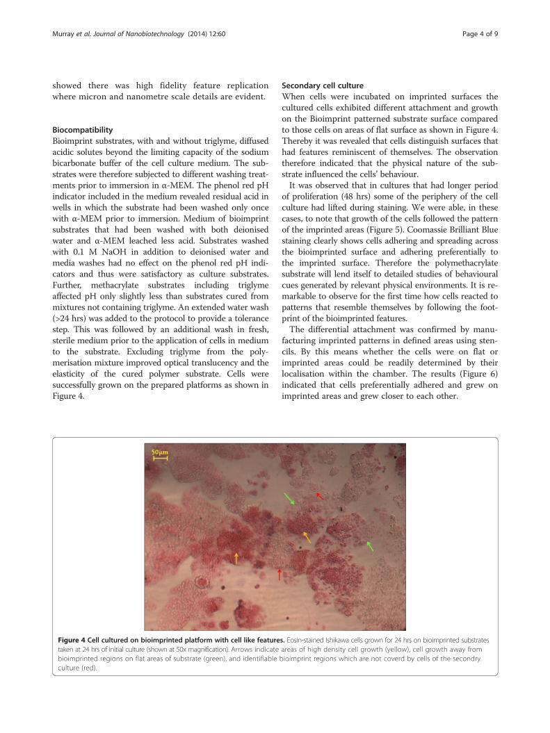
BiocompatibilityBioimprint substrates, with and without triglyme, diffusedacidic solutes beyond the limiting capacity of the sodiumbicarbonate buffer of the cell culture medium. The sub-strates were therefore subjected to different washing treat-ments prior to immersion in α-MEM. The phenol red pHindicator included in the medium revealed residual acid inwells in which the substrate had been washed only oncewith α-MEM prior to immersion. Medium of bioimprintsubstrates that had been washed with both deionisedwater and α-MEM leached less acid. Substrates washedwith 0.1 M NaOH in addition to deionised water andmedia washes had no effect on the phenol red pH indi-cators and thus were satisfactory as culture substrates.Further, methacrylate substrates including triglymeaffected pH only slightly less than substrates cured frommixtures not containing triglyme. An extended water wash(>24 hrs) was added to the protocol to provide a tolerancestep. This was followed by an additional wash in fresh,sterile medium prior to the application of cells in mediumto the substrate. Excluding triglyme from the poly-merisation mixture improved optical translucency and theelasticity of the cured polymer substrate. Cells weresuccessfully grown on the prepared platforms as shown inFigure 4.
Figure 4 Cell cultured on bioimprinted platform with cell like featuretaken at 24 hrs of initial culture (shown at 50x magnification). Arrows indicatebioimprinted regions on flat areas of substrate (green), and identifiableculture (red).
Secondary cell cultureWhen cells were incubated on imprinted surfaces thecultured cells exhibited different attachment and growthon the Bioimprint patterned substrate surface comparedto those cells on areas of flat surface as shown in Figure 4.Thereby it was revealed that cells distinguish surfaces thathad features reminiscent of themselves. The observationtherefore indicated that the physical nature of the sub-strate influenced the cells’ behaviour.It was observed that in cultures that had longer period
of proliferation (48 hrs) some of the periphery of the cellculture had lifted during staining. We were able, in thesecases, to note that growth of the cells followed the patternof the imprinted areas (Figure 5). Coomassie Brilliant Bluestaining clearly shows cells adhering and spreading acrossthe bioimprinted surface and adhering preferentially tothe imprinted surface. Therefore the polymethacrylatesubstrate will lend itself to detailed studies of behaviouralcues generated by relevant physical environments. It is re-markable to observe for the first time how cells reacted topatterns that resemble themselves by following the foot-print of the bioimprinted features.The differential attachment was confirmed by manu-
facturing imprinted patterns in defined areas using sten-cils. By this means whether the cells were on flat orimprinted areas could be readily determined by theirlocalisation within the chamber. The results (Figure 6)indicated that cells preferentially adhered and grew onimprinted areas and grew closer to each other.
s. Eosin-stained Ishikawa cells grown for 24 hrs on bioimprinted substratesareas of high density cell growth (yellow), cell growth away frombioimprint regions which are not coverd by cells of the secondry

Figure 5 Ishikawa cells grown on bioimprinted substrates for 48 hours, cells are stained with Commassie blue. Left image, unannotated.Right image, the bioimprint features are outlined in grey, the secondary cell culture (dark blue) is outlined in yellow. Arrows (red) note regions ofthe cell monolayer that peeled off and folded back. This figure illustrates that the growth of the secondary cell culture has been guided by thefootprint of the replicated cell patterns.
Murray et al. Journal of Nanobiotechnology (2014) 12:60 Page 5 of 9
Lithographically defined micro patterns of pillars andholes were prepared on platforms and cells were culturedon these substrates for comparison with the bioimprint.The dimensions of the lithographically defined patternswere chosen relative to typical Ishikawa cell size which isbetween 10–50 microns.Low magnification bright field imaging showed groups of
cells clustered across the lithography manufactured sub-strate, but there was no evidence for differential preferencebetween patterned and unpatterned surfaces (Figure 7).This is in contrast to the observation using Bioimprints.Figure 7 illustrates an array of 5 μm diamond shaped pillarsprepared on polymethacrylate substrate; Ishikawa cells werecultured and stained with Coomassie blue. The lithographicpatterns without cell-like features had no effect on cellspread between patterned and flat regions.
Figure 6 Stencilled Ishikawa Bioimprints showing designated regionscounterstained Ishikawa cells growing on stencilled substrates. Blue, nucleichannel (which allowed for a single focal plane). Green arrows indicate the(lower) areas. Insert showing a wide view of the border between the flat a
Interest in topography as an influence on cell behav-iour and thence potentially on a range of aspects ofhealth and treatments, independent of biochemical fac-tors, has recently increased. Thus it is important to ob-tain information on the contribution of cell environmentto pathologies such as cancer, as studied in this project,and vascular disease and to interventions that includeimplants and wound repair.To improve the effectiveness of inserted medical de-
vices which is expected to be used for monitoring, detec-tions and diagnostics of our health status the interfacebetween the body and foreign materials must be exam-ined and characterised. However we noted the absence ofmodels for in vitro investigations that incorporated phys-ical environments similar to those experienced in vivo.Here we have, for the first time, developed solid, robust
of bioimprint features. Left: Confocal fluorescence image of; Red, cytoplasm. Right Confocal imaging using bright field backgroundborder between the flat and bioimprinted (upper) areas and flatnd imprinted areas of a stencil.

Figure 7 Ishikawa cells grown on substrates with lithographically-manufactured patterns of similar overall size, cells are stained withCommassie blue. (A) - illustrates an array of 5 µm diamond shaped pillars prepared on polymethacrylate substrate, Ishikawa cell were cultured andstained with Coomassie blue. The cells show no evidence of preferential spread between patterned and flat regions; (B) is a high magnification image.
Murray et al. Journal of Nanobiotechnology (2014) 12:60 Page 6 of 9
substrates that have features similar to the cells beingstudied.Mixed methacrylate co-polymer was able to reliably
replicate high resolution Ishikawa cell surface features toless than 50 nm through fast, high intensity UV expos-ure methodology [17]. We have used Bioimprints dir-ectly for cell imaging studies previously [10,18]. In thisstudy, modifications to the Bioimprint pre-polymer com-position and protocol (i.e. removal of triglyme and im-proving its biocompatability) allow bioimprinted samplesto be used as cell culture platforms.The high light intensity required for the fast curing
produced two notable effects on the curing polymer:heat generation and induced stress. Heat generation didnot alter the bioimprint quality unless the refrigeratedtemplates of fixed cells were not allowed to acclimate toroom temperature prior to exposure and thence intro-duce defects, possibly resulting from bubbles at thebioimprint interface. The problem of induced crosslinkingstress was minimised by using circular sample geometryinstead of rectangular geometry. In the chosen geometrythe radial distribution of stress induced by high speed(30 seconds), high intensity (100w) UV source curingminimised the curving of the polymer and improved pla-narization of the bioimprinted surfaces.The Bioimprint provides a simple and readily-adaptable
platform to investigate cell behaviour by methods com-monly used for traditional in vitro cell culture. Themethod produces a substrate with nanometre reso-lution of cell surface features that has attributes thatare not provided by the soft surfaces of other cellimprinted templates [19,20]. Importantly the topography-related structures, obtained by the overlay imprinting ofthe method described here, are more comprehensive than,for example, those obtained from tissue sections [21]. Weobserved adherence and growth patterns of the cancer
cells on imprinted areas that were distinct from behaviouron flat polymethacrylate surface. Growth is recognised asoccurring in areas on the culture platform where cell via-bility and attachment is high. These results indicate thatthe cells identified differences in physical topography (flatcompared to imprinted) since the substrates, being on thesame culture chamber, had been treated identically. Wesuggest that investigations of guiding cell growth in areasthat are currently receiving extensive attention, such asstem cells development and tissue engineering, will alsobenefit from the method. Other advantages are the abilityto store cell details in a hard polymer and prepare cellculture platforms for controlled cell behaviour.The Bioimprint methodology provides a means of
studying cell behaviour in a physical environment thathas features of the order of those found in vivo and pro-vides a three-dimensional component to the cells’ envir-onment. This development is a step increase in biomimicryover that provided by geometrically manufactured sub-strates. It will be possible, when technical issues are opti-mised, to extend the concept to imprints in other formatssuch as cells replicated with structures convex to the base,flexible substrates and in a variety of polymers. It is likelyto become possible to manufacture a series of identicalsubstrates from a master mould so that pharmacologicaltreatments of cells on the same imprinted structures canbe undertaken. Additionally bioimprinted surfaces may bemodified using techniques already developed such as withprotein [4] or DNA [22], or adapted to be employed withparticulate entities such as viruses [23] to further increasetheir functionality.The method produces relevant topography in relation
to a cell’s micro and nanoenvironment in vivo. The reso-lution of the chosen polymethacrylate polymer is veryhigh (nanometre) and the role of these features that arereplicated at this level remain to be defined. The

Murray et al. Journal of Nanobiotechnology (2014) 12:60 Page 7 of 9
polymer with imprint is permanent and so can be poten-tially reused within an experiment, incorporated into alater study, or shared with other research laboratories.The process is easy, requires only simple equipment, isinexpensive and the substrate does not require molecu-lar modification. Thus the method provides unique plat-forms on which the effects of the physical shapes andtopography can be investigated. The role of mechano-transduction, the effects on cell behaviour of alteredmorphology, the cues by which the physical environ-ment either induces tumorigenesis or maintains homeo-stasis in cells, can all be subjects of study using thismethod. Importantly this study reported observations oncancer cells of morphological alteration and differentialadherence characteristics induced by cues provided byculturing cells on flat and on bioimprinted cell-like pat-terned platforms.Determining the effects of the micro-scale patterns
allowed us to separate observations of cell growth onflat, micro-patterned and bioimprinted substrates in-cluding the nano-scale topographical features. Whencells were cultured on the lithographically defined sub-strate, the pattern showed no effect on the overall cultureorganization and growth. Cell clusters were visible acrossthe diameter of the substrate irrespective of whether thereare patterns or not.
ConclusionsWe report development of a unique technique for print-ing a biological cell in hard polymer that provides highresolution replication and offers a cell culture environ-ment with cell-like features. This enabled us to observefor the first time how cells develop growth characteris-tics in response to an environment patterned with fea-tures that resemble themselves. This methodology hashigh potential for applications in tissue engineering,medical implants and in studying the influence of phys-ical environment on cell behaviour.
MethodsCell culture protocolIshikawa endometrial cancer cells were cultured in cir-cular chambers of polydimethylsiloxane (PDMS) on glasssubstrates. To fabricate the PDMS wells, liquid PDMS(Dow Corning, Midland, MI) was mixed at 10:1 elasto-mer to cross linker ratio, stirred thoroughly, and deaer-ated before curing. PDMS was poured into polystyrenedishes, which were levelled on a hot plate for curing at80°C for 2 hrs. Circular chambers were punched intocured PDMS sheets using a 14 mm cork borer. PDMSsheets were then cut to fit a microscope slide and con-formally sealed to the slide. PDMS/glass slide constructswere sterilised before use as cell culture substrates.
Ishikawa cells were seeded into the PDMS-borderedwells at 5.0 × 10⁴ cells/cm2 in α-minimum essentialmedium (α-MEM) supplemented with 2.2 g/L sodiumbicarbonate, 10% fetal bovine serum, 1% GlutaMAX, and1% penicillin/streptomycin. Ishikawa cells were incu-bated at 37°C and 5% CO2 for 24 hrs before mediumwas aspirated and replaced with 4% paraformaldehyde inPBS for at least 30 minutes for cell fixation prior tobioimprinting (all purchased from Life Technologies Co.,Carlsbad, CA). Fixative was removed and cultures wererinsed thoroughly in separate PBS and water washesbefore being placed in 4°C storage for at least 2 hrs toencourage drying of excess water before bioimprinting.Fixed Ishikawa cell cultures were removed from refriger-ated storage prior to polymer mixing to bring the sam-ples to room temperature before UV exposure tominimise condensation and bubble artefacts at the curedbioimprint-cell interface.
Bioimprint substratesThe liquid methacrylate co-polymer used for bioimprintsubstrate fabrication was adapted from previous work[18]. Ethylene glycol dimethacrylate (EGDMA) and meth-acrylic acid (MAA) (both purchased from Sigma Aldrich,St. Louis, MO) were mixed at the optimised ratio of600 μL to 300 μL with ~100 μL IRGAcure 2022 (CIBASpecialty Chemicals Basel, Switzerland) added as a photoi-nitiator. Triglyme was added to the mixture as a thicken-ing agent. The liquid methacrylate solution was mixed forat least 30 seconds with a vortex mixer before being pipet-ted into the PDMS-defined cell culture wells. The liquidpolymer solution was allowed to settle for 10–20 secondsbefore UV exposure to ensure maximum resolution ofsmall-scale cell features. Slides were placed 15 cm directlybeneath a UV light (Omni Cure series 1000 UV, 100w Hgarc lamp, 250-450 nm filter, EXFO Photonic SolutionsInc. Singapore) guide and exposed to UV at 40% apertureopening for 240 seconds. Cured imprints were removedfrom the PDMS/glass assembly to a water bath andmanually agitated to remove larger cell debris. The curedbioimprint was then transferred to an ultrasonic sodiumdodecyl sulphate bath (10% w/v in .01 M hydrochloricacid solution) and a 30 minute trypsin soak (0.05% trypsinin PBS) in order to minimise cell material remaining onthe bioimprinted polymer surface.
Patterned substrate fabricationTo directly compare the effects of geometrically pat-terned lithography with those of the bioimprint, differentpatterned substrates were fabricated. Patterned substratesconsisted of regular geometric arrays of pillar or hole pat-terns of 5–15 μm comparable to the size of the cells understudy. The patterns were initially fabricated in SU-8photoresist (MicroChem SU-8 2100) on silicon wafers

Murray et al. Journal of Nanobiotechnology (2014) 12:60 Page 8 of 9
using photolithography processes and inverse PDMSmoulds were made using soft lithography. The PDMSpatterned platforms were replicated in polymethacry-late substrates for cell culture experiments. Ishikawaendometrial cancer cells were cultured in the sameconditions as the bioimptinted platforms.
BiocompatibilityTo neutralise leaching of methacrylic acid the quenchingeffect of different washing techniques on the polymetha-crylate (EGDMA) substrates prior to use in cell culturewas investigated. Bioimprint samples were placed in 12wells of a 24-well polystyrene tissue culture plate. Bioim-prints were washed with (i) deionised water followed byα-MEM medium, (ii) only α-MEM medium, (iii) 0.1 MNaOH followed by deionised water and α-MEM medium,or (iv) left untreated. Washes were pipetted into each well,agitated for approximately 30 seconds, and aspirated. Afterremoval of wash conditions, each well was filled with freshα-MEM (without cells present) containing phenol red pHindicator.Cytotoxicity of bioimprinted polymethacrylate samples
was investigated by placing a cured bioimprint substrateat the bottom of 3 wells on a polystyrene 6-well plate; the3 wells without bioimprint samples were maintained ascontrol cultures. Ishikawa cells were seeded at 5.0 × 10⁴
cells/well in all 6 wells and incubated in accordance withthe previously outlined protocol for 24 hours, at whichpoint the substrates were removed for imaging.
Secondary cell cultureIshikawa cells were grown on bioimprinted substrates toverify the biocompatibility of the substrate and deter-mine the topographical influence of bioimprinted fea-tures on cell attachment and growth. Ishikawa cells wereseeded and cultured on bioimprinted polymethacrylatesubstrates placed on the bottom of 24-well polystyreneplates. These cells were referred to as secondary cellcultures in order to distinguish them from the initial cellcultures required for bioimprint substrate fabrication.Bioimprints were placed template-side-up and Ishikawacells were seeded at 5.0 × 10⁴ cells/well and maintainedin supplemented α-MEM at 37°C and 5% CO2 for24 hrs. At 24 hrs medium was aspirated and cells werefixed with 4% paraformaldehyde in PBS for at least30 minutes and then washed with PBS several times toremove trace fixative and salts. Cells were stainedwith Coomassie brilliant blue (Life Technologies Co.,Carlsbad, CA) for 5 minutes and washed at least twicewith PBS until wash solutions did not contain leachedstain.
Competing interestThe authors declare that they have no competing interests.
Authors’ contributionsLMM did the experimental work as part of her PhD project. VN was a postdoctorate fellow and contributed to the fabrication processes. MMA and JJEwere the supervisors of the project who provided the funding, facilities andguidance. All authors helped in the writing of the manuscript, discussions,and interpretation of data and approved the final version of the manuscript.
AcknowledgmentsThe authors wish to acknowledge the Marsden Fund for funding theproject and supporting the post doctorate position and the MacDiarmidInstitute for Advanced Materials and Nanotechnology for providing aPh.D scholarship.
Author details1The MacDiarmid Institute for Advanced Materials and Nanotechnology,Department of Electrical and Computer Engineering, University ofCanterbury, Christchurch 8140, New Zealand. 2The MacDiarmid Institute forAdvanced Materials and Nanotechnology, and Centre forNeuroendocrinology, Department of Obstetrics and Gynaecology, Universityof Otago, Christchurch 8011, New Zealand.
Received: 5 August 2014 Accepted: 12 December 2014
References1. Mills AM, Longacre TA: Endometrial hyperplasia. Semin Diagn Pathol 2010,
27:199–214.2. Bhattacharya N: Fetal cell/tissue therapy in adult disease: a new horizon
in regenerative medicine. Clin Exp Obstet Gynecol 2004, 31:167–173.3. Longmate WM, Dipersio CM: Integrin regulation of epidermal functions in
wounds. Adv Wound Care 2014, 3:229–246.4. Lindstrom S, Andersson-Svahn H: Overview of single-cell analyses: microdevices
and applications. Lab Chip 2010, 10:3363–3372.5. Kraning-Rush CM, Reinhart-King CA: Controlling matrix stiffness and
topography for the study of tumor cell migration. Cell Adh Migr 2012,6:274–279.
6. Blattler T, Huwiler C, Ochsner M, Stadler B, Solak H, Voros J, Grandin HM:Nanopatterns with biological functions. J Nanosci Nanotechnol 2006,6:2237–2264.
7. Lan H, Liu H: UV-nanoimprint lithography: structure, materials andfabrication of flexible molds. J Nanosci Nanotechnol 2013, 13:3145–3172.
8. Biggs MJ, Richards RG, Dalby MJ: Nanotopographical modification: aregulator of cellular function through focal adhesions. Nanomed 2010,6:619–633.
9. Nikkhah M, Edalat F, Manoucheri S, Khademhosseini A: Engineeringmicroscale topographies to control the cell-substrate interface.Biomaterials 2012, 33:5230–5246.
10. Muys JJ, Alkaisi MM, Evans JJ: Cancer imaging by atomic force microscopyusing a bioimprint cellular transfer technique. J Nanobiotechnol 2005, 4(1):10.
11. Muys JJ, Alkaisi MM, Evans JJ: Bioimprint: nanoscale analysis by replicationof cellular topography using soft lithography. J Biomed Nanotechnol 2006,2:1–5.
12. Murray LM, Nock V, Alkaisi MM, Lee JJM, Woodfield TBF: Fabrication ofpolymeric substrates with micro- and nanoscale topography bioimprintedat progressive cell morphologies. J Va Sci & Technol B: Microelectron andNanometer Struct 2012, 30:06F902–906F902–906.
13. Nock V, Murray L, Samsuri F, Alkaisi MM, Evans JJ: Microfluidics-assistedphoto nanoimprint lithography for the formation of cellular bioimprints.J Vac Sci & Technol B 2010, 28:C6K17–C16K22.
14. Nock V, Murray L, Samsuri F, Alkaisi MM, Evans JJ: Microfluidic arrays forbioimprint of cancer cells. Microelectron Eng 2011, 88:1828–1831.
15. Wang X, Zhang Y, Du K, Fang X: Atomic force microscope observation onultrastructures in plant cells. J Nanosci Nanotechnol 2010, 10:6624–6628.
16. Li JJ, Zhou XT, Shi J, Zhang F, Li X, Jiang LM, Chen Y: Upside and downsideviews of adherent cells on patterned substrates: three-dimensionalimage reconstruction. Microelectron Eng 2013, 110:365–368.
17. Samsuri F, Mitchell JS, Alkaisi MM, Evans JJ: Formation of NanoscaleBioimprints of Muscle Cells Using UV-Cured Spin-Coated Polymers.J Nanotech 2009, 2009:6.

Murray et al. Journal of Nanobiotechnology (2014) 12:60 Page 9 of 9
18. Samsuri F, Alkaisi MM, Evans JJ, Chitcholtan K, Mitchell JS: Detection ofchanges in cell membrane structures using the Bioimprint technique.Microelectron Eng 2011, 88:1871–1874.
19. DePorter SM, Luib I, McNaughton BR: Programmed cell adhesion andgrowth on cell-imprinted polyacrylamide hydrogels. Soft Matter 2012,8:10403–10408.
20. Jeon H, Kim G: Effects of a cell-imprinted poly(dimethylsiloxane) surfaceon the cellular activities of MG63 osteoblast-like cells: preparation of apatterned surface, surface characterization, and bone mineralization.Langmuir 2012, 28:13423–13430.
21. Tong WY, Shen W, Yeung CW, Zhao Y, Cheng SH, Chu PK, Chan D, Chan GC,Cheung KM, Yeung KW, Lam YW: Functional replication of the tendon tissuemicroenvironment by a bioimprinted substrate and the support oftenocytic differentiation of mesenchymal stem cells. Biomaterials 2012,33:7686–7698.
22. Spivaka DA, Shea KJ: Investigation into the scope and limitations ofmolecular imprinting with DNA molecules. Anal Chim Acta 2001,435:65–74.
23. Bolisay LD, Culver JN, Kofinas P: Molecularly imprinted polymers fortobacco mosaic virus recognition. Biomaterials 2006, 27:4165–4168.
Submit your next manuscript to BioMed Centraland take full advantage of:
• Convenient online submission
• Thorough peer review
• No space constraints or color figure charges
• Immediate publication on acceptance
• Inclusion in PubMed, CAS, Scopus and Google Scholar
• Research which is freely available for redistribution
Submit your manuscript at www.biomedcentral.com/submit