Biofunctionalization and immobilization of a membrane via peptide binding (CR3-1, S2) by a Monte...
6
Biofunctionalization and immobilization of a membrane via peptide binding „CR3-1, S2… by a Monte Carlo simulation R. B. Pandey, 1,a Hendrik Heinz, 2 Jie Feng, 2 and Barry L. Farmer 3 1 Department of Physics and Astronomy, University of Southern Mississippi, Hattiesburg, Mississippi 39406, USA 2 Department of Polymer Engineering, University of Akron, Akron, Ohio 44325, USA 3 Air Force Research Laboratory, Materials and Manufacturing Directorate, AFRL/RXBP, Wright-Patterson AFB, Ohio 45433, USA Received 22 April 2010; accepted 5 August 2010; published online 7 September 2010 A coarse-grained computer simulation model is used to study the immobilization of a dynamic tethered membrane representation of a clay platelet in a matrix of mobile peptide chains CR3-1: 1 Trpu 2 Prou 3 Seru 4 Seru 5 Tyru 6 Leuu 7 Seru 8 Prou 9 Ileu 10 Prou 11 Tyru 12 Ser and S2: 1 Hisu 2 Glyu 3 Ileu 4 Asnu 5 Thru 6 Thru 7 Lysu 8 Prou 9 Pheu 10 Lysu 11 Seru 12 Val on a cubic lattice. Each residue interacts with the membrane nodes with appropriate interaction and executes their stochastic motion with the Metropolis algorithm. Density profiles, binding energy of each residue, mobility, and targeted structural profile are analyzed as a function of peptide concentration. We find that the binding of peptides S2 is anchored by lysine residues 7 Lys, 10 Lys while peptides CR3-1 do not bind to membrane. The membrane slows down as peptides S2 continues to bind leading to its eventual pinning. How fast the immobilization of the membrane occurs depends on peptide concentration. Binding of peptide S2 modulates the morphology of the membrane. The immobilization of membrane occurs faster if peptides S2 are replaced by the homopolymer of lysine Lys 12 of the same molecular weight, the strongest binding residue. The surface of membrane can be patterned with somewhat reduced roughness with the homopolymer of lysine than that with peptide S2.© 2010 American Institute of Physics. doi:10.1063/1.3484241 I. INTRODUCTION Immobilization of cells via adhesive peptides 1 plays a very important role in cell adhesion to regenerate tissues such as corneal epithelium. 2 Membrane and nanoparticles e.g., gold and palladium biofunctionalized by appropriate peptides have enormous potential in such applications as de- signing multifunctional materials for sensors, tissue engi- neering, regenerative medicine, and drug delivery to desired targets. 3–8 The effect of interaction of membrane surface with appropriate peptide in a desired tissue culture is critical in controlling the dynamics and morphology of the membrane and cell. A cell is too complex to consider its full functional characteristics in such a computer simulation study presented here. Therefore, we focus on the basic characteristics of a tethered membrane as it is immersed in a peptide solution. The structural analysis of a tethered membrane including crumpling has been a subject of enormous interest for a long time. 9–12 The dynamics of a tethered membrane has been extensively studied by computer simulation. 13–15 A tethered membrane exhibits multiple dynamics 13,14 over a range of time scales, e.g., short time R t 1/8 to long time asymptotic diffusion R t 1/2 where R is the root mean square displace- ment of a local node and t is the time step. What happens when the membrane is placed in a host matrix consisting of peptides? Obviously the concentration of peptide is very im- portant in controlling the availability of free volume for the membrane to relax and execute its stochastic motion. In gen- eral, the higher the concentration of the underlying matrix, i.e., peptides, the longer the relaxation time is for constitu- ents, i.e., peptides and membrane, to reach asymptotic dy- namics. In addition to concentration, interaction between peptides and membrane may also affect the dynamics and self-assembly. How does the membrane move and relax in presence of peptides that bind in comparison to those that do not? Such questions are relevant, for example, in the context to binding of peptides S2 1 Hisu 2 Glyu 3 Ileu 4 Asnu 5 Thru 6 Thr u 7 Lysu 8 Prou 9 Pheu 10 Lysu 11 Seru 12 Val and CR3-1 1 Trp u 2 Pro u 3 Ser u 4 Ser u 5 Tyru 6 Leu u 7 Seru 8 Prou 9 Ileu 10 Prou 11 Tyru 12 Ser to clay platelets. 6–8 We have ex- amined the immobilization dynamics of the tethered mem- brane in the presence of peptides CR3-1 and S2 using a coarse-grained model, as described in the following. II. MODEL A cubic box of size L 3 is considered as a host space. The membrane, corresponding to a coarse-grained description of a bendable clay platelet, 13 is modeled by a set of nodes con- nected by flexible fluctuating bonds initially on a square grid. The membrane is placed at the center of the box fol- lowed by peptides inserted randomly in the box see Fig. 1. The peptide chain 16,17 is represented by a set of nodes, each representing the interaction characteristics of a correspond- ing amino acid, which are tethered by fluctuating covalent a Electronic mail: [email protected]. THE JOURNAL OF CHEMICAL PHYSICS 133, 095102 2010 0021-9606/2010/1339/095102/6/$30.00 © 2010 American Institute of Physics 133, 095102-1
Transcript of Biofunctionalization and immobilization of a membrane via peptide binding (CR3-1, S2) by a Monte...
Biofunctionalization and immobilization of a membrane via peptide binding„CR3-1, S2… by a Monte Carlo simulation
R. B. Pandey,1,a� Hendrik Heinz,2 Jie Feng,2 and Barry L. Farmer3
1Department of Physics and Astronomy, University of Southern Mississippi, Hattiesburg,Mississippi 39406, USA2Department of Polymer Engineering, University of Akron, Akron, Ohio 44325, USA3Air Force Research Laboratory, Materials and Manufacturing Directorate, AFRL/RXBP,Wright-Patterson AFB, Ohio 45433, USA
�Received 22 April 2010; accepted 5 August 2010; published online 7 September 2010�
A coarse-grained computer simulation model is used to study the immobilization of a dynamictethered membrane �representation of a clay platelet� in a matrix of mobile peptide chainsCR3-1: 1Trpu2Prou3Seru4Seru5Tyru6Leuu7Seru8Prou9Ileu10Prou11Tyru12Ser andS2: 1Hisu2Glyu3Ileu4Asnu5Thru6Thru7Lysu8Prou9Pheu10Lysu11Seru12Val on acubic lattice. Each residue interacts with the membrane nodes with appropriate interaction andexecutes their stochastic motion with the Metropolis algorithm. Density profiles, binding energy ofeach residue, mobility, and targeted structural profile are analyzed as a function of peptideconcentration. We find that the binding of peptides S2 is anchored by lysine residues �7Lys, 10Lys�while peptides CR3-1 do not bind to membrane. The membrane slows down as peptides �S2�continues to bind leading to its eventual pinning. How fast the immobilization of the membraneoccurs depends on peptide concentration. Binding of peptide �S2� modulates the morphology of themembrane. The immobilization of membrane occurs faster if peptides �S2� are replaced by thehomopolymer of lysine ��Lys�12 of the same molecular weight�, the strongest binding residue. Thesurface of membrane can be patterned with somewhat reduced roughness with the homopolymer oflysine than that with peptide �S2�. © 2010 American Institute of Physics. �doi:10.1063/1.3484241�
I. INTRODUCTION
Immobilization of cells via adhesive peptides1 plays avery important role in cell adhesion to regenerate tissuessuch as corneal epithelium.2 Membrane and nanoparticles�e.g., gold and palladium� biofunctionalized by appropriatepeptides have enormous potential in such applications as de-signing multifunctional materials for sensors, tissue engi-neering, regenerative medicine, and drug delivery to desiredtargets.3–8 The effect of interaction of membrane surface withappropriate peptide in a desired tissue culture is critical incontrolling the dynamics and morphology of the membraneand cell. A cell is too complex to consider its full functionalcharacteristics in such a computer simulation study presentedhere. Therefore, we focus on the basic characteristics of atethered membrane as it is immersed in a peptide solution.
The structural analysis of a tethered membrane includingcrumpling has been a subject of enormous interest for a longtime.9–12 The dynamics of a tethered membrane has beenextensively studied by computer simulation.13–15 A tetheredmembrane exhibits multiple dynamics13,14 over a range oftime scales, e.g., short time �R� t1/8� to long time asymptoticdiffusion �R� t1/2� where R is the root mean square displace-ment of a local node and t is the time step. What happenswhen the membrane is placed in a host matrix consisting ofpeptides? Obviously the concentration of peptide is very im-portant in controlling the availability of free volume for the
membrane to relax and execute its stochastic motion. In gen-eral, the higher the concentration of the underlying matrix,i.e., peptides, the longer the relaxation time is for constitu-ents, i.e., peptides and membrane, to reach asymptotic dy-namics. In addition to concentration, interaction betweenpeptides and membrane may also affect the dynamics andself-assembly.
How does the membrane move and relax in presence ofpeptides that bind in comparison to those that do not? Suchquestions are relevant, for example, in the context to bindingof peptides S2 �1Hisu2Glyu3Ileu4Asnu5Thru6Thru
7Lysu8Prou9Pheu10Lysu11Seru12Val� and CR3-1� 1Trpu 2Prou 3Seru 4Seru 5Tyru6Leuu 7Seru8Prou9Ileu10Prou11Tyru12Ser� to clay platelets.6–8 We have ex-amined the immobilization dynamics of the tethered mem-brane in the presence of peptides CR3-1 and S2 using acoarse-grained model, as described in the following.
II. MODEL
A cubic box of size L3 is considered as a host space. Themembrane, corresponding to a coarse-grained description ofa bendable clay platelet,13 is modeled by a set of nodes con-nected by flexible �fluctuating� bonds initially on a squaregrid. The membrane is placed at the center of the box fol-lowed by peptides inserted randomly in the box �see Fig. 1�.The peptide chain16,17 is represented by a set of nodes, eachrepresenting the interaction characteristics of a correspond-ing amino acid, which are tethered by fluctuating covalenta�Electronic mail: [email protected].
THE JOURNAL OF CHEMICAL PHYSICS 133, 095102 �2010�
0021-9606/2010/133�9�/095102/6/$30.00 © 2010 American Institute of Physics133, 095102-1

bonds. A node in both membrane and peptide occupies a unitcube i.e., its eight lattice sites; the bond length between con-secutive nodes can vary �fluctuate� between 2 and �10 withan exception of �8 in unit of lattice constant. Such a bond-fluctuation description is known to be computationallyefficient while incorporating ample degrees of freedom incomplex polymer systems18 and multicomponentnanocomposites.19,20
We consider a set of interactions among residues of thepeptide �intrachain and interchain� and with the membranenodes within a range of interaction. Only the excluded vol-ume interaction is however considered between nodes per-taining to the membrane. Since each peptide node representsa specific residue, it is essential to capture their specificity oruniqueness. The specificity of each residue is captured by aninteraction matrix �see below� method recently used to studyadsorption of peptides on gold and palladium substrates.16
This approach is briefly described as follows. Twenty aminoacids can be divided into three broad categories: hydropho-bic �H�, hydrophilic or polar �P�, and electrostatic �E�. Anamino acid within each group is further distinguished by itsrelative hydrophobicity, polar, or electrostatic strength. In ad-dition to peptide chains, there is a tethered membrane �S�.This constitutes four main components, H, P, E, and S inwhich there are three subgroups to characterize specificity ofeach residue:16 eight hydrophobic �H1 ,H2¯H8�, eight polar�P1 , P2¯P8�, and four electrostatic �E1 ,E2,E3 ,E4�. Eachnode �membrane and peptide� executes stochastic movementwith the Metropolis algorithm using a set of phenomenologi-cal interactions among these components as follows.
Interaction matrix. Interaction among four main interact-ing components �H , P ,E ,S� can be represented by a 4�4matrix.16 The interaction strength between two elements�nodes� at sites i and j separated by a distance rij is repre-sented by the standard Lennard-Jones potential
Uij = f�ij�� �
rij12
−�� �
rij6, rij � rc,
where rc= �8 �in unit of lattice constant� is the cutoff rangeand f�ij is the interaction strength with an arbitrary param-eter f that can be varied and �=1. On a cubic lattice the
distance between two sites is discrete and measured in unitsof the lattice constant. Note that the minimum distance be-tween two neighboring particles is two in units of the latticeconstant. The depth of the LJ potential is controlled by themagnitude of the pair interaction strength f�ij.
16 The numberof the pair interaction matrix elements is however reducedsomewhat due to symmetries, e.g., �ij =� ji.
The magnitude of each interaction elements is based onthe insight gained from all atomistic description and theirknown general characteristics.8,16 A typical set of values ofthe interaction matrix is presented in Table I. The interactionmatrix elements16 for residues are obtained by weighing thebroad interaction strength, �HS, �PS, and �ES, appropriatelyby their specific interaction characteristics, i.e., by their rela-tive hydrophobicity and polar and electrostatic strengthswithin each group. This leads to a larger interaction matrixwith �H1S ,�H2S , ¯�H8S ,�P1S ,�P2S , ¯�P8S ,�E1S ,�E2S , ¯�E4S. We have used f =100 the interaction energy factor toaccentuate the differences in adsorption �binding� probabili-ties for CR3-1 and S2 peptides. Interactions16 among the fourelectrostatic residues ��EE in Table I� are �11=�12=�22=�33
=�34=�44=0.1 and �13=�14=�23=�24=−0.4.Incorporating the specificity of individual amino acid
group for a particular system enhances the size of the inter-action matrix.16 The choices of the matrix elements presentedhere are primarily to illustrate how to predict the relativebinding of peptides CR3-1 and S2 with the clay plateletusing the coarse-grained model with phenomenologicalinteractions. The relative hydrophobic interaction strengthof S2 �1Hisu2Glyu3Ileu4Asnu5Thru6Thru7Lysu8Prou9Pheu10Lysu11Seru12Val� can be representedby �1P6u
2H8u3H1u
4P8u5P1u
6P1u7E3u
8P5u9H4u
10E3u11P2u
12H2� and that of CR3-1 �1Trpu2Prou3Seru4Seru5Tyru6Leuu7Seru8Prou9Ileu10Prou
11Tyru12Ser� by �1P3u2P5u
3P2u4P2u
5P4u6H3u
7P2u8P5u
9H1u10P5u
11P4u12P2�.
Stochastic movement. Each residue and membrane nodeexecutes their stochastic movement13,19,20 with the Metropo-lis algorithm. A particle �node� at a site i and one of its 26neighboring sites j are selected randomly. If site j is emptyand the change in the bond length with the proposed move iswithin the allowed range, then an attempt is made to move itfrom site i to site j. Provided the excluded volume conditionis satisfied, the energy in old �Ei� and new configuration �Ej�is compared and particle is moved from site i to j with prob-ability exp�−�Eij /T�, where �Eij =Ej −Ei, the temperature Tis in unit of the Boltzmann constant and the interaction en-ergy and fixed to unity here. Attempts to move each nodeonce defines one Monte Carlo step time. Simulation is per-formed for a sufficiently long time to achieve equilibration.
FIG. 1. A tethered membrane at the center surrounded by peptide chains ona cubic lattice.
TABLE I. A typical interaction matrix among H, P, E, and S components.
H P E S
H 0.0 0.0 0.0 0.1P 0.0 �0.2 �0.2 �0.2E 0.0 �0.2 �EE �0.5S 0.1 �0.2 �0.5 0.0
095102-2 Pandey et al. J. Chem. Phys. 133, 095102 �2010�
A number of physical quantities are evaluated during thesimulation including energy of each residue, their mobility,average number of components �membrane nodes and resi-dues� within the range of interaction, and the variation of themean square displacement of the center of mass of the pep-tide chains and that of the membrane with the time stepswhich are presented in the following.
III. RESULTS AND DISCUSSION
Starting with a homogeneous solution of the peptidescontaining the membrane, the immobilization process wasmonitored in the simulation. In the following, we report onthe displacement of the membrane and of the peptides, theirbinding strength, and mobility upon biofunctionalization.The method also allowed the variation of peptide concentra-tion to broaden the analysis.
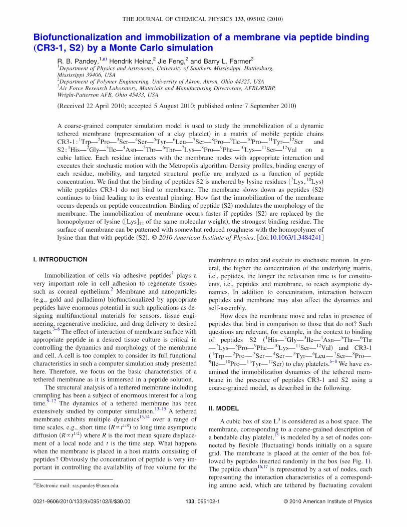
Let us first consider the variation of the root mean squaredisplacement �Rc� of the center of mass of the membrane andthat of the CR3-1 and S2 peptides as shown in Fig. 2 �toprow�. The slope of Rc versus time step �t� is the measure ofthe speed by which membrane and peptide move. We see thatthe membrane moves much faster in presence of CR3-1 thanin the presence of S2. As the time progresses, the membranebecomes immobilized in the presence of S2 peptides due tobinding. The membrane continues diffusion across the timerange in the presence of CR3-1 which binds weaker and hashardly any effect. The average mobility of the peptides ismuch less affected than that of the membrane due to a ma-jority of free S2 peptides even after tethering of many to themembrane. However, some reduction in average mobility inthe long time regime is clearly reflected due to the tether ofmany S2 molecules to the membrane.
In order to quantify the relative binding of peptides, weevaluate the average number �Ns� of membrane sites withinthe range of interaction to each residue of both CR3-1 and S2in Fig. 2 �bottom row�. The number of binding sites of themembrane to S2 is overwhelmingly larger than CR3-1 whichsuggests a much higher probability of binding of S2 than
binding of CR3-1. Note that the binding of S2 to the mem-brane is mediated by lysine residues �7Lys, 10Lys� which re-tain the largest number of membrane sites within their inter-action range over time. As we have identified that S2 bindsto the membrane �consistent with recent experiments�,7 wewill focus on the effect of its binding on the immobilizationof the membrane in the following.
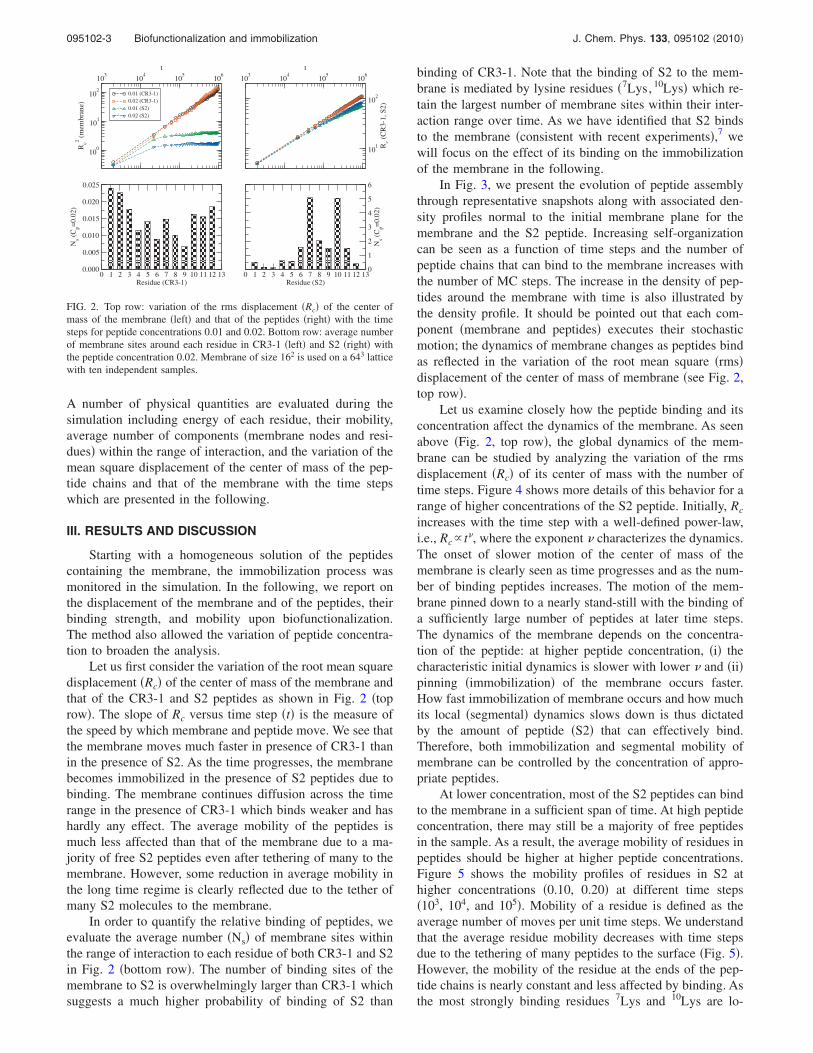
In Fig. 3, we present the evolution of peptide assemblythrough representative snapshots along with associated den-sity profiles normal to the initial membrane plane for themembrane and the S2 peptide. Increasing self-organizationcan be seen as a function of time steps and the number ofpeptide chains that can bind to the membrane increases withthe number of MC steps. The increase in the density of pep-tides around the membrane with time is also illustrated bythe density profile. It should be pointed out that each com-ponent �membrane and peptides� executes their stochasticmotion; the dynamics of membrane changes as peptides bindas reflected in the variation of the root mean square �rms�displacement of the center of mass of membrane �see Fig. 2,top row�.
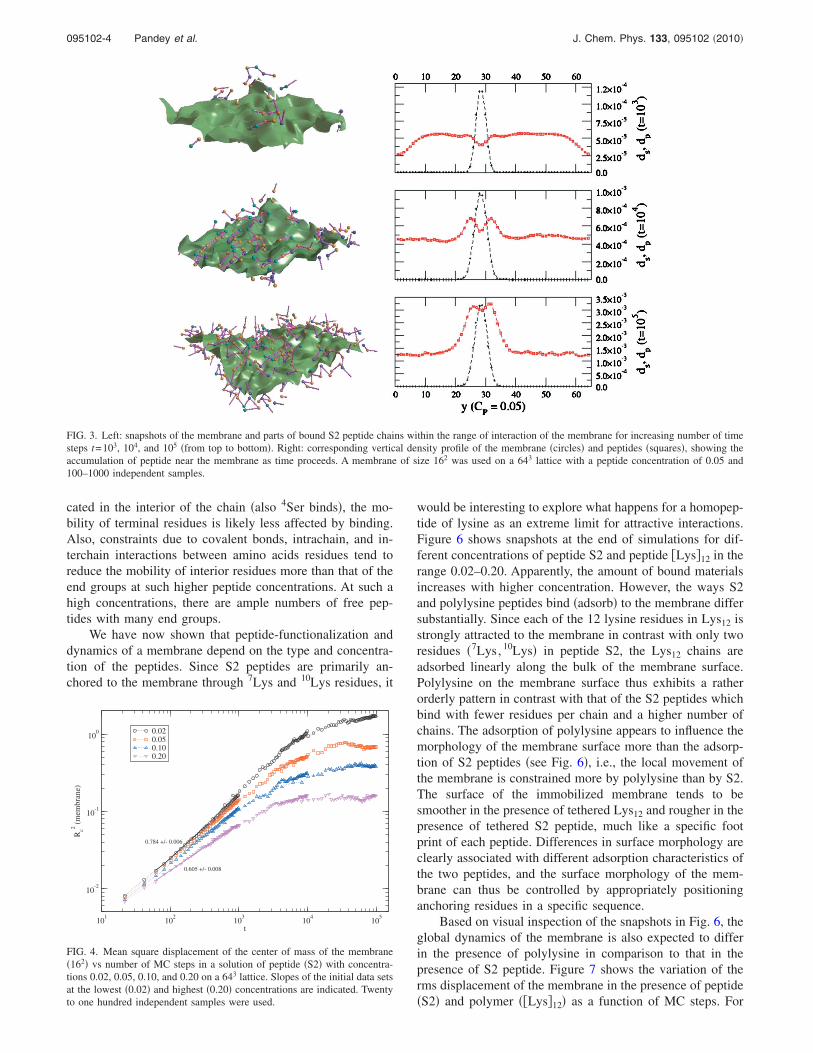
Let us examine closely how the peptide binding and itsconcentration affect the dynamics of the membrane. As seenabove �Fig. 2, top row�, the global dynamics of the mem-brane can be studied by analyzing the variation of the rmsdisplacement �Rc� of its center of mass with the number oftime steps. Figure 4 shows more details of this behavior for arange of higher concentrations of the S2 peptide. Initially, Rc
increases with the time step with a well-defined power-law,i.e., Rc� t, where the exponent characterizes the dynamics.The onset of slower motion of the center of mass of themembrane is clearly seen as time progresses and as the num-ber of binding peptides increases. The motion of the mem-brane pinned down to a nearly stand-still with the binding ofa sufficiently large number of peptides at later time steps.The dynamics of the membrane depends on the concentra-tion of the peptide: at higher peptide concentration, �i� thecharacteristic initial dynamics is slower with lower and �ii�pinning �immobilization� of the membrane occurs faster.How fast immobilization of membrane occurs and how muchits local �segmental� dynamics slows down is thus dictatedby the amount of peptide �S2� that can effectively bind.Therefore, both immobilization and segmental mobility ofmembrane can be controlled by the concentration of appro-priate peptides.
At lower concentration, most of the S2 peptides can bindto the membrane in a sufficient span of time. At high peptideconcentration, there may still be a majority of free peptidesin the sample. As a result, the average mobility of residues inpeptides should be higher at higher peptide concentrations.Figure 5 shows the mobility profiles of residues in S2 athigher concentrations �0.10, 0.20� at different time steps�103, 104, and 105�. Mobility of a residue is defined as theaverage number of moves per unit time steps. We understandthat the average residue mobility decreases with time stepsdue to the tethering of many peptides to the surface �Fig. 5�.However, the mobility of the residue at the ends of the pep-tide chains is nearly constant and less affected by binding. Asthe most strongly binding residues 7Lys and 10Lys are lo-
103
104
105
106
t
100
101
102
Rc2
(mem
bra
ne)
0.01 (CR3-1)
0.02 (CR3-1)
0.01 (S2)
0.02 (S2)
103
104
105
106
t
101
102
Rc
(CR
3-1
,S
2)
��������������������������
�������������
��������������������
��������������
����������������
������������
������������������
������������
����������
���������
������������������
����������������������
0 1 2 3 4 5 6 7 8 9 10 11 12 13Residue (CR3-1)
0.000
0.005
0.010
0.015
0.020
0.025
Ns
(Cp=
0.0
2)
����������
���
������
����������
������������
������������
��������
������������������������
��������
����
0 1 2 3 4 5 6 7 8 9 10 11 12 13Residue (S2)
0
1
2
3
4
5
6
Ns
(Cp=
0.0
2)
FIG. 2. Top row: variation of the rms displacement �Rc� of the center ofmass of the membrane �left� and that of the peptides �right� with the timesteps for peptide concentrations 0.01 and 0.02. Bottom row: average numberof membrane sites around each residue in CR3-1 �left� and S2 �right� withthe peptide concentration 0.02. Membrane of size 162 is used on a 643 latticewith ten independent samples.
095102-3 Biofunctionalization and immobilization J. Chem. Phys. 133, 095102 �2010�
cated in the interior of the chain �also 4Ser binds�, the mo-bility of terminal residues is likely less affected by binding.Also, constraints due to covalent bonds, intrachain, and in-terchain interactions between amino acids residues tend toreduce the mobility of interior residues more than that of theend groups at such higher peptide concentrations. At such ahigh concentrations, there are ample numbers of free pep-tides with many end groups.
We have now shown that peptide-functionalization anddynamics of a membrane depend on the type and concentra-tion of the peptides. Since S2 peptides are primarily an-chored to the membrane through 7Lys and 10Lys residues, it
would be interesting to explore what happens for a homopep-tide of lysine as an extreme limit for attractive interactions.Figure 6 shows snapshots at the end of simulations for dif-ferent concentrations of peptide S2 and peptide �Lys�12 in therange 0.02–0.20. Apparently, the amount of bound materialsincreases with higher concentration. However, the ways S2and polylysine peptides bind �adsorb� to the membrane differsubstantially. Since each of the 12 lysine residues in Lys12 isstrongly attracted to the membrane in contrast with only tworesidues �7Lys, 10Lys� in peptide S2, the Lys12 chains areadsorbed linearly along the bulk of the membrane surface.Polylysine on the membrane surface thus exhibits a ratherorderly pattern in contrast with that of the S2 peptides whichbind with fewer residues per chain and a higher number ofchains. The adsorption of polylysine appears to influence themorphology of the membrane surface more than the adsorp-tion of S2 peptides �see Fig. 6�, i.e., the local movement ofthe membrane is constrained more by polylysine than by S2.The surface of the immobilized membrane tends to besmoother in the presence of tethered Lys12 and rougher in thepresence of tethered S2 peptide, much like a specific footprint of each peptide. Differences in surface morphology areclearly associated with different adsorption characteristics ofthe two peptides, and the surface morphology of the mem-brane can thus be controlled by appropriately positioninganchoring residues in a specific sequence.
Based on visual inspection of the snapshots in Fig. 6, theglobal dynamics of the membrane is also expected to differin the presence of polylysine in comparison to that in thepresence of S2 peptide. Figure 7 shows the variation of therms displacement of the membrane in the presence of peptide�S2� and polymer ��Lys�12� as a function of MC steps. For
FIG. 3. Left: snapshots of the membrane and parts of bound S2 peptide chains within the range of interaction of the membrane for increasing number of timesteps t=103, 104, and 105 �from top to bottom�. Right: corresponding vertical density profile of the membrane �circles� and peptides �squares�, showing theaccumulation of peptide near the membrane as time proceeds. A membrane of size 162 was used on a 643 lattice with a peptide concentration of 0.05 and100–1000 independent samples.
101
102
103
104
105
t
10-2
10-1
100
Rc2
(mem
bra
ne)
0.020.050.100.20
0.784 +/- 0.006
0.605 +/- 0.008
FIG. 4. Mean square displacement of the center of mass of the membrane�162� vs number of MC steps in a solution of peptide �S2� with concentra-tions 0.02, 0.05, 0.10, and 0.20 on a 643 lattice. Slopes of the initial data setsat the lowest �0.02� and highest �0.20� concentrations are indicated. Twentyto one hundred independent samples were used.
095102-4 Pandey et al. J. Chem. Phys. 133, 095102 �2010�
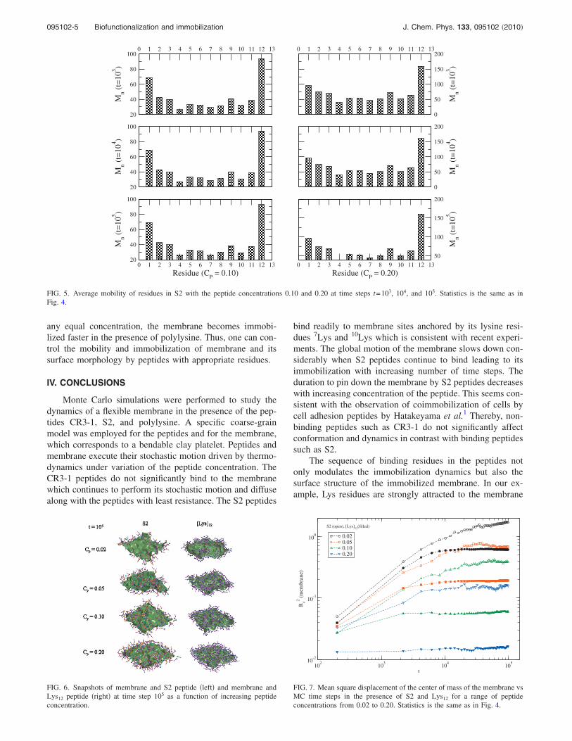
any equal concentration, the membrane becomes immobi-lized faster in the presence of polylysine. Thus, one can con-trol the mobility and immobilization of membrane and itssurface morphology by peptides with appropriate residues.
IV. CONCLUSIONS
Monte Carlo simulations were performed to study thedynamics of a flexible membrane in the presence of the pep-tides CR3-1, S2, and polylysine. A specific coarse-grainmodel was employed for the peptides and for the membrane,which corresponds to a bendable clay platelet. Peptides andmembrane execute their stochastic motion driven by thermo-dynamics under variation of the peptide concentration. TheCR3-1 peptides do not significantly bind to the membranewhich continues to perform its stochastic motion and diffusealong with the peptides with least resistance. The S2 peptides
bind readily to membrane sites anchored by its lysine resi-dues 7Lys and 10Lys which is consistent with recent experi-ments. The global motion of the membrane slows down con-siderably when S2 peptides continue to bind leading to itsimmobilization with increasing number of time steps. Theduration to pin down the membrane by S2 peptides decreaseswith increasing concentration of the peptide. This seems con-sistent with the observation of coimmobilization of cells bycell adhesion peptides by Hatakeyama et al.1 Thereby, non-binding peptides such as CR3-1 do not significantly affectconformation and dynamics in contrast with binding peptidessuch as S2.
The sequence of binding residues in the peptides notonly modulates the immobilization dynamics but also thesurface structure of the immobilized membrane. In our ex-ample, Lys residues are strongly attracted to the membrane
������������
���
������
������������������������
��������
����������������
0 1 2 3 4 5 6 7 8 9 10 11 12 13
20
40
60
80
100
Mn
(t=
10
3)
����
��������
������
��������
������
���
������
��������
������
������
��������������
0 1 2 3 4 5 6 7 8 9 10 11 12 13
0
50
100
150
200
Mn
(t=
10
3)
������������
���
������
��������������������������
��������
����������������
20
40
60
80
100
Mn
(t=
10
4)
�����
��������
��������
���
������
������
���
������
��������
������
��������
��������������
0
50
100
150
200
Mn
(t=
10
4)
������������
����
������
����������
������
��������������
��������
������������������
0 1 2 3 4 5 6 7 8 9 10 11 12 13
Residue (CP
= 0.10)
20
40
60
80
100
Mn
(t=
10
5)
����
������
������
���������������������
����������
��������������
0 1 2 3 4 5 6 7 8 9 10 11 12 13
Residue (CP
= 0.20)
50
100
150
200
Mn
(t=
10
5)
FIG. 5. Average mobility of residues in S2 with the peptide concentrations 0.10 and 0.20 at time steps t=103, 104, and 105. Statistics is the same as inFig. 4.
FIG. 6. Snapshots of membrane and S2 peptide �left� and membrane andLys12 peptide �right� at time step 105 as a function of increasing peptideconcentration.
102
103
104
105
t
10-2
10-1
100
Rc2
(mem
bra
ne)
0.020.050.100.20
S2 (open), [Lys]12
(filled)
FIG. 7. Mean square displacement of the center of mass of the membrane vsMC time steps in the presence of S2 and Lys12 for a range of peptideconcentrations from 0.02 to 0.20. Statistics is the same as in Fig. 4.
095102-5 Biofunctionalization and immobilization J. Chem. Phys. 133, 095102 �2010�
�clay� surface, so that polylysine Lys12 immobilizes themembrane faster and patterns the membrane surface withsomewhat lower roughness compared to the S2 peptidewhich only contains two Lys residues in the backbone.Therefore, the immobilization dynamics of a membrane, itssurface morphology, and the formation of specific biomo-lecular patterns on the surface can be controlled by designingpeptide sequences with appropriately distributed bindingresidues.
ACKNOWLEDGMENTS
Support from the Materials and Manufacturing Director-ate of the Air Force Research Laboratory is gratefullyacknowledged.
1 H. Hatakeyama, A. Kikuchi, M. Yamato, and T. Okano, Biomaterials 27,5069 �2006�.
2 K. Nishida, M. Yamato, Y. Hayashida, K. Watanabe, K. Yamamoto, E.Adachi, S. Nagai, A. Kikuchi, N. Maeda, H. Watanabe, T. Okano, and Y.Tano, N. Engl. J. Med. 351, 1187 �2004�.
3 M. Ebara, M. Yamato, T. Aoyagi, A. Kikuchi, K. Sakai, and T. Okano,Biomacromolecules 5, 505 �2004�.
4 M. Ebara, M. Yamato, T. Aoyagi, A. Kikuchi, K. Sakai, and T. Okano,
Tissue Eng. 10, 1125 �2004�.5 C. Tamerler and M. Sarikaya, ACS Nano 3, 1606 �2009�.6 M. B. Dickerson, K. H. Sandhage, and R. R. Naik, Chem. Rev. �Wash-ington, D.C.� 108, 4935 �2008�.
7 L. F. Drummy, S. E. Jones, R. B. Pandey, B. L. Farmer, R. A. Vaia, andR. R. Naik, ACS Appl. Mater. Interfaces 2, 1492 �2010�.
8 H. Heinz, H. Koerner, R. A. Vaia, K. L. Anderson, and B. L. Farmer,Chem. Mater. 17, 5658 �2005�.
9 Y. Kantor, M. Kardar, and D. R. Nelson, Phys. Rev. Lett. 57, 791 �1986�.10 F. F. Abraham, W. E. Rudge, and M. Plischke, Phys. Rev. Lett. 62, 1757
�1989�.11 Statistical Mechanics of Membranes and Interfaces, edited by D. R. Nel-
son, T. Piran, and S. Weinberg �World Scientific, Singapore, 1989�.12 J.-S. Ho and A. Baumgartner, Phys. Rev. Lett. 63, 1324 �1989�.13 R. B. Pandey, K. L. Anderson, and B. L. Farmer, Phys. Rev. E 75,
061913 �2007�.14 H. Popova and A. Milchev, Phys. Rev. E 77, 041906 �2008�.15 S. T. Knauert, J. F. Douglas, and F. W. Starr, Macromolecules 43, 3438
�2010�.16 R. B. Pandey, H. Heinz, J. Feng, B. L. Farmer, J. M. Slocik, L. F.
Drummy, and R. R. Naik, Phys. Chem. Chem. Phys. 11, 1989 �2009�.17 R. B. Pandey and B. L. Farmer, J. Chem. Phys. 130, 044906 �2009�.18 I. Carmesin and K. Kremer, Macromolecules 21, 2819 �1988�.19 R. B. Pandey and B. L. Farmer, Macromol. Theory Simul. 17, 208
�2008�.20 R. B. Pandey and B. L. Farmer, J. Polym. Sci., Part B: Polym. Phys. 46,
2696 �2008�.
095102-6 Pandey et al. J. Chem. Phys. 133, 095102 �2010�