PAMAM-functionalized water soluble quantum dots for cancer cell targeting
Upload
independentCategory
view
0download
0
Bioconjugated Quantum Dots for In Vivo Molecular and CellularImaging
Andrew M. Smith, Hongwei Duan, Aaron M. Mohs, and Shuming Nie*1Departments of Biomedical Engineering and Chemistry, Emory University and Georgia Institute ofTechnology, 101 Woodruff Circle, Suite 2001, Atlanta, GA 30322, USA.
AbstractSemiconductor quantum dots (QDs) are tiny light-emitting particles on the nanometer scale, and areemerging as a new class of fluorescent labels for biology and medicine. In comparison with organicdyes and fluorescent proteins, they have unique optical and electronic properties, with size-tunablelight emission, superior signal brightness, resistance to photobleaching, and broad absorption spectrafor simultaneous excitation of multiple fluorescence colors. QDs also provide a versatile nanoscalescaffold for designing multifunctional nanoparticles with both imaging and therapeutic functions.When linked with targeting ligands such as antibodies, peptides or small molecules, QDs can be usedto target tumor biomarkers as well as tumor vasculatures with high affinity and specificity. Here wediscuss the synthesis and development of state-of-the-art QD probes and their use for molecular andcellular imaging. We also examine key issues for in vivo imaging and therapy, such as nanoparticlebiodistribution, pharmacokinetics, and toxicology.
KeywordsQuantum dots; nanocrystals; nanoparticles; nanotechnology; fluorescence; molecular imaging;cellular imaging; drug delivery; cancer; biomarkers; toxicology
1. IntroductionThe development of biocompatible nanoparticles for molecular imaging and targeted therapyis an area of considerable current interest [1–9]. The basic rationale is that nanometer-sizedparticles have functional and structural properties that are not available from either discretemolecules or bulk materials [1–3]. When conjugated with biomolecular affinity ligands, suchas antibodies, peptides or small molecules, these nanoparticles can be used to target malignanttumors with high specificity [10–13]. Structurally, nanoparticles also have large surface areasfor the attachment of multiple diagnostic (e.g., optical, radioisotopic, or magnetic) andtherapeutic (e.g., anticancer) agents. Recent advances have led to the development ofbiodegradable nanostructures for drug delivery [14–18], iron oxide nanocrystals for magneticresonance imaging (MRI) [19,20], luminescent quantum dots (QDs) for multiplexed moleculardiagnosis and in vivo imaging [21–25], as well as nanoscale carriers for siRNA delivery [26,27].
*Author to whom correspondence should be addressed; e-mail: [email protected]'s Disclaimer: This is a PDF file of an unedited manuscript that has been accepted for publication. As a service to our customerswe are providing this early version of the manuscript. The manuscript will undergo copyediting, typesetting, and review of the resultingproof before it is published in its final citable form. Please note that during the production process errors may be discovered which couldaffect the content, and all legal disclaimers that apply to the journal pertain.
NIH Public AccessAuthor ManuscriptAdv Drug Deliv Rev. Author manuscript; available in PMC 2009 August 17.
Published in final edited form as:Adv Drug Deliv Rev. 2008 August 17; 60(11): 1226–1240. doi:10.1016/j.addr.2008.03.015.
NIH
-PA Author Manuscript
NIH
-PA Author Manuscript
NIH
-PA Author Manuscript
Due to their novel optical and electronic properties, semiconductor QDs are being intenselystudied as a new class of nanoparticle probe for molecular, cellular, and in vivo imaging [10–24]. Over the past decade, researchers have generated highly monodispersed QDs encapsulatedin stable polymers with versatile surface chemistries. These nanocrystals are brightlyfluorescent, enabling their use as imaging probes both in vitro and in vivo. In this article, wediscuss recent developments in the synthesis and modification of QD nanocrystals, and theiruse as imaging probes for living cells and animals. We also discuss the use of QDs as ananoscale carrier to develop multifunctional nanoparticles for integrated imaging and therapy.In addition, we describe QD biodistribution, pharmacokinetics, toxicology, as well as thechallenges and opportunities in developing nanoparticle agents for in vivo imaging and therapy.
2. QD Chemistry and Probe DevelopmentQDs are nearly spherical semiconductor particles with diameters on the order of 2–10nanometers, containing roughly 200–10,000 atoms. The semiconducting nature and the size-dependent fluorescence of these nanocrystals have made them very attractive for use inoptoelectronic devices, biological detection, and also as fundamental prototypes for the studyof colloids and the size-dependent properties of nanomaterials [28]. Bulk semiconductors arecharacterized by a composition-dependent bandgap energy, which is the minimum energyrequired to excite an electron to an energy level above its ground state, commonly through theabsorption of a photon of energy greater than the bandgap energy. Relaxation of the excitedelectron back to its ground state may be accompanied by the fluorescent emission of a photon.Small nanocrystals of semiconductors are characterized by a bandgap energy that is dependenton the particle size, allowing the optical characteristics of a QD to be tuned by adjusting itssize. Figure 1 shows the optical properties of CdSe QDs at four different sizes (2.2 nm, 2.9nm, 4.1 nm, and 7.3 nm). In comparison with organic dyes and fluorescent proteins, QDs areabout 10–100 times brighter, mainly due to their large absorption cross sections, 100–1000times more stable against photobleaching, and show narrower and more symmetric emissionspectra. In addition, a single light source can be used to excite QDs with different emissionwavelengths, which can be tuned from the ultraviolet [29], throughout the visible and near-infrared spectra [30–33], and even into the mid-infrared [34]. However QDs aremacromolecules that are an order of magnitude larger than organic dyes, which may limit theiruse in applications in which the size of the fluorescent label must be minimized. Yet, thismacromolecular structure allows the QD surface chemistry and biological functionality to bemodified independently from its optical properties.
2.1. QD SynthesisQD synthesis was first described in 1982 by Efros and Ekimov [35,36], who grew nanocrystalsand microcrystals of semiconductors in glass matrices. Since this work, a wide variety ofsynthetic methods have been devised for the preparation of QDs in different media, includingaqueous solution, high-temperature organic solvents, and solid substrates [28,37,38]. Colloidalsuspensions of QDs are commonly synthesized through the introduction of semiconductorprecursors under conditions that thermodynamically favor crystal growth, in the presence ofsemiconductor-binding agents, which function to kinetically control crystal growth andmaintain their size within the quantum-confinement size regime.
The size-dependent optical properties of QDs can only be harnessed if the nanoparticles areprepared with narrow size distributions. Major progress toward this goal was made in 1993 byBawendi and coworkers [39], with the introduction of a synthetic method for monodisperseQDs made from cadmium sulfide (CdS), cadmium selenide (CdSe), or cadmium telluride(CdTe). Following this report, the synthetic chemistry of CdSe QDs quickly advanced,generating brightly fluorescent QDs that can span the visible spectrum. As a result, CdSe hasbecome the most common chemical composition for QD synthesis, especially for biological
Smith et al. Page 2
Adv Drug Deliv Rev. Author manuscript; available in PMC 2009 August 17.
NIH
-PA Author Manuscript
NIH
-PA Author Manuscript
NIH
-PA Author Manuscript
applications. Many techniques have been implemented to post-synthetically modify QDs forvarious purposes, such as coating with a protective inorganic shell [40,41], surfacemodification to render colloidal stability [42,43], and direct linkage to biologically activemolecules [44,45]. QD production has now become an elaborate molecular engineeringprocess, best exemplified in the synthesis of polymer-encapsulated (CdSe)ZnS (core)shellQDs. In this method, CdSe cores are prepared in a nonpolar solvent, and a shell of zinc sulfide(ZnS) is grown on their surfaces. The QDs are then transferred to aqueous solution throughencapsulation with an amphiphilic polymer, which can then be cross-linked to biomoleculesto yield targeted molecular imaging agents.
In the design of a QD imaging probe, the selection of a QD core composition is determined bythe desired wavelength of emission. For example, CdSe QDs may be size-tuned to emit in the450–650 nm range, whereas CdTe can emit in the 500–750 nm range. QDs of this compositionare then grown to the appropriate wavelength-dependent size. In a typical synthesis of CdSe,a room-temperature selenium precursor (commonly trioctylphosphine-selenide ortributylphosphine-selenide) is swiftly injected into a hot (~300°C) solution containing both acadmium precursor (dimethylcadmium or cadmium oleate) and a coordinating ligand(trioctylphosphine oxide or hexadecylamine) under inert conditions (nitrogen or argonatmosphere). The cadmium and selenium precursors react quickly at this high temperature,forming CdSe nanocrystal nuclei. The coordinating ligands bind to metal atoms on the surfacesof the growing nanocrystals, stabilizing them colloidally in solution, and controlling their rateof growth. This injection of a cool solution quickly reduces the temperature of the reactionmixture, causing nucleation to cease. The remaining cadmium and selenium precursors thencan grow on the existing nuclei at a slower rate at lower temperature (240–270°C). Once theQDs have reached the desired size and emission wavelength, the reaction mixture may becooled to room temperature to arrest growth. The resulting QDs are coated in aliphaticcoordinating ligands and are highly hydrophobic, allowing them to be purified through liquid-liquid extractions or via precipitation from a polar solvent.
Because QDs have high surface area to volume ratios, a large fraction of the constituent atomsare exposed to the surface, and therefore have atomic or molecular orbitals that are notcompletely bonded. These “dangling” orbitals serve as defect sites that quench QDfluorescence. For this reason, it is advantageous to grow a shell of another semiconductor witha wider bandgap on the core surface after synthesis to provide electronic insulation. The growthof a shell of ZnS on the surface of CdSe cores has been found to dramatically enhancephotoluminescence efficiency [40,41]. ZnS is also less prone to oxidation than CdSe,increasing the chemical stability of the QDs, and greatly decreasing their rate of oxidativephotobleaching [46]. As well, the Zn2+ atoms on the surface of the QD bind more strongly thanCd2+ to most basic ligands, such as alkyl phosphines and alkylamines, increasing the colloidalstability of the nanoparticles [47]. In a typical shell growth of ZnS on CdSe, the purified coresare again mixed with coordinating ligands, and heated to an elevated temperature (140–240°C). Molecular precursors of the shell, usually diethylzinc and hexamethyldisilathiane dissolvedin TOP, are then slowly added [40]. The (CdSe)ZnS nanocrystals may then be purified justlike the cores.
More recently, it has become possible to widely engineer the fluorescence of QDs by changingthe material composition while maintaining the same size. The technological advances thatmade this possible were the development of alloyed QDs [29,30] and type-II heterostructures[32]. For example, homogeneously alloying the semiconductors CdTe and CdSe in differentratios allows one to prepare QDs of 5 nm diameter with emission wavelengths of 620 nm forCdSe, 700 nm for CdTe, and 800 nm for the CdSe0.34Te0.67 alloy [30]. Alternatively, type-IIQDs allow one to physically separate the charge carriers (the electron and its cationiccounterpart, known as the hole) into different regions of a QD by growing an appropriately
Smith et al. Page 3
Adv Drug Deliv Rev. Author manuscript; available in PMC 2009 August 17.
NIH
-PA Author Manuscript
NIH
-PA Author Manuscript
NIH
-PA Author Manuscript
chosen material on the QD as a shell [32]. For example, both the valence and conduction bandenergy levels of CdSe are lower in energy than those of CdTe. This means that in aheterostructure composed of CdTe and CdSe domains, electrons will segregate to the CdSeregion to the lowest energy of the conduction band, whereas the hole will segregate to the CdTeregion, where the valence band is highest in energy. This will effectively decrease the bandgapdue to the smaller energy separating the two charge carriers, and emission will occur at a longerwavelength. By using different sizes of the core and different shell thicknesses, one canengineer QDs with the same size but different wavelengths of emission.
2.2. Surface ModificationQDs produced in nonpolar solutions using aliphatic coordinating ligands are only soluble innonpolar organic solvents, making phase transfer an essential and nontrivial step for the QDsto be useful as biological reporters. Alternatively, QD syntheses have been performed directlyin aqueous solution, generating QDs ready to use in biological environments [48], but theseprotocols rarely achieve the level of monodispersity, crystallinity, stability, and fluorescentefficiency as the QDs produced in high-temperature coordinating solvents. Two generalstrategies have been developed to render hydrophobic QDs soluble in aqueous solution: ligandexchange, and encapsulation by an amphiphilic polymer. For ligand exchange, a suspensionof TOPO-coated QDs are mixed with a solution containing an excess of a heterobifunctionalligand, which has one functional group that binds to the QD surface, and another functionalgroup that is hydrophilic. Thereby, hydrophobic TOPO ligands are displaced from the QDthrough mass action, as the new bifunctional ligand adsorbs to render water solubility. Usingthis method, (CdSe)ZnS QDs have been coated with mercaptoacetic acid and (3-mercaptopropyl) trimethoxysilane, both of which contain basic thiol groups to bind to the QDsurface atoms, yielding QDs displaying carboxylic acids or silane monomers, respectively[44,45]. These methods generate QDs that are useful for biological assays, but ligand exchangeis commonly associated with decreased fluorescence efficiency and a propensity to aggregateand precipitate in biological buffers. More recently it has been shown that these problems canbe alleviated by retaining the native coordinating ligands on the surface, and covering thehydrophobic QDs with amphiphilic polymers [10,23,49]. This encapsulation method yieldsQDs that can be dispersed in aqueous solution and remain stable for long periods of time dueto a protective hydrophobic bilayer surrounding each QD through hydrophobic interactions.No matter what method is used to suspend the QDs in aqueous buffers, they should be purifiedfrom residual ligands and excess amphiphiles before use in biological assays, usingultracentrifugation, dialysis, or filtration. Also, when choosing a water solubilization method,it should be noted that many biological and physical properties of the QDs may be affected bythe surface coating, and the overall physical dimensions of the QDs are dependent on thecoating thickness. Typically the QDs are much larger when coated with amphiphiles, comparedto those coated with a monolayer of ligand.
2.3. BioconjugationWater-soluble QDs may be cross-linked to biomolecules such antibodies, oligonucleotides, orsmall molecule ligands to render them specific to biological targets. This may be accomplishedusing standard bioconjugation protocols, such as the coupling of maleimide-activated QDs tothe thiols of reduced antibodies [22]. The reactivities of many types of biomolecules have beenfound to remain after conjugation to nanoparticles surfaces, although possibly at a decreasedbinding strength. The optimization of surface immobilization of biomolecules is currently anactive area of research [50,51]. The surfaces of QDs may also be modified with bio-inert,hydrophilic molecules such as polyethylene glycol, to eliminate possible nonspecific binding,or to decrease the rate of clearance from the bloodstream following intravenous injection. QDshave also emerged as a new class of sensor, mediated by energy transfer to organic dyes(fluorescence resonance energy transfer, FRET) [52–54]. It has also recently been reported
Smith et al. Page 4
Adv Drug Deliv Rev. Author manuscript; available in PMC 2009 August 17.
NIH
-PA Author Manuscript
NIH
-PA Author Manuscript
NIH
-PA Author Manuscript
that QDs can emit fluorescence without an external source of excitation when conjugated toenzymes that catalyze bioluminescent reactions, due to bioluminescence resonance energytransfer (BRET) [55].
Figure 2 depicts the most commonly used and technologically advanced QD probes.Biologically nonfunctional QDs may be prepared by using a variety of methods. As shownfrom left to right (top), QDs coated with a monolayer of hydrophilic thiols (e.g. mercaptoaceticacid) are generally stabilized ionically in solution [45]; QDs coated with a cross-linked silicashell can be readily modified with a variety of organic functionalities using well developedsilane chemistry [44]; QDs encapsulated in amphiphilic polymers form highly stable, micelle-like structures [23,49]; and any of these QDs may be modified to contain polyethylene glycol(PEG) to decrease surface charge and increase colloidal stability [56]. Also, water-soluble QDsmay be covalently or electrostatically bound to a wide range of biologically active moleculesto render specificity to a biological target. As shown in Figure 2 (middle), QDs conjugated tostreptavidin may be readily bound to many biotinylated molecules of interest with high affinity[23]; QDs conjugated to antibodies can yield specificity for a variety of antigens, and are oftenprepared through the reaction between reduced antibody fragments with maleimide-PEG-activated QDs [22,57]; QDs cross-linked to small molecule ligands, inhibitors, peptides, oraptamers can bind with high specificity to many different cellular receptors and targets [58,59]; and QDs conjugated to cationic peptides, such as the HIV Tat peptide, can quicklyassociate with cells and become internalized via endocytosis [60]. Further, QDs have been usedto detect the presence of biomolecules using intricate probe designs incorporating energydonors or acceptors. For example, QDs can be adapted to sense the presence of the sugarmaltose by conjugating the maltose binding protein to the nanocrystal surface (Figure 2,bottom) [53]. By initially incubating the QDs with an energy-accepting dye that is conjugatedto a sugar recognized by the receptor, excitation of the QD (blue) yields little fluorescence, asthe energy is nonradiatively transferred (grey) to the dye. Upon addition of maltose, thequencher-sugar conjugate is displaced, restoring fluorescence (green) in a concentration-dependent manner. QDs can also be sensors for specific DNA sequences [52]. By mixing thessDNA to be detected with (a) an acceptor fluorophores conjugated to a DNA fragmentcomplementary to one end of the target DNA and (b) a biotinylated DNA fragmentcomplementary to the opposite end of the target DNA, these nucleotides hybridize to yield abiotin-DNA-fluorophore conjugate. Upon mixing this conjugate with QDs, QD fluorescence(green) is quenched via nonradiative energy transfer (grey) to the fluorophore conjugate. Thisdye acceptor then becomes fluorescent (red), specifically and quantitatively indicating thepresence of the target DNA. Finally, QDs conjugated to the luciferase enzyme cannonradiatively accept energy from the enzymatic bioluminescent oxidation of luciferins on theQD surface, exciting the QDs without the need for external illumination [55].
3. Live-Cell ImagingResearchers have achieved considerable success in using QDs for in vitro bioassays [61,62],labeling fixed cells [23] and tissue specimens [63,64], and for imaging membrane proteins onliving cells [58,65]. However, only limited progress has been made in developing QD probesfor imaging inside living cells. A major problem is the lack of efficient methods for deliveringmonodispersed (that is, single) QDs into the cytoplasms of living cells. A common observationis that QDs tend to aggregate inside cells, and are often trapped in endocytotic vesicles suchas endosomes and lysosomes.
3.1. Imaging and Tracking of Membrane ReceptorsQD bioconjugates have been found to be powerful imaging agents for specific recognition andtracking of plasma membrane antigens on living cells. In 2002 Lidke et al. coupled red-lightemitting (CdSe)ZnS QDs to epidermal growth factor, a small protein with a specific affinity
Smith et al. Page 5
Adv Drug Deliv Rev. Author manuscript; available in PMC 2009 August 17.
NIH
-PA Author Manuscript
NIH
-PA Author Manuscript
NIH
-PA Author Manuscript

for the erbB/HER membrane receptor [58]. After addition of these conjugates to culturedhuman cancer cells, receptor-bound QDs could be identified at the single-molecule level (singleQDs may be distinguished from aggregates because the fluorescent intensity from discrete dotsis intermittent, or “blinking”). The bright, stable fluorescence emitted from these QDs allowedthe continuous observation of protein diffusion on the cellular membrane, and could even bevisualized after the proteins were internalized. Dahan et al. similarly reported that QDsconjugated to an antibody fragment specific for glycine receptors on the membranes of livingneurons allowed tracking of single receptors [65]. These conjugates showed superiorphotostability, lateral resolution, and sensitivity relative to organic dyes. These applicationshave inspired the use QDs for monitoring other plasma membrane proteins such as integrins[50,66], tyrosine kinases [67,68], G-protein coupled receptors [69], and membrane lipidsassociated with apoptosis [70,71]. As well, detailed procedures for receptor labeling andvisualization of receptor dynamics with QDs have recently been published [72,73], and newtechniques to label plasma membrane proteins using versatile molecular biology methods havebeen developed [74,75].
3.2. Intracellular Delivery of QDsA variety of techniques have been explored to label cells internally with QDs, using passiveuptake, receptor-mediated internalization, chemical transfection, and mechanical delivery.QDs have been loaded passively into cells by exploiting the innate capacity of many cell typesto uptake their extracellular space through endocytosis [76–78]. It has been found that theefficiency of this process may be dramatically enhanced by coupling the QDs to membranereceptors. This is likely due to the avidity-induced increase in local concentration of QDs atthe surface of the cell, as well as an active enhancement caused by receptor-inducedinternalization [58,77,79]. However, these methods lead to sequestration of aggregated QDsin vesicles, showing strong colocalization with membrane dyes. Although these QDs cannotdiffuse to specific intracellular targets, this is a simple way to label cells with QDs, and an easymethod to fluorescently image the process of endocytosis. Nonspecific endocytosis was alsoutilized by Parak et al. to fluorescently monitor the motility of cells on a QD-coated substrate[78]. The path traversed by each cell became dark, and the cells increased in fluorescence asthey took up more QDs. Chemically-mediated delivery enhances plasma membranetranslocation with the use of cationic lipids or peptides, and was originally developed for theintracellular delivery of a wide variety of drugs and biomolecules [60,80–83]. The efficacy ofthese carriers for the intracellular deliver of QDs is discussed below (Section 3.3 and Section3.4). Mechanical delivery methods include microinjection of QDs into individual cells, andelectroporation of cells in the presence of QDs. Microinjection has been reported to deliverQDs homogeneously into the cytoplasms of cells [49,83], however this method is of lowstatistical value, as careful manipulation of single cells prevents the use of large sample sizes.Electroporation makes use of the increased permeability of cellular membranes under pulsedelectric fields to deliver QDs, but this method was reported to result in aggregation of QDs inthe cytoplasm [83], and generally results in widespread cell death.
Despite the current technical challenges, QDs are garnering interest as intracellular probes dueto their intense, stable fluorescence, and recent reports have demonstrated that intracellulartargeting is not far off. In 2004, Derfus et al. demonstrated that QDs conjugated to organelle-targeting peptides could specifically stain either cellular mitochondria or nuclei, followingmicroinjection into fibroblast cytoplasms [83]. Similarly, Chen et al. targeted peptide-QDconjugates to cellular nuclei, using electroporation to overcome the plasma membrane barrier[60]. These schemes have resulted in organelle-level resolution of intracellular targets for livingcells, yielding fluorescent contrast of vesicles, mitochondria, and nuclei, but not the ability tovisualize single molecules. Recently Courty et al. demonstrated the capacity to imageindividual kinesin motors in HeLa cells using QDs delivered into the cytoplasm via osmotic
Smith et al. Page 6
Adv Drug Deliv Rev. Author manuscript; available in PMC 2009 August 17.
NIH
-PA Author Manuscript
NIH
-PA Author Manuscript
NIH
-PA Author Manuscript
lysis of pinocytotic vesicles [84]. By incubating the cells in a hypertonic solution containingQDs, water efflux resulted in membrane invagination and pinocytosis, trapping extracellularQDs in endosomal vesicles. Then a brief incubation in hypotonic medium induced intracellularwater influx, rupturing the newly formed vesicles, and releasing single QDs into the cytosol.All of the QDs were observed to undergo random Brownian motion in the cytoplasm. Howeverif these QDs were first conjugated to kinesin motor proteins, a significant population of theQDs exhibited directional motion. The velocity of the directed motion and its processivity(average time before cessation of directed motion) were remarkably close to those observedfor the motion of these conjugates on purified microtubules in vitro. Although this workmanaged to overcome the plasma membrane diffusion barrier, it highlighted a differentproblem fundamental to intracellular imaging of living cells, which is the impossibility ofremoving probes that have not found their target. In this report, the behavior of the QDs wassufficient to distinguish bound QDs from those that were not bound, but this will not be thecase for the majority of other protein targets. Without the ability to wash away unbound probes,which is a crucial step for intracellular labeling of fixed, permeabilized cells, the need foractivateable probes that are ‘off’ until they reach their intended target is apparent. HoweverQDs have already found a niche for quantitative monitoring of motor protein transport and fortracking the fate of internalized receptors, allowing the study of downstream signalingpathways in real time with high signal-to-noise and high temporal and spatial resolution [58,67,68,85,86].
3.3. Tat-QD ConjugatesCell-penetrating peptides are a class of chemical transfectants that have garnered widespreadinterest due to the high transfection efficiency of their conjugated cargo, versatility ofconjugation, and low toxicity. For this reason, cell-penetrating peptides such as polyarginineand Tat have been investigated for their capacity to deliver QDs into living cells [81,85,87],but the delivery mechanism and the behavior of intracellular QDs are still a matter of debate.Considerable effort has been devoted to understanding the delivery mechanism of thesecationic carrier, especially the HIV-1-derived Tat peptide, which has emerged as a widely usedcellular delivery vector [88–93]. The delivery process was initially thought to be independentof endocytosis because of its apparent temperature-independence [89–93]. However, laterresearch showed that the earlier work failed to exclude the Tat peptide conjugated cargos boundto plasma membranes, and was largely an artifact caused by cellular fixation. More recentstudies based on improved experimental methods indicate that Tat peptide-mediated deliveryoccurs via macropinocytosis [94], a fluid-phase endocytosis process that is initiated by thebinding of Tat-QD to the cell surface [90]. These new results, however, did not shed any lighton the downstream events or the intracellular behavior of the internalized cargo. This kind ofdetailed and mechanistic investigation would be possible with QDs, which are sufficientlybright and photostable for extended imaging and tracking of intracellular events. In addition,most previous studies on Tat peptide-mediated delivery are based on the use of small dyemolecules and proteins as cargo [89–93], so it is not clear whether larger nanoparticles wouldundergo the same processes of cellular uptake and transport. This understanding is needed forthe design and development of imaging and therapeutic nanoparticles for biology and medicine.
Ruan et al. have recently used Tat peptide-conjugated QDs (Tat-QDs) as a model system toexamine the cellular uptake and intracellular transport of nanoparticles in live cells [95]. Theauthors used a spinning-disk confocal microscope for dynamic fluorescence imaging ofquantum dots in living cells at 10 frames per second. The results indicate that the peptide-conjugated QDs are internalized by macropinocytosis, in agreement with the recent work ofDowdy and coworkers [90]. It is interesting, however, that the internalized Tat-QDs aretethered to the inner surface of vesicles, and are trapped in intracellular organelles. Animportant finding is that the QD-loaded vesicles are actively transported by molecular
Smith et al. Page 7
Adv Drug Deliv Rev. Author manuscript; available in PMC 2009 August 17.
NIH
-PA Author Manuscript
NIH
-PA Author Manuscript
NIH
-PA Author Manuscript

machines (such as dyneins) along microtubule tracks to an asymmetric perinuclear regioncalled the microtubule organizing center (MTOC) [96]. Furthermore, it was found that Tat-QDs strongly bind to cellular membrane structures such as filopodia, and that large QD-containing vesicles are able to pinch off from the tips of filopodia. These results not onlyprovide new insight into the mechanisms of Tat peptide-mediated delivery, but also areimportant for the development of nanoparticle probes for intracellular targeting and imaging.
3.4. QDs with Endosome-Disrupting CoatingsDuan and Nie [97] developed a new class of cell-penetrating quantum dots (QDs) based on theuse of multivalent and endosome-disrupting (endosomolytic) surface coatings (Figure 3).Hyperbranched copolymer ligands such as PEG-grafted polyethylenimine (PEI-g-PEG) werefound to encapsulate and stabilize luminescent quantum dots in aqueous solution through directligand binding to the QD surface. Due to the cationic charges and a “proton spongeeffect” [98–100] associated with multivalent amine groups, these QDs could penetrate cellmembranes and disrupt endosomal organelles in living cells. This mechanism arises from thepresence of a large number of weak bases (with buffering capabilities at pH 5–6), which leadto proton absorption in acidic organelles, and an osmotic pressure buildup across the organellemembrane [100]. This osmotic pressure causes swelling and/or rupture of the acidic endosomesand a release of the trapped materials into the cytoplasm. PEI and other polycations are knownto be cytotoxic, however the grafted PEG segment was found to significantly reduce the toxicityand improve the overall nanoparticle stability and biocompatibility. In comparison withprevious QDs encapsulated with amphiphilic polymers, the cell-penetrating QDs were smallerin size and exceedingly stable in acidic environments [56]. Cellular uptake and imaging studiesrevealed that these dots were rapidly internalized by endocytosis, and the pathways of the QDsinside the cells showed dependence on the number of PEG grafts of the polymer ligands. Whilehigher PEG content led to QD sequestration in vesicles, the QDs coated by PEI-g-PEG withfewer PEG grafts are able to escape from endosomes and release into the cytoplasm.
Lovric et al. [101] recently reported that very small QDs (2.2 nm) coated with small moleculeligands (cysteamine) spontaneously translocated to the nuclei of murine microglial cellsfollowing cellular uptake through passive endocytosis. In contrast, larger QDs (5.5 nm) andsmall QDs bound to albumin remained in the cytosol only. This is fascinating because theseQDs could not only escape from endocytotic vesicles, but were also subjected to an unknowntype of active machinery that attracted the QDs to the nucleus. Nabiev et al. [102] studied asimilar trend of size-dependent QD segregation in human macrophages, and found that smallQDs may target nuclear histones and nucleoli after active transport across the nuclearmembrane. They found that the size cut-off for this effect was around 3.0 nm. Larger QDseventually ended up in vesicles in the MTOC region, although some QDs were found to befree in the cytoplasm. This group proposed that the proton sponge effect was also responsiblefor endosomal escape, as small carboxyl-coated QDs could buffer in the pH 5–7 range. Theseinsights are important for the design and development of nanoparticle agents for intracellularimaging and therapeutic applications.
4. In Vivo Animal ImagingCompared to the study of living cells in culture, different challenges arise with the increase incomplexity to a multicellular organism, and with the accompanying increase in size. Unlikemonolayers of cultured cells and thin tissue sections, tissue thickness becomes a major concernbecause biological tissue attenuates most signals used for imaging. Optical imaging, especiallyfluorescence imaging, has been used in living animal models, but it is still limited by the poortransmission of visible light through biological tissue. It has been suggested that there is a near-infrared optical window in most biological tissue that is the key to deep-tissue optical imaging[103]. The rationale is that Rayleigh scattering decreases with increasing wavelength, and that
Smith et al. Page 8
Adv Drug Deliv Rev. Author manuscript; available in PMC 2009 August 17.
NIH
-PA Author Manuscript
NIH
-PA Author Manuscript
NIH
-PA Author Manuscript
the major chromophores in mammals, hemoglobin and water, have local minima in absorptionin this window. Few organic dyes are available that emit brightly in this spectral region, andthey suffer from the same photobleaching problems as their visible counterparts, although thishas not prevented their successful use as contrast agents for living organisms [104]. One of thegreatest advantages of QDs for imaging in living tissue is that their emission wavelengths canbe tuned throughout the near-infrared spectrum by adjusting their composition and size,resulting in photostable fluorophores that are stable in biological buffers [24].
4.1 Biodistribution of QDsFor most in vivo imaging applications using QDs and other nanoparticle contrast agents,systemic intravenous delivery into the bloodstream will be the main mode of administration.For this reason, the interaction of the nanoparticles with the components of plasma, the specificand nonspecific adsorption to blood cells and the vascular endothelium, and the eventualbiodistribution in various organs are of great interest. Immediately upon exposure to blood,QDs may be quickly adsorbed by opsonins, in turn flagging them for phagocytosis. In addition,platelet coagulation may occur, the complement system may be activated, or the immunesystem can be stimulated or repressed (Figure 4). Although it is important for each of thesepotential biological effects to be addressed in detail, so far there are no studies that directlyexamine blood or immune system biocompatibility of QDs in vivo or ex vivo. However, a recentreview article by Dobrovolskaia and McNeil addresses the immunological properties ofpolymeric, liposomal, carbon-based, and magnetic nanoparticles [105]. Considering the manyfactors that may affect systemically administered QDs, such as size, shape, charge, targetingligands, etc., the two most important parameters that affect biodistribution are likely size andthe propensity for serum protein adsorption.
The number of papers published on quantum dot pharmacokinetics and biodistribution islimited, but several common trends can be identified. It has been consistently reported thatQDs are taken up nonspecifically by the reticuloendothelial system (RES), including the liverand spleen, and the lymphatic system [106–108]. These findings are not necessarily intrinsicto QDs, but are strictly predicated upon the size of the QDs and their surface coatings. Ballouand coworkers reported that (CdSe)ZnS QDs were rapidly removed from the bloodstream intoorgans of the RES, and remained there for at least 4 months with detectable fluorescence[107]. TEM of these tissues revealed that these QDs retained their morphology, suggesting thatgiven the proper coating, QDs are stable in vivo for very long periods of time withoutdegradation into their potentially toxic elemental components. A complimentary work byFischer, et al. showed that nearly 100% of albumin-coated QDs were removed from circulationand sequestered in the liver within hours after a tail vein injection, much faster than QDs thatwere not bound to albumin [108]. Within the liver, QDs conjugated to albumin were primarilyassociated with Kupffer cells (resident macrophages). From a clinical perspective, it may bepossible to completely inhibit the accumulation of QDs and avoid potential toxic effects if theyare within the size range of renal excretion. Recent publications have focused on this insight.Frangioni and coworkers demonstrated that the renal clearance of quantum dots is closelyrelated to the hydrodynamic diameter of the nanoparticle and the renal filtration threshold (~5–6 nm) [109]. Of equal importance to the QD size, is that the surface does not promote proteinadsorption, which could significantly increase QD size above that of the renal threshold, andpromote phagocytosis. However, it is unlikely that even small QDs could be entirely eliminatedfrom the kidneys, as it has also been found that small QDs (~9 nm) may directly extravasateout of blood vessels, into interstitial fluid [110].
For targeted imaging, specific modulation of the biodistribution of QD contrast agents is themain goal. One way to increase the probability of bioaffinity ligand-specific distribution is toincrease the circulation time of the contrast agent in the bloodstream. QD structure and surface
Smith et al. Page 9
Adv Drug Deliv Rev. Author manuscript; available in PMC 2009 August 17.
NIH
-PA Author Manuscript
NIH
-PA Author Manuscript
NIH
-PA Author Manuscript

properties have been found to strongly impact the plasma half-life. It was demonstrated byBallou et al. [107] that the lifetime of anionic, carboxylated QDs in the bloodstream of mice(4.6 minutes half-life) is significantly increased if the QDs are coated with PEG polymer chains(71 minutes half-life). This effect has also been documented for other types of nanoparticlesand small molecules, in part due to decreased nonspecific adsorption of the nanoparticles, anincrease in size, and decreased antigenicity [111]. In a more recent study using perfused porcineskin in vitro, Lee, et al. demonstrated that carboxylated QDs were extracted more rapidly fromcirculation, and had greater tissue deposition than PEG coated QDs [112]. It is important tonote that a bioaffinity molecule may also be prone to RES uptake, despite a strong affinity forits intended target. For example, Jayagopal et al. reported that QD-antibody conjugates havea significantly longer circulation time if the Fc antibody regions (non-antigen binding domains)are immunologically shielded to reduce nonspecific interactions [113].
4.2. In Vivo Vascular ImagingOne of the most immediately successful applications of QDs in vivo has been their use ascontrast agents for the two major circulatory systems of mammals, the cardiovascular systemand the lymphatic system. In 2003, Larson et. al demonstrated that green-light emitting QDsremained fluorescent and detectable in capillaries of adipose tissue and skin of a living mousefollowing intravenous injection [114]. This work was aided by the use of near-infrared two-photon excitation for deeper penetration of excitation light, and by the extremely large two-photon cross-sections of QDs, 100–20,000 times that of organic dyes [115]. In other work,Lim et al. used near-infrared QDs to image the coronary vasculature of a rat heart [116], andSmith et al. imaged the blood vessels of chicken embryos with a variety of near-infrared andvisible QDs [117]. The later report showed that QDs could be detected with higher sensitivitythan traditionally used fluorescein-dextran conjugates, and resulted in a higher uniformity inimage contrast across vessel lumena. Jayagopal et al. [113] recently demonstrated the potentialfor QDs to serve as molecular imaging agents for vascular imaging. Spectrally distinct QDswere conjugated to three different cell adhesion molecules (CAMs), and intravenously injectedin a diabetic rat model. Fluorescence angiography of the retinal vasculature revealed CAM-specific increases in fluorescence, and allowed imaging of the inflammation-specific behaviorof individual leukocytes, as they freely floated in the vessels, rolled along the endothelium,and underwent leukostasis. The unique spectral properties of QDs allowed the authors tosimultaneously image up to four spectrally distinct QD tags.
For imaging of the lymphatic system, the overall size of the probe is an important parameterfor determining biodistribution and clearance. For example, Kim et al. [24] intradermallyinjected ~16–19 nm near-infrared QDs in mice and pigs. QDs translocated to sentinel lymphnodes, likely due to a combination of passive flow in lymphatic vessels, and active migrationof dendritic cells that engulfed the nanoparticles. Fluorescence contrast of these nodes couldbe observed up to 1 cm beneath the skin surface. It was found that if these QDs were formulatedto have a smaller overall hydrodynamic size (~9 nm), they could migrate further into thelymphatic system, with up to 5 nodes showing fluorescence [110]. This technique could havegreat clinical impact due to the quick speed of lymphatic drainage and the ease of identificationof lymph nodes, enabling surgeons to fluorescently identify and excise nodes draining fromprimary metastatic tumors for the staging of cancer. This technique has been used to identifylymph nodes downstream from the lungs [106,118], esophagus [119], and from subcutaneoustumors [120]. Recently the multiplexing capabilities of QDs have been exploited for mappinglymphatic drainage networks. By injection of QDs of different color at different intradermallocations, these QDs could be fluorescently observed to drain to common nodes [121], or upto 5 different nodes in real time [122]. A current problem is that a major fraction of the QDsremain at the site of injection for an unknown length of time [123].
Smith et al. Page 10
Adv Drug Deliv Rev. Author manuscript; available in PMC 2009 August 17.
NIH
-PA Author Manuscript
NIH
-PA Author Manuscript
NIH
-PA Author Manuscript
4.3. In Vivo Tracking of QD-Loaded CellsCells can also be loaded with QDs in vitro, and then administered to an organism, providinga means to identify the original cells and their progeny within the organism. This was firstdemonstrated on a small organism scale by microinjecting QDs into the cytoplasms of singlefrog embryos [49]. As the embryos grew, the cells divided, and each cell that descended fromthe original labeled cell retained a portion of the fluorescent cytoplasm, which could befluorescently imaged in real time under continuous illumination. In reports by Hoshino et al.[124] and Voura et al. [82], cells loaded with QDs were injected intravenously into mice, andtheir distributions in the animals were later determined through tissue dissection, followed byfluorescence imaging. Also Gao et al. loaded human cancer cells with QDs, and injected thesecells subcutaneously in an immune-compromised mouse [10]. The cancer cells divided to forma solid tumor, which could be visualized fluorescently through the skin of the mouse. Rosenet al. recently reported that human mesenchymal stem cells loaded with QDs could beimplanted into an extracellular matrix patch for use as a regenerative implant for canine heartswith a surgically-induced defect [125]. Eight weeks following implantation, it was found thatthe QDs remained fluorescent within the cells, and could be used to track the locations andfates of these cells. This group also directly injected QD-labeled stem cells into the caninemyocardium, and used the fluorescence signals in cardiac tissue sections to elaboratelyreconstruct the locations of these cells in the heart. With reports that cells may be labeled withQDs at a high degree of specificity [80,81], it is foreseeable that multiple types of cells maybe simultaneously monitored in living organisms, and also identified using their distinct opticalcodes.
4.4. In Vivo Tumor ImagingImaging of tumors presents a unique challenge not only because of the urgent need for sensitiveand specific imaging agents of cancer, but also because of the unique biological attributesinherent to cancerous tissue. Blood vessels are abnormally formed during tumorinducedangiogenesis, having erratic architectures and wide endothelial pores. These pores are largeenough to allow the extravasation of large macromolecules up to ~400 nm in size, whichaccumulate in the tumor microenvironment due to a lack of effective lymphatic drainage[126–129]. This “enhanced permeability and retention” effect (EPR effect) has inspired thedevelopment of a variety of nanotherapeutics and nanoparticulates for the treatment andimaging of cancer (Figure 5). Because cancerous cells are effectively exposed to theconstituents of the bloodstream, their surface receptors may also be used as active targets ofbioaffinity molecules. In the case of imaging probes, active targeting of cancer antigens(molecular imaging) has become an area of tremendous interest to the field of medicine becauseof the potential to detect early stage cancers and their metastases. QDs hold great promise forthese applications mainly due to their intense fluorescent signals and multiplexing capabilities,which could allow a high degree of sensitivity and selectivity in cancer imaging with multipleantigens.
The first steps toward this goal were undertaken in 2002 by Akerman et al., who conjugatedQDs to peptides with affinity for various tumor cells and their vasculatures [130]. Afterintravenous injection of these probes into tumor-bearing mice, microscopic fluorescenceimaging of tissue sections demonstrated that the QDs specifically homed to the tumorvasculature. In 2004 Gao et al. demonstrated that tumor targeting with QDs could generatetumor contrast on the scale of whole-animal imaging [10]. QDs were conjugated to an antibodyagainst the prostate-specific membrane antigen (PSMA), and intravenously injected into micebearing subcutaneous human prostate cancers. Tumor fluorescence was significantly greaterfor the actively targeted conjugates compared to nonconjugated QDs, which also accumulatedpassively though the EPR effect. Using similar methods, Yu et al. were able to actively targetand image mouse models of human liver cancer with QDs conjugated to an antibody against
Smith et al. Page 11
Adv Drug Deliv Rev. Author manuscript; available in PMC 2009 August 17.
NIH
-PA Author Manuscript
NIH
-PA Author Manuscript
NIH
-PA Author Manuscript
alpha-fetoprotein [131], and Cai et al. showed that labeling QDs with RGD peptidesignificantly increased their uptake in human glioblastoma tumors [132].
The development of clinically relevant QD contrast agents for in vivo imaging is certain toencounter many roadblocks in the near future (see Section 5), however QDs can currently beused as powerful imaging agents for the study of the complex anatomy and pathophysiologyof cancer in animal models. Stroh et al. [133] demonstrated that QDs greatly enhance currentintravital microscopy techniques for the imaging of tumor microenvironment. The authors usedQDs as fluorescent contrast agents for blood vessels using two-photon excitation, andsimultaneously captured images of extracellular matrix from autofluorescent collagen, andperivascular cell contrast from fluorescent protein expression. The use of QDs allowed starkcontrast between the tumor constituents due to their intense brightness, tunable wavelengths,and reduced propensity to extravasate into the tumor, compared to organic dye conjugates. Inthis work, the authors also used QD-tagged beads with variable sizes to model the size-dependent distribution of various nanotherapeutics in tumors. As well, the authorsdemonstrated that bone marrow lineage-negative cells, which are thought to be progenitors forneovascular endothelium, were labeled ex vivo with QDs and imaged in vivo as they flowedand adhered to tumor blood vessels following intravenous administration. More recently, Tadaet al. used QDs to study the biological processes involved in active targeting of nanoparticles.The authors used QDs labeled with an antibody against human epidermal growth factorreceptor 2 (HER2) to target human breast cancer in a mouse model [134]. Through intravitalfluorescence microscopy of the tumor following systemic QD administration, the authors coulddistinctly observe individual QDs as they circulated in the bloodstream, extravasated into thetumor, diffused in extracellular matrix, bound to their receptors on tumor cells, and thentranslocated into the perinuclear region of the cells. The combination of sensitive QD probeswith powerful techniques like intravital microscopy and in vivo animal imaging could soonlead to major breakthroughs in the current understanding of tumor biology, improve earlydetection schemes, and guide new therapeutic designs.
5. Nanoparticle ToxicityGreat concern has been raised over the use of quantum dots in living cells and animals due totheir chemical composition of toxic heavy metal atoms (e.g. Cd, Hg, Pb, As, Pb). Presently themost commonly used QDs contain divalent cadmium, a nephrotoxin in its ionic form. Althoughthis element is incorporated into a nanocrystalline core, surrounded by biologically inert zincsulfide, and encapsulated within a stable polymer, it is still unclear if these toxic ions willimpact the use of QDs as clinical contrast agents. It may be of greater concern that QDs, andmany other types of nanoparticles, have been found to aggregate, bind nonspecifically tocellular membranes and intracellular proteins, and induce the formation of reactive oxygenspecies. As previously stated, QDs larger than the renal filtration threshold quickly accumulatein the reticuloendothelial system following intravenous administration. The eventual fate ofthese nanoparticles is of vital importance, but so far has yet to be elucidated.
5.1. Cadmium ToxicityIn the only long-term, quantitative study on QD biodistribution to date, Yang, et al. showedthat after intravenous administration of cadmium-based QDs, the concentration of cadmiumin the liver and kidneys gradually increased over the course of 28 days, as determined via ICP-MS [135]. The cadmium levels in the kidneys eventually reached nearly 10% of the injecteddose, compared to 40% in the liver. Although it was not apparent if the cadmium was in theform of a free ion, or remained in the nanocrystalline form, fluorescence microscopy revealedthe presence of intact QDs in both the liver and kidneys. However the redistribution of thecadmium over time may signify the degradation of QDs in vivo, since the natural accumulationsites of Cd2+ ions are the liver and kidneys [79,136,137]. In acute exposures, free cadmium
Smith et al. Page 12
Adv Drug Deliv Rev. Author manuscript; available in PMC 2009 August 17.
NIH
-PA Author Manuscript
NIH
-PA Author Manuscript
NIH
-PA Author Manuscript
also may be redistributed to the kidneys via hepatic production of metallothionein [138].Whether or not this is the specific mechanism observed in this report should be the focus ofdetailed in vivo validation studies. Nevertheless, these findings stress that (a) QD size andnonspecific protein interaction should be minimized to allow renal filtration, or else QDs willinevitably accumulate in organs and tissues of the RES, lung, and kidney, and (b) the potentialrelease of the elements of the QD and their distribution in specific organs, tissues, cell types,and subcellular locations must be well understood.
In general, most in vitro studies on the exposure of cells to QDs have attempted to relatecytotoxic events to the release of potentially toxic elements and/or to the size, shape, surface,and cellular uptake of QDs. Because the toxicity of Cd2+ ions is well documented, a significantbody of work has focused on the intracellular release of free cadmium from the QDs. Cd2+
ions can be released through oxidative degradation of the QD, and may then bind to sulfhydrylgroups on a variety of intracellular proteins, causing decreased functionality in manysubcellular organelles [139]. Several groups have investigated methods to quantify the amountof free Cd2+ ions released from QDs, either intracellularly or into culture media, by ICP-MSor fluorometric assays, leading to the conclusion that Cd2+ release correlates with cytotoxicmanifestations [79,140,141]. Derfus, et al. facilitated oxidative release of cadmium ions fromthe surface of CdSe QDs by exposure to air or ultraviolet irradiation [79]. Under theseconditions, CdSe QD cores coated with small thiolate ligands were toxic. Capping these QDswith ZnS shells or coating with BSA rendered the QD cores less susceptible to oxidativedegradation and less toxic to primary rat hepatocytes, implicating the potential role of cadmiumin QDs cytotoxicity. The decrease in QD cytotoxicity of CdSe QDs with the overgrowth of aZnS shell has since been verified in several reports [139,142]. If it is revealed in the future thatCd2+ release is a major hindrance for the use of QDs in cells and in animals, several new typesof QDs that have no heavy metals atoms may be useful for advancing this field [143,144].
5.2. Toxicity Induced by Colloidal InstabilityPresently it is nearly impossible to drawing firm conclusions about the toxicity of QDs incultured cells due to (a) the immense variety of QDs and variations of surface coatings usedby different labs and (b) a technical disparity in experimental conditions, such as the durationof the nanoparticle exposure, use of relevant cell lines, media choice (e.g. with or withoutserum), and even the units of concentration (e.g. mg/ml versus nM). Nonetheless, thecytotoxicity of QDs reported in the literature has strongly correlated with the stability andsurface coatings of these nanoparticles, which can be separated into three categories. (1) CoreCdTe QDs that are synthesized in aqueous solution and stabilized by small thiolate ligands(e.g. mercaptopropionic acid or mercaptoacetic acid). These QDs have been widely used dueto their ease of synthesis, low cost, and immediate utility in biological buffers. However,because these QDs are protected only by a weakly bound ligand, they are highly prone todegradation and aggregation, and their cytotoxicity toward cells in culture has been widelyreported [140,145]. (2) Core/shell CdSe/ZnS QDs synthesized in nonpolar solvents andtransferred to water using thiolate ligands. CdSe is less prone to oxidation than CdTe, and ZnSis even more inert, and therefore these QDs are much more chemically stable. With directcomparison to CdTe QDs, these nanocrystals are significantly less cytotoxic, although highconcentrations have been found to illicit toxic responses from cells [140]. Because these QDsare coated with a ZnS shell, the origin of this cytotoxicity is still unclear, whether it is fromdegradation of the shell, leading to cadmium release, or if it is caused by other effects. Whencoated with small ligands, these QDs have similar surface chemistries compared to aqueousCdTe QDs, burdened by significant dissociation of ligands from the QDs, rendering thenanoparticles colloidally unstable [146]. This propensity for aggregation may contribute totheir cytotoxicity, even if free cadmium is not released. Importantly for the comparison betweenCdSe/ZnS QDs and their cadmium-only counterparts (CdSe or CdTe core QDs), thiolate
Smith et al. Page 13
Adv Drug Deliv Rev. Author manuscript; available in PMC 2009 August 17.
NIH
-PA Author Manuscript
NIH
-PA Author Manuscript
NIH
-PA Author Manuscript
ligands bind more strongly to zinc than to cadmium, which may contribute colloidal stability.(3) Core/shell CdSe/ZnS QDs synthesized in nonpolar solvents and transferred to water viaencapsulation in amphiphilic polymers or cross-linked silica. These QDs have been found tobe significantly more stable colloidally, chemically, and optically when compared to theircounterparts coated in small ligands [56]. For this reason, they have been found to be nearlybiologically inert in both living cells and living animals [10,24,49,60,79,107,114,147]. Onlywhen exposed to extreme conditions or when directly injected into cells at immensely highconcentrations have these QDs been found to elicit toxic or inflammatory responses [49,142].
It is feasible that a significant amount of toxicological data obtained for QDs thus far has beenconsiderably influenced by the colloidal nature of these nanoparticles. The tendency fornanoparticles to aggregate, precipitate on cells in culture, nonspecifically adsorb tobiomolecules, and catalyze the formation of reactive oxygen species (ROS) may be just asimportant as heavy metal toxicity contributions to toxicity. For example, Kircher et al. foundthat CdSe/ZnS QDs coated with an amphiphilic polymer shell induced the detachment ofhuman breast cancer cells from their cell culture substrate [139]. This effect was found to alsooccur for biologically inert gold nanoparticles coated with the same polymer, thus ruling outthe possibility of heavy metal atom poisoning. Microscopic examination of the cells revealedthat the nanoparticles precipitated on the cells, causing physical harm. Indeed, carbonnanotubes, which are entirely composed of harmless carbon, have been found to be capable ofimpaling cells and causing major problems in the lungs of mammals [148]. Nonspecificadsorption to intracellular proteins may also impair cellular function, especially for very smallQDs (3 nm and below), which can invade the cellular nucleus [101], binding to histones andnucleosomes [102], and damage DNA in vitro [149,150]. QDs are also known to catalyze theformation of ROS [145,151], especially when exposed to ultraviolet radiation. In fact, Cho etal. exposed cells to CdTe QDs in cell culture and determined that their cytotoxicity could onlybe accounted for with the effects of ROS generation, as there was no dose-dependentrelationship with intracellular Cd2+ release, as determined with a cadmium-reactive dye[140]. However, protection of the surface of QDs with a thick ZnS shell may greatly reduceROS production [152,153]. Despite a significant surge of interest in the cytotoxicity ofnanoparticles, there is still much to learn about the cytological and physiological mediators ofnanoparticle toxicology. If it is determined that heavy metal composition plays a negligiblerole in QD toxicity, QDs will have as good of a chance as any other nanoparticle at being usedas clinical contrast agents.
6. Dual-Modality QDs for Imaging and TherapyIn comparison with small organic fluorophores, QDs have large surfaces that can be modifiedthrough versatile chemistry. This makes QDs convenient scaffolds to accommodate multipleimaging (e.g., radionuclide-based or paramagnetic probes) and therapeutic agents (e.g.anticancer drugs), through chemical linkage or by simple physical immobilization. This mayenable the development of a nearly limitless library of multifunctional nanostructures formultimodality imaging, as well as for integrated imaging and therapy.
6.1. Dual-Modality ImagingThe applications of QDs described above for in vivo imaging are limited by tissue penetrationdepth, quantification problems, and a lack of anatomic resolution and spatial information. Toaddress these limitations, several research groups have led efforts to couple QD-based opticalimaging with other imaging modalities that are not limited by penetration depth, such as MRI,positron emission tomography (PET) and single photon emission computed tomography(SPECT) [154–158]. For example, Mulder et al. [154] developed a dual-modality imagingprobe for both optical imaging and MRI by chemically incorporating paramagnetic gadoliniumcomplexes in the lipid coating layer of QDs [154,155]. In vitro experiments showed that
Smith et al. Page 14
Adv Drug Deliv Rev. Author manuscript; available in PMC 2009 August 17.
NIH
-PA Author Manuscript
NIH
-PA Author Manuscript
NIH
-PA Author Manuscript
labeling of cultured cells with these QDs led to significant T1 contrast enhancement with abrightening effect in MRI, as well as an easily detectable fluorescence signal from QDs.However, the in vivo imaging potential of this specific dual-modality contrast agent is uncertaindue to the unstable nature of the lipid coating that was used. More recently, Chen and coworkersused a similar approach to attach the PET-detectable radionuclide 64Cu to the polymeric coatingof QDs through a covalently bound chelation compound [158]. The use of this probe fortargeted in vivo imaging of a subcutaneous mouse tumor model was achieved by also attachingαvβ3 integrin-binding RGD peptides on the QD surface. The quantification ability and ultrahighsensitivity of PET imaging enabled the quantitative analysis of the biodistribution and targetingefficacy of this dual-modality imaging probe. However, the full potential of in vivo dual-modality imaging was not realized in this study, as fluorescence was only used as an ex vivoimaging tool to validate the in vivo results of PET imaging, primarily due to the lower sensitivityof optical imaging in comparison with PET. This imbalance in sensitivity is fundamental tothe differences in the physics of these imaging modalities, and points to an inherent difficultyin designing useful multimodal imaging probes. The majority of these probes are still at anearly stage of development. The clinical relevance of these nanoplatforms still needs furtherimprovement in sensitivity and better integration of different imaging modalities, as well asvalidation of their biocompatibility and safety.
It is also noteworthy that recent advances in the synthesis of QDs containing paramagneticdopants, such as manganese, have led to a new class of QDs that are intrinsically fluorescentand magnetic [159,160]. However the utility of these new probes for bioimaging applicationis unclear because they are currently limited to the ultraviolet and visible emission windows,and their stability (e.g., photochemical and colloidal) and biocompatibility have yet to besystematically investigated [144]. As well, inorganic heterodimers of QDs and magneticnanoparticles have generated dual-functional nanoparticles [161,162]. Although these newmaterials are of great interest, they are still in development and have only recently shownapplicability in cell culture, but not yet in living animals [160,163].
6.2. Integration of Imaging and TherapyDrug-containing nanoparticles have shown great promise for treating tumors in animal modelsand even in clinical trials [157]. Both passive and active targeting of nanotherapeutics havebeen used to increase the local concentration of chemotherapeutics in the tumor. Due to thesize and structural similarities between imaging and therapeutic nanoparticles, it is possiblethat their functions can be integrated to directly monitor therapeutic biodistribution, to improvetreatment specificity, and to reduce side effects. This synergy has become the principlefoundation for the development of multi-functional nanoparticles for integrated imaging andcancer treatment. Most studies are still at a proof-of-concept stage using cultured cancer cells,and are not immediately relevant to in vivo imaging and treatment of solid tumors. However,these studies will guide the future design and optimization of multifunctional nanoparticleagents for in vivo imaging and therapy [164–167].
In one example, Farokhzad et al. reported a ternary system composed of a QD, an aptamer,and the small molecular anticancer drug doxorubicin (Dox) for in vitro targeted imaging,therapy and sensing of drug release [165]. As illustrated in Figure 6, aptamers were conjugatedto QDs to serve as targeting units, and Dox was attached to the stem region of the aptamers,taking advantage of the nucleic acid binding ability of doxorubicin. Two donor-quencher pairsof fluorescence resonance energy transfer occurred in this construct, as the QD fluorescencewere quenched by Dox, and Dox was quenched by the double-stranded RNA aptamers. As aresult, gradual release of Dox from the conjugate was found to “turn on” the fluorescence ofboth QDs and Dox, providing a means to sense the release of the drug. However it is clear that
Smith et al. Page 15
Adv Drug Deliv Rev. Author manuscript; available in PMC 2009 August 17.
NIH
-PA Author Manuscript
NIH
-PA Author Manuscript
NIH
-PA Author Manuscript
the current design of this conjugate will not be sufficient for in vivo use unless the drug loadingcapacity can be greatly increased (currently 7–8 Dox molecules per QD).
6.3. QDs for siRNA Delivery and ImagingQDs also provide a versatile nanoscale scaffold to develop multifunctional nanoparticles forsiRNA delivery and imaging. RNA interference (RNAi) is a powerful technology for sequence-specific suppression of genes, and has broad applications ranging from functional gene analysisto targeted therapy [168–172]. However, these applications are limited by the same deliveryproblems that hinder intracellular imaging with QDs (Section 3.2), namely intracellulardelivery and endosomal escape, in addition to dissociation from the delivery vehicle (i.e.unpacking), and coupling with cellular machines (such as the RNA-induced silencing complexor RISC). For cellular and in vivo siRNA delivery, a number of approaches have been developed(see ref. [168] for a review), but these methods have various shortcomings and do not allow abalanced optimization of gene silencing efficacy and toxicity. For example, previous work hasused QDs and iron oxide nanoparticles for siRNA delivery and imaging [27,166,167,173], butthe QD probes were either mixed with conventional siRNA delivery agents [166] or anexogenous compound, such as the antimalaria drug chloroquine, was needed for endosomalrupture and gene silencing activity [173].
Gao et al. have recently fine-tuned the colloidal and chemical properties of QDs for use asdelivery vehicles for siRNA, resulting in highly effective and safe RNA interference, as wellas fluorescence contrast [174]. The authors balanced the proton-absorbing capacity of the QDsurface in order to induce endosomal release of the siRNA through the proton sponge effect(see Section 3.4). A major finding is that this effect can be precisely controlled by partiallyconverting the carboxylic acid groups on a QD into tertiary amines. When both are linked tothe surface of nanometer-sized particles, these two functional groups provide steric andelectrostatic interactions that are highly responsive to the acidic organelles, and are also wellsuited for siRNA binding and cellular entry. As a result, these conjugates can improve genesilencing activity by 10–20 fold, and reduce cellular toxicity by 5–6 fold, compared with currentsiRNA delivery agents (lipofectamine, JetPEI, and TransIT). In addition, QDs are inherentlydual-modality optical and electron microscopy probes, allowing real-time tracking andultrastructural localization of QDs during transfection.
7. Concluding RemarksQuantum dots have been received as technological marvels with characteristics that couldgreatly improve biological imaging and detection. In the near future, there are a number ofareas of research that are particularly promising but will require concerted effort for success:
(1) Design and development of nanoparticles with multiple functionsFor cancer and other medical applications, important functions include imaging (single or dual-modality), therapy (single drug or combination of two or more drugs), and targeting (one ormore ligands). With each added function, nanoparticles could be designed to have novelproperties and applications. For example, binary nanoparticles with two functions could bedeveloped for molecular imaging, targeted therapy, or for simultaneous imaging and therapy.Ternary nanoparticles with three functions could be designed for simultaneous imaging andtherapy with targeting, targeted dual-modality imaging, or for targeted dual-drug therapy.Quaternary nanoparticles with four functions can be conceptualized in the future to have thecapabilities of tumor targeting, dual-drug therapy and imaging.
Smith et al. Page 16
Adv Drug Deliv Rev. Author manuscript; available in PMC 2009 August 17.
NIH
-PA Author Manuscript
NIH
-PA Author Manuscript
NIH
-PA Author Manuscript
(2) Use of multiplexed QD bioconjugates for analyzing a panel of biomarkers and forcorrelation with disease behavior, clinical outcome, and treatment response
This application should begin with retrospective studies of archived specimens in which thepatient outcome is already known. A key hypothesis to be tested is that the analysis of a panelof tumor markers will allow more accurate correlations than single tumor markers. As well,the analysis of the relationship between gene expression from cancer cells and the host stromamay help to define important cancer subclasses, identify aggressive phenotypes of cancer, anddetermine the response of early stage disease to treatment (chemotherapy, radiation, orsurgery).
(3) Design and development of biocompatible nanoparticles to overcome nonspecific organuptake and RES scavenging
There is an urgent need to develop nanoparticles that are capable of escaping RES uptake, andable to target tumors by active binding mechanisms. This in vivo biodistribution barrier mightbe mitigated or overcome by systematically optimizing the size, shape, and surface chemistryof imaging and therapeutic nanoparticles.
(4) Penetration of imaging and therapeutic nanoparticles into solid tumors beyond thevascular endothelium
This task will likely require active pumping mechanisms such as caveolin transcytosis andreceptor-mediated endocytosis, or cell-based strategies such as nanoparticle-loadedmacrophages.
(5) Release of drug payloads inside targeted cells or organsThis task will likely require the development of biodegradable nanoparticle carriers that areresponsive to pH, temperature, or enzymatic reactions.
(6) Nanotoxicology studies including nanoparticle distribution, excretion, metabolism,pharmacokinetics, and pharmacodynamics in animal models in vivo
These investigations will be vital for the development of nanoparticles beyond their currentuse as research tools, toward clinical applications in cancer imaging and therapy.
AcknowledgementsThis work was supported by grants from the National Institutes of Health (P20 GM072069, R01 CA108468, andU01HL080711, U54CA119338), the US Department of Energy Genomes to Life Program, and the Georgia CancerCoalition (GCC). One of the authors (A.M.S.) acknowledges the Whitaker Foundation for generous fellowship support.
References1. Alivisatos AP. The use of nanocrystals in biological detection. Nat. Biotechnol 2004;22:47–52.
[PubMed: 14704706]2. Ferrari M. Cancer nanotechnology: Opportunities and challenges. Nat. Rev. Cancer 2005;5:161–171.
[PubMed: 15738981]3. Niemeyer CM. Nanoparticles, proteins, and nucleic acids: Biotechnology meets materials science.
Angew. Chem., Int. Ed 2001;40:4128–4158.4. Cao YWC, Jin RC, Mirkin CA. Nanoparticles with Raman spectroscopic fingerprints for DNA and
RNA detection. Science 2002;297:1536–1540. [PubMed: 12202825]5. Gao XH, Yang LL, Petros JA, Marshal FF, Simons JW, Nie SM. In vivo molecular and cellular imaging
with quantum dots. Curr. Opin. Biotechnol 2005;16:63–72. [PubMed: 15722017]
Smith et al. Page 17
Adv Drug Deliv Rev. Author manuscript; available in PMC 2009 August 17.
NIH
-PA Author Manuscript
NIH
-PA Author Manuscript
NIH
-PA Author Manuscript
6. Michalet X, Pinaud FF, Bentolila LA, Tsay JM, Doose S, Li JJ, Sundaresan G, Wu AM, Gambhir SS,Weiss S. Quantum dots for live cells, in vivo imaging, and diagnostics. Science 2005;307:538–544.[PubMed: 15681376]
7. Nie SM, Xing Y, Kim GJ, Simons JW. Nanotechnology applications in cancer. Annu. Rev. Biomed.Eng 2007;9:257–288. [PubMed: 17439359]
8. Rosi NL, Mirkin CA. Nanostructures in biodiagnostics. Chem. Rev 2005;105:1547–1562. [PubMed:15826019]
9. Yezhelyev MV, Gao X, Xing Y, Al-Hajj A, Nie SM, O'Regan RM. Emerging use of nanoparticles indiagnosis and treatment of breast cancer. Lancet Oncol 2006;7:657–667. [PubMed: 16887483]
10. Gao XH, Cui YY, Levenson RM, Chung LWK, Nie SM. In vivo cancer targeting and imaging withsemiconductor quantum dots. Nat. Biotechnol 2004;22:969–976. [PubMed: 15258594]
11. Liu Z, Cai WB, He LN, Nakayama N, Chen K, Sun XM, Chen XY, Dai HJ. In vivo biodistributionand highly efficient tumour targeting of carbon nanotubes in mice. Nat. Nanotech 2007;2:47–52.
12. Weissleder R, Kelly K, Sun EY, Shtatland T, Josephson L. Cell-specific targeting of nanoparticlesby multivalent attachment of small molecules. Nat. Biotechnol 2005;23:1418–1423. [PubMed:16244656]
13. Lee ES, Na K, Bae YH. Polymeric micelle for tumor pH and folate-mediated targeting. J. Control.Release 2003;91:103–113. [PubMed: 12932642]
14. Hood JD, Bednarski M, Frausto R, Guccione S, Reisfeld RA, Xiang R, Cheresh DA. Tumor regressionby targeted gene delivery to the neovasculature. Science 2002;296:2404–2407. [PubMed: 12089446]
15. Duncan R. Polymer conjugates as anticancer nanomedicines. Nat. Rev. Cancer 2006;6:688–701.[PubMed: 16900224]
16. Couvreur P, Vauthier C. Nanotechnology: Intelligent design to treat complex disease. Pharm. Res2006;23:1417–1450. [PubMed: 16779701]
17. Moghimi SM, Hunter AC, Murray JC. Long-circulating and target-specific nanoparticles: Theory topractice. Pharmacol. Rev 2001;53:283–318. [PubMed: 11356986]
18. Torchilin VP. Micellar nanocarriers: Pharmaceutical perspectives. Pharm. Res 2007;24:1–16.[PubMed: 17109211]
19. McCarthy JR, Kelly KA, Sun EY, Weissleder R. Targeted delivery of multifunctional magneticnanoparticles. Nanomedicine 2007;2:153–167. [PubMed: 17716118]
20. Harisinghani MG, Barentsz J, Hahn PF, Deserno WM, Tabatabaei S, van de Kaa CH, de la RosetteJ, Weissleder R. Noninvasive detection of clinically occult lymph-node metastases in prostate cancer.N. Engl. J. Med 2003;348:2491–2499. [PubMed: 12815134]
21. Rhyner MN, Smith AM, Gao XH, Mao H, Yang L, Nie SM. Quantum dots and multifunctionalnanoparticles: new contrast agents for tumor Imaging. Nanomedicine 2006;1:209–217. [PubMed:17716110]
22. Xing Y, Chaudry Q, Shen C, Kong KY, Zhau HE, Chung LW, Petros JA, O'Regan RM, YezhelyevMV, Simons JW, Wang MD, Nie SM. Bioconjugated quantum dots for multiplexed and quantitativeimmunohistochemistry. Nat. Protoc 2007;2:1152–1165. [PubMed: 17546006]
23. Wu XY, Liu HJ, Liu JQ, Haley KN, Treadway JA, Larson JP, Ge NF, Peale F, Bruchez MP.Immunofluorescent labeling of cancer marker Her2 and other cellular targets with semiconductorquantum dots. Nat. Biotechnol 2003;21:41–46. [PubMed: 12459735]
24. Kim S, Lim YT, Soltesz EG, De Grand AM, Lee J, Nakayama A, Parker JA, Mihaljevic T, LaurenceRG, Dor DM, Cohn LH, Bawendi MG, Frangioni JV. Near-infrared fluorescent type II quantum dotsfor sentinel lymph node mapping. Nat. Biotechnol 2004;22:93–97. [PubMed: 14661026]
25. Yezhelyev MV, Al-Hajj A, Morris C, Marcus AI, Liu T, Lewis M, Cohen C, Zrazhevskiy P, SimonsJW, Rogatko A, Nie S, Gao X, O'Regan RM. In situ molecular profiling of breast cancer biomarkerswith multicolor quantum dots. Adv. Mater 2007;19:3146–3151.
26. Woodle MC, Lu PY. Nanoparticles deliver RNAi therapy. NanoToday 2005;8:34–41.27. Medarova Z, Pham W, Farrar C, Petkova V, Moore A. In vivo imaging of siRNA delivery and
silencing in tumors. Nat. Med 2007;13:372–377. [PubMed: 17322898]28. Alivisatos AP. Semiconductor clusters, nanocrystals, and quantum dots. Science 1996;271:933–937.
Smith et al. Page 18
Adv Drug Deliv Rev. Author manuscript; available in PMC 2009 August 17.
NIH
-PA Author Manuscript
NIH
-PA Author Manuscript
NIH
-PA Author Manuscript
29. Zhong XH, Feng YY, Knoll W, Han MY. Alloyed ZnxCd1-xS nanocrystals with highly narrowluminescence spectral width. J. Am. Chem. Soc 2003;125:13559–13563. [PubMed: 14583053]
30. Bailey RE, Nie SM. Alloyed semiconductor quantum dots: Tuning the optical properties withoutchanging the particle size. J. Am. Chem. Soc 2003;125:7100–7106. [PubMed: 12783563]
31. Hines MA, Scholes GD. Colloidal PbS nanocrystals with size-tunable near-infrared emission:Observation of post-synthesis self-narrowing of the particle size distribution. Adv. Mater2003;15:1844–1849.
32. Kim S, Fisher B, Eisler HJ, Bawendi M. Type-II quantum dots: CdTe/CdSe(core/shell) and CdSe/ZnTe(core/shell) heterostructures. J. Am. Chem. Soc 2003;125:11466–11467. [PubMed: 13129327]
33. Qu LH, Peng XG. Control of photoluminescence properties of CdSe nanocrystals in growth. J. Am.Chem. Soc 2002;124:2049–2055. [PubMed: 11866620]
34. Pietryga J, Schaller R, Werder D, Stewart M, Klimov V, Hollingsworth J. Pushing the band gapenvelope: Mid-infrared emitting colloidal PbSe quantum dots. J. Am. Chem. Soc 2004;126:11752–11753. [PubMed: 15382884]
35. Efros AL, Efros AL. Interband absorption of light in a semiconductor sphere. Sov. Phys. Semicond1982;16:772–775.
36. Ekimov AI, Onushchenko AA. Quantum size effect in the optical-spectra of semiconductor micro-crystals. Sov. Phys. Semicond 1982;16:775–778.
37. Crouch D, Norager S, O'Brien P, Park JH, Pickett N. New synthetic routes for quantum dots, Philos.Trans. R. Soc. London, A 2003;361:297–310.
38. Bhattacharya P, Ghosh S, Stiff-Roberts AD. Quantum dot opto-electronic devices. Annu. Rev. Mater.Res 2004;34:1–40.
39. Murray CB, Norris DJ, Bawendi MG. Synthesis and characterization of nearly monodisperse CdE(E = S, Se, Te) semiconductor nanocrystallites. J. Am. Chem. Soc 1993;115:8706–8715.
40. Dabbousi BO, Rodriguez-Viejo J, Mikulec FV, Heine JR, Mattoussi H, Ober R, Jensen KF, BawendiMG. (CdSe)ZnS core-shell quantum dots: Synthesis and characterization of a size series of highlyluminescent nanocrystallites. J. Phys. Chem. B 1997;110:9463–9475.
41. Hines MA, Guyot-Sionnest P. Synthesis and characterization of strongly luminescing ZnS-cappedCdSe nanocrystals. J. Phys. Chem 1996;100:468–471.
42. Gerion D, Pinaud F, Williams SC, Parak WJ, Zanchet D, Weiss S, Alivisatos AP. Synthesis andproperties of biocompatible water-soluble silica-coated CdSe/ZnS semiconductor quantum dots. J.Phys. Chem 2001;105:8861–8871.
43. Guo W, Li JJ, Wang YA, Peng XG. Luminescent CdSe/CdS core/shell nanocrystals in dendron boxes:Superior chemical, photochemical and thermal stability. J. Am. Chem. Soc 2003;125:3901–3909.[PubMed: 12656625]
44. Bruchez M, Moronne M, Gin P, Weiss S, Alivisatos AP. Semiconductor nanocrystals as fluorescentbiological labels. Science 1998;281:2013–2016. [PubMed: 9748157]
45. Chan WCW, Nie SM. Quantum dot bioconjugates for ultrasensitive nonisotopic detection. Science1998;281:2016–2018. [PubMed: 9748158]
46. Talapin DV, Mekis I, Gotzinger S, Kornowski A, Benson O, Weller H. CdSe/CdS/ZnS and CdSe/ZnSe/ZnS core-shell-shell nanocrystals. J. Phys. Chem. B 2004;108:18826–18831.
47. Xie RG, Kolb U, Li JX, Basche T. A. Mews, Synthesis and characterization of highly luminescentCdSe-Core CdS/Zn0.5Cd0.5S/ZnS multishell nanocrystals. J. Am. Chem. Soc 2005;127:7480–7488.[PubMed: 15898798]
48. Gaponik N, Talapin DV, Rogach AL, Hoppe K, Shevchenko EV, Kornowski A, Eychmuller A, WellerH. Thiol-capping of CdTe nanocrystals: An alternative to organometallic synthetic routes. J. Phys.Chem. B 2002;106:7177–7185.
49. Dubertret B, Skourides P, Norris DJ, Noireaux V, Brivanlou AH, Libchaber A. In vivo imaging ofquantum dots encapsulated in phospholipid micelles. Science 2002;298:1759–1762. [PubMed:12459582]
50. Lieleg O, Lopez-Garcia M, Semmrich C, Auernheimer J, Kessler H, Bausch AR. Specific integrinLabeling in living Celts using functionalized nanocrystals. Small 2007;3:1560–1565. [PubMed:17705315]
Smith et al. Page 19
Adv Drug Deliv Rev. Author manuscript; available in PMC 2009 August 17.
NIH
-PA Author Manuscript
NIH
-PA Author Manuscript
NIH
-PA Author Manuscript
51. Pathak S, Davidson MC, Silva GA. Characterization of the functional binding properties of antibodyconjugated quantum dots. Nano Lett 2007;7:1839–1845. [PubMed: 17536868]
52. Zhang CY, Yeh HC, Kuroki MT, Wang TH. Single-quantum-dot-based DNA nanosensor. Nat. Mater2005;4:826–831. [PubMed: 16379073]
53. Medintz IL, Clapp AR, Mattoussi H, Goldman ER, Fisher B, Mauro JM. Self-assembled nanoscalebiosensors based on quantum dot FRET donors. Nat. Mater 2003;2:630–638. [PubMed: 12942071]
54. Tran PT, Goldman ER, Anderson GP, Mauro JM, Mattoussi H. Use of luminescent CdSe-ZnSnanocrystal bioconjugates in quantum dot-based nanosensors. Phys. Status Solidi B 2002;229:427–432.
55. So MK, Xu CJ, Loening AM, Gambhir SS, Rao JH. Self-illuminating quantum dot conjugates for invivo imaging. Nat. Biotechnol 2006;24:339–343. [PubMed: 16501578]
56. Smith AM, Duan HW, Rhyner MN, Ruan G, Nie SM. A systematic examination of surface coatingson the optical and chemical properties of semiconductor quantum dots. Phys. Chem. Chem. Phys2006;8:3895–3903.
57. Xing Y, Smith AM, Agrawal A, Ruan G, Nie SM. Molecular profiling of single cancer cells andclinical tissue specimens with semiconductor quantum dots. Int. J. Nanomedicine 2006;1:473–481.[PubMed: 17722280]
58. Lidke DS, Nagy P, Heintzmann R, Arndt-Jovin DJ, Post JN, Grecco HE, Jares-Erijman EA, JovinTM. Quantum dot ligands provide new insights into erbB/HER receptor-mediated signaltransduction. Nat. Biotechnol 2004;22:198–203. [PubMed: 14704683]
59. Bagwe RP, Zhao XJ, Tan WH. Bioconjugated luminescent nanoparticles for biological applications.J. Dispersion Sci. Technol 2003;24:453–464.
60. Chen F, Gerion D. Fluorescent CdSe/ZnS nanocrystal-peptide conjugates for long-term, nontoxicimaging and nuclear targeting in living cells. Nano Lett 2004;4:1827–1832.
61. Smith AM, Nie SM. Chemical analysis and cellular imaging with quantum dots. Analyst2004;129:672–677. [PubMed: 15344262]
62. Smith AM, Dave S, Nie SM, True L, Gao XH. Multicolor quantum dots for molecular diagnostics ofcancer. Expert Rev. Mol. Diagn 2006;6:231–244. [PubMed: 16512782]
63. Fountaine TJ, Wincovitch SM, Geho DH, Garfield SH, Pittaluga S. Multispectral imaging of clinicallyrelevant cellular targets in tonsil and lymphoid tissue using semiconductor quantum dots. Mod. Pathol2006;19:1181–1191. [PubMed: 16778828]
64. Ferrara DE, Weiss D, Carnell PH, Vito RP, Vega D, Gao XH, Nie SM, Taylor WR. Quantitative 3Dfluorescence technique for the analysis of en face preparations of arterial walls using quantum dotnanocrystals and two-photon excitation laser scanning microscopy. Am. J. Physiol.-Reg. I2006;290:R114–R123.
65. Dahan M, Levi S, Luccardini C, Rostaing P, Riveau B, Triller A. Diffusion dynamics of glycinereceptors revealed by single-quantum dot tracking. Science 2003;302:442–445. [PubMed:14564008]
66. Chen HF, Titushkin I, Stroscio M, Cho M. Altered membrane dynamics of quantum dot-conjugatedintegrins during osteogenic differentiation of human bone marrow derived progenitor cells. Biophys.J 2007;92:1399–1408. [PubMed: 17114225]
67. Echarte MM, Bruno L, Arndt-Jovin DJ, Jovin TM, Pietrasanta LI. Quantitative single particle trackingof NGF-receptor complexes: Transport is bidirectional but biased by longer retrograde run lengths.FEBS Lett 2007;581:2905–2913. [PubMed: 17543952]
68. Rajan SS, Vu TQ. Quantum dots monitor TrkA receptor dynamics in the interior of neural PC12 cells.Nano Lett 2006;6:2049–2059. [PubMed: 16968024]
69. Young SH, Rozengurt E. Qdot Nanocrystal Conjugates conjugated to bombesin or ANG II label thecognate G protein-coupled receptor in living cells. Am. J. Physiol.-Cell Ph 2006;290:C728–C732.
70. Le Gac S, Vermes I, van den Berg A. Quantum dots based probes conjugated to annexin V forphotostable apoptosis detection and imaging. Nano Lett 2006;6:1863–1869. [PubMed: 16967992]
71. Koeppel F, Jaiswal JK, Simon SM. Quantum dot-based sensor for improved detection of apoptoticcells. Nanomedicine 2007;2:71–78. [PubMed: 17716192]
72. Jaiswal JK, Goldman ER, Mattoussi H, Simon SM. Use of quantum dots for live cell imaging. Nat.Methods 2004;1:73–78. [PubMed: 16138413]
Smith et al. Page 20
Adv Drug Deliv Rev. Author manuscript; available in PMC 2009 August 17.
NIH
-PA Author Manuscript
NIH
-PA Author Manuscript
NIH
-PA Author Manuscript
73. Courty, S.; Bouzigues, C.; Luccardini, C.; Ehrensperger, MV.; Bonneau, S.; Dahan, M. Trackingindividual proteins in living cells using single quantum dot imaging. In: Inglese, J., editor. Methodsin Enzymology. Vol. Vol. 414. Amsterdam: Elsevier, Inc.; 2006. p. 211-228.
74. Howarth M, Takao K, Hayashi Y, Ting AY. Targeting quantum dots to surface proteins in living cellswith biotin ligase. Proc. Natl. Acad. Sci. U. S. A 2005;102:7583–7588. [PubMed: 15897449]
75. Bonasio R, Carman CV, Kim E, Sage PT, Love KR, Mempel TR, Springer TA. U.H. von Andrian,Specific and covalent labeling of a membrane protein with organic fluorochromes and quantum dots.Proc. Natl. Acad. Sci. U. S. A 2007;104:14753–14758. [PubMed: 17785425]
76. Hanaki K, Momo A, Oku T, Komoto A, Maenosono S, Yamaguchi Y, Yamamoto K. Semiconductorquantum dot/albumin complex is a long-life and highly photostable endosome marker. Biochem.Biophys. Res. Commun 2003;302:496–501. [PubMed: 12615061]
77. Jaiswal JK, Mattoussi H, Mauro JM, Simon SM. Long-term multiple color imaging of live cells usingquantum dot bioconjugates. Nat. Biotechnol 2003;21:47–51. [PubMed: 12459736]
78. Parak WJ, Boudreau R, Le Gros M, Gerion D, Zanchet D, Micheel CM, Williams SC, Alivisatos AP,Larabell C. Cell motility and metastatic potential studies based on quantum dot imaging ofphagokinetic tracks. Adv. Mater 2002;14:882–885.
79. Derfus AM, Chan WCW, Bhatia SN. Probing the cytotoxicity of semiconductor quantum dots. NanoLett 2004;4:11–18.
80. Mattheakis L, Dias J, Choi Y, Gong J, Bruchez M, Liu J, Wang E. Optical coding of mammalian cellsusing semiconductor quantum dots. Anal. Biochem 2004;327:200–208. [PubMed: 15051536]
81. Lagerholm B, Wang M, Ernst L, Ly D, Liu H, Bruchez M, Waggoner A. Multicolor coding of cellswith cationic peptide coated quantum dots. Nano Lett 2004;4:2019–2022.
82. Voura E, Jaiswal J, Mattoussi H, Simon S. Tracking metastatic tumor cell extravasation with quantumdot nanocrystals and fluorescence emission-scanning microscopy. Nat. Med 2004;10:993–998.[PubMed: 15334072]
83. Derfus AM, Chan WCW, Bhatia SN. Intracellular delivery of quantum dots for live cell labeling andorganelle tracking. Adv. Mater 2004;16:961–966.
84. Courty S, Luccardini C, Bellaiche Y, Cappello G, Dahan M. Tracking individual kinesin motors inliving cells using single quantum-dot imaging. Nano Lett 2006;6:1491–1495. [PubMed: 16834436]
85. Nan XL, Sims PA, Chen P, Xie XS. Observation of individual microtubule motor steps in living cellswith endocytosed quantum dots. J. Phys. Chem. B 2005;109:24220–24224. [PubMed: 16375416]
86. Cambi A, Lidke DS, Arndt-Jovin DJ, Figdor CG, Jovin TM. Ligand-conjugated quantum dots monitorantigen uptake and processing by dendritic cells. Nano Lett 2007;7:970–977. [PubMed: 17388641]
87. Delehanty JB, Medintz IL, Pons T, Brunel FM, Dawson PE, Mattoussi H. Self-assembled quantumdot-peptide bioconjugates for selective intracellular delivery. Bioconjugate Chem 2006;17:920–927.
88. Langel, U. Cell-penetrating peptides: processes and applications. Boca Raton, FL: CRC Press; 2002.89. Brooks H, Lebleu B, Vives E. Tat peptide-mediated cellular delivery: back to basics. Adv. Drug
Deliver. Rev 2005;57:559–577.90. Wadia JS, Dowdy SF. Transmembrane delivery of protein and peptide drugs by TAT-mediated
transduction in the treatment of cancer. Adv. Drug Deliver. Rev 2005;57:579–596.91. Gupta B, Levchenko TS, Torchilin VP. Intracellular delivery of large molecules and small particles
by cell-penetrating proteins and peptides. Adv. Drug Deliver. Rev 2005;57:637–651.92. Futaki S. Membrane-permeable arginine-rich peptides and the translocation mechanisms. Adv. Drug
Deliver. Rev 2005;57:547–558.93. Zorko M, Langel U. Cell-penetrating peptides: mechanism and kinetics of cargo delivery. Adv. Drug
Deliver. Rev 2005;57:529–545.94. Conner SD, Schmid SL. Regulated portals of entry into the cell. Nature 2003;422:37–44. [PubMed:
12621426]95. Ruan G, Agrawal A, Marcus AI, Nie SM. Imaging and tracking of Tat peptide-conjugated quantum
dots in living cells: new insights into nanoparticle uptake, intracellular transport, and vesicleshedding. J. Am. Chem. Soc 2007;129:14759–14766. [PubMed: 17983227]
Smith et al. Page 21
Adv Drug Deliv Rev. Author manuscript; available in PMC 2009 August 17.
NIH
-PA Author Manuscript
NIH
-PA Author Manuscript
NIH
-PA Author Manuscript
96. Gonczy P, Pichler S, Kirkham M, Hyman AA. Cytoplasmic dynein is required for distinct aspects ofMTOC positioning, including centrosome separation, in the one cell stage Caenorhabditis elegansembryo. J. Cell Biol 1999;147:135–150. [PubMed: 10508861]
97. Duan HW, Nie SM. Cell-penetrating quantum dots based on multivalent and endosome-disruptingsurface coatings. J. Am. Chem. Soc 2007;129:3333–3338. [PubMed: 17319667]
98. Neu M, Fischer D, Kissel T. Recent advances in rational gene transfer vector design based on poly(ethylene imine) and its derivatives. J. Gene Med 2005;7:992–1009. [PubMed: 15920783]
99. Boussif O, Lezoualch F, Zanta MA, Mergny MD, Scherman D, Demeneix B, Behr JP. A VersatileVector For Gene And Oligonucleotide Transfer Into Cells In Culture And In-Vivo -Polyethylenimine. Proc. Natl. Acad. Sci. U. S. A 1995;92:7297–7301. [PubMed: 7638184]
100. Pack DW, Hoffman AS, Pun S, Stayton PS. Design and development of polymers for gene delivery.Nat. Rev. Drug Discov 2005;4:581–593. [PubMed: 16052241]
101. Lovric J, Bazzi HS, Cuie Y, Fortin GRA, Winnik FM, Maysinger D. Differences in subcellulardistribution and toxicity of green and red emitting CdTe quantum dots. J. Mol. Med.-JMM2005;83:377–385.
102. Nabiev I, Mitchell S, Davies A, Williams Y, Kelleher D, Moore R, Gun'ko YK, Byrne S, RakovichYP, Donegan JF, Sukhanova A, Conroy J, Cottell D, Gaponik N, Rogach A, Volkov Y.Nonfunctionalized nanocrystals can exploit a cell's active transport machinery delivering them tospecific nuclear and cytoplasmic compartments. Nano Lett 2007;7:3452–3461. [PubMed:17949046]
103. Weissleder R. A clearer vision for in vivo imaging. Nat. Biotechnol 2001;19:316–317. [PubMed:11283581]
104. Frangioni JV. In vivo near-infrared fluorescence imaging. Curr. Opin. Chem. Biol 2003;7:626–634.[PubMed: 14580568]
105. Dobrovolskaia MA, Mcneil SE. Immunological properties of engineered nanomaterials. Nat.Nanotech 2007;2:469–478.
106. Soltesz E, Kim S, Laurence R, DeGrand A, Parungo C, Dor D, Cohn L, Bawendi M, Frangioni J,Mihaljevic T. Intraoperative sentinel lymph node mapping of the lung using near-infraredfluorescent quantum dots. Ann. Thorac. Surg 2005;79:269–277. [PubMed: 15620956]
107. Ballou B, Lagerholm BC, Ernst LA, Bruchez MP, Waggoner AS. Noninvasive imaging of quantumdots in mice. Bioconjugate Chem 2004;15:79–86.
108. Fischer HC, Liu LC, Pang KS, Chan WCW. Pharmacokinetics of nanoscale quantum dots: In vivodistribution, sequestration, and clearance in the rat. Adv. Funct. Mater 2006;16:1299–1305.
109. Choi HS, Liu W, Misra P, Tanaka E, Zimmer JP, Ipe BI, Bawendi MG, Frangioni JV. Renal clearanceof quantum dots. Nat. Biotechnol 2007;25:1165–1170. [PubMed: 17891134]
110. Zimmer JP, Kim SW, Ohnishi S, Tanaka E, Frangioni JV, Bawendi MG. Size series of small indiumarsenide-zinc selenide core-shell nanocrystals and their application to in vivo imaging. J. Am. Chem.Soc 2006;128:2526–2527. [PubMed: 16492023]
111. Roberts M, Bentley M, Harris J. Chemistry for peptide and protein PEGylation. Adv. Drug Deliver.Rev 2002;54:459–476.
112. Lee HA, Imran M, Monteiro-Riviere NA, Colvin VL, Yu WW, Rivlere JE. Biodistribution ofquantum dot nanoparticles in perfused skin: Evidence of coating dependency and periodicity inarterial extraction. Nano Lett 2007;7:2865–2870. [PubMed: 17685663]
113. Jayagopal A, Russ PK, Haselton FR. Surface engineering of quantum dots for in vivo vascularImaging. Bioconjugate Chem 2007;18:1424–1433.
114. Larson DR, Zipfel WR, Williams RM, Clark SW, Bruchez MP, Wise FW, Webb WW. Water-solublequantum dots for multiphoton fluorescence imaging in vivo. Science 2003;300:1434–1436.[PubMed: 12775841]
115. Rubart M. Two-photon microscopy of cells and tissue. Circ. Res 2004;95:1154–1166. [PubMed:15591237]
116. Lim YT, Kim S, Nakayama A, Stott NE, Bawendi MG, Frangioni JV. Selection of quantum dotwavelengths for biomedical assays and imaging. Mol. Imaging 2003;2:50–64. [PubMed: 12926237]
117. Smith JD, Fisher GW, Waggoner AS, Campbell PG. The use of quantum dots for analysis of chickCAM vasculature. Microvasc. Res 2007;73:75–83. [PubMed: 17070560]
Smith et al. Page 22
Adv Drug Deliv Rev. Author manuscript; available in PMC 2009 August 17.
NIH
-PA Author Manuscript
NIH
-PA Author Manuscript
NIH
-PA Author Manuscript
118. Parungo C, Colson Y, Kim S, Kim S, Cohn L, Bawendi M, Frangioni J. Sentinel lymph node mappingof the pleural space. Chest 2005;127:1799–1804. [PubMed: 15888861]
119. Parungo C, Ohnishi S, Kim S, Kim S, Laurence R, Soltesz E, Chen F, Colson Y, Cohn L, BawendiM, Frangioni J. Intraoperative identification of esophageal sentinel lymph nodes with near-infraredfluorescence imaging. J. Thorac. Cardiovasc. Surg 2005;129:844–850. [PubMed: 15821653]
120. Ballou B, Ernst LA, Andreko S, Harper T, Fitzpatrick JAJ, Waggoner AS, Bruchez MP. Sentinellymph node imaging using quantum dots in mouse tumor models. Bioconjugate Chem 2007;18:389–396.
121. Hama Y, Koyama Y, Urano Y, Choyke PL, Kobayashi H. Simultaneous two-color spectralfluorescence lymphangiography with near infrared quantum dots to map two lymphatic flows fromthe breast and the upper extremity. Breast Cancer Res. Treat 2007;103:23–28. [PubMed: 17028977]
122. Kobayashi H, Hama Y, Koyama Y, Barrett T, Regino CAS, Urano Y, Choyke PL. Simultaneousmulticolor imaging of five different lymphatic basins using quantum dots. Nano Lett 2007;7:1711–1716. [PubMed: 17530812]
123. Gopee NV, Roberts DW, Webb P, Cozart CR, Siitonen PH, Warbritton AR, Yu WW, Colvin VL,Walker NJ, Howard PC. Migration of intradermally injected quantum dots to sentinel organs inmice. Toxicol. Sci 2007;98:249–257. [PubMed: 17404394]
124. Hoshino A, Hanaki K, Suzuki K, Yamamoto K. Applications of T-lymphoma labeled withfluorescent quantum dots to cell tracing markers in mouse body. Biochem. Biophys. Res. Commun2004;314:46–53. [PubMed: 14715244]
125. Rosen AB, Kelly DJ, Schuldt AJT, Lu J, Potapova IA, Doronin SV, Robichaud KJ, Robinson RB,Rosen MR, Brink PR, Gaudette GR, Cohen IS. Finding fluorescent needles in the cardiac haystack:Tracking human mesenchymal stem cells labeled with quantum dots for quantitative in vivo three-dimensional fluorescence analysis. Stem Cells 2007;25:2128–2138. [PubMed: 17495112]
126. Jain RK. Delivery of molecular medicine to solid tumors: lessons from in vivo imaging of geneexpression and function. J. Control. Release 2001;74:7–25. [PubMed: 11489479]
127. Jain RK. Transport of molecules, particles, and cells in solid tumors. Annu. Rev. Biomed. Eng1999;1:241–263. [PubMed: 11701489]
128. Maeda H, Wu J, Sawa T, Matsumura Y, Hori K. Tumor vascular permeability and the EPR effectin macromolecular therapeutics: a review. J. Control. Release 2000;65:271–284. [PubMed:10699287]
129. Matsumura Y, Maeda H. A New Concept For Macromolecular Therapeutics In Cancer-Chemotherapy - Mechanism Of Tumoritropic Accumulation Of Proteins And The Antitumor AgentSmancs. Cancer Res 1986;46:6387–6392. [PubMed: 2946403]
130. Akerman ME, Chan WCW, Laakkonen P, Bhatia SN, Ruoslahti E. Nanocrystal targeting in vivo.Proc. Natl. Acad. Sci. U. S. A 2002;99:12617–12621. [PubMed: 12235356]
131. Yu XF, Chen LD, Li KY, Li Y, Xiao S, Luo X, Liu J, Zhou L, Deng YL, Pang DW, Wang QQ.Immunofluorescence detection with quantum dot bioconjugates for hepatoma in vivo. J. Biomed.Opt 2007;12:014008. [PubMed: 17343483]
132. Cai WB, Shin DW, Chen K, Gheysens O, Cao QZ, Wang SX, Gambhir SS, Chen XY. Peptide-labeled near-infrared quantum dots for imaging tumor vasculature in living subjects. Nano Lett2006;6:669–676. [PubMed: 16608262]
133. Stroh M, Zimmer JP, Duda DG, Levchenko TS, Cohen KS, Brown EB, Scadden DT, Torchilin VP,Bawendi MG, Fukumura D, Jain RK. Quantum dots spectrally distinguish multiple species withinthe tumor milieu in vivo. Nat. Med 2005;11:678–682. [PubMed: 15880117]
134. Tada H, Higuchi H, Wanatabe TM, Ohuchi N. In vivo real-time tracking of single quantum dotsconjugated with monoclonal anti-HER2 antibody in tumors of mice. Cancer Res 2007;67:1138–1144. [PubMed: 17283148]
135. Yang RH, Chang LW, Wu JP, Tsai MH, Wang HJ, Kuo YC, Yeh TK, Yang CS, Lin P. Persistenttissue kinetics and redistribution of nanoparticles, quantum dot 705, in mice: ICP-MS quantitativeassessment. Environ. Health Persp 2007;115:1339–1343.
136. Omarova A, Phillips CJC. A meta-analysis of literature data relating to the relationships betweencadmium intake and toxicity indicators in humans. Environ. Res 2007;103:432–440. [PubMed:17109840]
Smith et al. Page 23
Adv Drug Deliv Rev. Author manuscript; available in PMC 2009 August 17.
NIH
-PA Author Manuscript
NIH
-PA Author Manuscript
NIH
-PA Author Manuscript
137. Ikeda M, Watanabe T, Zhang ZW, Moon CS, Shimbo S. The integrity of the liver among peopleenvironmentally exposed to cadmium at various levels. Int. Arch. Occup. Environ. Health1997;69:379–385. [PubMed: 9215923]
138. Liu J, Liu YP, Michalska AE, Choo KHA, Klaassen CD. Distribution and retention of cadmium inmetallothionein I and II null mice. Toxicol. Appl. Pharmacol 1996;136:260–268. [PubMed:8619234]
139. Kirchner C, Liedl T, Kudera S, Pellegrino T, Javier AM, Gaub HE, Stolzle S, Fertig N, Parak WJ.Cytotoxicity of colloidal CdSe and CdSe/ZnS nanoparticles. Nano Lett 2005;5:331–338. [PubMed:15794621]
140. Cho SJ, Maysinger D, Jain M, Roder B, Hackbarth S, Winnik FM. Long-term exposure to CdTequantum dots causes functional impairments in live cells. Langmuir 2007;23:1974–1980. [PubMed:17279683]
141. Chang E, Thekkek N, Yu WW, Colvin VL, Drezek R. Evaluation of quantum dot cytotoxicity basedon intracellular uptake. Small 2006;2:1412–1417. [PubMed: 17192996]
142. Maysinger D, Behrendt M, Lalancette-Herbert M, Kriz J. Real-time imaging of astrocyte responseto quantum dots: In vivo screening model system for biocompatibility of nanoparticles. Nano Lett2007;7:2513–2520. [PubMed: 17638392]
143. Pradhan N, Battaglia DM, Liu YC, Peng XG. Efficient, stable, small, and water-soluble doped ZnSenanocrystal emitters as non-cadmium biomedical labels. Nano Lett 2007;7:312–317. [PubMed:17297994]
144. Pradhan N, Peng XG. Efficient and color-tunable Mn-doped ZnSe nanocrystal emitters: Control ofoptical performance via greener synthetic chemistry. J. Am. Chem. Soc 2007;129:3339–3347.[PubMed: 17311383]
145. Lovric J, Cho SJ, Winnik FM, Maysinger D. Unmodified cadmium telluride quantum dots inducereactive oxygen species formation leading to multiple organelle damage and cell death. Chem. Biol2005;12:1227–1234. [PubMed: 16298302]
146. Aldana J, Wang Y, Peng X. Photochemical instability of CdSe nanocrystals coated by hydrophilicthiols. J. Am. Chem. Soc 2001;123:8844–8850. [PubMed: 11535092]
147. Zhang TT, Stilwell JL, Gerion D, Ding LH, Elboudwarej O, Cooke PA, Gray JW, Alivisatos AP,Chen FF. Cellular effect of high doses of silica-coated quantum dot profiled with high throughputgene expression analysis and high content cellomics measurements. Nano Lett 2006;6:800–808.[PubMed: 16608287]
148. Lam CW, James JT, McCluskey R, Arepalli S, Hunter RL. A review of carbon nanotube toxicityand assessment of potential occupational and environmental health risks. Crit. Rev. Toxicol2006;36:189–217. [PubMed: 16686422]
149. Liang JG, He ZK, Zhang SS, Huang S, Ai XP, Yang HX, Han HY. Study on DNA damage inducedby CdSe quantum dots using nucleic acid molecular "light switches" as probe. Talanta2007;71:1675–1678. [PubMed: 19071507]
150. Green M, Howman E. Semiconductor quantum dots and free radical induced DNA nicking. Chem.Commun 2005:121–123.
151. Zhang Y, He J, Wang PN, Chen JY, Lu ZJ, Lu DR, Guo J, Wang CC, Yang WL. Time-dependentphotoluminescence blue shift of the quantum dots in living cells: Effect of oxidation by singletoxygen. J. Am. Chem. Soc 2006;128:13396–13401. [PubMed: 17031951]
152. Ipe BI, Lehnig M, Niemeyer CM. On the generation of free radical species from quantum dots. Small2005;1:706–709. [PubMed: 17193510]
153. Chan WH, Shiao NH, Lu PZ. CdSe quantum dots induce apoptosis in human neuroblastoma cellsvia mitochondrial-dependent pathways and inhibition of survival signals. Toxicol. Lett2006;167:191–200. [PubMed: 17049762]
154. Mulder WJM, Koole R, Brandwijk RJ, Storm G, Chin PTK, Strijkers GJ, Donega CD, Nicolay K,Griffioen AW. Quantum dots with a paramagnetic coating as a bimodal molecular imaging probe.Nano Lett 2006;6:1–6. [PubMed: 16402777]
155. van Tilborg GAF, Mulder WJM, Chin PTK, Storm G, Reutelingsperger CP, Nicolay K, StrijkersGJ. Annexin A5-conjugated quantum dots with a paramagnetic lipidic coating for the multimodaldetection of apoptotic cells. Bioconjugate Chem 2006;17:865–868.
Smith et al. Page 24
Adv Drug Deliv Rev. Author manuscript; available in PMC 2009 August 17.
NIH
-PA Author Manuscript
NIH
-PA Author Manuscript
NIH
-PA Author Manuscript
156. Bakalova R, Zhelev Z, Aoki I, Ohba H, Imai Y, Kanno I. Silica-shelled single quantum dot micellesas imaging probes with dual or multimodality. Anal. Chem 2006;78:5925–5932. [PubMed:16906742]
157. Cai WB, Chen X. Nanoplatforms for targeted molecular imaging in living subjects. Small2007;3:1840–1854. [PubMed: 17943716]
158. Cai WB, Chen K, Li Z, Gambhir SS, Chen X. Dual-functional probe for PET and near-infraredfluorescence imaging of tumor vasculature. J. Nucl. Med 2007;48:1862–1870. [PubMed:17942800]
159. Santra S, Yang HS, Holloway PH, Stanley JT, Mericle RA. Synthesis of water-dispersiblefluorescent, radio-opaque, and paramagnetic CdS : Mn/ZnS quantum dots: A multifunctional probefor bioimaging. J. Am. Chem. Soc 2005;127:1656–1657. [PubMed: 15700997]
160. Wang S, Jarrett BR, Kauzlarich SM, Louie AY. Core/shell quantum dots with high relaxivity andphotoluminescence for multimodality imaging. J. Am. Chem. Soc 2007;129:3848–3856. [PubMed:17358058]
161. Gu HW, Zheng RK, Zhang XX, Xu B. Facile one-pot synthesis of bifunctional heterodimers ofnanoparticles: A conjugate of quantum dot and magnetic nanoparticles. J. Am. Chem. Soc2004;126:5664–5665. [PubMed: 15125648]
162. Gu HW, Yang ZM, Gao JH, Chang CK, Xu B. Heterodimers of nanoparticles: Formation at a liquid-liquid interface and particle-specific surface modification by functional molecules. J. Am. Chem.Soc 2005;127:34–35. [PubMed: 15631435]
163. Selvan ST, Patra PK, Ang CY, Ying JY. Synthesis of silica-coated semiconductor and magneticquantum dots and their use in the imaging of live cells. Angew. Chem., Int. Ed 2007;46:2448–2452.
164. Manabe N, Hoshino A, Liang YQ, Goto T, Kato N, Yamamoto K. Quantum dot as a drug tracer invivo. IEEE T. Nanobiosci 2006;5:263–267.
165. Bagalkot V, Zhang L, Levy-Nissenbaum E, Jon S, Kantoff PW, Langer R, Farokhzad OC. Quantumdot - aptamer conjugates for synchronous cancer imaging, therapy, and sensing of drug deliverybased on bi-fluorescence resonance energy transfer. Nano Lett 2007;7:3065–3070. [PubMed:17854227]
166. Chen AA, Derfus AM, Khetani SR, Bhatia SN. Quantum dots to monitor RNAi delivery and improvegene silencing. Nucleic Acids Res 2005;33:e190. [PubMed: 16352864]
167. Tan WB, Jiang S, Zhang Y. Quantum-dot based nanoparticles for targeted silencing of HER2/neugene via RNA interference. Biomaterials 2007;28:1565–1571. [PubMed: 17161865]
168. Kim DH, Rossi JJ. Strategies for silencing human disease using RNA interference. Nat. Rev. Genet2007;8:173–184. [PubMed: 17304245]
169. Rana TM. Illuminating the silence: understanding the structure and function of small RNAs. Nat.Rev. Mol. Cell Biol 2007;8:23–36. [PubMed: 17183358]
170. Bumcrot D, Manoharan M, Koteliansky V, Sah DWY. RNAi therapeutics: a potential new class ofpharmaceutical drugs. Nat. Chem. Biol 2006;2:711–719. [PubMed: 17108989]
171. Meister G, Tuschl T. Mechanisms of gene silencing by double-stranded RNA. Nature 2004;431:343–349. [PubMed: 15372041]
172. Dykxhoorn DM, Lieberman J. The silent revolution: RNA interference as basic biology, researchtool, and therapeutic. Annu. Rev. Med 2005;56:401–423. [PubMed: 15660519]
173. Derfus AM, Chen AA, Min DH, Ruoslahti E, Bhatia SN. Targeted quantum dot conjugates forsiRNA delivery. Bioconjugate Chem 2007;18:1391–1396.
174. Gao XH, Yezhelyev MV, Qi L, O'Regan RM, Nie SM. Proton-sponge-coated quantum dots forsiRNA delivery and imaging. Nano Lett. 2008xx-xx
Smith et al. Page 25
Adv Drug Deliv Rev. Author manuscript; available in PMC 2009 August 17.
NIH
-PA Author Manuscript
NIH
-PA Author Manuscript
NIH
-PA Author Manuscript
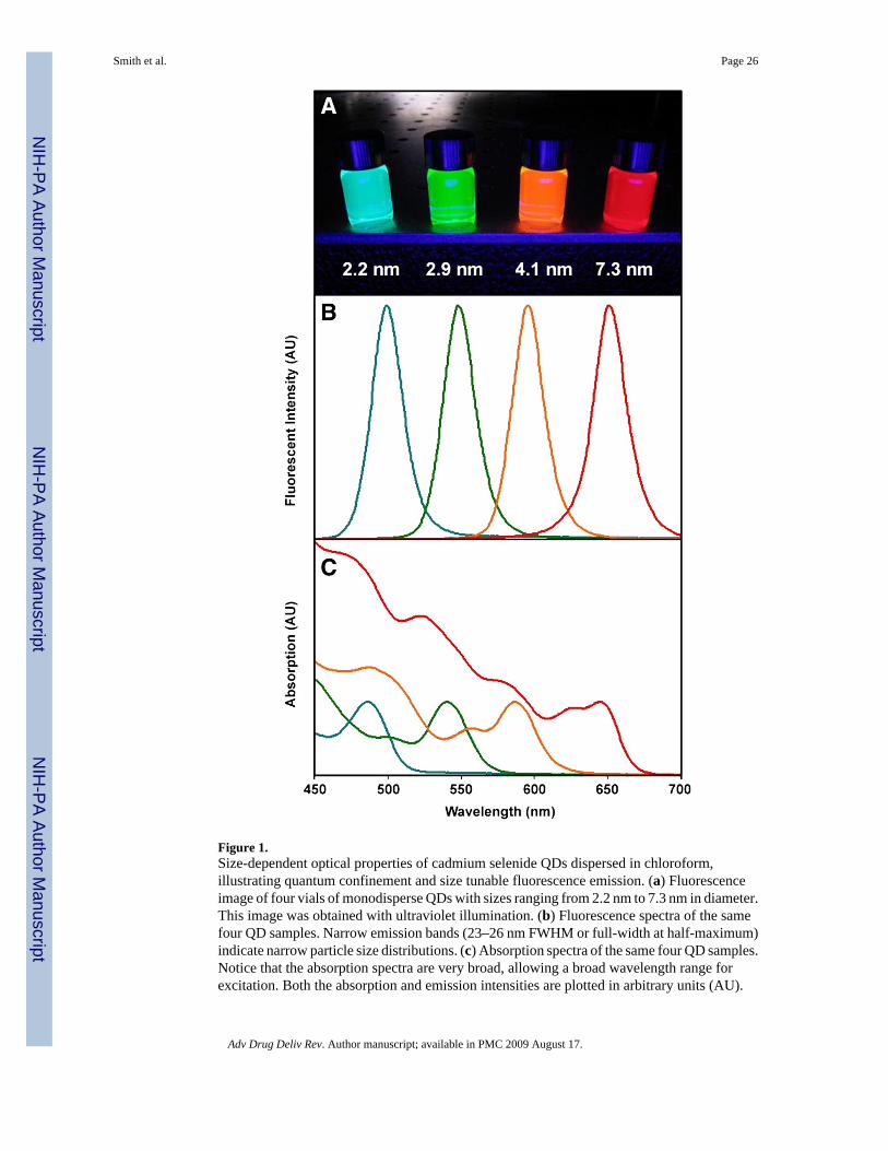
Figure 1.Size-dependent optical properties of cadmium selenide QDs dispersed in chloroform,illustrating quantum confinement and size tunable fluorescence emission. (a) Fluorescenceimage of four vials of monodisperse QDs with sizes ranging from 2.2 nm to 7.3 nm in diameter.This image was obtained with ultraviolet illumination. (b) Fluorescence spectra of the samefour QD samples. Narrow emission bands (23–26 nm FWHM or full-width at half-maximum)indicate narrow particle size distributions. (c) Absorption spectra of the same four QD samples.Notice that the absorption spectra are very broad, allowing a broad wavelength range forexcitation. Both the absorption and emission intensities are plotted in arbitrary units (AU).
Smith et al. Page 26
Adv Drug Deliv Rev. Author manuscript; available in PMC 2009 August 17.
NIH
-PA Author Manuscript
NIH
-PA Author Manuscript
NIH
-PA Author Manuscript
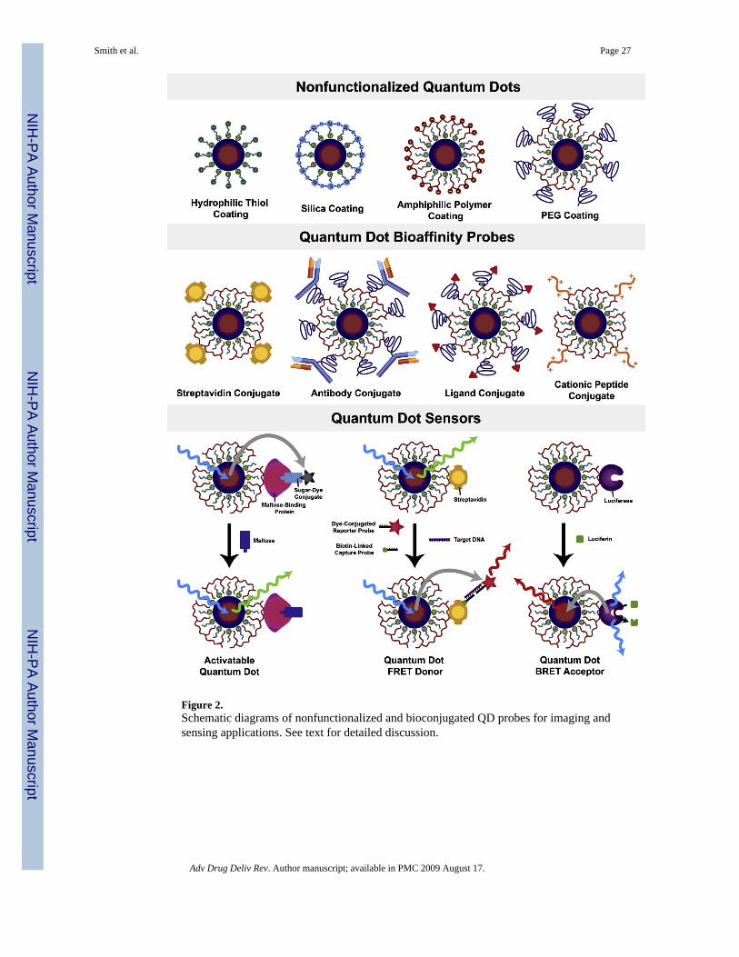
Figure 2.Schematic diagrams of nonfunctionalized and bioconjugated QD probes for imaging andsensing applications. See text for detailed discussion.
Smith et al. Page 27
Adv Drug Deliv Rev. Author manuscript; available in PMC 2009 August 17.
NIH
-PA Author Manuscript
NIH
-PA Author Manuscript
NIH
-PA Author Manuscript
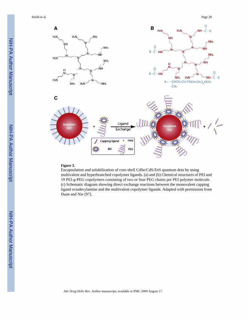
Figure 3.Encapsulation and solubilization of core-shell CdSe/CdS/ZnS quantum dots by usingmultivalent and hyperbranched copolymer ligands. (a) and (b) Chemical structures of PEI and19 PEI-g-PEG copolymers consisting of two or four PEG chains per PEI polymer molecule.(c) Schematic diagram showing direct exchange reactions between the monovalent cappingligand octadecylamine and the multivalent copolymer ligands. Adapted with permission fromDuan and Nie [97].
Smith et al. Page 28
Adv Drug Deliv Rev. Author manuscript; available in PMC 2009 August 17.
NIH
-PA Author Manuscript
NIH
-PA Author Manuscript
NIH
-PA Author Manuscript
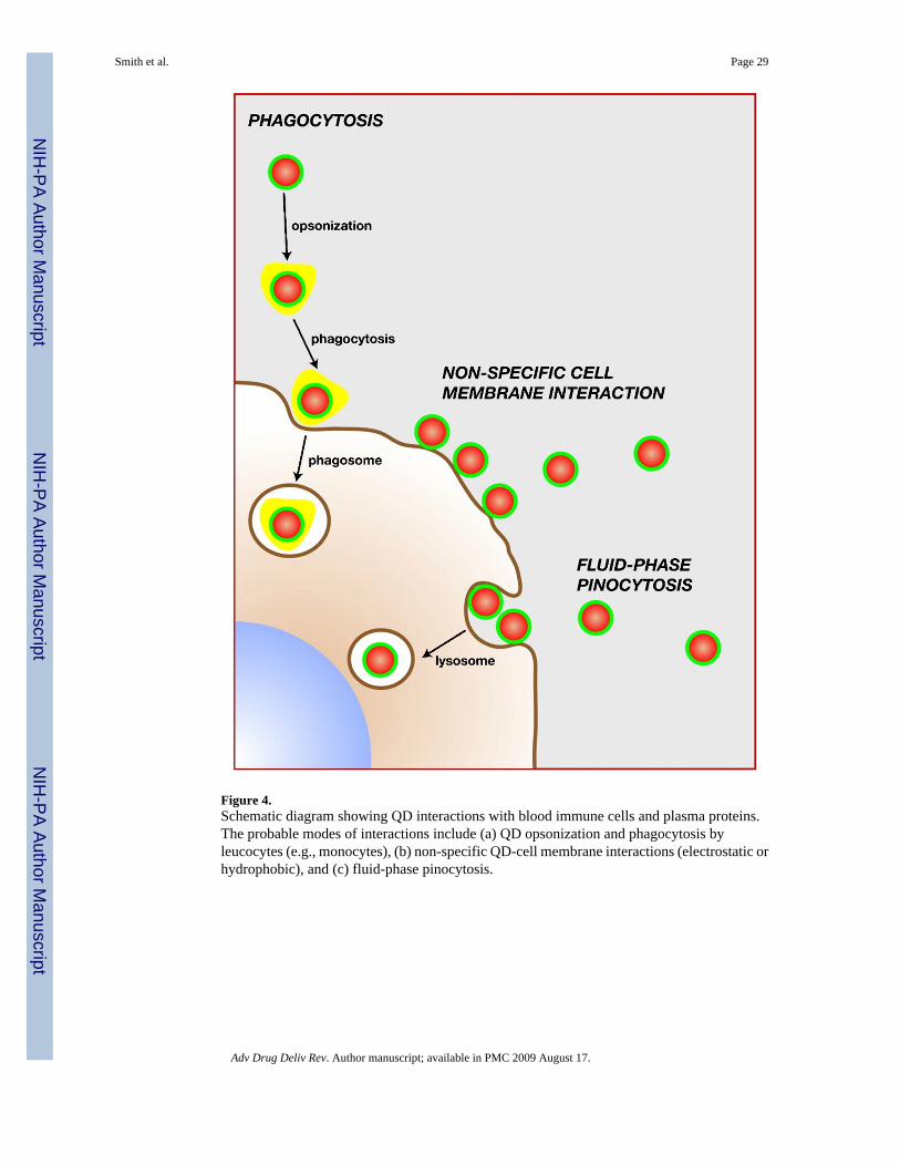
Figure 4.Schematic diagram showing QD interactions with blood immune cells and plasma proteins.The probable modes of interactions include (a) QD opsonization and phagocytosis byleucocytes (e.g., monocytes), (b) non-specific QD-cell membrane interactions (electrostatic orhydrophobic), and (c) fluid-phase pinocytosis.
Smith et al. Page 29
Adv Drug Deliv Rev. Author manuscript; available in PMC 2009 August 17.
NIH
-PA Author Manuscript
NIH
-PA Author Manuscript
NIH
-PA Author Manuscript
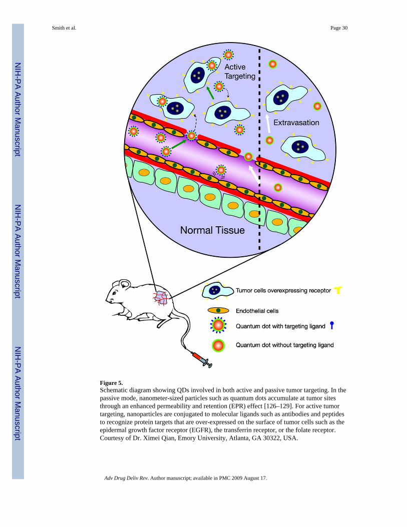
Figure 5.Schematic diagram showing QDs involved in both active and passive tumor targeting. In thepassive mode, nanometer-sized particles such as quantum dots accumulate at tumor sitesthrough an enhanced permeability and retention (EPR) effect [126–129]. For active tumortargeting, nanoparticles are conjugated to molecular ligands such as antibodies and peptidesto recognize protein targets that are over-expressed on the surface of tumor cells such as theepidermal growth factor receptor (EGFR), the transferrin receptor, or the folate receptor.Courtesy of Dr. Ximei Qian, Emory University, Atlanta, GA 30322, USA.
Smith et al. Page 30
Adv Drug Deliv Rev. Author manuscript; available in PMC 2009 August 17.
NIH
-PA Author Manuscript
NIH
-PA Author Manuscript
NIH
-PA Author Manuscript
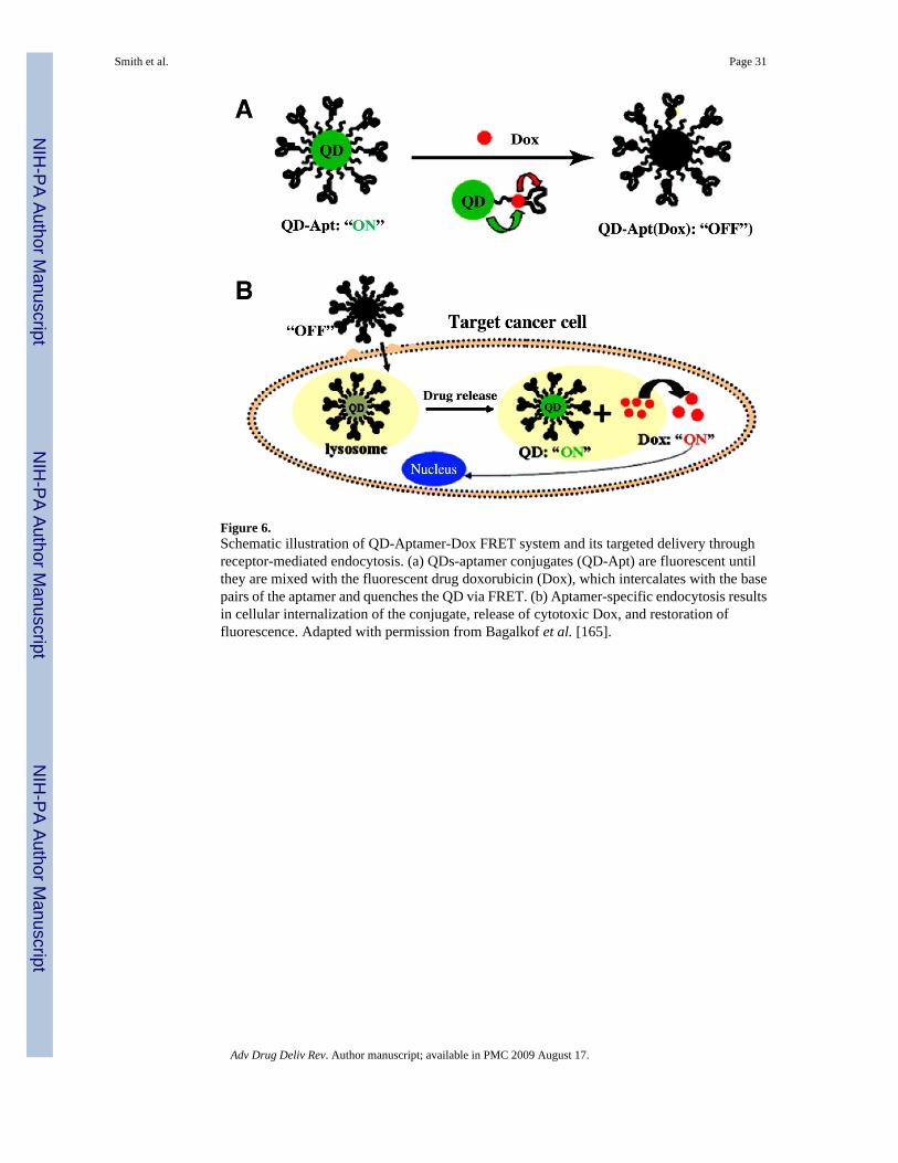
Figure 6.Schematic illustration of QD-Aptamer-Dox FRET system and its targeted delivery throughreceptor-mediated endocytosis. (a) QDs-aptamer conjugates (QD-Apt) are fluorescent untilthey are mixed with the fluorescent drug doxorubicin (Dox), which intercalates with the basepairs of the aptamer and quenches the QD via FRET. (b) Aptamer-specific endocytosis resultsin cellular internalization of the conjugate, release of cytotoxic Dox, and restoration offluorescence. Adapted with permission from Bagalkof et al. [165].
Smith et al. Page 31
Adv Drug Deliv Rev. Author manuscript; available in PMC 2009 August 17.
NIH
-PA Author Manuscript
NIH
-PA Author Manuscript
NIH
-PA Author Manuscript
Copyright © 2022 FDOKUMEN