T cell homeostatic proliferation elicits effective antitumor autoimmunity
Upload
independentCategory
view
0download
0
Biochemical Modulation of Cisplatin Mechanisms of Action: Enhancement ofAntitumor Activity and Circumvention of Drug Resistance
Miguel A. Fuertes,† Carlos Alonso,† and Jose. M. Perez*,‡
Centro de Biologıa Molecular “Severo Ochoa” (CSIC-UAM), and Departamento de Quımica Inorganica, Facultad de Ciencias, Universidad Autonomade Madrid 28049-Madrid, Spain
Received October 10, 2002
Contents1. Introduction 6452. Biochemical Mechanisms of Action of Cisplatin 646
2.1. Binding of Cisplatin to DNA Targets 6472.2. Binding of Cisplatin to Non-DNA Targets 6482.3. Biochemistry of Cisplatin-Induced Cell Death
Pathways648
3. Molecular Bases of Cisplatin Resistance 6503.1. Reduced Platinum Accumulation 6503.2. Cisplatin Inactivation by Thiol-Containing
Biomolecules650
3.3. The Role of DNA Adduct Structure andConformation in Cisplatin Resistance
651
3.4. Failure of Apoptotic Pathways 6524. Biochemical Modulation of Cisplatin
Chemosensitivity653
4.1. Cisplatin Accumulation 6534.2. Platinum Detoxification by Glutathione 6544.3. DNA Repair and Processing 6554.4. Cell Death Pathways 656
5. Concluding Remarks and Future Trends 6596. Acknowledgments 6607. References 660
1. IntroductionBiochemical modulation is the manipulation of
cellular biochemical pathways by chemical agents toproduce selective enhancement of the efficacy of anantitumor drug.1 Since the introduction of cisplatin(cis-DDP), cis-diamminedichloroplatinum(II), into gen-eral oncology practice, the studies dealing with the
molecular mechanism of action of the drug haveprovided considerable information as to how cisplatininduces its antitumor effects. The biochemical mech-anisms of cisplatin cytotoxicity involve the bindingof the drug to DNA and non-DNA targets and thesubsequent induction of cell death through apoptosis,necrosis, or both.2 In fact, it has been proposed thata functional cooperativity between these two formsof cell demise is regulated by the intracellular redoxpotential generated by the pyridine nucleotide pool(NAD+/NADH and NADP+/NADPH ratios) and thecellular free energy available from the ATP/ADPratio.3,4 Cisplatin is highly effective in the treatmentof testicular and ovarian cancers and is also employedfor treating bladder, cervical, head and neck, esoph-ageal, and small cell lung cancer.5 However, sometumors such as colorectal and nonsmall cell lungcancers have intrinsic resistance to cisplatin, whileothers such as ovarian or small cell lung cancersdevelop acquired resistance after the initial treat-ment.6 Biochemical studies have not clearly estab-lished the molecular bases of resistance to cisplatinin any type of cell, but, at least, they have identifiedseveral mechanisms that can contribute to thisphenomenon. Resistance to cisplatin is generallymultifactorial and has been shown to be due toreduced drug accumulation, inactivation by thiolcontaining species, increased repair/tolerance of plati-num-DNA adducts, and alterations in proteinsinvolved in apoptosis.7,8 One strategy to overcomecisplatin resistance is to design platinum complexesthat specifically deal with some or even all of theabove-mentioned resistance mechanisms. However,after more than 30 years of intensive research fromthe discovery of the antitumor activity of cisplatin,no more than 30 compounds have shown enoughpharmacological advantages relative to cisplatin tobe tested in clinical trials.9 Moreover, only four
* Corresponding Author. Phone: +34913978454. Fax: +34913874833. E-mail: [email protected].† Centro de Biologıa Molecular “Severo Ochoa” (CSIC-UAM).‡ Departamento de Quımica Inorganica, UAM.
Volume 103, Number 3
10.1021/cr020010d CCC: $44.00 © 2003 American Chemical SocietyPublished on Web 02/22/2003

platinum drugs are currently registered for clinicaluse (marketed drugs).10 Among the registered plati-num drugs (see Figure 1), carboplatin, [cis-diammine-1,1′-cyclobutane dycarboxilate platinum(II)], is lesstoxic than cisplatin but possesses the same spectrumof antitumor activity. The only registered platinumdrug that has consistently demonstrated antitumoractivity against cisplatin resistant tumors such ascolorectal cancers is oxaliplatin, [trans-L-1,2-diami-nocyclohexaneoxalatoplatinum(II)].11 However, therehave been recently reported several novel classes ofplatinum complexes able to circumvent cisplatinresistance in preclinical or even clinical studiesincluding cis-Pt(II) compounds with planar ligands,trans-Pt(II) and trans-Pt(IV) compounds, and poly-nuclear platinum complexes.9
Taking into account the above-mentioned consid-erations, there is little doubt that the search for novelplatinum compounds able to circumvent cisplatinresistance has proven to be a difficult task. An
alternative way based on the use of biochemicalmodulation strategies directed to circumvent cispla-tin resistance could enhance the antitumor activityof cisplatin improving the outcome of cancer patients.So far, an increasing number of drugs centered onthe biochemical mechanisms of modulation of cispla-tin resistance have been identified.6 The presentreview gives an update of the state-of-the-art of thebiochemical modulation of cisplatin mechanisms ofresistance and looks for future directions of researchon this important issue.
2. Biochemical Mechanisms of Action of CisplatinIt is generally accepted that binding of cisplatin to
genomic DNA (gDNA) in the cell nucleus is largelyresponsible for its antitumor properties.2 The damageinduced upon binding of cisplatin to gDNA mayinterfere with normal transcription and/or DNAreplication mechanisms. Eventually, these disrup-tions in DNA processing would trigger cytotoxicprocesses that lead to the death of the cancer cell.However, it is known that cisplatin forms a highamount of adducts in mitochondrial DNA (mtDNA)lacking histones.12 Moreover, mitochondria are un-able to carry out nucleotide excision repair (NER), amajor pathway for removing cisplatin-DNA ad-ducts.6 So, it should not be ruled out the possibilitythat mtDNA may also be an important pharmaco-logical target for cisplatin. In any event, previous tocisplatin binding to genomic or mitochondrial DNAa loss of chloride groups is required. However, the
Miguel Angel Fuertes Villadangos has been Research Associate ofMolecular Biology since 1981 at the Consejo Superior de InvestigacionesCientıficas (CSIC, Spain). In 1995 he joined the laboratory of ProfessorCarlos Alonso at the Centro de Biologıa Molecular “Severo Ochoa” ofthe Consejo Superior de Investigaciones Cientıficas-Universidad Autonomade Madrid (CSIC-UAM), Spain. His research interests center on structuralbiology, with particular focus on the interactions between metals andbiomolecules. He is a coauthor of 30 scientific articles.
Carlos Alonso Bedate is Research Professor of Molecular Biology at theCentro de Biologıa Molecular “Severo Ochoa” (CSIC-UAM). ProfessorCarlos Alonso is Reader in Macromolecular Interactions at the Departmentof Molecular Biology of the UAM. He was the Ph.D. supervisor of JoseManuel Perez Martın. His research interests cover molecular parasitology,immunology, platinum antitumor drugs, and structural biology. He is acoauthor of more than 150 scientific articles.
Jose Manuel Perez Martın obtained a degree in Biochemistry andMolecular Biology at the UAM in 1985. He completed his biochemicaltraining at the Centro de Biologıa Molecular “Severo Ochoa” (CSIC-UAM)and in 1992 obtained his Ph.D. in Bioinorganic Chemistry at theDepartment of Molecular Biology of the UAM. He received highest honors(summa cum laude) on his Ph.D. thesis entitled “Structure and AntitumorActivity of a Pt-pentamidine Complex”. Dr. Perez Martın had a postdoctoralposition from 1992 to 1996 at the Bristol-Myers-Squibb PharmaceuticalResearch Institute (Wallingford, CT, and Madrid, Spain) conducting clinicaland basic research with platinum and taxane antitumor drugs. In 1997he returned to the UAM, where he was recipient of a “Ramon y Cajal”postdoctoral fellowship at the Department of Inorganic Chemistry. From1999 up to the present, he has been Professor of Inorganic andBioinorganic Chemistry at the Department of Inorganic Chemistry of theUAM. In 2002, he was recipient of the Distinguished Leadership Awardof the American Biographical Institute for its outstanding contribution toInorganic Chemistry. He is a member of the Society of Biological InorganicChemistry. Professor Jose Manuel Perez Martın is coauthor of more than70 scientific papers and four patents. During his free time, he enjoysplaying classical guitar (mainly plays by Bach, Sors, Tarrega, and Hackett).
646 Chemical Reviews, 2003, Vol. 103, No. 3 Fuertes et al.

high chloride concentration in extracellular fluids(≈100 mM) suppresses the formation of mono- anddiaquo cis-Pt(II) species in which one or both chloridegroups are replaced by water molecules. In contrast,within the cell, the chloride concentration rangesbetween 2 and 30 mM. In this range of Cl- concen-trations, the hydrolysis of cis-DDP occurs efficientlyso that one or both chloride leaving groups arereplaced by water molecules, allowing the formationof aquo species. The final result is the formation ofthe [Pt(H2O)2(NH3)2]2+ cation. This diaquo species isvery reactive toward nucleophile centers of biomol-ecules because H2O is much better leaving groupthan Cl-.13
2.1. Binding of Cisplatin to DNA Targets
The N7 atoms of the imidazole rings of guanine andadenine located in the major groove of the doublehelix are the most accessible and reactive nucleo-philic sites for platinum binding to DNA.14 Thereaction of cisplatin with DNA may lead to theformation of various structurally different adducts.Initially, monofunctional DNA adducts are formed,but most of them further react to produce interstrandor intrastrand cross-links, which then block replica-tion and/or prevent transcription.15 It has been foundthat 60-65% of adducts formed by cisplatin are1,2-d(GpG) intrastrand cross-links and 20-25%1,2-d(ApG) intrastrand cross-links. Minor adducts,each accounting for a few percent, include 1,3-intra-strand cross-links and interstrand cross-links.16 DNA-protein cross-links have also been reported to beinduced by cis-DDP.17 Both the 1,2-d(GpG) and
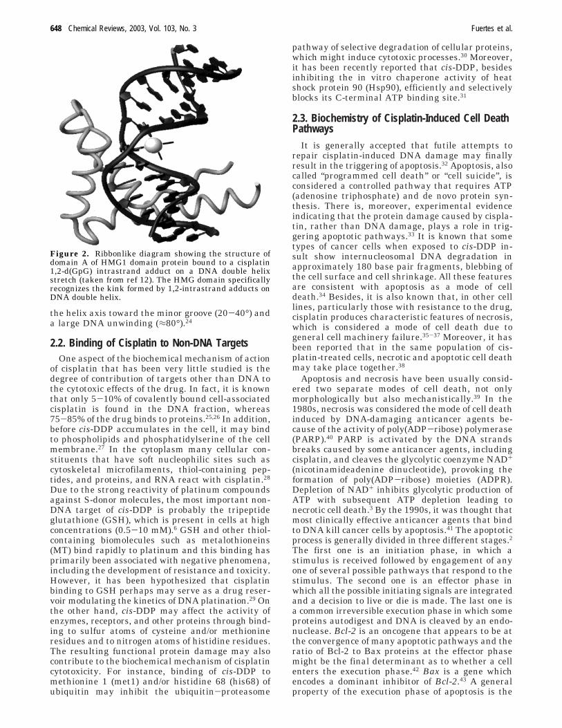
1,2-d(ApG) intrastrand cross-links unwind DNA by13°, while the 1,3-d(GpXpG) intrastrand cross-linksunwind DNA by 23°. Interestingly, however, bendingof the DNA double helix is similar (32-35°) for thesethree types of intrastrand adducts.18 There is stilldebate as to which types of cisplatin-DNA adductsare the most important in mediating the cytotox-icity of cis-DDP. Support for the role of the major 1,2-intrastrand adducts in cisplatin-induced tumor cellkilling arises from the fact that due to steric reasons,the inactive trans isomer of cisplatin, trans-DDP ortransplatin (see Figure 1), is unable to form theseadducts. In fact, trans-DDP mainly forms 1,3-intras-trand and interstrand cross-links.19 Moreover, it hasbeen found that the 1,2-intrastrand adducts are lesseffectively removed from DNA by repair enzymesthan 1,3-intrastrand adducts.20,21 Further support for1,2-intrastrand cross-links as the main adducts re-sponsible for the antitumor activity of cisplatin camefrom the discovery that some HMG (high mobilitygroup) domain proteins specifically recognize thistype of DNA adduct (see Figure 2). So, it has beenproposed that specific HMG proteins may be involvedin the cellular processing of the 1,2-intrastrand cross-links formed by cis-DDP.22 However, the possibleimportance of minor adducts, as interstrand andDNA-protein cross-links in the mechanism of cyto-toxic activity of cisplatin, should not be ruled out. Infact, some studies have shown a relationship betweencell killing or resistance and numbers or repair ofinterstrand and DNA-protein cross-links.23 It shouldbe pointed out that interstrand cross-links of cis-DDPinduce striking distortions on DNA with bending of
Figure 1. (a) Structures of the four platinum antitumor drugs currently registered for clinical use (marketed drugs) andof (b) two biologically inactive platinum compounds.
Cisplatin Mechanisms of Action Chemical Reviews, 2003, Vol. 103, No. 3 647
the helix axis toward the minor groove (20-40°) anda large DNA unwinding (≈80°).24
2.2. Binding of Cisplatin to Non-DNA TargetsOne aspect of the biochemical mechanism of action
of cisplatin that has been very little studied is thedegree of contribution of targets other than DNA tothe cytotoxic effects of the drug. In fact, it is knownthat only 5-10% of covalently bound cell-associatedcisplatin is found in the DNA fraction, whereas75-85% of the drug binds to proteins.25,26 In addition,before cis-DDP accumulates in the cell, it may bindto phospholipids and phosphatidylserine of the cellmembrane.27 In the cytoplasm many cellular con-stituents that have soft nucleophilic sites such ascytoskeletal microfilaments, thiol-containing pep-tides, and proteins, and RNA react with cisplatin.28
Due to the strong reactivity of platinum compoundsagainst S-donor molecules, the most important non-DNA target of cis-DDP is probably the tripeptideglutathione (GSH), which is present in cells at highconcentrations (0.5-10 mM).6 GSH and other thiol-containing biomolecules such as metalothioneins(MT) bind rapidly to platinum and this binding hasprimarily been associated with negative phenomena,including the development of resistance and toxicity.However, it has been hypothesized that cisplatinbinding to GSH perhaps may serve as a drug reser-voir modulating the kinetics of DNA platination.29 Onthe other hand, cis-DDP may affect the activity ofenzymes, receptors, and other proteins through bind-ing to sulfur atoms of cysteine and/or methionineresidues and to nitrogen atoms of histidine residues.The resulting functional protein damage may alsocontribute to the biochemical mechanism of cisplatincytotoxicity. For instance, binding of cis-DDP tomethionine 1 (met1) and/or histidine 68 (his68) ofubiquitin may inhibit the ubiquitin-proteasome
pathway of selective degradation of cellular proteins,which might induce cytotoxic processes.30 Moreover,it has been recently reported that cis-DDP, besidesinhibiting the in vitro chaperone activity of heatshock protein 90 (Hsp90), efficiently and selectivelyblocks its C-terminal ATP binding site.31
2.3. Biochemistry of Cisplatin-Induced Cell DeathPathways
It is generally accepted that futile attempts torepair cisplatin-induced DNA damage may finallyresult in the triggering of apoptosis.32 Apoptosis, alsocalled “programmed cell death” or “cell suicide”, isconsidered a controlled pathway that requires ATP(adenosine triphosphate) and de novo protein syn-thesis. There is, moreover, experimental evidenceindicating that the protein damage caused by cispla-tin, rather than DNA damage, plays a role in trig-gering apoptotic pathways.33 It is known that sometypes of cancer cells when exposed to cis-DDP in-sult show internucleosomal DNA degradation inapproximately 180 base pair fragments, blebbing ofthe cell surface and cell shrinkage. All these featuresare consistent with apoptosis as a mode of celldeath.34 Besides, it is also known that, in other celllines, particularly those with resistance to the drug,cisplatin produces characteristic features of necrosis,which is considered a mode of cell death due togeneral cell machinery failure.35-37 Moreover, it hasbeen reported that in the same population of cis-platin-treated cells, necrotic and apoptotic cell deathmay take place together.38
Apoptosis and necrosis have been usually consid-ered two separate modes of cell death, not onlymorphologically but also mechanistically.39 In the1980s, necrosis was considered the mode of cell deathinduced by DNA-damaging anticancer agents be-cause of the activity of poly(ADP-ribose) polymerase(PARP).40 PARP is activated by the DNA strandsbreaks caused by some anticancer agents, includingcisplatin, and cleaves the glycolytic coenzyme NAD+
(nicotinamideadenine dinucleotide), provoking theformation of poly(ADP-ribose) moieties (ADPR).Depletion of NAD+ inhibits glycolytic production ofATP with subsequent ATP depletion leading tonecrotic cell death.3 By the 1990s, it was thought thatmost clinically effective anticancer agents that bindto DNA kill cancer cells by apoptosis.41 The apoptoticprocess is generally divided in three different stages.2The first one is an initiation phase, in which astimulus is received followed by engagement of anyone of several possible pathways that respond to thestimulus. The second one is an effector phase inwhich all the possible initiating signals are integratedand a decision to live or die is made. The last one isa common irreversible execution phase in which someproteins autodigest and DNA is cleaved by an endo-nuclease. Bcl-2 is an oncogene that appears to be atthe convergence of many apoptotic pathways and theratio of Bcl-2 to Bax proteins at the effector phasemight be the final determinant as to whether a cellenters the execution phase.42 Bax is a gene whichencodes a dominant inhibitor of Bcl-2.43 A generalproperty of the execution phase of apoptosis is the
Figure 2. Ribbonlike diagram showing the structure ofdomain A of HMG1 domain protein bound to a cisplatin1,2-d(GpG) intrastrand adduct on a DNA double helixstretch (taken from ref 12). The HMG domain specificallyrecognizes the kink formed by 1,2-intrastrand adducts onDNA double helix.
648 Chemical Reviews, 2003, Vol. 103, No. 3 Fuertes et al.

specific degradation of a series of proteins by thecysteine aspartate-specific proteinases (caspases).Caspases are activated when an apoptotic stimulusinduces the release of cytochrome c from mitochon-dria.44 It has been recently found that mitochondriaplay a central role in apoptosis. As depicted in Figure3, cisplatin DNA damage induces a fall in themitochondrial permeability transition (MPT).45 Sub-sequently, the MPT fall releases factors that facilitatethe rupture of mitochondria such as reactive oxygenspecies (ROS), Bax, and Ca2+.46 Mitochondrial rup-ture releases cytochrome c and procaspase-9 thatbind to cytosolic Apaf-1 and ATP in an apoptosomecomplex, leading to the activation of caspase-9.Activated caspase-9 induces other caspases interac-tions, resulting in activation of caspase-3, caspase-6, and caspase-7 with the subsequent cleavage of keysubstrates.47 The final outcome is the dismantling ofthe cell by formation of apoptotic bodies. An alterna-tive pathway of apoptosis may be initiated by injuryof phospholipids of the cell membrane, which mayinduce the sphingomyelin-ceramide signaling sys-tem of cell death.48 A third possible apoptotic path-way is the one in which the activation of Fas receptorby Fas ligand (FasL) induces the formation of anapoptosome complex between Fas-associated deathdomain (FADD) and procaspase-8 that subsequentlyactivates caspase-8. Then caspase-8 activates thecaspase-3-6-7 system that finally cleaves key sub-strates, and the cell is digested through apoptosis.Caspase-8 may also activate the proapoptotic proteinBid that triggers apoptotic cell death through themitochondrial pathway.49
An outstanding contribution to the study of thebiochemical mechanisms of cell death was the dis-covery, by the end of the 1990s, that intracellularATP levels dictate whether antitumor drugs, includ-ing cisplatin as well as other chemical and physicalagents, induce cell death by necrosis or apoptosis andthat both processes of cell death are linked.50-52
Figure 3 shows in discontinuous arrows the currentlyknown interconnections between apoptotic and ne-crotic pathways. The cleavage of PARP by caspase-3, -6, or -7 switches the cell death mechanism formnecrosis to apoptosis. Thus, by inactivating PARP,caspase-3, -6, or -7 relieves necrotic-mediated celldeath by virtue of preventing the depletion in NAD+
and ATP. Caspase blocking by inhibitors of apoptosis(IAPs) plus continued activity by PARP and ATPdecrease, by the inhibition of electron transport inbroken mitochondria, lead the cell to necrosis becauseof continuation of PARP-induced ATP depletion.53
However, in some cases failure to cleave PARP canalso lead to apoptosis since depletion of NAD+/ATPmay increase the activity of MPT, thereby promotingROS, BAX, and Ca2+.54,55 Altogether, the above-mentioned data indicate that there is a functionalcooperativity between apoptotic and necrotic celldeath pathways. This hypothesis would explain someunusual observations, indicating that cells might alsodie as a result of an unfinished apoptotic program.For instance, it has been reported that cisplatin-induced cell death in the L1210 leukemic cell linemight be the consequence of a defective apoptoticprogram that lacks some morphological and biochemi-cal properties attributed to “classic” apoptosis.56
Figure 3. Schematic overview of the proposed biochemical pathways of cell death induced by cis-DDP showing theinterconnections between apoptosis and necrosis (discontinuous arrows). DNA strand breaks activate poly(ADP-ribose)polymerase (PARP), which cleaves NAD+ and provokes the formation of poly(ADP-ribose) moieties (ADPR). The result isa decrease in NAD+ with a concomitant fall of glycolysis and subsequent ATP depletion so that cell death by necrosistakes place. In contrast, if ATP levels are enough to sustain survival, caspase-3-6-7 cleaves PARP, necrosis is blocked, andapoptosis occurs. If PARP cleavage is prevented, the continued activity of PARP leads to enhancement of both necrosisand apoptosis. Apaf-1, apoptotic protease-activating factor-1; Bid, a type of proapoptotic protein; FAAD, Fas-associateddeath domain; Fas, cell surface membrane receptor; TNFR-1, tumor necrosis factor receptor; IAP, inhibitor of apoptosis;PAR, poly(ADP-ribose); ROS, reactive oxygen species; Cyto c, cytochrome c; Mito; mitochondrial; MPT, mitochondrialpermeability transition.
Cisplatin Mechanisms of Action Chemical Reviews, 2003, Vol. 103, No. 3 649

3. Molecular Bases of Cisplatin ResistanceThe occurrence of resistance is a common drawback
of cancer chemotherapy, and cis-DDP is no excep-tion.6 Moreover, the patterns of cisplatin resistancevary considerably between tumor types. Some tu-mors, such as colorectal cancer and nonsmall celllung cancer (NSCLC), are intrinsically resistant tocis-DDP chemotherapy.57,58 Other tumor types, suchas head and neck cancer, testicular cancer, ovariancancer, and small cell lung cancer (SCLC), are pre-dominantly sensitive to cis-DDP treatment. However,most of these sensitive tumors develop acquiredresistance after the initial treatment.6 In most pre-clinical models of cisplatin resistance (either acquiredor intrinsic), multiple mechanisms appear to operate.Thus, cancer cells that are resistant to cis-DDP oftenexhibit several resistance mechanisms acting simul-taneously. The molecular mechanisms of resistanceagainst cis-DDP can be divided into two maingroups: mechanisms that prevent cis-DDP reachingDNA as its main therapeutic target and mechanismsthat block the induction of cell death (apoptosis ornecrosis) after the formation of the cisplatin-DNAadduct.6,59 This is schematically outlined in Figure4.
3.1. Reduced Platinum AccumulationDecreased uptake/increased efflux of cis-DDP leads
to lower intracellular concentrations of drug.60 Mostin vitro models of acquired resistance to cisplatinexhibit a decrease in platinum accumulation between2- and 4-fold.8 It is generally accepted that reducedplatinum accumulation is due to reduce drug uptakerather than to increased drug efflux because the mainmultidrug resistance efflux pump, P-glycoprotein(Pgp), is not usually overexpressed in cisplatin-resistant tumors.61 At present, it is known that Pgp
is a complex multispanning membrane protein thatbelongs to the ABC (ATP-binding cassette) transport-ers. ABC utilizes ATP hydrolysis as fuel to export cis-DDP and other antineoplastics against a drug con-centration gradient and has been biochemical andpharmacologically characterized.62,63 However, newmultidrug resistance transporters are being charac-terized, as is the case of the multidrug resistance-associated protein group (MRP), which currently hasseven members and also belongs to the ABC familyof proteins. It has been recently found that MRPproteins preferably transport drugs (e.g., methotrex-ate, arsenite, or cis-DDP) outside the cell by conjuga-tion with sulfate, glucuronate, or GSH. In fact, theMRP1 and MRP2 proteins confer resistance to cis-DDP probably by transporting the drug in complexeswith GSH.64 MRP2 pump is also known as canalicu-lar MRP (cMRP) or canalicular multispecific organicanion transporter (cMOAT). Although the biochemi-cal mechanisms by which cis-DDP enters the cellsare not fully understood, it appears that passivediffusion is the main way of cis-DDP uptake. How-ever, some facilitated or active transport mechanismsmay contribute to cisplatin intracellular accumula-tion.65 In fact, although cis-DDP uptake is not satu-rable or inhibited by structural analogues, a certaindegree of uptake is, at least, energy-dependent andcan be modulated by pharmacological agents such asNa+/K+-ATPase inhibitors and membrane-interactivedrugs.61,65
3.2. Cisplatin Inactivation by Thiol-ContainingBiomolecules
A more established biochemical mechanism ofresistance to cisplatin is the intracellular inactivationof the cis-Pt(II) center prior to binding to DNA bycoordination to S-donor cysteine residues of thecytoplasmic tripeptide glutathione (see glutathionestructure in Figure 4) or metallothioneins (a class oflow-molecular-weight proteins).9 Glutathione (GSH)is a tripeptide of glutamate (Glu), cysteine (Cys), andglycine (Gly) that contains an unusual γ-peptide bondbetween glutamate and cysteine (γ-GluCysGly). Sucha bond prevents GSH from being hydrolyzed by mostpeptidases. Intracellularly, GSH is kept in its thiolform by glutathione disulfide reductase, a NADPH-dependent enzyme. GSH reacts with cisplatin andother electrophilic compounds to form deactivatedconjugates that are readily excreted by a GS-con-jugated export pump. This reaction may occur spon-taneously or with the help of the glutathione S-trans-ferase enzyme (GST).66 The removal of platinum byGSH depletes intracellular GSH levels. GSH deple-tion is known to sensitize cells to many cyto-toxic agents including cisplatin through activation ofsphingomyelinase (SMase), which increases ceramidelevels leading to SMase-induced apoptosis.67 On theother hand, high intracellular concentrations of GSH(up to 10 mM) often correlate with cis-DDP resis-tance. In fact, prominent GS-Pt-SG complexes(binding ratio of 1 mole of platinum per 2 mol ofglutathione) have been found in tumor cells.8 On theother hand, increased levels of metallothioneins havebeen also found in some cell lines with acquired
Figure 4. Schematic drawing of the major biochemicalmechanisms of resistance to cisplatin. Resistance mecha-nisms may operate prior to or after binding of cis-DDP toDNA. MMR ) mismatch repair; NER ) nucleotide excisionrepair; GSH ) glutathione.
650 Chemical Reviews, 2003, Vol. 103, No. 3 Fuertes et al.

resistance to cis-DDP.6,8 Mammalian metallothionein(MT) is a small protein of 62 amino acids whichcontains 20 cysteine residues. Mammalian MT hasbeen involved in intracellular detoxification of heavymetal ions such as Cd2+ and Zn2+. Cisplatin binds toMT, with a stoichiometry of 10 Pt atoms per MTmolecule and with a binding constant rate which issignificantly higher than that for GSH. When cis-DDP binds to MT, it loses its NH3 ligands anddisplaces from MT heavy-metal cations (e.g., Zn2+)according to the reaction
It is not yet clear whether MT plays a role in cis-DDP resistance. However, it has been recently re-ported that transfection of the human metallothio-nein MT-IIA cDNA into cells conferred over 4-foldresistance to cisplatin.68
3.3. The Role of DNA Adduct Structure andConformation in Cisplatin Resistance
It is known that even high levels of DNA platina-tion may not always induce cell death.6 Severalproteins have been described that recognize and bindto cisplatin-DNA adducts. These proteins are called“damage-recognition proteins” and include, amongothers, the XPA-RPA complex, nonhistone chroma-tin high mobility group HMG1 and HMG2, histoneH1, the TATA-box binding protein TBP, andHMSH2.12 Damage-recognition proteins may eitherassist in the repair of DNA lesions provoked by cis-DDP or, conversely, shield damage from repairproteins.69
An important biochemical mechanism of resistancethat occurs after platinum binding is the repair ofDNA damage. Nucleotide excision repair (NER) ap-pears to be a major mechanism of cisplatin-resis-tance. Increased NER in cisplatin-resistant cell lineshas been shown to occur both for intrastrand as wellas interstrand cisplatin-DNA adducts.2 For the 1,2-intrastrand adducts of cis-DDP, the NER system isof particular importance.70 NER is an ATP-dependentmultiprotein complex that recognizes the kink in-duced on DNA by 1,2-intrastrand cross-links andsubsequently excises the segment of the DNA thatincludes the kink, as a 27-29-base-pair oligonucle-otide. The gap that remains is then filled by DNApolymerase.71 Increased removal by NER of platinum-DNA adducts from the genome of resistant cell linesrelative to sensitive parent cell lines has been con-sistently observed in several models. The increasedNER activity in cisplatin-resistant cell lines appearsto be most strongly associated with increased levelsof expression of ERCC1 and XPA proteins.12,72 Con-versely, defective NER has been found in cell lineswith hypersensitivity to cis-DDP. In fact, it has beenrecently reported that the testis specific proteintsHMG, which belongs to the family of HMG domainproteins, might bind to 1,2-d(GpG) intrastrand cross-links blocking DNA repair by NER.73
As mentioned above, HMG-box proteins, includingHMG1 and HMG2, bind selectively to DNA modifiedby cis-DDP but not to that modified by biologicallyinactive trans-DDP.74 In contrast to NER proteins,HMG1 is able to inhibit the repair of the major 1,2-d(GpG) intrastrand cisplatin-DNA adduct by humanexcision nuclease in vitro.75 Several mechanisms havebeen proposed to explain how HMG domain proteinsmight modulate the sensitivity of cells to cis-DDP.Two of them seem to be the most feasible ones. The“repair shielding model” postulates that HMG pro-teins could protect cisplatin-DNA adducts fromrecognition by DNA repair enzymes.76 The secondone, the so-called “hijacking model”, establishes thatHMG proteins such as SSRP1 could modulate cellcycle events after DNA damage and trigger celldeath. Thus, this latter model postulates that therecognition by HMG cellular factors of cisplatin-DNA lesions would deviate them from their naturalbinding sites resulting in inhibition of vital cellularfunctions.77
The post-binding mechanism described as “in-creased tolerance” is probably one of the most generalbiochemical mechanisms of resistance encounteredin cancer chemotherapy of DNA-binding drugs.78
Post-replication repair is defined as the replicationof damaged DNA without the introduction of gapsinto the DNA and/or the repair of those discontinui-ties following replication.78 Since the presence of gapsor discontinuities in replicated DNA can be lethal,post-replication repair is a major mechanism of DNAdamage tolerance. In human cells, post-replicationappears to occur primarily during replication so thatit is often referred as a replicative bypass. Enhancedpost-replicative bypass, the ability of the replicationcomplex to synthesize DNA downstream a cisplatin-induced lesion, has been found in some cisplatin-resistant cells.79 The biochemical bases of increasedtolerance to damaged DNA are still unclear. How-ever, increased post-replicative bypass of cisplatin-DNA adducts has been observed in cell lines withdefects on a second DNA repair process namedmismatch repair (MMR).80 The mismatch repairsystem involves at least five proteins (MLH1, MSH2,MSH3, MSH6, and PMS2) and functions as anATP-dependent repair process that corrects misin-corporated nucleotides.81 The human MSH2 protein(hMSH2) recognizes 1,2-d(GpG) intrastrand cross-links of cis-DDP on DNA.82 MMR defects in hMutSR(a heterodimer of hMSH2 and hMSH6) or hMutLR(a heterodimer of hMLH1 and PMS2) have beenshown to contribute to an increase in replicativebypass of cisplatin-DNA adducts.8 In cisplatin-sensitive MMR-proficient cells, binding of hMutSRor hMutLR MMR complex to cisplatin-DNA adductsis thought to result in a continuous futile cycle ofrepair on the opposing DNA strand, ultimately lead-ing to cell death (see Figure 5).83 It has been recentlyreported that the registered drug oxaliplatin, whichcontains the bulky, nonpolar 1,2-diaminocyclohexane(DACH) ligand in place of the ammine ligands of cis-DDP (see Figure 1), is able to circumvent cisplatin-resistance in MMR-deficient tumor cell lines.84 Invitro, MutS binding assays have revealed that puri-
(Zn2+)7-MT + 10(NH3)2Pt2+ f
(Pt2+)10-MT + 20NH3 + 7Zn2+
Cisplatin Mechanisms of Action Chemical Reviews, 2003, Vol. 103, No. 3 651

fied MutS binds to cisplatin-modified DNA with2-fold greater affinity than DNA modified with oxa-liplatin.85 So it is likely that structural differencesin the conformation of DNA adducts formed byoxaliplatin relative to its analogue cis-DDP may beimportant in the induction of cell killing effects bydifferent biochemical mechanisms. These recent find-ings support the development of platinum drugsbased on their coordination chemistry to combat drugresistance in tumors.86
3.4. Failure of Apoptotic PathwaysTo trigger apoptosis it is believed that cellular
damage has to pass a certain threshold level.2 How-ever, damaged genes are common in cancer cells inwhich proteins involved in apoptotic pathways oftenmalfunction. This can make certain types of cancerrather insensitive to cis-DDP damage.87 Upstreamfactors involved in the cellular response to thedamaged DNA mediate the induction of a networkthat transmits both pro- and antiapoptotic signals.So, any interference that induces antiapoptotic signaltransduction or abrogates proapoptotic pathways,including transcriptional and translational responses,is a potential mechanism of cis-DDP resistance.
It is known that p53 protein plays a central rolein chemotherapy-induced apoptosis. The tumor sup-pressor gene p53 facilitates DNA repair before DNAreplication. p53, considered to be “guardian of the
genome”, functions as a transcription factor regulat-ing a host of other genes that lead to cell cycle arrestor induction of cell death.88 In fact, p53 is a strongtranscriptional activator of the gene encodingp21WAF1/CIP1, a protein that mediates cell cycle arrestand that may also protect cells from apoptosis.89 Ithas been recently found that p53 may be involved inthe development of cis-DDP resistance through theregulation of several genes involved in drug resis-tance and apoptosis (e.g, mismatch repair, bcl-2, highmobility group proteins, DNA polymerases R and â,PCNA, and insulin-like growth factors).78 However,a clear relationship between p53 cellular status andcisplatin-induced cytotoxicity has not been found yet.In fact, the cell type and the cellular context exert astrong influence in p53-mediated responses to cis-DDP damage of DNA.43,90
The bcl2 family of genes encodes a group ofproapoptotic (e.g., Bax, Bak, Bad) and antiapoptotic(e.g., Bcl-2, Bcl-XL) proteins, which form homo- andheterodimers with one another. It is currently thoughtthat the relative level of pro- and antiapoptoticproteins may function as a cell survival/cell deathrheostat to influence sensitivity and resistance tocisplatin-induced apoptosis. So high levels of Bcl-2may induce resistance to cis-DDP through inhibitionof apoptosis.91 On the other hand, Bcl-2 and p53 areoften overexpressed in resistant ovarian cancer celllines and it has been proposed that Bcl-2 might act
Figure 5. Proposed model for the contribution of mismatch-repair (MMR) activity to cis-DDP (CDDP) cytotoxicity. DNAreplication downstream the cisplatin 1,2-d(GpG) intrastrand cross-link results in imperfect base pairing. This alterationis recognized by the hMutLR/hMutSR MMR complex. Attempted MMR fails because it is directed at the daughter DNAstrand. So the newly synthesized DNA strand is removed, and the intrastrand cross-link on the parental DNA strandremains unexcised. The continued action of these futile replication/repair cycles results in the formation of gaps or strandbreaks. An inability to initiate mismatch correction results in cisplatin resistance because these futile repair attemptswould be avoided.
652 Chemical Reviews, 2003, Vol. 103, No. 3 Fuertes et al.

upstream of the p53 pathway.43 However, it has alsobeen found that high levels of Bcl-2 increase sensitiv-ity to cis-DDP in human ovarian cancer cells.92
Protein kinase A (PKA) may confer sensitivity tocis-DDP through apoptosis induction.93 Protein ki-nase A, also called cyclic AMP-dependent proteinkinase, participates in glycogen breakdown to glucose-1-phosphate (glycogenolysis). PKA is a tetramerhaving two catalytic subunits, C, and two regulatorysubunits, R. The tetramer, R2C2, is catalyticallyinactive, and binding of cyclic AMP (cAMP) to the Rsubunits causes the tetramer to dissociate, yieldingthe catalytically active monomer C.94 Besides, PDE(phosphodiesterase) negatively regulates PKA througha decrease in the intracellular concentrations ofcAMP. Decreased cAMP levels may produce resis-tance to cis-DDP through suppression of Bcl-2 ex-pression and inhibition of apoptosis.95
Sphingosine-1-phosphate phosphatase (S1PP) is anenzyme that produces sphingosine by cleavage of theester-phosphate bond of sphingosine-1-phosphate(S1P). The impairment of S1PP activity shifts thebalance between ceramide, which as mentioned aboveinduces apoptosis, and S1P, which promotes cellsurvival.96 Inhibition of S1PP will lead to accumula-tion of S1P, thereby protecting cells form apoptosis.However, S1P can also induce cell death in certaincells. So the modulation of the ceramide-sphingosine-sphingosine-1-phosphate rheostat may be an impor-tant factor with relation to potential resistance to cis-DDP.97
In somatic cells, the ends of chromosomes (thetelomeres) shorten with each cell division. However,in tumor cells, telomere length is maintained, mainlythrough activation of the reverse transcriptase en-zyme, telomerase.98 Telomeres are tandemly repeatedDNA sequences, comprising a G-rich strand and acomplementary C-rich strand, located at the end ofthe chromosomes. Telomerase is a ribonucleoproteinthat uses its RNA component as template to synthe-size the 5′-d(TTAGGG)-3′ repeats at the ends of thechromosomes. Thus, to maintain telomere length,malignant cells have to replicate the telomeric motifseveral times, yielding a d(TTAGGG)n telomericsequence.69 It has been reported that HeLa cellssensitive to low doses of cis-DDP may die throughapoptosis as a consequence of cisplatin binding totelomeres and subsequent telomere loss.99 Therefore,a putative resistance mechanism to cisplatin mightbe related with hyperactivation of telomerase andinability of the tumor cell to engage apoptosis.100
4. Biochemical Modulation of CisplatinChemosensitivity
Treatment of cisplatin-resistant tumors is a majordrawback which may, at least, be partially addressedby using biochemical modulation strategies directedto the enhancement of the activity of cis-DDP throughmanipulation of resistance pathways by pharmaco-logical agents. Table 1 summarizes the resistancemechanisms to cis-DDP which have been so farmanipulated with biochemical modulators. In addi-tion, Figure 6 shows the structure of several selectedbiochemical modulators of cis-DDP resistance.
4.1. Cisplatin Accumulation
Decreased accumulation of cis-DDP in the tumorcell is one of the most important mechanisms ofresistance to the drug both in preclinical and clinicalsettings.5,6 As previously mentioned, cis-DDP accum-ulation depends on two factors: drug uptake anddrug efflux. The most promising drugs in modulatingcisplatin uptake and/or efflux are dipyridamole, am-photericin B, and cyclosporine A.
Dipyridamole (Dpm) is a pyrimido-pyrimidine de-rivative that decreases cisplatin efflux and increasesdrug uptake.101 The mechanism by which Dpmdecreases cisplatin efflux is not yet fully understood.In animal cells a single protein, the nucleosidetransporter, appears to be responsible for uptake ofa wide variety of nucleosides including adenosine.Functioning of this protein is inhibited by Dpm,which blocks the uptake of adenosine.102 It is thoughtthat dypiridamole may exert an indirect effect in cis-DDP accumulation by decreasing ATP-dependentdrug efflux mechanisms associated to multidrugresistant transporters.103 In addition, Dpm mayenhance cis-DDP accumulation by increasing cellmembrane permeability. It has been reported thatDpm increases the cytotoxic activity of cisplatin inhuman colon carcinoma cells as well as in humanovarian cancer cells.103,104 Moreover, a combinationof 5-fluorouracil (5FU), cis-DDP, and Dpm has provento be highly effective in patients with advancedgastric cancer. 5FU is a clinically used antineoplasticdrug, which inhibits pyrimidine biosynthesis.105 Com-binations of 5FU plus cis-DDP have been givenclinically to treat solid tumors including, head andneck cancers and gastrointestinal malignances.101
Table 1. Selected Biochemical Modulators of CisplatinResistance Pathways
biochemical mechanismof resistance modulator
platinum accumulation dipyridamoleamphotericin Bcyclosporin
platinum detoxification L-buthionine sulfoximineby glutathione diazenes
ethacrynic acidrepair of Pt-DNA adducts
aphidicolingemcitabineazidothymidinecytarabinedideoxythymidinedeoxyazacytidinehydroxiureatrifluoperazinecamptothecinnalidixic acidnovobiocindoxorubicinetoposideestrogenprogesterone
cell death pathways 6-aminonicotinamide(ATP-depleting agents) 6-methylmercaptopurine
ribosideN-(phosphonacetyl)-
L-aspartic acid
Cisplatin Mechanisms of Action Chemical Reviews, 2003, Vol. 103, No. 3 653

Amphotericin B (AmphB) is a polyene antifungalantibiotic which is known to increase cis-DDP cyto-toxicity in some preclinical models.106 AmphB mol-ecule (see Figure 6) contains lactone and alcoholfunctions, conjugated double bonds, and a mycos-amine sugar ring. Conjugated double bonds and OHgroups allow AmphB to interact through van derWaals forces and hydrogen bonding with sterols ofthe cell membrane of animal cells. The result is theformation of membrane pores which provoke K+
efflux.107 The efflux and influx of K+ ions plays animportant role in the induction of apoptosis.108 It hasbeen recently reported that the combination of cis-DDP or carboplatin with amphB plus the Na+-K+/2Cl- cotransport blocker bumetadine potentiatesapoptosis in pulmonary mesothelioma P31 cells.109
Cyclosporine A (CsA), a nonpolar cyclic oligopeptideof 11 residues containing some rare amino acids, hasbeen traditionally used as an immunosupressor. The
combination of cis-DDP plus CsA has been shown tohave a promising degree of activity in patients withrecurrent and cisplatin-resistant ovarian cancers.110
CsA improves cis-DDP accumulation in culture cellsby inhibiting several ATP-dependent drug effluxpumps. Thus, CsA reverses the drug resistanceconferred by overexpression of both P-glycoprotein(PgP) and the canalicular multispecific organic aniontransporter (cMOAT).111,112 CsA has also been shownto decrease the resistance of cancer cells to cisplatin-modulating signal transduction pathways, whichinclude suppression of cisplatin-induced c-fos onco-gene expression.113
4.2. Platinum Detoxification by GlutathioneGlutathione (GSH) is an attractive target for
biochemical modulation because it potentially affectscis-DDP sensitivity through several mechanisms.GSH can bind to cisplatin in the cytoplasm or to
Figure 6. Structures of selected biochemical modulators of cis-DDP resistance. In some structures the aromatic rings arenumbered.
654 Chemical Reviews, 2003, Vol. 103, No. 3 Fuertes et al.
platinum-DNA monofunctional adducts in the nu-cleus preventing the formation of potentially cytotoxiccross-links.114,115 GSH-platinum complexes are ac-tively transported out of the cells (ATP-dependentefflux) contributing to reduced drug accumula-tion.116,117 In addition, GSH may directly or indirectlyparticipate in DNA repair because depletion of GSHby several drugs inhibits DNA repair.118 Moreover,active cysteine residues of HMG1 and HMG2 damagerecognition proteins must be in a reduced form to beable to recognize cisplatin-DNA intrastrand cross-links.119 GSH may also modulate the expression oftranscription factors that potentially affect DNArepair and apoptosis, such as c-fos and c-jun.6,120 GSHis synthesized intracellularly in a two-step pathwaythat is ATP-dependent (reactions 1 and 2):
The rate-limiting step in the synthesis of GSH isreaction 1, in which an amide bond between the NH2group of cysteine (Cys) and the γ-COOH group ofglutamate (Glu) is formed. The enzyme γ-glutamyl-cysteine synthetase (γ-GCS) catalyzes this first reac-tion and can be inhibited by L-S,R-buthionine sul-foximine (L-BSO). Reaction 2 uses the enzymeglutathione synthetase (GS) to complete the tripep-tide synthesis.121 L-BSO acts as transition stateanalogue so that γ-GCS catalyzes the Mg2+/ATP-dependent phosphorilation of L-BSO to yield L-BSOphosphate, a tightly bound enzyme inhibitor. Enzymeinhibition follows pseudo-first-order kinetics, is non-covalent, and is apparently irreversible in the pres-ence of Mg2+/ATP.122 It appears that enzyme inhibi-tion is stereospecific because only the L-S,R enantio-mer of BSO actually inhibits γ-GCS.123 Depletion ofGSH with L-BSO enhances the cytotoxicity of cispla-tin-resistant tumors in several in vitro and in vivopreclinical models.124,125 Moreover, it has been re-ported that L-BSO may be useful as a modulator ofcis-DDP cytotoxicity rhythms in mice bearing PO3pancreatic adenocarcinoma.126 The limited avail-ability of L-BSO for clinical use has so far impededrelevant attempts to define its clinical utility as amodulator of cis-DDP resistance. However, L-BSOhas proved its efficacy as an enhancer of the antitu-mor activity of the alkylating agent melphalan inPhase I and II clinical trials.127 Interestingly, alter-native inhibitors of the glutathione system such asdiazenes and ethacrynic acid are currently underpreclinical investigation.101,128
4.3. DNA Repair and ProcessingThe possible clinical role of increased repair/toler-
ance of cisplatin-DNA adducts, as a major mecha-nism of cis-DDP resistance, has led to attempts toenhance the therapeutic effect of cis-DDP by combi-nation with either DNA repair inhibitors or drugsinvolved in DNA processing and topology.129
Eukaryotic cells contain five distinct DNA poly-merases: R, â, γ, δ, and ε. These enzymes are
distinguished from each other by their intracellularlocations, kinetic properties, and responses to inhibi-tors.130 Polymerases R, â, and ε are located in the cellnucleus and all play an essential role in DNAreplication. Moreover, polymerases R, â, and ε aresensitive to inhibition to the diterpene antibioticaphidicolin (Aph).130 Polymerase R is responsible forthe replication of the DNA strand called “laggingstrand” that is synthesized in the opposite directionto the movement of the replication fork. The laggingstrand is synthesized as stretches of DNA known asOkazaki’s fragments, which are subsequently joinedby DNA ligase.131 The administration of aphidicolinglycinate, a water-soluble form of the steroid Aph,prior to administration of cis-DDP, markedly in-creased the survival rate of athymic nude micewith intraperitoneally implanted cisplatin-resistantOVCAR-3 ovarian cancer cells, as compared to eitheragent used alone.132 It has been also reported thatthe nucleoside analogue gemcitabine (Gem) is ableto enhance the antitumor efficacy of cisplatin. Gemis an inhibitor of DNA polymerase-mediated chainelongation and exonuclease repair.133 Other nucleo-side analogues are involved in inhibition of DNAreplication by stopping chain elongation in the “lead-ing strand” (DNA strand which is in the direction ofthe movement of the replication fork). These drugsmay also block nucleotide gap filling in the laggingstrand. On the other hand, nucleoside analogues suchas azidothymidine (AZT), arabinosylcytosine (cytara-bine, AraC), and dideoxythymidine (ddT) are able toincrease the cytotoxicity of cis-DDP in tumor cell linesresistant to the drug.6,129 The nucleoside analogues,lacking a 3′ hydroxyl terminus, function as blockersof DNA replication once they are converted withinthe cell to the corresponding triphosphate nucleotideby its incorporation into DNA.134 Increased cis-DDPbinding to DNA, through an alteration of DNAtopology, has been proposed as a biochemical mech-anism for the synergistic interaction of the DNAmethyltransferase inhibitor 2′-deoxy-5-azacytidine(DAC) with cis-DDP. The enhanced binding of cispla-tin to DAC-modified DNA was found to be indepen-dent of DNA hypomethylation because methylatedplasmid DNA bound more cis-DDP than unmethy-lated plasmid DNA.135
Hydroxyurea (H2NCONHOH) is a DNA repair in-hibitor, which acts by blocking ribonucleoside diphos-phate reductase (rNDP). This enzyme reduces all fourcommon ribonucleotide diphosphate substrates to thecorresponding 2′-deoxyribonucleotides.136 Mechanisti-cally, the reaction proceeds with retention of config-uration at C2′ of the sugar ring, which rules out thedisplacement of the hydroxyl group by a hydride ionin a SN2 reaction. rNDP contains catalytic residueson each of its subunits, which are redox-active thiolsof cysteine residues and a tyrosine free radical sta-bilized by an Fe3+-oxygen complex. The SH groupsundergo oxidation during the reaction. In addition,it is believed that the tyrosine free radical partici-pates in the reaction because the rNDP inhibitorhydroxyurea reversibly destroys the free radical.137
Combinations of cis-DDP and other platinum ana-logues with hydroxyurea have been shown to work
L-Glu + L-Cys + ATP T
L-γ-Glu-L-Cys + ADP + HPO42- (1)
L-γ-Glu-L-Cys + Gly + ATP T
GSH + ADP + HPO42- (2)
Cisplatin Mechanisms of Action Chemical Reviews, 2003, Vol. 103, No. 3 655
synergistically in experimental models of cisplatinresistance.5
Calcium channel blockers such as verapamil, nife-dipine, and the phenotiazine derivative trifluopera-zine (TFP) may increase the cytotoxic activity of cis-DDP in human ovarian cancer cells.138 TFP is acalmodulin antagonist, which exerts its pharmaco-logical action at several levels, including specificinteractions with calmodulin and/or P-glycoprotein,interactions with the tumor cell plasma membrane,and an indirect inhibition of DNA repair.101,139
Also, it is known that DNA supercoiling mayinfluence binding of cisplatin to DNA. In fact, theformation of DNA interstrand cross-links by cis-DDPis thermodynamically favored in negatively super-coiled DNA owed mainly to the relaxation of super-coils.140 Conversely, binding of cisplatin to DNA altersDNA supercoiling. For instance, binding of cis-DDPto negatively supercoiled plasmid DNA unwinds theDNA superhelix.141 In addition, it has been reportedthat cis-DDP inhibits DNA gyrase, alters DNA su-percoiling, and enhances DNA gyrase gene expres-sion.142 Topoisomerases (Topo) are enzymes that playan important role in DNA supercoiling and affectspecific processes including DNA transcription, DNAduplication, and chromosome segregation. Type ITopoisomerases break and reseal one DNA strand.Type II Topoisomerases catalyze double-strand break-age and rejoining of DNA.143 Topo I enzymes com-pletely wraps around its DNA substrate so that theDNA-protein contacts involve the DNA sugar-phosphate backbone. Camptothecin (CPT) is a TopoI inhibitor that binds within the DNA-protein con-tacts on the upstream 5′-side of the scissile phos-phodiester bond.144 Interestingly, the combination ofcis-DDP with CPT has shown to have a synergiceffect in human tumor xenografts and it is currentlyused in clinical trials.144,145 DNA gyrase is an E. colienzyme that belongs to the topoisomerase II familyand can relax supercoiled DNA, introducing negativesuperhelical turns. ATP hydrolysis is required duringthe catalytic cycle of DNA gyrase and most Topo IIenzymes. DNA gyrase is a tetramer, with two A andtwo B subunits. The A subunits bind and cleave DNA,while the B subunits carry out the energy transduc-tion resulting from ATP hydrolysis. The gyrase Asubunit is the target for the binding of nalidixic acid(NalA), a quinoline derivative that inhibits DNAreplication.146 Another replication inhibitor, the an-thracyclin novobiocin, binds to the B subunit ofgyrase and inhibits ATP cleavage.147 In a humanglioblastoma cell line, novobiocin pretreatment causeda 3-fold increase in the sensitivity of cells to cis-DDP.This increase in cisplatin cytotoxicity was associatedwith an enhancement of total genomic interstrandcross-links and a reduction in the rate and extent ofcross-link repair.148 Human Topo II enzymes arelocated in the cell nucleus and prevent the formationof “knots” in DNA by allowing the passage of anintact segment of the helical DNA through a tran-sient double strand break.148 There are two Topo IIenzymes in human beings, named R and â. Topo IIRis expressed preferentially in the G2/M phase of thecell cycle, while Topo IIâ is expressed constitutively
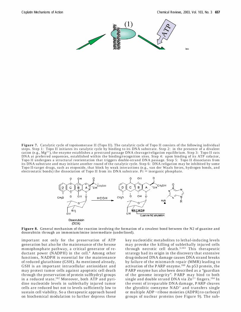
during the cell cycle.143 Human Topo II inhibitors,which stabilize the Topo II-DNA complex and in-terfere with DNA rebinding, are called Topo IIpoisons. Within these inhibitory drugs, anthracy-clines, as doxorubicin, and epipodophyllotoxins, asetoposide, are included (see Topo II catalytic cycleand etoposide structure in Figure 7). Doxorubicin(DOX), also known as adriamycin, is a planar aro-matic molecule that binds to DNA by intercalationbetween nucleobase pairs forming π-stacks.150 Threeprincipal functional components of DOX have beenidentified: (1) the intercalator (rings B-D), (2) theanchoring function associated with ring A (e.g.,C9-OH group), and (3) the amino sugar (see Figure8). It has been recently shown that under certainredox conditions DOX is capable of forming a covalentadduct with DNA using the daunosamine N3′ atomof the drug and the guanine N2 amino group.151 TheDOX-induced DNA cross-link may be the result ofthe action of HCOH (formaldehyde) generated fromthe DOX molecule via the Baeyer-Villiger reaction(see also Figure 8).152 The combination of DOX andcis-DDP has been successfully used in the clinic forthe treatment of advanced-stage ovarian cancer aswell as metastatic breast cancer.153 The Topo IIinhibitor etoposide, also called VP-16, binds to thecomplex formed by Topo II and the 5′-cleaved endsof the DNA, thus forming nonrepairable protein-linked DNA double strand breaks.154 VP-16 alsoexhibits a synergistic effect with cis-DDP. The com-bination of VP-16 with cis-DDP had greater cytotoxicactivity against four of five cell lines of cisplatin-resistant head and neck cancer, when compared toany single drug used alone.155 Moreover, the combi-nation of cis-DDP with VP-16 is widely used for thetreatment of patients with primary or recurrentsmall cell lung cancers.156
A novel and promising strategy for biochemicalmodulation of cisplatin resistance is the inhibitionof DNA repair through drugs which induce anincrease in the levels of HMG domain proteins intumor cells. For instance, it has been reported thattreatment of MCF-7 breast cancer cells, having thesteroid hormone receptors, with the appropriatehormone, estrogen, and/or progesterone, significantlyincreases the potency of cisplatin and its analoguecarboplatin by causing the overexpression of HMG1.157
4.4. Cell Death PathwaysAs mentioned already, cis-DDP provokes DNA
damage which initiates the cell death pathways ofapoptosis and necrosis.158 In certain cases of cisplatinresistance, apoptotic cell death pathways may beinhibited due to a drastic reduction of the bioener-getic cellular index (BEC index).159 Thus, tumor cellswith a low BEC index, as a result of a low mitochon-drial content and/or activity, would become moreresistant to programmed cell death. On the otherhand, in highly cisplatin-resistant sublines, necroticcell death may be blocked due to the inactivation ofpoly(ADP-ribose) polymerase (PARP) cleavage ofNAD+.160 As early as 1956, Otto Warburg reportedthat an elevated rate of glycolysis is a commonfeature of most tumors.161 Glycolytic metabolism is
656 Chemical Reviews, 2003, Vol. 103, No. 3 Fuertes et al.
important not only for the preservation of ATPgeneration but also for the maintenance of the hexosemonophosphate pathway, a critical generator of re-ductant power (NADPH) in the cell.4 Among otherfunctions, NADPH is essential for the maintenanceof reduced glutathione (GSH). As mentioned already,GSH is an important intracellular antioxidant andmay protect tumor cells against apoptotic cell deaththrough the preservation of protein sulfhydryl groupsin a reduced state.162 Moreover, both ATP and pyri-dine nucleotide levels in sublethally injured tumorcells are reduced but not to levels sufficiently low tosustain cell viability. So a therapeutic approach basedon biochemical modulation to further depress these
key nucleotidic metabolites to lethal-inducing levelsmay provoke the killing of sublethally injured cellsthrough necrotic cell death.3,163 This therapeuticstrategy had its origin in the discovery that extensivedrug-induced DNA damage causes DNA strand breaksby failure of the mistmatch repair (MMR) leading toactivation of the PARP enzyme.160 As p53 protein, thePARP enzyme has also been described as a “guardianof the genome integrity”. PARP may bind to bothsingle and double strand DNA via Zn2+ fingers.164 Inthe event of irreparable DNA damage, PARP cleavesthe glycolitic coenzyme NAD+ and transfers singleor multiple ADP-ribose moieties (ADPR) to carboxylgroups of nuclear proteins (see Figure 9). The sub-
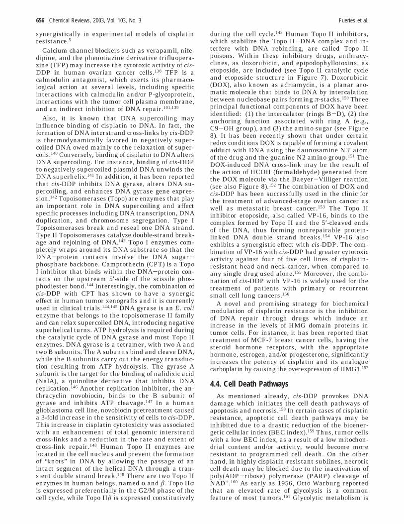
Figure 7. Catalytic cycle of topoisomerase II (Topo II). The catalytic cycle of Topo II consists of the following individualsteps. Step 1: Topo II initiates its catalytic cycle by binding to its DNA substrate. Step 2: in the presence of a divalentcation (e.g., Mg2+), the enzyme establishes a prestrand passage DNA cleavage/religation equilibrium. Step 3: Topo II cutsDNA at preferred sequences, established within the binding/recognition sites. Step 4: upon binding of its ATP cofactor,Topo II undergoes a structural reorientation that triggers double-strand DNA passage. Step 5: Topo II dissociates fromits DNA substrate and may initiate another round of the catalytic cycle. Step 6: DNA religation may be inhibited by someTopo II-target drugs, such as etoposide, that block by weak interactions (e.g., van der Waals forces, hydrogen bonds, andelectrostatic bonds) the dissociation of Topo II from its DNA substrate. Pi ) inorganic phosphate.
Figure 8. General mechanism of the reaction involving the formation of a covalent bond between the N2 of guanine anddoxorubicin through an immonium/imine intermediate (underlined).
Cisplatin Mechanisms of Action Chemical Reviews, 2003, Vol. 103, No. 3 657

sequent depletion of NAD+ inhibits glycolytic genera-tion of ATP with consequent ATP depletion. If ATPdepletion reaches lethal-inducing levels, then necroticcell death occurs.160 There is a likely rationale for theinduction of necrotic cell death via PARP activation.A severely damaged cell sustains such a large num-ber of mutations and metabolic alterations that it isseriously impaired in function. Therefore, poly(ADP)-ribosylation may be a system of cell death whichoperates when the cell is so badly damaged that ATPlevels are exhausted and when apoptotic pathwayscannot take place. PARP has been very recentlyidentified as a novel target for therapy of importantpathologies including cancer, neurologic alterations,and immunological diseases.164 For instance, it hasbeen reported that a concomitant ATP-depletingstrategy, called MAP regime, enhances antitumordrug-induced cell killing in sublethally injured cancercells through activation of the PARP-associatedbiochemical mechanism of necrotic cell death.3 MAPhas proven to enhance the cytotoxic activity of severalantitumor agents, including doxorubicin, etoposide,paclitaxel, 5-fluorouracil, and cisplatin. The MAP
regime is a combination of methylmercaptopurineriboside (MMPR) plus 6-aminonicotinamide (6-AN)plus N-(phosphonacetyl)-L-aspartic acid (PALA). 6-ANis a NAD+ antagonist, which inhibits glycolyticproduction of ATP.165,166 MMPR is an inhibitor of denovo purine biosynthesis and, therefore, limits ad-enine supplies for ATP production.167 PALA inhibitsaspartate transcarbamilase (ATCase) and selectivelylowers pyrimidine nucleotide levels in tumors.168
ATCase is a key enzyme in pyrimidine nucleotidesynthesis which catalyzes the formation of carbamoylaspartate from carbamoyl phosphate and aspartate.PALA acts as an analogue of the bisubstrate transi-tion-state complex formed between carbamoyl phos-phate and aspartate within the catalytic center ofATCase (see Figure 10).169 PALA, due to its highnegative charge, binds electrostatically to three argi-nine (Arg) residues and one lysine (Lys) residue inthe ATCase catalytic center. In addition, PALAinteracts through hydrogen bonding with other aminoacids of the catalytic site. Of particular interest isthe hydrogen bond formed between a nitrogen of theimidazol ring of histidine 134 (His134) and the oxygen
Figure 9. Cleavage of NAD+ and subsequent transfer of ADP-ribose moieties (ADPR) to carboxyl groups of nuclearproteins through the catalytic activity of poly(ADP-ribose) polymerase (PARP). Extensive DNA damage by cis-DDP mayinduce double strand breaks due, for instance, to mismatch repair (MMR) failure after futile replication/repair cycles.DNA double strand breaks activates PARP, which in turn binds to DNA through a zinc finger (the zinc finger may containthe following: L, leucine; F, phenylalanine; H, histidine; and C, cysteine). PARP also cleaves NAD+ and catalyzes theincorporation of ADPR units to nuclear proteins ([P] ) phosphate group). The PARP reaction involves the binding of theC1′ of ribose to the oxygen of the OH group of the carboxylic function of amino acids of nuclear proteins. NAD+ depletioninhibits glycolysis with subsequent depletion of ATP levels. If ATP depletion falls to lethal-inducing levels, then, necroticcell death occurs.
658 Chemical Reviews, 2003, Vol. 103, No. 3 Fuertes et al.
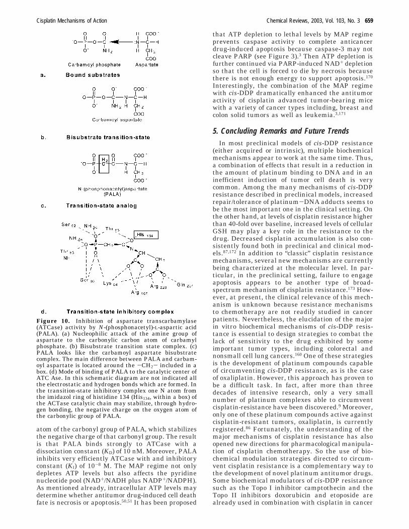
atom of the carbonyl group of PALA, which stabilizesthe negative charge of that carbonyl group. The resultis that PALA binds strongly to ATCase with adissociation constant (KD) of 10 nM. Moreover, PALAinhibits very efficiently ATCase with and inhibitoryconstant (KI) of 10-8 M. The MAP regime not onlydepletes ATP levels but also affects the pyridinenucleotide pool (NAD+/NADH plus NADP+/NADPH).As mentioned already, intracellular ATP levels maydetermine whether antitumor drug-induced cell deathfate is necrosis or apoptosis.50,51 It has been proposed
that ATP depletion to lethal levels by MAP regimeprevents caspase activity to complete anticancerdrug-induced apoptosis because caspase-3 may notcleave PARP (see Figure 3).3 Then ATP depletion isfurther continued via PARP-induced NAD+ depletionso that the cell is forced to die by necrosis becausethere is not enough energy to support apoptosis.170
Interestingly, the combination of the MAP regimewith cis-DDP dramatically enhanced the antitumoractivity of cisplatin advanced tumor-bearing micewith a variety of cancer types including, breast andcolon solid tumors as well as leukemia.3,171
5. Concluding Remarks and Future TrendsIn most preclinical models of cis-DDP resistance
(either acquired or intrinsic), multiple biochemicalmechanisms appear to work at the same time. Thus,a combination of effects that result in a reduction inthe amount of platinum binding to DNA and in aninefficient induction of tumor cell death is verycommon. Among the many mechanisms of cis-DDPresistance described in preclinical models, increasedrepair/tolerance of platinum-DNA adducts seems tobe the most important one in the clinical setting. Onthe other hand, at levels of cisplatin resistance higherthan 40-fold over baseline, increased levels of cellularGSH may play a key role in the resistance to thedrug. Decreased cisplatin accumulation is also con-sistently found both in preclinical and clinical mod-els.87,172 In addition to “classic” cisplatin resistancemechanisms, several new mechanisms are currentlybeing characterized at the molecular level. In par-ticular, in the preclinical setting, failure to engageapoptosis appears to be another type of broad-spectrum mechanism of cisplatin resistance.173 How-ever, at present, the clinical relevance of this mech-anism is unknown because resistance mechanismsto chemotherapy are not readily studied in cancerpatients. Nevertheless, the elucidation of the majorin vitro biochemical mechanisms of cis-DDP resis-tance is essential to design strategies to combat thelack of sensitivity to the drug exhibited by someimportant tumor types, including colorectal andnonsmall cell lung cancers.160 One of these strategiesis the development of platinum compounds capableof circumventing cis-DDP resistance, as is the caseof oxaliplatin. However, this approach has proven tobe a difficult task. In fact, after more than threedecades of intensive research, only a very smallnumber of platinum complexes able to circumventcisplatin-resistance have been discovered.9 Moreover,only one of these platinum compounds active againstcisplatin-resistant tumors, oxaliplatin, is currentlyregistered.86 Fortunately, the understanding of themajor mechanisms of cisplatin resistance has alsoopened new directions for pharmacological manipula-tion of cisplatin chemotherapy. So the use of bio-chemical modulation strategies directed to circum-vent cisplatin resistance is a complementary way tothe development of novel platinum antitumor drugs.Some biochemical modulators of cis-DDP resistancesuch as the Topo I inhibitor camptothecin and theTopo II inhibitors doxorubicin and etoposide arealready used in combination with cisplatin in cancer
Figure 10. Inhibition of aspartate transcarbamylase(ATCase) activity by N-(phosphonacetyl)-L-aspartic acid(PALA). (a) Nucleophilic attack of the amine group ofaspartate to the carbonylic carbon atom of carbamylphosphate. (b) Bisubstrate transition state complex. (c)PALA looks like the carbamoyl aspartate bisubstratecomplex. The main difference between PALA and carbam-oyl aspartate is located around the -CH2- included in abox. (d) Mode of binding of PALA to the catalytic center ofATC Ase. In this schematic diagram are not indicated allthe electrostatic and hydrogen bonds which are formed. Inthe transition-state inhibitory complex one N atom fromthe imidazol ring of histidine 134 (His134, within a box) ofthe ACTase catalytic chain may stabilize, through hydro-gen bonding, the negative charge on the oxygen atom ofthe carbonylic group of PALA.
Cisplatin Mechanisms of Action Chemical Reviews, 2003, Vol. 103, No. 3 659

patients producing good responses rates.153 In addi-tion, the modulator of cisplatin accumulation, cy-closporine A, increased the activity of cisplatin inpatients with recurrent and platinum-resistant ova-rian cancer.110 Moreover, depletion of GSH levels intumors by L-BSO administration has proven to workin the clinical setting, although the limited avail-ability of L-BSO has so far precluded its clinical useas a modulator of cis-DDP resistance.127 Of interestis the observation that ethacrynic acid, an inhibitorof glutathione transferase (GST) has shown a syn-ergistic effect with cis-DDP in preclinical modelsbeing a good candidate for clinical trials.174 Neverthe-less, and despite all these relevant clinical advances,a lot of biochemical modulators of cisplatin resistancestill remain in a preclinical phase of development.As noted above, this is mainly due to the great delaythat usually exists between the end of the preclinicalphase of drug research and the implementation ofclinical trials.
In recent years, increasing knowledge of the bio-chemical mechanisms of drug-induced tumor celldeath has opened novel and promising ways forbiochemical modulation of the activity of anticancerdrugs, particularly in highly resistant tumor cellpopulations. Of particular interest is the manipula-tion of tumor cell energy to increase the activity ofDNA-damaging antitumor drugs through the induc-tion of necrotic cell death. Thus, the biochemicalmodulation by MAP regime of cis-DDP activity toprovoke necrotic cell death in cisplatin-resistant cellsmerits further research.3,171 Interestingly, the com-bination of PALA with cisplatin does not provokesevere toxicity in mice. Moreover, 6-aminonicotina-mide (6-AN), as a single agent, has been safelyadministered to patients with disseminated cancerin Phase I clinical trials.3 In view of these data, wethink that future cancer chemotherapy must bedirected to look for adjuvant drugs that affect generalbiochemical mechanisms that can bypass drug resis-tance rather than to exclusively search for specificdrugs which target particular cellular constituents.Among others, it is the preclinically proven ATP-depleting modulatory concept what warrants andrequires appropriate clinical exploration not onlywhen PALA is combined with cis-DDP, but also whenother ATP-depleting agents are combined with an-ticancer drugs.
In summary, the data reviewed herein indicatethat biochemical modulation of cisplatin mechanismsof resistance offers multiple opportunities for futureapplications in clinical cancer research. In addition,biochemical modulation may constitute a therapeuticstrategy complementary to the discovery of novelplatinum complexes with activity in cisplatin-resis-tant tumors.
6. AcknowledgmentsThis work was supported by the European Coop-
eration in the Field of Scientific and TechnicalResearch Network (COST D20/003/00 Action: “Bio-chemistry, Structural and Cellular Biology of Non-Classical Antitumor Platinum Compounds”) andCICYT Bio99-1133. A specific and institutional grantfrom Fundacion Ramon Areces is also acknowledged.
7. References(1) Martin, D. S. In New Avenues in Developmental Cancer Che-
motherapy; Harrap, K. R., Ed.; Academic Press: London, 1986;p 113.
(2) Gonzalez, V. M.; Fuertes, M. A.; Alonso, C.; Perez, J. M. Mol.Pharmacol. 2001, 59, 657.
(3) Martin, D. S.; Bertino, J. R.; Koutcher, J. A. Cancer Res. 2000,60, 6776.
(4) Zhou, R.; Vander Heiden, M. G.; Rudin, C. M. Cancer Res. 2002,62, 3515.
(5) Giaccone, G. Drugs 2000, 59 (Suppl. 4), 9.(6) Perez, R. P. Eur. J. Cancer 1998, 34, 1535.(7) Chu, G. J. Biol. Chem. 1994, 269, 787.(8) Kelland, L. R. Drugs 2000, 59 (Suppl. 4), 1.(9) Fuertes, M. A.; Castilla, J.; Alonso, C.; Perez, J. M. Curr. Med.
Chem.-Anti Cancer Agents 2002, 2, 539.(10) Hudson, I.; Kelland, L. R. Drugs. 2000, 59 (Suppl. 4), 29.(11) Cvitkovic, E. Semin. Oncol. 1998, 25, 1.(12) Jamieson, E. R.; Lippard, S. J. Chem. Rev. 1999, 99, 2467.(13) Miller, S. E.; House, D. A. Inorg. Chim. Acta 1991, 187, 125.(14) Yang, X.-L.; Wang, A. H.-J. Pharmacol. Ther. 1999, 83, 181.(15) Payet, D.; Gaucheron, F.; Sip, M.; Leng, M. Nucleic Acids Res.
1993, 21, 5846.(16) Fichtinger-Schepman, A. M.; van der Veer, J. L.; den Hartog, J.
H. J.; Lohman, P. H. M.; Reedijk, J. Biochemistry 1985, 24, 707.(17) Auge, P.; Kozelka, J. Transition Met. Chem. (N.Y.) 1997, 22, 91.(18) Comess, K. M.; Lippard, S. J. In Molecular Aspects of Anticancer
Drug-DNA Interactions; Neidle, S., Waring, M., Eds.; Mac-Milllan Press: London, 1993; Vol. 1, p 134.
(19) Eastman, A.; Barry, M. A. Biochemistry 1987, 26, 3303.(20) Szymkowski, D. E.; Yarema, K.; Essigmann, J. M.; Lippard, S.
J.; Wood, R. D. Proc. Natl. Acad. Sci. U.S.A. 1992, 89, 10772.(21) Mu, D.; Shu, D. S.; Sancar, A. J. Biol. Chem. 1996, 271, 8285.(22) Pil, P. M.; Lippard, S. J. Science 1992, 256, 234.(23) Zwelling, L. A.; Anderson, T.; Kohn, K. W. Cancer Res. 1979,
39, 365.(24) Malinge, J.-M.; Giraud-Panis, M.-J.; Leng, M. J. Inorg. Biochem.
1999, 77, 23.(25) Akaboshi, M.; Kawai, K.; Maki, H.; Akuta, K.; Ujeno, Y.;
Miyahara, T. Jpn. J. Cancer Res. 1992, 83, 522.(26) Akaboshi, M.; Kawai, K.; Ujeno, Y.; Takada, S.; Miyahara, T.
Jpn. J. Cancer Res. 1994, 85, 106.(27) Speelmans, G.; Staffhorst, R. W. H. M.; Versluis, K.; Reedijk,
J.; de Kruijff, B. Biochemistry 1997, 36, 10545.(28) Jordan, P.; Carmo-Fonseca, M. Cell. Mol. Life. Sci. 2000, 57,
1229.(29) Reedijk, J. Chem. Rev. 1999, 99, 2499.(30) Peleg-Shulman, T.; Gibson, D. J. Am. Chem. Soc. 2001, 123,
3171.(31) Soti, C.; Racz, A.; Csermely, P. J. Biol. Chem. 2002, 277, 7066.(32) Barry, M. A.; Benhke, C. A.; Eastman, A. Biochem. Pharmacol.
1990, 40, 2353.(33) Kruidering, M.; van der Water, B.; Zhan, Y.; Baelde, J. J.; de
Heer, E.; Mulder, G. J.; Stevens, J. L.; Nagelkerke, J. F. CellDeath Differ. 1998, 5, 601.
(34) Henkels, K. M.; Turchi, J. J. Cancer Res. 1997, 57, 4488.(35) Matsumoto, M.; Tsuchida, T.; Kawamoto, K. Int. J. Oncol. 1997,
11, 1209.(36) Guchelaar, H. J.; Vermes, I.; Koopmans, R. P.; Reutelingsperger,
C. P. M.; Haanen, C. Cancer Chemother. Pharmacol. 1998, 42,77.
(37) Perez, J. M.; Montero, E. I.; Gonzalez, A. M.; Alvarez-Valdes,A.; Alonso, C.; Navarro-Ranninger, C. J. Inorg. Biochem. 1999,77, 37.
(38) Montero, E. I.; Perez, J. M.; Schwartz, A.; Fuertes, M. A.;Malinge, J.-M.; Alonso, C.; Leng, M.; Navarro-Ranninger, C.ChemBioChem. 2002, 3, 101.
(39) Wyllie, A. H. J. Pathol. 1987, 153, 313.(40) Tanizawa, A.; Kubota, M.; Hashimoto, H.; Shimizu, T.; Takimoto,
T.; Kitoh, T.; Akiyama, Y.; Mikama, H. Exp. Cell. Res. 1989, 185,237.
(41) Eastman, A. In Cisplatin, Chemistry and Biochemistry of aLeading Anticancer Drug; Lippert, B., Ed.; Wiley-VCH: Basel,Switzerland, 1999; p 111.
(42) Reed, J. C. J. Cell. Biol. 1994, 124, 1.(43) Eliopoulos, A. G.; Kerr, D. J.; Herod, J.; Hodgkins, L.; Krajewski,
S.; Reed, J. C.; Young, L. S. Oncogene 1995, 11, 1217.(44) Alnemri, E. S. J. Cell. Biochem. 1997, 64, 33.(45) Kroemer, G.; Zamzami, N.; Susin, S. A. Immunol. Today 1997,
18, 44.(46) Green, D. R. Cell 1998, 94, 695.(47) Reed, J. C. Nat. Rev.-Drug Discov. 2002, 1, 111.(48) Bose, R.; Verheij, M.; Haimovitz-Friedman, A.; Scotto, K.; Fucks,
Z.; Kolesnick, R. Cell. 1995, 82, 405.(49) Li, H.; Zhu, H.; Xu, C. J. Cell. 1998, 94, 491.(50) Eguchi, Y.; Shimizu, S.; Tsujimoto, Y. Cancer Res. 1997, 57, 835.
660 Chemical Reviews, 2003, Vol. 103, No. 3 Fuertes et al.

(51) Leist, M.; Single, B.; Castoldi, A. F.; Kuhnle, S.; Nicotera, P. J.Exp. Med. 1997, 185, 1481.
(52) Zhou, R.; Vander Heiden, M. G.; Rudin, C. M. Cancer Res. 2002,62, 3515.
(53) Green, D. R.; Reed, J. C. Science 1998, 281, 1309.(54) Herceg, Z.; Wang, Z. Q. Mol. Cell. Biol. 1999, 19, 5124.(55) Hirsch, T.; Marchetti, P.; Susin, S.; Dellaporta, B.; Zamzani, N.;
Marzo, I.; Geuskens, N.; Kroemer, G. Oncogene 1997, 15, 1573.(56) Segal-Bendirdjian, E.; Jacquemin-Sablon, A. Exp. Cell. Res.
1995, 218, 201.(57) Muggia, F. M.; Los, G. Stem Cells 1993, 11, 182.(58) Jassem, J. Ann. Oncol. 1999, 10, 77.(59) Johnson, S. W.; Ferry, K. V.; Hamilton, T. C. Drug Resist.
Updates 1998, 1, 243.(60) Wang, K.; Lu, J.; Li, R. Coord. Chem. Rev. 1996, 151, 53.(61) Andrews, P. A. In Platinum-Based Drugs in Cancer Chemo-
therapy; Kelland, L. R., Farrell, N. P., Eds.; Humana Press:Totowa, NJ, 2000, p 89.
(62) Gottesman, M. M.; Pastan, I. Annu. Rev. Biochem. 1993, 62, 385.(63) Bellamy, W. T. Annu. Rev. Pharmacol. Toxicol. 1996, 36, 161.(64) Uchiumi, T.; Hinoshita, E.; Haga, S.; Nakamura, T.; Tanaka,
T.; Toh, S.; Furukawa, M. K. T.; Wada, M.; Kagotani, K.;Okumura, K.; Kohno, K.; Akiyama, S.; Kuwano, M. Biochim.Biophys. Res. Commun. 1998, 252, 103.
(65) Gately, D. P.; Howell, S. B. Br. J. Cancer 1993, 67, 1171.(66) Wang, W.; Ballatori, N. Pharmacol. Rev. 1998, 50, 335.(67) Garcıa-Ruız, C.; Mari, M.; Morales, A.; Colell, A.; Ardite, E.;
Fernandez-Checa, J. C. Hepatology 2000, 32, 56.(68) Kelley, S. L.; Basu, A.; Teicher, B. A.; Hacker, M. P.; Hamer, D.
H.; Lazo, J. S. Science 1998, 241, 1813.(69) Cohen, S. M.; Lippard, S. J. Prog. Nucl. Acids Res. Mol. Biol.
2001, 67, 93.(70) Reardon, J. T.; Vaisman, A.; Chaney, S. G.; Sancar, A. Cancer
Res. 1999, 59, 3968.(71) Chaney, S. G.; Sancar, A. J. Natl. Cancer. Inst. 1996, 88, 1346.(72) Kelland, L. R.; Mistry, P.; Abel, G.; Friedlos, F.; Loh, S. Y.;
Roberts, J. J.; Harrap, K. R. Cancer Res. 1992, 52, 1710.(73) Zamble, D. B.; Mikata, Y.; Eng, C. H.; Sandman, K. E.; Lippard,
S. J. J. Inorg. Biochem. 2002, 91, 451.(74) Toney, J. H.; Donahue, B. A.; Kellet, P. J.; Bruhn, S. L.;
Essigman, J. M.; Lippard, S. J. Proc. Natl. Acad. Sci. U.S.A.1989, 86, 8328.
(75) Huang, J. C.; Zamble, D. B.; Reardon, J. T.; Lippard, S. J.;Sancar, A. Proc. Natl. Acad. Sci. U.S.A. 1994, 91, 10394.
(76) Brown, S. J.; Kellet, P. J.; Lippard, S. J. Science 1993, 261, 603.(77) Orphanides, G.; Wu, W. H.; Lane, W. S.; Hampsey, M.; Reinberg,
D. Nature 1999, 400, 284.(78) Dempke, W.; Voight, W.; Grothey, A.; Hill, B. T.; Schomll, H. J.
Anti-Cancer Drugs 2000, 11, 225.(79) Chaney, S. G.; Vaisman, A. In Platinum-Based Drugs in Cancer
Chemotherapy; Kelland, L. R., Farrell, N. P., Eds.; HumanaPress: Totowa, NJ, 2000, p 129.
(80) Vaisman, A.; Varchenko, M.; Umar, A.; Kunkel, T. A.; Risinger,J. L.; Barrett, J. C.; Hamilton, T. C.; Chaney, S. G. Cancer Res.1998, 58, 3579.
(81) Fishel, R. Cancer Res. 2001, 61, 7369.(82) Duckett, D. R.; Drummond, J. T.; Murchie, A. I. H.; Reardon,
J.T.; Sancar, A.; Lilley, D. M. J.; Modrich, P. Proc. Natl. Acad.Sci. U.S.A. 1996, 93, 6443.
(83) Chaney, S. G.; Vaisman, A. J. Inorg. Biochem. 1999, 77, 71.(84) Raymond, E.; Faivre, S.; Chaney, S.; Woynarowski, J.; Cvitkovic,
E. Mol. Cancer Ther. 2002, 1, 221.(85) Zdraveski, Z. Z.; Mello, J. A.; Farinelli, C. K.; Essigmann, J. M.;
Marinus M. G. J. Biol. Chem. 2002, 277, 1255.(86) Lloyd, D. Trends Pharmacol. Sci. 2002, 23, 158.(87) Niedner, H.; Christen, R.; Lin, X.; Kondo, A.; Howell, S. B. Mol.
Pharmacol. 2001, 60, 1153.(88) Fritche, M.; Haessler, C.; Brander, G. Oncogene 1993, 8, 307.(89) Gorospe, M.; Cirielli, C.; Wang, X.; Seth, P.; Capogrossi, M. C.;
Holbrook, N. J. Oncogene 1997, 14, 185.(90) Pestell, K. E.; Hobbs, S. M.; Titley, J. C.; Kelland, L. R.; Walton,
M. I. Mol. Pharmacol. 2000, 57, 503.(91) Reed, J. C. In Apoptosis and Cancer; Martin, S. J., Ed.; Basel:
Karger- Landes Systems, 1997, p 64.(92) Beale, P. J.; Rogers, P.; Boxall, F.; Sharp, S. Y.; Kelland, L. R.
Br. J. Cancer 2000, 82, 436.(93) Burger, H.; Capello, A.; Schenk, P. W.; Stoter, G.; Brouwer, J.;
Nooter, K. Biochem. Biophys. Res. Commun. 2000, 269, 767.(94) Knighton, D. R.; Zheng, J.; TenEyck, L. F.; Ashford, V. A.; Xuong,
N.; Taylor, S. S.; Sowadski, J. M. Science 1991, 253, 407.(95) Yoshida, Y.; Hosokawa, K.; Dantes, A.; Tajima, K.; Kotsuji, F.;
Amsterdan, A. Int. J. Oncol. 2000, 17, 227.(96) Pyne, S.; Pyne, N. J. Biochem. J. 2000, 349, 385.(97) Li, G.; Alexander, H.; Schneider, N.; Alexander, S. Microbiology
2000, 146, 2219.(98) Neidle, S.; Kelland, L. R. Anti-Cancer Drug Des. 1999, 14, 341.(99) Ishibashi, T.; Lippard, S. J. Proc. Natl. Acad. Sci. U.S.A. 1998,
95, 4219.(100) Kelland, L. R. Drug Resist Updat. 2000, 3 (Suppl. 3), 139.
(101) Gosland, M.; Lum, B.; Schimmelpfennig, J.; Baker, J.; Doukas,M. Pharmacotherapy 1996, 16, 16.
(102) Plagemann, P. G. W.; Wohlhueter, R. M.; Woffedin, C. Biochim.Biophys. Acta 1988, 947, 405.
(103) Berti, E.; Carrara, M.; Ragazzi, E.; D’Ancona, S.; Bertı, T. Int.J. Oncol. 1999, 15, 155.
(104) Jekunen, A.; Vick, J.; Sanga, R.; Chan, T. C.; Howell, S. B. CancerRes. 1992, 52, 3566.
(105) Kohnoe, S.; Maehara, Y.; Takahashi, I.; Emi, Y.; Baba, H.;Sugimachi, K. Int. J. Oncol. 1998, 13, 1203.
(106) Bergstrom, P.; Johnson, A.; Cavallin-Stahl, E.; Bergenheim, T.;Henriksson, R. Eur. J. Cancer. 1997, 33, 153.
(107) Sharp, S. Y.; Mistry, P.; Valenty, M. R.; Bryant, A. P.; Kelland,L. R. Cancer Chemother. Pharmacol. 1994, 35, 137.
(108) Hoffmann, E. K.; Dunham, P. B. Int. Rev. Cytol. 1995, 161, 173.(109) Marklund, L.; Henriksson, R.; Grankvist, K. Int. J. Cancer. 2001,
93, 577.(110) Manetta, A.; Blessing, J. A.; Hurteau, J. A. Gynecol. Oncol. 1998,
68, 45.(111) Foxwell, B. M.; Mackie, A.; Ling, V.; Ryffel, B. Mol. Pharmacol.
1989, 36, 543.(112) Chen, Z.-S.; Kawabe, T.; Ono, M.; Aoki, S.; Sumizawa, T.;
Furukawa, T.; Uchiumi, T.; Wada, M.; Kuwano, M.; Akiyama,S.-I. Mol. Pharmacol. 1999, 56, 1219.
(113) Kashani-Sabet, M.; Wang, W.; Sacanlon, K. J. J. Biol. Chem.1990, 265, 11285.
(114) Eastman, A. Chem. Biol. Interact. 1987, 61, 241.(115) Zhang, K.; Chew, M.; Yang, E. B.; Wong, K. P.; Mack, P. Mol.
Pharmacol. 2001, 59, 837.(116) Perez, R.; Johnson, S.; Andel, L.; O’Dwyer, P.; Hamilton, T.
Gynecol. Oncol. 1995, 58, 312.(117) Borst, P.; Evers, R.; Kool, M.; Wijnholds, J. J. Natl. Cancer Inst.
2000, 92, 1295.(118) Lertratanangkoon, K.; Savaraj, N.; Scimeca, J. M.; Thomas, M.
L. Biochim. Biophys. Res. Commun. 1997, 234, 470.(119) Billings, P. C.; Davis, R. J.; Engelsberg, B. N.; Skov, K. A.;
Hughes, E. N. Biochim. Biophys. Res. Comm. 1992, 188, 1286.(120) Zejia, Y.; Faustino, P. J.; Andrews, P. A.; Monastra, R.; Ras-
mussen, A. A.; Ellison, C. D.; Cullen, K. J. Cancer. Chemother.Pharmacol. 2000, 46, 255.
(121) Anderson, M. E. Chem. Biol. Interact. 1998, 111-112, 1.(122) Bailey, H. H. Chem. Biol. Interact. 1998, 111-112, 239.(123) Griffith, O. W. J. Biol. Chem. 1982, 257, 13704.(124) O’Dwyer, P. J.; Hamilton, T. C.; Young, R. C.; LaCreta, F. P.;
Carp, N.; Tew, K. D.; Padavic, K.; Comis, R. L.; Ozols, R. F. J.Natl. Cancer Inst. 1992, 84, 264.
(125) Perez, J. M.; Montero, E. I.; Quiroga, A. G.; Fuertes, M. A.;Alonso, C.; Navarro-Ranninger, C. Metal Based Drugs 2001, 8,29.
(126) Li, X. M.; Filipski, E.; Levi, F. Chronobiol. Int. 1998, 15, 323.(127) Calvert, P.; Yao, K.-S.; Hamilton, T.; O’Dwyer, P. J. Chem. Biol.
Interact. 1998, 111-112, 213.(128) Osmak, M.; Bordukalo, T.; Ristov, A. A.; Jerny, B.; Kosmrlj, J.;
Polanc, S. Neoplasma 2000, 47, 390.(129) Dabholkar, M.; Reed, E. In Cancer Chemotherapy and Biological
Response Modifiers; Pinedo, H. M., Longo, D. L., Chabner, B.A., Eds.; Elsevier Science: Amsterdam, 1996; Annual 16, p 88.
(130) Steitz, T. A. J. Biol. Chem. 1999, 274, 17395.(131) Singh, K.; Modak, M. J. Trends Biochem. Sci. 1998, 23, 277.(132) O’Dwyer, P. J.; Moyer, J. D.; Suddness, M.; Harrison, S. D., Jr.;
Cysyk, R.; Hamilton, T. C.; Plowman, J. Cancer Res. 1994, 54,724.
(133) Noble, S.; Goa, K. L. Drugs 1997, 54 (Suppl. 3), 447.(134) Arner, E. S. J.; Eriksson, S. Pharmacol. Ther. 1995, 67, 155.(135) Ellerhorst, J. A.; Frost, P.; Abbruzzese, J. L.; Newman, R. A.;
Chernajovsky, Y. Br. J. Cancer 1993, 67, 209.(136) Uhlin, U.; Eklund, H. Nature 1994, 370, 533.(137) Stubbe, J. Proc. Natl. Acad. Sci. U.S.A. 1998, 95, 2723.(138) Perez, R. P.; Handel, L. M.; Hamilton, T. C. Gynecol. Oncol. 1992,
46, 82.(139) Onoda, J. M.; Nelson, K. K.; Taylor, J. D.; Honn, K. V. Cancer
Res. 1989, 49, 2844.(140) Vrana, O.; Boudny, V.; Brabec, V. Nucl. Acids. Res. 1996, 24,
3918.(141) Perez, J. M.; Lopez-Solera, I.; Brana, M. F.; Alonso, C.; Robinson,
S. P.; Navarro-Ranninger, C. J. Med. Chem. 1999, 42, 5482.(142) Neumann, S.; Simon, H.; Zimmer, C.; Quinones, A. Biol. Chem.
1996, 377, 731.(143) Wang, J. C. Annu. Rev. Biochem. 1996, 65, 635.(144) Nash, H. A. Science. 1998, 279, 1490.(145) Kim, R.; Hirabayashi, N.; Nishiyama, M.; Jinuski, K.; Toge, T.;
Okada, K. Int. J. Cancer. 1992, 12, 760.(146) Takano, H.; Hohno, K.; Matsuo, K.; Matsuda, T.; Kuwano, M.
Anticancer Drugs 1992, 3, 323.(147) Liu, L. F. Annu. Rev. Biochem. 1989, 58, 351.(148) Ali-Osman, F.; Berger, M. S.; Rajagopal, S.; Spence, A.; Living-
ston, R. B. Cancer Res. 1993, 53, 5663.(149) Berger, J. M. Biochim. Biophys. Acta 1998, 1400, 3.
Cisplatin Mechanisms of Action Chemical Reviews, 2003, Vol. 103, No. 3 661

(150) Yang, X.-L.; Wang, A. H.-J. Pharmacol. Therapeut. 1998, 83,181.
(151) Taatjes, D. J.; Gaudiano, G.; Resing, K.; Koch, T. H. J. Med.Chem. 1997, 40, 1276.
(152) Cullinane, C.; Cutts, S. M.; van Rosmalen, A.; Phillips, D. R.Nucleic Acids Res. 1994, 22, 2296.
(153) O’Dwyer, P. J.; Stevenson, J. P.; Johnson, S. W. In Cisplatin,Chemistry and Biochemistry of a Leading Anticancer Drug;Lippert, B., Ed.; Wiley-VCH: Basel, Switzerland, 1999, p 31.
(154) Karpinich, N. O.; Tafani, M.; Rothman, R. J.; Russo, M. A.;Farber; J. L. J. Biol. Chem. 2002, 277, 16547.
(155) Haller, J.; Burgess, R.; Dawson, D. Laryngoscope 1993, 103,1081.
(156) Schuette, W. Lung Cancer. 2001, Suppl. 1, 99.(157) He, Q.; Liang, C. H.; Lippard, S. J. Proc. Natl. Acad. Sci. U.S.A.
2000, 97, 5768.(158) Fuertes, M. A.; Castilla, J.; Alonso, C.; Perez, J. M. Recent Res.
Devel. Mol. Pharmacol. 2002, 1, 37.(159) Cuezva, J. M.; Krajewska, M.; Lopez de Heredia, M.; Krajewski,
S.; Santamarıa, G.; Kim, H.; Zapata, J. M.; Marusawa, H.;Chamorro, M.; Reed, J. C. Cancer Res. 2002, 62, 6674.
(160) Fuertes, M. A.; Castilla, J.; Alonso, C.; Perez, J. M. Curr. Med.Chem. 2003, 10, 1241.
(161) Warburg, O. Science 1956, 123, 309.(162) Voehringer, D. Free Radical Biol. Med. 1999, 27, 945.(163) Nord, L. D.; Stolfi, R. L.; Alfieri, A. A.; Netto, G.; Reuter, V.;
Sternberg, S. S.; Colofiore, J. R.; Koutcher, J. A.; Martin, D. S.Cancer Chemother. Pharmacol. 1997, 40, 376.
(164) Simbulan-Rosenthal, C. M.; Rosenthal, D. S.; Haddad, B. H.; Ly,D.; Zhang, J.; Smulson, M. E. In Therapeutic Utilities of PARPInhibitors; Zhang, J., Ed.; CRC Press: Boca Raton, FL, 2002, p39.
(165) Dietrich, L. S.; Kaplan, L.; Friedland, I. M. J. Biol. Chem. 1958,233, 964.
(166) Street, J. C.; Mahmoud, V.; Ballon, D.; Alfieri, A. A.; Koutcher,J. A. J. Biol. Chem. 1996, 271, 4113.
(167) Shantz, G. D.; Smith, C. M.; Fontenella, L. J.; Lau, H. K. F.;Henderson, J. F. Cancer Res. 1973, 33, 2867.
(168) Martin, D. S.; Stofli, R. L.; Sawyer, R. C.; Spiegelman, S.; Casper,E. S.; Young, C. W. Cancer Res. 1983, 43, 2317.
(169) Voltz, K. W.; Krause, K. L.; Lipscomb, W. N. Biochim. Biophys.Res. Comm. 1986, 136, 822.
(170) Boulares, A. H.; Yokovlev, A. G.; Ivanova, V.; Stoica, B. A.; Wang,G.; Iyer, S.; Smulson, M. J. Biol. Chem. 1999, 274, 22932.
(171) Martin, D. S.; Spriggs, D.; Koutcher, J. A. Apoptosis. 2001, 6,125.
(172) Shen, D. W.; Goldenberg, S.; Pastan, I.; Gottesman, M. M. J.Cell. Physiol. 2000, 183, 108.
(173) Helmbach, H.; Kern, M. A.; Rossman, E.; Renz, K.; Kissel, C.;Gschwendt, B.; Schadendorf, D. J. Invest. Dermatol. 2002, 118,923.
(174) Zhang, K.; Chew, M.; Yang, E. B.; Wong, K. P.; Mack P. Mol.Pharmacol. 2001, 59, 837.
CR020010D
662 Chemical Reviews, 2003, Vol. 103, No. 3 Fuertes et al.
Copyright © 2022 FDOKUMEN