Bioassays to Monitor Taspase1 Function for the Identification of Pharmacogenetic Inhibitors
14
Bioassays to Monitor Taspase1 Function for the Identification of Pharmacogenetic Inhibitors Shirley K. Knauer 1. , Verena Fetz 2 , Jens Rabenstein 3 , Sandra Friedl 2 , Bettina Hofmann 4 , Samaneh Sabiani 3 , Elisabeth Schro ¨ der 1 , Lena Kunst 1 , Eugen Proschak 4 , Eckhard Thines 5 , Thomas Kindler 6 , Gisbert Schneider 4¤ , Rolf Marschalek 3 , Roland H. Stauber 2 *, Carolin Bier 2 * . 1 Institute for Molecular Biology, Centre for Medical Biotechnology (ZMB), University Duisburg-Essen, Essen, Germany, 2 Mainzer Screening Center (MSC), University Medical Center of the Johannes Gutenberg-University of Mainz, Mainz, Germany, 3 Institute of Pharmaceutical Biology/ZAFES, Goethe-University, Frankfurt/Main, Germany, 4 Institute Organic Chemistry and Chemical Biology/ZAFES, Goethe-University, Frankfurt/Main, Germany, 5 Institute of Biotechnology and Drug Research Kaiserslautern (IBWF), Kaiserslautern, Germany, 6 Department of Hematology/Oncology, University Medical Center of the Johannes Gutenberg-University of Mainz, Mainz, Germany Abstract Background: Threonine Aspartase 1 (Taspase1) mediates cleavage of the mixed lineage leukemia (MLL) protein and leukemia provoking MLL-fusions. In contrast to other proteases, the understanding of Taspase1’s (patho)biological relevance and function is limited, since neither small molecule inhibitors nor cell based functional assays for Taspase1 are currently available. Methodology/Findings: Efficient cell-based assays to probe Taspase1 function in vivo are presented here. These are composed of glutathione S-transferase, autofluorescent protein variants, Taspase1 cleavage sites and rational combinations of nuclear import and export signals. The biosensors localize predominantly to the cytoplasm, whereas expression of biologically active Taspase1 but not of inactive Taspase1 mutants or of the protease Caspase3 triggers their proteolytic cleavage and nuclear accumulation. Compared to in vitro assays using recombinant components the in vivo assay was highly efficient. Employing an optimized nuclear translocation algorithm, the triple-color assay could be adapted to a high- throughput microscopy platform (Z’factor = 0.63). Automated high-content data analysis was used to screen a focused compound library, selected by an in silico pharmacophor screening approach, as well as a collection of fungal extracts. Screening identified two compounds, N-[2-[(4-amino-6-oxo-3H-pyrimidin-2-yl)sulfanyl]ethyl]benzenesulfonamide and 2- benzyltriazole-4,5-dicarboxylic acid, which partially inhibited Taspase1 cleavage in living cells. Additionally, the assay was exploited to probe endogenous Taspase1 in solid tumor cell models and to identify an improved consensus sequence for efficient Taspase1 cleavage. This allowed the in silico identification of novel putative Taspase1 targets. Those include the FERM Domain-Containing Protein 4B, the Tyrosine-Protein Phosphatase Zeta, and DNA Polymerase Zeta. Cleavage site recognition and proteolytic processing of these substrates were verified in the context of the biosensor. Conclusions: The assay not only allows to genetically probe Taspase1 structure function in vivo, but is also applicable for high-content screening to identify Taspase1 inhibitors. Such tools will provide novel insights into Taspase1’s function and its potential therapeutic relevance. Citation: Knauer SK, Fetz V, Rabenstein J, Friedl S, Hofmann B, et al. (2011) Bioassays to Monitor Taspase1 Function for the Identification of Pharmacogenetic Inhibitors. PLoS ONE 6(5): e18253. doi:10.1371/journal.pone.0018253 Editor: Andy T. Y. Lau, University of Minnesota, United States of America Received November 29, 2010; Accepted February 28, 2011; Published May 25, 2011 Copyright: ß 2011 Knauer et al. This is an open-access article distributed under the terms of the Creative Commons Attribution License, which permits unrestricted use, distribution, and reproduction in any medium, provided the original author and source are credited. Funding: Grant support: German Cancer Aid - http://www.krebshilfe.de/ (FKZ102362 to R.S. and R.M.), Head and Neck Cancer Foundation - http://www.stiftung- tumorforschung.de/ (to C.B.), Wilhelm-Sander Foundation - http://www.sanst.de, Funds of the Chemical Industry - http://fonds.vci.de, Stiftung Rheinland-Pfalz fu ¨r Innovationen - http://www.stiftung-innovation.rlp.de, DFG KN973/1-1 and INST371/5-1FUGG, donation from R. Patzke, Alexander-Karl-Foundation - http://www. foerdern-und-stiften.uni-mainz.de/225.php, and the University Mainz Support Program. The funders had no role in study design, data collection and analysis, decision to publish, or preparation of the manuscript. Competing Interests: The authors have declared that no competing interests exist. * E-mail: [email protected] (CB); [email protected] (RHS) . These authors contributed equally to this work. ¤ Current address: ETH Zurich, Institute of Pharmaceutical Sciences, Zurich, Switzerland Introduction Besides their critical role in intra- and intercellular ‘‘waste management’’, proteases are currently accepted as important signaling molecules involved in numerous biological and patho- logical functions [1,2]. These include metabolism, tissue remod- eling, apoptosis, cell proliferation and migration [1,3]. Thus, protease signaling needs to be strictly regulated, and the deregulation of protease activity may contribute to various pathologies, including neoplastic diseases [1,2,4]. The human Threonine Aspartase 1/Taspase1 gene encodes a protein of 420 amino acids (aa), representing the proenzyme of the protease. In contrast to the other exclusively cis-active type 2 Asparaginases, only Taspase1 is also able to cleave other substrates PLoS ONE | www.plosone.org 1 May 2011 | Volume 6 | Issue 5 | e18253
Transcript of Bioassays to Monitor Taspase1 Function for the Identification of Pharmacogenetic Inhibitors

Bioassays to Monitor Taspase1 Function for theIdentification of Pharmacogenetic InhibitorsShirley K. Knauer1., Verena Fetz2, Jens Rabenstein3, Sandra Friedl2, Bettina Hofmann4, Samaneh
Sabiani3, Elisabeth Schroder1, Lena Kunst1, Eugen Proschak4, Eckhard Thines5, Thomas Kindler6, Gisbert
Schneider4¤, Rolf Marschalek3, Roland H. Stauber2*, Carolin Bier2*.
1 Institute for Molecular Biology, Centre for Medical Biotechnology (ZMB), University Duisburg-Essen, Essen, Germany, 2 Mainzer Screening Center (MSC), University
Medical Center of the Johannes Gutenberg-University of Mainz, Mainz, Germany, 3 Institute of Pharmaceutical Biology/ZAFES, Goethe-University, Frankfurt/Main,
Germany, 4 Institute Organic Chemistry and Chemical Biology/ZAFES, Goethe-University, Frankfurt/Main, Germany, 5 Institute of Biotechnology and Drug Research
Kaiserslautern (IBWF), Kaiserslautern, Germany, 6 Department of Hematology/Oncology, University Medical Center of the Johannes Gutenberg-University of Mainz, Mainz,
Germany
Abstract
Background: Threonine Aspartase 1 (Taspase1) mediates cleavage of the mixed lineage leukemia (MLL) protein andleukemia provoking MLL-fusions. In contrast to other proteases, the understanding of Taspase1’s (patho)biologicalrelevance and function is limited, since neither small molecule inhibitors nor cell based functional assays for Taspase1 arecurrently available.
Methodology/Findings: Efficient cell-based assays to probe Taspase1 function in vivo are presented here. These arecomposed of glutathione S-transferase, autofluorescent protein variants, Taspase1 cleavage sites and rational combinationsof nuclear import and export signals. The biosensors localize predominantly to the cytoplasm, whereas expression ofbiologically active Taspase1 but not of inactive Taspase1 mutants or of the protease Caspase3 triggers their proteolyticcleavage and nuclear accumulation. Compared to in vitro assays using recombinant components the in vivo assay washighly efficient. Employing an optimized nuclear translocation algorithm, the triple-color assay could be adapted to a high-throughput microscopy platform (Z’factor = 0.63). Automated high-content data analysis was used to screen a focusedcompound library, selected by an in silico pharmacophor screening approach, as well as a collection of fungal extracts.Screening identified two compounds, N-[2-[(4-amino-6-oxo-3H-pyrimidin-2-yl)sulfanyl]ethyl]benzenesulfonamide and 2-benzyltriazole-4,5-dicarboxylic acid, which partially inhibited Taspase1 cleavage in living cells. Additionally, the assay wasexploited to probe endogenous Taspase1 in solid tumor cell models and to identify an improved consensus sequence forefficient Taspase1 cleavage. This allowed the in silico identification of novel putative Taspase1 targets. Those include theFERM Domain-Containing Protein 4B, the Tyrosine-Protein Phosphatase Zeta, and DNA Polymerase Zeta. Cleavage siterecognition and proteolytic processing of these substrates were verified in the context of the biosensor.
Conclusions: The assay not only allows to genetically probe Taspase1 structure function in vivo, but is also applicable forhigh-content screening to identify Taspase1 inhibitors. Such tools will provide novel insights into Taspase1’s function and itspotential therapeutic relevance.
Citation: Knauer SK, Fetz V, Rabenstein J, Friedl S, Hofmann B, et al. (2011) Bioassays to Monitor Taspase1 Function for the Identification of PharmacogeneticInhibitors. PLoS ONE 6(5): e18253. doi:10.1371/journal.pone.0018253
Editor: Andy T. Y. Lau, University of Minnesota, United States of America
Received November 29, 2010; Accepted February 28, 2011; Published May 25, 2011
Copyright: � 2011 Knauer et al. This is an open-access article distributed under the terms of the Creative Commons Attribution License, which permitsunrestricted use, distribution, and reproduction in any medium, provided the original author and source are credited.
Funding: Grant support: German Cancer Aid - http://www.krebshilfe.de/ (FKZ102362 to R.S. and R.M.), Head and Neck Cancer Foundation - http://www.stiftung-tumorforschung.de/ (to C.B.), Wilhelm-Sander Foundation - http://www.sanst.de, Funds of the Chemical Industry - http://fonds.vci.de, Stiftung Rheinland-Pfalz furInnovationen - http://www.stiftung-innovation.rlp.de, DFG KN973/1-1 and INST371/5-1FUGG, donation from R. Patzke, Alexander-Karl-Foundation - http://www.foerdern-und-stiften.uni-mainz.de/225.php, and the University Mainz Support Program. The funders had no role in study design, data collection and analysis,decision to publish, or preparation of the manuscript.
Competing Interests: The authors have declared that no competing interests exist.
* E-mail: [email protected] (CB); [email protected] (RHS)
. These authors contributed equally to this work.
¤ Current address: ETH Zurich, Institute of Pharmaceutical Sciences, Zurich, Switzerland
Introduction
Besides their critical role in intra- and intercellular ‘‘waste
management’’, proteases are currently accepted as important
signaling molecules involved in numerous biological and patho-
logical functions [1,2]. These include metabolism, tissue remod-
eling, apoptosis, cell proliferation and migration [1,3]. Thus,
protease signaling needs to be strictly regulated, and the
deregulation of protease activity may contribute to various
pathologies, including neoplastic diseases [1,2,4].
The human Threonine Aspartase 1/Taspase1 gene encodes a
protein of 420 amino acids (aa), representing the proenzyme of the
protease. In contrast to the other exclusively cis-active type 2
Asparaginases, only Taspase1 is also able to cleave other substrates
PLoS ONE | www.plosone.org 1 May 2011 | Volume 6 | Issue 5 | e18253

in trans [5]. Therefore, Taspase1 represents a distinct class of
proteolytic enzymes. Taspase1 mediates cleavage of proteins by
recognizing a conserved peptide motif with an aspartate at the P1
position [5]. The N-terminal threonine (Thr234) is generated by
autoproteolysis of the Taspase1 proenzyme (cis-cleavage) into the
two subunits a and b, which appear to assemble into an
asymmetric 28 kDa/22 kDa a2/b2-heterotetramer, the active
protease [6]. The discovery of Taspase1 founded a new class of
endopeptidases that utilize the N-terminal threonine of its mature
b-subunit as the active site [5]. Mutation of this catalytic
nucleophile, Thr234, abolishes Taspase1’s proteolytic activity [5,6].
Taspase1 was first identified as the protease responsible for
cleavage of the Mixed Lineage Leukemia (MLL) protein at
conserved (Q3X2D1QG19) sites [5]. Proteolytic cleavage of MLL is
considered to stabilize the MLL protein [7,8] as a crucial event for
proper Hox gene expression and normal cell cycle [9,10].
However, MLL is also found as a translocation partner in a
variety of acute leukemias [5,9,10,11,12]. Interestingly, we
recently showed that only AF4NMLL but not the reciprocal
translocation product, MLLNAF4, lacking the Taspase1 cleavage
site, can cause proB ALL in a murine model [13].
Thus, proteolytic cleavage of MLL-fusion proteins by Taspase1
is considered a critical step for MLL-mediated tumorigenesis,
although the molecular details are not yet resolved [5,9,10,11,12].
Besides Taspase1’s role in leukemogenesis the protease was
suggested to be also overexpressed solid tumors [10]. In this
respect, recent data indicate that also other regulatory proteins,
such as the precursor of the Transcription Factor IIA (TFIIA)
or Drosophila HCF [7,14], are Taspase1 targets. Hence, there is
an increasing interest in defining novel Taspase1 targets. However,
the molecular mechanisms how Taspase1 affects biological
functions through site-specific proteolysis of its substrates and
what other cellular programs are regulated by Taspase1’s
degradome under normal or pathophysiological conditions is
completely unknown.
Besides genetic instruments, chemical decoys allowing the
targeted inhibition/activation of proteins are powerful tools to
dissect complex biological pathways. Small molecules that allow a
chemical knock out of a cellular reaction or a cell phenotype can
be selected by phenotypic screens, and used as molecular tools to
identify previously uncharacterized proteins and/or molecular
mechanisms. Hence, chemogenomics as studying the interaction
of biological systems with exogenous small molecules, i.e.,
analyzing the intersection of biological and chemical spaces
[15,16], seems an attractive approach to also dissect Taspase1
functions. Unfortunately, Taspase1’s catalytic activity is not
affected by common protease inhibitors and no small molecule
inhibitors for this enzyme are currently available to dissect
Taspase1’s function in vivo [5,17].
As biochemical data or potential drugs must be effective at the
cellular level, reliable cell-based assays (CBA) for Taspase1 are
urgently needed. Often, redistribution approaches, as cell-based
assay technology that uses protein translocation as the primary
readout have been used to study the activity of cellular signaling
pathways [18,19]. Protein targets are labeled with autofluorescent
proteins and are read using high-throughput, microscope-based
instruments [18,19]. Although, protein translocation assays have the
potential for high-content (HCS), high-throughput screening (HTS)
applications, such assays are generally not used for proteases.
Here, the spatial and functional division into the nucleus and
the cytoplasm was exploited to design a translocation-based
Taspase1-biosensor assay. The CBA was adapted on a HTS
platform, employed to identify potential Taspase1 small molecule
inhibitors, and was used to study Taspase1 function in living cells.
Results
Assays to study Taspase1 cleavage activityTo characterize the sequence and spatial requirements for
efficient Taspase1 processing as well as to screen for potential
Taspase1 inhibitors, we first tested an in vitro cleavage assay (Suppl.
Figure S1B). Attempts to express and purify Taspase1 under native
conditions as a GST-Taspase1-GFP fusion failed due to extensive
protein aggregation, which was evident already in bacteria (Suppl.
Figure S1A). Therefore, His-tagged Taspase1 (rTasp1) was
purified by imidazol and nickel chelating affinity chromatography.
Incubation of the substrate, GST-2Cl, containing the MLL
cleavage sites 1 and 2 (MLL aa 2650–2808), with increasing
amounts of rTasp1 resulted in the proteolytic cleavage of the
substrate as well as in the autocatalytic processing of the
proenzyme. However, cleavage occurred slowly, and a high ratio
of enzyme/substrate was required for complete substrate cleavage
(Suppl. Figure S1C and S1D). These results indicated the
possibility that bacterially expressed Taspase1 displays only an
attenuated catalytic activity.
To circumvent the limitations of the in vitro assay, we hence
focused on the most relevant test tube, the living cell. As shown
in our previous studies, translocation-based autofluorescent
biosensors are powerful tools to assess protein-protein interac-
tion as well as nucleo-cytoplasmic transport in vivo [20,21]. To
generate a Taspase1 biosensor, we integrated the Taspase1
cleavage site from MLL (CS2; aa 2713KISQLDGVDD2722) into
a biosensor backbone, composed of GST, GFP, the SV40 large
T-antigen nuclear import signal (NLS) and a Myc-epitope-
tagged nuclear export signal from the HIV-1 Rev protein
(NESRev) (Figure 1A). The rationale of this specific modular set-
up was that Taspase1-mediated cleavage of the biosensor should
liberate the NESRev triggering nuclear accumulation of the
fluorescent indicator protein. Indeed, the resulting NLS-GFP/
GST-CS2-NESRev fusion protein (TS-Cl2+) localizes predomi-
nantly to the cytoplasm (Figure 1B), since the NES activity is
dominant over the NLS. Though, TS-Cl2+ is continuously
shuttling between the nucleus and the cytoplasm, as confirmed
by treatment with the export inhibitor LeptomycinB (LMB),
which abrogates nuclear export leading to nuclear accumulation
of the biosensor (Suppl. Figure S2C). Similar results were
obtained for a biosensor containing the red fluorescent protein,
mCherry (mCh), instead of GFP (NLS-mCh/GST-CST-NESRev
= TS-Cl2+R) (Figure 1C).
Importantly, cotransfection of either the nuclear/nucleolar
Taspase1-BFP (Tasp-BFP) or -mCherry (Tasp-mCh) results in
the proteolytic cleavage of Myc-RevNES and the subsequent
nuclear accumulation of TS-Cl2+ in various epithelial and liquid
cancer cell lines (Figure 1B/C and Figure 2B). Cleavage which was
already evident 24 h post transfection and similar results were
obtained 48 h post transfection (data not shown). As a control,
a construct containing a non-functional Taspase1 cleavage site
(TS-Cl2+mut; aa 2713KISQLAAVDD2722) or a cleavage site for
Caspase3 (BS-Casp3; aa KRKGDEVDGVDE) remained cytoplas-
mic not only under identical experimental conditions (Figure 1E),
but also when cells were observed after 48 h (data not shown).
Also, no nuclear accumulation was observed upon coexpression of
the catalytically inactive TaspT234V-BFP fusion, in which Thr234
was changed into Val (TaspT234V) [5,6] or of the nucleolar HIV-1
Rev-BFP protein, underpinning the assay’s specificity (Figure 1E).
Proteolytic processing of the biosensor upon expression of
untagged or tagged Taspase1 was independently confirmed by
immunoblot analysis (Figure 1F and data not shown). Even
coexpression of an unrelated protease, such as Caspase-3 or -9, did
Taspase1 Biosensors
PLoS ONE | www.plosone.org 2 May 2011 | Volume 6 | Issue 5 | e18253
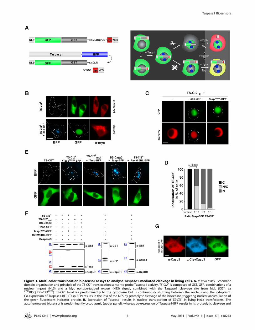
Figure 1. Multi-color translocation biosensor assays to analyse Taspase1-mediated cleavage in living cells. A. In vivo assay. Schematicdomain organization and principle of the TS-Cl2+ translocation sensor to probe Taspase1 activity. TS-Cl2+ is composed of GST, GFP, combinations of anuclear import (NLS) and a Myc epitope-tagged export (NES) signal, combined with the Taspase1 cleavage site from MLL (Cl2+; aa2713KISQLDGVDD2722). TS-Cl2+ localizes predominantly to the cytoplasm but is continuously shuttling between the nucleus and the cytoplasm.Co-expression of Taspase1-BFP (Tasp-BFP) results in the loss of the NES by proteolytic cleavage of the biosensor, triggering nuclear accumulation ofthe green fluorescent indicator protein. B. Expression of Taspase1 results in nuclear translocation of TS-Cl2+ in living HeLa transfectants. Theautofluorescent biosensor is predominantly cytoplasmic (upper panel), whereas co-expression of Taspase1-BFP results in its proteolytic cleavage and
Taspase1 Biosensors
PLoS ONE | www.plosone.org 3 May 2011 | Volume 6 | Issue 5 | e18253

not affect the cytoplasmic localization of the biosensor (Figure 1G
and data not shown).
Notably, in contrast to the high amounts of rTasp1 required for
cleavage in vitro (enzyme/substrate = 1:2), cotransfection of enzyme
(Taspase1-BFP) and substrate (TS-Cl2+) expression plasmid even
at a ratio of 1:10, was sufficient to catalyze efficient cleavage and
nuclear accumulation of the biosensor (Figure 1D).
Collectively, the results clearly underlined the practical
advantages and biological relevance of the cellular assay to search
for pharmacogenomic Taspase1 inhibitors.
nuclear accumulation (lower panel). Myc-NESRev was detected by indirect immunofluorescence using an a-myc-tag Ab. C. The translocationbiosensor is functional not only in adherent but also in leukemia cell models. K562 cells were transfected with expression plasmids encoding theindicated proteins. Coexpression of Tasp-GFP but not of inactive TaspT234V-GFP resulted in proteolytic cleavage and nuclear accumulation of the redfluorescent biosensor, TS-Cl2+
R. Biosensor localization was analyzed 48 h post transfection in at least 200 fluorescent living cells, and representativeimages are shown. Dashed lines mark nuclear/cytoplasmic cell boundaries obtained from the corresponding phase contrast images. D. Quantitationof TS-Cl2+ processing in vivo. HeLa cells were transfected with the indicated ratios of enzyme (Taspase1-BFP) and substrate (TS-Cl2+). 48 h later, thepercentage of cells showing cytoplasmic (C), cytoplasmic and nuclear (N/C) or nuclear (N) fluorescence was determined for at least 200 fluorescentcells. Cleavage-induced nuclear accumulation of the biosensor significantly increased already at a ratio of 1/10 (***: p,0.0001). Results from arepresentative experiment are shown. E. Biosensor assay specificity. Biosensors containing a non-functional Taspase1 cleavage site (TS-Cl2+
mut) or acleavage site for Caspase3 (BS-Casp3) remained cytoplasmic upon co-expression of Tasp-BFP. No nuclear accumulation of TS-Cl2+ was observed uponcoexpression of inactive TaspT234V-BFP or the nucleolar RevM10BL-BFP protein. F. Cleavage of TS-Cl2+, TS-Cl2+
mut or BS-Casp3 analyzed byimmunoblot. 293T cells were transfected with the indicated biosensors together with the indicated Taspase1 expression plasmids, the empty vector(control), RevM10BL-BFP or the protease Caspase3. Expression of proteins and cleavage products in cell lysates was visualized using a-GST, -Taspase1,-GFP or -Casp3 Abs. GAPDH served as loading control. G. Ectopic expression of Caspase3 does not induce cleavage and nuclear translocation of TS-Cl2+. Caspase3 expression was visualized by IF using a-Casp3 antibody, its activation by a-ClevCasp3 Ab. GFP/BFP were visualized by fluorescencemicroscopy. Scale bars, 10 mm.doi:10.1371/journal.pone.0018253.g001
Figure 2. Biosensor assay adaptation onto a HTS platform. A. Object selection parameters for nuclear (CIRC) and cytoplasmatic compartment(RING) analysis. B. Biosensor translocation assay analysis using the Cellomics ArrayscanH VTI platform. HeLa transfectants coexpressing TS-Cl2+ andactive or inactive (TaspT234V) mCherry fusions were fixed 48 h post transfection and nuclei marked by Hoechst 33342. The Hoechst 33342, GFP, andmCherry signals were recorded in channels 1, 2 and 3, respectively. Overlay with the CIRC mask and RING region is outlined for GFP (right panel).Representative images are shown. Scale bar, 10 mm. C. Translocation index (Ti = CIRC:RING) plotted for coexpression of Tasp1-mCh variants on asingle cell basis. Values were derived from analyzing ,400 cells/well. Mean from three wells is indicated. D. Ti was highly significantly increased uponcoexpression of active compared to inactive Taspase1 (***: p,0.0001). Columns, mean; bars, SD.doi:10.1371/journal.pone.0018253.g002
Taspase1 Biosensors
PLoS ONE | www.plosone.org 4 May 2011 | Volume 6 | Issue 5 | e18253
Triple-color biosensor-based high content screening forTaspase1 inhibitors
The robust performance of the TS-Cl2+ CBA met critical
requirements for high content screening: the biosensor was non-
toxic, localized to the cytoplasm in the absence of ectopically
expressed Taspase1, and efficiently accumulated in the nucleus
following Taspase1-specific cleavage. Hence, we tested whether
the assay can also be used on a high-throughput microscopy based
screening platform.
As cell lines inducibly expressing biosensors may facilitate
certain HCS/HTS applications, we generated stable Tet-off TS-
Cl2+TRE cell lines (Suppl. Figure S2A/B). The tetracycline
(doxycycline)-regulated system has been used successfully in
various applications [22]. Whereas expression of TS-Cl2+TRE was
blocked in the presence of doxycycline (Dox), Dox removal
induced TS-Cl2+TRE expression (Suppl. Figure S2B). Cleavage of
TS-Cl2+TRE by the endogenous Taspase1 subsequently resulted in
nuclear accumulation of the biosensor (Suppl. Figure S2B).
Although this cell system circumvents the need for cotransfection
of Taspase1, the levels of TS-Cl2+TRE are low compared to
transient expression, which we considered as a potential limitation
for HTS applications.
Thus, we decided to use transiently expressing transfectants for
the HTS assay. First, due to the low quantum yield of BFP, the
high cellular and sometimes observed compound autofluorescence
background upon UV-excitation, a green/red fluorescence dual-
color screening assay was developed. Therefore, BFP was replaced
by mCherry in the Taspase1 expression plasmids (Tasp-mCh).
Similar to GFP fusions, Tasp-mCh was biologically fully active,
whereas the inactive mutant TaspT234V-mCh did not cleave the
GFP-based biosensor (Figure 2B) even after 48 h.
Second, for HCS the assay was adapted to the Cellomics
ArrayscanHVTI platform. For this purpose, the molecular
translocation assay was adjusted by modifying several parameters
to ensure optimal object identification, including the adjustment of
the background correction and to define the threshold of pixels
derived from the Hoechst 33342 signal (Figure 2A). This
calculation resulted in an optimized object identification, capable
to automatically excluding ‘‘non cellular’’ irregular objects (too
small/big, debris, compound aggregates, etc.) in channel 1. By
adjusting object segmentation parameters, the fitting of the nuclear
mask to the Hoechst 33342 signal was further optimized. Also for
the channel 2 and 3, the background correction and values for the
threshold of the GFP and mCherry signal were defined to exclude
irregular and potentially false positive signals from the analysis.
The translocation index (Ti) was calculated as the ratio of the
nuclear to the cytoplasmic signal intensity (Ti = CIRC:RING) on a
single cell basis (Figure 2A–C). As shown in Figure 2C/D, the Ti
was highly significantly increased upon coexpression of Tasp-
compared to inactive TaspT234V-mCh. Notably, compared to
analyzing only GFP/Hoechst 33342 double-positive cells the
inclusion of GFP/mCherry/Hoechst 33342 triple-positive cells
resulted in an improved Ti. Based on the assay signal window and
Z9-factor of 0.63, the criterion for the primary screen was set at a
compound translocation index (Tic).2 (Tic = compound(CIR-
C:RING)/DMSO(CIRC:RING)). Only valid objects, i.e., cells that pass
the object selection criteria (Figure 2A) were included in the
analysis.
Next, we screened a focused compound library for potential
Taspase1 inhibitors. As Taspase1 is not affected by general
protease inhibitors [5,17], we used a pharmacophor screening
based on Taspase1’s crystal structure and the model suggested by
Khan et al. [6,23]. In an attempt to preclude the binding of the
peptidic substrate to Taspase1’s active centre, two classes of low
molecular weight (,500 Da) compounds were chosen (Suppl.
Figure S3). The first group was named ‘‘deep hole compounds’’
(DHCs), as these substances were selected to hypothetically fit into
the deep hydrophilic pocket of Taspase1 to prevent hydrolyzation
of the substrate [6] (Figure 3A). The second class, ‘‘chloride hole
compounds’’ (CHCs), should irreversibly occupy residues critical
for chloride ion and substrate binding in Taspase1’s active centre
(Figure 3B). In addition, as historically the majority of new drugs
have been derived from natural products [24] we also tested
lipophilic extracts prepared from 90 different types of fungi
obtained from the culture collection at the IBWF (http://www.
ibwf.de) [25].
Compounds and extracts were tested in 293T cells, which not
express detectable levels of endogenous Taspase1 (Figure 4B).
Cells coexpressing TS-Cl2+ and Tasp-mCh or TS-Cl2+R and
Tasp-GFP (Figure 3C) were challenged in 96-well plates and
analyzed 48 h after transfection to ensure that lack of inhibition is
not due to slow intracellular entry rates of the substances.
Although the majority of substances did not significantly affect
Taspase1’s trans cleavage activity at a concentration of 50 mM, we
identified two compounds, N-[2-[(4-amino-6-oxo-3H-pyrimidin-2-
yl)sulfanyl]ethyl]benzenesulfonamide (CHC-A4) and 2-benzyltria-
zole-4,5-dicarboxylic acid (DHC-C1), partially inhibiting biosen-
sor translocation (Figure 3E). In contrast, the tested fungal extracts
did not show detectable inhibition in our assay, although we
observed cytotoxicity for some extracts (data not shown). As we
previously identified transport inhibitors by chemicogenomic
screens [20], we first verified that CHC-A4 and DHC-C1 did
not affect nuclear import of the biosensor rather than cleavage.
Treatment with LMB resulted in nuclear accumulation of TS-
Cl2+R or TS-Cl2+ even in the presence of the inhibitors, excluding
interference with nuclear import (Suppl. Figure S2C and data not
shown). Taspase1 inhibition could be confirmed in other cell lines
using a compound concentration of 50 mM, although no inhibition
was detectable at a concentration of 5 mM (Figure 3D and data not
shown). Factors contributing to the weak inhibitory activity
observed may be compound instability and/or their inefficient
cell entry. Hence, to circumvent these limitations, we directly
microinjected both compounds into TS-Cl2+R/Tasp-GFP express-
ing transfectants. Compared to adding the compounds directly to
the cell culture medium, cytoplasmic injection of both compounds
resulted in improved Taspase1 inhibition reducing nuclear
translocation of the biosensor in the majority of cells (Figure 3F).
The coinjected fluorescent Ab allowed to select only healthy cells
for the analysis showing no signs of damage due to the
microinjection procedure. In order to allow a comparison of both
experimental approaches, the cells were inspected after 48 h. The
reason why inhibition did not occur in all injected cells is not
known, indicating that rational chemical modification of the
primary hits is required to improve their activity.
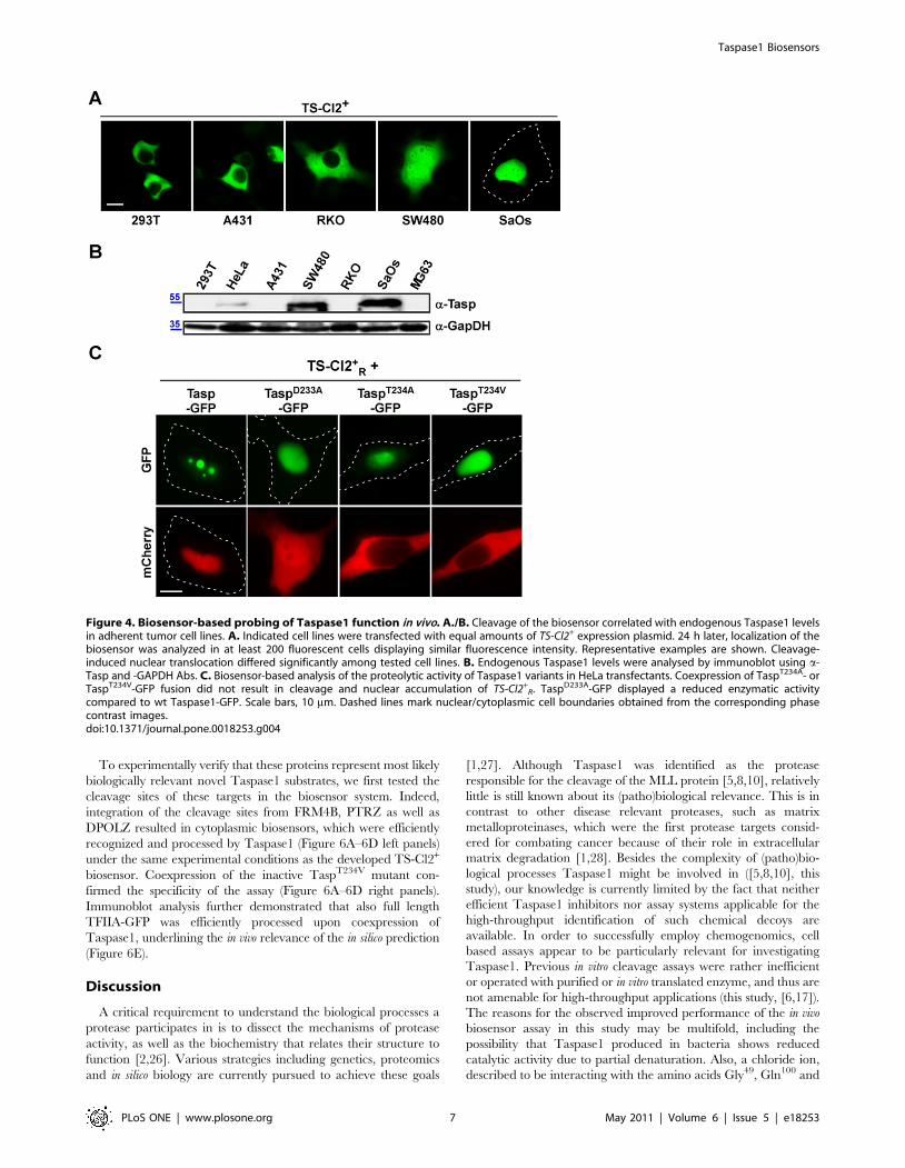
Biosensor-based probing of Taspase1 functionBesides their use in screening applications, we also exploited the
biosensors as genetic tools to characterize Taspase1’s biological
functions.
First, we used the biosensor to probe expression and biological
activity of endogenous Taspase1. As Taspase1 might also be
relevant for solid tumors, we tested several cancer cell models. As
depicted in Figure 4A/B, TS-Cl2+ remained cytoplasmic in cell
lines with low endogenous Taspase1 levels, whereas partial or
complete nuclear translocation was evident in cell lines expressing
high Taspase1 levels already after 24 h (for summary see Suppl.
Table S2). Later time points did not show a different localization.
Taspase1 Biosensors
PLoS ONE | www.plosone.org 5 May 2011 | Volume 6 | Issue 5 | e18253

Second, we analyzed the proteolytic acivity of Taspase1
mutants, in which the catalytic nucleophile, Thr234, was changed
into Val or Ala (TaspT234V, TaspT234A) or Asp233 was mutated into
Ala (TaspD233A). As shown in Figure 4C, coexpression of
TaspT234V- or TaspT234A-GFP fusion did not result in cleavage
and nuclear accumulation of TS-Cl2+R confirming that both
mutants are catalytically inactive [5]. Notably, although in vitro
studies reported a 1000-fold reduced activity for TaspD233A [5],
the in vivo data indicated that TaspD233A-GFP was still able to
recognize and process the biosensor albeit with a somehow
attenuated efficacy (Figure 4C).
Next, to uncover the sequence and spatial requirements for
Taspase1 processing in vivo, we performed Ala scan mutagenesis of
the MLL cleavage site (CS2; aa 2713KISQLDQGVDD2722) in the
biosensor background. As depicted in Figure 5, coexpression of the
TS-Cl2+ mutants (TS-Cl2+CSmut) with Tasp-mCh resulted in
proteolytic cleavage and nuclear accumulation of only those
biosensors in which non-essential residues were mutated. In
contrast, changing critical aa into Ala almost completely prevented
cleavage and nuclear accumulation of the autofluorescent proteins,
leading to the following consensus sequence: K6I5S4Q3L2D1Q
G19V29D39D49 (essential aa in bold; see Table 1 for summarized
results of targets). Notably, even replacing critical residues by
chemically similar aa could not rescue cleavage, exempt the
exchange of Leu for the also hydrophobic aa Ile or Val (Figure 5A/
B and Table 1). These results could be confirmed by immunoblot
analysis (Figure 5D/E). Again, specificity of the assay was verified
by cotransfection of inactive TaspT234V-mCh, which did neither
result in cleavage nor nuclear accumulation of the biosensors
(Suppl. Figure S2C). Nuclear accumulation of all the TS-Cl2+CSmut
variants upon LMB treatment further excluded the formal
possibility that mutagenesis had affected import of the biosensors
(Figure 5C). Collectively, these results underline the reliability and
practical advantages of our visual cell based assay to probe
Taspase1 function in living cells.
Identification of novel human Taspase1 targetsTo bioinformatically identify novel human Taspase1 targets, we
used the motifs Q3[I,L,V]2D1QG19X29X39D49 and Q3[I,L,V]2
D1QG19X29D39X49 obtained by our mutational analysis to scan
the Swiss-Prot database. Besides the expected Taspase1 targets,
MLL1 and MLL4, our analysis identified TF2A, the FERM
Domain-Containing Protein 4B (FRM4B), the Tyrosine-Protein
Phosphatase Zeta (PTRZ) and DNA Polymerase Zeta (DPOLZ) as
putative Taspase1 substrates (see Table 2 for verified, Suppl. Table
S3 for predicted targets).
Figure 3. In vivo analysis of potential Taspase1 inhibitors. A./B. Pharmacophore-queries for virtual screening. A. Four-point MOEpharmacophore model based on the docked substrate QLDGVDD (shown as sticks coloured by element) together with the assigned pharmacophoricfeatures (cyan: acceptor, green: hydrophobic, grey: acceptor and anion, left panel) B. Receptor-based SYBYL three-point pharmacophore modelrepresenting a negative charge (blue spheres) interacting with Lys57, hydrogen-bond donor (magenta) interacting with Ala50 and acceptor (green)interacting with Asn100 (right panel). The yellow surface indicates excluded volume. C. Expression of Tasp-GFP but not of inactive TaspT234V-GFPinduces nuclear accumulation of the red fluorescent biosensor, TS-Cl2+
R, in HeLa transfectants. D. CHC-A4 and DHC-C1 partially inhibited TS-Cl2+R
translocation. HeLa cell transfectants coexpressing TS-Cl2+R and Taspase1-GFP were treated with DMSO or compounds (50 mM final concentration),
and analyzed 48 h later. Representative examples are shown. Scale bars, 10 mm. Dashed lines mark nuclear/cytoplasmic cell boundaries obtainedfrom the corresponding phase contrast images. E. Structures and formulas of the respective compounds. F. Intracellular delivery of the compoundsby microinjection resulted in enhanced inhibition. Vero cell transfectants coexpressing TS-Cl2+
R and Taspase1-GFP were either treated with DMSO orcompounds (50 mM final concentration) or microinjected. 48 h later, the percentage of cells showing cytoplasmic (C), cytoplasmic and nuclear (N/C)or nuclear (N) fluorescence was determined for at least 100 sensor expressing cells.doi:10.1371/journal.pone.0018253.g003
Taspase1 Biosensors
PLoS ONE | www.plosone.org 6 May 2011 | Volume 6 | Issue 5 | e18253
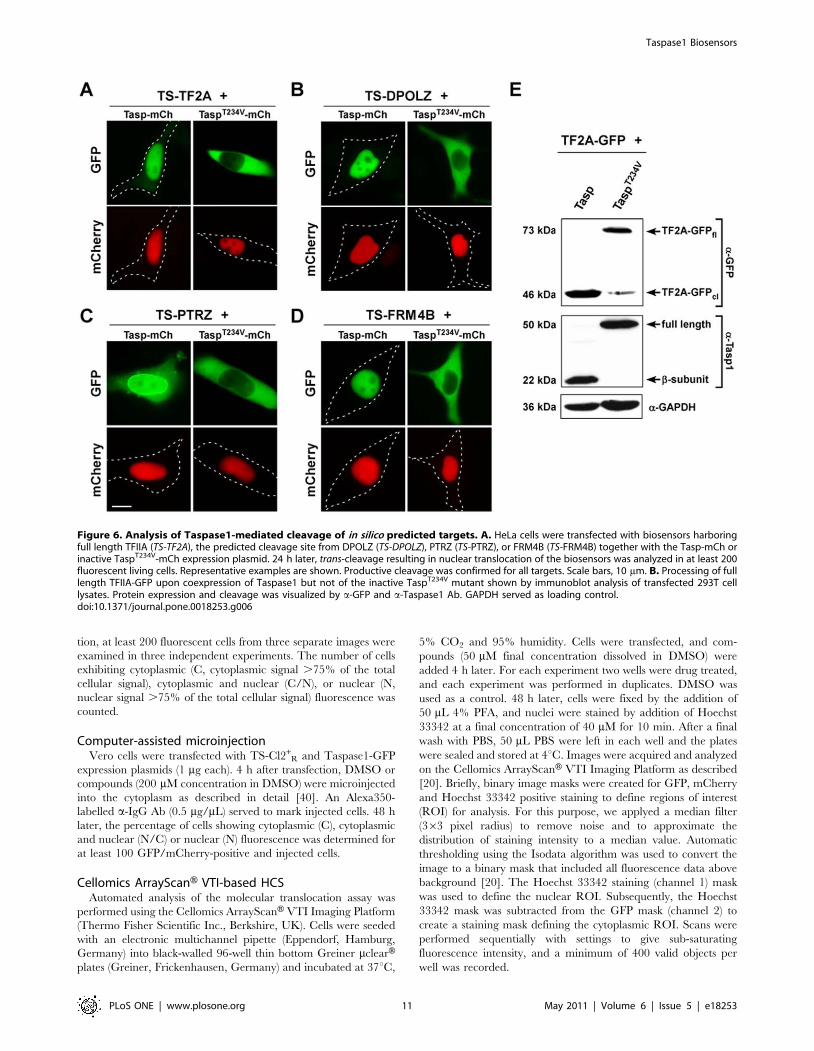
To experimentally verify that these proteins represent most likely
biologically relevant novel Taspase1 substrates, we first tested the
cleavage sites of these targets in the biosensor system. Indeed,
integration of the cleavage sites from FRM4B, PTRZ as well as
DPOLZ resulted in cytoplasmic biosensors, which were efficiently
recognized and processed by Taspase1 (Figure 6A–6D left panels)
under the same experimental conditions as the developed TS-Cl2+
biosensor. Coexpression of the inactive TaspT234V mutant con-
firmed the specificity of the assay (Figure 6A–6D right panels).
Immunoblot analysis further demonstrated that also full length
TFIIA-GFP was efficiently processed upon coexpression of
Taspase1, underlining the in vivo relevance of the in silico prediction
(Figure 6E).
Discussion
A critical requirement to understand the biological processes a
protease participates in is to dissect the mechanisms of protease
activity, as well as the biochemistry that relates their structure to
function [2,26]. Various strategies including genetics, proteomics
and in silico biology are currently pursued to achieve these goals
[1,27]. Although Taspase1 was identified as the protease
responsible for the cleavage of the MLL protein [5,8,10], relatively
little is still known about its (patho)biological relevance. This is in
contrast to other disease relevant proteases, such as matrix
metalloproteinases, which were the first protease targets consid-
ered for combating cancer because of their role in extracellular
matrix degradation [1,28]. Besides the complexity of (patho)bio-
logical processes Taspase1 might be involved in ([5,8,10], this
study), our knowledge is currently limited by the fact that neither
efficient Taspase1 inhibitors nor assay systems applicable for the
high-throughput identification of such chemical decoys are
available. In order to successfully employ chemogenomics, cell
based assays appear to be particularly relevant for investigating
Taspase1. Previous in vitro cleavage assays were rather inefficient
or operated with purified or in vitro translated enzyme, and thus are
not amenable for high-throughput applications (this study, [6,17]).
The reasons for the observed improved performance of the in vivo
biosensor assay in this study may be multifold, including the
possibility that Taspase1 produced in bacteria shows reduced
catalytic activity due to partial denaturation. Also, a chloride ion,
described to be interacting with the amino acids Gly49, Gln100 and
Figure 4. Biosensor-based probing of Taspase1 function in vivo. A./B. Cleavage of the biosensor correlated with endogenous Taspase1 levelsin adherent tumor cell lines. A. Indicated cell lines were transfected with equal amounts of TS-Cl2+ expression plasmid. 24 h later, localization of thebiosensor was analyzed in at least 200 fluorescent cells displaying similar fluorescence intensity. Representative examples are shown. Cleavage-induced nuclear translocation differed significantly among tested cell lines. B. Endogenous Taspase1 levels were analysed by immunoblot using a-Tasp and -GAPDH Abs. C. Biosensor-based analysis of the proteolytic activity of Taspase1 variants in HeLa transfectants. Coexpression of TaspT234A- orTaspT234V-GFP fusion did not result in cleavage and nuclear accumulation of TS-Cl2+
R. TaspD233A-GFP displayed a reduced enzymatic activitycompared to wt Taspase1-GFP. Scale bars, 10 mm. Dashed lines mark nuclear/cytoplasmic cell boundaries obtained from the corresponding phasecontrast images.doi:10.1371/journal.pone.0018253.g004
Taspase1 Biosensors
PLoS ONE | www.plosone.org 7 May 2011 | Volume 6 | Issue 5 | e18253

Thr234 of recombinant Taspase1 [6] may act as a competitive
inhibitor under in vitro assay conditions. Although we are currently
lacking experimental evidence it is suffice to speculate that
eukaryotic post-translational modifications and/or co-factors
may be required to render the enzyme fully active. Nevertheless,
our results underlined the practical advantages and biological
relevance of the cellular assay to investigate Taspase1 function.
A key part of understanding protease signaling in both health
and disease is to identify a protease’s physiological substrates.
Although the sequence Q3X2D1QG19 has been proposed as a
consensus cleavage site sequence for Taspase1 [6], employing this
motif for the bioinformatic identification of novel Taspase1 targets
is impractical, as more than 1000 putative substrates were
predicted. To improve our understanding of Taspase1’s substrate
specificity, we used our biosensor assay combined with positional
scanning mutagenesis to identify residues essential for Taspase1
cleavage activity in living cells. As expected, Asp at the P1 position
was required for cleavage by this aspartase, and Gly at P19 did not
even tolerate its replacement by Ala. Also, Gln at position P3 was
critical for substrate recognition, as an exchange of this uncharged
polar amino acid by the smaller hydrophobic residue Ala or even
the similar but smaller amino acid Asn completely blocks cleavage.
In contrast to previous studies [6], we found that albeit position P2
can hold hydrophobic residues of similar size (Leu, Ile, Val), other
amino acids such as the smaller hydrophobic amino acid Ala were
not tolerated. Hence, hydrophobicity in combination with certain
size are likely to be structural requirements for productive
cleavage. Position P29 was found to be flexible, whereas the amino
acids at P39 and P49 seem to be interdependent. At least one of
these residues needed to be Asp, although a small residue at the
other position, like Gly or Ala, was tolerated. Glu at either position
however impaired cleavage, indicating that not only charge but
Figure 5. Identification of residues required for productive Taspase1 cleavage in living cells. A.–C. Nuclear translocation of the indicatedbiosensor cleavage site mutants (TS-Cl2+
CSmut) was analyzed in HeLa transfectants coexpressing the indicated biosensors together with Tasp- orinactive TaspT234V-mCherry. At least 200 fluorescent living cells were inspected, and representative examples are shown. Whereas substitution of Leu2
with Ile did not affect cleavage (A.), exchange of Asp1 with Ala completely abrogated cleavage (B.) LMB treatment verified that nuclear import of thevariants was not affected. (C.) Scale bars, 10 mm. D./E. Cleavage of indicated cleavage site mutants by Tasp- (D.) or inactive TaspT234V-mCh (E.)analyzed by immunoblot. Notably, D19A, D39A and D49A mutants run lower in the gel, most likely due to the loss of the negative charge. Expressionof Taspase1 proteins as well as of cleavage products in 293T cell lysates was visualized using a-GST or -Taspase1 Abs. GAPDH served as loadingcontrol.doi:10.1371/journal.pone.0018253.g005
Taspase1 Biosensors
PLoS ONE | www.plosone.org 8 May 2011 | Volume 6 | Issue 5 | e18253

also size is important for productive processing. Taken together,
we defined the sequence motif Q3[I,L,V]2D1QG19V29D39D49 as an
improved consensus recognition site for Taspase1.
Employing this motif, we bioinformatically identified not only
known Taspase1 substrates, such as MLL1 and MLL4, but also
proteins, which have not been considered as potential targets for
this protease. These include the FERM Domain-Containing
Protein 4B (FRM4B), the Tyrosine-Protein Phosphatase Zeta
(PTRZ) and DNA Polymerase Zeta (DPOLZ), suggested to be
relevant for various biological processes (Table 2). Although we are
currently lacking experimental evidence how Taspase1-mediated
processing of these targets contributes to their functional
regulation, we could confirm that the cleavage sites of these
proteins are recognized and processed by Taspase1 in vivo. The
potential impact of Taspase1 for neoplastic diseases extrapolated
from its processing of leukemia inducing MLL fusion proteins
containing a functional Taspase1 cleavage site is further supported
by our identification of these substrates. We just showed that only
AF4NMLL but not the reciprocal translocation product,
MLLNAF4, lacking the Taspase1 cleavage site, can cause proB
ALL in a murine model [13]. Albeit the exact biological relevance
of PTRZ for disease and development is not yet resolved, this
phosphatase was suggested as a therapeutic target for glioblastoma
and glioblastoma-derived stem cells [29,30]. Likewise, although
the function of FRM4B is unknown, other members of the protein
4.1 superfamily such as FRMD4A or FRMD3 have been
implicated in oncogenic signaling [31,32,33]. Notably, DPOLZ
is not only essential during embryogenesis but also important in
defense against genotoxins. As recent evidence indicates that
reduced DPOLZ levels enhance spontaneous tumorigenesis, it is
tempting to speculate that Taspase1 might participate in
controlling DPOLZ levels and thus, disease [34,35]. Notably, we
found that Taspase1 is expressed in several solid tumor cell
models. Whether the differences in Taspase1 expression levels
detected have implications also on the (patho)biological charac-
teristics of the tumor cell lines as well as for the primary disease
remains to be investigated.
Nevertheless, there is increasing evidence that Taspase1 may be
critically contributing to disease, underlining its pathobiological
and potentially therapeutic relevance. However, we still do not
comprehense the processes and molecular mechanisms Taspase1
might be involved in. Thus, besides genetic and biochemical
approaches, small molecules allowing a (transient) chemical
knockout of Taspase1 in a specific biological system or disease
model would be highly valuable. These needs underline the
relevance of the developed translocation biosensor for the
identification and validation of inhibitors in living cells. Impor-
tantly, the biosensors can operate with red or green autofluor-
escent proteins, which can be optimally detected even by high-
throughput fluorescence microscopy, and are not restricted to a
specific cell type. The assay strictly depends on the presence of
catalytically active Taspase1 and occurs with a high signal-to-noise
ratio, allowing its use in HTS/HCS applications of large or
focused compound libraries.
As a proof of principle, we screened a collection of small
molecules, which were chosen based on a pharmacophore
screening relying on the published crystal structure of Taspase1
[6]. The low molecular weight compounds were selected by
virtual screening to prevent substrate cleavage and/or arrest the
enzyme in an inactive state. Noteworthy, we identified two
substances showing inhibitory activity in living cells, which would
represent a primary hit rate of 3%. The reasons why other
compounds were not active in our assay are versatile, including
their potential inability to penetrate cell membranes. Also, the
accuracy of virtual screening might have been flawed as details in
the published crystal structure of Taspase1 are missing and the
catalytic mechanism of Taspase1 is not yet resolved in detail. The
first hit compound, N-[2-[(4-amino-6-oxo-3H-pyrimidin-2-yl)
sulfanyl]ethyl]benzenesulfonamide (CHC-A4), was retrieved
by SYBYL UNITY-Flex similarity searching (receptor-derived
pharmacophore model). The second, 2-benzyltriazole-4,5-dicar-
boxylic acid (DHC-C1), was selected based on the four-point
substrate pharmacophore model using the software Molecular
Operating Environment. Both compounds are small and polar,
with a pronounced hydrogen-bonding potential, which can be
readily explained by the requirements of the pharmacophore
queries. Although we controlled that the compounds do not
unspecifically act by blocking nuclear import of the biosensors,
significant Taspase1 inhibition in vivo required relative high
inhibitor concentrations (50 mM). Notably, we observed im-
proved inhibition upon direct delivery of both compounds into
the cells by microinjection, indicating that the weak inhibitory
activity observed may be due to compound instability and/or
their inefficient cell entry. Recently, Lee et al. [17] designed
chemically modified peptidic derivates of a Taspase1 cleavage
substrate. Although some of these compounds displayed mild
inhibitory activity using in vitro Taspase1 assays (e.g., yzm18
IC5029.4 mM), these peptide-based inhibitors have not shown
efficacy in living cells, in contrast to our low molecular weight
inhibitors.
Although natural products appear to interrogate a different area
of chemical space than synthetic compounds [36], the tested
lipophilic fungal extracts showed no inhibitory activity. Failure
may be due to the fact that albeit such extracts contain a mixture
of many different substances, the concentration of potentially
active ingredients may be too low or outweighed by toxic effects of
other components. Also, the numbers of samples which have to be
screened in unfocussed natural product libraries are usually high,
and hit rates are mostly below 0.01% [20,24].
Hence, as future strategies to identify potent Taspase1 inhibitors
we suggest to focus on a rational synthesis of derivates based on the
Table 1. Cleavage-site residues critical for Taspase1processing in vivo.
Cleavage-site mutation In cell cleavage by Taspase1
K6A +
I5A +
S4A +
Q3A 2
Q3N 2
L2A 2
L2I +
D1A 2
G19A 2
V29A +
D39A (2)
D39E (2)
D49A (2)
D49E (2)
Summary of results obtained from the biosensor-based mapping.Cleavage site aa residues: K6I5S4Q3L2D1QG19V29D39D49 (essential aa in bold).2: no cleavage, (2): reduced cleavage, +: cleavage.doi:10.1371/journal.pone.0018253.t001
Taspase1 Biosensors
PLoS ONE | www.plosone.org 9 May 2011 | Volume 6 | Issue 5 | e18253

structures of our primary hits combined with HTS of large
natural/synthetic compound libraries.
Materials and Methods
Antibodies (Ab), reagents, compounds and fungalextracts
Ab used: a-GST (sc-57753), a-Taspase1 (sc-85945), a-GAPDH
(sc-47724) and a-GFP (sc-8334) (Santa Cruz Biotechnology,
Heidelberg, Germany); a-myc-tag (NEB GmbH, Frankfurt am
Main, Germany). Appropriate HRP-, Cy3- or AlexaDye-conju-
gated secondary antibodies (Sigma Aldrich, Munich, Germany;
Santa Cruz Biotechnology, Heidelberg, Germany) were used.
Reagents were from Sigma Aldrich (Sigma Aldrich, Munich,
Germany) unless stated otherwise. Cells were treated with
leptomycin B (LMB) (10 nM) as described in [37]. Potential
Taspase1 inhibitors (Suppl. Figure S3) were purchased from
ASINEX Ltd (Moscow, Russia). Fungal extracts were obtained
from submerged cultures of higher fungi, preferentially from asco-
and basidiomycetes, deposited in the culture collection at the
IBWF, as described [25]. Briefly, the fermentation of the fungi was
stopped as soon as free glucose in the growth medium was
depleted, and mycelia were separated by filtration. Lipophilic
molecules were extracted from the culture broth with ethyl acetate.
The extracts were dried in vacuo, redissolved in 25 mL DMSO, and
aliquots of these extracts were used in the assays at a dilution of
1:2000.
Cell culture, microscopy and fluorescence imaging ofcells
Cell lines used in the study were maintained and transfected as
described [20,37]. MEF3T3 stably expressing the Dox-inducible
TS-Cl2+TRE were established by G418- (800 mg/mL) and puro-
mycin- (2 mg/mL) selection, and fluorescence activated cell sorting
as reported [20]. Cells were cultured in medium containing 1 mg/
mL doxycycline (Dox) [20]. Twelve-bit black and white images
were captured using a digital Axiocam CCD camera (Carl Zeiss,
Jena, Germany). Quantitation, image analysis and presentation
were performed as described [18,38]. The nuclear signal was
similarly obtained by measuring the pixel intensity in the nucleus.
Nuclei were marked by Hoechst 33258 staining as described
[18,39]. To determine the average intracellular protein localiza-
Table 2. Characteristics of verified Taspase1 target genes according to their GO term classifications.
Gene Locus GO : biological process GO : cellular component GO : molecular function
ID description ID description ID description
MLL1 0006366 transcription 0071339 nucleoplasm 0003680 DNA binding
0006461 protein complex assembly 0003700 nucleic acid binding
0006915 cell death 0003702 transcription regulator activity
0035162 developmental process 0008270 ion binding
0043984 histone acetylation 0042800 N-methyltransferase activity
0051568 histone methylation 0042803 protein homodimerization activity
0045322 transcription factor activity
0070577 histone binding
MLL4 0006350 transcription 0035097 nucleoplasm 0005515 protein binding
0016568 chromatin modification 0008270 ion binding
0008284 cell proliferation 0010843 DNA binding
0033148 estrogen signalling 0018024 N-methyltransferase activity
0010552 transcription
0043627 estrogen signalling
FRM4B - unknown 0005737 cytoplasm 0005488 binding
0005856 cytoskeleton
PTRZ 0006470 metabolic process 0005887 integral tomembrane
0005001 phosphatase activity
0007417 developmental process 0005515 protein binding
0008330 phosphatase activity
TF2AA 0006367 transcription initiation 0005634 nucleus 0003677 DNA binding
0006368 RNA elongation 0005672 nucleoplasm 0003713 transcription regulator activity
0032568 transcription 0005737 cytoplasm 0016251 transcription regulator activity
0045449 transcription 0017025 TATA-binding protein binding
0046982 protein heterodimerization activity
DPOLZ 0006261 DNA replication 0005634 nucleus 0000166 nucleotide binding
0006281 DNA repair 0003677 DNA polymerase activity
0003887 DNA polymerase activity
0008270 ion binding
doi:10.1371/journal.pone.0018253.t002
Taspase1 Biosensors
PLoS ONE | www.plosone.org 10 May 2011 | Volume 6 | Issue 5 | e18253
tion, at least 200 fluorescent cells from three separate images were
examined in three independent experiments. The number of cells
exhibiting cytoplasmic (C, cytoplasmic signal .75% of the total
cellular signal), cytoplasmic and nuclear (C/N), or nuclear (N,
nuclear signal .75% of the total cellular signal) fluorescence was
counted.
Computer-assisted microinjectionVero cells were transfected with TS-Cl2+
R and Taspase1-GFP
expression plasmids (1 mg each). 4 h after transfection, DMSO or
compounds (200 mM concentration in DMSO) were microinjected
into the cytoplasm as described in detail [40]. An Alexa350-
labelled a-IgG Ab (0.5 mg/mL) served to mark injected cells. 48 h
later, the percentage of cells showing cytoplasmic (C), cytoplasmic
and nuclear (N/C) or nuclear (N) fluorescence was determined for
at least 100 GFP/mCherry-positive and injected cells.
Cellomics ArrayScanH VTI-based HCSAutomated analysis of the molecular translocation assay was
performed using the Cellomics ArrayScanH VTI Imaging Platform
(Thermo Fisher Scientific Inc., Berkshire, UK). Cells were seeded
with an electronic multichannel pipette (Eppendorf, Hamburg,
Germany) into black-walled 96-well thin bottom Greiner mclearHplates (Greiner, Frickenhausen, Germany) and incubated at 37uC,
5% CO2 and 95% humidity. Cells were transfected, and com-
pounds (50 mM final concentration dissolved in DMSO) were
added 4 h later. For each experiment two wells were drug treated,
and each experiment was performed in duplicates. DMSO was
used as a control. 48 h later, cells were fixed by the addition of
50 mL 4% PFA, and nuclei were stained by addition of Hoechst
33342 at a final concentration of 40 mM for 10 min. After a final
wash with PBS, 50 mL PBS were left in each well and the plates
were sealed and stored at 4uC. Images were acquired and analyzed
on the Cellomics ArrayScanH VTI Imaging Platform as described
[20]. Briefly, binary image masks were created for GFP, mCherry
and Hoechst 33342 positive staining to define regions of interest
(ROI) for analysis. For this purpose, we applyed a median filter
(363 pixel radius) to remove noise and to approximate the
distribution of staining intensity to a median value. Automatic
thresholding using the Isodata algorithm was used to convert the
image to a binary mask that included all fluorescence data above
background [20]. The Hoechst 33342 staining (channel 1) mask
was used to define the nuclear ROI. Subsequently, the Hoechst
33342 mask was subtracted from the GFP mask (channel 2) to
create a staining mask defining the cytoplasmic ROI. Scans were
performed sequentially with settings to give sub-saturating
fluorescence intensity, and a minimum of 400 valid objects per
well was recorded.
Figure 6. Analysis of Taspase1-mediated cleavage of in silico predicted targets. A. HeLa cells were transfected with biosensors harboringfull length TFIIA (TS-TF2A), the predicted cleavage site from DPOLZ (TS-DPOLZ), PTRZ (TS-PTRZ), or FRM4B (TS-FRM4B) together with the Tasp-mCh orinactive TaspT234V-mCh expression plasmid. 24 h later, trans-cleavage resulting in nuclear translocation of the biosensors was analyzed in at least 200fluorescent living cells. Representative examples are shown. Productive cleavage was confirmed for all targets. Scale bars, 10 mm. B. Processing of fulllength TFIIA-GFP upon coexpression of Taspase1 but not of the inactive TaspT234V mutant shown by immunoblot analysis of transfected 293T celllysates. Protein expression and cleavage was visualized by a-GFP and a-Taspase1 Ab. GAPDH served as loading control.doi:10.1371/journal.pone.0018253.g006
Taspase1 Biosensors
PLoS ONE | www.plosone.org 11 May 2011 | Volume 6 | Issue 5 | e18253

PlasmidsTo generate plasmid p_NLS-GFP/GST-CS2-NESRev (p_TS-
Cl2+), encoding a fusion composed of the SV40 large T-antigen
NLS, GST, GFP, the Taspase1 cleavage site from MLL (CS2; aa2713KISQLDGVDD2722), and a Myc-epitope-tagged NES from
the HIV-1 Rev protein (NESRev) [41], the CS2 coding sequence
was inserted into vector pNLS-GFP/GST-CS3-RevNES (p_BS-
Casp3), replacing CS3. p_BS-Casp3 encodes a biosensor harbor-
ing the cleavage site for Caspase3 (CS3: aa KRKGDEVDGVDE)
[18]. p_TS-Cl2+R encodes a red fluorescent biosensor (NLS-
mCherry/GST-CS2-NESRev), in which GFP was replaced by
mCherry [18]. Expression plasmids for TS-Cl2+ variants, in which
CS2 was mutated (p_TS-Cl2+mut; see Table 2, and Suppl. Table S1
for oligonucleotides used) were generated by oligonucleotide-
annealing and cloning into the NotI/XhoI-restriction sites of p_TS-
Cl2+ as described [18]. Likewise, the coding sequence for full
length TFIIA (p_TS-TF2A), or the cleavage sites from DPOLZ
(p_TS-DPOLZ), PTRZ (p_TS-PTRZ) or FRM4B (p_TS-FRM4B)
were inserted into p_TS-Cl2+, thereby replacing the CS2. pTRE-
NLS-GFP/GST-CS2-NESRev (p_TS-Cl2+TRE) allows the inducible
expression of the biosensor (‘‘tet-off’’) and was constructed by
inserting the NLS-GFP/GST-CS2-NESRev coding sequence into
pTRE-NLS-GFP/GST-NESRev [18].
The Taspase1 or TFIIA coding sequence was amplified from
cDNA obtained from a human head and neck tumor. mRNA
preparation and cDNA synthesis from tumor tissue was performed
as described [42]. Cloning of the Taspase1 coding sequence into
expression vectors pc3, pc3-GFP, pc3-BFP, and pc3-mCherry
using BamHI/EcoRI- or BamHI/NheI-restriction sites, respectively,
allowed the expression of Taspase1, alone or as a fusion with
fluorescent proteins as described [21,43]. Plasmid p_TaspT234V-
GFP, p_TaspT234A-GFP, p_TaspD233A-GFP and p_TaspT234V-
mCherry or –BFP encoding the catalytically inactive Taspase1
mutant, were generated by splice overlap extension polymerase
chain reaction as reported [18,44]. p_TFIIA-GFP, encoding a
TFIIA-GFP fusion, was generated by PCR amplification and
cloning into pc3-GFP as reported [45]. pc3_RevM10BL-BFP,
encoding a mutant HIV-1 Rev protein, was described [40].
Bacterial expression plasmid pGEX_GST-Tasp1-GFP encod-
ing a GST-Tasp1-GFP fusion protein and pET22-Tasp, encoding
a His-tagged Taspase1 protein, were generated by inserting the
Taspase1 coding sequence into pGEX-GFP or pET22b+,
respectively, as reported [46]. The coding sequence for the MLL
aa 2650–2808 (2Cl) was inserted into vector pGEX5T to generate
pGEX5T_GST-2Cl, encoding a His-tagged-GST-2Cl fusion
protein.
Plasmids were verified by sequence analysis as described [47].
Oligonucleotides used for PCR amplification and cloning are
listed in Suppl. Table S1.
Protein extraction, immunoblot analysis andimmunofluorescence
Preparation of whole cell lysates and immunoblotting were
carried out as described [39,48]. Immunofluorescence was
performed as reported in detail [18,49].
In vitro Taspase1 cleavage assayHis-tagged Taspase1, GST-Taspase1-GFP and His-tagged
GST-2Cl substrates were expressed in BL21 bacteria and purified
by nickel chelating or glutathione affinity chromatography as
described [18,46]. Fractions were eluted (50 mM NaH2PO4,
300 mM NaCl, 250 mM Imidazol, pH 8.0) and dialyzed against
Taspase1 cleavage buffer (200 mM Hepes/KOH pH 7.9, 10 mM
MgCl2, 40 mM KCl, 20% Saccharose, 10 mM DTT). Trans-
cleavage assays were performed in cleavage buffer adding
recombinant Taspase1 to 5 mM of GST-2Cl. Analysis of cleavage
was carried out by SDS page followed by Coomassie staining as
outlined in [49].
Virtual screening and database searchesAn X-ray structure of the inactive autocatalytically processed
Taspase 1 dimer (Protein Data Bank ID 2a8j, 1.9 A resolution [6]
served as the basis for pharmacophore model generation and
computer-based similarity searching in a commercial screening
compound collection (Asinex Gold collection nov. 2005: 231,812
compounds; ASINEX Ltd, Moscow, Russia) [23]. Briefly,
screening compounds were reduced to ‘‘druglike’’ compounds
(clogS.4, no rule-of-five violation) using Molecular Operating
Environment (MOE) 2005.06 (Chemical Computing Group, Mon-
treal, Canada), and for the remaining 181,403 substances single
conformers were computed using CORINA 3.20 (Molecular
Networks GmbH, Erlangen, Germany). Bases were de-protonated,
acid groups were protonated (‘‘wash’’ function in MOE). Two
types of pharmacophore hypotheses were generated: (i) ‘‘ligand-
based’’ models from hypothetical binding mode of the Taspase1
cleavage site substrate QLDQGVDD [5], (pre-docking of the
substrate by GOLD 3.0.1; force-field relaxation using AMBER99
in MOE; manual assignment of potential pharmacophoric points
in MOE; similarity searching with MOE), and (ii) a ‘‘receptor-
based’’ model of a hypothetical ligand pharmacophore using
SYBYL 7.1 (Tripos Inc, Missouri, U.S.A.), with UNITY-Flex
search option. The resulting ligand-based pharmacophore models
yielded a total of 62 perfect matches in the screening compound
collection, and the receptor-based model retrieved 209 perfect
matches. From these hits, compounds were selected for testing.
For the bioinformatic identification of potential human
Taspase1 targets, ScanProsite searches were performed in the human
taxon of the UniProtKB/SwissProt database using the patterns Q-
[IVL]-D-G-X-D-D, Q-[IVL]-D-G-X-X-D and Q-[IVL]-D-G-X-
D-X as queries.
Statistical analysisFor experiments stating p-values, a paired Student’s t-test was
performed. Unless stated otherwise, p-values represent data
obtained from three independent experiments done in triplicate.
p-values,0.05 were considered as significant.
Supporting Information
Figure S1 In vitro Taspase1 cleavage assay. A. Extensive
aggregation of GST-Tasp1-GFP expressed in BL21 bacteria
visualized by fluorescence microscopy. In contrast, GST-GFP
showed no aggregation and was efficiently expressed. Images were
taken with identical CCD camera settings. Scale bars, 1 mm. B.Schematic representation of expression constructs for His-tagged
Taspase1 (rTasp) and the substrate GST-2Cl, containing the MLL
cleavage sites CS1 and CS2 (MLL aa 2650–2808). Molecular
weight of the expected cleavage products is indicated. C.Concentration dependent processing of GST-2Cl by recombinant
Taspase1 (rTasp). GST-2Cl (5 mM) was incubated with increasing
amounts of rTasp (lane1: 2.5 mM, 2: 1.25 mM, 3: 0.63 mM, 4:
0.32 mM, 5: 0.16 mM, 6: 0.08 mM, 7: 0.04 mM, 8: 0.02 mM) for
60 min. Cleavage was visualized by SDS-PAGE and Coomassie
staining. Uncleaved and cleaved proteins are indicated. D. Time
dependent processing of GST-2Cl by recombinant Taspase1.
GST-2Cl (5 mM) was incubated with 2.5 mM Taspase1, and
cleavage was monitored over time. Cleavage was visualized by
Taspase1 Biosensors
PLoS ONE | www.plosone.org 12 May 2011 | Volume 6 | Issue 5 | e18253

SDS-PAGE and Coomassie staining. Uncleaved and cleaved
proteins are indicated.
(CVX)
Figure S2 In vivo screening for inhibitors of Taspase1activity. A. Principle of the inducible biosensor system, Tet-off
TS-Cl2+TRE. Dox interacts with tTA preventing its binding and
thus activation of the TRE-containing CMV promoter. Removal
of Dox allows tTA binding, triggering transcriptional activation
and expression of the shuttling biosensor, which predominately
localizes to the cytoplasm. Dox, Doxycylin; pCMV/pCMVmin,
(minimal) CMV promoter; TRE, Tetracycline-responsive promot-
er element; tTA, Tet-controlled transactivator. B. Dox-induced
biosensor expression. MEF3T3 cells stably expressing TS-Cl2+TRE
were cultured in the presence or absence of Dox. In presence of
Dox, no expression of the biosensor is detectable. Three days after
Dox removal, expression of cytoplasmic TS-Cl2+TRE is visible
(2Dox 3d), and cleavage by endogenous Taspase1 results in its
nuclear accumulation 24 h later (2Dox 4d). Living cells were
analyzed by fluorescence microscopy and images taken with
identical CCD camera settings. Scale bars, 10 mm. C. CHC-A4 or
DHC-C1 do not interfere with nuclear import of the biosensor.
HeLa transfectants were treated with DMSO or compounds
(50 mM final concentration) for 12 h. Treatment with the export
inhibitor LMB (10 nM, 2 h) resulted in efficient nuclear
accumulation of TS-Cl2+R even in the presence of the compounds.
Localization of TS-Cl2+R was analyzed in at least 200 fluorescent
cells. Representative examples are shown. Scale bars, 10 mm.
(TIF)
Figure S3 Chloride and deep hole compounds analyzedin HCS assay. Chemical structures and formulas are shown.
Abbreviations: CHC, chloride hole compound; DHC, deep hole
compound.
(TIF)
Table S1 Oligonucleotides used for PCR amplificationand cloning.(DOC)
Table S2 Taspase1 expression levels and proteolyticactivity in solid cancer cell line models.(DOC)
Table S3 List of potential human Taspase1 targetspredicted by ScanProsite.(DOC)
Acknowledgments
We thank N. Riewe for excellent technical assistance. Chemical
Computing Group Inc. and Merz Pharmaceuticals are thanked for
supplying MOE and SYBYL licences and the ChemBioNet (www.
chembionet.de).
Author Contributions
Conceived and designed the experiments: SKK BH ET GS RM RHS CB.
Performed the experiments: SKK VF JR SF BH SS ES LK EP CB.
Analyzed the data: SKK VF JR EP GS RM RHS CB. Contributed
reagents/materials/analysis tools: ET TK GS RM. Wrote the paper: SKK
GS RM RHS CB.
References
1. Turk B (2006) Targeting proteases: successes, failures and future prospects. Nat
Rev Drug Discov 5: 785–799.
2. Lopez-Otin C, Bond JS (2008) Proteases: multifunctional enzymes in life and
disease. J Biol Chem 283: 30433–30437.
3. Overall CM, Dean RA (2006) Degradomics: systems biology of the protease
web. Pleiotropic roles of MMPs in cancer. Cancer Metastasis Rev 25: 69–75.
4. Lopez-Otin C, Matrisian LM (2007) Emerging roles of proteases in tumour
suppression. Nat Rev Cancer 7: 800–808.
5. Hsieh JJ, Cheng EH, Korsmeyer SJ (2003) Taspase1: a threonine aspartase required
for cleavage of MLL and proper HOX gene expression. Cell 115: 293–303.
6. Khan JA, Dunn BM, Tong L (2005) Crystal structure of human Taspase1, a
crucial protease regulating the function of MLL. Structure 13: 1443–1452.
7. Capotosti F, Hsieh JJ, Herr W (2007) Species selectivity of mixed-lineage leukemia/
trithorax and HCF proteolytic maturation pathways. Mol Cell Biol 27: 7063–7072.
8. Hsieh JJ, Ernst P, Erdjument-Bromage H, Tempst P, Korsmeyer SJ (2003)
Proteolytic cleavage of MLL generates a complex of N- and C-terminal
fragments that confers protein stability and subnuclear localization. Mol Cell
Biol 23: 186–194.
9. Liu H, Cheng EH, Hsieh JJ (2007) Bimodal degradation of MLL by SCFSkp2
and APCCdc20 assures cell cycle execution: a critical regulatory circuit lost in
leukemogenic MLL fusions. Genes Dev 21: 2385–2398.
10. Takeda S, Chen DY, Westergard TD, Fisher JK, Rubens JA, et al. (2006)
Proteolysis of MLL family proteins is essential for taspase1-orchestrated cell cycle
progression. Genes Dev 20: 2397–2409.
11. Marschalek R (2010) Mixed lineage leukemia: roles in human malignancies and
potential therapy. Febs J 277: 1822–1831.
12. Meyer C, Schneider B, Jakob S, Strehl S, Attarbaschi A, et al. (2006) The MLL
recombinome of acute leukemias. Leukemia 20: 777–784.
13. Bursen A, Schwabe K, Ruster B, Henschler R, Ruthardt M, et al. (2010) The
AF4.MLL fusion protein is capable of inducing ALL in mice without
requirement of MLL.AF4. Blood 115: 3570–3579.
14. Zhou H, Spicuglia S, Hsieh JJ, Mitsiou DJ, Hoiby T, et al. (2006) Uncleaved
TFIIA is a substrate for taspase 1 and active in transcription. Mol Cell Biol 26:
2728–2735.
15. Bredel M, Jacoby E (2004) Chemogenomics: an emerging strategy for rapid
target and drug discovery. Nat Rev Genet 5: 262–275.
16. Doucet A, Overall CM (2008) Protease proteomics: revealing protease in vivo
functions using systems biology approaches. Mol Aspects Med 29: 339–358.
17. Lee JT, Chen DY, Yang Z, Ramos AD, Hsieh JJ, et al. (2009) Design,
syntheses, and evaluation of Taspase1 inhibitors. Bioorg Med Chem Lett 19:
5086–5090.
18. Knauer SK, Moodt S, Berg T, Liebel U, Pepperkok R, et al. (2005)
Translocation biosensors to study signal-specific nucleo-cytoplasmic transport,
protease activity and protein-protein interactions. Traffic 6: 594–606.
19. Giuliano KA (2007) Optimizing the integration of immunoreagents and
fluorescent probes for multiplexed high content screening assays. Methods
Mol Biol 356: 189–193.
20. Fetz V, Knauer SK, Bier C, Kriess JP, Stauber RH (2009) Translocation
Biosensors – Cellular System Integrators to Dissect CRM1-Dependent Nuclear
Export by Chemicogenomics. Sensors 7: 5423–5445.
21. Knauer SK, Stauber RH (2005) Development of an autofluorescent transloca-
tion biosensor system to investigate protein-protein interactions in living cells.
Anal Chem 77: 4815–4820.
22. Gossen M, Freundlieb S, Bender G, Muller G, Hillen W, et al. (1995)
Transcriptional activation by tetracyclines in mammalian cells. Science 268:
1766–1769.
23. Schneider G (2010) Virtual screening: an endless staircase? Nat Rev Drug
Discov 9: 273–276.
24. Li JW, Vederas JC (2009) Drug discovery and natural products: end of an era or
an endless frontier? Science 325: 161–165.
25. Liermann JC, Kolshorn H, Opatz T, Thines E, Anke H (2009) Xanthepinone,
an antimicrobial polyketide from a soil fungus closely related to Phoma
medicaginis. J Nat Prod 72: 1905–1907.
26. Schilling O, Overall CM (2008) Proteome-derived, database-searchable peptide
libraries for identifying protease cleavage sites. Nat Biotechnol 26: 685–694.
27. Quesada V, Ordonez GR, Sanchez LM, Puente XS, Lopez-Otin C (2009) The
Degradome database: mammalian proteases and diseases of proteolysis. Nucleic
Acids Res 37: D239–243.
28. Ugalde AP, Ordonez GR, Quiros PM, Puente XS, Lopez-Otin C (2010)
Metalloproteases and the degradome. Methods Mol Biol 622: 3–29.
29. Muller S, Lamszus K, Nikolich K, Westphal M (2004) Receptor protein tyrosine
phosphatase zeta as a therapeutic target for glioblastoma therapy. Expert Opin
Ther Targets 8: 211–220.
30. He J, Liu Y, Xie X, Zhu T, Soules M, et al. (2010) Identification of cell surface
glycoprotein markers for glioblastoma-derived stem-like cells using a lectin
microarray and LC-MS/MS approach. J Proteome Res 9: 2565–2572.
31. An N, Blumer JB, Bernard ML, Lanier SM (2008) The PDZ and band 4.1
containing protein Frmpd1 regulates the subcellular location of activator of G-
protein signaling 3 and its interaction with G-proteins. J Biol Chem 283:
24718–24728.
32. Haase D, Meister M, Muley T, Hess J, Teurich S, et al. (2007) FRMD3, a novel
putative tumour suppressor in NSCLC. Oncogene 26: 4464–4468.
Taspase1 Biosensors
PLoS ONE | www.plosone.org 13 May 2011 | Volume 6 | Issue 5 | e18253

33. Ikenouchi J, Umeda M (2010) FRMD4A regulates epithelial polarity by
connecting Arf6 activation with the PAR complex. Proc Natl Acad Sci U S A
107: 748–753.
34. Wittschieben JP, Patil V, Glushets V, Robinson LJ, Kusewitt DF, et al. (2010)
Loss of DNA polymerase zeta enhances spontaneous tumorigenesis. Cancer Res
70: 2770–2778.
35. Lin X, Trang J, Okuda T, Howell SB (2006) DNA polymerase zeta accounts for
the reduced cytotoxicity and enhanced mutagenicity of cisplatin in human colon
carcinoma cells that have lost DNA mismatch repair. Clin Cancer Res 12:
563–568.
36. Ganesan A (2008) The impact of natural products upon modern drug discovery.
Curr Opin Chem Biol 12: 306–317.
37. Knauer SK, Heinrich U-R, Habtemichael N, Docter D, Helling K, et al. (2010)
An otoprotective role for the apoptosis inhibitor protein survivin. Cell Death and
Disease;doi:10.1038/cddis.2010.25.
38. Habtemichael N, Heinrich UR, Knauer SK, Schmidtmann I, Bier C, et al.
(2010) Expression analysis suggests a potential cytoprotective role of Birc5 in the
inner ear. Mol Cell Neurosci 45: 297–305.
39. Engels K, Knauer SK, Loibl S, Fetz V, Harter P, et al. (2008) NO signaling
confers cytoprotectivity through the survivin network in ovarian carcinomas.
Cancer Res 68: 5159–5166.
40. Knauer SK, Carra G, Stauber RH (2005) Nuclear export is evolutionarily
conserved in CVC paired-like homeobox proteins and influences protein
stability, transcriptional activation, and extracellular secretion. Mol Cell Biol 25:
2573–2582.
41. Heger P, Lohmaier J, Schneider G, Schweimer K, Stauber RH (2001)
Qualitative highly divergent nuclear export signals can regulate export by thecompetition for transport cofactors in vivo. Traffic 2: 544–555.
42. Schlingemann J, Habtemichael N, Ittrich C, Toedt G, Kramer H, et al. (2005)
Patient-based cross-platform comparison of oligonucleotide microarray expres-sion profiles. Lab Investigation 85: 1024–1039.
43. Knauer SK, Mann W, Stauber RH (2007) Survivin’s dual role: an export’s view.Cell Cycle 6: 518–521.
44. Knauer SK, Kramer OH, Knosel T, Engels K, Rodel F, et al. (2007) Nuclear
export is essential for the tumor-promoting activity of survivin. Faseb J 21:207–216.
45. Engels K, Knauer SK, Metzler D, Simf C, Struschka O, et al. (2007) Dynamicintracellular survivin in oral squamous cell carcinoma: underlying molecular
mechanism and potential as an early prognostic marker. J Pathol 211: 532–540.46. Rucker E, Schneider G, Steinhauser K, Lower R, Hauber J, et al. (2001) Rapid
evaluation and optimization of recombinant protein production using GFP
tagging. Protein Expression and Purification 21: 220–223.47. Habtemichael N, Wunsch D, Bier C, Tillmann S, Unruhe B, et al. (2010)
Cloning and functional characterization of the guinea pig apoptosis inhibitorprotein Survivin. Gene 469: 9–17.
48. Kramer OH, Knauer SK, Greiner G, Jandt E, Reichardt S, et al. (2009) A
phosphorylation-acetylation switch regulates STAT1 signaling. Genes Dev 23:223–235.
49. Knauer SK, Bier C, Habtemichael N, Stauber RH (2006) The Survivin-Crm1interaction is essential for chromosomal passenger complex localization and
function. EMBO Rep 7: 1259–1265.
Taspase1 Biosensors
PLoS ONE | www.plosone.org 14 May 2011 | Volume 6 | Issue 5 | e18253