Analysis of Tryptic Digests of a Monoclonal Antibody and an ...
Upload
independentCategory
view
1download
0
FOCUS: PEPTIDE FRAGMENTATION
Bifurcating Fragmentation Behavior ofGas-Phase Tryptic Peptide Dications inCollisional Activation
Mikhail M. Savitski, Maria Fälth, Y. M. Eva Fung,Christopher M. Adams, and Roman A. ZubarevInstitute for Cell and Molecular Biology, Uppsala University, Uppsala, Sweden
Collision-activated dissociation (CAD) of tryptic peptides is a cornerstone of mass spectrometry-based proteomics research. Principal component analysis of a database containing 15,000high-resolution CADmass spectra of gas-phase tryptic peptide dications revealed that they fallinto two classes with a good separation between the classes. The main factor determining theclass identity is the relative abundance of the peptide bond cleavage after the first twoN-terminal residues. A possible scenario explaining this bifurcation involves trans- tocis-isomerization of the N-terminal peptide bond, which facilitates solvation of the N-terminalcharge on the second backbone amide and formation of stable b2 ions in the form of protonateddiketopiperazines. Evidence supporting this scenario is derived from statistical analysis of thehigh-resolution CAD MS/MS database. It includes the observation of the strong deficit of a3ions and anomalous amino acid preferences for b2 ion formation. (J Am Soc Mass Spectrom2008, 19, 1755–1763) © 2008 American Society for Mass Spectrometry
Collision-activated dissociation (CAD) of trypticpeptides is a cornerstone of mass spectrometry-based proteomics, in which proteins are identi-
fied by matching experimental datasets obtained withtandem mass spectrometry (MS/MS) of their peptideswith predicted MS/MS patterns of theoretical peptidesderived from a sequence database. Matching algorithmsused in such popular search engines as Mascot [1] andSequest [2] do not predict relative abundances of frag-ment ions and are mostly concerned with matchingionic masses of certain fragment types. At the sametime, fragment ion abundances in CAD MS/MSspectra can be relatively reproducible and are far fromhomogeneous—and to use the patterns of these abun-dances for sequence verification has for a long timebeen a tempting idea. Two alternative approaches werepursued in implementation of this idea. One approachwas to collect a massive database of tandem massspectra of all possible peptides and to make directcomparison between the standard MS/MS datasets andthe experimental ones [3]. Although this approach hasmany merits, it faces a huge challenge in the form of anastronomical number of peptide sequences existing innature. The alternative approach is to predict fragmentabundances for a given sequence. The abundance-predicting algorithm by Zhang has more than 200adjustable parameters but still exhibits only 70% accu-racy [4, 5]. The global model of CAD fragmentation
developed by us for tryptic peptide dications uses only20 parameters, one for each amino acid [6]. In prelimi-nary assessment of the prediction accuracy of thatmodel we have found a bifurcating behavior. It ap-peared that, although a large group of mass spectrabehaved in good agreement with the model’s predic-tions, another large group showed significantlydeviating features. Thus the question arose as towhether all tryptic peptide dications constitute onegroup in the statistical sense. This question is criticalfor every abundance-predicting algorithm because dif-ferent models of fragmentation should be applied todescribe the behavior of each group. The obvious hy-pothesis that one group represented peptides endingwith Lys and another one with Arg was quickly testedand rejected. Another suggestion was that the divisionline was the presence or absence of a particular aminoacid, e.g., proline, histidine, or aspartic acid, which areknown for their effect on bond fragmentation [7, 8]. Thishypothesis was also tested and rejected. Another anom-aly in fragmentation could arise from the presence ofbasic residues in internal parts of the sequence forpeptides with cleavage sites missed by trypsin. Thus thesequences with missed cleavages were also removedfrom consideration.Herein we present evidence for the presence of
two statistical ensembles in the CAD MS/MS data-base even after removal of all known sources ofartifacts. We also suggest and discuss a hypothesisthat is capable of explaining the bifurcating behaviorof CAD mass spectra.
Address reprint requests to Prof. Roman A. Zubarev, Uppsala University,Division of Molecular Biometry, Institute for Cell and Molecular Biology,Box 596, SE-75 124 Uppsala, Sweden. E-mail: [email protected]
Published online August 9, 2008© 2008 American Society for Mass Spectrometry. Published by Elsevier Inc. Received April 11, 20081044-0305/08/$32.00 Revised August 6, 2008doi:10.1016/j.jasms.2008.08.003 Accepted August 6, 2008
Experimental
MS/MS data were collected from the proteomics anal-ysis of lysates of human cell lines obtained from SantaCruz Biotechnology (Santa Cruz, CA, USA) and Esche-richia coli was obtained from Sigma performed on alinear ion trap/Fourier transform (LTQ-FT) mass spec-trometer (ThermoFisher Scientific, Braunschweig, Ger-many) using consecutive electron capture dissociation(ECD) and CAD fragmentation of peptides eluting fromthe analytical column of a nano-LC (liquid chromatog-raphy) system (Agilent 1100, Agilent Technologies,Santa Clara, CA, USA). The peptides were identified bythe Mascot search engine (Matrix Sciences, London,UK). The details of the experimental procedure anddata processing are described in Nielsen and colleagues[9] and Savitski and colleagues [10].
Results and Discussion
Distribution of the positions of the maximum-intensitycleavage sites in tryptic peptides is presented inFigure 1a. Compared to Figure 1 in Savitski et al. [11],sequences were removed that contain internal basicresidues Arg and Lys, appearing as a result of imperfecttrypsin action (missed cleavage) and occurrence ofArg/Lys-Pro combinations. The bimodal character ofthe distribution manifests itself through the presenceof two local maxima corresponding to the loss of twoand four amino acids from the N-terminus, respec-tively. However, by itself such bimodality is not adefinite proof of the presence of two distinct compo-nents. As illustrated in Figure 1b, such a distributioncan be obtained both from the spectra of largely thesame character (left panel) and from those of vastlydifferent characters (right panel). In the first case, eachspectrum exhibits intrinsic heterogeneity in the form ofbimodality, with local maxima at ym�2 and ym�4 or ym�5.In the alternative case there are two different classes of
spectra, each of a homogeneous nature, one peaking atym�2 and the other one at ym�4 or ym�5. Both cases leadto the same distribution. To determine which of the twoalternatives is present in reality, we applied principalcomponent analysis (PCA) to the CAD mass spectra inthe database.
Principal Component Analysis
PCA (SIMCA-P software version 11, Umetrics AB,Umeå, Sweden) is used to reduce the number of dimen-sions of the analytical data in such a way as to maximizethe variance of the reduced data. In our case, the wholespace had 10 dimensions as we analyzed ym�n ions,where m is the peptide length and 1 � n � 10. Forpeptides with the length m � 10, the abundances ofym�n ions with n � m were set to zero. Normalizationwas performed by dividing each ym�n abundance bythat of the most abundant ym�n ion. PCA replaced theoriginal 10 intensities by a single global variable, alinear combination of the 10 intensities. In a “blind”reduction, no assumption on the nature of data wasmade—i.e., it was implicitly assumed that all datapoints belong to the same statistical group. To testwhether this assumption held, the histogram of the datapoints was investigated (Figure 2a). The clearly bimodalshape of the histogram revealed the presence of twomajor components: (1) a broad distribution that peaks atthe value of approximately �0.6 of the new global PCAvariable and (2) a sharp distribution peaking at about1.5. Since the new global variable was a linear combi-nation of ym�n ion abundances, the coefficients of thatcombination reflected the relative contributions of eachcleavage. The vector of the linear coefficients was [0.172,0.410, 0.185, �0.044, �0.265, �0.367, �0.403, �0.395,�0.368, �0.327]. The largest absolute value, 0.410, wasthus acquired by the second coefficient in the vector,which corresponds to the ym�2 ion. Thus in the simplest
Figure 1. (a) Distribution of the positions of maximum intensity cleavage sites in the database ofCAD mass spectra of tryptic peptides without missed cleavages. Each bin corresponds to a ym�n ionwhere m is the peptide length, and n is the cleavage site counting from the N-terminus. (b) Examplesof a homogeneous (left) and a heterogeneous with two classes (right) subsets of spectra that could giverise to the distribution in (a).
1756 SAVITSKI ET AL. J Am Soc Mass Spectrom 2008, 19, 1755–1763
interpretation, the sharp distribution should corre-spond to the spectra with ym�2 ion being the mostabundant of all y species, whereas the broad distribu-tion should correspond to all other spectra. The validityof this interpretation was clearly confirmed when thedataset was split according to that principle (“the ym�2
rule”) and corresponding distributions were plottedseparately (Figure 2b).If the ym�2 ion abundance was the only feature
separating the two classes of datasets, its removal fromthe input data for PCA would eliminate the bimodalityof the output. However, upon removal of the ym�2 ionabundances and renormalizing the nine remainingabundances by their highest value, the bimodality wasreduced but did not disappear: the peak of the sharpdistribution was still higher than the peak of the broadcomponent by a factor of 2.5. Thus not only the ym�2 ionabundance but the whole fragmentation pattern in anygiven MS/MS spectrum bears the signature of one ofthe two classes.
To reveal the patterns characteristic for both classes,we plotted the distributions of the average cleavageabundances in the two datasets separated by the ym�2
rule (Figure 3). In class 1 (�35% of the data), the n � 2ions in the ym�n series are the most abundant (Figure3a), with monotonously decreasing abundances forn � 2. In class 2 (65% of the data, Figure 3b), there is abroad distribution of ion abundances with maximum atn � 4–5.
Possible Origin of the Two Classes
The stark difference between the classes strongly sug-gests a single and persistent reason for their existence.Since this reason is not related to the presence orabsence of a single amino acid (see preceding text), it ismost likely structural. The most straightforward struc-tural parameter of a linear peptide is its length. Indeed,in terms of the peptide lengths, class 1 peptides areshorter (average length 8.4 residues) than class 2 pep-
Figure 2. Results of the principal component analysis yielding one global parameter instead of the10-dimensional input vector ym�1, ym�2, . . . , ym�10 (same data as in Figure 1a): (a) overall distribution;(b) black columns, CAD mass spectra with ym�2 as the most abundant peak; gray columns, all otherspectra.
Figure 3. Average ym�n ion abundances in CAD mass spectra where (a) ym�2 is the most abundantpeak, and (b) all other spectra.
1757J Am Soc Mass Spectrom 2008, 19, 1755–1763 TWO CLASSES OF CAD SPECTRA
tides (average length 12.4 residues). Even for peptidesof similar lengths (e.g., 8–10 residues), however, PCAshowed strong division between the classes (data notshown).It was reasonable to suggest that the fragmentation
behavior of peptides is linked to their secondary struc-ture, which for linear gas-phase peptides is held to-gether by hydrogen bonds that can have both a neutraland a charged nature. Neutral H-bonds are muchweaker than covalent bonds, but by acting in concertthey can give rise to more or less defined gas-phasestructures, such as �-helices [12]. However, the pres-ence of a mobile proton near the N-terminus plays adestabilizing role for helical structures, especially inshorter peptides [12]. At the same time, based on thewidths of the distributions in Figures 2 and 3, theconclusion can be made that it is the generally shorterclass 1 peptides (narrow distributions) that possess acommon, well-defined structure, whereas the generallylonger class 2 peptides (broad distributions) lack adefined common conformation. Furthermore, colli-sional excitation initiates unfolding of gas-phase ionsand thus tends to erase or severely mask the secondarystructure differences [13]. Thus the emergence of twoclasses has to be attributed to a more energeticallystable structural feature than hydrogen bonding.After reviewing other possible sources of structural
differences between peptides, we arrived at the hypoth-esis that the stark effect on the fragmentation patternmay arise from the cis/trans isomerization of the firstN-terminal peptide bond. The transition state forisomerization requires the partial double bond to bebroken, with the activation energy in a free linearpeptide chain of roughly 20 kcal/mol [14]. Such a largeenergy barrier may be the reason for the stark separa-tion between the classes.
Initiation of Peptide Bond Isomerization
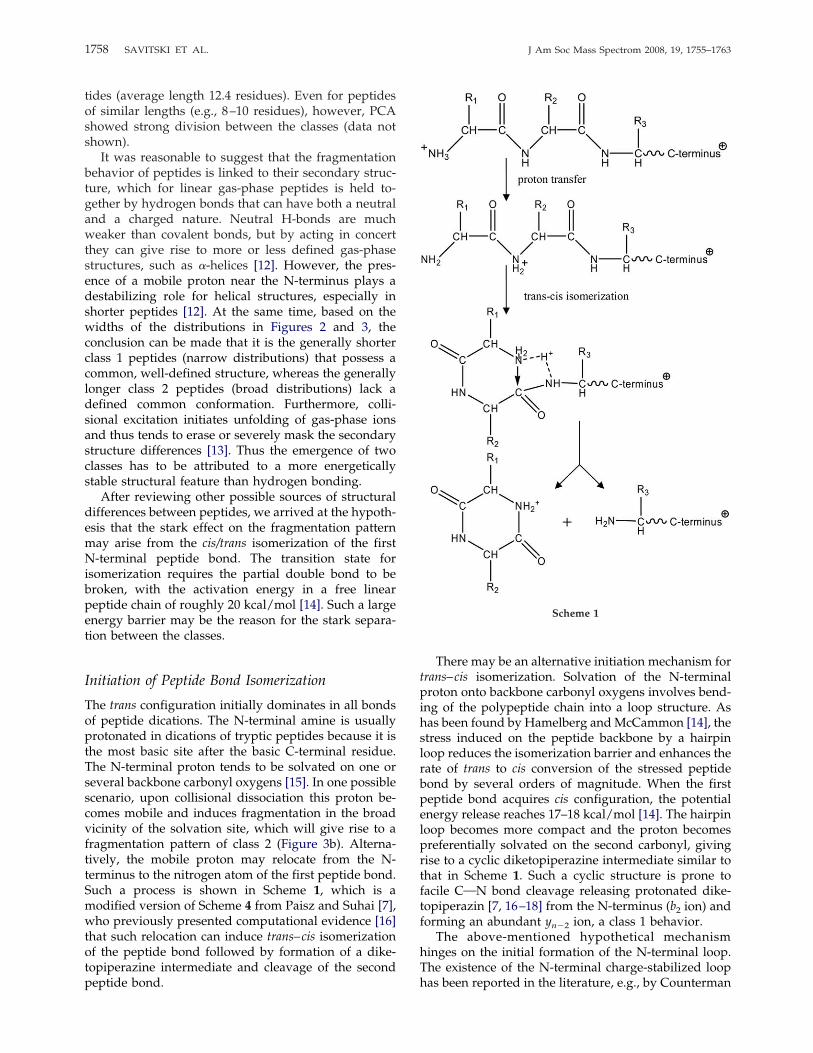
The trans configuration initially dominates in all bondsof peptide dications. The N-terminal amine is usuallyprotonated in dications of tryptic peptides because it isthe most basic site after the basic C-terminal residue.The N-terminal proton tends to be solvated on one orseveral backbone carbonyl oxygens [15]. In one possiblescenario, upon collisional dissociation this proton be-comes mobile and induces fragmentation in the broadvicinity of the solvation site, which will give rise to afragmentation pattern of class 2 (Figure 3b). Alterna-tively, the mobile proton may relocate from the N-terminus to the nitrogen atom of the first peptide bond.Such a process is shown in Scheme 1, which is amodified version of Scheme 4 from Paisz and Suhai [7],who previously presented computational evidence [16]that such relocation can induce trans–cis isomerizationof the peptide bond followed by formation of a dike-topiperazine intermediate and cleavage of the secondpeptide bond.
There may be an alternative initiation mechanism fortrans–cis isomerization. Solvation of the N-terminalproton onto backbone carbonyl oxygens involves bend-ing of the polypeptide chain into a loop structure. Ashas been found by Hamelberg andMcCammon [14], thestress induced on the peptide backbone by a hairpinloop reduces the isomerization barrier and enhances therate of trans to cis conversion of the stressed peptidebond by several orders of magnitude. When the firstpeptide bond acquires cis configuration, the potentialenergy release reaches 17–18 kcal/mol [14]. The hairpinloop becomes more compact and the proton becomespreferentially solvated on the second carbonyl, givingrise to a cyclic diketopiperazine intermediate similar tothat in Scheme 1. Such a cyclic structure is prone tofacile CON bond cleavage releasing protonated dike-topiperazin [7, 16–18] from the N-terminus (b2 ion) andforming an abundant yn�2 ion, a class 1 behavior.The above-mentioned hypothetical mechanism
hinges on the initial formation of the N-terminal loop.The existence of the N-terminal charge-stabilized loophas been reported in the literature, e.g., by Counterman
Scheme 1
1758 SAVITSKI ET AL. J Am Soc Mass Spectrom 2008, 19, 1755–1763

and Clemmer [19], who simulated a singly protonatedGPHH peptide in both cis and trans isoforms. In thelowest-energy trans-Pro isomer, the loop was largebecause the proton was shared between the N-terminusand the third and fourth carbonyls. The cis-conformerwas lower in energy and the loop in it was morecompact. The N-terminal proton in that structure wasshared with the second carbonyl, whereas the third andfourth carbonyls were involved in charge solvation to asignificantly lesser degree than in the trans isoform.With ion mobility spectroscopy, Counterman andClemmens observed both cis and trans isomers forproline-containing peptides and only one isomer forpeptides without a proline. Their measurements couldnot distinguish which (cis or trans) configuration theunique isomer has acquired. All 645 peptides theyinvestigated were short (�10 residues long) and themajority were singly charged. Thus it is still an openquestion whether proline-free tryptic peptide dicationscan produce a mixture of gas-phase cis- and trans-isomers in comparable proportions.
Proline Effect in b2 Ion Formation
Research linking b2/yn�2 fragment formation with dike-topiperazine formation in proline-containing peptideshas been done by several authors, e.g., Smith et al. [17].Proline is in general known to induce very strongcleavage at its N-terminal site [20], whereas the cleav-age at the C-terminal site is suppressed [21]. Smith et al.[17] found that in the cationic peptide VPDPR cleavageC-terminal to the proline in the second position wasunusually abundant and attributed this fact to cycliza-tion of the first two residues into a diketopiperazinestructure. It has been pointed out by Paizs and Suhaithat the necessary condition for cyclization is the cisisomerization of the first peptide bond [16].The enhanced cleavage after Pro-2 turned out to be
a general phenomenon. We calculated the average
cleavage preferences C-terminal to proline in differ-ent positions n from our extensive dataset. As ex-pected, the C-terminal cleavage to proline was veryscarce for n � 2, whereas for n � 2 the cleavage wasvery prominent—in fact, more prominent than theC-terminal cleavage to glycine and serine in the sameposition (data not shown). Thus for the Xaa-ProN-terminal sequences, abundant b2/yn�2 cleavage is aclear signature of cis/trans isomerization of the firstpeptide bond. Our data, however, support the hy-pothesis [18, 22] that such an isomerization fre-quently occurs for residues other than proline.
Proton Retention
Paisz and Suhai developed their model of trans/cis bondisomerization for neutral loss of the diketopiperazinefragment [7, 16], whereas our data point toward facileformation of ionic b2. For the proton to be retained by b2fragment, the diketopiperazine derivative should bemore basic than the ym�2
� ion. We found a strongcorrelation between the basicity of the two N-terminalamino acids and ym�2
� formation frequencies (Figure 4,left). For reliable statistics, amino acids exhibitingstrong ammonia and water losses (E, D, N, Q, S, and T)were omitted, as were amino acids rarely appearing ininternal or N-terminal regions of tryptic peptides (P, C,W, M, R, and K). The dependence on the amino acidbasicity is much stronger for n � 2 than that for n � 2,for which case backbone carbonyl basicities are of primeimportance [6].The loss of a neutral diketopiperazine could be
relatively more important for nonbasic N-termini.However, of the 78 spectra found for the extreme G1G2case (glycine is the least basic amino acid), ym�2
� ionswere present in 14 cases and ym�2
2� ions were found inonly 8 cases. Thus even for least basic N-terminalsequences, formation of protonated diketopiperazine isrelatively more important than neutral diketopipera-
Figure 4. Left panel: dependence of the appearance frequency of dominant ym�2� ions as a function
of the proton affinity of the first two N-terminal amino acids, aa1 and aa2. Right panel: appearancefrequency of dominant ym�2
� ions multiplied by the number of peptide bonds (m � 1).
1759J Am Soc Mass Spectrom 2008, 19, 1755–1763 TWO CLASSES OF CAD SPECTRA
zine loss. This result raises the issue of the reason for thesurprisingly high basicity of the N-terminal leavinggroup. One reason for that effect could be the coulombicrepulsion between the two ionizing protons. In agree-ment with the preference of shorter peptides for class 1behavior, the dominance of ym�2
� ions was founddiminished for peptides with m � 10 (right panel inFigure 4). The potential coulombic energy in a fullystretched 10-residue peptide (an average size for atryptic peptide) protonated at both termini is approxi-mately 9 kcal/mol, a significant value according to theleft panel in Figure 4. The fall-off in the right panel ofFigure 4 resembles a parabola, exactly as expected forCoulomb’s law.
Distinguishing Between the Cyclic and Oxazolonb2
� Structures
One way the cyclic and oxazolon b2� structures are
expected to differ from each other is in the effect of thechemical nature of the first and second amino acids onthe b2
� formation probability. Oxazolone is formed viaan asymmetric intermediate, which should reflect invastly different, and indeed opposite, reactivities ofthese two residues during the bond cleavage, whereasin a cyclic diketopiperazine intermediate the differencebetween the first and second residues should be small.Influence of an amino acid in a specific position onbond cleavage can be evaluated through variance anal-ysis [6]. The most important residue determining thefrequency of a CON bond cleavage is the one on itsN-terminal side, with the same variance v � 0.030 forboth aa2 in ym�2
� and aa3 in ym�3�. For n � 2, the effect
of the next preceding amino acid is an order of magni-tude smaller (v � 0.003 for aa2 in ym�3
�), whereas forn � 2 it is comparable (v � 0.014 for aa1 in ym�2
�). Thecomparable variances mean that both N-terminal resi-dues are involved in the (b2
�, ym�2�) ion pair formation
to a similar extent, as would be expected for dike-topiperazine, but not for oxazolone (otherwise the vari-ance for aa2 in ym�3
� would also be large).Even more telling than the variance analysis is the
direct assessment of the effect of the amino acid nature
on the ym�2� formation probability. As would be ex-
pected for a symmetric b2�, the N-terminal sequences
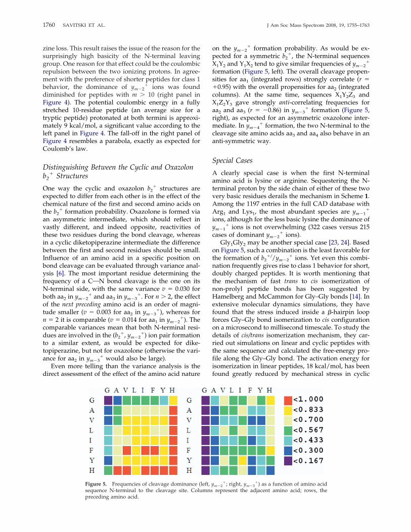
X1Y2 and Y1X2 tend to give similar frequencies of ym�2�
formation (Figure 5, left). The overall cleavage propen-sities for aa1 (integrated rows) strongly correlate (r ��0.95) with the overall propensities for aa2 (integratedcolumns). At the same time, sequences X1Y2Z3 andX1Z2Y3 gave strongly anti-correlating frequencies foraa2 and aa3 (r � �0.86) in ym�3
� formation (Figure 5,right), as expected for an asymmetric oxazolone inter-mediate. In ym�4
� formation, the two N-terminal to thecleavage site amino acids aa3 and aa4 also behave in ananti-symmetric way.
Special Cases
A clearly special case is when the first N-terminalamino acid is lysine or arginine. Sequestering the N-terminal proton by the side chain of either of these twovery basic residues derails the mechanism in Scheme 1.Among the 1197 entries in the full CAD database withArg1 and Lys1, the most abundant species are ym�1
�
ions, although for the less basic lysine the dominance ofym�1
� ions is not overwhelming (322 cases versus 215cases of dominant ym�2
� ions).Gly1Gly2 may be another special case [23, 24]. Based
on Figure 5, such a combination is the least favorable forthe formation of b2
�//ym�2� ions. Yet even this combi-
nation frequently gives rise to class 1 behavior for short,doubly charged peptides. It is worth mentioning thatthe mechanism of fast trans to cis isomerization ofnon-prolyl peptide bonds has been suggested byHamelberg and McCammon for Gly–Gly bonds [14]. Inextensive molecular dynamics simulations, they havefound that the stress induced inside a �-hairpin loopforces Gly–Gly bond isomerization to cis configurationon a microsecond to millisecond timescale. To study thedetails of cis/trans isomerization mechanism, they car-ried out simulations on linear and cyclic peptides withthe same sequence and calculated the free-energy pro-file along the Gly–Gly bond. The activation energy forisomerization in linear peptides, 18 kcal/mol, has beenfound greatly reduced by mechanical stress in cyclic
Figure 5. Frequencies of cleavage dominance (left, ym�2�; right, ym�3
�) as a function of amino acidsequence N-terminal to the cleavage site. Columns represent the adjacent amino acid; rows, thepreceding amino acid.
1760 SAVITSKI ET AL. J Am Soc Mass Spectrom 2008, 19, 1755–1763
peptides. As a result, the calculated rate of isomeriza-tion in cyclic peptides has been found to increase by fiveorders of magnitude compared to linear peptides. Thuswe cannot exclude that even Gly1Gly2 combinationleads in some cases to protonated diketopiperazineformation.
a3 Anomaly
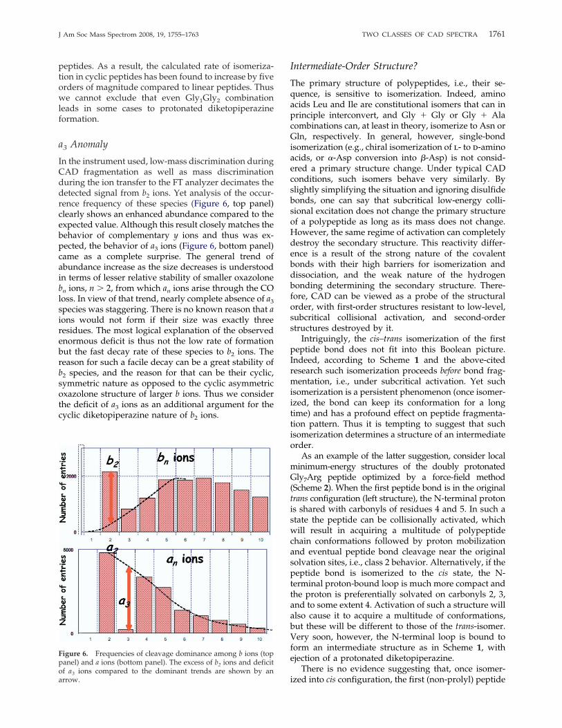
In the instrument used, low-mass discrimination duringCAD fragmentation as well as mass discriminationduring the ion transfer to the FT analyzer decimates thedetected signal from b2 ions. Yet analysis of the occur-rence frequency of these species (Figure 6, top panel)clearly shows an enhanced abundance compared to theexpected value. Although this result closely matches thebehavior of complementary y ions and thus was ex-pected, the behavior of a3 ions (Figure 6, bottom panel)came as a complete surprise. The general trend ofabundance increase as the size decreases is understoodin terms of lesser relative stability of smaller oxazolonebn ions, n � 2, from which an ions arise through the COloss. In view of that trend, nearly complete absence of a3species was staggering. There is no known reason that aions would not form if their size was exactly threeresidues. The most logical explanation of the observedenormous deficit is thus not the low rate of formationbut the fast decay rate of these species to b2 ions. Thereason for such a facile decay can be a great stability ofb2 species, and the reason for that can be their cyclic,symmetric nature as opposed to the cyclic asymmetricoxazolone structure of larger b ions. Thus we considerthe deficit of a3 ions as an additional argument for thecyclic diketopiperazine nature of b2 ions.
Intermediate-Order Structure?
The primary structure of polypeptides, i.e., their se-quence, is sensitive to isomerization. Indeed, aminoacids Leu and Ile are constitutional isomers that can inprinciple interconvert, and Gly � Gly or Gly � Alacombinations can, at least in theory, isomerize to Asn orGln, respectively. In general, however, single-bondisomerization (e.g., chiral isomerization of l- to d-aminoacids, or �-Asp conversion into �-Asp) is not consid-ered a primary structure change. Under typical CADconditions, such isomers behave very similarly. Byslightly simplifying the situation and ignoring disulfidebonds, one can say that subcritical low-energy colli-sional excitation does not change the primary structureof a polypeptide as long as its mass does not change.However, the same regime of activation can completelydestroy the secondary structure. This reactivity differ-ence is a result of the strong nature of the covalentbonds with their high barriers for isomerization anddissociation, and the weak nature of the hydrogenbonding determining the secondary structure. There-fore, CAD can be viewed as a probe of the structuralorder, with first-order structures resistant to low-level,subcritical collisional activation, and second-orderstructures destroyed by it.Intriguingly, the cis–trans isomerization of the first
peptide bond does not fit into this Boolean picture.Indeed, according to Scheme 1 and the above-citedresearch such isomerization proceeds before bond frag-mentation, i.e., under subcritical activation. Yet suchisomerization is a persistent phenomenon (once isomer-ized, the bond can keep its conformation for a longtime) and has a profound effect on peptide fragmenta-tion pattern. Thus it is tempting to suggest that suchisomerization determines a structure of an intermediateorder.As an example of the latter suggestion, consider local
minimum-energy structures of the doubly protonatedGly7Arg peptide optimized by a force-field method(Scheme 2). When the first peptide bond is in the originaltrans configuration (left structure), the N-terminal protonis shared with carbonyls of residues 4 and 5. In such astate the peptide can be collisionally activated, whichwill result in acquiring a multitude of polypeptidechain conformations followed by proton mobilizationand eventual peptide bond cleavage near the originalsolvation sites, i.e., class 2 behavior. Alternatively, if thepeptide bond is isomerized to the cis state, the N-terminal proton-bound loop is much more compact andthe proton is preferentially solvated on carbonyls 2, 3,and to some extent 4. Activation of such a structure willalso cause it to acquire a multitude of conformations,but these will be different to those of the trans-isomer.Very soon, however, the N-terminal loop is bound toform an intermediate structure as in Scheme 1, withejection of a protonated diketopiperazine.There is no evidence suggesting that, once isomer-
ized into cis configuration, the first (non-prolyl) peptide
Figure 6. Frequencies of cleavage dominance among b ions (toppanel) and a ions (bottom panel). The excess of b2 ions and deficitof a3 ions compared to the dominant trends are shown by anarrow.
1761J Am Soc Mass Spectrom 2008, 19, 1755–1763 TWO CLASSES OF CAD SPECTRA

bond would easily return back to the trans form. If suchreversed isomerization were a fast process, two ionpopulations would mix and no stark division betweenthe two statistical groups would exist. Facile formationof b2 ions is the “escape route” for cis forms; reverseisomerization must be entropically disfavored. Thepresence of separate, largely isolated ion populations isthe argument for considering cis and trans isomers asdifferent structures of intermediate order.
Conclusions
We have presented evidence that CAD mass spectra oftryptic peptide dications show bifurcating behavior.Class 1 spectra are dominated by b2/yn�2 cleavage,whereas class 2 spectra show a broad distribution ofcleavages preferentially peaking at residue 4 or 5 fromthe N-terminus. Such a bifurcating behavior is hypoth-esized to be linked to the trans-to-cis izomerization ofthe first peptide bond and subsequent formation of b2
ions in the form of cyclic, symmetrical diketopipera-zines. Our data clearly show preference of shorterpeptides for the class 1 behavior. Several effects contrib-ute to this preference. In shorter dications, coulombicrepulsion between the N- and C-terminal charges re-duces the size of the loop formed by the protonatedN-terminal amine solvated onto backbone carbonyls. Ina smaller loop, an enhanced stress in the peptide bondsreduces the barrier for cis–trans isomerization that thusoccurs faster. Additionally, the shorter distance be-tween the charges increases the basicity of the N-terminus and the balance between the loss of a neutraland protonated diketopiperazine shifts toward chargeseparation.The balance between isomerization before fragmen-
tation (class 1) and direct fragmentation (class 2) mustalso depend on the ionic temperature after excitation,i.e., the type of the tandem mass spectrometer used. Inlinear ion traps, one third of all MS/MS spectra of
tryptic peptide dications are found to belong to class 1;in other instruments this fraction may be smaller orlarger. In this work we have argued for the plausibilityof the izomerization hypothesis and provided support-ing arguments. However, we recognize that these argu-ments may not be enough to resolve the issue once andfor all. At any rate, realization of the bifurcating frag-mentation behavior of peptides must have an impact onthe efforts to create a quantitative model for CADfragmentation.
AcknowledgmentsThis work was supported by the Knut and Alice WallenbergFoundation, European Union (grant to consortium EPITOPE) aswell as the Swedish research council (Grants 621-2004-4897 and621-2003-4877 to RZ).
References1. Perkins, D. N.; Pappin, D. J. C.; Creasy, D. M.; Cottrell, J. S. Probability-based Protein Identification by Searching Sequence Databases UsingMass Spectrometry Data. Electrophoresis 1999, 20, 3551–3567.
2. Eng, J. K.; McCormack, A. L.; Yates, J. R. An Approach to CorrelateTandemMass-Spectral Data of Peptides with Amino-Acid-Sequences ina Protein Database. J. Am. Soc. Mass Spectrom. 1994, 5, 976–989.
3. Lam, H.; Deutsch, E. W.; Eddes, J. S.; Eng, J. K.; King, N.; Stein, S. E.;Aebersold, R. Development and Validation of a Spectral Library Search-ing Method for Peptide Identification from MS/MS. Proteomics 2007, 7,655–667.
4. Zhang, Z. Q. Prediction of Low-energy Collision-induced DissociationSpectra of Peptides. Anal. Chem. 2004, 76, 3908–3922.
5. Zhang, Z. Q. Prediction of Low-energy Collision-induced DissociationSpectra of Peptides with Three or More Charges. Anal. Chem. 2005, 77,6364–6373.
6. Savitski, M. M.; Kjeldsen, F.; Nielsen, M. L.; Garbuzynskiy, S. O.;Galzitskaya, O. V.; Surin, A. K.; Zubarev, R. A. Backbone CarbonylGroup Basicities Are Related to Gas-phase Fragmentation of Peptidesand Protein Folding. Angew. Chem. Int. Ed. Engl. 2007, 46, 1481–1484.
7. Paizs, B.; Suhai, S. Fragmentation Pathways of Protonated Peptides.Mass Spectrom. Rev. 2005, 24, 508–548.
8. Wysocki, V. H.; Tsaprailis, G.; Smith, L. L.; Breci, L. A. Special Feature:Commentary—Mobile and Localized Protons: A Framework for Under-standing Peptide Dissociation. J. Mass Spectrom. 2000, 35, 1399–1406.
9. Nielsen, M. L.; Savitski, M. M.; Zubarev, R. A. Improving ProteinIdentification Using Complementary Fragmentation Techniques in Fou-rier Transform Mass Spectrometry. Mol. Cell. Proteomics 2005, 4, 835–845.
10. Savitski, M. M.; Nielsen, M. L.; Zubarev, R. A. New Data Base-independent, Sequence Tag-based Scoring of Peptide MS/MS DataValidates Mowse Scores, Recovers below Threshold Data, Singles OutModified Peptides, and Assesses the Quality of MS/MS Techniques.Mol. Cell. Proteomics 2005, 4, 1180–1188.
11. Savitski, M. M.; Kjeldsen, F.; Nielsen, M. L.; Zubarev, R. A. Comple-mentary Sequence Preferences of Electron-Capture Dissociation andVibrational Excitation in Fragmentation of Polypeptide Polycations.Angew. Chem. Int. Ed. Engl. 2006, 45, 5301–5303.
12. Shelimov, K. B.; Jarrold, M. F. Conformations, Unfolding, and Refoldingof Apomyoglobin in Vacuum: An Activation Barrier for Gas-PhaseProtein Folding. J. Am. Chem. Soc. 1997, 119, 2987–2994.
13. Burlet, O.; Yang, C. Y.; Gaskell, S. J. Influence of Cysteine to CysteicAcid Oxidation on the Collision-Activated Decomposition of Proton-ated Peptides - Evidence for Intraionic Interactions. J. Am. Soc. MassSpectrom. 1992, 3, 337–344.
14. Hamelberg, D.; McCammon, J. A. Fast Peptidyl cis-trans Isomerizationwithin the Flexible Gly-rich Flaps of HIV-1 Protease. J. Am. Chem. Soc.2005, 127, 13778–13779.
15. Samuelson, S.; Martyna, G. J. Computer Simulation Studies of FiniteTemperature Conformational Equilibrium in Alanine-based Peptides. J.Phys. Chem. B 1999, 103, 1752–1766.
16. Paizs, B.; Suhai, S. Combined Quantum Chemical and RRKMModelingof the Main Fragmentation Pathways of Protonated GGG. I. Cis-transIsomerization Around Protonated Amide Bonds. Rapid Commun. MassSpectrom. 2001, 15, 2307–2323.
17. Smith, L. L.; Herrmann, K. A.; Wysocki, V. H. Investigation of GasPhase ion Structure for Proline-Containing b(2) Ion. J. Am. Soc. MassSpectrom. 2006, 17, 20–28.
Scheme 2
1762 SAVITSKI ET AL. J Am Soc Mass Spectrom 2008, 19, 1755–1763

18. Farrugia, J. M.; O’Hair, R. A. J.; Reid, G. E. Do All b(2) Ions HaveOxazolone Structures? Multistage Mass Spectrometry and Ab InitioStudies on Protonated N-Acyl Amino Acid Methyl Ester Model Sys-tems. Int. J. Mass Spectrom. 2001, 210, 71–87.
19. Counterman, A. E.; Clemmer, D. E. Anhydrous Polyproline Helices andGlobules. J. Phys. Chem. B 2004, 108, 4885–4898.
20. Hunt, D. F.; Yates, Y. R.; Shabanowitz, J.; Winston, S.; Hauer, C. R.Protein Sequencing by TandemMass Spectrometry. Proc. Natl. Acad. Sci.U. S. A. 1986, 83, 6233–6237.
21. Huang, Y.; Triscari, J. M.; Wysocki, V. H.; Pasa-Tolic, L.; Anderson,G. A.; Lipton, M. S.; Smith, R. D. Dissociation Behaviors of Doubly-
Charged Tryptic Peptides: Correlation of Gas-Phase Cleavage Abun-dance with Ramachandran Plots. J. Am. Chem. Soc. 2004, 126, 3034–3035.
22. Cordero, M. M.; Houser, J. J.; Wesdemiotis, C. The Neutral ProductsFormed during Backbone Fragmentations of Protonated Peptides inTandem Mass Spectrometry. Anal. Chem. 1993, 65, 1594–1601.
23. Paizs, B.; Suhai, S. Combined Quantum Chemical and RRKMModeling ofthe Main Fragmentation Pathways of Protonated GGG. II. Formation ofb(2), y(1), and y(2) Ions. Rapid Commun. Mass Spectrom. 2002, 16, 375–389.
24. Paizs, B.; Suhai, S. Towards Understanding the TandemMass Spectra ofProtonated Oligopeptides. 1: Mechanism of Amide Bond Cleavage.J. Am. Soc. Mass Spectrom. 2004, 15, 103–113.
1763J Am Soc Mass Spectrom 2008, 19, 1755–1763 TWO CLASSES OF CAD SPECTRA
Copyright © 2022 FDOKUMEN