Bias Associated with Peripheral Non-Invasive Compared to ...
13
Citation: Pratt, S.; Barnes, T.S.; Cowling, N.; de Klerk, K.; Rainger, J.; Sole-Guitart, A.; Woldeyohannes, S.; Goodwin, W. Bias Associated with Peripheral Non-Invasive Compared to Invasive Arterial Blood Pressure Monitoring in Healthy Anaesthetised and Standing Horses Using the Bionet BM7Vet. Vet. Sci. 2022, 9, 52. https://doi.org/10.3390/vetsci9020052 Academic Editor: Patrick Butaye Received: 16 December 2021 Accepted: 26 January 2022 Published: 28 January 2022 Publisher’s Note: MDPI stays neutral with regard to jurisdictional claims in published maps and institutional affil- iations. Copyright: © 2022 by the authors. Licensee MDPI, Basel, Switzerland. This article is an open access article distributed under the terms and conditions of the Creative Commons Attribution (CC BY) license (https:// creativecommons.org/licenses/by/ 4.0/). veterinary sciences Article Bias Associated with Peripheral Non-Invasive Compared to Invasive Arterial Blood Pressure Monitoring in Healthy Anaesthetised and Standing Horses Using the Bionet BM7Vet Shaun Pratt 1, * , Tamsin S. Barnes 1,2 , Nicholas Cowling 1 , Karla de Klerk 1 , Joanne Rainger 1 , Albert Sole-Guitart 1 , Solomon Woldeyohannes 1 and Wendy Goodwin 1 1 School of Veterinary Science, Gatton Campus, The University of Queensland, Gatton 4343, Australia; [email protected] (T.S.B.); [email protected] (N.C.); [email protected] (K.d.K.); [email protected] (J.R.); [email protected] (A.S.-G.); [email protected] (S.W.); [email protected] (W.G.) 2 Queensland Alliance for Agriculture and Food Innovation, The University of Queensland, Gatton 4343, Australia * Correspondence: [email protected] Abstract: To compare arterial blood pressure (ABP) measured invasively (IBP) to ABP measured non-invasively (NIBP) via oscillometry in healthy anaesthetised and standing horses using the Bionet BM7Vet. Fourteen horses were anaesthetised for elective procedures (anaesthetised group) and 10 horses were enrolled for standing blood pressure manipulation (standing group). In both groups, IBP and NIBP-corrected to heart level were measured every 3 min using the Bionet BM7Vet. The overall mean difference (bias), standard deviation and limits of agreement (LOA) were calculated for paired IBP and NIBP systolic (SAP), mean (MAP) and diastolic (DAP) blood pressure measurements. In anaesthetised horses, the NIBP cuff was placed at either the proximal tail base or the metacarpus. Invasive MAP was used to retrospectively characterise measurements into hypotensive (≤70 mm Hg), normotensive (71–110 mm Hg) or hypertensive (≥111 mm Hg) subgroups. In standing horses, the NIBP cuff was placed at the tail base only and invasive MAP was manipulated to achieve hypertension (≥126 mm Hg) and hypotension (≤90 mm Hg) using phenylephrine and acepromazine, respectively. When measuring NIBP at the tail in anaesthetised horses, the Bionet BM7Vet failed on 8/185 occasions and overestimated SAP, MAP and DAP during hypotension and normotension. The biases (lower, upper LOA) for MAP were -11.4 (-33.3, 10.5) and -6.0 (-25.8, 13.8) mm Hg, respectively. Hypertension could not be evaluated. When measuring NIBP at the metacarpus in anaesthetised horses, the Bionet BM7Vet failed on 24/65 occasions and underestimated SAP, MAP and DAP when all ABP subgroups were combined. The bias (lower, upper LOA) for pooled MAP was 3.6 (-44.3, 51.6) mm Hg. When measuring NIBP at the tail in standing horses, the Bionet BM7Vet failed on 64/268 occasions and underestimated SAP, MAP and DAP during hypotension, normotension and hypertension. The biases (lower, upper LOA) for MAP were 16.3 (-10.5, 43.1), 16.6 (-19.5, 52.7) and 30.0 (-8.1, 68.0) mm Hg, respectively. Monitoring NIBP on the Bionet BM7Vet in anaesthetised horses overestimated ABP at the tail and underestimated ABP at the metacarpus. The device inaccurately detected hypotension and should be used cautiously. In standing horses, the Bionet BM7Vet underestimated ABP at the tail, especially during pharmacologically induced hypertension. Keywords: ABP; NIBP; IBP; horse; anaesthesia; Bionet BM7Vet 1. Introduction Arterial blood pressure (ABP) measurements provide important cardiovascular in- formation and are especially useful in hemodynamically unstable and anaesthetised pa- tients [1–4]. In horses, ABP measured invasively (IBP) is considered the gold standard ABP measurement technique. An arterial catheter is connected via non-distensible, heparinised Vet. Sci. 2022, 9, 52. https://doi.org/10.3390/vetsci9020052 https://www.mdpi.com/journal/vetsci
-
Upload
khangminh22 -
Category
Documents
-
view
4 -
download
0
Transcript of Bias Associated with Peripheral Non-Invasive Compared to ...

�����������������
Citation: Pratt, S.; Barnes, T.S.;
Cowling, N.; de Klerk, K.; Rainger, J.;
Sole-Guitart, A.; Woldeyohannes, S.;
Goodwin, W. Bias Associated with
Peripheral Non-Invasive Compared
to Invasive Arterial Blood Pressure
Monitoring in Healthy Anaesthetised
and Standing Horses Using the
Bionet BM7Vet. Vet. Sci. 2022, 9, 52.
https://doi.org/10.3390/vetsci9020052
Academic Editor: Patrick Butaye
Received: 16 December 2021
Accepted: 26 January 2022
Published: 28 January 2022
Publisher’s Note: MDPI stays neutral
with regard to jurisdictional claims in
published maps and institutional affil-
iations.
Copyright: © 2022 by the authors.
Licensee MDPI, Basel, Switzerland.
This article is an open access article
distributed under the terms and
conditions of the Creative Commons
Attribution (CC BY) license (https://
creativecommons.org/licenses/by/
4.0/).
veterinarysciences
Article
Bias Associated with Peripheral Non-Invasive Compared toInvasive Arterial Blood Pressure Monitoring in HealthyAnaesthetised and Standing Horses Using the Bionet BM7VetShaun Pratt 1,* , Tamsin S. Barnes 1,2, Nicholas Cowling 1 , Karla de Klerk 1, Joanne Rainger 1,Albert Sole-Guitart 1 , Solomon Woldeyohannes 1 and Wendy Goodwin 1
1 School of Veterinary Science, Gatton Campus, The University of Queensland, Gatton 4343, Australia;[email protected] (T.S.B.); [email protected] (N.C.); [email protected] (K.d.K.);[email protected] (J.R.); [email protected] (A.S.-G.); [email protected] (S.W.);[email protected] (W.G.)
2 Queensland Alliance for Agriculture and Food Innovation, The University of Queensland,Gatton 4343, Australia
* Correspondence: [email protected]
Abstract: To compare arterial blood pressure (ABP) measured invasively (IBP) to ABP measurednon-invasively (NIBP) via oscillometry in healthy anaesthetised and standing horses using the BionetBM7Vet. Fourteen horses were anaesthetised for elective procedures (anaesthetised group) and10 horses were enrolled for standing blood pressure manipulation (standing group). In both groups,IBP and NIBP-corrected to heart level were measured every 3 min using the Bionet BM7Vet. Theoverall mean difference (bias), standard deviation and limits of agreement (LOA) were calculated forpaired IBP and NIBP systolic (SAP), mean (MAP) and diastolic (DAP) blood pressure measurements.In anaesthetised horses, the NIBP cuff was placed at either the proximal tail base or the metacarpus.Invasive MAP was used to retrospectively characterise measurements into hypotensive (≤70 mm Hg),normotensive (71–110 mm Hg) or hypertensive (≥111 mm Hg) subgroups. In standing horses,the NIBP cuff was placed at the tail base only and invasive MAP was manipulated to achievehypertension (≥126 mm Hg) and hypotension (≤90 mm Hg) using phenylephrine and acepromazine,respectively. When measuring NIBP at the tail in anaesthetised horses, the Bionet BM7Vet failedon 8/185 occasions and overestimated SAP, MAP and DAP during hypotension and normotension.The biases (lower, upper LOA) for MAP were −11.4 (−33.3, 10.5) and −6.0 (−25.8, 13.8) mm Hg,respectively. Hypertension could not be evaluated. When measuring NIBP at the metacarpus inanaesthetised horses, the Bionet BM7Vet failed on 24/65 occasions and underestimated SAP, MAP andDAP when all ABP subgroups were combined. The bias (lower, upper LOA) for pooled MAP was 3.6(−44.3, 51.6) mm Hg. When measuring NIBP at the tail in standing horses, the Bionet BM7Vet failedon 64/268 occasions and underestimated SAP, MAP and DAP during hypotension, normotensionand hypertension. The biases (lower, upper LOA) for MAP were 16.3 (−10.5, 43.1), 16.6 (−19.5, 52.7)and 30.0 (−8.1, 68.0) mm Hg, respectively. Monitoring NIBP on the Bionet BM7Vet in anaesthetisedhorses overestimated ABP at the tail and underestimated ABP at the metacarpus. The deviceinaccurately detected hypotension and should be used cautiously. In standing horses, the BionetBM7Vet underestimated ABP at the tail, especially during pharmacologically induced hypertension.
Keywords: ABP; NIBP; IBP; horse; anaesthesia; Bionet BM7Vet
1. Introduction
Arterial blood pressure (ABP) measurements provide important cardiovascular in-formation and are especially useful in hemodynamically unstable and anaesthetised pa-tients [1–4]. In horses, ABP measured invasively (IBP) is considered the gold standard ABPmeasurement technique. An arterial catheter is connected via non-distensible, heparinised
Vet. Sci. 2022, 9, 52. https://doi.org/10.3390/vetsci9020052 https://www.mdpi.com/journal/vetsci
Vet. Sci. 2022, 9, 52 2 of 13
tubing to a transducer placed at the phlebostatic axis. Pressure wave changes within thetransducer transmit electrical signals for interpretation and display on the patient sidemonitor. Despite arterial catheters being relatively simple to place in anaesthetised horses inhospital, for the monitoring of standing sedated, critically ill and anaesthetised horses in thefield, the risk for contamination, arteritis and equipment failure is high [5]. Consequently,devices measuring ABP non-invasively might be beneficial in horses.
Devices which measure ABP non-invasively (NIBP) are routinely used in humanand small domestic animal patients, however, inaccuracy and poor reliability comparedwith IBP mean they remain underused in horses. Oscillometric NIBP devices utilise aperipherally placed arterial bladder cuff to record pressure wave oscillations induced byblood flow turbulence during vascular release. The monitor measures MAP at maximaloscillation amplitude, then uses proprietary algorithms to calculate SAP and DAP. Vet-erinary specific oscillometric devices have been studied in conscious and anaesthetisedadult horses [6–12] and foals [13,14] across variably defined hypotensive and hypertensiveABP states. Estimates for accuracy and reliability vary considerably in these studies, withthe underestimation and overestimation of ABP by the non-invasive method reported.Additionally, as mathematical algorithms used for oscillometric calculation are monitorspecific, inter-monitor extrapolation is inappropriate.
The accuracy and precision of oscillometric NIBP monitoring using the Bionet BM7Vethas not been evaluated in horses. The purpose of this study was to compare ABP measuredinvasively and non-invasively in anaesthetised and standing horses to provide recommen-dations on the clinical use of the Bionet BM7Vet. It was hypothesised that the greatestvariability in accuracy and precision would occur during hypotension and hypertension.
2. Materials and Methods
All procedures were performed with approval of The University of Queensland’sProduction and Companion Animal Ethics Committee (SVS/449/17). The study enrolledtwo populations of horses; client-owned horses anaesthetised for elective surgery (observa-tional study) and an experimental group of university-owned horses for standing bloodpressure manipulation.
2.1. Anaesthetised Group2.1.1. Animals
Fourteen client-owned horses presenting for surgery at The University of Queensland’sEquine Specialist Hospital were enrolled into the observational study arm. Demographicdata included 9 Thoroughbreds, 2 Quarter Horses, 1 Arabian, 1 Australian Stock Horse and1 Irish Sport Horse (10 females, 4 males). Horses were aged (mean ± SD) 22 ± 20 monthsand weighed 370 ± 107 kg at the time of surgery. Procedures included arthroscopicsurgery for osteochondritis dissecans (tarsus n = 6, stifle n = 3, fetlock n = 1), woundexploration (n = 1) and transphyseal bridging of the distal radius (n = 3). All horseswere an American Society of Anaesthesiologists (ASA) classification I or II based on theresults of a physical examination and pre-anaesthetic biochemical and haematologicalanalysis. Animals were housed in individual stalls for varying durations dependant ontheir medical care requirements. Water was available ad libitum and grassy lucerne wasprovided morning and night.
2.1.2. Anaesthesia
The skin overlying the left jugular vein was aseptically prepared and following thesubcutaneous deposition of 20 mg lidocaine 2% (Lignocaine 20; Troy Laboratories, NSW,Australia), a 14 G, 3-inch catheter (BD Angiocath; Becton Dickinson Rowa Australia,QLD, Australia) was placed for the delivery of all intravenous (IV) agents. Anaesthetictechnique was at the discretion of the primary anaesthetist. Thirteen horses were premed-icated with medetomidine (Medetomidine; Troy Laboratories, NSW, Australia) 5 µg/kgIV, methadone (Methone; Ceva Animal Health, NSW, Australia) 0.1 mg/kg IV and ace-
Vet. Sci. 2022, 9, 52 3 of 13
promazine (Acemav-10; Mavlab, QLD, Australia) 0.02 mg/kg IV. General anaesthesia wasinduced 5 to 10 min later with ketamine (Ketamil; Troy Laboratories, NSW, Australia)2.2 mg/kg IV and midazolam (Hypnovel; Roche Products, NSW, Australia) 0.06 mg/kg IV.One horse was premedicated with butorphanol (Butogesic; Troy Laboratories, NSW, Aus-tralia) 0.04 mg/kg IV, xylazine (Xylazil-100; Troy Laboratories, NSW, Australia) 0.5 mg/kgIV and medetomidine 1 µg/kg IV and was induced with propofol (Fresofol; Fresenius Kabi,NSW, Australia) 2 mg/kg IV.
Once recumbency was achieved and orotracheal intubation performed, all horses werehoisted onto a padded equine surgical table and positioned either in dorsal (n = 12) orlateral (n = 2) recumbency in the surgical suite. Partial IV anaesthesia was maintained witha medetomidine (2–3.5 µg/kg/hr IV) infusion (CRI) and either isoflurane (Isofol; ZoetisAustralia, NSW, Australia) (n = 10) or sevoflurane (Sevorane; AbbVie, NSW, Australia)(n = 4) in oxygen. A large animal ventilator (Mallard; Mallard Medical/AB MedicalTechnologies, CA, United States of America) was used for controlled ventilation and lactateringers’ solution (Compounds Sodium Lactate; Multipoint Technologies, VIC, Australia)was provided at approximately 5 mL/kg/hr IV throughout anaesthesia. A dobutamine(Dobutamine; Sandoz, NSW, Australia) infusion (0.25–1 µg/kg/min IV) was available torectify trending- or to treat hypotension if required.
2.1.3. Catheterisation and Invasive Blood Pressure Apparatus
Following the aseptic preparation of the skin, a 20 G, 1.4 inch catheter (Optiva; SmithsMedical International, Lanks, United Kingdom) was placed into either the facial artery(n = 13) or transverse facial artery (n = 1) and connected via a non-distensible tubing to adisposable transducer and a pressurised (300 mm Hg), heparinised 1 L saline bag (2 IU/mLheparin in 0.9% saline). The transducer (Disposable Blood Pressure Transducer; UnimedMedical Supplies, SZ, China) was placed at the level of the right atrium (sternal manubriumfor lateral recumbency and humeral tuberosity for dorsal recumbency). The transducerwas connected via a transducer cable to the Bionet BM7Vet and was calibrated (zeroed)to atmospheric pressure. The IBP system was assessed for accuracy by comparing thepressures with those concurrently measured using a manual mercury manometer pressuresystem (Mercurial Sphygmomanometer, A.C. Cossor & Son Surgical Ltd, London). Bothsystems were pressurized, and agreement between the transducer and the manometer wasconfirmed at pressures of 0, 50, 100, 150 and 200 mm Hg.
2.1.4. Non-Invasive Blood Pressure Apparatus
The NIBP arterial cuff was placed at the most proximal section of the tail base with thebladder centred over the middle coccygeal artery during all periods of anaesthesia (n = 14).Following approximately 10 recordings and if it did not impose on the sterile field, thecuff was moved to the distal metacarpus with the bladder centred over the medial andlateral palmar metacarpal arteries (n = 9). If in lateral recumbency, the non-dependantlimb was used. The cuff size was determined to be approximately 40% of the appendagecircumference and was connected directly to the Bionet BM7Vet, set to large cuff size andto auto inflate to maximum pressure.
2.1.5. Arterial Blood Pressure Assessment
Cardiorespiratory variables and clinical observations of anaesthetic depth were moni-tored per hospital protocol. The Bionet BM7Vet device was used to simultaneously recordIBP and NIBP readings every 3 min. The IBP was noted at the completion of the NIBP cycleor when the NIBP device displayed an error message. The IBP trace was visually assessedevery 3 min and described as good (prominent dicrotic notch with optimal dynamic pres-sure oscillations following fast flush) or poor (overdampened or underdampened). If poor,the invasive tubing was inspected for line abnormalities and the apparatus flushed.
Arterial blood pressure state classification in anaesthetised horses was extrapolatedfrom previous studies [6,7,9,11,15,16] and can be found in Table 1.
Vet. Sci. 2022, 9, 52 4 of 13
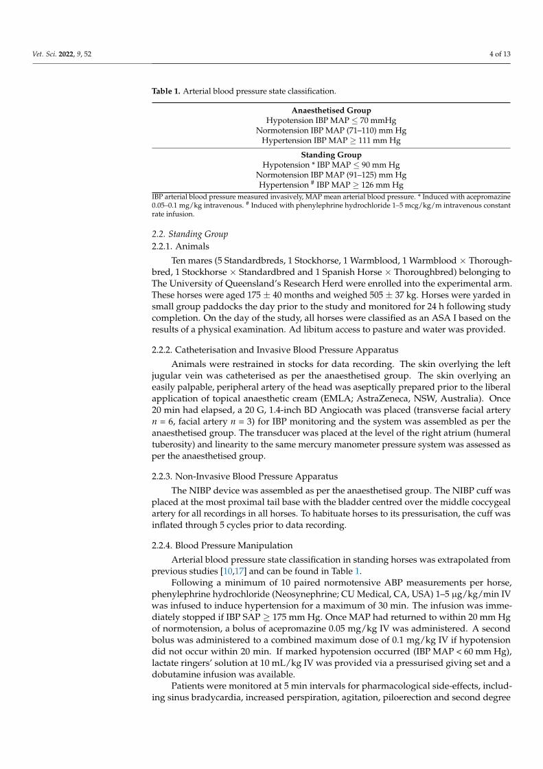
Table 1. Arterial blood pressure state classification.
Anaesthetised GroupHypotension IBP MAP ≤ 70 mmHg
Normotension IBP MAP (71–110) mm HgHypertension IBP MAP ≥ 111 mm Hg
Standing GroupHypotension * IBP MAP ≤ 90 mm Hg
Normotension IBP MAP (91–125) mm HgHypertension # IBP MAP ≥ 126 mm Hg
IBP arterial blood pressure measured invasively, MAP mean arterial blood pressure. * Induced with acepromazine0.05–0.1 mg/kg intravenous. # Induced with phenylephrine hydrochloride 1–5 mcg/kg/m intravenous constantrate infusion.
2.2. Standing Group2.2.1. Animals
Ten mares (5 Standardbreds, 1 Stockhorse, 1 Warmblood, 1 Warmblood × Thorough-bred, 1 Stockhorse × Standardbred and 1 Spanish Horse × Thoroughbred) belonging toThe University of Queensland’s Research Herd were enrolled into the experimental arm.These horses were aged 175 ± 40 months and weighed 505 ± 37 kg. Horses were yarded insmall group paddocks the day prior to the study and monitored for 24 h following studycompletion. On the day of the study, all horses were classified as an ASA I based on theresults of a physical examination. Ad libitum access to pasture and water was provided.
2.2.2. Catheterisation and Invasive Blood Pressure Apparatus
Animals were restrained in stocks for data recording. The skin overlying the leftjugular vein was catheterised as per the anaesthetised group. The skin overlying aneasily palpable, peripheral artery of the head was aseptically prepared prior to the liberalapplication of topical anaesthetic cream (EMLA; AstraZeneca, NSW, Australia). Once20 min had elapsed, a 20 G, 1.4-inch BD Angiocath was placed (transverse facial arteryn = 6, facial artery n = 3) for IBP monitoring and the system was assembled as per theanaesthetised group. The transducer was placed at the level of the right atrium (humeraltuberosity) and linearity to the same mercury manometer pressure system was assessed asper the anaesthetised group.
2.2.3. Non-Invasive Blood Pressure Apparatus
The NIBP device was assembled as per the anaesthetised group. The NIBP cuff wasplaced at the most proximal tail base with the bladder centred over the middle coccygealartery for all recordings in all horses. To habituate horses to its pressurisation, the cuff wasinflated through 5 cycles prior to data recording.
2.2.4. Blood Pressure Manipulation
Arterial blood pressure state classification in standing horses was extrapolated fromprevious studies [10,17] and can be found in Table 1.
Following a minimum of 10 paired normotensive ABP measurements per horse,phenylephrine hydrochloride (Neosynephrine; CU Medical, CA, USA) 1–5 µg/kg/min IVwas infused to induce hypertension for a maximum of 30 min. The infusion was imme-diately stopped if IBP SAP ≥ 175 mm Hg. Once MAP had returned to within 20 mm Hgof normotension, a bolus of acepromazine 0.05 mg/kg IV was administered. A secondbolus was administered to a combined maximum dose of 0.1 mg/kg IV if hypotensiondid not occur within 20 min. If marked hypotension occurred (IBP MAP < 60 mm Hg),lactate ringers’ solution at 10 mL/kg IV was provided via a pressurised giving set and adobutamine infusion was available.
Patients were monitored at 5 min intervals for pharmacological side-effects, includ-ing sinus bradycardia, increased perspiration, agitation, piloerection and second degree
Vet. Sci. 2022, 9, 52 5 of 13
atrial-ventricular block during induced hypertension and sinus tachycardia, depression,weakness, and ataxia during induced hypotension.
2.3. Data Management and Statistical Analysis
To account for the effects of hydrostatic pressure on the position of the NIPB cuff, thedifference (∆) in vertical distance between the transducer and the NIBP cuff was calculated(in both anaesthetised and standing horses). A correction factor was applied to the NIBPdata (0.078 mm Hg × ∆ mm) [18] and only these values are here presented in the study.Data points were excluded from the analysis when the pulse rate measured using the NIBPmethod differed by ≥20% to the pulse rate on the IBP trace or heart rate on the ECG trace;whichever was found to be manually accurate.
Horse demographic data and SAP, MAP and DAP measurements made using eachmethod (IBP and NIBP) were summarised using descriptive statistics. Non-normally dis-tributed data was summarised as median (minimum–maximum) and normally distributeddata as mean ± SD. Trajectory plots for paired SAP, MAP and DAP measurements werecreated for each horse and the data was checked for outliers.
Arterial blood pressure data comprised linked replicate measures, in that for eachhorse, the first replicate measure using the IBP method was recorded simultaneously withthe first replicate measure using the NIBP method. Consequently, a proportion of variationbetween replicated measurements within horses was common to both methods. To accountfor repeated measurements and the linked nature of the data, methods for ABP assessmentwere compared using linear mixed models, with random effects fitted for method-by-horseinteraction and horse-by-replicate interaction [19]. For each dataset, separate models werefitted for paired SAP, MAP and DAP values and Bland–Altman plots were used to visualisethe results. Bias (IBP—NIBP), standard deviation (SD) and limits of agreement (LOA) werecalculated from the model output. A positive bias denotes an underestimation of ABPby the NIBP method, whereas a negative bias denotes an overestimation of ABP by theNIBP method. LOA were derived from the 95% confidence intervals of repeat mean biasmeasurements on the same animal.
In reference to the only veterinary specific guidelines for IBP and NIBP evaluation [20]accuracy standards were defined as bias estimates < 10 mm Hg and precision standards asSD estimates < 15mm Hg for SAP, MAP and DAP.
Models were fitted using the MethComp package in R [21]. Stata/SE 15.1® was used forall other analyses (Stata Statistical Software; Stata Corporation, College Station, TX, USA)
3. Results
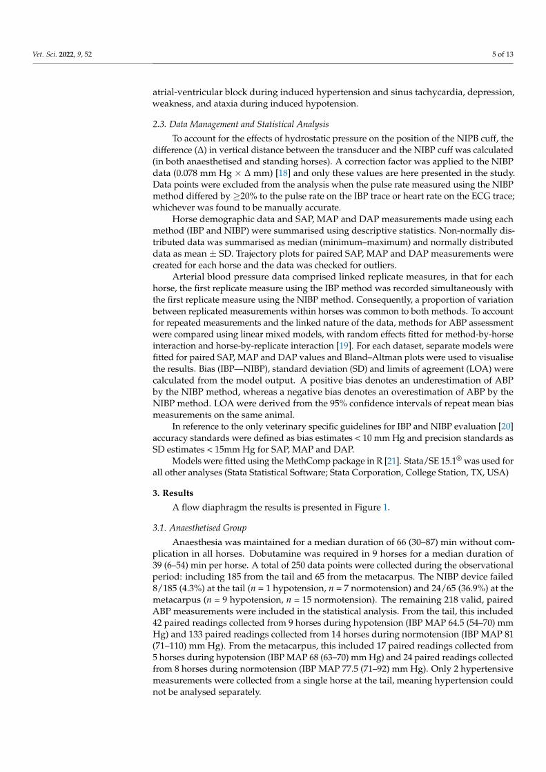
A flow diaphragm the results is presented in Figure 1.
3.1. Anaesthetised Group
Anaesthesia was maintained for a median duration of 66 (30–87) min without com-plication in all horses. Dobutamine was required in 9 horses for a median duration of39 (6–54) min per horse. A total of 250 data points were collected during the observationalperiod: including 185 from the tail and 65 from the metacarpus. The NIBP device failed8/185 (4.3%) at the tail (n = 1 hypotension, n = 7 normotension) and 24/65 (36.9%) at themetacarpus (n = 9 hypotension, n = 15 normotension). The remaining 218 valid, pairedABP measurements were included in the statistical analysis. From the tail, this included42 paired readings collected from 9 horses during hypotension (IBP MAP 64.5 (54–70) mmHg) and 133 paired readings collected from 14 horses during normotension (IBP MAP 81(71–110) mm Hg). From the metacarpus, this included 17 paired readings collected from5 horses during hypotension (IBP MAP 68 (63–70) mm Hg) and 24 paired readings collectedfrom 8 horses during normotension (IBP MAP 77.5 (71–92) mm Hg). Only 2 hypertensivemeasurements were collected from a single horse at the tail, meaning hypertension couldnot be analysed separately.
Vet. Sci. 2022, 9, 52 6 of 13Vet. Sci. 2022, 9, x FOR PEER REVIEW 6 of 13
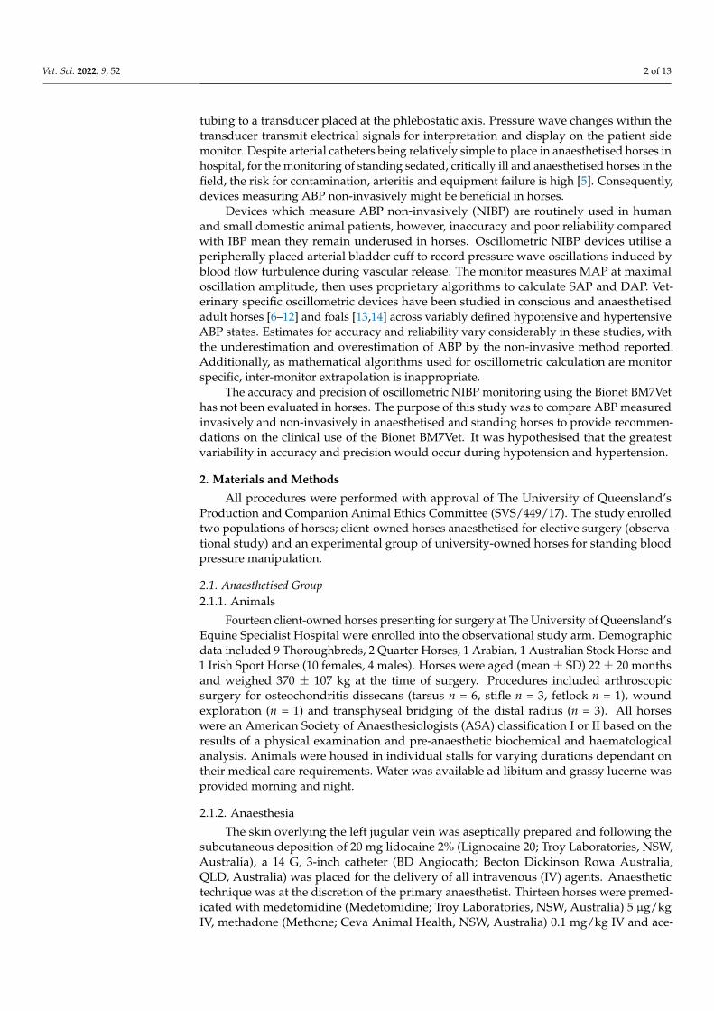
Figure 1. Pictorial representation of the methodology for comparing arterial blood pressure meas‐
ured non‐invasively to arterial blood pressure measured invasively using the Bionet BM7Vet in 14
healthy anaesthetised and 9 standing horses. Failures occurred when the Bionet BM7Vet failed to
generate a non‐invasive arterial blood pressure measurement by the completion of the inflation cy‐
cle. Paired measurements were excluded from the analysis when the pulse rate measured by the
non‐invasive method differed by ≥ 20% to the true pulse rate. Anaesthetised group: hypotension ≤
70 mm Hg, normotension 71–110 mm Hg, hypertension ≥ 111 mm Hg. Standing group: hypotension
≤ 90 mm Hg, normotension 91–125 mm Hg, hypertension ≥ 126 mm Hg.
3.1. Anaesthetised Group
Anaesthesia was maintained for a median duration of 66 (30–87) min without com‐
plication in all horses. Dobutamine was required in 9 horses for a median duration of 39
(6–54) min per horse. A total of 250 data points were collected during the observational
period: including 185 from the tail and 65 from the metacarpus. The NIBP device failed
8/185 (4.3%) at the tail (n = 1 hypotension, n = 7 normotension) and 24/65 (36.9%) at the
metacarpus (n = 9 hypotension, n = 15 normotension). The remaining 218 valid, paired
ABP measurements were included in the statistical analysis. From the tail, this included
42 paired readings collected from 9 horses during hypotension (IBP MAP 64.5 (54–70) mm
Hg) and 133 paired readings collected from 14 horses during normotension (IBP MAP 81
(71–110) mm Hg). From the metacarpus, this included 17 paired readings collected from
5 horses during hypotension (IBP MAP 68 (63–70) mm Hg) and 24 paired readings col‐
lected from 8 horses during normotension (IBP MAP 77.5 (71–92) mm Hg). Only 2 hyper‐
tensive measurements were collected from a single horse at the tail, meaning hypertension
could not be analysed separately.
Individual horse trajectory plots (Supplementary Figure S1) indicated trends of IBP‐
NIBP variation differed between NIBP cuff location. As a result, paired ABP measure‐
ments were separated by NIBP location (tail, metacarpus) for the Bland–Altman analysis
presented in Table 2, Figures 2 and 3 and Supplementary Figures S2 and S3. Due to the
limited number of paired ABP measurements, metacarpal data remained pooled for the
statistical analysis.
Table 2. Bland–Altmann analyses for 218 paired arterial blood pressure measurements made non‐
invasively compared to those made invasively using the Bionet BM7Vet in 14 healthy, anaesthetised
horses.
No. Paired
Observations
No.
Horses
Bias
(mm Hg)
SD
(mm Hg)
LOA‐L
(mm Hg)
LOA‐U
(mm Hg)
Proximal Tail Base
Systolic
Pooled 177 14 −12.9 14.1 −41.0 15.2
Hypotension 42 9 −15.4 13.9 −43.1 12.4
Normotension 133 14 −12.8 14.4 −41.5 16.0
Figure 1. Flow diaphragm for comparing arterial blood pressure measured non-invasively to arterialblood pressure measured invasively using the Bionet BM7Vet in 14 healthy anaesthetised and 9 stand-ing horses. Failures occurred when the Bionet BM7Vet failed to generate a non-invasive arterial bloodpressure measurement by the completion of the inflation cycle. Paired measurements were excludedfrom the analysis when the pulse rate measured by the non-invasive method differed by ≥20% to thetrue pulse rate. Anaesthetised group: hypotension ≤ 70 mm Hg, normotension 71–110 mm Hg, hy-pertension ≥ 111 mm Hg. Standing group: hypotension ≤ 90 mm Hg, normotension 91–125 mm Hg,hypertension ≥ 126 mm Hg.
Individual horse trajectory plots (Supplementary Figure S1) indicated trends of IBP-NIBP variation differed between NIBP cuff location. As a result, paired ABP measurementswere separated by NIBP location (tail, metacarpus) for the Bland–Altman analysis presentedin Table 2, Figures 2 and 3 and Supplementary Figures S2 and S3. Due to the limited numberof paired ABP measurements, metacarpal data remained pooled for the statistical analysis.
Vet. Sci. 2022, 9, x FOR PEER REVIEW 7 of 13
Hypertension 2 1
Mean
Pooled 177 14 −6.9 10.7 −28.3 14.5
Hypotension 42 9 −11.4 11.0 −33.3 10.5
Normotension 133 14 −6.0 9.9 −25.8 13.8
Hypertension 2 1
Diastolic
Pooled 177 14 −5.4 10.3 −25.9 15.2
Hypotension 42 9 −10.2 10.4 −30.9 10.6
Normotension 133 14 −4.0 9.7 −23.4 15.4
Hypertension 2 1
Metacarpus
Systolic 41 9 9.0 22.1 −35.1 53.2
Mean 41 9 3.6 24.0 −44.3 51.6
Diastolic 41 9 0.8 26.0 −51.1 52.7
No. number, SD standard deviation, LOA limits of agreement, U upper and L lower, MAP mean
arterial pressure. Bias defines accuracy and SD defines precision. The non‐invasive arterial blood
pressure cuff was placed either at the proximal tail base or the metacarpus. Combined recordings
on the proximal tail base (pooled) were separated into hypotension (MAP ≤ 70 mm Hg), normoten‐
sion (71 ≤ MAP ≤ 110 mm Hg) and hypertension (MAP ≥ 111 mmHg).
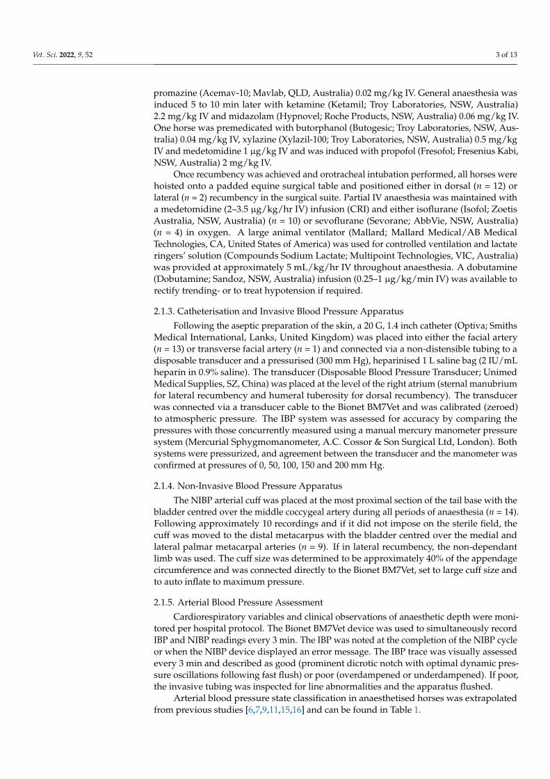
Figure 2. Bland–Altman plot of agreement for 177 paired mean arterial blood pressure measure‐
ments (mm Hg) made non‐invasively compared to those made invasively using the Bionet BM7Vet
in 14 healthy, anaesthetised horses. The non‐invasive arterial blood pressure cuff was placed at the
proximal tail base. Repeated observations from the same horse are plotted using the same colour‐
shape combination. The solid black line indicates mean bias, and the dashed lines indicate lower
and upper limits of agreement. The solid red line represents zero bias. IBP invasive blood pressure,
NIBP non‐invasive blood pressure. Bias defines accuracy.
Figure 3. Bland–Altman plot of agreement for 41 paired mean arterial blood pressure measurements
(mm Hg) made non‐invasively compared to those made invasively using the Bionet BM7Vet in 9
healthy, anaesthetised horses. The non‐invasive arterial blood pressure cuff was placed at the
-6.9
14.5
-28.3
40 60 80 100 120(IBP mean + NIBP mean) / 2
3.6
51.6
-44.3
40 60 80 100 120(IBP mean + NIBP mean) / 2
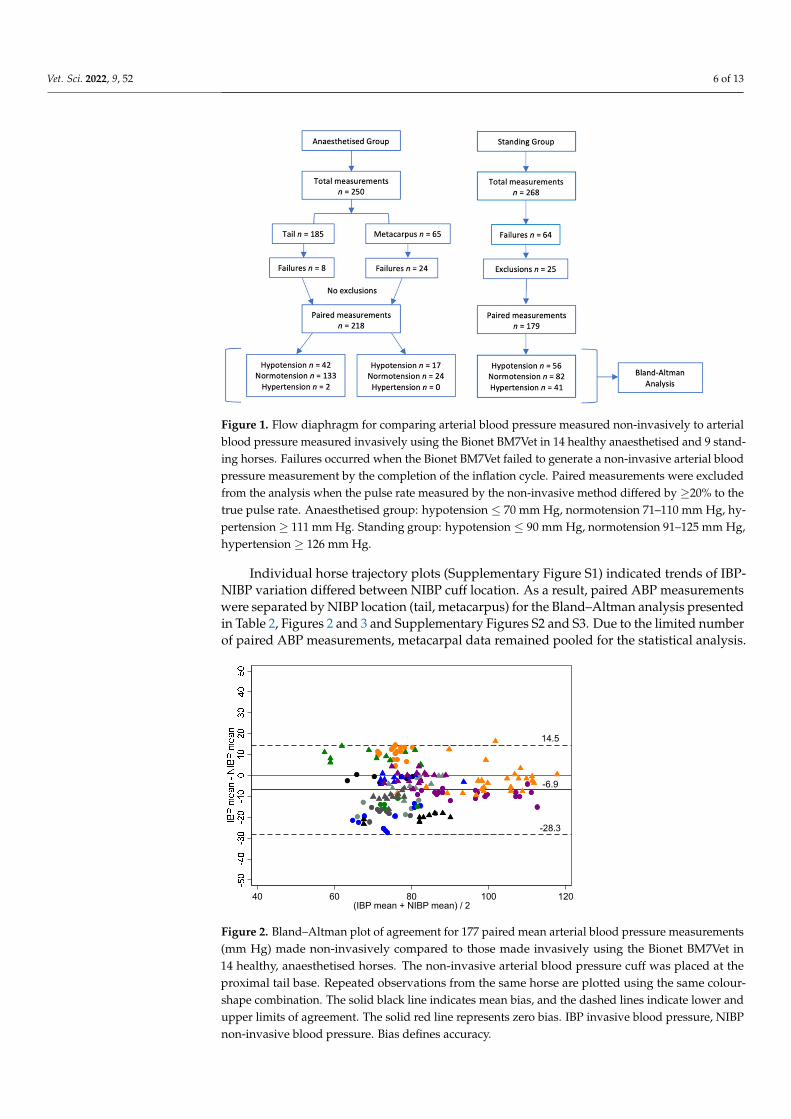
Figure 2. Bland–Altman plot of agreement for 177 paired mean arterial blood pressure measurements(mm Hg) made non-invasively compared to those made invasively using the Bionet BM7Vet in14 healthy, anaesthetised horses. The non-invasive arterial blood pressure cuff was placed at theproximal tail base. Repeated observations from the same horse are plotted using the same colour-shape combination. The solid black line indicates mean bias, and the dashed lines indicate lower andupper limits of agreement. The solid red line represents zero bias. IBP invasive blood pressure, NIBPnon-invasive blood pressure. Bias defines accuracy.
Vet. Sci. 2022, 9, 52 7 of 13
Vet. Sci. 2022, 9, x FOR PEER REVIEW 7 of 13
Hypertension 2 1
Mean
Pooled 177 14 −6.9 10.7 −28.3 14.5
Hypotension 42 9 −11.4 11.0 −33.3 10.5
Normotension 133 14 −6.0 9.9 −25.8 13.8
Hypertension 2 1
Diastolic
Pooled 177 14 −5.4 10.3 −25.9 15.2
Hypotension 42 9 −10.2 10.4 −30.9 10.6
Normotension 133 14 −4.0 9.7 −23.4 15.4
Hypertension 2 1
Metacarpus
Systolic 41 9 9.0 22.1 −35.1 53.2
Mean 41 9 3.6 24.0 −44.3 51.6
Diastolic 41 9 0.8 26.0 −51.1 52.7
No. number, SD standard deviation, LOA limits of agreement, U upper and L lower, MAP mean
arterial pressure. Bias defines accuracy and SD defines precision. The non‐invasive arterial blood
pressure cuff was placed either at the proximal tail base or the metacarpus. Combined recordings
on the proximal tail base (pooled) were separated into hypotension (MAP ≤ 70 mm Hg), normoten‐
sion (71 ≤ MAP ≤ 110 mm Hg) and hypertension (MAP ≥ 111 mmHg).
Figure 2. Bland–Altman plot of agreement for 177 paired mean arterial blood pressure measure‐
ments (mm Hg) made non‐invasively compared to those made invasively using the Bionet BM7Vet
in 14 healthy, anaesthetised horses. The non‐invasive arterial blood pressure cuff was placed at the
proximal tail base. Repeated observations from the same horse are plotted using the same colour‐
shape combination. The solid black line indicates mean bias, and the dashed lines indicate lower
and upper limits of agreement. The solid red line represents zero bias. IBP invasive blood pressure,
NIBP non‐invasive blood pressure. Bias defines accuracy.
Figure 3. Bland–Altman plot of agreement for 41 paired mean arterial blood pressure measurements
(mm Hg) made non‐invasively compared to those made invasively using the Bionet BM7Vet in 9
healthy, anaesthetised horses. The non‐invasive arterial blood pressure cuff was placed at the
-6.9
14.5
-28.3
40 60 80 100 120(IBP mean + NIBP mean) / 2
3.6
51.6
-44.3
40 60 80 100 120(IBP mean + NIBP mean) / 2
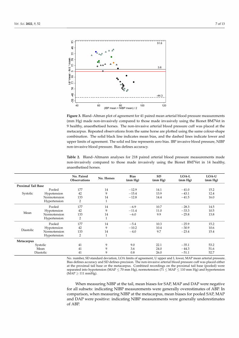
Figure 3. Bland–Altman plot of agreement for 41 paired mean arterial blood pressure measurements(mm Hg) made non-invasively compared to those made invasively using the Bionet BM7Vet in9 healthy, anaesthetised horses. The non-invasive arterial blood pressure cuff was placed at themetacarpus. Repeated observations from the same horse are plotted using the same colour-shapecombination. The solid black line indicates mean bias, and the dashed lines indicate lower andupper limits of agreement. The solid red line represents zero bias. IBP invasive blood pressure, NIBPnon-invasive blood pressure. Bias defines accuracy.
Table 2. Bland–Altmann analyses for 218 paired arterial blood pressure measurements madenon-invasively compared to those made invasively using the Bionet BM7Vet in 14 healthy,anaesthetised horses.
No. PairedObservations No. Horses Bias
(mm Hg)SD
(mm Hg)LOA-L
(mm Hg)LOA-U
(mm Hg)
Proximal Tail Base
SystolicPooled 177 14 −12.9 14.1 −41.0 15.2
Hypotension 42 9 −15.4 13.9 −43.1 12.4Normotension 133 14 −12.8 14.4 −41.5 16.0Hypertension 2 1
Mean
Pooled 177 14 −6.9 10.7 −28.3 14.5Hypotension 42 9 −11.4 11.0 −33.3 10.5
Normotension 133 14 −6.0 9.9 −25.8 13.8Hypertension 2 1
Diastolic
Pooled 177 14 −5.4 10.3 −25.9 15.2Hypotension 42 9 −10.2 10.4 −30.9 10.6
Normotension 133 14 −4.0 9.7 −23.4 15.4Hypertension 2 1
MetacarpusSystolic 41 9 9.0 22.1 −35.1 53.2Mean 41 9 3.6 24.0 −44.3 51.6
Diastolic 41 9 0.8 26.0 −51.1 52.7
No. number, SD standard deviation, LOA limits of agreement, U upper and L lower, MAP mean arterial pressure.Bias defines accuracy and SD defines precision. The non-invasive arterial blood pressure cuff was placed eitherat the proximal tail base or the metacarpus. Combined recordings on the proximal tail base (pooled) wereseparated into hypotension (MAP ≤ 70 mm Hg), normotension (71 ≤ MAP ≤ 110 mm Hg) and hypertension(MAP ≥ 111 mmHg).
When measuring NIBP at the tail, mean biases for SAP, MAP and DAP were negativefor all subsets: indicating NIBP measurements were generally overestimates of ABP. Incomparison, when measuring NIBP at the metacarpus, mean biases for pooled SAP, MAPand DAP were positive: indicating NIBP measurements were generally underestimatesof ABP.
Vet. Sci. 2022, 9, 52 8 of 13
3.2. Standing Group
A total of 268 data points were collected from 9 conscious horses. One horse was with-drawn from the study following two failed arterial catheterisation attempts. Hypertensionand hypotension were achieved in all 9 horses. The NIBP device failed on 64/268 (23.9%)occasions. This included 5/62 (8.1%) failures during hypotension, 11/100 (11%) failuresduring normotension and 48/106 (45.3%) failures during hypertension. The ≥20% pulserate-based exclusion criteria removed a further 25 of the remaining 204 data points (12.3%).This included 1/57 (1.8%) during hypotension, 7/89 (7.9%) during normotension and17/58 (29.3%) during hypertension. The remaining 179 valid, paired ABP measurementswere included in the statistical analysis. This included 56 paired readings collected duringhypotension (IBP MAP 78 (59, 90) mm Hg), 82 paired readings collected during normoten-sion (IBP MAP 110 (91, 125) mm Hg) and 41 paired readings collected during hypertension(IBP MAP 146 (126, 182) mm Hg). During the phenylephrine infusion, second degree atrial-ventricular block and piloerection were identified in 5 horses and all 9 horses experiencedagitation (trunk swaying, weight shifting, vocalisation), increased perspiration and sinusbradycardia. All horses experienced sinus tachycardia during induced hypotension. Fol-lowing collection of the final hypotensive measurement, one horse developed pronouncedhypotension, weakness, and depression (acepromazine 0.1 mg/kg IV was administered intotal). This horse required fluid support and additional close monitoring for a short period,however, made a full recovery.
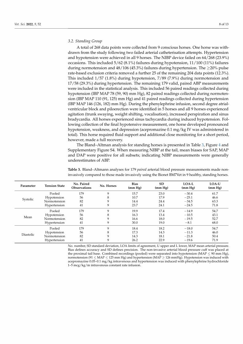
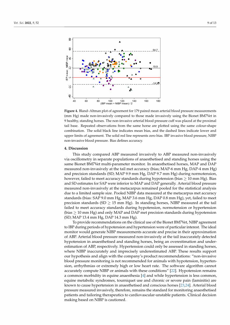
The Bland–Altman analysis for standing horses is presented in Table 3, Figure 4 andSupplementary Figure S4. When measuring NIBP at the tail, mean biases for SAP, MAPand DAP were positive for all subsets; indicating NIBP measurements were generallyunderestimates of ABP.
Table 3. Bland–Altmann analyses for 179 paired arterial blood pressure measurements made non-invasively compared to those made invasively using the Bionet BM7Vet in 9 healthy, standing horses.
Parameter Tension State No. PairedObservations No. Horses Bias
(mm Hg)SD
(mm Hg)LOA-L
(mm Hg)LOA-U
(mm Hg)
Systolic
Pooled 179 9 15.7 23.0 −30.4 61.7Hypotension 56 8 10.7 17.9 −25.1 46.6
Normotension 82 9 14.4 24.4 −34.5 63.3Hypertension 41 9 23.7 24.1 −24.5 71.8
Mean
Pooled 179 9 19.9 17.4 −14.9 54.7Hypotension 56 8 16.3 13.4 −10.5 43.1
Normotension 82 9 16.6 18.0 −19.5 52.7Hypertension 41 9 30.0 19.0 −8.1 68.0
Diastolic
Pooled 179 9 18.4 18.2 −18.0 54.7Hypotension 56 8 17.3 14.3 −11.3 46.0
Normotension 82 9 14.3 18.1 −21.8 50.4Hypertension 41 9 26.2 22.9 −19.6 71.9
No. number, SD standard deviation, LOA limits of agreement, U upper and L lower, MAP mean arterial pressure.Bias defines accuracy and SD defines precision. The non-invasive arterial blood pressure cuff was placed atthe proximal tail base. Combined recordings (pooled) were separated into hypotension (MAP ≤ 90 mm Hg),normotension (91 ≤ MAP ≤ 125 mm Hg) and hypertension (MAP ≥ 126 mmHg). Hypotension was induced withacepromazine 0.05–0.1 mg/kg intravenous and hypertension was induced with phenylephrine hydrochloride1–5 mcg/kg/m intravenous constant rate infusion.
Vet. Sci. 2022, 9, 52 9 of 13
Vet. Sci. 2022, 9, x FOR PEER REVIEW 9 of 13
No. number, SD standard deviation, LOA limits of agreement, U upper and L lower, MAP mean
arterial pressure. Bias defines accuracy and SD defines precision. The non‐invasive arterial blood
pressure cuff was placed at the proximal tail base. Combined recordings (pooled) were separated
into hypotension (MAP ≤ 90 mm Hg), normotension (91 ≤ MAP ≤ 125 mm Hg) and hypertension
(MAP ≥ 126 mmHg). Hypotension was induced with acepromazine 0.05–0.1 mg/kg intravenous
and hypertension was induced with phenylephrine hydrochloride 1–5 mcg/kg/m intravenous con‐
stant rate infusion.
Figure 4. Bland–Altman plot of agreement for 179 paired mean arterial blood pressure measure‐
ments (mm Hg) made non‐invasively compared to those made invasively using the Bionet BM7Vet
in 9 healthy, standing horses. The non‐invasive arterial blood pressure cuff was placed at the prox‐
imal tail base. Repeated observations from the same horse are plotted using the same colour‐shape
combination. The solid black line indicates mean bias, and the dashed lines indicate lower and up‐
per limits of agreement. The solid red line represents zero bias. IBP invasive blood pressure, NIBP
non‐invasive blood pressure. Bias defines accuracy.
4. Discussion
This study compared ABP measured invasively to ABP measured non‐invasively via
oscillometry in separate populations of anaesthetised and standing horses using the same
Bionet BM7Vet multi‐parameter monitor. In anaesthetised horses, MAP and DAP meas‐
ured non‐invasively at the tail met accuracy (bias; MAP‐6 mm Hg, DAP‐4 mm Hg) and
precision standards (SD; MAP 9.9 mm Hg, DAP 9.7 mm Hg) during normotension, how‐
ever, failed to meet accuracy standards during hypotension (bias ≥ 10 mm Hg). Bias and
SD estimates for SAP were inferior to MAP and DAP generally. Arterial blood pressure
measured non‐invasively at the metacarpus remained pooled for the statistical analysis
due to a limited sample size. Pooled NIBP data measured at the metacarpus met accuracy
standards (bias: SAP 9.0 mm Hg, MAP 3.6 mm Hg, DAP 0.8 mm Hg), yet, failed to meet
precision standards (SD ≥ 15 mm Hg). In standing horses, NIBP measured at the tail failed
to meet accuracy standards during hypotension, normotension or hypertension (bias ≥ 10
mm Hg) and only MAP and DAP met precision standards during hypotension (SD; MAP
13.4 mm Hg, DAP 14.3 mm Hg).
To provide recommendations on the clinical use of the Bionet BM7Vet, NIBP agree‐
ment to IBP during periods of hypotension and hypertension were of particular interest.
The ideal monitor would generate NIBP measurements accurate and precise in their ap‐
proximation of ABP. Arterial blood pressure measured non‐invasively at the tail inaccu‐
rately detected hypotension in anaesthetised and standing horses, being an overestima‐
tion and underestimation of ABP, respectively. Hypertension could only be assessed in
standing horses, where NIBP inaccurately and imprecisely underestimated ABP. These
results support our hypothesis and align with the company’s product recommendations:
“non‐invasive blood pressure monitoring is not recommended for animals with hypoten‐
sion, hypertension, arrhythmias or extremely high or low heart rate. The software algo‐
rithm cannot accurately compute NIBP or animals with these conditions” [22]. Hypoten‐
sion remains a common morbidity in equine anaesthesia [4] and while hypertension is
19.9
54.7
-14.9
40 60 80 100 120 140 160 180(IBP mean + NIBP mean) / 2
Figure 4. Bland–Altman plot of agreement for 179 paired mean arterial blood pressure measurements(mm Hg) made non-invasively compared to those made invasively using the Bionet BM7Vet in9 healthy, standing horses. The non-invasive arterial blood pressure cuff was placed at the proximaltail base. Repeated observations from the same horse are plotted using the same colour-shapecombination. The solid black line indicates mean bias, and the dashed lines indicate lower andupper limits of agreement. The solid red line represents zero bias. IBP invasive blood pressure, NIBPnon-invasive blood pressure. Bias defines accuracy.
4. Discussion
This study compared ABP measured invasively to ABP measured non-invasivelyvia oscillometry in separate populations of anaesthetised and standing horses using thesame Bionet BM7Vet multi-parameter monitor. In anaesthetised horses, MAP and DAPmeasured non-invasively at the tail met accuracy (bias; MAP-6 mm Hg, DAP-4 mm Hg)and precision standards (SD; MAP 9.9 mm Hg, DAP 9.7 mm Hg) during normotension,however, failed to meet accuracy standards during hypotension (bias ≥ 10 mm Hg). Biasand SD estimates for SAP were inferior to MAP and DAP generally. Arterial blood pressuremeasured non-invasively at the metacarpus remained pooled for the statistical analysisdue to a limited sample size. Pooled NIBP data measured at the metacarpus met accuracystandards (bias: SAP 9.0 mm Hg, MAP 3.6 mm Hg, DAP 0.8 mm Hg), yet, failed to meetprecision standards (SD ≥ 15 mm Hg). In standing horses, NIBP measured at the tailfailed to meet accuracy standards during hypotension, normotension or hypertension(bias ≥ 10 mm Hg) and only MAP and DAP met precision standards during hypotension(SD; MAP 13.4 mm Hg, DAP 14.3 mm Hg).
To provide recommendations on the clinical use of the Bionet BM7Vet, NIBP agreementto IBP during periods of hypotension and hypertension were of particular interest. The idealmonitor would generate NIBP measurements accurate and precise in their approximationof ABP. Arterial blood pressure measured non-invasively at the tail inaccurately detectedhypotension in anaesthetised and standing horses, being an overestimation and under-estimation of ABP, respectively. Hypertension could only be assessed in standing horses,where NIBP inaccurately and imprecisely underestimated ABP. These results supportour hypothesis and align with the company’s product recommendations: “non-invasiveblood pressure monitoring is not recommended for animals with hypotension, hyperten-sion, arrhythmias or extremely high or low heart rate. The software algorithm cannotaccurately compute NIBP or animals with these conditions” [22]. Hypotension remainsa common morbidity in equine anaesthesia [4] and while hypertension is less common,equine metabolic syndromes, tourniquet use and chronic or severe pain (laminitis) areknown to cause hypertension in anaesthetised and conscious horses [23,24]. Arterial bloodpressure measured invasively, therefore, remains the standard for monitoring anaesthetisedpatients and tailoring therapeutics to cardiovascular-unstable patients. Clinical decisionmaking based on NIBP is cautioned.
Vet. Sci. 2022, 9, 52 10 of 13
Mean arterial blood pressure is the average arterial pressure over the cardiac cycle andmost reliably indicates central tissue perfusion pressure [17]. Clinicians use MAP to adjustinterventional therapies and deliver anaesthetic agents appropriately, especially, wherecardiac output and micro perfusion (lactate, base excess, oxygen extraction) monitoringis unavailable. Measuring NIBP at the tail in anaesthetised horses during normotension,was the only subset where MAP satisfied both accuracy (bias—6.0 mm Hg) and precision(SD 9.9 mm Hg) standards simultaneously. Estimates of bias and SD for MAP gener-ally worsened during hypotension (anaesthetised and standing horses) and hypertension(standing horses). This trend of IBP-NIBP variability dependant on ABP state classification(hypotension, normotension or hypertension), is inconsistently reported within the litera-ture. Certain manuscripts describe suboptimal NIBP agreement during hypotension [9,11],while others report good agreement in adults [6,12] and foals [13,14]. Until a monitor canreliably generate accurate NIBP estimates of ABP, the use of this monitoring modality willremain underused in horses.
The inferior precision estimates for NIBP measured at the metacarpus compared withthe tail in anaesthetised horses, may reflect greater limb manipulation during surgeryor poorer cuff contact. The proximal tail base has historically been used for NIBP stud-ies in horses [6–14], and is preferable for its even contouring, limited compression dur-ing recumbency and limited manipulation during surgery. Despite returning accurateunder-estimates of ABP, NIBP measured at the metacarpus was significantly imprecise(SD > 22 mm Hg). The effects of cuff location and patient recumbency on NIBP approx-imation of ABP has been investigated previously. Drynan, Schier and Raisis [6] foundpoorer agreement in dorsally recumbent horses generally yet were unable to isolate a cuff-recumbency effect. Hatz, Hartnack, Kummerle, Hassig and Bettschart-Wolfensberger [8]concluded that the accuracy of the Sentinel device was optimised with the cuff placed overthe coccygeal artery (tail base) in lateral recumbency and over the palmar digital artery(metacarpus) or dorsal metatarsal artery (metatarsus) in dorsal recumbency. However,their methodology did not correct for hydrostatic pressure; being the force acting on acolumn of blood due to the weight of the blood above it [18]. Hydrostatic pressure is zeroas blood drains into the right atrium, providing a reference point for correct placementof the arterial transducer (phlebostatic axis) and being the basis for NIBP data correc-tion (0.078 mm Hg × ∆ mm) [18]. Despite incorporating a hydrostatic correction factorin the current study, NIBP devices are often placed over peripheral arteries unalignedwith the right atrium, complicating the translation of results into clinical settings. Regard-less, measuring NIBP with the Bionet BM7Vet at the metacarpus in anaesthetised horsesis cautioned.
Oscillometric devices may incorrectly identify pulse flow in certain situations, forexample, when the rate of cuff deflation to patient heart rate is too great, during arrythmiasor in situations of reduced peripheral blood flow [25,26]. Many of the standing horsesdeveloped sinus bradycardia and second-degree atrioventricular block; likely the resultof phenylephrine induced hypertension. The alpha-1 adrenergic alterations in peripheralblood flow may explain the reduction in MAP accuracy (∆ 13.4 mm Hg) between nor-motension and hypertension. Non-invasive blood pressure failure rates for standing horsesare not reported in the literature, however, patient compliance, agitation, movement, andmuscle tone, likely contributed to the greater rate of measurement failures in standing(64/268, 23.9%) compared with anaesthetised (32/250, 12.8%) horses, especially duringperiods of induced hypertension (48/106, 45.3%). In comparison, acepromazine inducedhypotension in standing horses, may have improved flow volume through the coccygealand metacarpal arteries and, thus, reduced NIBP failures in this subgroup (5/62, 8.1%).
A medetomidine infusion was delivered to all anaesthetised horses to provide intra-operative analgesia and improve anaesthesia recovery quality. Its impact on the detection ofperipheral pulse flow using the non-invasive method is unconfirmed [8] and is difficult topredict, given alpha-2 agents are biphasic in their haemodynamic effects. Previous reports
Vet. Sci. 2022, 9, 52 11 of 13
of NIBP failure rates in anaesthetised horses vary between monitors [7,8] and, betweenpharmacologically altered ABP states (hypotension, normotension and hypertension) [11].
The 20% pulse rate-based exclusion criterion excluded 25/204 (12.3%) data points fromstanding horses, including a large proportion of hypertensive measurements 17/58 (29.3%).It is likely that these excluded measurements would show greater variability than thosewhich met the inclusion criteria; meaning the estimates of precision (SD and LOA) derivedfrom the Bland–Altman analysis are likely underestimates of their true value. It is alsopossible that these excluded measurements may have underestimated or overestimatedABP to a larger degree than those which met the inclusion criteria; meaning the biasestimates may also be underestimates of their true value. Unfortunately, there is a lack ofstandardised exclusion (or inclusion) criteria used in IBP-NIBP comparative studies [6,7,9],and this complicates how clinicians interpret these studies.
Using the Bionet BM7Vet, Jacobs-Fohrman et al. [27] compared ABP measured in-vasively to ABP measured non-invasively in anaesthetised bitches undergoing electiveovariohysterectomy. They found that NIBP underestimated SAP, MAP and DAP duringhypotension, normotension and, with the greatest variability, hypertension. The guidelinesfor IBP-NIBP evaluation [20] were satisfied by SAP, MAP and DAP during hypotensionand MAP and DAP during normotension. These results are similar to other studies usingveterinary specific multi-parameter monitors in dogs [28,29], with estimates for bias andSD comparably more accurate and precise than those reported in horses.
This study has limitations involving sample size and limited replicate measurements.It is statistically preferrable to use fewer replicate measurements collected from a largernumber of horses, than to use more replicate measurements collected from a small numberof horses. Access to large numbers of horses is always a challenging factor in veterinaryresearch, however, this is the first study to investigate ABP in anaesthetised and standinghorses (pharmacologically manipulated) using the Bionet BM7Vet.
Vasopressor and ionotropic therapies are frequently administered within a multi-modal anaesthetic approach to standing and anaesthetised horses. Their summativeeffect on oscillometric peripheral blood flow detection is largely uncategorised. Theco-administration of dobutamine in 9/14 horses (being at the discretion of the primaryanaesthetist) better reflects clinical situations, however, does introduce variability. Finally,ABP state manipulation to hypotension and hypertension in standing horses was achievedpharmacologically. How the accuracy and precision of the Bionet BM7Vet may differ duringhaemodynamic crises is unknown.
5. Conclusions
In conclusion, monitoring ABP via non-invasive oscillometry on the Bionet BM7Vetin anaesthetised horses overestimated ABP at the tail and underestimated ABP at themetacarpus. The device inaccurately detected hypotension at the tail when evaluatedagainst the accepted veterinary guidelines and, therefore, should be used cautiously. Instanding horses, the Bionet BM7Vet underestimated ABP at the tail, especially duringpharmacologically induced hypertension. High failure rates discourage monitoring NIBPat the metacarpus in anaesthetised horses and at the tail in conscious horses with theBionet BM7Vet.
Supplementary Materials: The following supporting information can be downloaded at: https://www.mdpi.com/article/10.3390/vetsci9020052/s1, Figure S1. Individual horse trajectory plotsfor 218 paired mean arterial blood pressure (MAP) measurements made non-invasively (NIBP)compared to those made invasively (IBP) using the Bionet BM7Vet in 14 healthy, anaesthetisedhorses. The non-invasive arterial cuff was placed either at the proximal tail base or the metacarpus.Figure S2. Bland–Altman plots of agreement for 177 paired (a) systolic and (b) diastolic arterialblood pressure measurements (mm Hg) made non-invasively compared to those made invasivelyusing the Bionet BM7Vet in 14 healthy, anaesthetised horses. The non-invasive arterial cuff wasplaced at the proximal tail base. Repeated observations from the same horse are plotted using thesame colour-shape combination. The solid black line indicates mean bias, and the dashed lines
Vet. Sci. 2022, 9, 52 12 of 13
indicate lower and upper limits of agreement. The solid red line represents zero bias. Figure S3.Bland–Altman plots of agreement for 41 paired (a) systolic and (b) diastolic arterial blood pressuremeasurements (mm Hg) made non-invasively compared to those made invasively using the BionetBM7Vet in 9 healthy, anaesthetised horses. The non-invasive arterial cuff was placed at the metacarpus.Repeated observations from the same horse are plotted using the same colour-shape combination.The solid black line indicates mean bias, and the dashed lines indicate lower and upper limits ofagreement. The solid red line represents zero bias. Figure S4. Bland–Altman plots of agreementfor 179 paired (a) systolic and (b) diastolic arterial blood pressure measurements (mm Hg) madenon-invasively compared to those made invasively using the Bionet BM7Vet in 9 healthy, standinghorses. The non-invasive arterial cuff was placed at the proximal tail base. Repeated observationsfrom the same horse are plotted using the same colour-shape combination. The solid black lineindicates mean bias, and the dashed lines indicate lower and upper limits of agreement. The solidred line represents zero bias.
Author Contributions: S.P. and W.G. contributed to conceptualization, methodology, formal analysis,investigation, data curation, writing—original draft preparation, review and editing and visualisation.T.S.B. contributed to methodology, software, formal analysis, data curation and writing—originaldraft preparation, review and editing. S.W. contributed to methodology, software, validation, andwriting—review and editing. J.R. contributed to conceptualization, methodology, investigation, andwriting—review and editing. A.S.-G. contributed to formal analysis and writing—original draftpreparation, review and editing. N.C. and K.d.K. contributed to investigation and writing—reviewand editing. W.G. supervised the project. All authors gave their final approval of the manuscript. Allauthors have read and agreed to the published version of the manuscript.
Funding: This research received no external funding.
Institutional Review Board Statement: The animal study protocol was approved by The Universityof Queensland’s Production and Companion Animal Ethics Committee (SVS/449/17).
Informed Consent Statement: Informed consent was obtained from all subjects involved in the study.
Data Availability Statement: Datasets analysed in this study are available upon request from thecorresponding author. The data is stored on UQRDM and mediated access is available upon request.
Acknowledgments: The authors would like to thank Professor Nigel Perkins from The University ofQueensland, School of Veterinary Science for his statistical expertise, Mr Mitchell Coyle from TheUniversity of Queensland’s Equine Unit for his dedication to research and assistance with animalmanagement and Mediquip Ltd Pty, Australia for the use of the Bionet BM7Vet.
Conflicts of Interest: The authors declare no conflict of interest.
References1. Bidwell, L.A.; Bramlage, L.R.; Rood, W.A. Equine perioperative fatalities associated with general anaesthesia at a private
practice–a retrospective case series. Vet. Anaesth. Analg. 2007, 34, 23–30. [CrossRef]2. Grandy, J.L.; Steffey, E.P.; Hodgson, D.S.; Woliner, M.J. Arterial hypotension and the development of postanaesthetic myopathy in
halothane-anaesthetised horses. Am. J. Vet. Res. 1987, 2, 192–197.3. Magdesian, K.G. Monitoring the critically ill equine patient. Vet. Clin. N. Am. 2004, 20, 11–39. [CrossRef]4. Young, S.S.; Taylor, P.M. Factors influencing the outcome of equine anaesthesia: A review of 1,314 cases. Equine Vet. J. 1993,
25, 147–151. [CrossRef]5. Hardy, J. Venous and arterial catheterisation and fluid thearpy. In Equine Anaesthesia, 2nd ed; Muir, W.W., Hubbel, J.A.E., Eds.;
W.B. Saunders: Philadelphia, PA, USA, 2009; pp. 131–148.6. Drynan, E.A.; Schier, M.; Raisis, A.L. Comparison of invasive and noninvasive blood pressure measurements in anaesthetized
horses using the Surgivet V9203. Vet. Anaesth Analg 2016, 43, 301–308. [CrossRef]7. Fouche, A.; Auer, U.; Iff, I. Comparison of non-invasive and invasive blood pressure measuremeants in horses during anaesthesia
using the oscillometric blood pressure monitor S/5 Datex Ohmeda. Pferdeheilkunde 2016, 32, 479–484. [CrossRef]8. Hatz, L.A.; Hartnack, S.; Kummerle, J.; Hassig, M.; Bettschart-Wolfensberger, R. A study of measurement of noninvasive blood
pressure with the oscillometric device, Sentinel, in isoflurane-anaesthetized horses. Vet. Anaesth. Analg. 2015, 42, 369–376.[CrossRef]
9. Heliczer, N.; Lorello, O.; Casoni, D.; Navas de Solis, C. Accuracy and Precision of Noninvasive Blood Pressure in Normo-, Hyper-,and Hypotensive Standing and Anesthetized Adult Horses. J. Vet. Intern. Med. 2016, 30, 866–872. [CrossRef]

Vet. Sci. 2022, 9, 52 13 of 13
10. Olsen, E.; Pedersen, T.L.S.; Robinson, R.; Haubro Andersen, P. Accuracy and precision of oscillometric blood pressure in standingconscious horses. J. Vet. Emerg. Crit. Care 2016, 26, 85–92. [CrossRef]
11. Tunsmeyer, J.; Hopster, K.; Feige, K.; Kastner, S.B. Agreement of high definition oscillometry with direct arterial blood pressuremeasurement at different blood pressure ranges in horses under general anaesthesia. Vet. Anaesth. Analg. 2015, 42, 286–291.[CrossRef]
12. Yamaoka, T.T.; Flaherty, D.; Pawson, P.; Scott, M.; Auckburally, A. Comparison of arterial blood pressure measurements obtainedinvasively or oscillometrically using a Datex S/5 Compact monitor in anaesthetised adult horses. Vet. Anaes. Analg. 2017,44, 492–501. [CrossRef]
13. Giguere, S.; Knowles, J.A.; Valverde, A.; Bucki, E.; Young, L. Accuracy of indirect measurement of blood pressure in neonatalfoals. J. Vet. Intern. Med. 2005, 19, 571–576. [CrossRef]
14. Nout, Y.S.; Corley, T.T.; Donaldson, L.L.; Furr, M.O. Indirect oscillometric and direct blood pressure measurements in anaesthetisedand conscious neonatal foals. J. Vet. Emerg. Crit. Care 2002, 12, 75–80. [CrossRef]
15. Duke, T.; Filzek, U.; Read, M.R.; Read, E.K.; Ferguson, J.G. Clinical observations surrounding an increased incidence ofpostanesthetic myopathy in halothane-anesthetized horses. Vet. Anaesth. Analg. 2006, 33, 122–127. [CrossRef]
16. Hubbell, J.A.E.; Muir, W.W. Monitoring anaesthesia. In Equine Anaesthesia, 2nd ed.; Muir, W.W., Hubbell, J.A.E., Eds.;W.B. Saunders: Philadelphia, PA, USA, 2009; pp. 149–170.
17. Schwarzwald, C.C.; Bonagura, J.D.; Muir, W.W. The cardiovascular system. In Equine Anaesthesia, 2nd ed.; Muir, W.W.,Hubbell, J.A.E., Eds.; W.B. Saunders: Philadelphia, PA, USA, 2009; pp. 37–100.
18. Gornik, H.L.; Garcia, B.; Wolski, K.; Jones, D.C.; Macdonald, K.A.; Fronek, A. Validation of a method for determination of theankle-brachial index in the seated position. J. Vasc. Surg. Cases 2008, 48, 1204–1210. [CrossRef]
19. Carstensen, B. Comparing Clinical Measurement Methods: A Practical Guide; John Wiley & Sons, Ltd.: West Sussex, UK, 2010.20. Brown, S.; Atkins, C.; Bagley, R.; Carr, A.; Cowgill, L.; Davidson, M.; Egner, B.; Elliott, J.; GHenik, R.; Labato, M.; et al. Guidlines
for the identification, evaluation and management of systemic hypertension in dogs and cats. J. Vet. Intern. Med. 2007, 21, 542–558.[CrossRef]
21. Carstensen, B. The MethComp Package for R: Statistical Analysis of Method Comparison Studies. Available online: https://cran.r-project.org/web/packages/MethComp/MethComp.pdf (accessed on 8 April 2018).
22. Bionetus. BM7VET Operation Manual. 2016, p. 139. Available online: https://www.bionetus.com/wp-content/file/2018/01/BM7Vet_User_Manual.pdf (accessed on 28 October 2021).
23. Bailey, S.R.; Harbershon-Butcher, J.L.; Ransom, K.J.; Elliott, J.; Menzies-Gow, N.J. Hypertension and insulin resistance in amixed-breed population of ponies predisposed to laminitis. Am. J. Vet. Res. 2008, 69, 122–129. [CrossRef]
24. Hubbell, J.A.E.; Muir, W.W. Anaesthetic associated complications. In Equine Anaesthesia, 2nd ed.; Muir, W.W., Hubbell, J.A.E., Eds.;W.B. Saunders: Philadelphia, PA, USA, 2009; pp. 397–417.
25. Amoore, J.N.; Geake, W.B.; Scott, D.H.T. The effects of pulse rate, artifact and pulse strength on oscillometric non-invasive bloodpressure measurements. J. Clin. Eng. 1998, 23, 103–109. [CrossRef]
26. Shuai, W.; Wang, X.X.; Hong, K.; Peng, Q.; Li, J.X.; Li, P.; Cheng, X.S.; Su, H. How to estimate heart rate from pulse rate reportedby oscillometric method in atrial fibrillation: The value of pulse rate variation. Int. J. Cardiol. 2016, 222, 1022–1026. [CrossRef]
27. Jacobs-Fohrman, Z.R.; Barnes, T.S.; McEwen, M.M.; Goodwin, W.A. Clinical evaluation of arterial blood pressure in anesthetizeddogs by use of a veterinary-specific multiparameter monitor. Am. J. Vet. Res. 2020, 81, 635–641. [CrossRef]
28. Da Cunha, A.F.; Ramos, S.J.; Domingues, M.; Beaufrere, H.; Shelby, A.; Stout, R.; Acierno, M.J. Agreement between twooscillometric blood pressure technologies and invasively measured arterial pressure in the dog. Vet. Anaesth. Analg. 2016,43, 199–203. [CrossRef] [PubMed]
29. Vachon, C.; Belanger, M.C.; Burns, P.M. Evaluation of oscillometric and Doppler ultrasonic devices for blood pressure measure-ments in anesthetized and conscious dogs. Res. Vet. Sci. 2014, 97, 111–117. [CrossRef] [PubMed]