Cancer preventive agents 9. Betulinic acid derivatives as potent cancer chemopreventive agents
Upload
independentCategory
view
0download
0
Betulinic Acid Suppresses Constitutive and TNFα-induced NF-κBActivation and Induces Apoptosis in Human Prostate CarcinomaPC-3 Cells
Thangaiyan Rabi1,2, Sanjeev Shukla1,2, and Sanjay Gupta1,2,3,*1 Department of Urology, The James & Eillen Dicke Research Laboratory, Case Western ReserveUniversity, Cleveland, Ohio 441062 University Hospitals Case Medical Center, Cleveland, Ohio 441063 Case Comprehensive Cancer Center, Cleveland, Ohio 44106
AbstractDevelopment of chemoresistance in androgen-refractory prostate cancer cells is partly due toconstitutive activation of Rel/NF-κB transcription factors that regulate several cell survival and anti-apoptotic genes. In this study we examined whether betulinic acid (BetA), a pentacyclic triterpenefrom the bark of white birch, is effective in inhibiting NF-κB expression in androgen-refractoryhuman prostate cancer cells exhibiting high constitutive NF-κB expression. Treatment of PC-3 cellswith BetA inhibited DNA binding and reduced nuclear levels of the NF-κB/p65. BetA-mediated NF-κB inhibition involved decreased IKK activity and phosphorylation of IκBα at serine 32/36 followedby its degradation. Reporter assays revealed that NF-κB inhibition by BetA is transcriptionally active.These effects were found to correlate with a shift in Bax/Bcl-2 ratio and cleavage of poly(ADP)ribosepolymerase more towards apoptosis. BetA also inhibited TNFα-induced activation of NF-κB via theIκBα pathway, thereby sensitizing the cells to TNFα-induced apoptosis. Our studies demonstrate thatBetA effectively inhibits constitutive NF-κB activation and supports the rationale for targeting NF-κB through combination protocols with BetA in androgen-refractory prostate cancer.
Keywordstranscription factor; betulinic acid; IκBα; IKK; prostate cancer
INTRODUCTIONNuclear Factor-kappaB (NF-κB), a member of Rel transcription factor family, controlsexpression of a multitude of critical genes that regulate cell survival, proliferation, apoptosis,immune responses and adaptive responses to changes in cellular redox balance [1–3]. NF-κBconsists of homo- and hetero-dimers formed by several subunits: NF-κB1 (p50/p105), NF-κB2 (p52/100), RelA (p65), RelB, and c-Rel proteins [4,5]. The NF-κB proteins are regulatedby inhibitors of the IκB family which includes IκBα, IκBβ, IκBε, IkBγ, Bcl-3, p100 and p105[6]. In an inactive state, NF-κB is present in the cytoplasm as a heterodimer composed of p65,p50 and IκB subunits. In response to various stimuli, the IκBα subunit is phosphorylated byan upstream IKKα at serine residues 32 and 36, triggering ubiquitination and proteasomal
*Correspondence to: Sanjay Gupta, Ph.D., Department of Urology, The James & Eillen Dicke Research Laboratory, Case WesternReserve University, 10900 Euclid Avenue, Cleveland, OH 44106, Phone: (216) 368 6162, Fax: (216) 368 0213, [email protected].
NIH Public AccessAuthor ManuscriptMol Carcinog. Author manuscript; available in PMC 2010 May 5.
Published in final edited form as:Mol Carcinog. 2008 December ; 47(12): 964–973. doi:10.1002/mc.20447.
NIH
-PA Author Manuscript
NIH
-PA Author Manuscript
NIH
-PA Author Manuscript

degradation of IκBα, thereby facilitating the translocation of p50–p65 heterodimer into thenucleus [4–6]. Phosphorylation of p65 facilitates its binding to a specific sequence in DNA,which in turn results in gene transcription [4]. In recent years, increasing evidence indicatesthat activation of NF-κB plays an important role in coordinating the control of apoptotic celldeath, consistent with reports that NF-κB can promote apoptosis under certain circumstances[5,6]. Aberrant NF-κB activation has been implicated in the pathogenesis of several humanmalignancies, including various types of carcinoma, and hematological cancers includingleukemia and lymphoma [7]. We and others have reported that NF-κB/p65/RelA isconstitutively activated in human prostate cancer and prostate cancer xenografts [8–10].Nuclear localization of NF-κB/p65 in primary prostate cancer is highly predictive of metastasis,disease relapse and resistance to chemotherapy [11,12]. NF-κB has been shown to activate atranscription regulatory element of the prostate-specific antigen-encoding gene, a marker ofprostate cancer development and progression [13]. Increased NF-κB activity in androgen-refractory human prostate carcinoma PC-3 cells contributes directly to its aggressive behavior[14]. Conversely, blockade of NF-κB activity in human prostate cancer cells is associated withsuppression of angiogenesis, invasion and metastasis [15]. Therefore, agents that can suppressNF-κB activation are attractive candidates for development as therapeutic agents for themanagement of prostate cancer.
Betulinic acid (BetA) is a pentacyclic triterpene discovered in 1995 in the stem bark of theplant Zizyphus mauritiana; it was found to be a melanoma-specific cytotoxic agent that inhibitsthe growth of human melanoma tumors in athymic mice [16]. BetA is also found in variousother plants widespread in tropical regions including Tryphyllum peltaum, Ancistrocladusheyneaus, Zizyphus joazeiro, Diospyoros leucomelas, Tetracera boliviana, and Syzygiumformosanum [17]. BetA has been shown to induce apoptosis in neuroblastomas andglioblastomas through the mitochondrial activation pathway [18,19]. BetA also appears to beactive against HIV [17,20], and it has displayed anti-inflammatory activities in variousexperimental systems [21]. Reports have demonstrated that apoptosis induced by BetAinvolved mitogen-activated protein kinase activation without involvement of caspases [22].The cytotoxicity research on BetA showed that it had selective cytotoxicity on tumor cell linesbut not on normal cells [23], suggesting that it may have potential for development as atherapeutic agent. Recent evidence indicates that the anticancer activity of BetA can bemarkedly increased when it is used in combination with chemotherapy, ionizing radiation orTRAIL [24–26]. BetA has been shown to suppress carcinogen-induced NF-κB activationthrough inhibition of IκBα kinase and p65 phosphorylation in epithelial cells and throughinhibition of the transcription factors specificity protein1 (Sp1), Sp3, and Sp4 which regulateVEGF and survivin expression [27,28]. We investigated the effect of BetA on constitutive NF-κB inhibition and in the TNFα-induced NF-κB activation pathway. Our results demonstratethat BetA inhibits both constitutive and TNFα-induced NF-κB activation, and that BetAsensitizes PC-3 cells to TNFα-induced apoptosis.
MATERIALS AND METHODSCell Lines and Reagents
Androgen-refractory human prostate cancer PC-3 cells were obtained from ATCC (Manassas,VA). RPMI 1640 medium and all other cell culture materials were obtained from LifeTechnologies Inc., (Gaithersburg, MD). Betulinic acid (≥ 98% purity) was obtained from A.G. Scientific, Inc. (San Diego, CA). NF-κB-dependent reporter plasmid (PathDetect® NF-κBcis-Reporter System) having p-NF-κB-Luc plasmid and pFC-MEKK positive control plasmidwas obtained from Stratagene, (La Jolla, CA). The Luciferase Assay System and Beta-Glo™
Assay System were purchased from Promega Corporation (Madison, WI). Proteasomalinhibitor MG132 was obtained from Calbiochem San Diego, CA. Human recombinant tumor
Rabi et al. Page 2
Mol Carcinog. Author manuscript; available in PMC 2010 May 5.
NIH
-PA Author Manuscript
NIH
-PA Author Manuscript
NIH
-PA Author Manuscript

necrosis factor-α (TNFα) was purchased from Roche Applied Sciences (Indianapolis, IN).Anti-NF-κB/p65, anti-IκBα, anti-p-IκBα, anti-IKKα, anti-Bax, anti-Bcl-2 and anti-PARPantibodies were purchased from Santa Cruz Biotechnologies Inc. (Santa Cruz, CA). The CellDeath detection ELISAPLUS kit was obtained from Roche (Mannheim, Germany) and theApopNexin® FITC apoptosis detection kit containing FITC-labeled annexin V and PI wasobtained from Millipore (Billerica, MA).
Cell CultureThe cells were cultured under standard culture condition in RPMI 1640 supplemented with10% fetal bovine serum (FBS) and 1% penicillin-streptomycin. The cells were maintained at37°C and 5% CO2 in a humid environment. At 60% confluency, cultures were switched to 1%FBS for 12 h, and then treated with specified doses of BetA in DMSO or DMSO alone(maximum final concentration 0.1%, v/v), or with/without TNFα, or with/without MG-132 forindicated times. After desired treatments, medium was aspirated and cells were harvested bythe addition of Trypsin-EDTA, and cytosolic and nuclear extracts were prepared as previouslydescribed [29].
Protein Extraction and Western Blot AnalysisFollowing treatment of cells as described above, media was aspirated, cells were washed withcold PBS (pH 7.4) and ice cold lysis buffer [50 mM Tris-HCl, 150 mM NaCl, 1 mM EGTA,1 mM EDTA, 20 mM NaF, 100 mM Na3VO4, 0.5% NP-40, 1% Triton X-100, 1 mM PMSF(pH 7.4)] with freshly added protease inhibitor cocktail (Protease Inhibitor Cocktail Set III,Calbiochem, La Jolla, CA) was added and it was incubated over ice for 30 min. The cells werescraped, lysate was collected in a microfuge tube and passed through a 21½ G needle to breakup cell aggregates. The lysate was cleared by centrifugation at 14,000 × g for 15 min at 4°Cand supernatant (total cell lysate) was used or immediately stored at −80°C. The proteinconcentration was determined by DC Bio-Rad assay using manufacturer’s protocol (Bio RadLaboratories, Hercules, CA).
For Western blot analysis, appropriate amount of cell lysates (25–50 μg protein) were resolvedover 4–20% Tris-Glycine polyacrylamide gel and then transferred onto the nitrocellulosemembrane. The blots were blocked using 5% non-fat dry milk and probed using appropriateprimary antibodies in blocking buffer overnight at 4°C. The membrane was then incubatedwith appropriate secondary antibody conjugated with horseradish peroxidase (HRP)(Amersham Life Sciences Inc., Arlington Heights, IL) followed by detection usingchemiluminescence ECL kit (Amersham Life Sciences Inc., Arlington Heights, IL). To ensureequal protein loading, the membrane was stripped and reprobed with anti-Oct-1 and anti-α-tubulin antibodies (Santa Cruz Biotechnologies).
Electrophoretic Mobility Shift Assay (EMSA)EMSA for NF-κB was performed using Lightshift™ Chemiluminiscent EMSA kit (Pierce,Rockford, IL) following manufacturer’s protocol. Briefly, DNA was biotin-labeled using theBiotin 3′ end labeling kit (Pierce, Rockford, IL) in a 50 μl reaction buffer, 5 pmol of doublestranded NF-κB oligonucleotide (5′-AGTTGAGGGGACTTTCCCAGGC-3′ and 3′-TCAACTCCCCTGAAAGGGTCCG-5′) incubated in a microfuge tube with 10 μl of 5x TdT(terminal deoxynucleotidyl transferase) buffer, 5 μl of 5 μM biotin-N4-CTP, 10 U of dilutedTdT, 25 μl of ultrapure water at 37°C for 30 min. The reaction was stopped with 2.5 μl of 0.2M EDTA. To extract labeled DNA, 50 μl of chloroform:isoamyl alcohol (24:1) was added toeach tube and centrifuged at 13,000 × g. The top aqueous phase containing the labeled DNAwas further used for binding reactions. Each binding reaction contained 1x-binding buffer (100mM Tris, 500 mM KCl, 10 mM Dithiothretol, pH 7.5), 2.5% glycerol, 5 mM MgCl2, 50 ng/μl poly (dI-dC), 0.05% NP-40, 2.5 μg of nuclear extract and 20–50 fm of biotin-end-labeled
Rabi et al. Page 3
Mol Carcinog. Author manuscript; available in PMC 2010 May 5.
NIH
-PA Author Manuscript
NIH
-PA Author Manuscript
NIH
-PA Author Manuscript
target DNA. The contents were incubated at room temperature for 20 min. To this reactionmixture, 5 μl of 5x loading buffer was added, subjected to gel electrophoresis on a nativepolyacrylamide gel and transferred to a nylon membrane. After transfer was completed, DNAwas cross-linked to the membrane at 120 mJ/cm2 using a UV cross-linker equipped with 254-nm bulb. The biotin end-labeled DNA was detected using streptavidin-horseradish peroxidaseconjugate and a chemiluminiscent substrate. The membrane was exposed to X-ray film (XAR-5Amersham Life Science Inc., Arlington Height, IL) and developed using a Kodak filmprocessor.
Cell Survival AssayThe effect of BetA on the viability of cells was determined by MTT (3-[4, 5-dimethylthiazol-2-yl]-2, 5-diphenyl tetrazoliumbromide) assay. Briefly, the cells were plated at 1 × 10⁴ cells perwell in 200 μl of complete culture medium containing desired concentrations of BetA preparedin DMSO and diluted with the culture media to achieve 1–40 μM final concentrations. Theconcentration of DMSO remained within maximum permissible concentration of 0.1% in bothcontrol and treated samples. After incubation for desired times at 37°C in a humidifiedincubator, cell viability was determined. 50 μl MTT (5 mg/ml in phosphate buffered salinestock, diluted to working strength 1mg/ml with media) was added to each well and incubatedfor 2 h after which the plate was centrifuged at X 600g for 5 min at 4°C. The MTT solutionwas removed from the wells by aspiration. After careful removal of the medium, 0.1 ml ofbuffered DMSO was added to each well, and plates were shaken. The absorbance was recordedon a microplate reader at the wavelength of 540 nm. The effect of BetA on growth inhibitionwas assessed as percent cell viability where vehicle-treated cells were taken as 100% viable.
Apoptosis Detection by ELISAApoptosis was assessed by Cell Death Detection ELISAPLUS kit according to themanufacturer’s protocol. Briefly, cells were treated with 0.1% DMSO or 5–20 μM BetA for48 h, after which the media was aspirated, the cells were washed with cold PBS and incubatedon ice for 30 min. in cell lysis buffer. The cells were then scraped and the lysate was collectedin a microfuge tube and vortex to break up the cell aggregates. The lysate was cleared bycentrifugation at 5,000 rpm for 10 min at 4°C and the supernatant was stored at −80°C. Proteinconcentration was determined by DC Bio-Rad protein assay. The cell lysate with 50 μg proteinwere added to lysis buffer provided with the kit and pipetted on a streptavidin-coated 96-wellmicrotiter plate to which immunoreagent mix was added and incubated for 2 h at roomtemperature with continuous shaking at 500 rpm. The wells were then washed with washingbuffer, and color was developed by addition of substrate solution, which was read at 405 nMagainst the blank, reference wavelength of 490 nm after 10–15 min. The enrichment factor(total amount of apoptosis) was calculated by dividing the absorbance of the sample (A405 nm)by the absorbance of the controls without treatment (A490 nm).
Transfection and Reporter AssayTo examine the effect of BetA on TNFα-induced reporter gene expression, PC-3 cells weretransiently transfected using Lipofectamine™ 2000 reagent (Invitrogen Corporation, Carlsbad,CA) with 1.6 μg p-NF-κB-Luc plasmid for 6 h in serum free medium. Later these cells wereswitched to 5% FBS medium treated either with DMSO or BetA (20μM in vehicle) for 16 h,or with TNFα (10 ng/ml) for 30 min without or with pre-BetA treatment (20μM) for 16 h. Thecells were processed to obtain total cell lysate for luciferase reporter gene assay and β-galactosidase assay (Promega, Madison, WI) according to vendor’s protocol. Luciferaseactivity was normalized to β-galactosidase activity to control for transfection efficiency.
Rabi et al. Page 4
Mol Carcinog. Author manuscript; available in PMC 2010 May 5.
NIH
-PA Author Manuscript
NIH
-PA Author Manuscript
NIH
-PA Author Manuscript
Detection of Apoptosis by FACS AnalysisPC-3 cells (30–40% confluent) grown in 6-well plates were switched to 1% FBS media for 12h and then treated either with TNFα (10 ng/ml) only, or pretreated with BetA (20μM) only for16 h and later with TNFα for 30 min. After these treatments, cells were harvested and processedfor annexin V-PI staining using ApopNexin® FITC apoptosis detection kit as permanufacturer’s protocol. The samples were then analyzed for apoptotic cells by FACS analysis.
Statistical AnalysisDensitometric measurements of the bands in Western blot analysis were performed usingdigitalized scientific software program on Kodak 2000R Image Station. Statistical analysis wasperformed using one-way analysis of variance (ANOVA) with 95% confidence limits. Dataare presented in the figures as mean ± SE.
RESULTSSince inappropriate regulation of NF-κB/p65/RelA correlates with prostate cancer progression,therefore, as a first step to determine the constitutive activation of NF-κB, we analyzed theexpression of NF-κB/p65 and IκBα in various human prostate cancer cells. As shown in figure1A, protein expression of NF-κB/p65 and IκBα was higher in androgen-refractory humanprostate cancer DU145 and PC-3 cells compared to androgen-responsive LNCaP cells. SinceNF-κB activation requires IκBα phosphorylation at Ser32/36 followed by ubiquitination anddegradation, we determined the phosphorylated levels of IκBα in LNCaP, DU145 and PC-3cells. IκBα is more highly phosphorylated in androgen-refractory DU145 and PC-3 cells thanin androgen-responsive LNCaP cells. A modest decrease in the levels of IKKα was observedin DU145 and PC-3 cells compared to LNCaP cells (Figure 1A). Next we determined DNAbinding for NF-κB/p65, which was higher in DU145 and PC-3 cells compared to LNCaP cells(Figure 1B). It is well recognized that degradation of phosphorylated IκB by the 26 Sproteasome regulates the DNA binding of NF-κB subunits therefore; next we used MG132, apharmacological inhibitor of proteasome activity, to determine the effect of treatment in thesecells. Previous study has demonstrated that use of proteasomal inhibitor MG132 blocksdegradation of phosphorylated IκBα [30]. Exposure of LNCaP, DU145 and PC-3 cells to 20μM MG132 for 6 and 12 h decreased the rate of IκBα phosphorylation, similar to publishedobservations (Figure 1C). Exposure of cells to MG132 for 12 h resulted in accumulation ofphosphorylated IκBα protein in all three cell lines; however, the rate of p-IκBα accumulationwas faster in PC-3 cells whereas the final amount of the p-IκBα protein was higher in DU145and PC-3 cells compared to LNCaP cells, as evidenced by time-dependent studies (Figure 1C).MG132 treatment significantly inhibited the DNA binding activity of NF-κB/p65 at 12 h in allthree prostate cancer cell lines (Figure 1D). Based on the rapid turnover of IκBα in PC-3 cellswe considered this cell line for further studies with BetA.
Our next aim was to investigate whether BetA treatment imparts anti-proliferative effectsagainst androgen-refractory human prostate carcinoma PC-3 cells. BetA is a naturallyoccurring pentacyclic triterpenoid which has shown to possess anti-viral, anti-inflammatoryand anticancer properties (Figure 2A). Exposure of PC-3 cells to 20 μM concentration of BetAfor 24 h resulted in loss of adhesion to substrate, and altered morphology, consistent withapoptosis (Figure 2B). Furthermore, BetA treatment (1 to 40 μM) resulted in a dose-dependentinhibition of cell growth, as compared to vehicle-treated controls. Exposure to BetA at thehighest doses of 40 μM resulted in 91.2% to 96.7% decrease in cell viability between 24 to 72h. BetA treatment for 24 h resulted in dose-dependent reduction in cell viability which rangedfrom 2.9% to 91.2% in PC-3 cells at 1 to 40 μM concentrations. BetA treatment also resultedin time-dependent inhibition of cell growth. This effect was more pronounced at 48 and 72 hpost-treatment with BetA (Figure 2C). We next investigated whether BetA-mediated loss of
Rabi et al. Page 5
Mol Carcinog. Author manuscript; available in PMC 2010 May 5.
NIH
-PA Author Manuscript
NIH
-PA Author Manuscript
NIH
-PA Author Manuscript
cell viability in human prostate cancer PC-3 cells is a result of apoptosis. To demonstrate theseeffects we performed cell death detection by ELISA. Compared to vehicle treated controls,1.4, 2.5 and 4.8 fold increases in induction of apoptosis were observed with BetA in PC-3 cells,following 24 h of treatment (Figure 2D). Since BetA treatment to PC-3 cells resulted indecreased cell survival and induction of apoptosis therefore, next we studied its effect on NF-κB signaling.
To examine the effect of BetA on NF-κB constitutive activation, PC-3 cells were treated with10 and 20 μM BetA for 12, 24 and 48 h. As shown in figure 3A, BetA treated cells showedexclusive cytoplasmic accumulation of NF-κB/p65 protein inhibiting its translocation into thenucleus in time and dose dependent manner. This effect correlated with significant dose andtime dependent increase in the levels of total IκBα. To further determine whether inhibition ofBetA on constitutive activation of NF-κB was caused by inhibition of IκBα degradation, weexamined the cells for p-IκBα status in the cytoplasm. The level of serine 32-phosphorylatedIκBα (p-IκBα) exhibited a dose and time dependent decrease in the phospho-protein expressionafter BetA treatment. Because phosphorylation of IκBα is mediated by IKKs, we nextdetermined whether the inhibitory effect of BetA on IκBα phosphorylation is mediated viaIKKα. BetA treatment markedly decreased the protein level of IKKα over 48 h. This findingraised the possibility that the primary target of BetA in the regulation of the IκB/NF-κB pathwaymight be regulated via IKKα. Furthermore, we examined the effect of BetA on DNA bindingactivities of NF-κB by EMSA assay in nuclear extracts of PC-3 cells. Treatment with 20 μMBetA for 24 h resulted in a decrease of NF-κB DNA binding (Figure 3B). Taken together, theseexperiments demonstrate that BetA inhibits constitutive activation of NF-κB in human prostatecancer PC-3 cells.
Since BetA prevents the entry of NF-κB/p65 into the nucleus, it is plausible that it might inducealterations in survival factors such as Bcl-2; therefore we next studied the time and dosedependent effects of BetA on the constitutive protein levels of Bax and Bcl-2 in PC-3 cells.The time-dependent treatment with 10 and 20 μM concentration of BetA resulted in the increaseaccumulation of Bax protein, which was observed up to 48 h post-treatment compared to thebasal levels. In sharp contrast, the protein expression of Bcl-2 was significantly decreased byBetA treatment in a dose and time dependent fashion (Figure 4A). This resulted in a significanttime and dose dependent shift in the ratio of Bax to Bcl-2 after BetA treatment which correlatedwith the cleavage of poly(ADP)ribose polymerase, indicative of induction of the apoptoticprocess (Figure 4B).
Since cytokine stimulation leads to NF-κB activation, we assessed the effect of BetA onTNFα-induced activation of NF-κB. PC-3 cells were serum starved and pretreated with 20μM BetA for 16 h followed by TNFα (10 ng/ml) treatment for 30 min. Vehicle treated controlsand cultures exposed only to TNFα were used as controls. Cytoplasmic and nuclear extractswere prepared from these cultures, and nuclear extracts were analyzed by EMSA for NF-κBDNA binding activity. Compared to vehicle treated control, treatment of cells with TNFαresulted in a significant increase in NF-κB/p65 DNA binding activity. Pre-treatment of cellswith BetA significantly reduced NF-κB/p65 DNA binding activity, suggesting that BetAinhibits TNFα-induced NF-κB activation (Figure 5A). Further investigation on cytoplasmicextracts from this experiment, for total IκBα and p-IκBα, revealed that treatment with TNFαresulted in an increase in total IκBα, which was further increased after pretreatment with BetA.However, TNFα-induced p-IκBα levels were strongly inhibited by treatment with BetA (Figure5B).
Although we have shown by EMSA that BetA blocks NF-κB activation, however, DNAbinding does not always correlate with NF-κB-dependent gene transcription, suggesting thereare additional regulatory steps. To determine the effect of BetA on TNFα-induced NF-κB-
Rabi et al. Page 6
Mol Carcinog. Author manuscript; available in PMC 2010 May 5.
NIH
-PA Author Manuscript
NIH
-PA Author Manuscript
NIH
-PA Author Manuscript
dependent reporter gene expression, we transiently transfected the cells with the NF-κB-regulated reporter construct, incubated them with 20 μM BetA for 16 h, and then stimulatedthe cells with 10 ng/ml TNFα for 30 min. A 4.4-fold increase in NF-κB-Luc activity over thevector control was observed upon stimulation with TNFα, and BetA completely suppressedthe TNFα-induced stimulation (Figure 5C). These results demonstrate that BetA also repressesNF-κB-dependent reporter gene expression induced by TNFα.
Since TNFα-induced NF-κB activity is the major factor in PC-3 cells that make them resistantto apoptosis, we tested if BetA is effective in sensitizing these cells to TNFα-induced apoptosis,possibly by virtue of its ability to inhibit NF-κB activity. As observed by annexin V-PI staining,TNFα treatment of PC-3 cells resulted in apoptotic cell death rates that were comparable tothose observed in vehicle treated controls; BetA exposure alone resulted in a 33% increase inrate of apoptotic cell death over controls. However, pre-treatment of cells with BetA followedby TNFα treatment resulted in a significant increase (44%; p<0.001) in the percentage ofapoptotic cells (Figure 5D). These observations clearly suggest that BetA has the potential tosensitize highly resistant PC-3 cells to TNFα-induced apoptosis and that this effect of BetAmay be mediated via inhibition of TNFα-induced NF-κB activation in these cells.
DISCUSSIONBetA is a natural product identified in various bark extracts and is readily synthesized frombetulin, a major component in the bark of birch trees [17]. The initial report of the efficacy ofBetA as a selective inhibitor of human melanoma cells was published in 1995 [16].Subsequently, BetA was found to be active against neuroectodermal tumors (neuroblastoma,medulloblastoma, Ewing’s sarcoma), malignant brain tumors, and several types of humanepithelial malignancies, including carcinoma of the ovary, colon, breast, and lung, andsquamous cell carcinoma of the head and neck [18,19,31–33]. Currently BetA is undergoingdevelopment as a therapeutic agent against melanoma with assistance from the Rapid Accessto Intervention Development (RAID) program of the National Cancer Institute [34]. In thisstudy we demonstrate that BetA causes growth inhibition and apoptosis in human prostatecancer cells.
It has been demonstrated that BetA induces apoptosis in several different cancer cell linesthrough multiple pathways, which include mitochondrial pathways, p53-independent inductionof p21/Waf1, upregulation of death receptors, inhibition of Specificity Protein (Sp)transcription factors and interaction with other agents [16–28]. Studies have demonstrated thatBetA-induced apoptosis in human prostate cancer LNCaP cells may be due to decreased ARexpression, leading to decreased expression of VEGF and survivin and caspase-dependentPARP cleavage [28]. However, it is unclear how BetA causes apoptosis in androgen-refractorycell lines, which are highly resistant to pro-apoptotic stimuli. In this study we demonstrate thatBetA induces apoptosis in androgen-refractory PC-3 human prostate cancer cells, and inaddition sensitizes these cells to TNFα-induced apoptosis through suppression of NF-κB.
The transcription factor, NF-κB is a key mediator of the cellular stress response and in cellsexposed to anticancer therapy NF-κB typically activates survival pathways [1–3]. Recentfindings also suggest an inhibitory role of NF-κB in carcinogenesis and tumorigenesis [35]. Ithas been demonstrated that inhibition of NF-κB activity in cancer cell lines could reduces cellproliferation and metastatic capabilities in vivo [4–6,15]. We and others have shown that NF-κB/p65/RelA is constitutively activated during prostate cancer progression and in androgen-refractory human prostate cancer cells [8–10]. The major mechanism of NF-κB activation inprostate cancer cells involves aberrant activation of IKK, resulting in increasedphosphorylation and instability of IκB proteins. It is logical to devise strategies to manipulatethe NF-κB pathway in order to induce androgen-refractory prostate carcinoma cells to undergo
Rabi et al. Page 7
Mol Carcinog. Author manuscript; available in PMC 2010 May 5.
NIH
-PA Author Manuscript
NIH
-PA Author Manuscript
NIH
-PA Author Manuscript
apoptosis. Previous strategies have employed protein synthesis inhibitors, cyclophosphamide,proteasomal inhibitor, ALLN and MG132 treatment, all of which are highly cytotoxic to normalcells [36,37]. BetA, in contrast, is a natural minimally-toxic, non-mutagenic pentacyclictriterpenoid proven to inhibit growth and induce apoptosis in both early and advanced stagecancers [38]. In the present study we have demonstrated the capability of BetA to inhibit DNAbinding of NF-κB and its translocation to the nucleus, IκBα phosphorylation, and IKKactivation. Our results provide convincing evidence that treatment of cells with BetA resultedin significant inhibition of NF-κB activation, and that this effect was mediated via the IκBαpathway. Our findings indicate that BetA shows potential for use as an agent targeting NF-κBblockage in androgen-refractory human prostate cancer cells. Although a related study suggeststhat blocking NF-κB activation by mutant IκBα is not sufficient to induce apoptosis or modifyresponse to cytotoxic drugs [39]; it can be hypothesized that along with IκBα inhibition, BetAmight interfere with other signaling events in the NF-κB pathway. Additional detailed studiesare required to further evaluate these possibilities.
Studies have shown that androgen-refractory prostate cancer DU145 and PC-3 cells areresistant to TNFα treatment [40]. This resistance may be due to constitutive activation of NF-κB, a characteristic that has been observed in these cell lines and that may help these cellssurvive damage induced by pro-apoptotic stimuli [41]. Activation of NF-κB also rendersTNFα ineffective in inducing apoptotic death of cancer cells and may cause the induction of atoxic TNFα environment in prostate cancer patients [40,41]. The most significant finding ofthis study is that pretreatment of PC-3 cells with a combination of BetA and TNFα make themmore sensitive to apoptosis, since the anti-apoptotic signaling elicited by TNFα-induced NF-κB activation is effectively blocked by BetA. This could be one of the major mechanismsthrough which BetA might overcome TNFα insensitivity in human prostate cancer cells.Previous reports have shown that a combination of BetA and the cytotoxic ligand TRAILsignificantly enhanced BetA-induced apoptosis [26]. Furthermore, BetA has been reported toact in concert with ionizing radiation or anticancer agents, suggesting that NF-κB inhibitionmight lead to such responses [24,25]. However, a recent study has demonstrated that BetAactivates NF-κB in various tumor cell lines derived from neuroblastoma, glioblastoma andmelanoma, unexpectedly promoting BetA-induced apoptosis [42]. A detailed study on BetA-induced NF-κB-mediated cell growth inhibitory responses in different cell cancer cell types isneeded to resolve the issue of cell type-specific responses to BetA exposure. Because the NF-κB pathway plays such important roles in prostate cancer cell survival, proliferation andresistance to chemotherapy and radiation, our findings suggest that this signaling pathwayrepresents a key molecular target for anticancer strategies, and that BetA represents a noveltranscription factor targeting agent. It is tempting to speculate that combinations of BetA withchemopreventive/therapeutic agents working through different pathways might prove usefulin providing new treatment approaches for prostate cancer. Further mechanism-based studieswith BetA directing NF-κB signaling pathways in preclinical models of prostate cancer areneeded to validate these findings.
AcknowledgmentsResearch Support: This work was supported by grants from United States Public Health Service RO1CA108512 andRO1CA115491.
This work was conducted in The James & Eilleen Dicke Research Laboratory, Department of Urology, Case WesternReserve University, Cleveland, Ohio.
Abbreviations
NF-κB Nuclear Factor-kappaB
Rabi et al. Page 8
Mol Carcinog. Author manuscript; available in PMC 2010 May 5.
NIH
-PA Author Manuscript
NIH
-PA Author Manuscript
NIH
-PA Author Manuscript
EMSA electrophoretic-mobility shift assay
ALLN N-acetyl leucyl leucyl norleucinal
Sp transcription factors specificity protein
References1. Luo JL, Kamata H, Karin M. IKK/NF-kappaB signaling: balancing life and death--a new approach to
cancer therapy. J Clin Invest 2005;115:2625–2632. [PubMed: 16200195]2. Li Q, Verma IM. NF-kappaB regulation in the immune system. Nat Rev Immunol 2002;2:725–734.
[PubMed: 12360211]3. Karin M. Nuclear factor-kappaB in cancer development and progression. Nature 2006;441:431–436.
[PubMed: 16724054]4. Gilmore TD. Introduction to NF-kappaB: players, pathways, perspectives. Oncogene 2006;2:6680–
6684. [PubMed: 17072321]5. Li X, Stark GR. NFkappaB-dependent signaling pathways. Exp Hematol 2002;30:285–296. [PubMed:
11937262]6. Tergaonkar V, Correa RG, Ikawa M, Verma IM. Distinct roles of IkappaB proteins in regulating
constitutive NF-kappaB activity. Nat Cell Biol 2005;7:921–923. [PubMed: 16136188]7. Rayet B, Gélinas C. Aberrant rel/nfkb genes and activity in human cancer. Oncogene 1999;18:6938–
6947. [PubMed: 10602468]8. Shukla S, MacLennan GT, Fu P, Patel J, Marengo SR, Resnick MI, Gupta S. Nuclear factor-kappaB/
p65 (Rel A) is constitutively activated in human prostate adenocarcinoma and correlates with diseaseprogression. Neoplasia 2004;6:390–400. [PubMed: 15256061]
9. Lessard L, Mes-Masson AM, Lamarre L, Wall L, Lattouf JB, Saad F. NF-kappa B nuclear localizationand its prognostic significance in prostate cancer. BJU Int 2003;91:417–420. [PubMed: 12603426]
10. Suh J, Payvandi F, Edelstein LC, Amenta PS, Zong WX, Gélinas C, Rabson AB. Mechanisms ofconstitutive NF-kappaB activation in human prostate cancer cells. Prostate 2002;52:183–200.[PubMed: 12111695]
11. Fradet V, Lessard L, Bégin LR, Karakiewicz P, Masson AM, Saad F. Nuclear factor-kappaB nuclearlocalization is predictive of biochemical recurrence in patients with positive margin prostate cancer.Clin Cancer Res 2004;10:8460–8464. [PubMed: 15623625]
12. Lessard L, Karakiewicz PI, Bellon-Gagnon P, Alam-Fahmy M, Ismail HA, Mes-Masson AM, SaadF. Nuclear localization of nuclear factor-kappaB p65 in primary prostate tumors is highly predictiveof pelvic lymph node metastases. Clin Cancer Res 2006;12:5741–5745. [PubMed: 17020979]
13. Chen CD, Sawyers CL. NF-kappa B activates prostate-specific antigen expression and is upregulatedin androgen-independent prostate cancer. Mol Cell Biol 2002;22:2862–2870. [PubMed: 11909978]
14. Lindholm PF, Bub J, Kaul S, Shidham VB, Kajdacsy-Balla A. The role of constitutive NF-kappaBactivity in PC-3 human prostate cancer cell invasive behavior. Clin Exp Metastasis 2000;18:471–479. [PubMed: 11592304]
15. Huang S, Pettaway CA, Uehara H, Bucana CD, Fidler IJ. Blockade of NF-kappaB activity in humanprostate cancer cells is associated with suppression of angiogenesis, invasion, and metastasis.Oncogene 2001;20:4188–4197. [PubMed: 11464285]
16. Pisha E, Chai H, Lee IS, Chagwedera TE, Farnsworth NR, Cordell GA, Beecher CW, Fong HH,Kinghorn AD, Brown DM. Discovery of betulinic acid as a selective inhibitor of human melanomathat functions by induction of apoptosis. Nat Med 1995;1:1046–1051. [PubMed: 7489361]
17. Vlietinck AJ, De Bruyne T, Apers S, Pieters LA. Plant-derived leading compounds for chemotherapyof human immunodeficiency virus (HIV) infection. Planta Med 1998;64:97–109. [PubMed:9525100]
18. Fulda S, Friesen C, Los M, Scaffidi C, Mier W, Benedict M, Nuñez G, Krammer PH, Peter ME,Debatin KM. Betulinic acid triggers CD95 (APO-1/Fas)- and p53-independent apoptosis via
Rabi et al. Page 9
Mol Carcinog. Author manuscript; available in PMC 2010 May 5.
NIH
-PA Author Manuscript
NIH
-PA Author Manuscript
NIH
-PA Author Manuscript
activation of caspases in neuroectodermal tumors. Cancer Res 1997;57:4956–4964. [PubMed:9354463]
19. Jeremias I, Steiner HH, Benner A, Debatin KM, Herold-Mende C. Cell death induction by betulinicacid, ceramide and TRAIL in primary glioblastoma multiforme cells. Acta Neurochir (Wien)2004;146:721–729. [PubMed: 15197616]
20. Mayaux JF, Bousseau A, Pauwels R, Huet T, Hénin Y, Dereu N, Evers M, Soler F, Poujade C, DeClercq E, Le Pecq JB. Triterpene derivatives that block entry of human immunodeficiency virus type1 into cells. Proc Natl Acad Sci U S A 1994;91:3564–3568. [PubMed: 8170948]
21. Honda T, Liby KT, Su X, Sundararajan C, Honda Y, Suh N, Risingsong R, Williams CR, Royce DB,Sporn MB, Gribble GW. Design, synthesis, and anti-inflammatory activity both in vitro and in vivoof new betulinic acid analogues having an enone functionality in ring A. Bioorg Med Chem Lett2006;16:6306–6309. [PubMed: 16996735]
22. Tan Y, Yu R, Pezzuto JM. Betulinic acid-induced programmed cell death in human melanoma cellsinvolves mitogen-activated protein kinase activation. Clin Cancer Res 2003;9:2866–2875. [PubMed:12855667]
23. Zuco V, Supino R, Righetti SC, Cleris L, Marchesi E, Gambacorti-Passerini C, Formelli F. Selectivecytotoxicity of betulinic acid on tumor cell lines, but not on normal cells. Cancer Lett 2002;175:17–25. [PubMed: 11734332]
24. Fulda S, Debatin KM. Sensitization for anticancer drug-induced apoptosis by betulinic Acid.Neoplasia 2005;7:162–170. [PubMed: 15802021]
25. Selzer E, Pimentel E, Wacheck V, Schlegel W, Pehamberger H, Jansen B, Kodym R. Effects ofbetulinic acid alone and in combination with irradiation in human melanoma cells. J Invest Dermatol2000;114:935–940. [PubMed: 10771474]
26. Fulda S, Jeremias I, Debatin KM. Cooperation of betulinic acid and TRAIL to induce apoptosis intumor cells. Oncogene 2004;23:7611–7620. [PubMed: 15361826]
27. Takada Y, Aggarwal BB. Betulinic acid suppresses carcinogen-induced NF-kappa B activationthrough inhibition of I kappa B alpha kinase and p65 phosphorylation: abrogation ofcyclooxygenase-2 and matrix metalloprotease-9. J Immunol 2003;171:3278–3286. [PubMed:12960358]
28. Chintharlapalli S, Papineni S, Ramaiah SK, Safe S. Betulinic acid inhibits prostate cancer growththrough inhibition of specificity protein transcription factors. Cancer Res 2007;67:2816–2823.[PubMed: 17363604]
29. Shukla S, Gupta S. Suppression of constitutive and tumor necrosis factor alpha-induced nuclear factor(NF)-kappaB activation and induction of apoptosis by apigenin in human prostate carcinoma PC-3cells: correlation with down-regulation of NF-kappaB-responsive genes. Clin Cancer Res2004;10:3169–3178. [PubMed: 15131058]
30. Sun L, Carpenter G. Epidermal growth factor activation of NF-kappaB is mediated throughIkappaBalpha degradation and intracellular free calcium. Oncogene 1998 Apr 23;16(16):2095–102.[PubMed: 9572490]
31. Baglin I, Poumaroux A, Nour M, Tan K, Mitaine-Offer AC, Lacaille-Dubois MA, Chauffert B, CavéC. New ursolic and betulinic derivatives as potential cytotoxic agents. J Enzyme Inhib Med Chem2003;18:111–117. [PubMed: 12943194]
32. Meng RD, El-Deiry WS. p53-independent upregulation of KILLER/DR5 TRAIL receptor expressionby glucocorticoids and interferon-gamma. Exp Cell Res 2001;262:154–169. [PubMed: 11139340]
33. Kessler JH, Mullauer FB, de Roo GM, Medema JP. Broad in vitro efficacy of plant-derived betulinicacid against cell lines derived from the most prevalent human cancer types. Cancer Lett2007;251:132–145. [PubMed: 17169485]
34. Udeani GO, Zhao GM, Geun Shin Y, Cooke BP, Graham J, Beecher CW, Kinghorn AD, PezzutoJM. Pharmacokinetics and tissue distribution of betulinic acid in CD-1 mice. Biopharm Drug Dispos1999;20:379–383. [PubMed: 10870094]
35. Chen F, Castranova V. Nuclear Factor-kappaB, an unappreciated tumor suppressor. Cancer Res2007;67:11093–11098. [PubMed: 18056430]
Rabi et al. Page 10
Mol Carcinog. Author manuscript; available in PMC 2010 May 5.
NIH
-PA Author Manuscript
NIH
-PA Author Manuscript
NIH
-PA Author Manuscript
36. Shumway SD, Miyamoto S. A mechanistic insight into a proteasome-independent constitutiveinhibitor kappaBalpha (IkappaBalpha) degradation and nuclear factor kappaB (NF-kappaB)activation pathway in WEHI-231 B-cells. Biochem J 2004;380:173–180. [PubMed: 14763901]
37. Meyer S, Kohler NG, Joly A. Cyclosporine A is an uncompetitive inhibitor of proteasome activityand prevents NF-kappaB activation. FEBS Lett 1997;413:354–358. [PubMed: 9280312]
38. Eiznhamer DA, Xu ZQ. Betulinic acid: a promising anticancer candidate. IDrugs 2004;7:359–373.[PubMed: 15057642]
39. Bentires-Alj M, Hellin AC, Ameyar M, Chouaib S, Merville MP, Bours V. Stable inhibition of nuclearfactor kappaB in cancer cells does not increase sensitivity to cytotoxic drugs. Cancer Res1999;59:811–815. [PubMed: 10029068]
40. Muenchen HJ, Lin DL, Walsh MA, Keller ET, Pienta KJ. Tumor necrosis factor-alpha-inducedapoptosis in prostate cancer cells through inhibition of nuclear factor-kappaB by an IkappaBalpha“super-repressor”. Clin Cancer Res 2006:1969–1977.
41. Gasparian AV, Yao YJ, Kowalczyk D, Lyakh LA, Karseladze A, Slaga TJ, Budunova IV. The roleof IKK in constitutive activation of NF-kappaB transcription factor in prostate carcinoma cells. JCell Sci 2002;115:141–151. [PubMed: 11801732]
42. Kasperczyk H, La Ferla-Brühl K, Westhoff MA, Behrend L, Zwacka RM, Debatin KM, Fulda S.Betulinic acid as new activator of NF-kappaB: molecular mechanisms and implications for cancertherapy. Oncogene 2005;24:6945–6956. [PubMed: 16007147]
Rabi et al. Page 11
Mol Carcinog. Author manuscript; available in PMC 2010 May 5.
NIH
-PA Author Manuscript
NIH
-PA Author Manuscript
NIH
-PA Author Manuscript
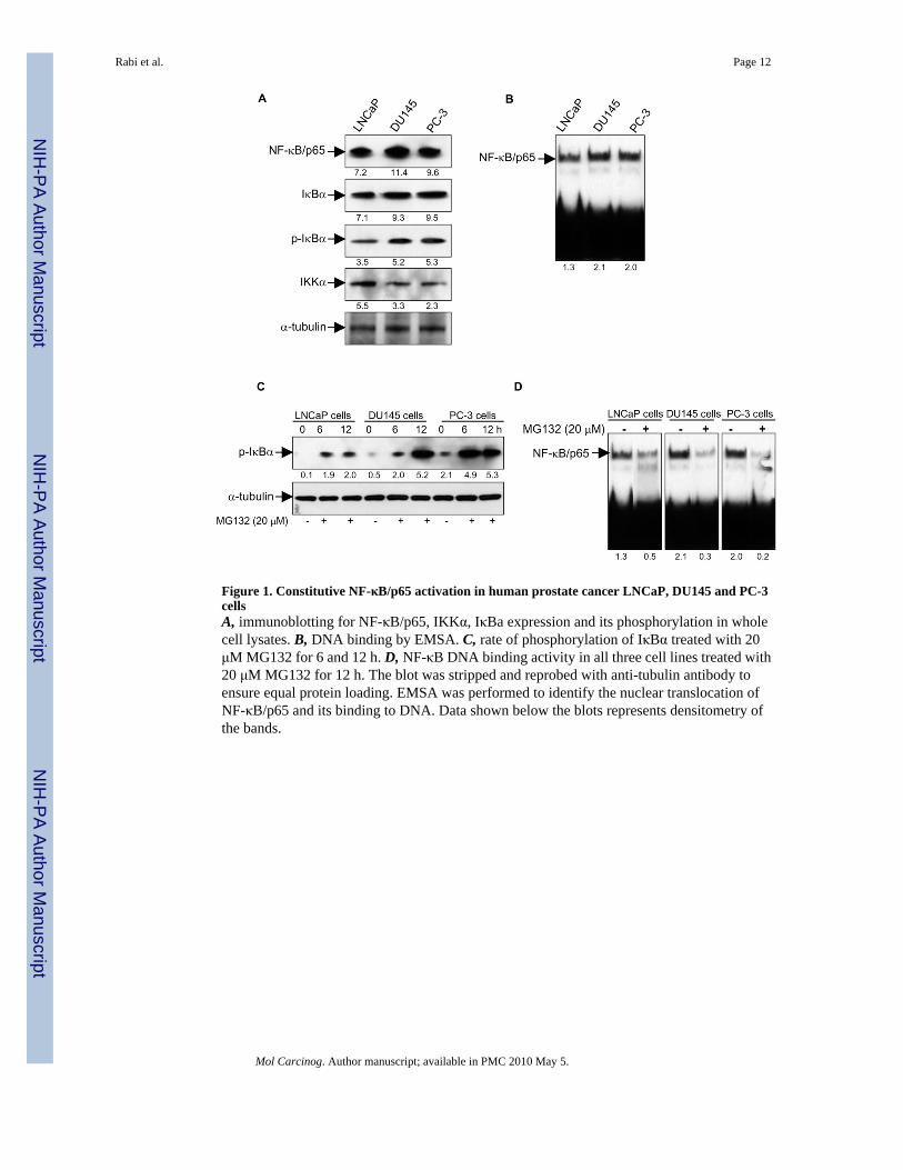
Figure 1. Constitutive NF-κB/p65 activation in human prostate cancer LNCaP, DU145 and PC-3cellsA, immunoblotting for NF-κB/p65, IKKα, IκBa expression and its phosphorylation in wholecell lysates. B, DNA binding by EMSA. C, rate of phosphorylation of IκBα treated with 20μM MG132 for 6 and 12 h. D, NF-κB DNA binding activity in all three cell lines treated with20 μM MG132 for 12 h. The blot was stripped and reprobed with anti-tubulin antibody toensure equal protein loading. EMSA was performed to identify the nuclear translocation ofNF-κB/p65 and its binding to DNA. Data shown below the blots represents densitometry ofthe bands.
Rabi et al. Page 12
Mol Carcinog. Author manuscript; available in PMC 2010 May 5.
NIH
-PA Author Manuscript
NIH
-PA Author Manuscript
NIH
-PA Author Manuscript
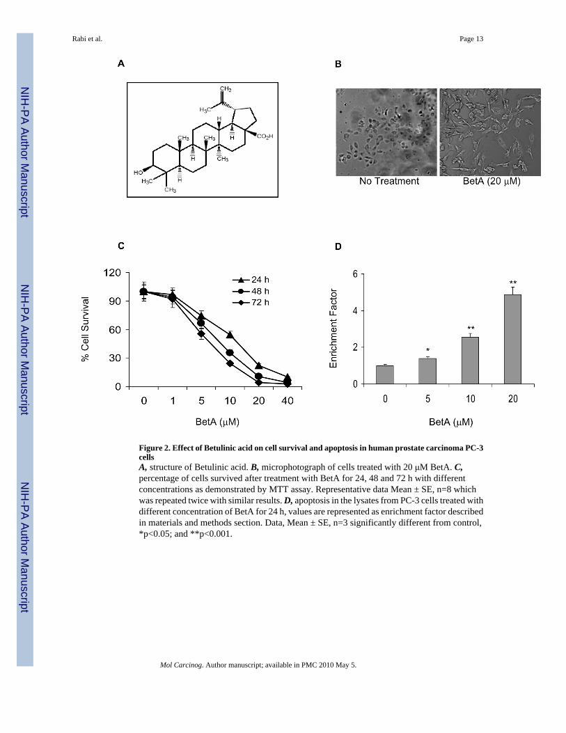
Figure 2. Effect of Betulinic acid on cell survival and apoptosis in human prostate carcinoma PC-3cellsA, structure of Betulinic acid. B, microphotograph of cells treated with 20 μM BetA. C,percentage of cells survived after treatment with BetA for 24, 48 and 72 h with differentconcentrations as demonstrated by MTT assay. Representative data Mean ± SE, n=8 whichwas repeated twice with similar results. D, apoptosis in the lysates from PC-3 cells treated withdifferent concentration of BetA for 24 h, values are represented as enrichment factor describedin materials and methods section. Data, Mean ± SE, n=3 significantly different from control,*p<0.05; and **p<0.001.
Rabi et al. Page 13
Mol Carcinog. Author manuscript; available in PMC 2010 May 5.
NIH
-PA Author Manuscript
NIH
-PA Author Manuscript
NIH
-PA Author Manuscript
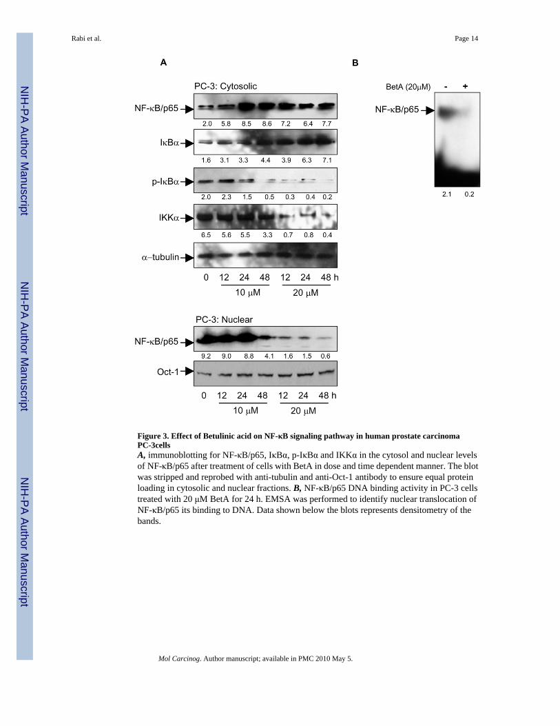
Figure 3. Effect of Betulinic acid on NF-κB signaling pathway in human prostate carcinomaPC-3cellsA, immunoblotting for NF-κB/p65, IκBα, p-IκBα and IKKα in the cytosol and nuclear levelsof NF-κB/p65 after treatment of cells with BetA in dose and time dependent manner. The blotwas stripped and reprobed with anti-tubulin and anti-Oct-1 antibody to ensure equal proteinloading in cytosolic and nuclear fractions. B, NF-κB/p65 DNA binding activity in PC-3 cellstreated with 20 μM BetA for 24 h. EMSA was performed to identify nuclear translocation ofNF-κB/p65 its binding to DNA. Data shown below the blots represents densitometry of thebands.
Rabi et al. Page 14
Mol Carcinog. Author manuscript; available in PMC 2010 May 5.
NIH
-PA Author Manuscript
NIH
-PA Author Manuscript
NIH
-PA Author Manuscript
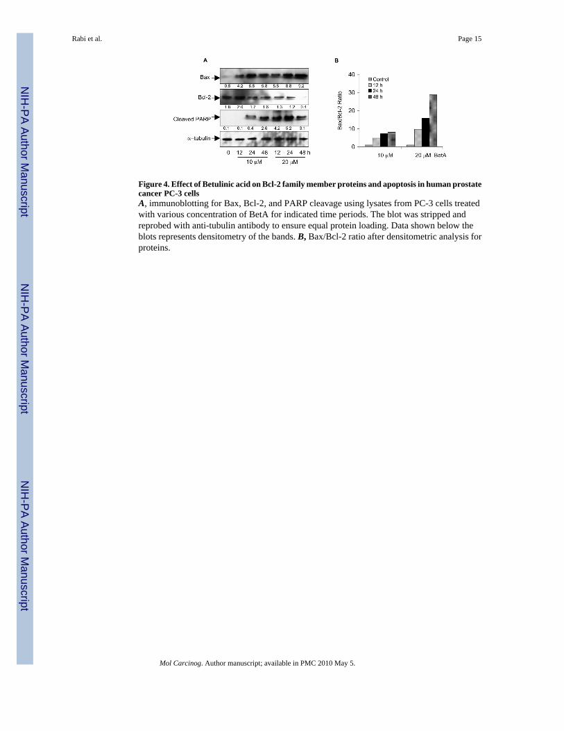
Figure 4. Effect of Betulinic acid on Bcl-2 family member proteins and apoptosis in human prostatecancer PC-3 cellsA, immunoblotting for Bax, Bcl-2, and PARP cleavage using lysates from PC-3 cells treatedwith various concentration of BetA for indicated time periods. The blot was stripped andreprobed with anti-tubulin antibody to ensure equal protein loading. Data shown below theblots represents densitometry of the bands. B, Bax/Bcl-2 ratio after densitometric analysis forproteins.
Rabi et al. Page 15
Mol Carcinog. Author manuscript; available in PMC 2010 May 5.
NIH
-PA Author Manuscript
NIH
-PA Author Manuscript
NIH
-PA Author Manuscript

Figure 5. Effect of Betulinic acid on TNFα-induced NF-κB activation and apoptosis in humanprostate cancer PC-3 cellsA, NF-κB/p65 DNA binding activity in PC-3 cells pretreated with 20 μM BetA for 16 hfollowed by TNFα (10 ng/ml) treatment for 30 min. B, immunoblotting for IκBα and p-IκBαin PC-3 cells after similar treatments. The blot was stripped and reprobed with anti-tubulinantibody to ensure equal protein loading. Data shown below the blots represents densitometryof the bands. C, NF-κB-dependent reporter gene assay after transient transfection of cells withthe NF-κB-regulated reporter construct followed by incubation with 20 μM BetA for 16 h, andthen stimulation with 10 ng/ml TNFα for 30 min. D, estimation of apoptosis by FACS analysisof annexin V-PI stained cells following similar treatment. Data, Mean ± SE, n=3 significantlydifferent from control, **p<0.001.
Rabi et al. Page 16
Mol Carcinog. Author manuscript; available in PMC 2010 May 5.
NIH
-PA Author Manuscript
NIH
-PA Author Manuscript
NIH
-PA Author Manuscript
Copyright © 2022 FDOKUMEN