Analysis of one-dimesional wave propagation in a fluid-saturated finite soil column
Upload
independentCategory
view
1download
0
Subscriber access provided by BUPMC - Bibliothèque Universitaire Pierre et Marie Curie
The Journal of Physical Chemistry C is published by the American Chemical Society.1155 Sixteenth Street N.W., Washington, DC 20036Published by American Chemical Society. Copyright © American Chemical Society.However, no copyright claim is made to original U.S. Government works, or worksproduced by employees of any Commonwealth realm Crown government in the courseof their duties.
Article
Benzaldehyde on Water-Saturated Si(001): Reaction with IsolatedSilicon Dangling Bonds versus Concerted Hydrosilylation
Debora Pierucci, Ahmed Naitabdi, Fabrice Bournel, Jean-Jacques Gallet, Héloïse Tissot, Stephane Carniato,Francois Rochet, Ulrich Köhler, Daniel Laumann, Stefan Kubsky, Mathieu Gael Silly, and Fausto Sirotti
J. Phys. Chem. C, Just Accepted Manuscript • DOI: 10.1021/jp4077678 • Publication Date (Web): 15 Apr 2014
Downloaded from http://pubs.acs.org on April 21, 2014
Just Accepted
“Just Accepted” manuscripts have been peer-reviewed and accepted for publication. They are postedonline prior to technical editing, formatting for publication and author proofing. The American ChemicalSociety provides “Just Accepted” as a free service to the research community to expedite thedissemination of scientific material as soon as possible after acceptance. “Just Accepted” manuscriptsappear in full in PDF format accompanied by an HTML abstract. “Just Accepted” manuscripts have beenfully peer reviewed, but should not be considered the official version of record. They are accessible to allreaders and citable by the Digital Object Identifier (DOI®). “Just Accepted” is an optional service offeredto authors. Therefore, the “Just Accepted” Web site may not include all articles that will be publishedin the journal. After a manuscript is technically edited and formatted, it will be removed from the “JustAccepted” Web site and published as an ASAP article. Note that technical editing may introduce minorchanges to the manuscript text and/or graphics which could affect content, and all legal disclaimersand ethical guidelines that apply to the journal pertain. ACS cannot be held responsible for errorsor consequences arising from the use of information contained in these “Just Accepted” manuscripts.

1
Benzaldehyde on Water-Saturated Si(001):
Reaction with Isolated Silicon Dangling Bonds
versus Concerted Hydrosilylation
D. Pierucci1,2,4
, A. Naitabdi1,2#
, F. Bournel1,2#
, J.-J. Gallet 1,2#
, H. Tissot1,2,4
, S. Carniato1,2
and F. Rochet 1,2#
*
1 Sorbonne Universités, UPMC Univ Paris 06, UMR 7614, Laboratoire de Chimie Physique
Matière et Rayonnement, 11 rue P. et M. Curie, 75005 Paris, France
2 CNRS, UMR 7614, Laboratoire de Chimie Physique Matière et Rayonnement, 75005 Paris,
France
U. Köhler3and D. Laumann3
3 Fakultät für Physik und Astronomie, Institut für Experimentalphysik IV AG
Oberflächenphysik, Ruhr-Universität Bochum,D-44780 Bochum, Germany
S.Kubsky4, M. G. Silly
4 and F.Sirotti
4
4 Synchrotron SOLEIL, L’Orme des Merisiers, Saint-Aubin, 91192 Gif sur Yvette, France
# Associated to Synchrotron SOLEIL
* Corresponding author. Email: [email protected]
Page 1 of 36
ACS Paragon Plus Environment
The Journal of Physical Chemistry
123456789101112131415161718192021222324252627282930313233343536373839404142434445464748495051525354555657585960

2
ABSTRACT
Despite strong similarities due to the common presence of silicon monohydrides and isolated
silicon dangling bonds (silicon radicals), the water-saturated Si(001)-2×1 surface and the
hydrogen-terminated Si(001)-2×1 surface show very different reactivities with respect to
benzaldehyde. By using real-time scanning tunneling microscopy, synchrotron radiation
photoemission, x-ray absorption, and high resolution electron energy loss spectroscopies in
combination, we demonstrated that benzaldehyde reacts with the silicon dangling bonds of
water-saturated Si (001). As we found no evidence for the abstraction of a nearby H leading to
the formation of a new dangling bond, the formation of a stable radical adduct is a plausible
explanation. This observation contrasts with the H-terminated case for which benzaldehyde
grafting occurs via a radical chain reaction that can propagate after abstraction of a nearby H
by the radical adduct. Also at odds with the H-terminated case, a second chemisorption
channel is observed, i.e. a concerted hydrosilylation reaction between a surface monohydride
(SiH) and the carbonyl moiety, without any participation of the silicon dangling bond. We
discuss how the presence of hydroxyls on water-saturated Si(001)-2×1 could make its
reactivity markedly different from that of H-terminated Si(001)-2×1.
KEYWORDS surface hydride, surface hydroxyl, Radical, Chidsey mechanism, STM,
HREELS, Photoemission, x-ray absorption spectroscopy, XPS, NEXAFS, Density functional
calculation, vibrational mode simulation.
1. INTRODUCTION
The water-saturated Si(001)-2×1 surface (denoted in the following (H,OH)-Si(001)-2×1)
has been extensively studied,1,2,3,4,5,6,7 to determine its chemistry and the spatial arrangement
of silicon monohydrides (SiH) and hydroxyls (SiOH) resulting from the dissociative reaction
Page 2 of 36
ACS Paragon Plus Environment
The Journal of Physical Chemistry
123456789101112131415161718192021222324252627282930313233343536373839404142434445464748495051525354555657585960
3
of water with the silicon dimers. A “perfect” (H,OH)-Si(001)-2×1 surface would consist in
silicon dimers whose dangling bonds are all capped by an H and an OH moiety. However,
scanning tunneling microscopy (STM) showed that the surface is not fully passivated, as
about
of a monolayer (ML) of tri-coordinated silicon atoms (termed isolated dangling
bonds, IDB) are left,2 even after prolonged exposures to water at room temperature.
Because of obvious similarities with hydroxylated silica surfaces,8,9,10 and the H-terminated
Si(001)-2×1 surface,11,12,13,14 the reactivity of (H,OH)-Si(001)-2×1 begins to stimulate interest
within the surface science community. Most of the experimental and theoretical works
concern the reactivity of (H,OH)-Si(001)-2×1 with organometallics, 15,16,17 as this surface can
be the starting substrate in the atomic layer deposition of high κ dielectrics. On the other
hand, little is known about its interaction with π-bonded organic molecules.
To date, only the adsorption of 4-nitrobenzoic acid18 and styrene19 on (H,OH)-Si(001)-2×1
are reported. Both hydroxyls and monohydrides are reactive, depending on the functionality
of the molecule. 4-nitrobenzoic acid is thought to be grafted to the surface via a water-
elimination reaction involving the carboxylic moiety and a surface hydroxyl. On the other
hand, styrene bonds to the surface via the formation of a Si-C bond, after abstraction of a
surface hydrogen by the molecule. However STM data are still needed to state whether the
bonding of styrene occurs via a direct hydrosilylation reaction,20 or via a silicon radical
mediated process, as observed for H-terminated Si(001)-2×1.11 In this latter case the π-bonded
molecule attaches to the silicon dangling bond to give a radical adduct, that then captures a H
atom from a nearby SiH (the so-called Chidsey mechanism21).
In the wake of our preceding work on styrene adsorption,19 we present here a study of the
reaction of another π-bonded molecule, benzaldehyde (C6H5CHO), with (lightly doped p-
Page 3 of 36
ACS Paragon Plus Environment
The Journal of Physical Chemistry
123456789101112131415161718192021222324252627282930313233343536373839404142434445464748495051525354555657585960

4
type) (H,OH)-Si(001)-2×1. Indeed, the hydrosylilation of aldehydes (acetaldehyde and
benzaldehyde) on H-terminated Si(001)-2×1 via a radical chain reaction was observed by
STM.12 The limiting step in the radical chain reaction being the H abstraction from a nearby
hydride, the permanence time of the radical adduct on the surface must be sufficiently long.
Density functional theory (DFT) slab calculations by Kanai et al.22 for the H-terminated
surface show indeed that the bonding energy of the radical benzaldehyde adduct is 0.94 eV.
This value is greater than that of formaldehyde, 0.78 eV, due to charge delocalization in the
benzene ring. The calculated H-abstraction barriers are 0.79 eV (0.35 eV) for benzaldehyde
(formaldehyde). Kanai et al. do not describe the formation of the radical adduct, that should
be barrierless, according to the DFT slab calculation by Tan and Pei (on the water-terminated
surface).23 Indeed the experimental measurements of rate constants and activation energies of
an aldehyde (propionaldehyde) reacting with silyl radicals in solution point to a very low
action barrier energy, i.e. 0.99 ± 0.41 kcal.mol-1 (0.043 ± 0.018 eV).24
Besides hydrosilylation reactions involving the surface SiH, competing reactions with the
surface SiOH may be a distinctive feature of (H,OH)-Si(001)-2×1. A direct reaction of the
aldehyde with an SiOH to give a silyl-hemiacetal seems unlikely, as on silica the interaction
of aldehydes with surface OH groups is limited to hydrogen bonding.25,26 The capture of a
nearby OH by the radical molecular adduct can be envisaged as a possibility, but
investigations on such a mechanism was not observed experimentally for styrene.19 Indeed the
barrier is prohibitive for benzaldehyde (in the 1.5 - 2 eV range), as shown by Tan and Pei23 in
their recent density functional theory (DFT) periodic calculation of the reactivity of (H,OH)-
Si(001)-2×1 with alkenes and aldehydes. A third possibility, not envisaged in our preceding
work on styrene,19 but worth considering, is the capture, by the radical adduct, of a hydrogen
provided by a nearby hydroxyl. Tan and Pei show that the activation barrier of this process (in
the 0.9 - 1.1 eV range for benzaldehyde) is comparable, or even lower, than the hydrogen
Page 4 of 36
ACS Paragon Plus Environment
The Journal of Physical Chemistry
123456789101112131415161718192021222324252627282930313233343536373839404142434445464748495051525354555657585960

5
capture from a silicon monohydride. As the oxygen adatom then inserts into a Si-Si bond, a
new Si dangling bond is created, and the chain reaction can propagate.23
To draw a complete picture of the molecule/surface interactions and, specifically, to
examine whether the dangling bonds play a role in the adsorption process, we used real-time
STM imagery in combination with space-averaging spectroscopies, that is, synchrotron
radiation x-ray photoelectron spectroscopy (XPS), near-edge x-ray absorption fine structure
(NEXAFS) spectroscopy, and high resolution energy electron loss spectroscopy (HREELS)
supported by vibrational mode DFT simulations. We find that the molecule does react with
the silicon dangling bond. As we do not observe H abstraction from nearby SiH or SiOH, we
discuss the possibility of a radical adduct. Moreover, we observe another reaction channel
giving a Si-O-CH2-C6H5 product, via a concerted hydrosilylation process between the
molecule and a surface SiH.
2. EXPERIMENTS AND SIMULATIONS
The experiments were carried out in three independent ultrahigh vacuum (UHV) chambers,
dedicated to STM, synchrotron radiation XPS/NEXAFS, and HREELS experiments. For STM
and synchrotron radiation experiments, the Si (001) samples were cut from the same wafer, a
lightly p-doped (boron) Si (001) wafer with a resistivity ρ = 18 Ω × cm, corresponding to
boron concentration NBoron∼ 7.5 × 10¹⁴ atoms/cm3. For HREELS intrinsic wafers of resistivity
ρ = 2000 Ω × cm were used to optimize the spectral resolution.27 Lightly p-doped samples
were also used for the sake of comparison with the STM and XPS/NEXAFS experiments.
Identical cleaning procedures of the Si (001) samples were used in all cases. The Si (001)
samples were cleaned from their native oxide by one flash annealing (Joule effect) at 1150°C
(measured by an infrared pyrometer) under ultra-high vacuum (UHV) after carefully
Page 5 of 36
ACS Paragon Plus Environment
The Journal of Physical Chemistry
123456789101112131415161718192021222324252627282930313233343536373839404142434445464748495051525354555657585960

6
degassing the sample at 650° for 20 h. (H,OH)-Si(001)-2×1 surfaces were prepared by
exposing the clean surface to H2O at room temperature to 4.5 L (1 langmuir (L) = 10-6 Torr×
s) of water (under a nominal pressure of 0.75 × 10⁻⁸ Torr). This exposure was sufficient to
saturate the surface. Then the water-covered surface was exposed to benzaldehyde at room
temperature. The pressure gauges in the different experimental setups were not calibrated
against one another. Therefore the respective nominal working pressures may differ (they are
noted , , respectively). The “dosing-while-scanning” approach we used in
the STM experiments creates further uncertainty about the “real” pressure and benzaldehyde
flux, due to surface shadowing by the tip.28
STM images were obtained at room temperature using a VT XA STM setup (Omicron
NanoTechnology). The filled-states (empty-states) STM images were obtained in constant
current mode (I = 100 pA) at a sample bias of Vbias = -2V (+1.5 V). The chemically etched W
tip was cleaned by heating in UHV. A scanning while dosing approach was used to monitor in
the real-time the adsorption of molecules on the surface.
XPS/NEXAFS experiments were carried out at TEMPO beamline29 (SOLEIL French
synchrotron facility). The photon source is a HU80 Apple II undulator set to deliver linearly
polarized light. The end-station is fitted with a modified 200 nm hemispheric electron
analyzer (Scienta 200) equipped with a delay line detector.30 The X-ray spot (normally
focused in a spot 100 µm long in the horizontal direction by 80 µm wide in the vertical
dimension) was deliberately defocused to a spot of dimensions 390 ×200 µm2, without losing
photoelectron count rate, to reduce beam damage. Moreover the sample was moved by 500
µm after each acquisition in order to collect subsequent data on a fresh area.
During the XPS measurements, the photoelectrons were detected at 0° from the sample
surface normal and at 46° from the polarization vector . The Si 2p spectra were measured
Page 6 of 36
ACS Paragon Plus Environment
The Journal of Physical Chemistry
123456789101112131415161718192021222324252627282930313233343536373839404142434445464748495051525354555657585960

7
at a photon energy hν = 150 eV (overall resolution ~70 meV), the C 1s spectra at hν = 350 eV
(overall resolution ~100 meV) and the O 1s spectra at hν = 640 eV (overall resolution ~140
meV). The zero binding energy (BE) (i.e. the Fermi level) was taken at the leading edge of a
clean molybdenum foil in electrical contact with the silicon sample. Details on the fitting
procedure are reported in the Supporting Information §S1. The Si 2p3/2 binding energy was
found at 99.26 eV for the pristine (H,OH)-Si(001)-2×1 surface and at 99.29 eV after reaction
with benzaldehyde.
The C 1s NEXAFS spectra of the adsorbed layer were recorded in the Auger yield mode31
using an energy window of 13 eV, centered at 260 eV. The measurement of the C 1s edge due
to about a monolayer of carbon was made feasible thanks to a procedure allowing the cleaning
of the mirrors and leading to a minimization of the absorption dips in the photon flux around
285 eV.32 The measurements were done at three different angles θ between the normal to the
sample surface and the polarization vector : θ = 10° (grazing incidence, nearly
perpendicular to the surface), θ = 44°, and θ = 90° (normal incidence, parallel to the
surface). The spectra were normalized by dividing the “surface plus adsorbate” spectrum by
that of the pristine (H,OH)-Si(001)-2×1 surface. The normalized Auger yield is set to zero
before the C 1s (O 1s) edge and to one at hν = 320 eV (hν = 580eV).
The HREELS-experiments were performed in a separate chamber equipped with a toroid-
spectrometer (Gossmann electron optics; design H. Froitzheim) capable of achieving an
energy resolution of 1 meV. In the present experimental conditions a resolution of the
spectrometer of 4-5meV was used, resulting in a full width at half maximum (fwhm) of the
elastically reflected peak around 10-12 meV. All vibrational spectra are taken at a sample
temperature of 300 K in the specular configuration (θi = θf = 60° with respect to the surface
normal) with a primary beam energy Ep of 5 eV. Low doped samples (ρ = 2000 Ω×cm) were
Page 7 of 36
ACS Paragon Plus Environment
The Journal of Physical Chemistry
123456789101112131415161718192021222324252627282930313233343536373839404142434445464748495051525354555657585960
8
used to avoid plasmon excitations which result in a broadening of the quasielastic peak.33,34 In
all HREEL-spectra shown in this work the loss intensities are normalized with respect to the
intensity of the elastically reflected intensity.
The geometries of the H-Si-Si-OH decorated dimer and of the envisioned benzaldehyde
configurations were optimized at a DFT level of theory, using a silicon cluster (single dimer
Si9H12) that mimics the surface and three subsurface layers. The latter ones were hydrogen
terminated to preserve the sp3 hybridization of the bulk diamond lattice. Optimizations were
performed with the GAMESS software35 package, using the Becke three-parameter exchange
functional,36 along with the Lee-Yang-Parr37 gradient-corrected correlation functional [the so
called Becke three-parameter Lee-Yang-Parr (B3LYP) functional]. The following basis sets
were used:
- oxygens: a 6-311G* basis set (plus diffuse s and p orbitals);
- carbons: a 6-31G* basis set;
- hydrogens (except OH): a 631G basis set plus polarization p orbital (exponent=1);
- hydrogen (OH): 631G+3 polarization p orbitals whose exponents are 1, 0.275 et 0.075
respectively;
- silicons: effective core potentials (SBJK).4
Then we used the GAMESS-US package to calculate the vibrational spectra of the H-Si-
SiOH unit and of the adduct. The frequencies were determined from the Hessian matrix (the
hydrogen atoms bonded to the Si clusters were frozen out). As HREELS allows to probe only
vibration modes implying dynamic dipole moments normal to the Si surface,27 we calculated
the vibrational spectra corresponding to such a polarization. A Lorentzian broadening of 5
Page 8 of 36
ACS Paragon Plus Environment
The Journal of Physical Chemistry
123456789101112131415161718192021222324252627282930313233343536373839404142434445464748495051525354555657585960

9
meV fwhm (about half the value of the experimental broadening) enables a better distinction
of the various components in the plotted spectrum.
3. RESULTS AND DISCUSSION
We show in figure 1a and 1b the dual bias (Vbias = -2 V and +1.5 V) STM images of a lightly
p-doped (NBoron = 7.5×10¹⁴ atoms/cm3, ρ = 18 Ω×cm) Si(001)-21 surface passivated with 4.5
L of water at room temperature ( 0.75×10⁻⁸ Torr). Isolated dangling bonds (IDBs)
appear as bright features, both in occupied and unoccupied state images, with an areal density
of 0.020 ± 0.005 defects/Si atoms Their apparent height is ~ 0.8 Å (Å) in the filled-state
image and ~1.6 Å (Å) in the empty-state images. The dual-bias images presented here are
identical to previously published dual-bias images of IDB on low-doped n-type.38,39 and p-
type14 high-doped H-terminated Si(001)-2×1. A downward band bending of -0.27 eV is
measured via Si 2p XPS (Supporting Information, §2, Table S2). Knowing the defect density,
the positive average charge per IDB is 4 × 10⁻³ unit charge. The IDB is therefore quasi-neutral
(i.e. singly occupied). However, during STM measurements the IDB occupation can be
modified by tip induced band bending (TIBB) effects40,41, especially at low doping,42 and non-
equilibrium defect charging due to the different tunneling rates between tip and defect, on the
one hand, and defect and bulk, on the other one.43,44,45
In atomically resolved images (Figures 1a, 1b), the spatial arrangement of H and OH
terminations can also be determined. In filled state images (figure 1a), medium bright
elongated features placed every five dimers in a row (on average), are due to SiOH pairs on
the same dimer (likely stabilized by hydrogen bonding).6,7 SiH and SiOH can also be
distinguished, due to a greater apparent height of the latter.6,7 Their arrangement is better seen
in the high contrast occupied state image of figure 1c, which shows clearly that the OH
Page 9 of 36
ACS Paragon Plus Environment
The Journal of Physical Chemistry
123456789101112131415161718192021222324252627282930313233343536373839404142434445464748495051525354555657585960

10
species tend to be aligned on the same side of the dimer row (as a consequence the H are also
aligned, but on the opposite side).
Page 10 of 36
ACS Paragon Plus Environment
The Journal of Physical Chemistry
123456789101112131415161718192021222324252627282930313233343536373839404142434445464748495051525354555657585960

11
Figure 1. STM images of the water-saturated (4.5 L) p-doped (NBoron = 7.5 × 10¹⁴atoms/cm3,
ρ = 18 Ω × cm)-Si(001)-2×1 surface. (a) and (b) were acquired on the same area at filled
(Vbias = -2 V) and empty-state (Vbias = +1.5 V) conditions, respectively. The setpoint current I
was 100 pA. The neutral isolated dangling bond (IDB) appears bright in both conditions.
Their areal density is 0.020 0.005 defects /Si atoms. (c) is a high-contrast filled-state image
(same imaging conditions as (a)) illustrating the surface distribution of hydroxyls and
monohydrides.
The (H,OH)-Si(001)-2×1 surface was exposed to benzaldehyde under a nominal pressure
3.75×10 Torr. The occupied-state images of figures 2a, 2b and 2c were obtained
using the “scanning-while-dosing” procedure. The pristine water-covered surface (figure 2a)
was exposed to a maximum benzaldehyde nominal dose of 31.5 L during 14 min (figure 2c).
Figure 2. Real-time (“scanning-while-dosing”) filled-state STM images of (H,OH)-Si(001)-
2×1 (lightly p-doped ρ = 18 Ω × cm) exposed to benzaldehyde at nominal PSTM = 3.75 × 10⁻⁸
Page 11 of 36
ACS Paragon Plus Environment
The Journal of Physical Chemistry
123456789101112131415161718192021222324252627282930313233343536373839404142434445464748495051525354555657585960

12
Torr: (a) pristine surface; (b) after 7 min dosing; (c) after 14 min dosing. (a’), (b’) and (c’) are
duplicated from (a), (b) and (c), respectively but with a grid to facilitate the localization of the
adsorption events. The white circles indicate isolated IDB, before (dashed circle) and after
(solid circle) reaction with the molecule. The arrow indicates the transformation of an IDB
into a double-dot like feature (two events). A zoomed region, centered on a reacted IDB, is
shown in Figure 3. Vbias = -2 V and I = 100 pA.
Figure 3. Zoomed region (taken from the area enclosed in the green square in the STM
images shown in Fig. 2b and Fig. 2c) featuring an IDB before (a) and after reaction (b). (c)
Page 12 of 36
ACS Paragon Plus Environment
The Journal of Physical Chemistry
123456789101112131415161718192021222324252627282930313233343536373839404142434445464748495051525354555657585960

13
and (d) are duplicated from (a) and (b) for clarity. The solid circles indicate the area of the
isolated dangling bond (IDB) while the dotted circles show the region of the halo resulting
from the adsorption of the Benzaldehyde molecule. The solid lines (i) and (ii) are cross
section profiles which are plotted in (g). (e) and (f) are schematics shown to illustrate the
position of the IDB and its disappearance after the adsorption of a Benzaldehyde molecule.
The small grey circles indicate reacted Si atoms: Si-H or Si-OH. Note that the halo is not
centered on the IDB, the asymmetry is discussed in the text. Scanning conditions are Vbias = -2
V and I = 100 pA.
The real-time STM images show that the preferential reaction sites are the IDBs (figures 3a,
3b). In filled state images, the IDB changes from a bright protrusion to a circular dark halo
after reaction with the molecule. This depression has a radius (~ 2.5 nm) greater than the
width (~ 1 nm) of the intact IDB, as it can be seen in the profile given in figure 3g (see height
width profiles (ii) and (i) respectively). The center of the halo is not exactly confounded with
the location of the reacted dangling bond. As shown in figure 3, the halo extends over the
trench separating two dimer rows, nearest to the reacted IDB. The latter is still seen as a faint
protrusion (indicated by the arrow in figures 3b and 3g). Therefore the molecular footprint
could well extend over the trench. Note that no IDB identified as a bright protrusion is re-
created in the vicinity of a reacted IDB. After the 14 min time span of the scanning, 16 IDBs
over a total of 272 IDBs have reacted (statistics made on the whole 50×50 nm2 images from
which the images (zoom) shown here are adapted), that is ~ 6 % of the initial value. There are
rare events (2 in the whole 50×50 nm2 image) corresponding to the transformation of an IDB
into double-dot feature (the arrows in images 2b and 2c). The latter is an O-bridge dimer •Si-
O-Si• (where • is an unpaired electron) that likely results from the reaction HO-Si-Si•→•Si-
Page 13 of 36
ACS Paragon Plus Environment
The Journal of Physical Chemistry
123456789101112131415161718192021222324252627282930313233343536373839404142434445464748495051525354555657585960
14
O-Si• + H (the loss of a H from a surface silanol could lead to an instable non-bonding O, that
then inserts into the silicon dimer bond23).
The advantage of the “scanning-while-dosing” method is that one can follow in real-time the
chemical evolution of a given dimer/dangling bond interacting with benzaldehyde. On the
other hand, the presence of the tip may diminish the effective molecular flux. Due to
shadowing effects, only a fraction ! of the molecular flux reaches the scanned region. ! is
estimated to be about 0.14 for a tip radius of 50 nm and a tip-surface distance of 0.5 nm.28 It is
therefore crucial to image areas positioned at macroscopic distances from the “scanning-
while-dosing areas”. The dual-bias images of an unshadowed area presented in figure 4 were
obtained after an overall dose of 58.7 L ( 3.75×10 Torr, duration 26 min).
Page 14 of 36
ACS Paragon Plus Environment
The Journal of Physical Chemistry
123456789101112131415161718192021222324252627282930313233343536373839404142434445464748495051525354555657585960

15
Figure 4. STM images acquired on the same area of (H,OH)-Si(001)-2×1 (lightly p-doped ρ
= 18 Ω × cm) exposed to 58.7 L of benzaldehyde under a pressure PSTM = 3.75 × 10⁻⁸ Torr. (a)
filled-state (Vbias = -2V) and (b) empty-state (Vbias = +1.5 V) images at a current setpoint I
=100 pA. Green diamonds: bright (in filled state)/bright (in empty state) corresponding to
Page 15 of 36
ACS Paragon Plus Environment
The Journal of Physical Chemistry
123456789101112131415161718192021222324252627282930313233343536373839404142434445464748495051525354555657585960

16
unreacted dangling bonds; Pink circles: dark (in filled-state image)/bright (in empty-state
image) adsorption site corresponding to reacted dangling bonds; blue squares: bright (in filled
state)/dark (in empty state) adsorption site of a closed-shell adduct resulting from a concerted
hydrosilylation. (c) zoomed part showing the empty state image of the reacted dangling bond;
(d) height profiles for the filled-state and empty-state image of the reacted IDB.
First let us focus on the reacted IDB for which we have now empty-state images. In figure 4
they inscribed within a pink circle to distinguish them from the unreacted IDB (inscribed in
green diamonds). The faint protrusion (apparent height ~0.8 Å) associated with an off-
centered wide dark depression encompassing an adjacent dimer row seen in filled state
images, turns into a bright protrusion of apparent height 1.3 Å in the empty-state image (see
figures 4c-d for a zoomed-in image and height profile). Its width is ~5.5 Å,smaller than that of
the unreacted IDB, i.e. ~ 8.7 Å. The positions of the faint (filled-state) and bright protrusion
(empty-state) coincide and are found at the position of the unreacted dangling bond. The
position of the reacted IDB is well identified, and its bias-dependent features are distinctively
different from that of the unreacted one. There is no hint for the re-formation of an IDB (a
bright protrusion in dual-bias images) in the vicinity of the reacted IDB, that would result
from the abstraction of nearby H, in contrast to the case of H-terminated Si(001)-2×1.12 The
dark depression off-centered with respect to the position of the reacted IDB seen in filled
states images suggests that the molecule (and particularly its bulky phenyl ring) extend over
the trench separating two dimer rows. The last piece of information comes from the band
bending measured by Si 2p XPS (see §S2 and Table S2 of the Supporting Information). After
exposure to the molecule gives complementary information on the reacted IDB, the observed
downward band bending (-0.30 eV) is in fact practically equal to that of the pristine (H,OH)-
Si(001) surface. This is indicative that the reacted IDB remains electrically active (with states
Page 16 of 36
ACS Paragon Plus Environment
The Journal of Physical Chemistry
123456789101112131415161718192021222324252627282930313233343536373839404142434445464748495051525354555657585960
17
within the surface gap). The formation of closed-shell adduct via e.g. formation a Si-O bond
should produce bonding and antibonding states out of the gap,46 and therefore the bands
should go flat, which is not observed.
What bonding geometries could explain the STM images of the reacted IDB? First, the IDB
are sufficiently far apart (see figure 2) to make impossible the grafting of a closed-shell
adduct on a pair of dangling bonds via its oxygen and (phenyl) carbon ends. A closed-shell Si-
O-CH2-C6H5 adduct (on an adjacent dimer in the same row, or on an nearby silicon of the
adjacent row) is also excluded because in occupied state images of the H-terminated surface
the adduct appears as a protrusion, and because the new IDB formed in its vicinity the IDB
should also appear as a bright protrusion,12 which is not observed. One may speculate that the
new IDB is not seen because it makes a bond with the phenyl ring. It is however well known
that aromatic molecules interact strongly with two dangling bonds but not with one.47,48
Dissociative adsorption (via C-H or C-C bond breaking) could also be considered, but it
should also require at least two silicon dangling bonds. In any case we find no indication for
molecular fragmentation on several sites from the dual-bias images, that shows essentially one
protrusion at the position of the unreacted IDB.
Let us now consider the hypothesis of an open-shell radical molecular species, formed after
the attack of HSi-Si• or HOSi-Si• moieties (• denotes the unpaired electron) by the carbonyl
and formation of a Si-O-CH•-C6H5 species (the CαH bears formally an unpaired electron).
This is indeed the simplest bonding geometry one can imagine, given that the pristine IDB is
surrounded only by silicon atoms capped either by H or OH. The absence of IDB re-formation
in the vicinity of the reacted IDB is an argument in favor of a radical adduct. The remaining
band bending observed by XPS also point to the formation of an electrically active defect (a
donor), explainable by an open-shell adduct. Recent DFT calculations by Tan and Pei23 are in
Page 17 of 36
ACS Paragon Plus Environment
The Journal of Physical Chemistry
123456789101112131415161718192021222324252627282930313233343536373839404142434445464748495051525354555657585960

18
support of this hypothesis, as they point to the stability of the radical adduct. Tan and Pei find
a large adsorption energy of 1.175 eV (1.57 eV, when dispersion corrections are included) for
the radical adduct on (H,OH)-Si(001)-2×1. Therefore it is expected that the molecule does not
return to the gas phase. Moreover an activation barrier of ~1 eV must be surmounted for a H
transfer from an OH (at intra dimer, intra row or cross-trench positions) to produce a closed-
shell Si-O-CH2-C6H5 (the latter is only 0.1-0.2 eV lower in energy than the radical species).
Taking a pre-exponential frequency of 1013 Hz and a barrier of 1 eV (DFT calculation
including dispersion), a lifetime of 6.5 hours for the radical species is predicted, to be
compared with the observed time stability of the reacted IDB (at least greater than 8 hours).
While the preceding arguments can call for the radical adduct, the fact remains that the dark
halo observed at Vbias<0 needs further explanations. The most pressing question is why a wide
depression is seen. A depression in constant current image means that the density of filed
states is locally diminished, e.g. by a local downward band bending around a positively
charged defect. The defect may reach a non-equilibrium charge state when it is not aligned
with valence band states. This is likely the case of the single occupied molecular orbital of the
radical, that should lie at about mid-gap given the surface Fermi level position measured by Si
2p XPS (Supporting Information). Defect imaging can be considered a double barrier junction
with different tunneling rates between the bulk and the defect, on the one hand, and the defect
and the tip, on the other hand.43,44,45 With a negative Vbias, the band bending is further
increased42 with respect to the zero-bias situation, and a wide space layer develops. While this
decouples the reacted IDB level from the bulk, and strongly decreases the tunneling
bulk/defect tunneling rate, the defect/tip tunneling rate is high. As a consequence, the defect
becomes positively charged. The dark depression in the filled state-state is off-centered with
respect to the unreacted IDB position. This suggests that the phenyl ring lies flat over the
trench. We note that a bent geometry is energetically favored, according to Kanai et al..22 Due
Page 18 of 36
ACS Paragon Plus Environment
The Journal of Physical Chemistry
123456789101112131415161718192021222324252627282930313233343536373839404142434445464748495051525354555657585960

19
to steric hindrance with the H/OH termination of the second silicon atom of the reacted dimer,
the rotation around the Si-O axis should be limited, explaining why the filled-state depression
is displaced over the trench.
In the un-shadowed zone, most of the IDBs have reacted after a dose of 58.7 L: we count 35
reacted IDBs over a total of 45 IDB (in an area of 2040 nm2), that is about 80% of the initial
amount. Therefore the maximum surface density of benzaldehyde having reacted with the
IDB is equal to the initial density of these defects, that is, 2×10⁻² of a ML. This low maximum
surface density makes its characterization by XPS and HREELS difficult, if not impossible
(see below).
Most interestingly, the dual-bias analysis enables the detection of a second reaction channel
that would have gone unnoticed without the empty-state image. The defect inscribed in a light
blue square in figure 4, appears, like the IDB, as a bright protrusion in the filled-state image,
but in the empty-state image, it shows up as a depression, while the IDB remains bright. This
defect was absent on pristine (H,OH)-Si(001)-2×1, for which we have obtained extended
scanned areas. It is not correlated to the presence of dangling bonds, therefore its surface
concentration should be limited only by the number of available reaction sites, i.e. the silicon
dimers, corresponding to 0.5 ML, making it characterizable by our spectroscopic techniques.
In the XPS setup we have exposed the surface to 67.5 L of benzaldehyde, under a nominal
pressure 7.5×10"8 Torr. The measurement of the normalized O 1s area before and
after exposure to benzaldehyde indicates an increase in the oxygen coverage (see Supporting
Information §S3) of 0.070 ± 0.015 ML with respect to the initial 0.5 ML of the pristine
surface. The O coverage increase corresponds exactly to the amount of adsorbed
benzaldehyde molecules, if no oxygenated species are eliminated during the reaction (it is
naturally the case for a hydrosilylation reaction). The C 1s XPS and NEXAFS spectra given
Page 19 of 36
ACS Paragon Plus Environment
The Journal of Physical Chemistry
123456789101112131415161718192021222324252627282930313233343536373839404142434445464748495051525354555657585960
20
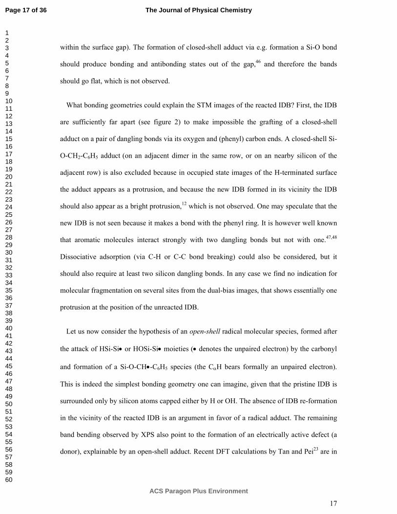
in figure 5 and 6, respectively, should be essentially representative of the bright/dark feature
observed by STM.
Figure 5. High resolution C 1s spectrum (hv = 350 eV) of lightly p-doped (NBoron = 7.5×10¹⁴
atoms/cm3, ρ = 18 Ω × cm) Si(001)-21 surface passivated with 4.5 L of water at room
temperature and then exposed to 67.5 L of benzaldehyde ( 7.5×10"8$%&&, 900().
The C 1s XPS spectrum (figure 5) exhibits two peaks that can be fitted with Voigt
components (full width at half-maximum, fwhm = 0.86 eV), one at binding energy BE ~
284.83 eV and the other at BE ~ 286.30 eV (93 % and 7 % of spectral weight, respectively).
The low binding energy (BE) peak at 284.83 eV is attributed to the aromatic carbons.49,50,51,19
The high BE peak at 286.30 eV is distant from the phenyl component by 1.47 eV, a value
much smaller than that separating the phenyl and carbonyl C 1s in the intact molecule (2.7
eV).52 Therefore the conservation of the carbonyl is excluded. As the XPS literature,53
indicates that (in polymers) the BE shift between CHx and COC is 1.44 eV, the component at
286.3 eV can be attributed to the α C in a Si-O-C linkage. A Si-O-C-O linkage is excluded, as
Page 20 of 36
ACS Paragon Plus Environment
The Journal of Physical Chemistry
123456789101112131415161718192021222324252627282930313233343536373839404142434445464748495051525354555657585960
21
(in polymers) the OCO moiety component is 2.9 eV higher in BE than the CHx one. Therefore
there is no indication of benzaldehyde oligomers (starting from the radical adduct) nor of
hemiacetal-like adducts, resulting from the capture of a nearby OH by the radical adduct, in
agreement with the calculations of Tan and Pei.23
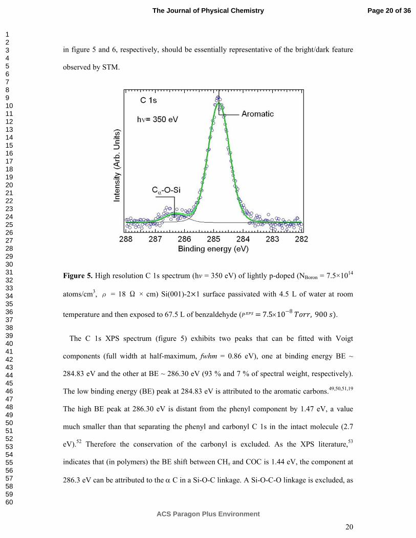
Figure 6. Bottom curve: C 1s edge NEXAFS spectrum (Auger Yield Mode) of benzaldehyde
on (H,OH)-Si(001)-2×1 (θ = 44°), see caption of figure 6. Middle curve: the inner-shell
electron energy loss spectrum (ISEELS) of gas-phase benzaldehyde (from Ref. 54). Top curve:
ISEELS spectrum of gas phase benzene (from Ref. 55).
In figure 6 (bottom curve) we show the C 1s NEXAFS spectrum (θ = 44°), corresponding to
the XPS spectrum of figure 5. For the sake of comparison, we show in the same figure the
inner-shell electron energy loss spectra (ISEELS) of gas phase benzaldehyde (middle curve)
and that of gas phase benzene (top curve). In gas phase benzaldehyde, the main absorption
Page 21 of 36
ACS Paragon Plus Environment
The Journal of Physical Chemistry
123456789101112131415161718192021222324252627282930313233343536373839404142434445464748495051525354555657585960
22
line is found at an excitation energy of 285.1 eV, with a shoulder at ~ 286 eV.54 Conjugation
between the π*ring and π*
C=O orbitals causes the splitting of the two degenerate π1*(e2u) levels
of benzene into levels 18a’ and 19a’. The component at 286 eV is therefore ascribed to a
transition from C 1s (C-R) (where C-R denotes the ring carbon atom next to the carbonyl) to
the 19 a’ level.54 The peak at ~ 287.5 eV corresponds to excitations from C1s (C=O) to
orbitals of primarily π*C=O character.
For benzaldehyde adsorbed on (H,OH)-Si(001)-2×1, XPS suggests the breaking of the C=O
double bond. As a consequence the π1* splitting due to conjugation in gas phase benzaldehyde
should be eliminated, and the C1s (C=O) →π*C=O transition should be quenched. This is
indeed confirmed by the NEXAFS spectrum where we see a single narrow component (fwhm
= 0.6) at hν = 285 eV corresponding to the C 1s (ring)→π*ring transition. Consequently the
NEXAFS spectrum of adsorbed benzaldehyde presents the same components as that of gas-
phase benzene55 (see figure 6). By varying θ from 10° (grazing light incidence) to 90°
(normal incidence), we have observed no dichroic effect in the absorption intensities (see
Supporting Information, §S4). Given the (two-domain) surface has a fourfold rotation
symmetry along the surface normal, the π-system may be either randomly oriented, or the C
2p component makes an angle of 54.7° (magic angle) with respect to the surface normal.31
Thus XPS and NEXAFS prove that the carbonyl bond opens and that Si-O-C units are
formed. A closed shell adduct Si-O-CH2-C6H5 can be formed by direct hydrosilylation20 of
the molecule. This adduct is depicted in figure 5b showing that the HOMO is localized on the
molecule. However in contrast to the SOMO of the radical , the HOMO is positioned below
the valence band maximum (the carbon π level are found at about 3.5 eV below the valence
band maximum56). Therefore in filled state images, electrons picked up by the tip from the
molecule should be easily replenished by electrons coming from silicon, in contrast to the
radical adduct case, whose localized level is in the surface gap. A steady-state charging of the
Page 22 of 36
ACS Paragon Plus Environment
The Journal of Physical Chemistry
123456789101112131415161718192021222324252627282930313233343536373839404142434445464748495051525354555657585960

23
molecule is not expected for the closed-shell adduct, and hence the molecule appears as a
protrusion.
Illustrative HREELS spectra (measured in specular geometry) of the (H,OH)-Si(001)-2×1
surface exposed to increasing doses of benzaldehyde are presented in figure 7. They are
obtained using an intrinsic silicon wafer (ρ =2000 Ω × cm) to optimize the resolution.
Complementary measurements were also carried out on the substrates used for STM and
XPS/NEXAFS experiments (p-doped, ρ = 18 Ω × cm), with qualitatively identical results
(see Supporting Information §S5).
Page 23 of 36
ACS Paragon Plus Environment
The Journal of Physical Chemistry
123456789101112131415161718192021222324252627282930313233343536373839404142434445464748495051525354555657585960

24
Figure 7. HREELS spectra of (H,OH)-Si(001)-2×1 exposed to increasing doses of
benzaldehyde. The assignment of the different loss intensities is indicated by the blue dotted
lines. We use an intrinsic substrate for better spectral resolution. The quasielastic peak shows
a width of 12 meV.
Theoretical vibration energies are calculated for the H-Si-Si-OH unit, for the
hydrosilylation product HOSi-Si-OCH2-C6H5. The vibration modes of the radical adduct
HOSi-Si-OCH•-C6H5, is also calculated to look for specific features that could permit its
identification at low coverage. No scaling of the theoretical energy values is made. In figure 8
we plot the spectral intensities (calculated with the dipolar moment perpendicular to the
surface) of the various modes.
We start with the experimental spectrum of the pristine (H,OH)-Si(001)-2×1 surface, we
interpret thanks to the DFT cluster calculation. The component at 258 meV is attributed to the
Si↔H stretching mode (calculated at 268 meV) and that found at 451 meV to the SiO↔H
stretching mode (calculated at 489 meV). According to the calculation (figure 9), the
experimental components at 101 meV corresponds in fact to three unresolved modes of the
hydroxyl, a SiH bending mode coupled with a Si↔OH stretching mode (calculated at 85
meV), a Si↔OH stretching mode (calculated at 89 meV) and a swinging mode SiOH
(calculated at 108 meV). The component at 203 meV is a double loss.57
HREELS (figure 7) gives also evidence for the adsorption of the benzaldehyde molecule on
the surface. With increasing doses, one observes the growth of an intense peak at 128 meV.
This loss is attributed to a stretching C↔O mode. Indeed the calculation points to one C↔O
(+bending SiOH) mode at 133 meV for the hydrosilylation (closed-shell) product. For the
radical adduct two C↔O modes are calculated at somewhat higher energy, 147 meV and 150
Page 24 of 36
ACS Paragon Plus Environment
The Journal of Physical Chemistry
123456789101112131415161718192021222324252627282930313233343536373839404142434445464748495051525354555657585960
25
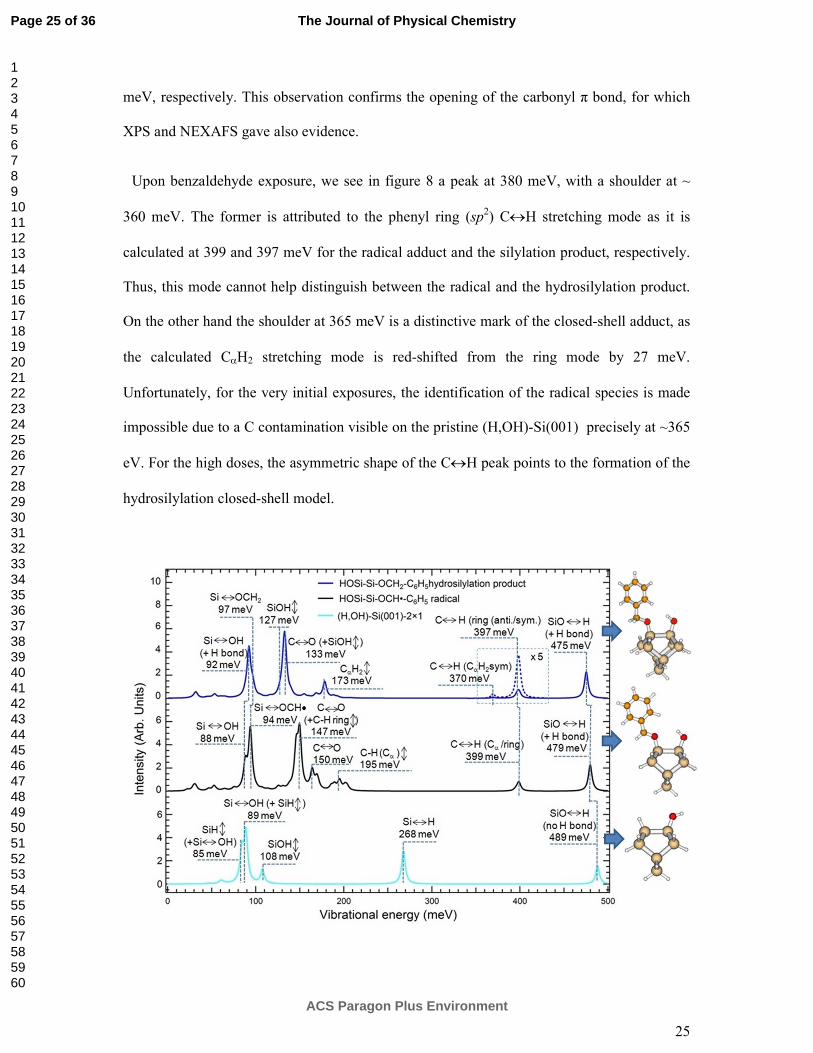
meV, respectively. This observation confirms the opening of the carbonyl π bond, for which
XPS and NEXAFS gave also evidence.
Upon benzaldehyde exposure, we see in figure 8 a peak at 380 meV, with a shoulder at ~
360 meV. The former is attributed to the phenyl ring (sp2) C↔H stretching mode as it is
calculated at 399 and 397 meV for the radical adduct and the silylation product, respectively.
Thus, this mode cannot help distinguish between the radical and the hydrosilylation product.
On the other hand the shoulder at 365 meV is a distinctive mark of the closed-shell adduct, as
the calculated CαH2 stretching mode is red-shifted from the ring mode by 27 meV.
Unfortunately, for the very initial exposures, the identification of the radical species is made
impossible due to a C contamination visible on the pristine (H,OH)-Si(001) precisely at ~365
eV. For the high doses, the asymmetric shape of the C↔H peak points to the formation of the
hydrosilylation closed-shell model.
Page 25 of 36
ACS Paragon Plus Environment
The Journal of Physical Chemistry
123456789101112131415161718192021222324252627282930313233343536373839404142434445464748495051525354555657585960

26
Figure 8. Calculated infrared spectra intensities (arbitrary units) of the H-Si-Si-OH unit
(bottom), of the radical adduct sitting next to a hydroxyl (middle), of the silylation product
sitting next to a hydroxyl (top). No scaling of the DFT energy values is made. The
corresponding ball-and-stick models are given. To ease comparison with the HREELS spectra
of figure 7 measured in specular geometry, the dipolar moment is placed perpendicularly to
the surface. A Lorentzian broadening of 5 meV is used to plot the intensities.
DFT cluster calculations provide also interesting qualitative information on the intensity and
energy change of the hydroxyl modes when a hydrogen bonding is established with the
hydrosilylation product. As it can be seen in the ball and stick models of figure 8, the proton
of the hydroxyl is attracted by the π system of the ring and the oxygen of the molecule.
Calculations show that this dative H-bonding causes a red shift of the SiO↔H stretching
mode from 489 meV (no H bond) to 475 meV (-14 meV). As a matter of fact, the HREELS
mode observed at 451 meV (figure 8) of the pristine surface strongly decreases in intensity
after prolonged dosing (670 L), while a new component shows up at ~ 434 eV, redshifted by -
17 eV, in excellent accord with the theoretical redshift value. In essence, HREELS indicates
that a H bond is established between the surface OH and the benzaldehyde molecule.
Now can we use the HREELS mode intensities to monitor the molecular attachment?
If we consider first the SiO↔H stretching mode, its intensity depends on the orientation of
the O-H bond axis with respect to the surface normal. For the HO-Si-Si-H unit the calculation
is made with a single-dimer cluster that may be not representative of the average O-H
orientation on the real surface, as interactions with nearby hydroxyls are not taken into
account. Therefore the observed decrease of the SiO↔H HREELS intensity does not
necessarily mean that OH moieties are consumed by the reaction. Neither is the decrease in
intensity of the 101 meV HREELS peak evidence of OH reacting with the aldehyde. Indeed,
Page 26 of 36
ACS Paragon Plus Environment
The Journal of Physical Chemistry
123456789101112131415161718192021222324252627282930313233343536373839404142434445464748495051525354555657585960

27
according to theory, the SiOH bending mode, at 108 meV in the absence of H-bonding,
shifts to 127 meV when the H bond is established, merging with the coupled C↔O (+SiOH)
mode calculated at 133 meV. Therefore the intensity is simply transferred to higher energy.
On the other hand, the intensity of the monohydride SiH neither presents this orientational
issue, nor is sensitive to the presence of the aldehyde. Therefore the plot of the Si↔H mode
intensity (at 258 meV) against the C↔H (ring) intensity (380 meV) can be a valuable
indicator of a hydrosylilation reaction involving the monohydride. The plot is given in figure
9. We see a clear correlation between the decrease of the Si↔H intensity and the increase of
the C↔H (ring) intensity. This gives a strong evidence that H from SiH units are consumed in
a hydrosilylation reaction.
Figure 9. Plot of Si↔H stretching mode intensity (258 meV) versus C↔H (ring) intensity
(380 meV), for the intrinsic substrate (blue filled circles). The black line is a linear fit of the
experimental data.
Page 27 of 36
ACS Paragon Plus Environment
The Journal of Physical Chemistry
123456789101112131415161718192021222324252627282930313233343536373839404142434445464748495051525354555657585960
28
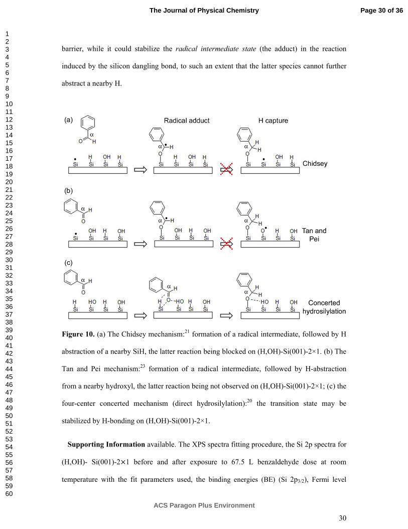
4. Conclusions
Although the water-covered Si (001) surface, (H,OH)-Si(001)-2×1, presents strong
analogies with the H-terminated Si(001)-2×1 surface due to the existence of monohydrides
and of isolated dangling bonds, their reactivity with a carbonyl-bearing molecule,
benzaldehyde, shows noticeable differences. As shown by STM, benzaldehyde reacts with the
quasi-neutral isolated silicon dangling bond of (H,OH)-Si(001)-2×1 formed on a lightly p-
doped sample. However, in contrast to the H-Si(001)-2×1 case where the Chidsey
mechanism21,12 is operative, we do not see the reformation of an isolated dangling bond via
the abstraction of a nearby H atom from a silicon monohydride (see figure 10a). Nor is the
Tan and Pei mechanism23 observed, where the H atom is abstracted from an OH neighbor.
The nature of the product formed by the reaction of the molecule with the isolated dangling
bond is discussed. We tentatively propose that a radical molecular adduct is formed. It is clear
that further studies are necessary to confirm this hypothesis, for instance the use of ultra-high
vacuum surface electron spin resonance.58
STM shows also the existence of a second type of molecular species located on the silicon
dimers, with dual-bias footprints markedly different from that of the preceding species.
Apparently its bonding needs not to be mediated by a silicon dangling bond. The chemical
nature of this second species, whose maximum areal density should be limited only by the
density of dimers (0.5 monolayer), is revealed by electron (XPS/NEXAFS) and vibrational
(HREELS) surface spectroscopies. Surface spectroscopies concur to show that the π-bond of
the carbonyl unit opens to form a Si-O-C unit. XPS indicates that the α carbon has only one
oxygen neighbor, excluding oligomerization or capture of surface hydroxyls. The attachment
via the benzene ring is also excluded. HREELS, in combination with vibrational mode
calculations, indicates that the energies of the C-H stretching and bending modes of the α
Page 28 of 36
ACS Paragon Plus Environment
The Journal of Physical Chemistry
123456789101112131415161718192021222324252627282930313233343536373839404142434445464748495051525354555657585960

29
carbon are well accounted for if we consider the hydrosilylation product. A strong piece of
evidence for a concerted hydrosilylation reaction is also provided by the correlation between
the decrease of the Si-H stretching intensity and the increase of the (ring) C-H stretching
intensities. Finally the evolution of the hydroxyl modes (energies and intensities) with
increasing benzaldehyde coverage are interpreted, through the DFT simulation, in terms of a
hydrogen bonding established between the reaction product and a nearby OH.
The absence of radical chain reaction and the observation of concerted hydrosilylation
reaction are the two main features opposing (H,OH)-Si(001)-2×1 and H-Si(001)-2×1.
Concerning the non-propagation of the radical reaction (figure 10 a, 10b), the chemical
disorder of (H,OH)-Si(001)-2×1 could be invoked. However figure 1c shows that relatively
long sections of aligned SiH are present on the surface, similarly to H-Si (001)-2×1. If we
take into consideration the possible formation of a radical adduct, the latter should be trapped
in a minimum of energy, with a high barrier to surmount to capture a nearby hydrogen, from
an OH or a SiH, as suggested by recent calculations.23 H bonding with a nearby OH could
provide an extra stabilization energy and increase the barrier.
On H-terminated surfaces, the concerted hydrosilylation reaction is generally considered as
less likely than the Chidsey radical reaction, due to kinetic barriers considerations (figure
10a).21 In their DFT calculation of the direct reaction of an alkene with an SiH, Coletti et al.20
considered a direct concerted pathway that passes through a four-center transition state (figure
10c). For ethylene, they calculated a very large barrier of ~ 2.6 eV. However benzaldehyde is
a much bigger molecule than ethylene (London interactions are expected to be more intense)
and, above all, it can make a hydrogen bond with an OH of the same dimer. Therefore we
propose that, when the reactivity of benzaldehyde with (H,OH)-Si(001)-2×1 is compared to
that with H-Si(001)-2×1, it is the hydrogen bond that makes the difference. Indeed H-bonding
could lower the transition state energy of the direct hydrosilylation process, and the activation
Page 29 of 36
ACS Paragon Plus Environment
The Journal of Physical Chemistry
123456789101112131415161718192021222324252627282930313233343536373839404142434445464748495051525354555657585960
30
barrier, while it could stabilize the radical intermediate state (the adduct) in the reaction
induced by the silicon dangling bond, to such an extent that the latter species cannot further
abstract a nearby H.
Figure 10. (a) The Chidsey mechanism:21 formation of a radical intermediate, followed by H
abstraction of a nearby SiH, the latter reaction being blocked on (H,OH)-Si(001)-2×1. (b) The
Tan and Pei mechanism:23 formation of a radical intermediate, followed by H-abstraction
from a nearby hydroxyl, the latter reaction being not observed on (H,OH)-Si(001)-2×1; (c) the
four-center concerted mechanism (direct hydrosilylation):20 the transition state may be
stabilized by H-bonding on (H,OH)-Si(001)-2×1.
Supporting Information available. The XPS spectra fitting procedure, the Si 2p spectra for
(H,OH)- Si(001)-21 before and after exposure to 67.5 L benzaldehyde dose at room
temperature with the fit parameters used, the binding energies (BE) (Si 2p3/2), Fermi level
Page 30 of 36
ACS Paragon Plus Environment
The Journal of Physical Chemistry
123456789101112131415161718192021222324252627282930313233343536373839404142434445464748495051525354555657585960

31
position at the surface +, " +-./0
, surface band bending qVbb, surface charge density, the
determination of the molecular coverage from O 1s XPS normalized intensities, the angular
dependent C 1s NEXAFS spectra and the HREELS spectra of the lightly doped surface. This
information is available free of charge via the Internet at http://pubs.acs.org.
TOC
Page 31 of 36
ACS Paragon Plus Environment
The Journal of Physical Chemistry
123456789101112131415161718192021222324252627282930313233343536373839404142434445464748495051525354555657585960
32
References
(1) Poncey, C.; Rochet, F.; Dufour, G.; Roulet, H.; Sirotti, F.; Panaccione, G. Adsorption of Water on Si(001)-2x1 and Si(111)-7x7 Surfaces at 90 and 300 K: a Si2p Core-Level and Valence Band Study with Synchrotron Radiation. Surf. Sci. 1995, 338, 143–156. DOI: 10.1016/0039-6028(95)00501-3
(2) Andersohn, L.; Köhler, U. In Situ Observation of Water Adsorption on Si(100) with Scanning Tunneling Microscopy. Surf. Sci. 1993, 284, 77–90. DOI: 10.1016/0039-6028(93)90526-P
(3) Henderson, M. The Interaction of Water with Solid Surfaces: Fundamental Aspects Revisited. Surf. Sci. Rep. 2002, 46, 1–308. DOI: 10.1016/S0167-5729(01)00020-6s
(4) Carniato, S.; Gallet, J.-J.; Rochet, F.; Dufour, G.; Bournel, F.; Rangan, S.; Verdini, A.; Floreano, L. Characterization of Hydroxyl Groups on Water-Reacted Si(001)-2×1 Using Synchrotron Radiation O 1s Core-Level Spectroscopies and Core-Excited State Density-Functional Calculations. Phys. Rev. B 2007, 76, 085321. DOI: 10.1103/PhysRevB.76.085321
(5) Giustino, F.; Pasquarello, A. First-Principles Theory of Infrared Absorption Spectra at Surfaces and Interfaces: Application to the Si(100):H2O Surface. Phys. Rev. B 2008, 78, 075307. DOI: 10.1103/PhysRevB.78.075307
(6) Skliar, D. B.; Willis, B. G. The Role of Dangling Bonds in H2O-Induced Oxidation of Si(100)-2 × 1. J. Phys. Chem. C 2008, 112, 9434–9442. DOI: 10.1021/jp8010519
(7) Gallet, J.-J.; Bournel, F.; Rochet, F.; Kohler, U.; Kubsky, S.; Silly, M. G.; Sirotti, F.; Pierucci, D. Isolated Silicon Dangling Bonds on a Water-Saturated n+ -Doped Si(001)-2 × 1 Surface: An XPS and STM Study. J. Phys. Chem. C 2011, 115, 7686–7693. DOI: 10.1021/jp201262x
(8) Morrow, B. A.; McFarlan, A. J. Chemical Reactions at Silica Surfaces. J. Non-Cryst.
Solids 1990, 120, 61–71. DOI: 10.1016/0022-3093(90)90191-N
(9) Blitz, J. P.; Christensen, J. M.; Deakyne, C. A.; Gunko, V. M. Silica Surface Modification Reactions with Aluminum and Boron Alkyls and (Alkyl) Chlorides: Reactivities and Surface Nanostructures. J. Nanosci. Nanotechnol. 2008, 8, 660–666. DOI: 10.1166/jnn.2008.Cl99
(10) Lowen, W. K.; Broge, E. C. Effects of Dehydration and Chemisorbed Materials on the Surface Properties of Amorphous Silica. J. Phys. Chem. 1961, 65, 16–19. DOI: 10.1021/j100819a006
(11) Lopinski, G.; Wayner, D.; Wolkow, R. Self-Directed Growth of Molecular Nanostructures on Silicon. Nature 2000, 406, 48–51. DOI: 10.1038/35017519
Page 32 of 36
ACS Paragon Plus Environment
The Journal of Physical Chemistry
123456789101112131415161718192021222324252627282930313233343536373839404142434445464748495051525354555657585960

33
(12) Pitters, J. L.; Dogel, I.; DiLabio, G. A.; Wolkow, R. A. Linear Nanostructure Formation of Aldehydes by Self-Directed Growth on Hydrogen-Terminated Silicon(100). J. Phys. Chem. B 2006, 110, 2159–2163. DOI: 10.1021/jp055153t
(13) Buriak, J. M. Organometallic Chemistry on Silicon and Germanium Surfaces. Chem.
Rev. 2002, 102, 1271–1308. DOI: 10.1021/cr000064s
(14) Liu, L.; Yu, J.; Lyding, J. W. Scanning Tunneling Microscopy Observation of Single Dangling Bonds On The Si(100)2x1:H Surface. Mater. Res. Soc. Symp. Proc. 2002, 705, 187–192. DOI: http://dx.doi.org/10.1557/PROC-705-Y6.6
(15) Willis, B. G.; Mathew, A.; Wielunski, L. S.; Opila, R. L. Adsorption and Reaction of HfCl4 with H2O-Terminated Si(100)-2 × 1. J. Phys. Chem. C 2008, 112, 1994–2003. DOI: 10.1021/jp0758317
(16) Lee, S. S.; Baik, J. Y.; An, K.-S.; Suh, Y. D.; Oh, J.-H.; Kim, Y. Reduction of Incubation Period by Employing OH-Terminated Si(001) Substrates in the Atomic Layer Deposition of Al2O3. J. Phys. Chem. B 2004, 108, 15128–15132. DOI: 10.1021/jp048038b
(17) Kim, D.-H.; Baek, S.-B.; Kim, Y.-C. Energy Barriers for Trimethylaluminum Reaction with Varying Surface Hydroxyl Density. Appl. Surf. Sci. 2011, 258, 225–229. DOI: 10.1016/j.apsusc.2011.08.035
(18) Ihm, K.; Kang, T.-H.; Han, J. H.; Moon, S.; Hwang, C. C.; Kim, K.-J.; Hwang, H.-N.; Jeon, C.-H.; Kim, H.-D.; Kim, B.; et al. Hydroxyl Group-Induced Adsorptions of 4-Nitro Benzoic Acid on the Si(100) Surface. J. Electron Spectros. Relat. Phenomena 2005, 144-147, 397–400. DOI: 10.1016/j.elspec.2005.01.108
(19) Bournel, F.; Gallet, J.-J.; Pierucci, D.; Khaliq, A.; Rochet, F.; Pietzsch, A. Hydrosilylation of Styrene on Water-Saturated Si(001)-2×1 at Room Temperature. J.
Phys. Chem. C 2011, 115, 14827–14833. DOI: 10.1021/jp202913y
(20) Coletti, C.; Marrone, A.; Giorgi, G.; Sgamellotti, A.; Cerofolini, G.; Re, N. Nonradical Mechanisms for the Uncatalyzed Thermal Functionalization of Silicon Surfaces by Alkenes and Alkynes: a Density Functional Study. Langmuir 2006, 22, 9949–9956. DOI: 10.1021/la060013b
(21) Cicero, R. L.; Chidsey, C. E. D.; Lopinski, G. P.; Wayner, D. D. M.; Wolkow, R. A. Olefin Additions on H−Si(111): Evidence for a Surface Chain Reaction Initiated at Isolated Dangling Bonds. Langmuir 2002, 18, 305–307. DOI: 10.1021/la010823h
(22) Kanai, Y.; Takeuchi, N.; Car, R.; Selloni, A. Role of Molecular Conjugation in the Surface Radical Reaction of Aldehydes with H-Si(111): First Principles Study. J. Phys.
Chem. B 2005, 109, 18889–18894. DOI: 10.1021/jp0527610
(23) Tan, Y.; Pei, Y. Radical Chain-Reaction of Terminal-Unsaturated Organic Molecules on Water-Saturated Si(100)-(2×1): The Role of Surface Hydroxyl Groups. J. Phys.
Chem. C 2013, 117, 14032-14042. DOI: 10.1021/jp403101v
Page 33 of 36
ACS Paragon Plus Environment
The Journal of Physical Chemistry
123456789101112131415161718192021222324252627282930313233343536373839404142434445464748495051525354555657585960

34
(24) Chatgilialoglu, C.; Ingold, K. U.; Scaiano, J. C. Absolute Rate Constants for the Addition of Triethylsilyl Radicals to the Carbonyl Group. J. Am. Chem. Soc. 1982, 104, 5119–5123. DOI: 10.1021/ja00383a021
(25) Busca, G.; Lamotte, J.; Lavalley, J. C.; Lorenzelli, V. FT-IR Study of the Adsorption and Transformation of Formaldehyde on Oxide Surfaces. J. Am. Chem. Soc. 1987, 109, 5197–5202. DOI: 10.1021/ja00251a025
(26) Hill, W.; Miessner, H.; Öhlmann, G. Fourier Transform Infrared Study of the Adsorption and of Reactions of Acetaldehyde on Dispersed Silica. J. Chem. Soc.
Faraday Trans. 1 1989, 85, 691-697. DOI: 10.1039/f19898500691
(27) Ibach, H.; Mills, D. Electron Energy Loss Spectroscopy; 2nd ed.; 1982 Academic: NewYork.
(28) Naitabdi, A.; Bournel, F.; Gallet, J.-J.; Markovits, A.; Rochet, F.; Borensztein, Y.; Silly, M. G.; Sirotti, F. Triethylamine on Si(001)-(2 × 1) at 300 K: Molecular Adsorption and Site Configurations Leading to Dissociation. J. Phys. Chem. C 2012, 116, 16473–16486. DOI: 10.1021/jp303002c
(29) Polack, F.; Silly, M.; Chauvet, C.; Lagarde, B.; Bergeard, N.; Izquierdo, M.; Chubar, O.; Krizmancic, D.; Ribbens, M.; Duval, J.-P.; et al. TEMPO: a New Insertion Device Beamline at SOLEIL for Time Resolved Photoelectron Spectroscopy Experiments on Solids and Interfaces. AIP Conference Proceedings 2010, 185, 185–188. DOI: 10.1063/1.3463169
(30) Bergeard, N.; Silly, M. G.; Krizmancic, D.; Chauvet, C.; Guzzo, M.; Ricaud, J. P.; Izquierdo, M.; Stebel, L.; Pittana, P.; Sergo, R.; et al. Time-Resolved Photoelectron Spectroscopy Using Synchrotron Radiation Time Structure. J. Synchrotron Radiat. 2011, 18, 245–250. DOI: 10.1107/S0909049510052301
(31) Stöhr, J. NEXAFS Spectroscopy; Springer: NewYork, 1992.
(32) Chauvet, C.; Polack, F.; Silly, M. G.; Lagarde, B.; Thomasset, M.; Kubsky, S.; Duval, J. P.; Risterucci, P.; Pilette, B.; Yao, I.; et al. Carbon Contamination of Soft X-Ray Beamlines: Dramatic Anti-Reflection Coating Effects Observed in the 1 keV Photon Energy Region. J. Synchrotron Radiat. 2011, 18, 761–764. DOI: 10.1107/S0909049511023119
(33) Tautz, F. S.; Schaefer, J. a. Ultimate Resolution Electron Energy Loss Spectroscopy at H/Si(100) Surfaces. J. Appl. Phys. 1998, 84, 6636. DOI: 10.1063/1.369038
(34) Polyakov, V.; Elbe, a; Wu, J.; Lapeyre, G.; Schaefer, J. Silicon Spreading in Delta -Doped GaAs(100): A High-Resolution Electron-Energy-Loss-Spectroscopy Study. Phys. Rev. B. 1996, 54, 2010–2018. DOI: 10.1103/PhysRevB.54.2010
(35) Gordon Group/GAMESS Homepage http://www.msg.chem.iastate.edu/gamess/index.html.
Page 34 of 36
ACS Paragon Plus Environment
The Journal of Physical Chemistry
123456789101112131415161718192021222324252627282930313233343536373839404142434445464748495051525354555657585960

35
(36) Becke, A. D. Density-Functional Thermochemistry. III. The Role of Exact Exchange. J. Chem. Phys. 1993, 98, 5648-5652. DOI: 10.1063/1.464913
(37) Lee, C.; Yang, W.; Parr, R. G. Development of the Colle-Salvetti Correlation-Energy Formula into a Functional of the Electron Density. Phys. Rev. B 1988, 37, 785–789. DOI: 10.1103/PhysRevB.37.785
(38) Piva, P. G.; DiLabio, G. a; Pitters, J. L.; Zikovsky, J.; Rezeq, M.; Dogel, S.; Hofer, W. a; Wolkow, R. a. Field Regulation of Single-Molecule Conductivity by a Charged Surface Atom. Nature 2005, 435, 658–661. DOI: 10.1038/nature03563
(39) Haider, M. B.; Pitters, J. L.; Dilabio, G. A.; Livadaru, L.; Mutus, J. Y.; Wolkow, R. A. Controlled Coupling and Occupation of Silicon Atomic Quantum Dots at Room Temperature. Phys. Rev. Lett. 2009, 102, 046805 . DOI: 10.1103/PhysRevLett.102.046805
(40) Feenstra, R. M.; Dong, Y.; Semtsiv, M.P.; Masselink, W. T. Influence of Tip-Induced Band Bending on Tunnelling Spectra of Semiconductor Surfaces. Nanotechnology 2007, 18, 044015. DOI: 10.1088/0957-4484/18/4/044015
(41) McEllistrem, M.; Haase, G.; Chen, D.; Hamers, R. Electrostatic Sample-Tip Interactions in the Scanning Tunneling Microscope. Phys. Rev. Lett. 1993, 70, 2471–2474. DOI: 10.1103/PhysRevLett.70.2471
(42) Hirayama, M.; Nakamura, J.; Natori, A. Band-Bending Effects on Scanning Tunneling Microscope Images of Subsurface Dopants: First-Principles Calculations. J. Appl.
Phys. 2009, 105, 083720. DOI: 10.1063/1.3110070
(43) Livadaru, L.; Pitters, J.; Taucer, M.; Wolkow, R. A. Theory of Nonequilibrium Single-Electron Dynamics in STM Imaging of Dangling Bonds on a Hydrogenated Silicon Surface. Phys. Rev. B 2011, 84, 205416. DOI: 10.1103/PhysRevB.84.205416
(44) Pitters, J. L.; Piva, P. G.; Wolkow, R. A. Dopant Depletion in the Near Surface Region of Thermally Prepared Silicon (100) in UHV. J. Vac. Sci. Technol. B 2012, 30, 021806. DOI: 10.1116/1.3694010
(45) Schofield, S. R.; Studer, P.; Hirjibehedin, C. F.; Curson, N. J.; Aeppli, G.; Bowler, D. R. Quantum Engineering at the Silicon Surface Using Dangling Bonds. Nat. Commun. 2013, 4, 1649. DOI: 10.1038/ncomms2679
(46) Nishida, M. Origin of Gap States at Initial Stage Oxidation on Si(001)2×1:H and Water Adsorption on Si(001)2×1: A Theoretical Study. Appl. Phys. Lett. 2002, 81, 1827-1829. DOI: 10.1063/1.1505123
(47) Miwa, J. A.; Eves, B. J.; Rosei, F.; Lopinski, G. P. Selective Adsorption of Pyridine at Isolated Reactive Sites on Si(100). J. Phys. Chem. B 2005, 109, 20055–20059. DOI: 10.1021/jp0537658
(48) Ample, F.; Joachim, C. The Chemisorption of Polyaromatic Hydrocarbons on Si(100)H Dangling Bonds. Surf. Sci. 2008, 602, 1563–1571. DOI: 10.1016/j.susc.2008.02.020
Page 35 of 36
ACS Paragon Plus Environment
The Journal of Physical Chemistry
123456789101112131415161718192021222324252627282930313233343536373839404142434445464748495051525354555657585960

36
(49) Cao, X.; Coulter, S. K.; Ellison, M. D.; Liu, H.; Liu, J.; Hamers, R. J. Bonding of Nitrogen-Containing Organic Molecules to the Silicon(001) Surface: The Role of Aromaticity. J. Phys. Chem. B 2001, 105, 3759–3768. DOI: 10.1021/jp003329f
(50) Coulter, S. K.; Schwartz, M. P.; Hamers, R. J. Sulfur Atoms as Tethers for Selective Attachment of Aromatic Molecules to Silicon(001) Surfaces. J. Phys. Chem. B 2001, 105, 3079–3087. DOI: 10.1021/jp004016n
(51) Beamson, G.; Clark, D. T.; Kendrick, J.; Briggs, D. Observation of Vibrational Asymmetry in the High Resolution Monochromatized XPS of Hydrocarbon Polymers. J. Electron Spectros. Relat. Phenomena 1991, 57, 79–90. DOI: 10.1016/0368-2048(91)85015-L
(52) Huang, H. G.; Zhang, Y. P.; Cai, Y. H.; Huang, J. Y.; Yong, K. S.; Xu, G. Q. Selective Attachment of Benzaldehyde on Si(100)-2 x 1: Structure, Selectivity, and Mechanism. J. Chem. Phys. 2005, 123, 104702. DOI: 10.1063/1.2035095
(53) Briggs, D.; Beamson, G. Primary and Secondary Oxygen-Induced C1s Binding Energy Shifts in x-Ray Photoelectron Spectroscopy of Polymers. Anal. Chem. 1992, 64, 1729–1736. DOI: 10.1021/ac00039a018
(54) Hitchcock, A. Inner-Shell Spectroscopy of Benzaldehyde, Terephthalaldehyde, Ethylbenzoate, Terephthaloyl Chloride and Phosgene: Models for Core Excitation of Poly (ethylene. J. Phys. Chem. 1992, 96, 8736–8750. DOI: 10.1021/j100201a015
(55) Horsley, J. a.; Stohr, J.; Hitchcock, a. P.; Newbury, D. C.; Johnson, a. L.; Sette, F. Resonances in the K Shell Excitation Spectra of Benzene and Pyridine: Gas Phase, Solid, and Chemisorbed States. J. Chem. Phys. 1985, 83, 6099-6107. DOI: 10.1063/1.449601
(56) Rochet, F.; Bournel, F.; Gallet, J.-J.; Dufour, G.; Lozzi, L.; Sirotti, F. Electronic Structure of 1,3,5,7-Cyclooctatetraene Chemisorbed on Si(001)-2×1 at 300 K Studied by PES, NEXAFS, and Resonant Valence Band Spectroscopy. J. Phys. Chem. B 2002, 106, 4967–4973. DOI: 10.1021/jp013780m
(57) Kato, H. S.; Kawai, M.; Akagi, K.; Tsuneyuki, S. Interaction of Condensed Water Molecules with Hydroxyl and Hydrogen Groups on Si(001). Surf. Sci. 2005, 587, 34–40. DOI: 10.1016/j.susc.2005.04.032
(58) Futako, W.; Nishizawa, M.; Yasuda, T.; Isoya, J.; Yamasaki, S. In Situ Electron Spin Resonance Observation of Si(111) 7×7 Surface During Hydrogenation Process. J. Vac.
Sci. Technol. B 2001, 19, 1898-1900. DOI: 10.1116/1.1403439
Page 36 of 36
ACS Paragon Plus Environment
The Journal of Physical Chemistry
123456789101112131415161718192021222324252627282930313233343536373839404142434445464748495051525354555657585960
Copyright © 2022 FDOKUMEN


![Excited-State Diproton Transfer in [2, 2′-Bipyridyl]-3, 3′-diol: the Mechanism Is Sequential, Not Concerted](https://static.fdokumen.com/doc/165x107/6332daff5f7e75f94e094ac2/excited-state-diproton-transfer-in-2-2-bipyridyl-3-3-diol-the-mechanism.jpg)