Behavioral correlates of the distributed coding of spatial context
13
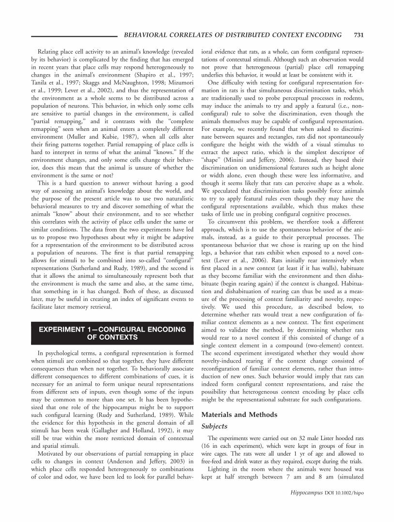
Behavioral Correlates of the Distributed Coding of Spatial Context Michael I. Anderson, Sarah Killing, Caitlin Morris, Alan O’Donoghue, Dikennam Onyiagha, Rosemary Stevenson, Madeleine Verriotis, and Kathryn J. Jeffery* ABSTRACT: Hippocampal place cells respond heterogeneously to ele- mental changes of a compound spatial context, suggesting that they form a distributed code of context, whereby context information is shared across a population of neurons. The question arises as to what this distributed code might be useful for. The present study explored two possibilities: one, that it allows contexts with common elements to be disambiguated, and the other, that it allows a given context to be associated with more than one outcome. We used two naturalistic measures of context processing in rats, rearing and thigmotaxis (bound- ary-hugging), to explore how rats responded to contextual novelty and to relate this to the behavior of place cells. In experiment 1, rats showed dishabituation of rearing to a novel reconfiguration of familiar context elements, suggesting that they perceived the reconfiguration as novel, a behavior that parallels that of place cells in a similar situation. In experiment 2, rats were trained in a place preference task on an open-field arena. A change in the arena context triggered renewed thig- motaxis, and yet navigation continued unimpaired, indicating simultane- ous representation of both the altered contextual and constant spatial cues. Place cells similarly exhibited a dual population of responses, con- sistent with the hypothesis that their activity underlies spatial behavior. Together, these experiments suggest that heterogeneous context encod- ing (or ‘‘partial remapping’’) by place cells may function to allow the flexible assignment of associations to contexts, a faculty that could be useful in episodic memory encoding. V V C 2006 Wiley-Liss, Inc. KEY WORDS: hippocampus; place cells; context; episodic memory; behavioral analysis; navigation; remapping; cognitive map INTRODUCTION Episodic, or autobiographical, memory is one of the great mysteries of cognition. The ability to recollect events from their own past endows humans with an enormously useful capacity to reflect on experience, learn from it and transmit it, via language, to others of our species. Understanding episodic memory will thus shed light on an important aspect of human experience, as well as opening many therapeutic doors to the treatment of amnesic disorders such as Alzheimer’s disease. To this end, neural studies of the analogue of episodic memory that is postu- lated to exist in other mammals (Morris, 2001) and birds (Clayton and Dickinson, 1998) potentially provide important infor- mation regarding the nature of episodic encoding. All events occur in a place (in fact we even use the term ‘‘take place’’ to describe events), and the place of occurrence seems to be an important part of an epi- sodic memory. For example, returning to a place often brings back a flood of related memories of events that happened there previously, and voluntary episodic recall in humans frequently involves first bringing to mind the location of the episode, and then recalling the sequence of events that occurred there. Thus, it seems that where something happened is very much interwoven with what happened, and recall involves retrieving both (Tulving, 1983). It has long been known that the hippocampus has an important role in the subtype of human memory that we now, following Tulving’s seminal proposal, call ‘‘episodic’’ (Scoville and Milner, 1957; Burgess et al., 2002). The finding that it also plays a particularly im- portant role in spatial representation (O’Keefe and Nadel, 1978) raises the possibility that the ‘‘where’’ part of an episodic memory is encoded by the spa- tially specific hippocampal neurons known as place cells. The purpose of the present article is to focus on the functional role of the hippocampal place cells in representing the background against which events occur. How does place cell activity relate to what the animal ‘‘knows’’ about the world, and how might it enhance the efficacy of episodic memory encoding? The background cues in an animal’s environment are often referred to as the ‘‘context,’’ and we have previously followed Mizumori et al. in using the term ‘‘spatial context’’ to describe the combination of spatial and contextual cues that make up a given environ- ment (Mizumori et al., 1999; Jeffery et al., 2004). By our view, spatial context has two separable compo- nents: geometric (comprising information about the metric features of the environment, such as its shape and orientation) and contextual (nonmetric informa- tion such as color and odor). Together, these cues are able to tell the animal both which environment it is in, and where it is within it. Since place cells respond to both metric (for review see Barry et al., 2006) and contextual (Jeffery et al., 2004) information, and hip- pocampal lesions impair processing of spatial context, a reasonable hypothesis is that place cells are the site of this which/where spatial-contextual ‘‘knowledge’’ that the animal has about the environment. The pres- ent study aimed to explore this hypothesis. This article is dedicated to the memory of Dikennam Onyiagha. Institute of Behavioural Neuroscience, Department of Psychology, Uni- versity College London, London, United Kingdom Grant sponsor: Biotechnology and Biological Sciences Research Coun- cil(BBSRC); Grant number: BBS/B1566X. *Correspondence to: Dr K.J. Jeffery, Institute of Behavioural Neuroscience, Department of Psychology, University College London, 26 Bedford Way, London WC1H OAP, United Kingdom. E-mail: [email protected] S.K., C.M., A.O., D.O., R.S., and M.V. contributed equally to this work. Accepted for publication 10 June 2006 DOI 10.1002/hipo.20206 Published online 18 August 2006 in Wiley InterScience (www.interscience. wiley.com). HIPPOCAMPUS 16:730–742 (2006) V V C 2006 WILEY-LISS, INC.
Transcript of Behavioral correlates of the distributed coding of spatial context
Behavioral Correlates of the Distributed Coding of Spatial Context
Michael I. Anderson, Sarah Killing, Caitlin Morris, Alan O’Donoghue, Dikennam Onyiagha,Rosemary Stevenson, Madeleine Verriotis, and Kathryn J. Jeffery*
ABSTRACT: Hippocampal place cells respond heterogeneously to ele-mental changes of a compound spatial context, suggesting that theyform a distributed code of context, whereby context information isshared across a population of neurons. The question arises as to whatthis distributed code might be useful for. The present study exploredtwo possibilities: one, that it allows contexts with common elements tobe disambiguated, and the other, that it allows a given context to beassociated with more than one outcome. We used two naturalisticmeasures of context processing in rats, rearing and thigmotaxis (bound-ary-hugging), to explore how rats responded to contextual novelty andto relate this to the behavior of place cells. In experiment 1, ratsshowed dishabituation of rearing to a novel reconfiguration of familiarcontext elements, suggesting that they perceived the reconfiguration asnovel, a behavior that parallels that of place cells in a similar situation.In experiment 2, rats were trained in a place preference task on anopen-field arena. A change in the arena context triggered renewed thig-motaxis, and yet navigation continued unimpaired, indicating simultane-ous representation of both the altered contextual and constant spatialcues. Place cells similarly exhibited a dual population of responses, con-sistent with the hypothesis that their activity underlies spatial behavior.Together, these experiments suggest that heterogeneous context encod-ing (or ‘‘partial remapping’’) by place cells may function to allow theflexible assignment of associations to contexts, a faculty that could beuseful in episodic memory encoding. VVC 2006 Wiley-Liss, Inc.
KEY WORDS: hippocampus; place cells; context; episodic memory;behavioral analysis; navigation; remapping; cognitive map
INTRODUCTION
Episodic, or autobiographical, memory is one of the great mysteriesof cognition. The ability to recollect events from their own past endowshumans with an enormously useful capacity to reflect on experience,learn from it and transmit it, via language, to others of our species.Understanding episodic memory will thus shed light on an importantaspect of human experience, as well as opening many therapeutic doorsto the treatment of amnesic disorders such as Alzheimer’s disease. To thisend, neural studies of the analogue of episodic memory that is postu-lated to exist in other mammals (Morris, 2001) and birds (Clayton and
Dickinson, 1998) potentially provide important infor-mation regarding the nature of episodic encoding.
All events occur in a place (in fact we even use theterm ‘‘take place’’ to describe events), and the place ofoccurrence seems to be an important part of an epi-sodic memory. For example, returning to a place oftenbrings back a flood of related memories of events thathappened there previously, and voluntary episodicrecall in humans frequently involves first bringing tomind the location of the episode, and then recallingthe sequence of events that occurred there. Thus, itseems that where something happened is very muchinterwoven with what happened, and recall involvesretrieving both (Tulving, 1983).
It has long been known that the hippocampus hasan important role in the subtype of human memorythat we now, following Tulving’s seminal proposal, call‘‘episodic’’ (Scoville and Milner, 1957; Burgess et al.,2002). The finding that it also plays a particularly im-portant role in spatial representation (O’Keefe andNadel, 1978) raises the possibility that the ‘‘where’’part of an episodic memory is encoded by the spa-tially specific hippocampal neurons known as placecells. The purpose of the present article is to focus onthe functional role of the hippocampal place cells inrepresenting the background against which eventsoccur. How does place cell activity relate to what theanimal ‘‘knows’’ about the world, and how might itenhance the efficacy of episodic memory encoding?
The background cues in an animal’s environmentare often referred to as the ‘‘context,’’ and we havepreviously followed Mizumori et al. in using the term‘‘spatial context’’ to describe the combination of spatialand contextual cues that make up a given environ-ment (Mizumori et al., 1999; Jeffery et al., 2004). Byour view, spatial context has two separable compo-nents: geometric (comprising information about themetric features of the environment, such as its shapeand orientation) and contextual (nonmetric informa-tion such as color and odor). Together, these cues areable to tell the animal both which environment it isin, and where it is within it. Since place cells respondto both metric (for review see Barry et al., 2006) andcontextual (Jeffery et al., 2004) information, and hip-pocampal lesions impair processing of spatial context,a reasonable hypothesis is that place cells are the siteof this which/where spatial-contextual ‘‘knowledge’’that the animal has about the environment. The pres-ent study aimed to explore this hypothesis.
This article is dedicated to the memory of Dikennam Onyiagha.
Institute of Behavioural Neuroscience, Department of Psychology, Uni-versity College London, London, United Kingdom
Grant sponsor: Biotechnology and Biological Sciences Research Coun-cil(BBSRC); Grant number: BBS/B1566X.*Correspondence to: Dr K.J. Jeffery, Institute of Behavioural Neuroscience,Department of Psychology, University College London, 26 Bedford Way,LondonWC1H OAP, United Kingdom. E-mail: [email protected]., C.M., A.O., D.O., R.S., and M.V. contributed equally to this work.Accepted for publication 10 June 2006DOI 10.1002/hipo.20206Published online 18 August 2006 in Wiley InterScience (www.interscience.wiley.com).
HIPPOCAMPUS 16:730–742 (2006)
VVC 2006 WILEY-LISS, INC.
Relating place cell activity to an animal’s knowledge (revealedby its behavior) is complicated by the finding that has emergedin recent years that place cells may respond heterogeneously tochanges in the animal’s environment (Shapiro et al., 1997;Tanila et al., 1997; Skaggs and McNaughton, 1998; Mizumoriet al., 1999; Lever et al., 2002), and thus the representation ofthe environment as a whole seems to be distributed across apopulation of neurons. This behavior, in which only some cellsare sensitive to partial changes in the environment, is called‘‘partial remapping,’’ and it contrasts with the ‘‘completeremapping’’ seen when an animal enters a completely differentenvironment (Muller and Kubie, 1987), when all cells altertheir firing patterns together. Partial remapping of place cells ishard to interpret in terms of what the animal ‘‘knows.’’ If theenvironment changes, and only some cells change their behav-ior, does this mean that the animal is unsure of whether theenvironment is the same or not?
This is a hard question to answer without having a goodway of assessing an animal’s knowledge about the world, andthe purpose of the present article was to use two naturalisticbehavioral measures to try and discover something of what theanimals ‘‘know’’ about their environment, and to see whetherthis correlates with the activity of place cells under the same orsimilar conditions. The data from the two experiments have ledus to propose two hypotheses about why it might be adaptivefor a representation of the environment to be distributed acrossa population of neurons. The first is that partial remappingallows for stimuli to be combined into so-called ‘‘configural’’representations (Sutherland and Rudy, 1989), and the second isthat it allows the animal to simultaneously represent both thatthe environment is much the same and also, at the same time,that something in it has changed. Both of these, as discussedlater, may be useful in creating an index of significant events tofacilitate later memory retrieval.
EXPERIMENT 1—CONFIGURAL ENCODINGOF CONTEXTS
In psychological terms, a configural representation is formedwhen stimuli are combined so that together, they have differentconsequences than when not together. To behaviorally associatedifferent consequences to different combinations of cues, it isnecessary for an animal to form unique neural representationsfrom different sets of inputs, even though some of the inputsmay be common to more than one set. It has been hypothe-sized that one role of the hippocampus might be to supportsuch configural learning (Rudy and Sutherland, 1989). Whilethe evidence for this hypothesis in the general domain of allstimuli has been weak (Gallagher and Holland, 1992), it maystill be true within the more restricted domain of contextualand spatial stimuli.
Motivated by our observations of partial remapping in placecells to changes in context (Anderson and Jeffery, 2003) inwhich place cells responded heterogeneously to combinationsof color and odor, we have been led to look for parallel behav-
ioral evidence that rats, as a whole, can form configural represen-tations of contextual stimuli. Although such an observation wouldnot prove that heterogeneous (partial) place cell remappingunderlies this behavior, it would at least be consistent with it.
One difficulty with testing for configural representation for-mation in rats is that simultaneous discrimination tasks, whichare traditionally used to probe perceptual processes in rodents,may induce the animals to try and apply a featural (i.e., non-configural) rule to solve the discrimination, even though theanimals themselves may be capable of configural representation.For example, we recently found that when asked to discrimi-nate between squares and rectangles, rats did not spontaneouslyconfigure the height with the width of a visual stimulus toextract the aspect ratio, which is the simplest descriptor of‘‘shape’’ (Minini and Jeffery, 2006). Instead, they based theirdiscrimination on unidimensional features such as height aloneor width alone, even though these were less informative, andthough it seems likely that rats can perceive shape as a whole.We speculated that discrimination tasks possibly force animalsto try to apply featural rules even though they may have theconfigural representations available, which thus makes thesetasks of little use in probing configural cognitive processes.
To circumvent this problem, we therefore took a differentapproach, which is to use the spontaneous behavior of the ani-mals, instead, as a guide to their perceptual processes. Thespontaneous behavior that we chose is rearing up on the hindlegs, a behavior that rats exhibit when exposed to a novel con-text (Lever et al., 2006). Rats initially rear intensively whenfirst placed in a new context (at least if it has walls), habituateas they become familiar with the environment and then disha-bituate (begin rearing again) if the context is changed. Habitua-tion and dishabituation of rearing can thus be used as a meas-ure of the processing of context familiarity and novelty, respec-tively. We used this procedure, as described below, todetermine whether rats would treat a new configuration of fa-miliar context elements as a new context. The first experimentaimed to validate the method, by determining whether ratswould rear to a novel context if this consisted of change of asingle context element in a compound (two-element) context.The second experiment investigated whether they would shownovelty-induced rearing if the context change consisted ofreconfiguration of familiar context elements, rather than intro-duction of new ones. Such behavior would imply that rats canindeed form configural context representations, and raise thepossibility that heterogeneous context encoding by place cellsmight be the representational substrate for such configurations.
Materials and Methods
Subjects
The experiments were carried out on 32 male Lister hooded rats(16 in each experiment), which were kept in groups of four inwire cages. The rats were all under 1 yr of age and allowed tofree-feed and drink water as they required, except during the trials.
Lighting in the room where the animals were housed waskept at half strength between 7 am and 8 am (simulated
BEHAVIORAL CORRELATES OF DISTRIBUTED CONTEXT ENCODING 731
Hippocampus DOI 10.1002/hipo
dawn), full strength between 8 am and 7 pm (simulated daytime), half strength between 7 pm and 8 pm (simulated dusk).They were completely turned off between 8 pm and 7 am(simulated night). All procedures in this study were licensed bythe UK Home Office, subject to the restrictions and provisionscontained in the Animals (Scientific Procedures) Act of 1986.
Apparatus
The apparatus was the same ‘‘context box’’ as that used in apreviously published study of place cells (Anderson and Jeffery,2003). Briefly, it consisted of two wooden boxes, one paintedblack and one white (Fig. 1A), into which could be placed oneof two Plexiglas inserts of dimensions 60 3 60 cm 3 50 cmhigh. One of the Plexiglas inserts was scented with lemon andone with vanilla. This meant that four compound contextscould be created, black-lemon, black-vanilla, white-lemon, andwhite-vanilla. At the start of each trial, the selected insert wasrefreshed with the appropriate scented food flavoring byextracting 1.0 ml of the flavoring from the bottle using a clean
syringe, applying this to a paper towel and then wiping thetowel over the inner surface of the Plexiglas insert. The odor wasreapplied before every trial, and the walls of the box were wipedin a different order each time. The scented insert was then placedinside the corresponding wooden box. The context box was situ-ated in the center of a well-lit laboratory room, the rest of whichcontained ordinary laboratory furniture: a table and chair, com-puter, electrophysiology equipment, etc.
Procedure
The procedure for habituation and dishabituation was thesame for both experiments. Rearing (Fig. 1B) was scored by twoexperimenters, who counted the number of times the rat rearedin the first 4 min after being placed in the context box, and sub-sequently averaged their scores. Each rat received a total of nine4-min exposures to the box, with an interexposure interval ofonly 1 or 2 min, this being the amount of time it took to pre-pare the next configuration of the casing and insert. After beingplaced in the box, the rat was free to do as it chose and was not
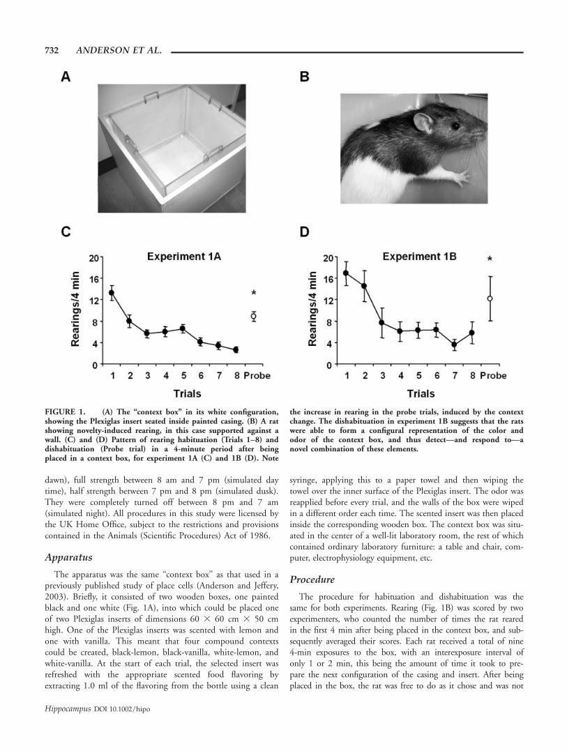
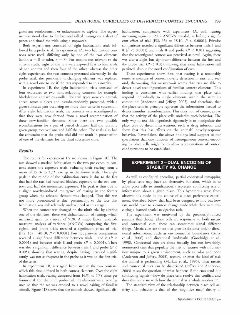
FIGURE 1. (A) The ‘‘context box’’ in its white configuration,showing the Plexiglas insert seated inside painted casing. (B) A ratshowing novelty-induced rearing, in this case supported against awall. (C) and (D) Pattern of rearing habituation (Trials 1–8) anddishabituation (Probe trial) in a 4-minute period after beingplaced in a context box, for experiment 1A (C) and 1B (D). Note
the increase in rearing in the probe trials, induced by the contextchange. The dishabituation in experiment 1B suggests that the ratswere able to form a configural representation of the color andodor of the context box, and thus detect—and respond to—anovel combination of these elements.
732 ANDERSON ET AL.
Hippocampus DOI 10.1002/hipo
given any reinforcement or inducements to explore. The experi-menters stood close to the box and tallied rearings on a sheet ofpaper, and timed the trials using a stopwatch.
Both experiments consisted of eight habituation trials fol-lowed by a probe trial. In experiment 1A, two habituation con-texts were used, differing only by one of the two elements(color, n ¼ 8 or odor, n ¼ 8). For reasons not relevant to thecurrent study, eight of the rats were exposed first to four trialsof one context and then four of the other, whereas the othereight experienced the two contexts presented alternately. In theprobe trial, the previously unchanging element was replacedwith a novel one to see if the rats responded to this novelty.
In experiment 1B, the eight habituation trials consisted offour exposures to two nonoverlapping contexts: for example,black-lemon and white-vanilla. The trial types were counterbal-anced across subjects and pseudo-randomly presented, with agiven stimulus pair occurring no more than twice in succession.After eight habituation trials, the contexts were reconfigured sothat they were now formed from a novel recombination ofthese now-familiar elements. Since there are two possiblerecombinations for a pair of paired elements, half the rats in agiven group received one and half the other. The trials also hadthe constraint that the probe trial did not result in presentationof one of the elements for the third successive time.
Results
The results for experiment 1A are shown in Figure 1C. Therats showed a marked habituation to the two pre-exposure con-texts across the exposure trials, reducing their rearing from amean of 15.16 to 2.72 rearings in the 4-min trials. The slightpeak in the middle of the habituation curve is due to the factthat half the rats had received blocked exposure to the two con-texts and half the intermixed exposure. The peak is thus due toa slight novelty-induced resurgence of rearing in the formergroup when the relevant element was changed. That this wasnot more pronounced is due, presumably, to the fact thathabituation was still relatively undeveloped at this stage.
When the context was changed on the ninth trial by alteringone of the elements, there was dishabituation of rearing, whichincreased again to a mean of 9.28. A single factor repeated-measures analysis of variance (ANOVA) comparing the first,eighth, and probe trials revealed a significant effect of trial[F(2, 15) ¼ 40.10, P < 0.0001]. Post hoc pairwise comparisonsrevealed a significant difference between trials 1 and 8 (P <0.0001) and between trials 8 and probe (P < 0.0001). Therewas also a significant difference between trials 1 and probe (P <0.005), showing that rearing, despite having increased signifi-cantly, was not as frequent in the probe as it was on the first trialof the series.
In experiment 1B, rats again habituated to the two contexts,which this time differed in both context elements. Over the eighthabituation trials, rearing decreased from 16.91 to 5.78 times per4-min trial. On the ninth probe-trial, the elements were reconfig-ured so that the rat was exposed to a novel pairing of familiarstimuli. Figure 1D shows that the animals showed significant dis-
habituation, comparable with experiment 1A, with rearingincreasing again to 12.16. ANOVA revealed, as before, a signifi-cant effect of trial [F(2, 15) ¼ 18.10, P < 0.0001]. Pairwisecomparisons revealed a significant difference between trials 1 and8 (P < 0.0001) and trials 8 and probe (P < 0.01) suggestingthat the reconfigured context was perceived as novel. Again, therewas also a slight but significant difference between the first andthe probe trial (P < 0.05), showing that some habituation stillpersisted, despite the novel configuration of cues.
These experiments show, first, that rearing is a reasonablysensitive measure of context novelty detection in rats, and sec-ond, that—using this measure—it seems that rats are able todetect novel reconfigurations of familiar context elements. Thisfinding is consonant with earlier findings that place cellsrespond individually to single elements within the stimuluscompound (Anderson and Jeffery, 2003), and therefore, thatthe place cells in principle represent the information needed todetect stimulus recombinations. This does not, of course, provethat the activity of the place cells underlies such behavior. Theonly way to test this hypothesis rigorously is to manipulate theplace cells by direct interventions, such as drug infusion, andshow that this has effects on the animals’ novelty-responsebehavior. Nevertheless, the above findings lend support to ourspeculation that one function of heterogeneous context encod-ing by place cells might be to allow representations of contextconfigurations to be established.
EXPERIMENT 2—DUAL ENCODING OFSTABILITY VS. CHANGE
As well as configural encoding, partial contextual remappingby place cells may have an alternative function, which is toallow place cells to simultaneously represent conflicting sets ofinformation about a given place. This hypothesis arose fromobservations made in the course of a place preference experi-ment, described below, that had been designed to find out howrats would react to a context change made while they were exe-cuting a learned spatial navigation task.
The experiment was motivated by the previously-noticedparadox that though place cells are responsive to both metricand contextual cues, these can sometimes signal differentthings. Metric cues are those that provide distance and/or direc-tional information: such as environmental boundaries (Barryet al., 2006) and directional landmarks (Goodridge et al.,1998). Contextual cues are those (usually, but not invariably,nonmetric) cues that populate the metric features with informa-tion unique to a given environment, such as color and odor(Anderson and Jeffery, 2003), texture, or even the kind of taskthe animal is performing (Markus et al., 1995). That metricand contextual cues can be dissociated (Jeffery and Anderson,2003) raises the question of what happens if the cues send outconflicting signals—how do place cells resolve this conflict, anddoes this correlate with how the animal as a whole resolves it?
The standard view of the relationship between place cell ac-tivity and behavior is that of the ‘‘cognitive map’’ theory of
BEHAVIORAL CORRELATES OF DISTRIBUTED CONTEXT ENCODING 733
Hippocampus DOI 10.1002/hipo
O’Keefe and Nadel (1978), which proposes that the activity ofthe place cells underlies navigational behavior: implying, inessence, that the collective activity of the place cells constitutesthe animal’s ‘‘knowledge’’ about where it is. Introducing a con-flict in the cues that drive place cells, and which also drivebehavior (perhaps via the place cells, but perhaps not) offersthe possibility of eliciting dissociations between the neural ac-tivity and behavior, and thus the opportunity to falsify the cog-nitive map theory. In other words, by dissociating environmen-tal cues, it may be possible to disconnect place cell activityfrom behavior. If this happened, the implication would be thateither place cells are not the repository of the animal’s spatial‘‘map,’’ or that the animal uses more than just this map to nav-igate with.
A previous attempt to explore the results of such a conflict foundthat a change in context, in the face of unchanged spatial cues,caused place cells to alter their firing patterns, apparently completely,and yet the rats continued to navigate reliably to a previously learnedgoal (Jeffery et al., 2003). This at first glance looks like a falsificationof the place-cells ¼ place-knowledge hypothesis—it seems to implythat the rats could not have been using their place cells to performthe navigational task, even though acquisition of the task was shownto be hippocampal dependent. However, one possible explanationfor this is that the task had been so over-learned that it was no longerhippocampal dependent at the time of testing. In other words, therats had learned a habit-based strategy: coming, over time, to useindividual cues in the room to disambiguate the four corners of thebox. The present experiment was designed to foil this strategy byproviding a much more unconstrained environment in which, wehoped, individual cues could not be used to solve the task. Navigat-ing in such an environment would, according to theoretical consid-erations, be more likely to require the hippocampus, and—if thetheory is correct—the place cells.
In the place preference task used here, rats were trained, in aparticular context, to visit an unmarked zone located on thesurface of a 1 m diameter arena. This task ought to be hippo-campal dependent, because its successful execution is com-pletely dependent on processing of the spatial relations amongdistal environmental cues, and provides few or no opportunitiesfor more cue-based, ‘‘taxon’’ navigation. If the cognitive maphypothesis is correct, then altering contextual cues should there-fore, alter both place cell activity (i.e., induce remapping) anddisrupt navigation.
Once the rats had been well trained on the place preferencetask, some of them were implanted for the recording of placecells. The purpose of this was to see how the cells responded toa change in context (i.e., change in the color of the arena fromblack to white or vice versa), and how this change related tothe behavior of the animals. Other rats were implanted withinjection cannulae for hippocampal inactivation, with theintention of revealing the hippocampal dependence of the task.Since this procedure proved to be ineffective, and since welacked the ability at that time to determine whether this wasbecause the task is hippocampal independent or (more likely)because the injections failed to inactivate the hippocampus,only the behavioral data are reported from those animals.
Materials and Methods
Subjects
The subjects were eight male Lister-hooded rats (weighingapproximately 250 g on arrival) obtained from the BiologicalServices Unit at UCL, and housed in same facility as described inexperiment 1. They were fed standard rat food to maintain 90%of their free-feeding weight and were allowed unlimited access towater. Initially rats were housed in groups of four in Plexiglascages; after surgery they were housed in separate cages to ensuretheir well-being. All procedures in this study were licensed by theUK Home Office, subject to the restrictions and provisions con-tained in the Animals (Scientific Procedures) Act of 1986.
Experimental apparatus and protocol
The rats were trained in a standard place preference protocol(Fig. 2) in a quiet room containing a rich array of cues. Thetask required the rats to locate an unmarked zone in a large cir-
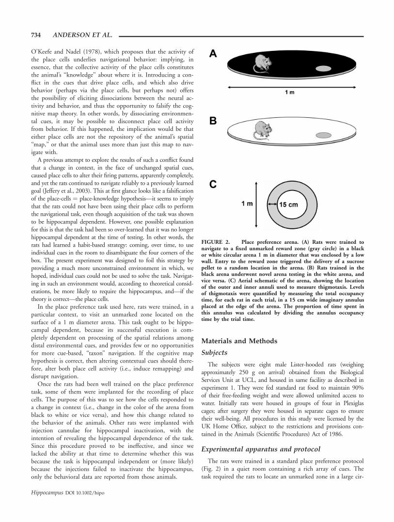
FIGURE 2. Place preference arena. (A) Rats were trained tonavigate to a fixed unmarked reward zone (gray circle) in a blackor white circular arena 1 m in diameter that was enclosed by a lowwall. Entry to the reward zone triggered the delivery of a sucrosepellet to a random location in the arena. (B) Rats trained in theblack arena underwent novel arena testing in the white arena, andvice versa. (C) Aerial schematic of the arena, showing the locationof the outer and inner annuli used to measure thigmotaxis. Levelsof thigmotaxis were quantified by measuring the total occupancytime, for each rat in each trial, in a 15 cm wide imaginary annulusplaced at the edge of the arena. The proportion of time spent inthis annulus was calculated by dividing the annulus occupancytime by the trial time.
734 ANDERSON ET AL.
Hippocampus DOI 10.1002/hipo
cular arena surrounded by extra-arena cues and in which intra-arena cues had been rendered irrelevant for the purposes ofnavigation. In the present experiment, the rats were trained tonavigate reliably in an arena of one color (e.g. black; the ‘famil-iar arena’), then tested on their navigational ability in a differ-ently colored arena (e.g. white; the ‘novel arena’). The experi-ment was counterbalanced so that four rats were trained in theblack floored arena (the white arena was therefore the novelcontext for these rats), and the other rats were trained in thewhite floored arena (the black arena was the novel context forthese rats). Each rat within these groups was trained toapproach a different reward zone. Because of time constraints,the two groups of rats were tested at different times by differ-ent experimenters. There were slight differences in the proce-dures: rats in the first group were trained in the familiar con-text and then tested in the novel context prior to surgery; theywere then retrained in the familiar context and retested in thenovel context. Rats in the second group were trained in the fa-miliar context, underwent surgery, were then retrained in thefamiliar context and then tested in the novel context. Since allcomparisons between familiar and novel environments weremade within a given subject, these slight between-subject differ-ences would not have been pertinent.
The details of the arena are as follows: it was a circular plat-form 100 cm in diameter (Fig. 2), which was painted black onone side and white on the other, and had a 5 cm wall all theway round on both sides to prevent the food pellets from roll-ing off but without obstructing the rats’ view of the extra-arenacues. The arena was raised 40 cm above the floor to preventthe rats from escaping. A circular 25 cm diameter unmarkedzone was allocated to each of the four quadrants of the arena,centered on the mid-point of the radius. For each rat, one ofthese was defined as the reward zone, one as the clockwise adja-cent zone, one as the anticlockwise adjacent zone, and one asthe opposite zone.
A monochrome video camera positioned directly above thearena recorded the movement of the rat in the arena by track-ing an infrared light strapped to the rat by an elastic harness.The video image was passed to a tracking system (Axona,Herts, UK), which extracted the position of the LED in x–ycoordinates and stored the data for later analysis. Path trajecto-ries, the time elapsed since the beginning of the trial, and thenumber of entries to each zone in the arena were also stored.Entry to the reward zone, as detected by the tracking system,led to the sounding of a 150 ls 750 Hz tone together withthe dispensing of a small food pellet to a random location inthe arena from a pellet-hopper located in the ceiling above thearena (but with the criterion that 3 s must have elapsed sincethe last entry). Because of technical problems with the hopper,pellets were sometimes dispensed manually.
All rats underwent 3 days of pretraining prior to place pref-erence training. Each group of rats was placed together in thearena for 15 min on the first day; food pellets scattered all overthe floor encouraged the rats to forage. On the next 2 days,rats were introduced individually into the pellet-filled arena for15 min. No place-preference training was conducted on these
days. Beginning on the next day, rats underwent place-prefer-ence training in two 15-min trials per day. Each day the ratswere brought individually into the experimental room, fittedwith the harness bearing the LED, then placed in the center ofthe arena to begin the first trial. The rats were always placed inthe same location in the arena, facing away from their rewardzone. At the end of the first trial, the rat was removed andplaced in a holding box at the side of the room. The arena wasthen cleaned, rotated (to prevent the use of olfactory cues aslandmarks), and the rat began its second trial. The intertrialinterval was *4 min. Rats were run in a pseudorandom ordereach day. Training continued until rats were performing consis-tently. When all rats had developed a clear preference for theirdesignated reward zone, they each underwent two 15-min trialson the other side of the arena (i.e., in the novel color; the‘‘novel arena test day’’). Nothing other than the color of thearena differed between familiar and novel context trials.
It was hypothesized that the arena color change would causeplace cells to remap, and that any detriment in place preferenceperformance might therefore be attributable to the disruption ofthe hippocampal cognitive map. No detriment in performancewould suggest either (a) that the task was not hippocampal-de-pendent, (b) that the cognitive map somehow remained intactdespite the remapping, or (c) that the rats were able to rapidlyrelearn the task in the novel arena. To refute the last possibility,on the day after the novel arena test day, rats were tested withtwo additional 15-min trials in the familiar arena in a novelroom with different extra-arena cues (the ‘‘novel room test day’’).
Behavioral analyses
Each rat’s performance on the place preference task wasscored using the formula
ðEr�meanðEcwþEacwþEoppÞÞ=ðErþmeanðEcwþEacwþEoppÞÞ
where Er ¼ number of entries to the reward zone, Ecw ¼ num-ber of entries to the clockwise zone (relative to the rewardzone), Eacw ¼ number of entries to the anticlockwise zone, andEopp ¼ number of entries to the opposite zone. This scoreranged between +1 (when the rat only makes entries to thereward zone) and �1 (when the rat never enters the rewardzone and makes at least one entry to one of the other zones); ascore of zero indicated no preference for the reward zone overthe other zones.
The trajectory of the rats in the arena was also examined bymeasuring the proportion of time rats spent near the edges ofthe platform (thigmotaxis). An imaginary annulus with externaldiameter equal to the arena diameter and internal diameter 30cm less was positioned over the arena and the amount of timerats spent within this 15 cm circular corridor in each trial wascalculated (Fig. 2C). The proportion of the trial time spent inthis annulus was calculated by dividing the annulus occupancytime by the total trial time to give a score which rangedbetween 0 (no time spent in the annulus) and +1 (all trial timespent in annulus). This measure also indicated whether or not
BEHAVIORAL CORRELATES OF DISTRIBUTED CONTEXT ENCODING 735
Hippocampus DOI 10.1002/hipo
the rats responded to the novel stimuli during testing (arenaand room).
Surgery
Two rats from each group were unilaterally implanted withtetrodes for the purpose of recording hippocampal place cells;the other two rats from each group were bilaterally implantedwith stainless steel cannulae to permit infusion of an inactivat-ing agent into the hippocampus (though, as mentioned above,the infusion data were noncontributory: thus, these rats servedas sham controls for the behavioral procedure). One rat didnot survive the surgery and therefore data for this rat areincluded in the first training stage only.
During surgery, rats were anesthetized with isoflurane andoxygen, and given a subcutaneous injection of antibiotic (enro-floxacin). They were mounted in a stereotaxic frame, the scalpwas shaved and cleaned, and the skull was exposed by means ofa midline incision followed by cleaning. Designated rats wereimplanted with tetrodes mounted on a microdrive, others withstainless steel cannulae. The tetrodes were implanted in theright hemisphere 3.8 mm posterior to bregma, 2.5 mm lateralto the midline, and 1.5 mm ventral to the brain surface; can-nulae were implanted bilaterally at the same coordinates. Theinjection cannulae extended 1.1 mm below the tip of the im-plantation cannulae and were aimed at the middle of the hip-pocampus, between CA1 and CA3. Jewellery screws and dentalacrylic were used to securely attach the microdrive or cannulaeto the skull. At the end of surgery, rats were given an intramus-cular injection of an analgesic (buprenorphine) and returned totheir home cages. Rats recovered for at least 5 days beforetraining recommenced.
Place cell recording and analysis
Four of the rats were implanted with tetrodes mounted on amicrodrive that allowed the tetrodes to be advanced togetherthrough the brain in steps as small as 25 lm. To obtain placecell recordings, each rat was screened daily after recovery fromsurgery. Full details of the screening and recording procedurehave been described previously (Anderson and Jeffery, 2003)and will only be given briefly here. Before the first trial of theday, rats were connected to the recording equipment (Axona,Herts, UK), and activity on the tetrodes was monitored forboth complex spike activity and ripples, both reliable indicatorsof intrahippocampal electrode positioning. If neither sign wasevident, the microdrive was advanced by 25–200 lm per day,and the trials were run without place cell data recording.Because of the inherent difficulty in obtaining place cell record-ings at the same time as maintaining place preference perform-ance, no strict criteria signaled the end of training and the be-ginning of testing. Instead rats within each group were trainedfor the time it took to obtain stable place cells in as many ratsas possible. Once place cells were detected, recordings weremade in the familiar arena on what became the final day oftraining, followed on the next day by recording in the novelarena. Cells were not recorded in the novel room trials. For
unknown reasons, no place cells at all could be recorded in twoof the four rats implanted with tetrodes; once it became clearthat no cells would be obtained from these rats, they weretested in the novel arena without further recordings.
Single neuron data were isolated using cluster cutting soft-ware (Tint: Axona, Herts, UK). Firing rate maps were thenconstructed by dividing the arena floor into square pixels ofside *2 cm: the firing rate for a given cell in each pixel wasdetermined by dividing the number of spikes in that pixel bythe amount of time the rat spent there. The firing rate mapswere smoothed using an algorithm that replaced the value ineach pixel with the average of the value in that plus the adja-cent eight pixels. Place fields were visualized as contour mapswith five levels, each level representing a 20% portion of thepeak firing rate for that map. Putative interneurons and com-plex spiking cells were discriminated on the basis of spikeshape, firing rate, and place field size.
Additional analyses were conducted using programs custom-written in Matlab (The MathWorks, Natick, MA). A cell wasdefined as having a place field if its peak rate after smoothing(taken from the pixel with the highest rate) was �1 Hz andthe number of spikes in its cluster was �20. Place fields wereassessed for remapping or nonremapping using pixel-to-pixelcorrelations between place cell firing rate maps (for details seeAnderson and Jeffery, 2003). Only pixels in which the cell hadfired in at least one of the maps entered the correlation, to pre-vent artificially high correlations from being generated by largenumbers of zero-zero correlations. Cells classed as ‘remappingcells’ had differences between mean same and different condi-tion r values of �0.4 (see Results); nonremapping cells typi-cally had values close to zero (for rat 2, only those place fieldsthat remained stable in the same-condition trials were includedin this analysis).
RESULTS
Eight rats were trained in the place preference task, but therat that died during surgery only contributed training data.The first group of rats was trained for 42 days and was thentested in the novel arena and, on the following day, in thenovel room; this was then followed by surgery and recovery,then a further 11 days of retraining and then retesting in thenovel arena. The second group of rats was trained for 33 daysprior to surgery. After surgery and recovery, the rats wereretrained for 19 days, and then tested in the novel arena and,on the following day, in the novel room.
The novel arena behavioral data for the first group presentedhere were obtained in the first exposure (i.e., prior to surgery);the place cell recordings were made during the secondexposure.
Behavioral Analysis
Figure 3 shows the mean (6, the standard error of the mean,SEM) for all rats of both the place preference and thigmotaxis
736 ANDERSON ET AL.
Hippocampus DOI 10.1002/hipo
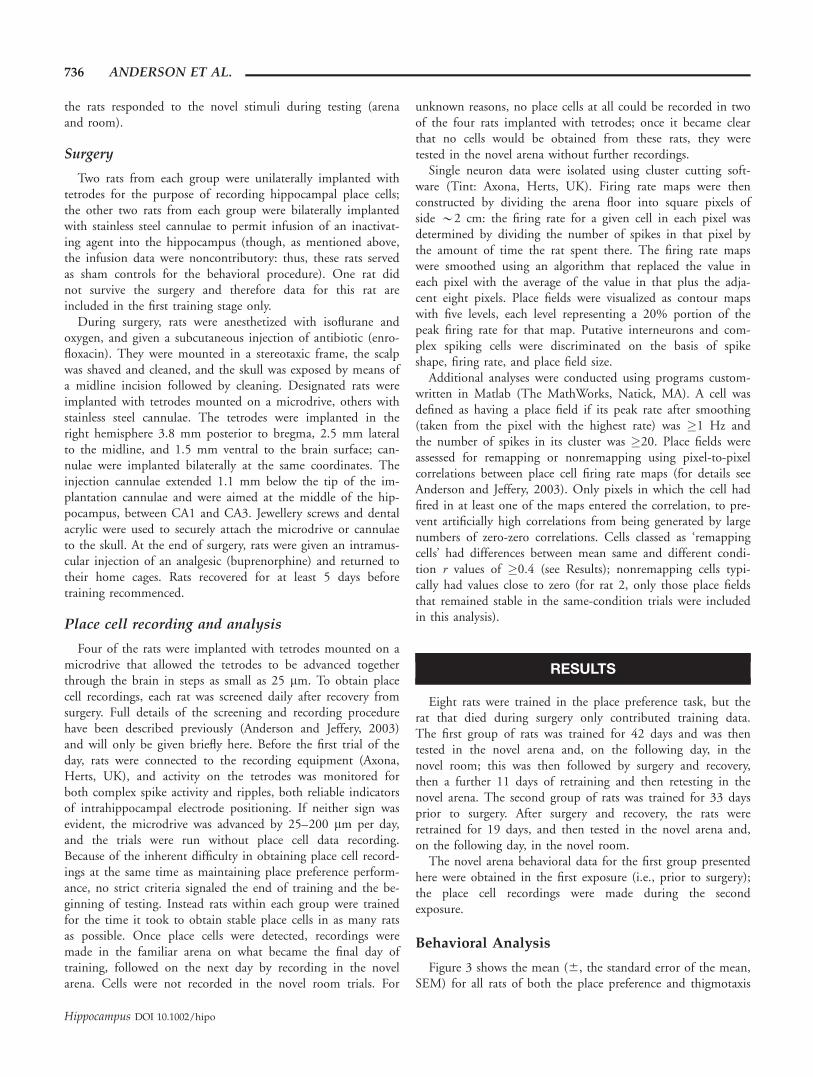
measure for the first week of training, the three weeks of trainingprior to testing, and the testing in the novel arena and then in thenovel room. Rats initially, as expected, showed no preferencefor their reward zones and high levels of thigmotaxis. By the endof training, place preference performance levels rose comparedwith the first day of training, while levels of thigmotaxis fell.Scores on the first trial for the pertinent training days (first andlast) and testing days (novel arena and novel room) are displayedin Figure 4.
Place Preference Analysis
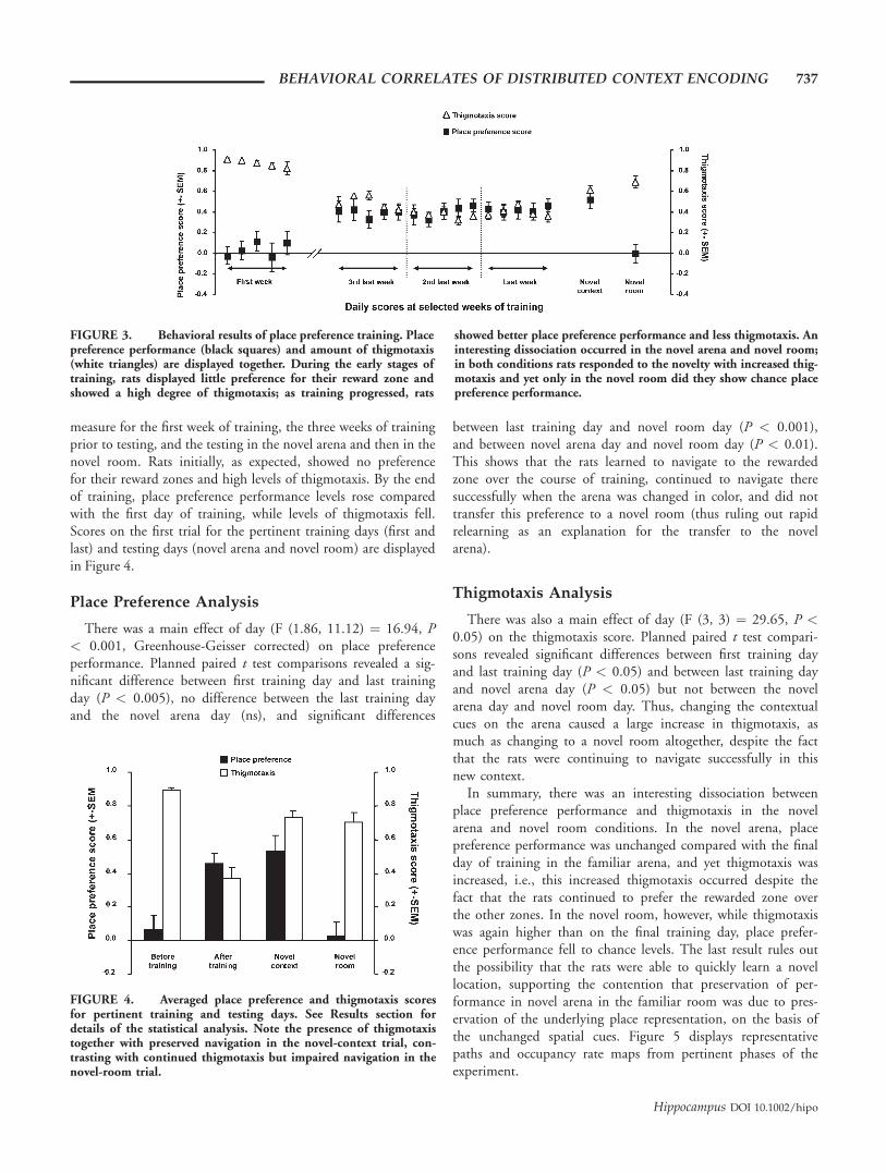
There was a main effect of day (F (1.86, 11.12) ¼ 16.94, P< 0.001, Greenhouse-Geisser corrected) on place preferenceperformance. Planned paired t test comparisons revealed a sig-nificant difference between first training day and last trainingday (P < 0.005), no difference between the last training dayand the novel arena day (ns), and significant differences
between last training day and novel room day (P < 0.001),and between novel arena day and novel room day (P < 0.01).This shows that the rats learned to navigate to the rewardedzone over the course of training, continued to navigate theresuccessfully when the arena was changed in color, and did nottransfer this preference to a novel room (thus ruling out rapidrelearning as an explanation for the transfer to the novelarena).
Thigmotaxis Analysis
There was also a main effect of day (F (3, 3) ¼ 29.65, P <0.05) on the thigmotaxis score. Planned paired t test compari-sons revealed significant differences between first training dayand last training day (P < 0.05) and between last training dayand novel arena day (P < 0.05) but not between the novelarena day and novel room day. Thus, changing the contextualcues on the arena caused a large increase in thigmotaxis, asmuch as changing to a novel room altogether, despite the factthat the rats were continuing to navigate successfully in thisnew context.
In summary, there was an interesting dissociation betweenplace preference performance and thigmotaxis in the novelarena and novel room conditions. In the novel arena, placepreference performance was unchanged compared with the finalday of training in the familiar arena, and yet thigmotaxis wasincreased, i.e., this increased thigmotaxis occurred despite thefact that the rats continued to prefer the rewarded zone overthe other zones. In the novel room, however, while thigmotaxiswas again higher than on the final training day, place prefer-ence performance fell to chance levels. The last result rules outthe possibility that the rats were able to quickly learn a novellocation, supporting the contention that preservation of per-formance in novel arena in the familiar room was due to pres-ervation of the underlying place representation, on the basis ofthe unchanged spatial cues. Figure 5 displays representativepaths and occupancy rate maps from pertinent phases of theexperiment.
FIGURE 3. Behavioral results of place preference training. Placepreference performance (black squares) and amount of thigmotaxis(white triangles) are displayed together. During the early stages oftraining, rats displayed little preference for their reward zone andshowed a high degree of thigmotaxis; as training progressed, rats
showed better place preference performance and less thigmotaxis. Aninteresting dissociation occurred in the novel arena and novel room;in both conditions rats responded to the novelty with increased thig-motaxis and yet only in the novel room did they show chance placepreference performance.
FIGURE 4. Averaged place preference and thigmotaxis scoresfor pertinent training and testing days. See Results section fordetails of the statistical analysis. Note the presence of thigmotaxistogether with preserved navigation in the novel-context trial, con-trasting with continued thigmotaxis but impaired navigation in thenovel-room trial.
BEHAVIORAL CORRELATES OF DISTRIBUTED CONTEXT ENCODING 737
Hippocampus DOI 10.1002/hipo
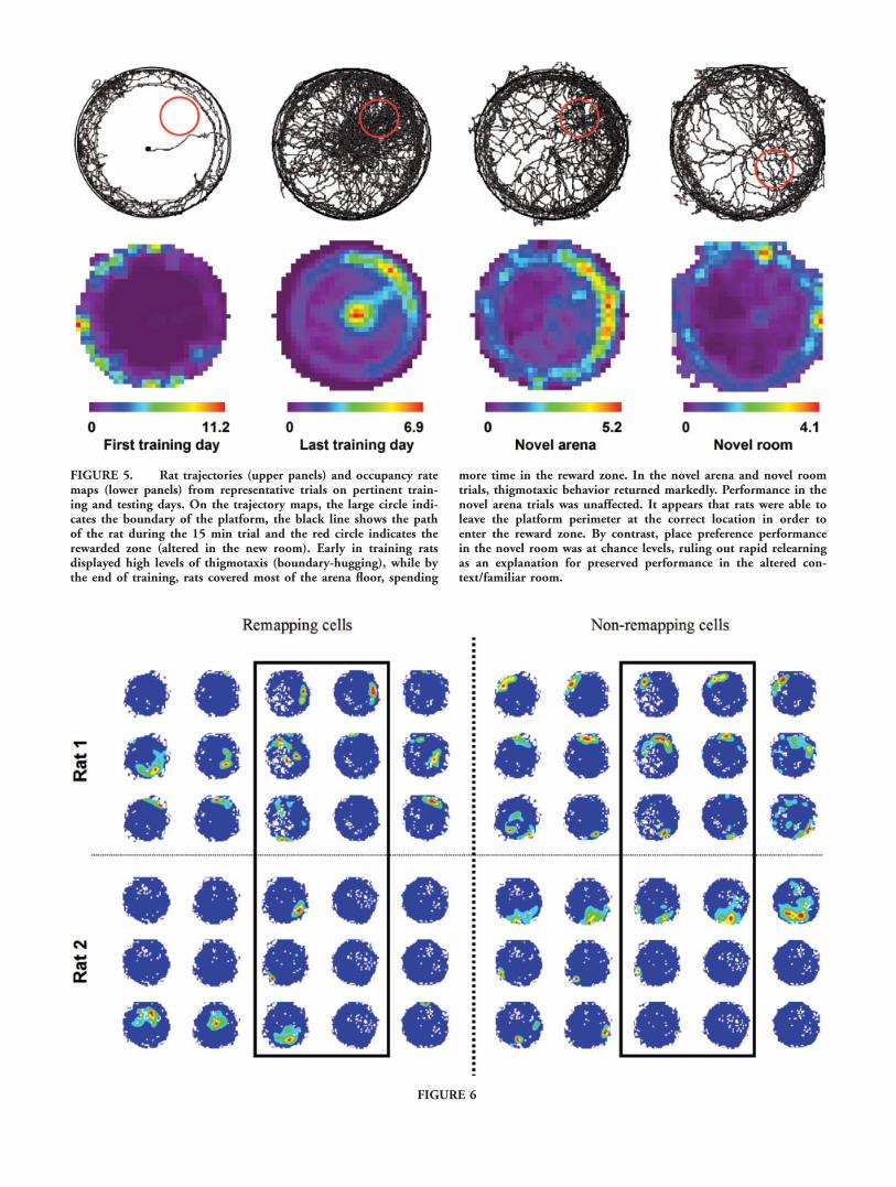
FIGURE 5. Rat trajectories (upper panels) and occupancy ratemaps (lower panels) from representative trials on pertinent train-ing and testing days. On the trajectory maps, the large circle indi-cates the boundary of the platform, the black line shows the pathof the rat during the 15 min trial and the red circle indicates therewarded zone (altered in the new room). Early in training ratsdisplayed high levels of thigmotaxis (boundary-hugging), while bythe end of training, rats covered most of the arena floor, spending
more time in the reward zone. In the novel arena and novel roomtrials, thigmotaxic behavior returned markedly. Performance in thenovel arena trials was unaffected. It appears that rats were able toleave the platform perimeter at the correct location in order toenter the reward zone. By contrast, place preference performancein the novel room was at chance levels, ruling out rapid relearningas an explanation for preserved performance in the altered con-text/familiar room.
FIGURE 6
Thigmotaxis might be expected to, nevertheless, have somedeleterious effects on the navigation accuracy measure. Indeed,correlation of the two scores did reveal a significant negativerelationship between place preference and thigmotaxis (�0.67,P < 0.01), with the propensity for rats to circle the arena beingin inverse proportion to the number of visits to the rewardedzone. That the rats nevertheless continued to spend significantlymore time in the rewarded zone lends even stronger support tothe notion that the processing of spatial cues in this taskremained intact after the context change.
Place Cell Analysis
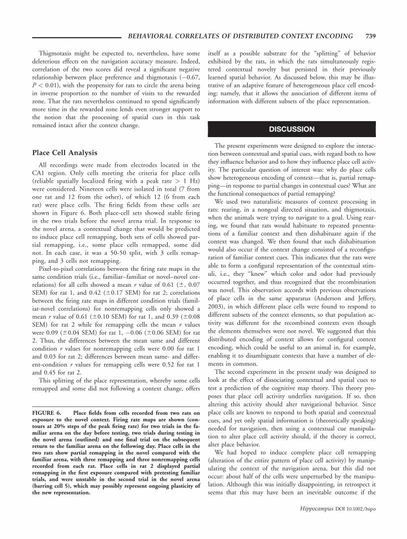
All recordings were made from electrodes located in theCA1 region. Only cells meeting the criteria for place cells(reliable spatially localized firing with a peak rate > 1 Hz)were considered. Nineteen cells were isolated in total (7 fromone rat and 12 from the other), of which 12 (6 from eachrat) were place cells. The firing fields from these cells areshown in Figure 6. Both place-cell sets showed stable firingin the two trials before the novel arena trial. In response tothe novel arena, a contextual change that would be predictedto induce place cell remapping, both sets of cells showed par-tial remapping, i.e., some place cells remapped, some didnot. In each case, it was a 50-50 split, with 3 cells remap-ping, and 3 cells not remapping.
Pixel-to-pixel correlations between the firing rate maps in thesame condition trials (i.e., familiar–familiar or novel–novel cor-relations) for all cells showed a mean r value of 0.61 (6, 0.07SEM) for rat 1, and 0.42 (60.17 SEM) for rat 2; correlationsbetween the firing rate maps in different condition trials (famil-iar-novel correlations) for nonremapping cells only showed amean r value of 0.61 (60.10 SEM) for rat 1, and 0.39 (60.08SEM) for rat 2 while for remapping cells the mean r valueswere 0.09 (60.04 SEM) for rat 1, �0.06 (60.06 SEM) for rat2. Thus, the differences between the mean same and differentcondition r values for nonremapping cells were 0.00 for rat 1and 0.03 for rat 2; differences between mean same- and differ-ent-condition r values for remapping cells were 0.52 for rat 1and 0.45 for rat 2.
This splitting of the place representation, whereby some cellsremapped and some did not following a context change, offers
itself as a possible substrate for the ‘‘splitting’’ of behaviorexhibited by the rats, in which the rats simultaneously regis-tered contextual novelty but persisted in their previouslylearned spatial behavior. As discussed below, this may be illus-trative of an adaptive feature of heterogeneous place cell encod-ing: namely, that it allows the association of different items ofinformation with different subsets of the place representation.
DISCUSSION
The present experiments were designed to explore the interac-tion between contextual and spatial cues, with regard both to howthey influence behavior and to how they influence place cell activ-ity. The particular question of interest was: why do place cellsshow heterogeneous encoding of context—that is, partial remap-ping—in response to partial changes in contextual cues? What arethe functional consequences of partial remapping?
We used two naturalistic measures of context processing inrats: rearing, in a nongoal directed situation, and thigmotaxis,when the animals were trying to navigate to a goal. Using rear-ing, we found that rats would habituate to repeated presenta-tions of a familiar context and then dishabituate again if thecontext was changed. We then found that such dishabituationwould also occur if the context change consisted of a reconfigu-ration of familiar context cues. This indicates that the rats wereable to form a configural representation of the contextual stim-uli, i.e., they ‘‘knew’’ which color and odor had previouslyoccurred together, and thus recognized that the recombinationwas novel. This observation accords with previous observationsof place cells in the same apparatus (Anderson and Jeffery,2003), in which different place cells were found to respond todifferent subsets of the context elements, so that population ac-tivity was different for the recombined contexts even thoughthe elements themselves were not novel. We suggested that thisdistributed encoding of context allows for configural contextencoding, which could be useful to an animal in, for example,enabling it to disambiguate contexts that have a number of ele-ments in common.
The second experiment in the present study was designed tolook at the effect of dissociating contextual and spatial cues totest a prediction of the cognitive map theory. This theory pro-poses that place cell activity underlies navigation. If so, thenaltering this activity should alter navigational behavior. Sinceplace cells are known to respond to both spatial and contextualcues, and yet only spatial information is (theoretically speaking)needed for navigation, then using a contextual cue manipula-tion to alter place cell activity should, if the theory is correct,alter place behavior.
We had hoped to induce complete place cell remapping(alteration of the entire pattern of place cell activity) by manip-ulating the context of the navigation arena, but this did notoccur: about half of the cells were unperturbed by the manipu-lation. Although this was initially disappointing, in retrospect itseems that this may have been an inevitable outcome if the
FIGURE 6. Place fields from cells recorded from two rats onexposure to the novel context. Firing rate maps are shown (con-tours at 20% steps of the peak firing rate) for two trials in the fa-miliar arena on the day before testing, two trials during testing inthe novel arena (outlined) and one final trial on the subsequentreturn to the familiar arena on the following day. Place cells in thetwo rats show partial remapping in the novel compared with thefamiliar arena, with three remapping and three nonremapping cellsrecorded from each rat. Place cells in rat 2 displayed partialremapping in the first exposure compared with pretesting familiartrials, and were unstable in the second trial in the novel arena(barring cell 5), which may possibly represent ongoing plasticity ofthe new representation.
BEHAVIORAL CORRELATES OF DISTRIBUTED CONTEXT ENCODING 739
Hippocampus DOI 10.1002/hipo
place cells do indeed underlie an animal’s spatial knowledge.Given that we provided the rat with sufficient spatial cues tocontinue navigating with, it is possible that under these circum-stances, complete remapping is impossible, i.e., the very infor-mation that enables the rat to navigate to the same place neces-sarily also provides the place cells with enough information toretain some of their fields. In fact, one could go further andpropose that this spatial information enables continued naviga-tion by keeping some of the place fields unchanged. If this istrue, then the only way to induce complete remapping in theplace cells would have been to alter so much environmental in-formation that the rat could no longer navigate, either. Thus,the present results can be viewed as being consistent with cog-nitive mapping theory. Our interpretation of the results is thatby splitting their representation in the way that they did, theplace cells were able to encode different aspects of the sameenvironment, using the same population of neurons. Thus,with a distributed code, it is possible to simultaneously repre-sent, for example, both that some aspects of the environmenthave changed and that some have remained the same.
A similar proposal was recently advanced by Leutgeb et al.(2005), who observed that local changes to an environment(such as to color, what we have here called ‘‘contextual’’)tended to induce changes only in firing rate, particularly inCA3 neurons, and they proposed that this ‘‘rate remapping’’could allow the discrimination of different events occurring ata given location. By contrast, global environmental changes(moving the recording apparatus to a different room, a changethat we would call ‘‘metric’’) induced changes in field location,perhaps implying recruitment of a new spatial map. We gener-ally do not see rate remapping in response to change of localcues, and did not in this experiment either, but this may bebecause our recordings are from CA1 neurons. Our interpreta-tion of the rate/location dichotomy is that rate remappingreflects a modulation of drive, or ‘‘confidence,’’ of a particularfield, whereas location remapping reflects a switch to a differentfield (via different inputs). We have previously proposed thatthe function of the context cues is to, as it were, set this switch(Jeffery et al., 2004).
Although our results are consistent with the cognitive maptheory, it should be noted that they by no means prove it. It ispossible that the rats, after the prolonged training required toreach above-chance performance, had managed to develop ahabit-based strategy for locating the goal area, and were nolonger using the place cells at all. This is one of the possibleexplanations for why a previous study of ours found that evencomplete place cell remapping was associated with only aminor decrement in navigational performance (Jeffery et al.,2003). Although we attempted to thwart this in the presentstudy by using an open-field arena, in which habit-based navi-gation is much harder to acquire, this possibility could only becompletely ruled out by disrupting hippocampal activity afterthe animals had learned the task, to see whether they werenow impaired (if the task was still hippocampal dependent) orif they could still navigate (suggesting use of another system,such as striatum). Such a test was the motivation behind the
implantation of injection cannulae in some of the animals, asit happened, the injections, as far as we could tell, were inef-fective. We lacked the means to distinguish, however, whetherthis was because the hippocampus was not successfully inacti-vated or because it was no longer needed for the task. Thisissue needs to be explored in future experiments because theinterplay between hippocampal and nonhippocampal naviga-tion systems is complex, and without knowing which systemthe animals are using at any given moment, it is hard to drawconclusions about the concomitant neural activity and how itrelates. Until then, the present results must be regarded asmerely being ‘‘not inconsistent’’ with cognitive mappingtheory. Nevertheless, the observation of a representational split(partial remapping) in the place cells that paralleled the splitbehavior of the animals is suggestive and deserves furtherstudy.
Some Speculations About the Role of the SpatialContext Representation in Episodic Memory
The above experiments show that the place cell representa-tion of spatial context has properties consistent with thoseof the spatial-contextual behavior of the animals, suggestingthat the one may underlie the other. Given that the place cellshave these properties, why, then, is the hippocampus so cru-cially important in human (and probably animal) episodicmemory? It is hard to link the activity of place cells, whosefiring is predominantly driven by metric and contextual cuesand is remarkably stable over long periods of time, to theencoding of episodes, which by their nature involve many dif-ferent kinds of stimuli and are fleeting, one-off occurrences.Do place cells also have a role in the encoding, storage and/orretrieval of episodic memories?
A number of studies have, indeed, reported place cell activityoccurring in association with important events. In an early se-ries of studies, Berger and Thompson showed that hippocampalneurons in rabbits developed conditioned responding to pairedpresentations of a tone with a corneal airpuff (Berger et al.,1976). Eichenbaum and colleagues have reported that hippo-campal neurons fire in response to behavioral events such ascue-sampling and goal approach (Eichenbaum et al., 1987;Wiener et al., 1989; Wood et al., 1999), and a number ofstudies have found activity of place cells that seems to berelated to the goal area in an environment, a place where im-portant events (goal discovery) repeatedly occur (Breese et al.,1989; Wiebe and Staubli, 1999; Hollup et al., 2001). Intrigu-ingly, in-field modulation of place fields by events was reportedby Moita et al. (2003), who found an increase in place cell ac-tivity evoked by an auditory conditioned stimulus when the ani-mal was in the cell’s place field, but not when it was outside it.
The above studies are certainly suggestive of the possibilitythat place cells encode events. However, it is of potential signif-icance that these studies have all involved events which occur,repeatedly, at the same place in the environment. Thus, itmight be possible to make the argument that the events had,by virtue of their spatial constancy and repetition, come to
740 ANDERSON ET AL.
Hippocampus DOI 10.1002/hipo
form part of the descriptor of the place, in other words, part ofthe context. It is certainly the case that place cells are often notactive in event-rich situations: for example, even in environ-ments in which important (i.e., behavior-modifying) events areoccurring, if these are not occurring in a constant location thenplace cells rarely show much activity outside the zone of theirown firing fields (e.g., Jeffery et al., 2003). Thus, if place cellsencode events too, then such activity must be sparse. This maynot be unreasonable, however, given the number of events thatthe hippocampus must—if it encodes events at all—encodeover the lifetime of an animal.
The above notwithstanding, an alternative, and perhaps moreplausible, possibility is that the role of the hippocampus is notto encode the events themselves, but rather to form a link, orindex, between the representation of the place in which theevents occurred and the memory traces of these events else-where in the brain (presumably neocortex). This would meanthat when the animal returned to that place, and reactivatedthat representation, the memory itself would also be reacti-vated. Such memory-index theories have been proposed a num-ber of times, though first and most elegantly by Teyler andDiScenna (1986). The role of the hippocampus in their modelis to bind together the disparate elements of an experience,which belong with each other by virtue of having occurred atthe same time and place. Burgess and co-workers (Burgess,2002; Burgess et al., 2002) have taken this a step further, andproposed that the hippocampus is also required for the imposi-tion of a viewpoint onto the retrieved memory, so that theevent is able to be re-experienced, as it were, from a particularviewpoint.
To conclude, then: studies of place cells in rats suggest a rolein the encoding of spatial context, a proposal that is supportedby correspondences such as the ones we demonstrated here,between the properties of the place cell representation (partialremapping) and the properties of spatial-contextual behavior. Itis suggested that this representation, as well as providing infor-mation useful for navigation, also serves as an index of eventsthat occurred there, with the memory traces of the eventsthemselves being stored in neocortex. When the animal returnsto a place these memories are thus reactivated. In humans,returning to the place may be a mental rather than physicalevent; in this case, the hippocampus can be used to impose aviewpoint on the mental image, so as to enable reconstructionof the locations of salient object and people in the memory. Tothe extent that this is the case, recall of episodic memory is nota veridical replay of the original sensory stream, but is really areconstruction of the events comprising a sensory experience.The heterogeneous, distributed encoding of context, contrib-uted by the place cells, may allow flexibility in how associationsare made between contexts and their associated events.
Acknowledgments
The authors thank Colin Lever for suggesting rearing as acontextual novelty measure, for helping with a pilot version ofthe rearing experiment, and for providing helpful comments on
the manuscript. The authors are also grateful to Roger Buncefor help with the place preference apparatus, and to Jim Don-nett for help with software.
REFERENCES
Anderson MI, Jeffery KJ. 2003. Heterogeneous modulation of placecell firing by changes in context. J Neurosci 23:8827–8835.
Barry C, Lever C, Hayman R, Hartley T, Burton S, O’Keefe J, JefferyKJ, Burgess N. 2006. The boundary vector cell model of place cellfiring and spatial memory. Rev Neurosci 17:71–97.
Berger TW, Alger B, Thompson RF. 1976. Neuronal substrate of clas-sical conditioning in the hippocampus. Science 192:483–485.
Breese CR, Hampson RE, Deadwyler SA. 1989. Hippocampal placecells: Stereotypy and plasticity. J Neurosci 9:1097–1111.
Burgess N. 2002. The hippocampus, space, and viewpoints in episodicmemory. Q J Exp Psychol A 55:1057–1080.
Burgess N, Maguire EA, O’Keefe J. 2002. The human hippocampusand spatial and episodic memory. Neuron 35:625–641.
Clayton NS, Dickinson A. 1998. Episodic-like memory during cacherecovery by scrub jays. Nature 395:272–274.
Eichenbaum H, Kuperstein M, Fagan A, Nagode J. 1987. Cue-sam-pling and goal-approach correlates of hippocampal unit activity inrats performing an odor-discrimination task. J Neurosci 7:716–732.
Gallagher M, Holland PC. 1992. Preserved configural learning andspatial learning impairment in rats with hippocampal damage. Hip-pocampus 2:81–88.
Goodridge JP, Dudchenko PA, Worboys KA, Golob EJ, Taube JS.1998. Cue control and head direction cells. Behav Neurosci 112:749–761.
Hollup SA, Molden S, Donnett JG, Moser MB, Moser EI. 2001.Accumulation of hippocampal place fields at the goal location inan annular watermaze task. J Neurosci 21:1635–1644.
Jeffery KJ, Anderson MI. 2003. Dissociation of the geometric andcontextual influences on place cells. Hippocampus 13:868–872.
Jeffery KJ, Gilbert A, Burton S, Strudwick A. 2003. Preserved per-formance in a hippocampal dependent spatial task despite completeplace cell remapping. Hippocampus 13:175–189.
Jeffery KJ, Anderson MI, Hayman R, Chakraborty S. 2004. A pro-posed architecture for the neural representation of spatial context.Neurosci Biobehav Rev 28:201–218.
Leutgeb S, Leutgeb JK, Barnes CA, Moser EI, McNaughton BL, MoserMB. 2005. Independent codes for spatial and episodic memory inhippocampal neuronal ensembles. Science 309:619–623.
Lever C, Wills T, Cacucci F, Burgess N, O’Keefe J. 2002. Long-termplasticity in hippocampal place-cell representation of environmentalgeometry. Nature 416:90–94.
Lever C, Burton S, O’Keefe J. 2006. Rearing on hind legs, environ-mental novelty, and the hippocampal formation. Rev Neurosci17:111–133.
Markus EJ, Qin YL, Leonard B, Skaggs WE, McNaughton BL, Barnes CA.1995. Interactions between location and task affect the spatial and direc-tional firing of hippocampal neurons. J Neurosci 15:7079–7094.
Minini L, Jeffery KJ. 2006. Do rats use shape to solve ‘‘shape discrimi-nations’’? Learn Mem 13:287–297.
Mizumori SJ, Ragozzino KE, Cooper BG, Leutgeb S. 1999. Hippo-campal representational organization and spatial context. Hippo-campus 9:444–451.
Moita MA, Rosis S, Zhou Y, LeDoux JE, Blair HT. 2003. Hippocam-pal place cells acquire location-specific responses to the conditionedstimulus during auditory fear conditioning. Neuron 37:485–497.
Morris RG. 2001. Episodic-like memory in animals: Psychological cri-teria, neural mechanisms and the value of episodic-like tasks toinvestigate animal models of neurodegenerative disease. PhilosTrans R Soc Lond B Biol Sci 356:1453–1465.
BEHAVIORAL CORRELATES OF DISTRIBUTED CONTEXT ENCODING 741
Hippocampus DOI 10.1002/hipo
Muller RU, Kubie JL. 1987. The effects of changes in the environ-ment on the spatial firing of hippocampal complex-spike cells.J Neurosci 7:1951–1968.
O’Keefe J, Nadel L. 1978. The Hippocampus as a Cognitive Map.Oxford: Clarendon Press.
Rudy JW, Sutherland RJ. 1989. The hippocampal formation is neces-sary for rats to learn and remember configural discriminations.Behav Brain Res 34:97–109.
Scoville WB, Milner B. 1957. Loss of recent memory after bilateral hip-pocampal lesions. J Neuropsychiatry Clin Neurosci 12:103–113.
Shapiro ML, Tanila H, Eichenbaum H. 1997. Cues that hippocampalplace cells encode: Dynamic and hierarchical representation of localand distal stimuli. Hippocampus 7:624–642.
Skaggs WE, McNaughton BL. 1998. Spatial firing properties of hippo-campal CA1 populations in an environment containing two visu-ally identical regions. J Neurosci 18:8455–8466.
Sutherland RJ, Rudy JW. 1989. Configural association theory: Therole of the hippocampal formation in learning, memory, and amne-sia. Psychobiology 17:129–144.
Tanila H, Shapiro ML, Eichenbaum H. 1997. Discordance of spatialrepresentation in ensembles of hippocampal place cells. Hippocam-pus 7:613–623.
Teyler TJ, DiScenna P. 1986. The hippocampal memory indexingtheory. Behav Neurosci 100:147–154.
Tulving E. 1983. Elements of Episodic Memory. Oxford: ClarendonPress.
Wiebe SP, Staubli UV. 1999. Dynamic filtering of recognition mem-ory codes in the hippocampus. J Neurosci 19:10562–10574.
Wiener SI, Paul CA, Eichenbaum H. 1989. Spatial and behavioral corre-lates of hippocampal neuronal activity. J Neurosci 9:2737–2763.
Wood ER, Dudchenko PA, Eichenbaum H. 1999. The global record ofmemory in hippocampal neuronal activity. Nature 397:613–616.
742 ANDERSON ET AL.
Hippocampus DOI 10.1002/hipo