Select overexpression of homer1a in dorsal hippocampus impairs spatial working memory
Upload
independentCategory
view
1download
0
RESEARCH ARTICLE Open Access
Balanced Shh signaling is required for properformation and maintenance of dorsaltelencephalic midline structuresDiana S Himmelstein1†, Chunming Bi1†, Brian S Clark1,2, Brian Bai3, Jhumku D Kohtz1*
Abstract
Background: The rostral telencephalic dorsal midline is an organizing center critical for the formation of the futurecortex and hippocampus. While the intersection of WNTs, BMPs, and FGFs establishes boundaries within this criticalcenter, a direct role of Shh signaling in this region remains controversial. In this paper we show that bothincreased and decreased Shh signaling directly affects boundary formation within the telencephalic dorsal midline.
Results: Viral over-expression of Shh in the embryonic telencephalon prevents formation of the cortical hem andchoroid plexus, while expanding the roof plate. In a transgenic model where cholesterol-lacking ShhN is expressedfrom one allele (ShhN/+), genes expressed in all three domains, cortical hem, choroid plexus and roof plateexpand. In Gli1/2 -/- mutant brains, where Shh signaling is reduced, the roof plate expands, again at the expense ofcortical hem and plexus. Cell autonomous activation of Shh signaling in the dorsal midline through Gdf7-drivenactivated Smoothened expression results in expansion of the Wnt3a-expressing cortical hem into the plexusdomain. In addition, developmental stage determines dorsal midline responsiveness to Shh.
Conclusions: Together, these data demonstrate that balanced Shh signaling is critical for maintaining regionalboundaries within the dorsal midline telencephalic organizing center.
BackgroundThe telencephalic dorsal midline contains two organiz-ing centers: the roof plate and cortical hem [1,2]. Theroof plate is initially induced by signals from the overly-ing epidermal ectoderm, and once established, providesa secondary source of secreted TGFb-family membersalong the entire dorsal midline of the developing neuraltube [3]. Fate-mapping experiments show that the roofplate is derived from Wnt1-expressing cells in the over-lying neuroectoderm, and develops from lineage-restricted cellular compartments [4]. Evidence that theroof plate may be an organizer stems from genetic abla-tion experiments demonstrating roof plate-dependentdorsal interneuron specification in the spinal cord, andcortical and choroid plexus development in the
telencephalon [5-7]. In addition, it has been shown thatthe roof plate directs choroid plexus formation througha cell non-autonomous mechanism [5]. The corticalhem, originally identified as an embryonic structuremarked by Wnt expression [8], exhibits hippocampalorganizer activity [9]. Targeted inactivation of Wnt3aand the signaling co-factor Lef-1 confirm the hem’s rolein hippocampal growth and development [10,11].The intersection of multiple secreted factors expressed
in the telencephalic midline are known to contribute tothe patterning of this region. These include BMPs,WNTs and FGFs [12-14]. FGF signals from the rostralforebrain regulate anterior-posterior regionalization inthe telencephalon [15]. The current model suggests thata number of FGFs, specifically FGF8, function to coordi-nate the competing morphogenic signals expressed fromthe dorsal and ventral midlines [16].However, recent questions have been raised as to a
direct role of the secreted signaling protein Shh in pat-terning the dorsal telencephalic midline. While Shh isexpressed in the ventral telencephalic midline as early as
* Correspondence: [email protected]† Contributed equally1Developmental Biology and Department of Pediatrics, Children’s MemorialResearch Center and Feinberg School of Medicine, Northwestern University,Chicago, IL, USAFull list of author information is available at the end of the article
Himmelstein et al. BMC Developmental Biology 2010, 10:118http://www.biomedcentral.com/1471-213X/10/118
© 2010 Himmelstein et al; licensee BioMed Central Ltd. This is an Open Access article distributed under the terms of the CreativeCommons Attribution License (http://creativecommons.org/licenses/by/2.0), which permits unrestricted use, distribution, andreproduction in any medium, provided the original work is properly cited.
E10.5, a time when the first invagination of the dorsalmidline region occurs, it is not detectable in the dorsaltelencephalic midline. However, disruption of Shhsignaling through mutation of Shh or its downstreamtargets affects dorsal midline patterning in both humansand mice [17-19]. Genetic interactions between Shh andGli3 suggested a mechanism [20] whereby Shh expres-sion in the telencephalic ventral midline represses thetranscriptional repressor Gli3 through an activity gradi-ent [21]. However, loss of both Shh copies in Gli3mutants fails to rescue telencephalic dorsal midlinedefects, challenging the idea that Shh acts solely througha Gli3-dependent mechanism [22]. Together these datasuggest that the role of Shh in dorsal telencephalicmidline patterning is still not well understood.In this paper we show that Shh expression from the
dorsal extent of the zona limitans intrathalamica (ZLI,[23]) is positioned correctly to directly influence geneexpression boundaries critical for telencephalic dorsalmidline formation, specifically the choroid plexus andcortical hem. Using mouse models that containincreased or decreased Shh activity in the developingforebrain, we show that balanced Shh signaling isrequired for proper telencephalic dorsal midline devel-opment. Further, we show that disruption of FGF signal-ing does not result in dorsal midline patterning defectsresembling the Shh loss- or gain-of-function mousemodels, suggesting that Shh signaling in the dorsal mid-line is not mediated by FGFs. Together, these datasupport a direct role of Shh in patterning the dorsaltelencephalic midline.
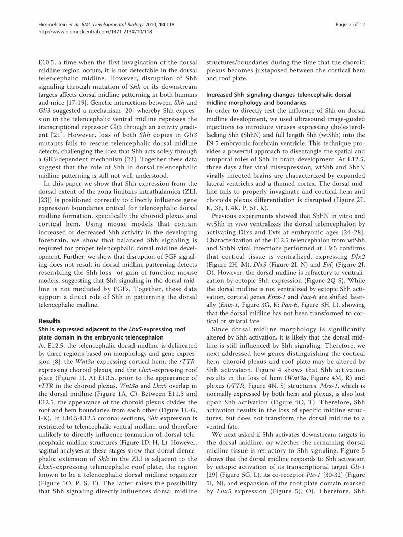
ResultsShh is expressed adjacent to the Lhx5-expressing roofplate domain in the embryonic telencephalonAt E12.5, the telencephalic dorsal midline is delineatedby three regions based on morphology and gene expres-sion [8]: the Wnt3a-expressing cortical hem, the rTTR-expressing choroid plexus, and the Lhx5-expressing roofplate (Figure 1). At E10.5, prior to the appearance ofrTTR in the choroid plexus, Wnt3a and Lhx5 overlap inthe dorsal midline (Figure 1A, C). Between E11.5 andE12.5, the appearance of the choroid plexus divides theroof and hem boundaries from each other (Figure 1E-G,I-K). In E10.5-E12.5 coronal sections, Shh expression isrestricted to telencephalic ventral midline, and thereforeunlikely to directly influence formation of dorsal tele-ncephalic midline structures (Figure 1D, H, L). However,sagittal analyses at these stages show that dorsal dience-phalic extension of Shh in the ZLI is adjacent to theLhx5-expressing telencephalic roof plate, the regionknown to be a telencephalic dorsal midline organizer(Figure 1O, P, S, T). The latter raises the possibilitythat Shh signaling directly influences dorsal midline
structures/boundaries during the time that the choroidplexus becomes juxtaposed between the cortical hemand roof plate.
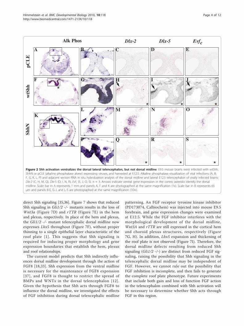
Increased Shh signaling changes telencephalic dorsalmidline morphology and boundariesIn order to directly test the influence of Shh on dorsalmidline development, we used ultrasound image-guidedinjections to introduce viruses expressing cholesterol-lacking Shh (ShhN) and full length Shh (wtShh) into theE9.5 embryonic forebrain ventricle. This technique pro-vides a powerful approach to disentangle the spatial andtemporal roles of Shh in brain development. At E12.5,three days after viral misexpression, wtShh and ShhNvirally infected brains are characterized by expandedlateral ventricles and a thinned cortex. The dorsal mid-line fails to properly invaginate and cortical hem andchoroids plexus differentiation is disrupted (Figure 2F,K, 3E, I, 4K, P, 5F, K).Previous experiments showed that ShhN in vitro and
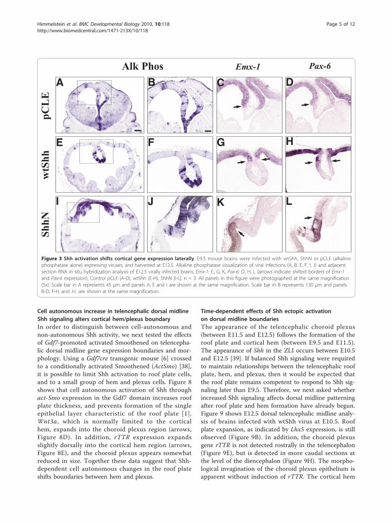
wtShh in vivo ventralizes the dorsal telencephalon byactivating Dlxs and Evfs at embryonic ages [24-28].Characterization of the E12.5 telencephalon from wtShhand ShhN viral infections performed at E9.5 confirmsthat cortical tissue is ventralized, expressing Dlx2(Figure 2H, M), Dlx5 (Figure 2I, N) and Evfc (Figure 2J,O). However, the dorsal midline is refractory to ventrali-zation by ectopic Shh expression (Figure 2Q-S). Whilethe dorsal midline is not ventralized by ectopic Shh acti-vation, cortical genes Emx-1 and Pax-6 are shifted later-ally (Emx-1, Figure 3G, K; Pax-6, Figure 3H, L), showingthat the dorsal midline has not been transformed to cor-tical or striatal fate.Since dorsal midline morphology is significantly
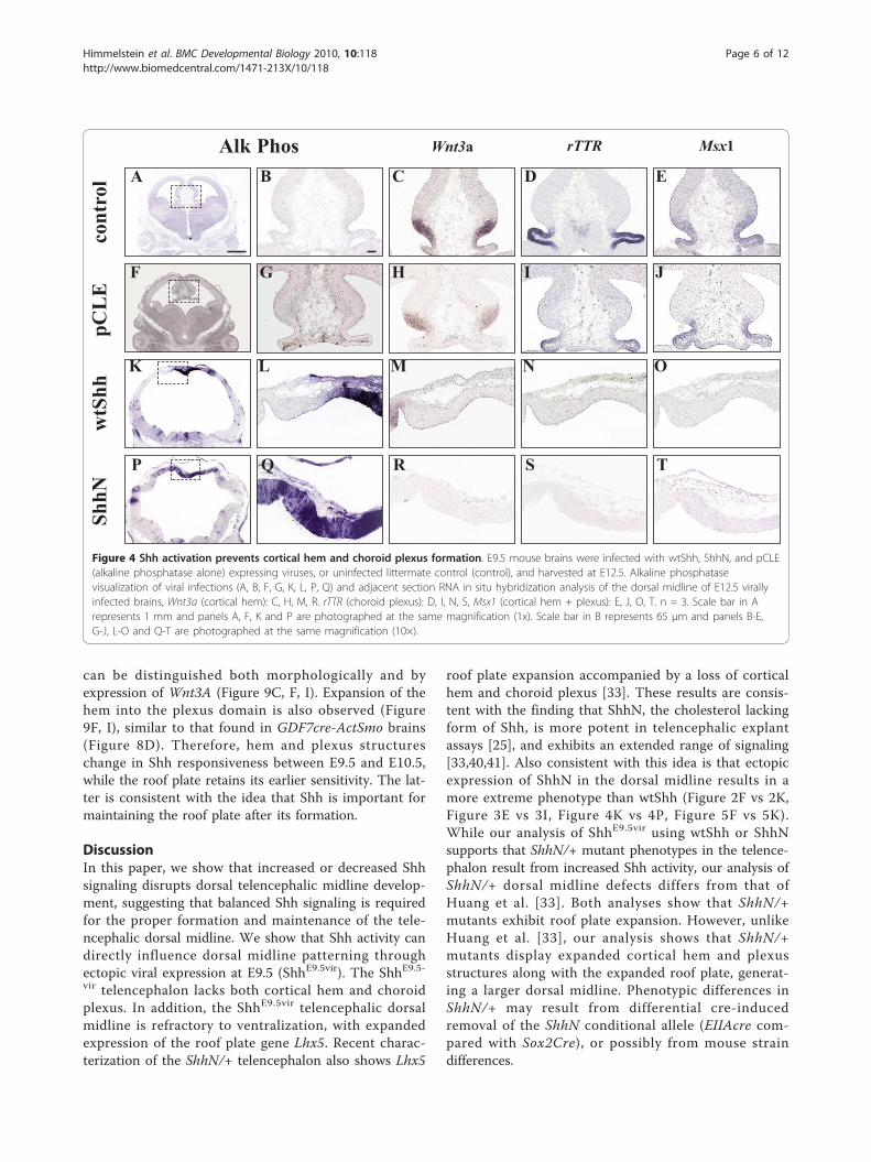
altered by Shh activation, it is likely that the dorsal mid-line is still influenced by Shh signaling. Therefore, wenext addressed how genes distinguishing the corticalhem, choroid plexus and roof plate may be altered byShh activation. Figure 4 shows that Shh activationresults in the loss of hem (Wnt3a, Figure 4M, R) andplexus (rTTR, Figure 4N, S) structures. Msx-1, which isnormally expressed by both hem and plexus, is also lostupon Shh activation (Figure 4O, T). Therefore, Shhactivation results in the loss of specific midline struc-tures, but does not transform the dorsal midline to aventral fate.We next asked if Shh activates downstream targets in
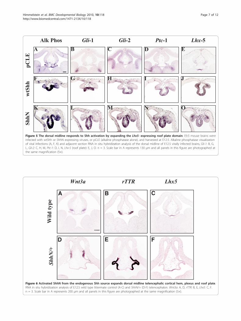
the dorsal midline, or whether the remaining dorsalmidline tissue is refractory to Shh signaling. Figure 5shows that the dorsal midline responds to Shh activationby ectopic activation of its transcriptional target Gli-1[29] (Figure 5G, L), its co-receptor Ptc-1 [30-32] (Figure5I, N), and expansion of the roof plate domain markedby Lhx5 expression (Figure 5J, O). Therefore, Shh
Himmelstein et al. BMC Developmental Biology 2010, 10:118http://www.biomedcentral.com/1471-213X/10/118
Page 2 of 12
activation causes roof plate expansion at the expense ofchoroid plexus and cortical hem.The experiments in Figure 2, 3, 4, and 5 show the
effects of ectopic Shh activation by viral delivery to E9.5telencephalic dorsal midline. In order to address ifincreased Shh signaling from its normal source (ventralmidline or ZLI) would affect telencephalic dorsal mid-line formation, we utilized an activated Shh transgenicmodel where one allele of Shh is replaced by ShhN(ShhN/+) [33]. In this model, ShhN is proposed to travelfurther, resulting in Shh activation in the dorsal telence-phalon. Figure 6 shows that all three telencephalic dor-sal midline boundaries are expanded in ShhN/+ mice(Figure 6D-F). While viral activation and genetic modifi-cation both cause roof plate Lhx5 expansion, rTTR andWnt3a expand in ShhN/+ mice (Figure 6) and areabsent in brains injected with Shh virus (Figure 4).Together, these data not only support the idea that
increased Shh signaling alters telencephalic dorsal mid-line patterning, but that timing and dose are critical fac-tors in determining the outcome.
Telencephalic dorsal midline defects result fromdecreased Shh signalingIncreased Shh signaling, both indirectly through loss ofa repressor (Gli3, [8]) and directly through transgenic orviral expression (this paper) can affect telencephalic dor-sal midline boundaries. However, it is unknown whetherShh normally plays a direct role in patterning the tele-ncephalic dorsal midline. The dramatic loss of bothdorsal and ventral telencephalic midline structures inShh -/- mutants supports the idea that Shh is critical forproper formation of both regions [34]. Since Shh hasbeen shown to activate transcriptional targets throughthe action of Gli1 and Gli2, combined loss of these twotranscription factors reduces, but does not eliminate
Wnt3a rTTR Lhx5 ShhSa
gitt
al
Cor
onal
Cor
onal
C
oron
al
E12
.5
E11
.5
E10
.5
Choroid roof/plaqueChoroid plexusCortical hemCortexGanglionic eminencesVentral midline
A B C D
E F G H
I J K L
M N O P
Q R S T
ZLI
ZLI
U
R C
D
V
Figure 1 Relationship of Shh and dorsal telencephalic midline markers at E10.5, E11.5, and E12.5. RNA in situ hybridization analysis ofdorsal midline telencephalic genes, Wnt3a, rTTR, Lhx5, and ventral midline expression of Shh. Coronal sections [A-L], sagittal sections (M-T). E10.5(A-D), E11.5 (E-H), and E12.5 (I-T). Scale bars = 200 μm. U is a schematic depicting the regional boundaries corresponding to the observed geneexpression domains in a coronal section at mouse embryonic day 12.5. D, Dorsal, V, Ventral, R, Rostral, C, Caudal.
Himmelstein et al. BMC Developmental Biology 2010, 10:118http://www.biomedcentral.com/1471-213X/10/118
Page 3 of 12
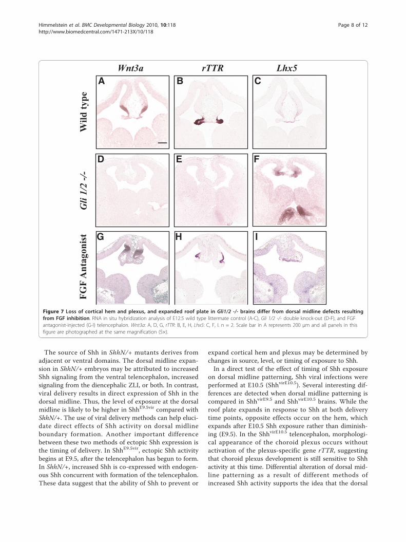
direct Shh signaling [35,36]. Figure 7 shows that reducedShh signaling in Gli1/2 -/- mutants results in the loss ofWnt3a (Figure 7D) and rTTR (Figure 7E) in the hemand plexus, respectively. In place of the hem and plexus,the Gli1/2 -/- mutant telencephalic dorsal midline nowexpresses Lhx5 throughout (Figure 7F), without properthinning to a single epithelial layer characteristic of theroof plate [1]. This suggests that Shh signaling isrequired for inducing proper morphology and geneexpression boundaries that establish the hem, plexusand roof relationships.The current model predicts that Shh indirectly influ-
ences dorsal midline development through the action ofFGF8 [18,33]. Shh expression from the ventral midlineis necessary for the maintenance of FGF8 expression[37], and FGF8 is thought to restrict the spread ofBMPs and WNTs in the dorsal telencephalon [12].Given the hypothesis that Shh acts through FGF8 toinfluence the dorsal midline, we investigated the effectsof FGF inhibition during dorsal telencephalic midline
patterning. An FGF receptor tyrosine kinase inhibitor(PD173074, Calbiochem) was injected into mouse E9.5forebrain, and gene expression changes were examinedat E12.5. While the FGF inhibitor interferes with themorphological development of the dorsal midline,Wnt3A and rTTR are still expressed in the cortical hemand choroid plexus structures, respectively (Figure7G, H). In addition, Lhx5 expansion and thickening ofthe roof plate is not observed (Figure 7I). Therefore, thedorsal midline defects resulting from reduced Shhsignaling (Gli1/2 -/-) are distinct from reduced FGF sig-naling, raising the possibility that Shh signaling in thetelencephalic dorsal midline may be independent ofFGF. However, we cannot rule out the possibility thatFGF inhibition is incomplete, and then fails to generatethe complete roof plate phenotype. Future experimentsthat include both gain and loss of function FGF actionin the telencephalon combined with Shh activation willbe necessary to determine whether Shh acts throughFGF in this region.
C D EA B
M N OL
Dlx-2 Dlx-5 EvfcAlk Phos
ShhN
H I JGF
wtS
hhpC
LE
K
Q R SP
*
* * *
* * *Figure 2 Shh activation ventralizes the dorsal lateral telencephalon, but not dorsal midline. E9.5 mouse brains were infected with wtShh,ShhN or pCLE (alkaline phosphatase alone) expressing viruses, and harvested at E12.5. Alkaline phosphatase visualization of viral infections (A, B,F, G, K, L, P) and adjacent section RNA in situ hybridization analysis of the dorsal midline and lateral E12.5 telencephalon of virally infected brains:Dlx-2 (C, H, M, Q), Dlx-5 (D, I, N, R), Evfc (E, J, O, S). n = 3. Arrows indicate ventral gene expression in the cortex; asterisks identify the dorsalmidline. Scale bar in A represents 1 mm and panels A, F and K are photographed at the same magnification (1x). Scale bar in B represents 65μm and panels B-E, G-J, and L-S are photographed at the same magnification (10×).
Himmelstein et al. BMC Developmental Biology 2010, 10:118http://www.biomedcentral.com/1471-213X/10/118
Page 4 of 12
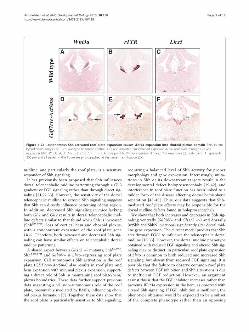
Cell autonomous increase in telencephalic dorsal midlineShh signaling alters cortical hem/plexus boundaryIn order to distinguish between cell-autonomous andnon-autonomous Shh activity, we next tested the effectsof Gdf7-promoted activated Smoothened on telencepha-lic dorsal midline gene expression boundaries and mor-phology. Using a Gdf7cre transgenic mouse [6] crossedto a conditionally activated Smoothened (ActSmo) [38],it is possible to limit Shh activation to roof plate cells,and to a small group of hem and plexus cells. Figure 8shows that cell autonomous activation of Shh throughact-Smo expression in the Gdf7 domain increases roofplate thickness, and prevents formation of the singleepithelial layer characteristic of the roof plate [1].Wnt3a, which is normally limited to the corticalhem, expands into the choroid plexus region (arrows,Figure 8D). In addition, rTTR expression expandsslightly dorsally into the cortical hem region (arrows,Figure 8E), and the choroid plexus appears somewhatreduced in size. Together these data suggest that Shh-dependent cell autonomous changes in the roof plateshifts boundaries between hem and plexus.
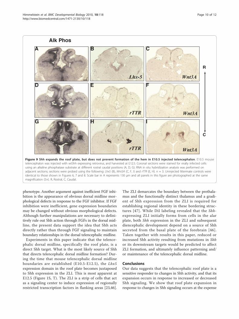
Time-dependent effects of Shh ectopic activationon dorsal midline boundariesThe appearance of the telencephalic choroid plexus(between E11.5 and E12.5) follows the formation of theroof plate and cortical hem (between E9.5 and E11.5).The appearance of Shh in the ZLI occurs between E10.5and E12.5 [39]. If balanced Shh signaling were requiredto maintain relationships between the telencephalic roofplate, hem, and plexus, then it would be expected thatthe roof plate remains competent to respond to Shh sig-naling later than E9.5. Therefore, we next asked whetherincreased Shh signaling affects dorsal midline patterningafter roof plate and hem formation have already begun.Figure 9 shows E12.5 dorsal telencephalic midline analy-sis of brains infected with wtShh virus at E10.5. Roofplate expansion, as indicated by Lhx5 expression, is stillobserved (Figure 9B). In addition, the choroid plexusgene rTTR is not detected rostrally in the telencephalon(Figure 9E), but is detected in more caudal sections atthe level of the diencephalon (Figure 9H). The morpho-logical invagination of the choroid plexus epithelium isapparent without induction of rTTR. The cortical hem
Emx-1 Pax-6
wtS
hhpC
LE
ShhN
Alk Phos
I J K L
E F G H
A B C D
Figure 3 Shh activation shifts cortical gene expression laterally. E9.5 mouse brains were infected with wtShh, ShhN or pCLE (alkalinephosphatase alone) expressing viruses, and harvested at E12.5. Alkaline phosphatase visualization of viral infections (A, B, E, F, I, J) and adjacentsection RNA in situ hybridization analysis of E12.5 virally infected brains, Emx-1: C, G, K, Pax-6: D, H, L (arrows indicate shifted borders of Emx-1and Pax-6 expression). Control pCLE (A-D), wtShh (E-H), ShhN [I-L]. n = 3. All panels in this figure were photographed at the same magnification(5x). Scale bar in A represents 45 μm and panels A, E and I are shown at the same magnification. Scale bar in B represents 130 μm and panelsB-D, F-H, and J-L are shown at the same magnification.
Himmelstein et al. BMC Developmental Biology 2010, 10:118http://www.biomedcentral.com/1471-213X/10/118
Page 5 of 12
can be distinguished both morphologically and byexpression of Wnt3A (Figure 9C, F, I). Expansion of thehem into the plexus domain is also observed (Figure9F, I), similar to that found in GDF7cre-ActSmo brains(Figure 8D). Therefore, hem and plexus structureschange in Shh responsiveness between E9.5 and E10.5,while the roof plate retains its earlier sensitivity. The lat-ter is consistent with the idea that Shh is important formaintaining the roof plate after its formation.
DiscussionIn this paper, we show that increased or decreased Shhsignaling disrupts dorsal telencephalic midline develop-ment, suggesting that balanced Shh signaling is requiredfor the proper formation and maintenance of the tele-ncephalic dorsal midline. We show that Shh activity candirectly influence dorsal midline patterning throughectopic viral expression at E9.5 (ShhE9.5vir). The ShhE9.5-vir telencephalon lacks both cortical hem and choroidplexus. In addition, the ShhE9.5vir telencephalic dorsalmidline is refractory to ventralization, with expandedexpression of the roof plate gene Lhx5. Recent charac-terization of the ShhN/+ telencephalon also shows Lhx5
roof plate expansion accompanied by a loss of corticalhem and choroid plexus [33]. These results are consis-tent with the finding that ShhN, the cholesterol lackingform of Shh, is more potent in telencephalic explantassays [25], and exhibits an extended range of signaling[33,40,41]. Also consistent with this idea is that ectopicexpression of ShhN in the dorsal midline results in amore extreme phenotype than wtShh (Figure 2F vs 2K,Figure 3E vs 3I, Figure 4K vs 4P, Figure 5F vs 5K).While our analysis of ShhE9.5vir using wtShh or ShhNsupports that ShhN/+ mutant phenotypes in the telence-phalon result from increased Shh activity, our analysis ofShhN/+ dorsal midline defects differs from that ofHuang et al. [33]. Both analyses show that ShhN/+mutants exhibit roof plate expansion. However, unlikeHuang et al. [33], our analysis shows that ShhN/+mutants display expanded cortical hem and plexusstructures along with the expanded roof plate, generat-ing a larger dorsal midline. Phenotypic differences inShhN/+ may result from differential cre-inducedremoval of the ShhN conditional allele (EIIAcre com-pared with Sox2Cre), or possibly from mouse straindifferences.
Alk Phos
pCL
E
rTTRWnt3a
wtS
hh
Msx1
ShhN
cont
rol A B C D E
F G H I J
K L M N O
P Q R S T
Figure 4 Shh activation prevents cortical hem and choroid plexus formation. E9.5 mouse brains were infected with wtShh, ShhN, and pCLE(alkaline phosphatase alone) expressing viruses, or uninfected littermate control (control), and harvested at E12.5. Alkaline phosphatasevisualization of viral infections (A, B, F, G, K, L, P, Q) and adjacent section RNA in situ hybridization analysis of the dorsal midline of E12.5 virallyinfected brains, Wnt3a (cortical hem): C, H, M, R. rTTR (choroid plexus): D, I, N, S, Msx1 (cortical hem + plexus): E, J, O, T. n = 3. Scale bar in Arepresents 1 mm and panels A, F, K and P are photographed at the same magnification (1x). Scale bar in B represents 65 μm and panels B-E,G-J, L-O and Q-T are photographed at the same magnification (10×).
Himmelstein et al. BMC Developmental Biology 2010, 10:118http://www.biomedcentral.com/1471-213X/10/118
Page 6 of 12
Gli-1 Lhx-5Ptc-1Gli-2Alk Phos
pCL
Ew
tShh
ShhN
C EDBA
H JIGF
M ONLK
Figure 5 The dorsal midline responds to Shh activation by expanding the Lhx5- expressing roof plate domain. E9.5 mouse brains wereinfected with wtShh or ShhN expressing viruses, or pCLE (alkaline phosphatase alone), and harvested at E12.5. Alkaline phosphatase visualizationof viral infections (A, F, K) and adjacent section RNA in situ hybridization analysis of the dorsal midline of E12.5 virally infected brains, Gli-1: B, G,L, Gli-2: C, H, M, Ptc-1: D, I, N, Lhx-5 (roof plate): E, J, O. n = 3. Scale bar in A represents 130 μm and all panels in this figure are photographed atthe same magnification (5×).
Wnt3a rTTR Lhx5
A B C
FED
Wild
type
ShhN
/+
Figure 6 Activated ShhN from the endogenous Shh source expands dorsal midline telencephalic cortical hem, plexus and roof plate.RNA in situ hybridization analysis of E12.5 wild type littermate control (A-C) and ShhN/+ (D-F) telencephalon. Wnt3a: A, D, rTTR: B, E, Lhx5: C, F.n = 3. Scale bar in A represents 200 μm and all panels in this figure are photographed at the same magnification (5×).
Himmelstein et al. BMC Developmental Biology 2010, 10:118http://www.biomedcentral.com/1471-213X/10/118
Page 7 of 12
The source of Shh in ShhN/+ mutants derives fromadjacent or ventral domains. The dorsal midline expan-sion in ShhN/+ embryos may be attributed to increasedShh signaling from the ventral telencephalon, increasedsignaling from the diencephalic ZLI, or both. In contrast,viral delivery results in direct expression of Shh in thedorsal midline. Thus, the level of exposure at the dorsalmidline is likely to be higher in ShhE9.5vir compared withShhN/+. The use of viral delivery methods can help eluci-date direct effects of Shh activity on dorsal midlineboundary formation. Another important differencebetween these two methods of ectopic Shh expression isthe timing of delivery. In ShhE9.5vir, ectopic Shh activitybegins at E9.5, after the telencephalon has begun to form.In ShhN/+, increased Shh is co-expressed with endogen-ous Shh concurrent with formation of the telencephalon.These data suggest that the ability of Shh to prevent or
expand cortical hem and plexus may be determined bychanges in source, level, or timing of exposure to Shh.In a direct test of the effect of timing of Shh exposure
on dorsal midline patterning, Shh viral infections wereperformed at E10.5 (ShhvirE10.5). Several interesting dif-ferences are detected when dorsal midline patterning iscompared in ShhvirE9.5 and ShhvirE10.5 brains. While theroof plate expands in response to Shh at both deliverytime points, opposite effects occur on the hem, whichexpands after E10.5 Shh exposure rather than diminish-ing (E9.5). In the ShhvirE10.5 telencephalon, morphologi-cal appearance of the choroid plexus occurs withoutactivation of the plexus-specific gene rTTR, suggestingthat choroid plexus development is still sensitive to Shhactivity at this time. Differential alteration of dorsal mid-line patterning as a result of different methods ofincreased Shh activity supports the idea that the dorsal
A C
E
B
D
Wnt3a rTTR Lhx5
F
Wild
type
Gli
1/2
-/-
HG I
FGF
Ant
agon
ist
Figure 7 Loss of cortical hem and plexus, and expanded roof plate in Gli1/2 -/- brains differ from dorsal midline defects resultingfrom FGF inhibition. RNA in situ hybridization analysis of E12.5 wild type littermate control (A-C), Gli 1/2 -/- double knock-out (D-F), and FGFantagonist-injected (G-I) telencephalon. Wnt3a: A, D, G, rTTR: B, E, H, Lhx5: C, F, I. n = 2. Scale bar in A represents 200 μm and all panels in thisfigure are photographed at the same magnification (5×).
Himmelstein et al. BMC Developmental Biology 2010, 10:118http://www.biomedcentral.com/1471-213X/10/118
Page 8 of 12
midline, and particularly the roof plate, is a sensitiveresponder of Shh signaling.It has previously been proposed that Shh influences
dorsal telencephalic midline patterning through a Gli3gradient or FGF signaling rather than through direct sig-naling [21,22,33]. However, the sensitivity of the dorsaltelencephalic midline to ectopic Shh signaling suggeststhat Shh can directly influence patterning of this region.In addition, decreased Shh signaling in mice lackingboth Gli1 and Gli2 results in dorsal telencephalic mid-line defects similar to that found when Shh is increased(ShhE9.5vir): loss of cortical hem and choroid plexus,with a concomitant expansion of the roof plate geneLhx5. Therefore, both increased and decreased Shh sig-naling can have similar effects on telencephalic dorsalmidline patterning.A shared aspect between Gli1/2 -/- mutants, ShhE9.5vir,
ShhE10.5vir and ShhN/+ is Lhx5-expressing roof plateexpansion. Cell autonomous Shh activation in the roofplate (GDF7cre-ActSmo) also results in roof plate andhem expansion with minimal plexus expansion, support-ing a direct role of Shh in maintaining roof plate/hem/plexus boundaries. These data further support previousdata suggesting a cell-non-autonomous role of the roofplate, presumably mediated by BMPs, influencing chor-oid plexus formation [5]. Together, these data show thatthe roof plate is particularly sensitive to Shh signaling,
requiring a balanced level of Shh activity for propermorphology and gene expression. Interestingly, muta-tions in Shh or its downstream targets result in thedevelopmental defect holoprosencephaly [19,42], andinterference in roof plate function has been linked to amilder form of the disease affecting dorsal hemisphericseparation [43-45]. Thus, our data suggests that Shh-mediated roof plate effects may be responsible for thedorsal midline defects found in holoprosencephaly.We show that both increases and decreases in Shh sig-
naling ventrally (ShhN/+ and Gli1/2 -/-) and dorsally(wtShh and ShhN injections) significantly alter dorsal mid-line gene expression. The current model predicts that Shhacts through FGF8 to influence the telencephalic dorsalmidline [18,33]. However, the dorsal midline phenotypeobtained with reduced FGF signaling and altered Shh sig-naling may be distinct. In particular, roof plate expansionof Lhx5 is common to both reduced and increased Shhsignaling, but absent from reduced FGF signaling. It ispossible that the failure to observe common roof platedefects between FGF inhibition and Shh alterations is dueto inefficient FGF reduction. However, an argumentagainst this is that the FGF inhibitor increases rather thanprevents Wnt3a expression in the hem, as observed withaltered Shh signaling. If FGF inhibition is inefficient, thephenotype obtained would be expected to be a subsetof the complete phenotype rather than an opposing
Wnt3a rTTR Lhx5
F
C
E
B
D
AG
df7c
re-A
ctSm
o W
ild t
ype
Figure 8 Cell autonomous Shh-activated roof plate expansion causes Wnt3a expansion into choroid plexus domain. RNA in situhybridization analysis of E12.5 wild type littermate control (A-C) and activated Smoothened expressed in the roof plate through GDF7creregulation (D-F). Wnt3a: A, D, rTTR: B, E, Lhx5: C, F. n = 3. Arrows point to Wnt3a expansion (D) and rTTR expansion (E). Scale bar in A represents200 μm and all panels in this figure are photographed at the same magnification (5×).
Himmelstein et al. BMC Developmental Biology 2010, 10:118http://www.biomedcentral.com/1471-213X/10/118
Page 9 of 12
phenotype. Another argument against inefficient FGF inhi-bition is the appearance of obvious dorsal midline mor-phological defects in response to the FGF inhibitor. If FGFinhibition were inefficient, gene expression boundariesmay be changed without obvious morphological defects.Although further manipulations are necessary to defini-tively rule out Shh action through FGFs in the dorsal mid-line, the present data support the idea that Shh actsdirectly rather than through FGF signaling to maintainboundary relationships in the dorsal telencephalic midline.Experiments in this paper indicate that the telence-
phalic dorsal midline, specifically the roof plate, is adirect Shh target. What is the most likely source of Shhthat directs telencephalic dorsal midline formation? Dur-ing the time that mouse telencephalic dorsal midlineboundaries are established (E10.5-E12.5), the Lhx5expression domain in the roof plate becomes juxtaposedto Shh expression in the ZLI. This is most apparent atE12.5 (Figure 1S, T). The ZLI is a strip of cells that actas a signaling center to induce expression of regionallyrestricted transcription factors in flanking areas [23,46].
The ZLI demarcates the boundary between the prethala-mus and the functionally distinct thalamus and a gradi-ent of Shh expression from the ZLI is required forestablishing regional identity in these bordering struc-tures [47]. While DiI labeling revealed that the Shh-expressing ZLI initially forms from cells in the alarplate, both Shh expression in the ZLI and subsequentdiencephalic development depend on a source of Shhsecreted from the basal plate of the forebrain [46].Taken together with results in this paper, reduced orincreased Shh activity resulting from mutations in Shhor its downstream targets would be predicted to affectZLI formation, and ultimately influence patterning and/or maintenance of the telencephalic dorsal midline.
ConclusionsOur data suggests that the telencephalic roof plate is asensitive responder to changes in Shh activity, and that itsexpansion occurs in response to increased or decreasedShh signaling. We show that roof plate expansion inresponse to changes in Shh signaling occurs at the expense
Figure 9 Shh expands the roof plate, but does not prevent formation of the hem in E10.5 injected telencephalon. E10.5 mousetelencephalon was injected with wtShh expressing retrovirus, and harvested at E12.5. Coronal sections were stained for virally infected cellsusing an alkaline phosphatase substrate at different rostral caudal positions (A, D, G). RNA in situ hybridization analysis was performed onadjacent sections; sections were probed using the following: Lhx5 (B), Wnt3A (C, F, I) and rTTR (E, H). n = 3. Uninjected littermate controls wereidentical to those shown in Figures 6, 7 and 8. Scale bar in A represents 130 μm and all panels in this figure are photographed at the samemagnification (5×). R, Rostral, C, Caudal.
Himmelstein et al. BMC Developmental Biology 2010, 10:118http://www.biomedcentral.com/1471-213X/10/118
Page 10 of 12
of the hem and plexus or can lead to expansion of the hemand plexus. In addition, responsiveness of the telencepha-lic dorsal midline to Shh signaling changes between E9.5and E10.5 in mice. The structural relationship of Shh anddorsal midline markers raises the possibility that dorsallyexpressed Shh from the diencephalic ZLI is a source ofShh signaling to the telencephalic dorsal midline. We pro-pose that imbalanced Shh signaling directly changes dorsalmidline boundaries between the roof plate, choroid plexusand cortical hem.
MethodsAnimals and SurgeryShhN floxed mice [48] were obtained from Dr. A. McMa-hon, and crossed to EIIAcre mice (Jackson Lab) to gener-ate ShhN/+ embryos at E12.5. ActSmo floxed mice [38]were crossed to GDF7cre mice [6] to generate GDF7cre-ActSmo E12.5 embryos. Animals used in these studieswere maintained according to protocols approved byInstitutional Animal Care and Use Committee at Chil-dren’s Memorial Hospital Research Center. Timed-pregnant Swiss Webster mice used for injections wereobtained from Taconic breeding laboratories. Embryonicday 0.5 is defined as noon of the day a vaginal plug wasfound after overnight mating. Detailed animal care, pre-paration for surgery and the use of the ultrasound scan-ner (UBM scanner) have been previously described [49].
Viral expression and embryonic injectionsShh full-length cDNA (wtShh) and the N-terminal frag-ment (ShhN) were inserted into the pCLE viral vectorbackbone, and pseudo-typed retrovirus was produced aspreviously described [26]. Viruses were titered on theC17 neural cell line [50]. Western analysis (not shown)on C17 infected extracts was performed with rabbitanti-Shh antibody (1:5000, R&D). Viruses were dilutedto 5 × 10⁷ cfu/ml in PBS containing 80 μg/ml polybrene(Sigma). FGF receptor tyrosine kinase inhibitorPD173074, Calbiochem) was dissolved in DMSO anddiluted to 25 μM in PBS containing 80 μg/ml polybrene(sigma). FGF receptor tyrosine inhibitor PD173074, cal-biochem) was dissolved in DMSO and diluted to 25 μMin PBS containing 80 μg/ml polybrene. 1-1.5 μl of virusor FGF inhibitor was injected into the E9.5 or E10.5mouse telencephalon using ultrasound-guided in uteroinjection, as previously described [26,49,51]. Embryoswere harvested 2 or 3 days after injection, as indicated.
In situ hybridizationIn situ hybridization was performed by modification ofSchaeren-Wiemers and Gerfin-Moser [52] as previouslydescribed [27], with the exception of the rTTR probe,which was visualized using the method of Tekki-Kessariset al. [53]. The Lhx5 in situ probe was made from
pGEM-Teasy (Promega) after subcloning an RT-PCRfragment made from E12.5 mouse brain RNA using thefollowing primers:5’ Primer - 5’ACA TGA GGG TCA TTC AGG TGT
GGT 3’3’ Primer - 5’ TGT GCT TGG AAT CTC GAC CCT
TCA 3’
AcknowledgementsWe thank Kathy Millen for helpful discussions, Raj Awatramani. for GDF7cre-ActSmo embryos, and the following investigators for probes: Wnt3A(P. Salinas), rTTR (W. Duan), Msx1 (C. Abate-Shen), Emx1 (E. Boccinelli), Pax-6(P. Gruss), Dlx-2, Dlx-5 (J. Rubenstein), Gli1, Gli2, Ptc1 (M. Matise). This workwas funded by NICHHD RO1 HD044745 and HD056504 and IllinoisExcellence in Academic Medicine to J.D.K.
Author details1Developmental Biology and Department of Pediatrics, Children’s MemorialResearch Center and Feinberg School of Medicine, Northwestern University,Chicago, IL, USA. 2Department of Cell Biology, Neurobiology and Anatomy,Medical College of Wisconsin, Milwaukee, WI, USA. 3Department of Genetics,Case Western Reserve University, Cleveland, OH, USA.
Authors’ contributionsDSH performed experiments and prepared the manuscript. CB and BCperformed experiments. BB contributed Gli1/2 -/- embryos. JDK designedexperiments and prepared the manuscript. All authors read and approvedthe manuscript.
Received: 28 June 2010 Accepted: 29 November 2010Published: 29 November 2010
References1. Chizhikov VV, Millen KJ: Roof plate-dependent patterning of the
vertebrate dorsal central nervous system. Dev Biol 2005, 277:287-95.2. O’Leary DD, Sahara S: Genetic regulation of arealization of the neocortex.
Curr Opin Neurobiol 2008, 18:90-100.3. Liem KF, Tremml G, Jessell TM: A role for the roof plate and its resident
TGFbeta-related proteins in neuronal patterning in the dorsal spinalcord. Cell 1997, 91:127-38.
4. Awatramani R, Soriano P, Rodriguez C, Mai JJ, Dymecki SM: Crypticboundaries in roof plate and choroid plexus identified by intersectionalgene activation. Nat Genet 2003, 35:70-5.
5. Currle DS, Cheng X, Hsu CM, Monuki ES: Direct and indirect roles of CNSdorsal midline cells in choroid plexus epithelia formation. Development2005, 132:3549-59.
6. Lee KJ, Dietrich P, Jessell TM: Genetic ablation reveals that the roof plateis essential for dorsal interneuron specification. Nature 2000, 403:734-40.
7. Monuki ES, Porter FD, Walsh CA: Patterning of the dorsal telencephalonand cerebral cortex by a roof plate-Lhx2 pathway. Neuron 2001,32:591-604.
8. Grove EA, Tole S, Limon J, Yip L, Ragsdale CW: The hem of the embryoniccerebral cortex is defined by the expression of multiple Wnt genes andis compromised in Gli3-deficient mice. Development 1998, 125:2315-25.
9. Mangale VS, Hirokawa KE, Satyaki PR, Gokulchandran N, Chikbire S,Subramanian L, Shetty AS, Martynoga B, Paul J, Mai MV, et al: Lhx2 selectoractivity specifies cortical identity and suppresses hippocampal organizerfate. Science 2008, 319:304-9.
10. Galceran J, Miyashita-Lin EM, Devaney E, Rubenstein JL, Grosschedl R:Hippocampus development and generation of dentate gyrus granulecells is regulated by LEF1. Development 2000, 127:469-82.
11. Lee SM, Tole S, Grove E, McMahon AP: A local Wnt-3a signal is requiredfor development of the mammalian hippocampus. Development 2000,127:457-67.
12. Shimogori T, Banuchi V, Ng HY, Strauss JB, Grove EA: Embryonic signalingcenters expressing BMP, WNT and FGF proteins interact to pattern thecerebral cortex. Development 2004, 131:5639-47.
Himmelstein et al. BMC Developmental Biology 2010, 10:118http://www.biomedcentral.com/1471-213X/10/118
Page 11 of 12
13. Takahashi H, Liu FC: Genetic patterning of the mammalian telencephalonby morphogenetic molecules and transcription factors. Birth Defects Res CEmbryo Today 2006, 78:256-66.
14. Wilson L, Maden M: The mechanisms of dorsoventral patterning in thevertebrate neural tube. Dev Biol 2005, 282:1-13.
15. Fukuchi-Shimogori T, Grove EA: Neocortex patterning by the secretedsignaling molecule FGF8. Science 2001, 294:1071-4.
16. Storm EE, Garel S, Borello U, Hebert JM, Martinez S, McConnell SK,Martin GR, Rubenstein JL: Dose-dependent functions of Fgf8 in regulatingtelencephalic patterning centers. Development 2006, 133:1831-44.
17. Hayhurst M, McConnell SK: Mouse models of holoprosencephaly. CurrOpin Neurol 2003, 16:135-41.
18. Monuki ES: The morphogen signaling network in forebrain developmentand holoprosencephaly. J Neuropathol Exp Neurol 2007, 66:566-75.
19. Muenke M, Beachy PA: Genetics of ventral forebrain development andholoprosencephaly. Curr Opin Genet Dev 2000, 10:262-9.
20. Litingtung Y, Chiang C: Specification of ventral neuron types is mediatedby an antagonistic interaction between Shh and Gli3. Nat Neurosci 2000,3:979-85.
21. Rallu M, Machold R, Gaiano N, Corbin JG, McMahon AP, Fishell G:Dorsoventral patterning is established in the telencephalon of mutantslacking both Gli3 and Hedgehog signaling. Development 2002,129:4963-74.
22. Rash BG, Grove EA: Patterning the dorsal telencephalon: a role for sonichedgehog? J Neurosci 2007, 27:11595-603.
23. Kiecker C, Lumsden A: Hedgehog signaling from the ZLI regulatesdiencephalic regional identity. Nat Neurosci 2004, 7:1242-9.
24. Feng J, Bi C, Clark BS, Mady R, Shah P, Kohtz JD: The Evf-2 noncoding RNAis transcribed from the Dlx-5/6 ultraconserved region and functions as aDlx-2 transcriptional coactivator. Genes Dev 2006, 20:1470-84.
25. Feng J, White B, Tyurina OV, Guner B, Larson T, Lee HY, Karlstrom RO,Kohtz JD: Synergistic and antagonistic roles of the Sonic hedgehog N-and C-terminal lipids. Development 2004, 131:4357-70.
26. Gaiano N, Kohtz JD, Turnbull DH, Fishell G: A method for rapid gain-of-function studies in the mouse embryonic nervous system. Nat Neurosci1999, 2:812-9.
27. Kohtz JD, Baker DP, Corte G, Fishell G: Regionalization within themammalian telencephalon is mediated by changes in responsiveness toSonic Hedgehog. Development 1998, 125:5079-89.
28. Kohtz JD, Lee HY, Gaiano N, Segal J, Ng E, Larson T, Baker DP, Garber EA,Williams KP, Fishell G: N-terminal fatty-acylation of sonic hedgehogenhances the induction of rodent ventral forebrain neurons.Development 2001, 128:2351-63.
29. Lee J, Platt KA, Censullo P, Ruiz i Altaba A: Gli1 is a target of Sonichedgehog that induces ventral neural tube development. Development1997, 124:2537-52.
30. Goodrich LV, Johnson RL, Milenkovic L, McMahon JA, Scott MP:Conservation of the hedgehog/patched signaling pathway from flies tomice: induction of a mouse patched gene by Hedgehog. Genes Dev 1996,10:301-12.
31. Marigo V, Davey RA, Zuo Y, Cunningham JM, Tabin CJ: Biochemicalevidence that patched is the Hedgehog receptor. Nature 1996, 384:176-9.
32. Stone DM, Hynes M, Armanini M, Swanson TA, Gu Q, Johnson RL, Scott MP,Pennica D, Goddard A, Phillips H, et al: The tumour-suppressor genepatched encodes a candidate receptor for Sonic hedgehog. Nature 1996,384:129-34.
33. Huang X, Litingtung Y, Chiang C: Ectopic sonic hedgehog signalingimpairs telencephalic dorsal midline development: implication forhuman holoprosencephaly. Hum Mol Genet 2007, 16:1454-68.
34. Chiang C, Litingtung Y, Lee E, Young KE, Corden JL, Westphal H, Beachy PA:Cyclopia and defective axial patterning in mice lacking Sonic hedgehoggene function. Nature 1996, 383:407-13.
35. Bai CB, Auerbach W, Lee JS, Stephen D, Joyner AL: Gli2, but not Gli1, isrequired for initial Shh signaling and ectopic activation of the Shhpathway. Development 2002, 129:4753-61.
36. Park HL, Bai C, Platt KA, Matise MP, Beeghly A, Hui CC, Nakashima M,Joyner AL: Mouse Gli1 mutants are viable but have defects in SHHsignaling in combination with a Gli2 mutation. Development 2000,127:1593-605.
37. Ohkubo Y, Chiang C, Rubenstein JL: Coordinate regulation and synergisticactions of BMP4, SHH and FGF8 in the rostral prosencephalon regulate
morphogenesis of the telencephalic and optic vesicles. Neuroscience2002, 111:1-17.
38. Hynes M, Ye W, Wang K, Stone D, Murone M, Sauvage F, Rosenthal A: Theseven-transmembrane receptor smoothened cell-autonomously inducesmultiple ventral cell types. Nat Neurosci 2000, 3:41-6.
39. Echelard Y, Epstein DJ, St-Jacques B, Shen L, Mohler J, McMahon JA,McMahon AP: Sonic hedgehog, a member of a family of putativesignaling molecules, is implicated in the regulation of CNS polarity. Cell1993, 75:1417-30.
40. Huang X, Litingtung Y, Chiang C: Region-specific requirement forcholesterol modification of sonic hedgehog in patterning thetelencephalon and spinal cord. Development 2007, 134:2095-105.
41. Li Y, Zhang H, Litingtung Y, Chiang C: Cholesterol modification restrictsthe spread of Shh gradient in the limb bud. Proc Natl Acad Sci USA 2006,103:6548-53.
42. Shiota K, Yamada S, Komada M, Ishibashi M: Embryogenesis ofholoprosencephaly. Am J Med Genet A 2007, 143A:3079-87.
43. Barkovich AJ, Quint DJ: Middle interhemispheric fusion: an unusualvariant of holoprosencephaly. AJNR Am J Neuroradiol 1993, 14:431-40.
44. Cheng X, Hsu CM, Currle DS, Hu JS, Barkovich AJ, Monuki ES: Central rolesof the roof plate in telencephalic development and holoprosencephaly.J Neurosci 2006, 26:7640-9.
45. Simon EM, Hevner RF, Pinter JD, Clegg NJ, Delgado M, Kinsman SL,Hahn JS, Barkovich AJ: The middle interhemispheric variant ofholoprosencephaly. AJNR Am J Neuroradiol 2002, 23:151-6.
46. Zeltser LM: Shh-dependent formation of the ZLI is opposed by signalsfrom the dorsal diencephalon. Development 2005, 132:2023-33.
47. Vieira C, Martinez S: Sonic hedgehog from the basal plate and the zonalimitans intrathalamica exhibits differential activity on diencephalicmolecular regionalization and nuclear structure. Neuroscience 2006,143:129-40.
48. Tian H, Jeong J, Harfe BD, Tabin CJ, McMahon AP: Mouse Disp1 is requiredin sonic hedgehog-expressing cells for paracrine activity of thecholesterol-modified ligand. Development 2005, 132:133-42.
49. Liu A, Joyner AL, Turnbull DH: Alteration of limb and brain patterning inearly mouse embryos by ultrasound-guided injection of Shh-expressingcells. Mech Dev 1998, 75:107-15.
50. Snyder EY, Deitcher DL, Walsh C, Arnold-Aldea S, Hartwieg EA, Cepko CL:Multipotent neural cell lines can engraft and participate in developmentof mouse cerebellum. Cell 1992, 68:33-51.
51. Olsson M, Campbell K, Turnbull DH: Specification of mouse telencephalicand mid-hindbrain progenitors following heterotopic ultrasound-guidedembryonic transplantation. Neuron 1997, 19:761-72.
52. Schaeren-Wiemers N, Gerfin-Moser A: A single protocol to detecttranscripts of various types and expression levels in neural tissue andcultured cells: in situ hybridization using digoxigenin-labelled cRNAprobes. Histochemistry 1993, 100:431-40.
53. Tekki-Kessaris N, Woodruff R, Hall AC, Gaffield W, Kimura S, Stiles CD,Rowitch DH, Richardson WD: Hedgehog-dependent oligodendrocytelineage specification in the telencephalon. Development 2001,128:2545-54.
doi:10.1186/1471-213X-10-118Cite this article as: Himmelstein et al.: Balanced Shh signaling isrequired for proper formation and maintenance of dorsal telencephalicmidline structures. BMC Developmental Biology 2010 10:118.
Himmelstein et al. BMC Developmental Biology 2010, 10:118http://www.biomedcentral.com/1471-213X/10/118
Page 12 of 12
Copyright © 2022 FDOKUMEN