Kv3.1b and Kv3.3 channel subunit expression in murine spinal dorsal horn GABAergic interneurones
16
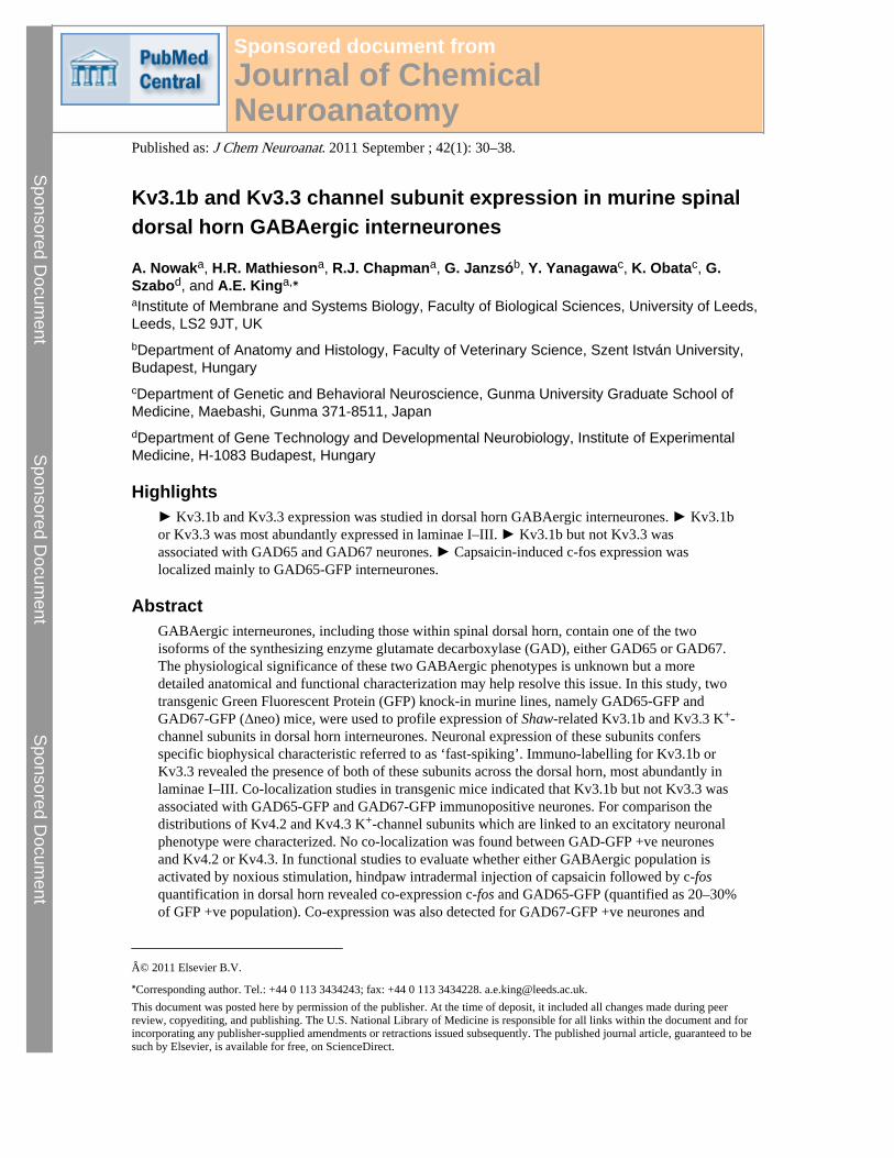
Kv3.1b and Kv3.3 channel subunit expression in murine spinal dorsal horn GABAergic interneurones A. Nowak a , H.R. Mathieson a , R.J. Chapman a , G. Janzsó b , Y. Yanagawa c , K. Obata c , G. Szabo d , and A.E. King a,⁎ a Institute of Membrane and Systems Biology, Faculty of Biological Sciences, University of Leeds, Leeds, LS2 9JT, UK b Department of Anatomy and Histology, Faculty of Veterinary Science, Szent István University, Budapest, Hungary c Department of Genetic and Behavioral Neuroscience, Gunma University Graduate School of Medicine, Maebashi, Gunma 371-8511, Japan d Department of Gene Technology and Developmental Neurobiology, Institute of Experimental Medicine, H-1083 Budapest, Hungary Highlights ► Kv3.1b and Kv3.3 expression was studied in dorsal horn GABAergic interneurones. ► Kv3.1b or Kv3.3 was most abundantly expressed in laminae I–III. ► Kv3.1b but not Kv3.3 was associated with GAD65 and GAD67 neurones. ► Capsaicin-induced c-fos expression was localized mainly to GAD65-GFP interneurones. Abstract GABAergic interneurones, including those within spinal dorsal horn, contain one of the two isoforms of the synthesizing enzyme glutamate decarboxylase (GAD), either GAD65 or GAD67. The physiological significance of these two GABAergic phenotypes is unknown but a more detailed anatomical and functional characterization may help resolve this issue. In this study, two transgenic Green Fluorescent Protein (GFP) knock-in murine lines, namely GAD65-GFP and GAD67-GFP (Δneo) mice, were used to profile expression of Shaw-related Kv3.1b and Kv3.3 K + - channel subunits in dorsal horn interneurones. Neuronal expression of these subunits confers specific biophysical characteristic referred to as ‘fast-spiking’. Immuno-labelling for Kv3.1b or Kv3.3 revealed the presence of both of these subunits across the dorsal horn, most abundantly in laminae I–III. Co-localization studies in transgenic mice indicated that Kv3.1b but not Kv3.3 was associated with GAD65-GFP and GAD67-GFP immunopositive neurones. For comparison the distributions of Kv4.2 and Kv4.3 K + -channel subunits which are linked to an excitatory neuronal phenotype were characterized. No co-localization was found between GAD-GFP +ve neurones and Kv4.2 or Kv4.3. In functional studies to evaluate whether either GABAergic population is activated by noxious stimulation, hindpaw intradermal injection of capsaicin followed by c-fos quantification in dorsal horn revealed co-expression c-fos and GAD65-GFP (quantified as 20–30% of GFP +ve population). Co-expression was also detected for GAD67-GFP +ve neurones and © 2011 Elsevier B.V. ⁎ Corresponding author. Tel.: +44 0 113 3434243; fax: +44 0 113 3434228. [email protected]. This document was posted here by permission of the publisher. At the time of deposit, it included all changes made during peer review, copyediting, and publishing. The U.S. National Library of Medicine is responsible for all links within the document and for incorporating any publisher-supplied amendments or retractions issued subsequently. The published journal article, guaranteed to be such by Elsevier, is available for free, on ScienceDirect. Sponsored document from Journal of Chemical Neuroanatomy Published as: J Chem Neuroanat. 2011 September ; 42(1): 30–38. Sponsored Document Sponsored Document Sponsored Document
-
Upload
independent -
Category
Documents
-
view
1 -
download
0
Transcript of Kv3.1b and Kv3.3 channel subunit expression in murine spinal dorsal horn GABAergic interneurones
Kv3.1b and Kv3.3 channel subunit expression in murine spinaldorsal horn GABAergic interneurones
A. Nowaka, H.R. Mathiesona, R.J. Chapmana, G. Janzsób, Y. Yanagawac, K. Obatac, G.Szabod, and A.E. Kinga,⁎
aInstitute of Membrane and Systems Biology, Faculty of Biological Sciences, University of Leeds,Leeds, LS2 9JT, UKbDepartment of Anatomy and Histology, Faculty of Veterinary Science, Szent István University,Budapest, HungarycDepartment of Genetic and Behavioral Neuroscience, Gunma University Graduate School ofMedicine, Maebashi, Gunma 371-8511, JapandDepartment of Gene Technology and Developmental Neurobiology, Institute of ExperimentalMedicine, H-1083 Budapest, Hungary
Highlights► Kv3.1b and Kv3.3 expression was studied in dorsal horn GABAergic interneurones. ► Kv3.1bor Kv3.3 was most abundantly expressed in laminae I–III. ► Kv3.1b but not Kv3.3 wasassociated with GAD65 and GAD67 neurones. ► Capsaicin-induced c-fos expression waslocalized mainly to GAD65-GFP interneurones.
AbstractGABAergic interneurones, including those within spinal dorsal horn, contain one of the twoisoforms of the synthesizing enzyme glutamate decarboxylase (GAD), either GAD65 or GAD67.The physiological significance of these two GABAergic phenotypes is unknown but a moredetailed anatomical and functional characterization may help resolve this issue. In this study, twotransgenic Green Fluorescent Protein (GFP) knock-in murine lines, namely GAD65-GFP andGAD67-GFP (Δneo) mice, were used to profile expression of Shaw-related Kv3.1b and Kv3.3 K+-channel subunits in dorsal horn interneurones. Neuronal expression of these subunits confersspecific biophysical characteristic referred to as ‘fast-spiking’. Immuno-labelling for Kv3.1b orKv3.3 revealed the presence of both of these subunits across the dorsal horn, most abundantly inlaminae I–III. Co-localization studies in transgenic mice indicated that Kv3.1b but not Kv3.3 wasassociated with GAD65-GFP and GAD67-GFP immunopositive neurones. For comparison thedistributions of Kv4.2 and Kv4.3 K+-channel subunits which are linked to an excitatory neuronalphenotype were characterized. No co-localization was found between GAD-GFP +ve neuronesand Kv4.2 or Kv4.3. In functional studies to evaluate whether either GABAergic population isactivated by noxious stimulation, hindpaw intradermal injection of capsaicin followed by c-fosquantification in dorsal horn revealed co-expression c-fos and GAD65-GFP (quantified as 20–30%of GFP +ve population). Co-expression was also detected for GAD67-GFP +ve neurones and
© 2011 Elsevier B.V.⁎Corresponding author. Tel.: +44 0 113 3434243; fax: +44 0 113 3434228. [email protected] document was posted here by permission of the publisher. At the time of deposit, it included all changes made during peerreview, copyediting, and publishing. The U.S. National Library of Medicine is responsible for all links within the document and forincorporating any publisher-supplied amendments or retractions issued subsequently. The published journal article, guaranteed to besuch by Elsevier, is available for free, on ScienceDirect.
Sponsored document fromJournal of ChemicalNeuroanatomy
Published as: J Chem Neuroanat. 2011 September ; 42(1): 30–38.
Sponsored Docum
ent Sponsored D
ocument
Sponsored Docum
ent
capsaicin-induced c-fos but at a much reduced level of 4–5%. These data suggest that whilst bothGAD65-GFP and GAD67-GFP +ve neurones express Kv3.1b and therefore may share certainbiophysical traits, their responses to peripheral noxious stimulation are distinct.
KeywordsGAD65; GAD67; Immunohistochemistry; c-fos; Capsaicin; Nociception; Potassium channel
1 IntroductionGlutamate decarboxylase (GAD) synthesizes GABA, the main inhibitory neurotransmitter inthe adult central nervous system, and exists as two isoforms, named GAD65 or GAD67 onthe basis of their respective molecular weights. Each isoform is expressed in differentamounts within GABAergic interneurones and although the physiological significance ofthis difference is not established, it has been suggested to be related to factors such asafferent terminal GABA release mechanisms or ‘tonic’ versus ‘phasic’ firing properties ofsingle neurones (Soghomonian and Martin, 1998). In the rat dorsal horn (DH) GAD65immunoreactivity is abundant in superficial laminae with decreasing amounts localized tothe deeper DH laminae (Mackie et al., 2003). GAD67 immunoreactive profiles areconcentrated within laminae I–III with moderate amounts also in laminae IV–VI (Mackie etal., 2003). These data on spinal GAD65 and GAD67 infer a heterogeneous distribution ofGABAergic neurones in the DH with the highest numbers localized to laminae I–III, an areathat is richly innervated by nociceptive sensory afferents. It is estimated that about 30% ofneurones in LI–II are GABAergic (Todd and Sullivan, 1990) and these can induce eitherpre- or postsynaptic inhibition of primary afferents (Barber et al., 1978). In DH, thereappears to be specific patterns of postsynaptic targeting by classes of GABAergic neurones(Puskar et al., 2001), this presumably shaping the output to other laminae or projectionpathways. GABAA and GABAB receptors are localized to primary afferents where they actto modulate glutamate and peptidergic transmitter release (Malcangio and Bowery, 1996).Inhibitory neurones in DH are activated by noxious stimuli, as evidenced by enhancedexpression of the immediate early gene c-fos in GABA-immunoreactive neurones (Todd etal., 1994) but it is unknown as to whether this is mainly within the GAD65 or GAD67interneurone population.
Phenotypically distinct populations of GABAergic interneurones have been characterized insome detail for both brain and spinal cord (Todd and Spike, 1993; Kawaguchi et al., 1995).The functional importance of anatomical and biophysical heterogeneities is emerging fromdata that reveal specific roles for GABAergic interneuronal subtypes within the circuitry thatthey are embedded (McBain and Fisahn, 2001). For example, in cortical and hippocampalnetworks GABAergic interneurones with a fast-spiking phenotype are crucially involved indetection and promotion of synchronous activity (Galarreta and Hestrin, 2001). As inferredby the name, fast-spiking cells are able to maintain high frequency firing with little evidentadaptation (Kawaguchi, 2001). This striking biophysical characteristic is due to theexpression of K+-channel subunits belonging to the Shaw-related Kv3 family (Rudy et al.,1999). The expression profiles of Kv3 subunits in either GAD65 or GAD67 populations ofGABAergic interneurones in the DH are currently unknown although it is known thatKv3.1b and Kv3.3 but not Kv3.2 are localized to DH somata (Deuchars et al., 2001; Brookeet al., 2006). A more complete phenotypic characterization of GAD65 compared to GAD67interneurone populations, in particular the expression of Kv3 subunits, will provide insightinto their possible function within DH circuitry.
Nowak et al. Page 2
Published as: J Chem Neuroanat. 2011 September ; 42(1): 30–38.
Sponsored Docum
ent Sponsored D
ocument
Sponsored Docum
ent
In this study, we have used two transgenic Green Fluorescent Protein (GFP) knock-in mouselines, namely GAD65-GFP and GAD67-GFP (Δneo) knock-in mice, to profile expression ofKv3 subunits in identified populations of GABAergic DH interneurones. For comparisonpurposes, we determined the expression levels of the shal-related K+ channel subunits Kv4.2and Kv4.3 which are also localized to DH but putatively within a subset of excitatoryinterneurones (Hu et al., 2006; Huang et al., 2005). To evaluate whether there is activationof either of these two classes of GABAergic interneurones following peripheral noxiousstimulation, we used paw intradermal injection of capsaicin followed by the quantificationof nuclear c-fos expression in DH.
2 Materials and methods2.1 Animals
All procedures were carried out in accordance with the UK Animals (Scientific Procedures)Act of 1986 and experimental protocols were approved by the local Faculty of BiologicalSciences Ethics Committee. Experiments utilized mainly either GAD65-GFP or GAD67-GFP (Δneo) mice bred as heterozygous strains (due to the lethality of homozygotes) on aC57BL6 background (Harlan, UK). In some studies, C57BL6 GAD65-GFP or GAD67-GFP−/− littermates were used. The generation and characterization of the GAD67-GFP (Δneo)knock-in mouse has been described previously (Tamamaki et al., 2003; Tsunekawa et al.,2005) but briefly, GFP cDNA was targeted to the GAD67 locus and GFP expressed underthe control of the endogenous GAD67 promoter. Similarly, GAD65-GFP mice expressedGFP under the control of the GAD65 gene promoter (Lopez-Bendito et al., 2004; Labrakakiset al., 2009). Neonatal mice were phenotyped by the observation of GFP fluorescence in theintact brains of mice exposed to ultra-violet illumination.
2.2 Kv subunit immunohistochemistryAdult mice were deeply anaesthetised with urethane (2 g/kg, i.p.) and trans-cardiallyperfused with fixative containing 4% paraformaldehyde (PFA; in 0.1 M phosphate buffer,pH 7.4). The spinal cords were removed and post-fixed in 4% PFA overnight at 4 °C.Transverse sections (50 μm) from lumbar spinal cord were cut on a Vibratome (Leica,Milton Keynes, UK) and collected into phosphate buffered saline (PBS; pH 7.2). Sectionswere permeabilised by the inclusion of 0.1% Triton X-100 (Sigma, UK) in the primaryantibody solution, washed in PBS (3 × 10 min) and transferred to either anti-Kv3.1b(Alomone Labs, rabbit polyclonal, 1:1000), anti-Kv3.3 (Alomone Labs, rabbit polyclonal,1:1000), anti-Kv4.2 (NeuroMab, mouse monoclonal, 1:100) or anti-Kv4.3 (NeuroMab,mouse monoclonal, 1:100) primary antibodies for 12–36 h at 4 °C. Prior to secondaryantibody application, all sections were washed in PBS (3 × 10 min). Anti-Kv3.1b and anti-Kv3.3 were visualised using Alexa488-conjugated donkey anti-rabbit (1:1000, Invitrogen) orusing Cy3-conjugated anti-rabbit (1:1000; Jackson ImmunoResearch Laboratories). Anti-Kv4.2 and anti-Kv4.3 were visualised using Alexa488-conjugated donkey anti-mouse(1:1000, Invitrogen) or using Cy3-conjugated anti-mouse (1:1000, JacksonImmunoResearch Laboratories). Secondary antibodies were applied for 2–3 h (4 °C) andsections washed in PBS before drying, mounting and cover slipping using Vectashield(Vector laboratories, Peterborough, UK). As a procedural control for antiserum specificity,primary antibodies were replaced by PBS and under these conditions no staining occurred.The % co-localization of Kv3.1b and GAD65-GFP or GAD67-GFP was calculated from thetotal number GFP-immunopositive cells, counted in non-consecutive longitudinal lumbarsections, relative to the number of neurones that were surrounded by Kv3.1b puncta.
Nowak et al. Page 3
Published as: J Chem Neuroanat. 2011 September ; 42(1): 30–38.
Sponsored Docum
ent Sponsored D
ocument
Sponsored Docum
ent
2.3 c-fos immunohistochemistry and quantificationTo evaluate c-fos expression in response to peripheral noxious stimuli, adult GAD65-GFP orGAD67-GFP mice were deeply anaesthetised with urethane (2 g/kg, i.p.) and following theloss of the paw withdrawal reflex, capsaicin (3%, 10 μl) was injected into the intraplantarsurface of one hind paw. For comparison, capsaicin was also injected into one paw ofC57BL6 GAD65-GFP or GAD67-GFP −/− littermates and in a separate control group(n = 3) only saline (10 μl) was injected into the paw. Animals were maintained under lightanaesthesia for 45–60 min before being trans-cardially perfused with fixative containing 4%paraformaldehyde (PFA; in 0.1 M phosphate buffer, pH 7.4). This end-point was selected onthe basis of published literature indicating that c-fos levels in superficial laminae peak at thistime (Hunt et al., 1987). The spinal cords were removed and post-fixed in 4% PFAovernight at 4 °C. Longitudinal and transverse sections (50 μm) from the lumbar region(L3–L6) of the spinal cord were cut on a Vibratome (Leica, Milton Keynes, UK) andcollected into PBS (pH 7.2). Sections were permeabilised by the inclusion of 0.1% TritonX-100 (Sigma, UK) in the primary antibody solution, washed in PBS (3 × 10 min) andtransferred to anti-c-fos (Oncogene, rabbit polyclonal, 1:10,000) for 12–36 h at 4 °C. Prior tosecondary antibody application, all sections were washed in PBS (3 × 10 min). Anti-c-foswas visualised using Cy3-conjugated anti-rabbit (1:1000, Jackson ImmunoResearchLaboratories). Secondary antibodies were applied for 2–3 h (4 °C) and sections washed inPBS before drying, mounting and cover slipping using Vectashield (Vector Laboratories,Peterborough, UK). As a control to ensure that the antiserum detected the appropriateantigen, replacement of the primary antibodies with PBS eliminated immuno-staining.
For each animal, non-consecutive longitudinal sections (minimum = 6) from lumbar spinalcord, ipsilateral and contralateral to the site of capsaicin injection were randomly selectedfor nuclear c-fos counts. Counts from each section were taken from an equal-sized region(650 μm2) across both the superficial and deep dorsal horn under 40× microscopicmagnification. Total cell counts were calculated for both superficial (laminae I/II) and deep(laminae III–VI) DH in both ipsilateral and contralateral sections. All data sets were testedfor normality before continuing with a t-test, or for comparison of multiple data sets, one-way ANOVA (Minitab version 13). Data are expressed as means ± standard error of themean (S.E.M.) where n represents the number of animals. The % co-localisation of c-fos andGAD65-GFP or GAD67-GFP cells was calculated according to the number of GFPimmunoreactive cells relative to the number of c-fos expressing cells.
2.4 Image capture and manipulationLow- and high-power images were captured using a Nikon (Surrey, UK) Eclipse E600epifluorescence microscope and AcQuis image capture software (Synoptics, Cambridge,UK). For figure production, CorelDraw13 software was used to adjust brightness, contrastand intensity, if appropriate.
3 Results3.1 Distribution of GAD65-GFP and GAD67-GFP expression in murine lumbar spinal cord
In both GAD65-GFP and GAD67-GFP (Δneo) transgenic mouse tissues, GFP +ve neuroneswere localized throughout the DH but were particularly abundant in the superficial (I–II)compared to deep laminae (III–VI) (Fig. 1B and C). For each mouse strain, GFPfluorescence was observed in both unidentified processes and somata of presumedGABAergic interneurones (Fig. 1D and E). For GAD65-GFP mice, the highest density ofGFP +ve fluorescence was within laminae I–III with more moderate expression scatteredthroughout the deeper laminae IV–VI (Fig. 1B, D and F) and especially within the medialaspect of laminae VI. Within the superficial DH, there was a distinctly strong band of GFP
Nowak et al. Page 4
Published as: J Chem Neuroanat. 2011 September ; 42(1): 30–38.
Sponsored Docum
ent Sponsored D
ocument
Sponsored Docum
ent
+ve cells within lamina I and the inner region of lamina II (IIi). For GAD67-GFP (Δneo)mice, GFP +ve profiles were also numerous throughout laminae I–III and were particularlyabundant within laminae I and IIi (Fig. 1C, E and G). GAD67 GFP +ve profiles werelocalized to deeper laminae IV–VI, particularly the medial aspect of these laminae but theoverall expression levels in these deeper laminae was considerably reduced compared tolaminae I and LII. Visualization of GFP +ve neurons in longitudinal sections of lumbarspinal cord (L3–L6) revealed a uniform rostro-caudal distribution (Fig. 1F and G).
3.2 Distribution of Kv3 and Kv4 subunits in lumbar spinal cord of GAD65-GFP andGAD67-GFP (Δneo) mice
To determine the extent to which Kv3 or Kv4 subunits were expressed in GABAergicneurons with either a GAD65- or GAD67-GFP phenotype, immuno-labelling for Kv3 andKv4 was performed using spinal cords from GAD65-GFP (Fig. 2) and GAD67-GFP (Δneo)(Fig. 3) mice. In both GFP reporter mouse strains, the distribution of Kv3 and Kv4 receptorsubunits in the DH was similar to that observed in GFP −/−ve litter mates.
Strong immunofluorescence for Kv3.1b or Kv3.3 subunits, associated with both somata andfibres, was detected throughout the DH of the lumbar spinal cords of GAD65-GFP andGAD67-GFP (Δneo) mice. In both mouse strains, immuno-labelling for these Kv3 subunitsappeared as punctate fluorescence surrounding somata and in some instances labellingextended along fibres, presumed to be axons or dendrites, emanating from the labelled cellbody (Figs. 2A–F and 3A–F). For Kv3.1b, immunoreactivity was intense across laminae I–III and was closely associated with numerous cell bodies within this region (Figs. 2B and3B). More moderate immuno-labelling for Kv3.1b was evident in deeper laminae (notillustrated). For Kv3.3 subunits, immunoreactivity was observed within fibres of theneuropil surrounding neurons and as distinct puncta closely associated with somata withinlaminae I–III (Figs. 2E and 3E). Kv3.3 immuno-positive structures were observed alsowithin deep DH laminae (not illustrated).
In comparing the distribution of immunoreactivity for Kv3.1b subunits and GAD65-GFP+ve neurones, whilst significant numbers of Kv3.1b immunoreactive profiles were not GFP+ve, a proportion of GAD65-GFP neurons were surrounded by Kv3.1b immunoreactivepuncta (Fig. 2A–C). However, not all GAD65-GFP +ve somata were associated withKv3.1b immunoreactivity. Quantification of Kv3.1b and GAD65-GFP co-localizationindicated a co-expression level of 25% (278/1109 neurones, n = 3). Similarly, a proportionof GAD67-GFP +ve somata were associated with Kv3.1b immunoreactive puncta (Fig. 3A–C) and Kv3.1b profiles that did not associate with GFP were observed. Quantification ofKv3.1b and GAD67-GFP co-localization indicated a co-expression level of 31% (634/2039neurones, n = 4). The % co-localization values calculated for Kv3.1b in the two mousestrains were not significantly different. Whilst Kv3.3. immunoreactivity was observed in theDH of both strains there was no clear association between GFP +ve and Kv3.3immunoreactive profiles (Figs. 2D–F and 3D–F).
In contrast to this rather dispersed Kv3 immunoreactivity distribution, the pattern for Kv4subunits was more restricted with immunoreactive structures for either Kv4.2 or Kv4.3predominantly in laminae I–III, with particularly strong immunoreactivity in lamina II.Kv4.2 immunoreactivity was observed either as intense labelling across numerous cellsomata (Fig. 2H) or as distinct puncta tightly surrounding cell bodies (Fig. 3H). The latterpunctate profile of immunolabelling typified Kv4.3 (Figs. 2K and 3K) immunoreactivity, aswas found for Kv3 immunolabelling. For GAD65-GFP (Fig. 2G–L) and GAD67-GFP(Δneo) mice (Fig. 3G–L), no association could be established between GFP +ve cells andKv4.2 or 4.3 subunit expression in the DH.
Nowak et al. Page 5
Published as: J Chem Neuroanat. 2011 September ; 42(1): 30–38.
Sponsored Docum
ent Sponsored D
ocument
Sponsored Docum
ent
3.3 Nociceptive-induced c-fos expression in lumbar spinal cord of GAD65-GFP andGAD67-GFP (Δneo) mice
To determine whether GAD65- or GAD67-containing GABAergic interneurones werereactive to peripheral noxious stimulation, the extent to which c-fos co-localized with GFP+ve neurons after hindpaw injection of capsaicin was examined for the two transgenicstrains. In control mice (GAD65- or GAD67-GFP (Δneo) −/− littermates) that received onlya saline injection, modest c-fos immunoreactivity was observed in superficial and deep DHlaminae on the ipsilateral side of the spinal cord with the highest counts associated withlaminae I–II (mean cell counts: 59 ± 6 cells in laminae I–II; 36 ± 4 cells in laminae III–IV;Fig. 4A, n = 3). Low levels (<10 cells) of c-fos expressing cells were evident in superficialand deep laminae contralateral to saline injection (Fig. 4A). In wild-type (GAD65- orGAD67-GFP −/−) littermates), intradermal capsaicin-injection increased the number of c-fos-expressing cells significantly both ipsilaterally and contralaterally (Fig. 4A). However,the superficial laminae of the ipsilateral spinal cord showed the greatest augmentation of c-fos immunoreactivity rising by 140% compared to the saline-injected control group (meancell count: 141.3 ± 14.4 cells in laminae I–II; 50 ± 5 cells or 38% in laminae III–IV; Fig. 4A,P < 0.05, n = 3). In both strains of GAD-GFP mice, after capsaicin injection the highestlevels of c-fos expression were in ipsilateral superficial laminae. Quantified data forGAD65-GFP mice (Fig. 4A) reveal a mean cell count of 97 ± 5 (n = 6) which compared tothe control saline-injected group represents an increase of 64% for GAD65-GFP mice. ForGAD67-GFP (Δneo) transgenic mice, the ipsilateral laminae I–II mean cell count followingcapsaicin was 138 ± 13.2, representing a 134% rise in the number of c-fos expressingneurons (Fig. 4A, P < 0.05, n = 5). For both strains of GAD-GFP mice, smaller but stillsignificant capsaicin-induced increases in c-fos were quantified for ipsilateral deep laminae.In contralateral spinal cord, the overall expression levels of c-fos were much reducedcompared to the ipsilateral cord and in GAD65-GFP but not GAD67-GFP (Δneo) micecounts were significantly increased by intradermal capsaicin.
In longitudinal sections of dorsal horn, c-fos immunoreactivity appears as a distinct bandlocalized to superficial dorsal horn (Fig. 5A). With respect to co-localization of c-fos andGAD65- or GAD67-GFP +ve neurons, this was evident at modest levels (Fig. 5C, F and I).Co-expression of GAD65-GFP and c-fos was observed within superficial and deep laminaeof both ipsi- and contralateral DHs at quantified levels of between 20 and 30% (Fig. 4B). Inlongitudinal sections from GAD65-GFP mice treated with capsaicin (Fig. 5), many neuronswere c-fos immunopositive but GFP −ve indicating significant activation of an alternativenon-GABAergic neuronal phenotype. Conversely, a proportion of GAD65-GFP +ve neuronswere immuno-negative for c-fos (Fig. 5) suggesting only partial recruitment of this neuronalphenotype by peripheral chemical nociception. For GAD67-GFP mice, there was littleoverlap between GFP +ve neurons and the c-fos immunopositive population (not illustrated).Quantified data indicate a GAD67-GFP and c-fos co-localization levels of 4–5% both ipsi-and contralateral. Data values calculated for GAD67-GFP and c-fos were significantly lowerthan values quantified for GAD65-GFP (Fig. 4B).
4 DiscussionThe use of two GAD-GFP reported mouse strains in this study has confirmed the presenceof distinct populations of GAD65- and GAD67-containing interneurones within the murinelumbar DH. Both GAD isoforms were particularly abundant in superficial laminae I–IIIalthough expression was not exclusively restricted to these laminae since GFP +ve profileswere scattered, albeit at much lower densities, across laminae III–VI. These data accord withthe distribution pattern reported for the rat spinal cord on the basis of immunocytochemicalstudies that also revealed co-localization of the neuronal glycine transporter GLYT2 in GAD+ve boutons (Mackie et al., 2003). The expression of GFP in superficial laminae of the
Nowak et al. Page 6
Published as: J Chem Neuroanat. 2011 September ; 42(1): 30–38.
Sponsored Docum
ent Sponsored D
ocument
Sponsored Docum
ent
GAD67-GFP (Δneo) mouse strain resembles that reported for another equivalent transgenicmouse line, referred to as the GIN (GFP-expressing inhibitory neurones) mouse (Heinke etal., 2004) and the concentrated superficial DH distribution of GAD65-GFP has also beendescribed (Hughes et al., 2005; Labrakakis et al., 2009). GAD-GFP expressing neurones intransgenic mouse strains have been confirmed as GABAergic (Makinae et al., 2000; Heinkeet al., 2004; Huang et al., 2008). Further analysis of the neurochemical phenotype ofGAD67-GFP neurones revealed that they constitute a heterogeneous group differentially co-expressing parvalbumin, glycine but not PKCγ (Heinke et al., 2004; Dougherty et al., 2009).
Analysis of the distribution of the expression of Kv4 potassium subunits in the murine DHindicated a profile of expression similar to that reported for adult rat DH. Kv4.2 and Kv4.3subunit expression was localized to superficial laminae with no evident co-expression ineither GAD65-GFP or GAD67-GFP +ve neurones, a finding that is consistent with thereported presence of Kv4 in a population of μ-opioid receptor +ve excitatory interneuroneswithin lamina II (Huang et al., 2005). As has been reported for rat sensory neurones (Huanget al., 2005), in some dorsal horn neurones cytoplasmic rather than cell-surface Kv4.2subunit expression was observed. Trafficking of Kv4 receptor subunits from the cytoplasmicdomain to the membrane surface is promoted by several factors, e.g. Kv channel-interactingproteins (KChIPs) and potassium channel accessory protein (KChAP), it is suggested to beone mechanism of activity-induced neuroplasticity. Kv4.2 mediates the majority of A-typepotassium currents in dorsal horn and an increase in A-type currents would be predicted toreduce overall neuronal excitability (Hu et al., 2006).
In rat thoracic spinal cord, Kv3.1b has been localized to cell bodies and fibres withinsuperficial and deep DH (Deuchars et al., 2001) whilst Kv3.3 is expressed more sparsely inlaminae I–III compared to laminae IV/V (Brooke et al., 2006). In this study, Kv3.1b andKv3.3 subunits were found across the mouse DH and immuno-labelling was associated withsomata and unidentified fibres. Expression of Kv3.1b was observed in proximity to GAD65-GFP or GAD67-GFP +ve neurones in the transgenic mouse strains whereas Kv3.3immunoreactivity and GFP fluorescence did not significantly overlap. The absence of co-localization of Kv3.3 in GFP +ve neurones is in contrast to the reported co-expression ofthese two subunits and the potential for heteromultimeric Shaw-related potassium channelformation in rat spinal cord (Brooke et al., 2006). However, both subunits present uniquepatterns of expression in central neurones and the presence in thoracic (Brooke et al., 2006)or lumbar (this study) spinal cord of single-labelled Kv3.1b or Kv3.3 infers the putativeexistence of homomeric Kv3 channels. An association between Kv3.1b and GAD67-GFPexpressing neurones has been demonstrated for both the medial septum diagonal band(Henderson et al., 2010) and cerebellar nuclei (Alonso-Espinaco et al., 2008) but to ourknowledge this has not previously been reported for GAD65 GABAergic neurones or inspinal cord dorsal horn. However, given the limitations of immunofluorescence, electronmicroscopy studies will be required to fully determine the extent to which Kv3 subunits andGAD65 or GAD67 synthesizing enzymes co-exist in dorsal horn neurones.
In terms of physiology, by enhancing membrane repolarization and limiting after-hyperpolarization duration, Kv3 subunits confer on neurones a fast-spiking phenotype suchthat firing frequencies of 200–400 Hz are achieved and maintained for several seconds(Erisir et al., 1999). However, there are certainly examples of Kv3.1b expressing cells wherea fast-spiking phenotype with such high frequencies are less evident (Dallas et al., 2005;Deuchars et al., 2001; Shevchenko et al., 2004). Electrophysiological analyses of GAD67-GFP interneurones in mouse lamina II (Heinke et al., 2004) revealed a degree ofheterogeneity in recorded firing patterns although the most dominant was described as‘initial bursting’ in which a short volley of action potentials occurred at the onset ofdepolarization. However, other studies have reported that a tonic firing pattern, where action
Nowak et al. Page 7
Published as: J Chem Neuroanat. 2011 September ; 42(1): 30–38.
Sponsored Docum
ent Sponsored D
ocument
Sponsored Docum
ent
potentials fire throughout the duration of depolarization, is more common in interneuronesof superficial laminae expressing GAD67 (Daniele and MacDermott, 2009) or GAD65-GFP(Labrakakis et al., 2009). The physiological importance of specific neuronal firing patternsis unknown but neurones that are able to follow high frequencies may operate as‘coincidence detectors’ or ‘high-pass filters’ whereas other neurone types act as ‘low-pass-filters’ or ‘integrators’ (Schneider, 2003; Prescott and DeKoninck, 2002). Speculatively,these biophysical properties allow for the control of transmission of high- and low-thresholdprimary afferent inputs by DH neurones in ways that are, as yet, poorly understood. Inforebrain, many so-called fast-spiking interneurones that express Kv3 subunits have aparvalbumin phenotype (PV) (Chow et al., 1999). PV-containing cells, a proportion ofwhich are GABAergic, are found in laminae II–III of the rat DH (Laing et al., 1994). Anassociation between PV and GABA has been reported for GAD67-GFP transgenic mice(Dougherty et al., 2009) but the population as a whole was phenotypically quite diverse.These findings taken together with the fact that not all GAD65- or GAD67-GFP +veinterneurones examined here expressed either Kv3.1b or Kv3.3 suggest that theseGABAergic cells present as a heterogeneous population. This is borne out by the mixedneurochemical phenotype and morphological diversity of GAD65 or GAD67 expressing DHinterneurones seen in other transgenic mouse strains (Heinke et al., 2004; Labrakakis et al.,2009).
The extent to which these populations of GABAergic interneurones may be recruited afternoxious peripheral stimuli was evaluated for both GAD65- and GAD67-GFP (Δneo) mousestrains. Expression of c-fos or Fos protein has been used extensively as an indicator ofneuronal activation and the patterns of expression in response to a variety of noxious stimulihave been described (Williams et al., 1990; Bullitt, 1990). Capsaicin-induced noxiouschemical stimulation of the hindpaw elevated the expression of c-fos in the DH of −/−littermates, GAD65- and GAD67-GFP mice with similar distribution patterns across DHlaminae. In accord with previous studies of rat spinal cord DH, the highest increases in c-fosexpression were associated with the ipsilateral superficial DH laminae although someaugmentation was observed in the contralateral DH (Hunt et al., 1987; Jinks et al., 2002).Co-localization of GFP and c-fos expression was evident in the GAD65-GFP strain, albeit ata modest level of ∼20–30%. Significantly lower levels of co-localization were calculated forall laminae in GAD67-GFP (Δneo) mice. These data suggest differential recruitment ofGAD65- or GAD67-expressing populations in response to peripheral short-lasting chemicalnociception. Further studies will be required to determine relative levels of recruitment ofGAD65- or GAD67-expressing populations in response to acute mechanical or thermalnociceptive stimuli not tested here. With respect to models of tonic pain, induction of Fos inGABAergic interneurones of rat DH in response to formalin has been profiled (Todd et al.,1994) and revealed that of the total population expressing Fos ∼20% of neurones wereGABA-immunoreactive, some of these were also glycine-immunoreactive. Comparativeanalyses of c-fos in glycine and/or GABAergic containing neurones in acute and chronicpain models indicated significant activation of inhibitory interneurones though for the latter,c-fos expression was more abundant within laminae III–VI (Hossaini et al., 2010). Thesedata taken together with the current finding of c-fos expression in mainly GAD65-GFPexpressing interneurones indicates clearly that at least some inhibitory interneurones arerecruited by noxious peripheral stimuli although the vast majority are not GABAergic. Thereceptive field properties of inhibitory neurones in DH are unknown but mono- andpolysynaptic high threshold Aδ or C fibre drive onto inhibitory neurones within the DH hasbeen described (Lu and Perl, 2003; Heinke et al., 2004; Hantman et al., 2004). A proportionof GABAergic interneurones, including the GAD67 +ve population, may also receiveconvergent inputs from low threshold Aβ afferents (Daniele and MacDermott, 2009). Spinaldisinhibition is proposed to pathologically enhance pain sensitivity thereby contributing tohyperalgesia or allodynia (Sivilotti and Woolf, 1994; Zeilhofer, 2008). It is interesting that
Nowak et al. Page 8
Published as: J Chem Neuroanat. 2011 September ; 42(1): 30–38.
Sponsored Docum
ent Sponsored D
ocument
Sponsored Docum
ent
after neuropathic injury which elicits a reduction of GABAergic inhibition in neurones ofLII there is a concomitant fall in ipsilateral levels of GAD65 but not GAD67 (Moore et al.,2002). Taken together these data suggest as yet unidentified and possibly distinctive rolesfor GAD65- and GAD67-expressing inhibitory neurones in the processing of afferent inputsand the response to peripheral nociception. Further physiological studies of these twopopulations are clearly required to clarify this more fully.
ReferencesAlonso-Espinaco V. Elezgarai I. Diez-Garcia J. Puente N. Knöpfel T. Grandes P. Subcellular
localization of the voltage-gated potassium channels Kv3.1b and Kv3.3 in the cerebellar dentatenucleus of glutamic acid decarboxylase 67-green fluorescent protein transgenic mice. Neuroscience.2008; 155:1059–1069. [PubMed: 18682278]
Barber R.P. Vaughn J.E. Saito K. McLaughlin B.J. Roberts E. GABAergic terminals are presynaptic toprimary afferent terminals in the substantia gelatinosa of the rat spinal cord. Brain Res.. 1978;141:35–55. [PubMed: 624076]
Brooke R.E. Atkinson L. Edwards I. Parson S.H. Deuchars J. Immunohistochemical localisation of thevoltage-gated potassium ion channel subunit Kv3.3 in the rat medulla oblongata and thoracic spinalcord. Brain Res.. 2006; 1070:101–115. [PubMed: 16403474]
Bullitt E. Expression of c-fos-like protein as a marker for neuronal activity following noxiousstimulation in the rat. J. Comp. Neurol.. 1990; 296:517–530. [PubMed: 2113539]
Chow A. Erisir A. Farb C. Nadal M.S. Ozaita A. Lau D. Welker E. Rudy B. K(+) channel expressiondistinguishes subpopulations of parvalbumin- and somatostatin-containing neocortical interneurons.J. Neurosci.. 1999; 19:9332–9345. [PubMed: 10531438]
Dallas M.L. Atkinson L. Milligan C.J. Morris N.P. Lewis D.I. Deuchars S.A. Deuchars J. Localizationand function of the Kv3.1b subunit in the rat medulla oblongata: focus on the nucleus tractussolitarii. J. Physiol.. 2005; 562:655–672. [PubMed: 15528247]
Daniele C.A. MacDermott A.B. Low-threshold primary afferent drive onto GABAergic interneuronsin the superficial dorsal horn of the mouse. J. Neurosci.. 2009; 29:686–695. [PubMed: 19158295]
Deuchars S.A. Brooke R.E. Frater B. Deuchars J. Properties of interneurones in the intermediolateralcell column of the rat spinal cord: role of the potassium channel subunit Kv3.1. Neuroscience. 2001;106:433–446. [PubMed: 11566512]
Dougherty K.J. Sawchuk M.A. Hochman S. Phenotypic diversity and expression of GABAergicinhibitory interneurons during postnatal development in lumbar spinal cord of glutamic aciddecarboxylase 67-green fluorescent protein mice. Neuroscience. 2009; 163:909–919. [PubMed:19560523]
Erisir A. Lau D. Rudy B. Leonard C.S. Function of specific K(+) channels in sustained high-frequencyfiring of fast-spiking neocortical interneurons. J. Neurophysiol.. 1999; 82:2476–2489. [PubMed:10561420]
Galarreta M. Hestrin S. Spike transmission and synchrony detection in networks of GABAergicinterneurons. Science. 2001; 292:2295–2299. [PubMed: 11423653]
Hantman A.W. Van den Pol A.N. Perl E.R. Morphological and physiological features of a set of spinalsubstantia gelatinosa neurons defined by green fluorescent protein expression. J. Neurosci.. 2004;24:836–842. [PubMed: 14749428]
Heinke B. Ruscheweyh R. Forsthuber L. Wunderbaldinger G. Sandkuhler J. Physiological,neurochemical and morphological properties of a subgroup of GABAergic spinal lamina IIneurones identified by expression of green fluorescent protein in mice. J. Physiol.. 2004; 560:249–266. [PubMed: 15284347]
Henderson Z. Lu C.B. Janzsó G. Matto N. McKinley C.E. Yanagawa Y. Halasy K. Distribution androle of Kv3.1b in neurons in the medial septum diagonal band complex. Neuroscience. 2010;166:952–969. [PubMed: 20083165]
Hossaini M. Duruka L.S. Saraç Ç. Jongen J.L.M. Holstege J.C. Differential distribution of activatedspinal neurons containing glycine and/or GABA and expressing c-fos in acute and chronic painmodels. Pain. 2010; 151:356–365. [PubMed: 20727678]
Nowak et al. Page 9
Published as: J Chem Neuroanat. 2011 September ; 42(1): 30–38.
Sponsored Docum
ent Sponsored D
ocument
Sponsored Docum
ent
Hu H.J. Carrasquillo Y. Karim F. Jung W.E. Nerbonne J.M. Schwarz T.L. Gereau R.W. The kv4.2potassium channel subunit is required for pain plasticity. Neuron. 2006; 50:89–100. [PubMed:16600858]
Huang H.Y. Cheng J.K. Shih Y.H. Chen P.H. Wang C.L. Tsaur M.L. Expression of A-type K channelalpha subunits Kv 4.2 and Kv 4.3 in rat spinal lamina II excitatory interneurons and colocalizationwith pain-modulating molecules. Eur. J. Neurosci.. 2005; 22:1149–1157. [PubMed: 16176357]
Huang J. Wang Y. Wang W. Wei Y. Li Y. Wu S. Preproenkephalin mRNA is expressed in asubpopulation of GABAergic neurons in the spinal dorsal horn of the GAD67-GFP knock-inmouse. Anat. Rec. (Hoboken). 2008; 291:1334–1341. [PubMed: 18780300]
Hughes D.I. Mackie M. Nagy G.G. Riddell J.S. Maxwell D.J. Szabo G. Erdelyi F. Veress G. Szucs P.Antal M. Todd A.J. P boutons in lamina IX of the rodent spinal cord express high levels ofglutamic acid decarboxylase-65 and originate from cells in deep medial dorsal horn. Proc. Natl.Acad. Sci. U.S.A.. 2005; 102:9038–9043. [PubMed: 15947074]
Hunt S.P. Pini A. Evan G. Induction of c-fos-like protein in spinal cord neurons following sensorystimulation. Nature. 1987; 328:632–634. [PubMed: 3112583]
Jinks S.L. Simons C.T. Dessirier J.M. Carstens M.I. Antognini J.F. Carstens E. C-fos induction in ratsuperficial dorsal horn following cutaneous application of noxious chemical or mechanical stimuli.Exp. Brain Res.. 2002; 145:261–269. [PubMed: 12110967]
Kawaguchi Y. Distinct firing patterns of neuronal subtypes in cortical synchronized activities. J.Neurosci.. 2001; 21:7261–7272. [PubMed: 11549736]
Kawaguchi Y. Wilson C.J. Augood S.J. Emson P.C. Striatal interneurones: chemical, physiologicaland morphological characterization. Trends Neurosci.. 1995; 18:527–535. [PubMed: 8638293]
Labrakakis C. Lorenzo L.E. Bories C. Ribeiro-da-Silva A. De K.Y. Inhibitory coupling betweeninhibitory interneurons in the spinal cord dorsal horn. Mol. Pain. 2009; 5:24. [PubMed: 19432997]
Laing I. Todd A.J. Heizmann C.W. Schmidt H.H.H.W. Subpopulations of GABAergic neurons inlaminae I–III of rat spinal dorsal horn defined by coexistence with classical transmitters, peptides,nitric oxide synthase or parvalbumin. Neuroscience. 1994; 61:123–132. [PubMed: 7526265]
Lopez-Bendito G. Sturgess K. Erdelyi F. Szabo G. Molnar Z. Paulsen O. Preferential origin and layerdestination of GAD65-GFP cortical interneurons. Cereb. Cortex. 2004; 14:1122–1133. [PubMed:15115742]
Lu Y. Perl E.R. A specific inhibitory pathway between substantia gelatinosa neurons receiving directC-fiber input. J. Neurosci.. 2003; 23:8752–8758. [PubMed: 14507975]
Mackie M. Hughes D.I. Maxwell D.J. Tillakaratne N.J. Todd A.J. Distribution and colocalisation ofglutamate decarboxylase isoforms in the rat spinal cord. Neuroscience. 2003; 119:461–472.[PubMed: 12770560]
Makinae K. Kobayashi T. Kobayashi T. Shinkawa H. Sakagami H. Kondo H. Tashiro F. Miyazaki J.Obata K. Tamura S. Yanagawa Y. Structure of the mouse glutamate decarboxylase 65 gene and itspromoter: preferential expression of its promoter in the GABAergic neurons of transgenic mice. J.Neurochem.. 2000; 75:1429–1437. [PubMed: 10987822]
Malcangio M. Bowery N.G. GABA and its receptors in the spinal cord. Trends Pharmacol. Sci.. 1996;17:457–462. [PubMed: 9014500]
McBain C.J. Fisahn A. Interneurons unbound. Nat. Rev. Neurosci.. 2001; 2:11–23. [PubMed:11253355]
Moore K.A. Kohno T. Karchewski L.A. Scholz J. Baba H. Woolf C.J. Partial peripheral nerve injurypromotes a selective loss of GABAergic inhibition in the superficial dorsal horn of the spinal cord.J. Neurosci.. 2002; 22:6724–6731. [PubMed: 12151551]
Prescott S.A. DeKoninck Y. Four cell types with distinctive membrane properties and morphologies inlamina I of the spinal dorsal horn of the adult rat. J. Physiol.. 2002; 539:817–836. [PubMed:11897852]
Puskar Z. Polgar E. Todd A.J. A population of large lamina I projection neurons with selectiveinhibitory input in rat spinal cord. Neuroscience. 2001; 102:167–176. [PubMed: 11226680]
Rudy B. Chow A. Lau D. Amarillo Y. Ozaita A. Saganich M. Moreno H. Nadal M.S. Hernandez-Pineda R. Hernandez-Cruz A. Erisir A. Leonard C. Vega-Saenz D.M. Contributions of Kv3
Nowak et al. Page 10
Published as: J Chem Neuroanat. 2011 September ; 42(1): 30–38.
Sponsored Docum
ent Sponsored D
ocument
Sponsored Docum
ent
channels to neuronal excitability. Ann. N. Y. Acad. Sci.. 1999; 868:304–343. [PubMed:10414303]
Schneider S.P. Spike frequency adaptation and signaling properties of identified neurons in rodentdeep spinal dorsal horn. J. Neurophysiol.. 2003; 90:245–258. [PubMed: 12634280]
Shevchenko T. Teruyama R. Armstrong W.E. High-threshold, Kv3-like potassium currents inmagnocellular neurosecretory neurons and their role in spike repolarization. J. Neurophysiol..2004; 92:3043–3055. [PubMed: 15240761]
Sivilotti L. Woolf C.J. The contribution of GABAA and glycine receptors to central sensitization:disinhibition and touch-evoked allodynia in the spinal cord. J. Neurophysiol.. 1994; 72:169–179.[PubMed: 7965003]
Soghomonian J.J. Martin D.L. Two isoforms of glutamate decarboxylase: why? Trends Pharmacol.Sci.. 1998; 19:500–505. [PubMed: 9871412]
Tamamaki N. Yanagawa Y. Tomioka R. Miyazaki J. Obata K. Kaneko T. Green fluorescent proteinexpression and colocalization with calretinin, parvalbumin, and somatostatin in the GAD67-GFPknock-in mouse. J. Comp. Neurol.. 2003; 467:60–79. [PubMed: 14574680]
Todd A.J. Spike R.C. The localization of classical transmitters and neuropeptides within neurons inlaminae I–III of the mammalian spinal dorsal horn. Prog. Neurobiol.. 1993; 41:609–645.[PubMed: 7904359]
Todd A.J. Spike R.C. Brodbelt A.R. Price R.F. Shehab S.A. Some inhibitory neurons in the spinal corddevelop c-fos-immunoreactivity after noxious stimulation. Neuroscience. 1994; 63:805–816.[PubMed: 7898680]
Todd A.J. Sullivan A.C. Light microscope study of the coexistence of GABA-like and glycine-likeimmunoreactivities in the spinal cord of the rat. J. Comp. Neurol.. 1990; 296:496–505. [PubMed:2358549]
Tsunekawa N. Yanagawa Y. Obata K. Development of GABAergic neurons from the ventricular zonein the superior colliculus of the mouse. Neurosci. Res.. 2005; 51:243–251. [PubMed: 15710488]
Williams S. Evan G. Hunt S.P. Spinal c-fos induction by sensory stimulation in neonatal rats.Neurosci. Lett.. 1990; 109:309–314. [PubMed: 2109845]
Zeilhofer H.U. Loss of glycinergic and GABAergic inhibition in chronic pain—contributions ofinflammation and microglia. Int. Immunopharmacol.. 2008; 8:182–187. [PubMed: 18182224]
AcknowledgmentsFinancially supported by The Wellcome Trust. Technical support from Mrs B. Frater is gratefully acknowledged.
Nowak et al. Page 11
Published as: J Chem Neuroanat. 2011 September ; 42(1): 30–38.
Sponsored Docum
ent Sponsored D
ocument
Sponsored Docum
ent
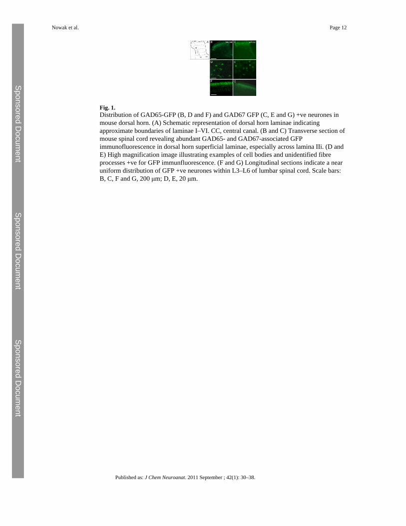
Fig. 1.Distribution of GAD65-GFP (B, D and F) and GAD67 GFP (C, E and G) +ve neurones inmouse dorsal horn. (A) Schematic representation of dorsal horn laminae indicatingapproximate boundaries of laminae I–VI. CC, central canal. (B and C) Transverse section ofmouse spinal cord revealing abundant GAD65- and GAD67-associated GFPimmunofluorescence in dorsal horn superficial laminae, especially across lamina IIi. (D andE) High magnification image illustrating examples of cell bodies and unidentified fibreprocesses +ve for GFP immunfluorescence. (F and G) Longitudinal sections indicate a nearuniform distribution of GFP +ve neurones within L3–L6 of lumbar spinal cord. Scale bars:B, C, F and G, 200 μm; D, E, 20 μm.
Nowak et al. Page 12
Published as: J Chem Neuroanat. 2011 September ; 42(1): 30–38.
Sponsored Docum
ent Sponsored D
ocument
Sponsored Docum
ent
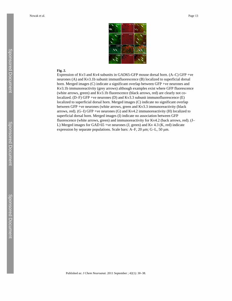
Fig. 2.Expression of Kv3 and Kv4 subunits in GAD65-GFP mouse dorsal horn. (A–C) GFP +veneurones (A) and Kv3.1b subunit immunfluorescence (B) localized to superficial dorsalhorn. Merged images (C) indicate a significant overlap between GFP +ve neurones andKv3.1b immunoreactivity (grey arrows) although examples exist where GFP fluorescence(white arrows, green) and Kv3.1b fluorescence (black arrows, red) are clearly not co-localized. (D–F) GFP +ve neurones (D) and Kv3.3 subunit immunofluorescence (E)localized to superficial dorsal horn. Merged images (C) indicate no significant overlapbetween GFP +ve neurones (white arrows, green and Kv3.3 immunoreactivity (blackarrows, red). (G–I) GFP +ve neurones (G) and Kv4.2 immunoreactivity (H) localized tosuperficial dorsal horn. Merged images (I) indicate no association between GFPfluorescence (white arrows, green) and immunoreactivity for Kv4.2 (back arrows, red). (J–L) Merged images for GAD 65 +ve neurones (J, green) and Kv 4.3 (K, red) indicateexpression by separate populations. Scale bars: A–F, 20 μm; G–L, 50 μm.
Nowak et al. Page 13
Published as: J Chem Neuroanat. 2011 September ; 42(1): 30–38.
Sponsored Docum
ent Sponsored D
ocument
Sponsored Docum
ent
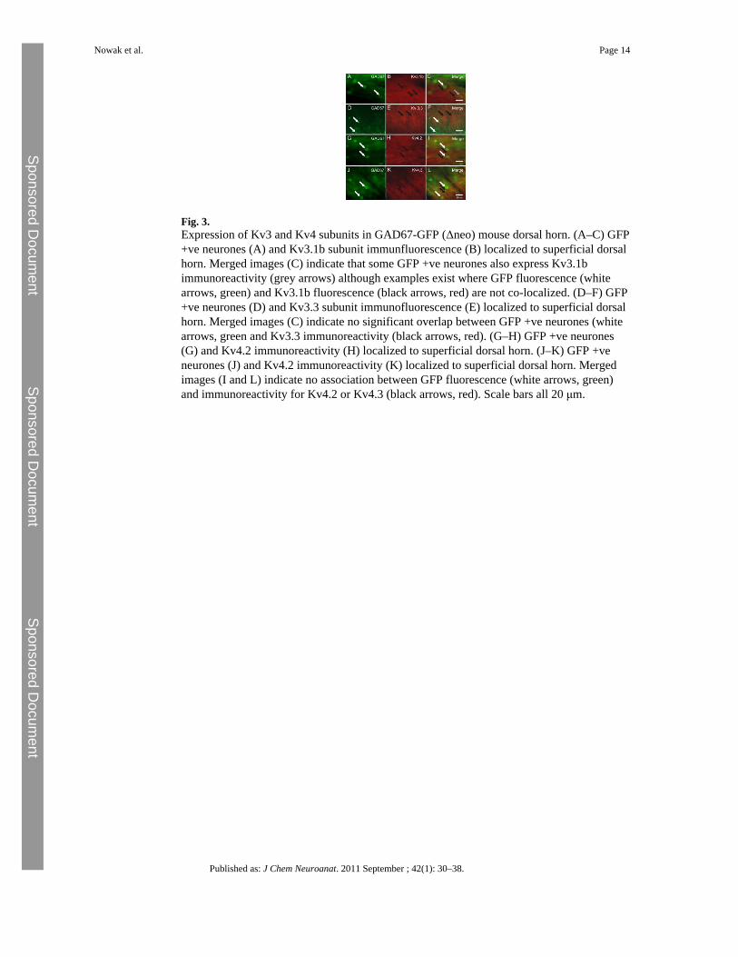
Fig. 3.Expression of Kv3 and Kv4 subunits in GAD67-GFP (Δneo) mouse dorsal horn. (A–C) GFP+ve neurones (A) and Kv3.1b subunit immunfluorescence (B) localized to superficial dorsalhorn. Merged images (C) indicate that some GFP +ve neurones also express Kv3.1bimmunoreactivity (grey arrows) although examples exist where GFP fluorescence (whitearrows, green) and Kv3.1b fluorescence (black arrows, red) are not co-localized. (D–F) GFP+ve neurones (D) and Kv3.3 subunit immunofluorescence (E) localized to superficial dorsalhorn. Merged images (C) indicate no significant overlap between GFP +ve neurones (whitearrows, green and Kv3.3 immunoreactivity (black arrows, red). (G–H) GFP +ve neurones(G) and Kv4.2 immunoreactivity (H) localized to superficial dorsal horn. (J–K) GFP +veneurones (J) and Kv4.2 immunoreactivity (K) localized to superficial dorsal horn. Mergedimages (I and L) indicate no association between GFP fluorescence (white arrows, green)and immunoreactivity for Kv4.2 or Kv4.3 (black arrows, red). Scale bars all 20 μm.
Nowak et al. Page 14
Published as: J Chem Neuroanat. 2011 September ; 42(1): 30–38.
Sponsored Docum
ent Sponsored D
ocument
Sponsored Docum
ent
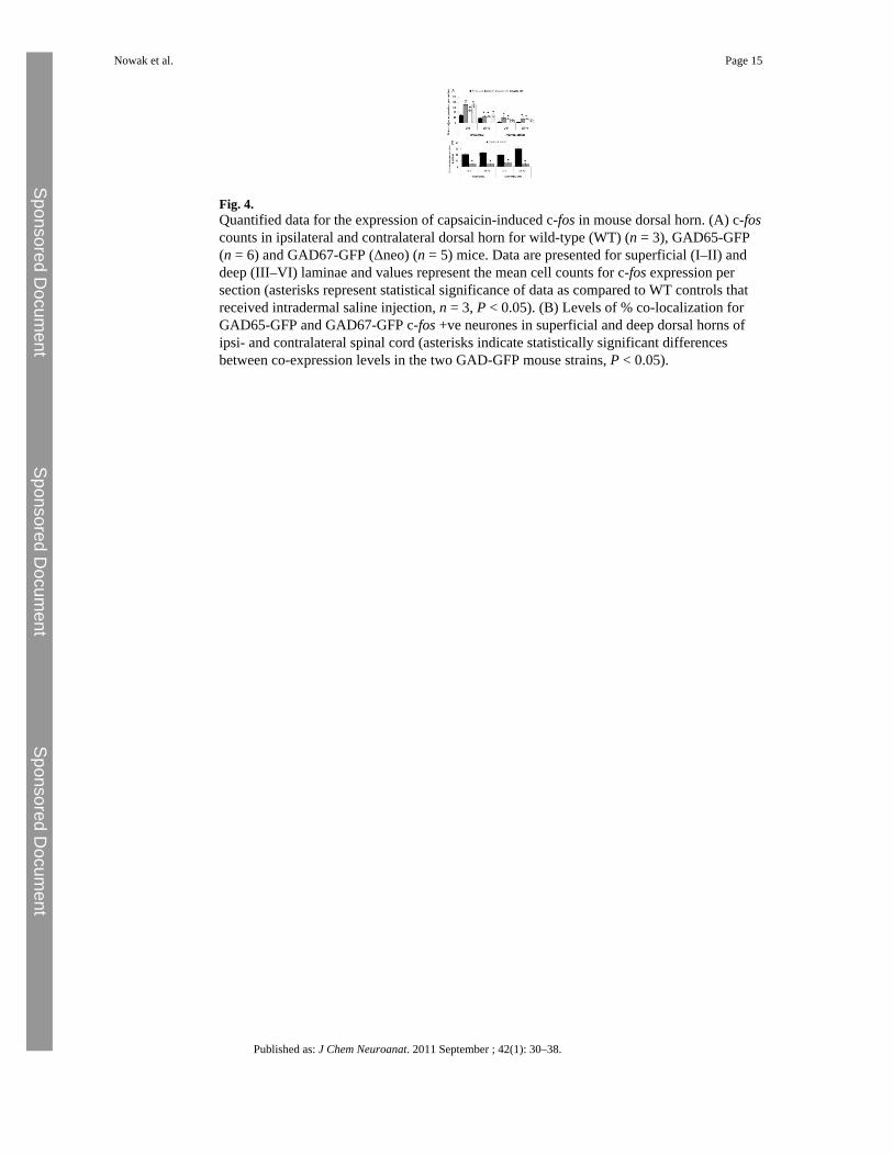
Fig. 4.Quantified data for the expression of capsaicin-induced c-fos in mouse dorsal horn. (A) c-foscounts in ipsilateral and contralateral dorsal horn for wild-type (WT) (n = 3), GAD65-GFP(n = 6) and GAD67-GFP (Δneo) (n = 5) mice. Data are presented for superficial (I–II) anddeep (III–VI) laminae and values represent the mean cell counts for c-fos expression persection (asterisks represent statistical significance of data as compared to WT controls thatreceived intradermal saline injection, n = 3, P < 0.05). (B) Levels of % co-localization forGAD65-GFP and GAD67-GFP c-fos +ve neurones in superficial and deep dorsal horns ofipsi- and contralateral spinal cord (asterisks indicate statistically significant differencesbetween co-expression levels in the two GAD-GFP mouse strains, P < 0.05).
Nowak et al. Page 15
Published as: J Chem Neuroanat. 2011 September ; 42(1): 30–38.
Sponsored Docum
ent Sponsored D
ocument
Sponsored Docum
ent
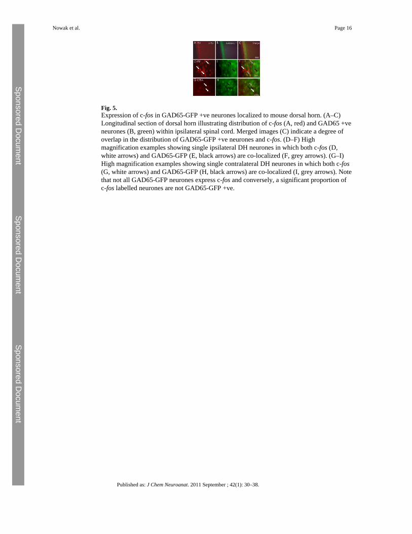
Fig. 5.Expression of c-fos in GAD65-GFP +ve neurones localized to mouse dorsal horn. (A–C)Longitudinal section of dorsal horn illustrating distribution of c-fos (A, red) and GAD65 +veneurones (B, green) within ipsilateral spinal cord. Merged images (C) indicate a degree ofoverlap in the distribution of GAD65-GFP +ve neurones and c-fos. (D–F) Highmagnification examples showing single ipsilateral DH neurones in which both c-fos (D,white arrows) and GAD65-GFP (E, black arrows) are co-localized (F, grey arrows). (G–I)High magnification examples showing single contralateral DH neurones in which both c-fos(G, white arrows) and GAD65-GFP (H, black arrows) are co-localized (I, grey arrows). Notethat not all GAD65-GFP neurones express c-fos and conversely, a significant proportion ofc-fos labelled neurones are not GAD65-GFP +ve.
Nowak et al. Page 16
Published as: J Chem Neuroanat. 2011 September ; 42(1): 30–38.
Sponsored Docum
ent Sponsored D
ocument
Sponsored Docum
ent