Bacterial sinusitis can be a focus for initial lung colonisation ...
118
PhD thesis By Kasper Aanæs Bacterial sinusitis can be a focus for initial lung colonisation and chronic lung infection in patients with cystic fibrosis Academic advisors: Helle Krogh Johansen MD, DMSc & Professor Christian von Buchwald MD, DMSc Submitted: 28 th of September 2012
-
Upload
khangminh22 -
Category
Documents
-
view
0 -
download
0
Transcript of Bacterial sinusitis can be a focus for initial lung colonisation ...

PhD thesis
By Kasper Aanæs
Bacterial sinusitis can be a focus for initial lung colonisation and chronic lung infection in
patients with cystic fibrosis
Academic advisors:
Helle Krogh Johansen MD, DMSc & Professor Christian von Buchwald MD, DMSc
Submitted: 28th of September 2012
2
Institutnavn: Øre-næse-halskirurgisk og Audiologisk Klinik, Rigshospitalet og Det
Sundhedsvidenskabelige fakultet, Københavns Universitet.
Name of department: Department of Otorhinolaryngology, Head and Neck Surgery and
Audiology, Rigshospitalet and Faculty of Health and Medical Sciences,
University of Copenhagen
Author: Kasper Aanæs
Dansk titel: Bakterier i bihulerne kan være fokus for den initiale lunge-kolonisering
og kroniske lunge-infektion hos patienter med cystisk fibrose.
English title: Bacterial sinusitis can be a focus for initial lung colonisation and chronic
lung infection in patients with cystic fibrosis.
Subject description: Describing interactions between the paranasal sinuses and lungs in
patients with cystic fibrosis.
Academic advisors: Professor Christian von Buchwald, MD, DMSc
Department of Otolaryngology, Head and Neck Surgery and Audiology
Rigshospitalet, Copenhagen University Hospital, Denmark.
Helle Krogh Johansen, MD, DMSc
Department of Clinical Microbiology
Rigshospitalet, Copenhagen University Hospital, Denmark.
Assessment committee: Professor Wytske J. Fokkens, MD, PhD. Department of
Otorhinolaryngology, Academic Medical Centre, Amsterdam,
The Netherlands.
Jochen G. Mainz, MD, Dr.med., Cystic Fibrosis Center, Department of
Paediatrics, Paediatric Pulmonology, Jena University Hospital, Jena,
Germany.
Professor Vibeke Backer, MD, DMSc. Department of Respiratory
Medicine L, Copenhagen University Hospital, Bispebjerg, Denmark.

3
Table of Content
List of Publications p. 5
List of abbreviations p.6
English summary p.7
Danish summary / Dansk resumé p.9
1. Introduction p.11
1.1 Aims of thesis p.11
2. Background p.12
2.1 Cystic fibrosis (CF) p.12
2.2 Lower airways p.12
2.2.1 Grading of pulmonary infection p.14
2.3 Pseudomonas aeruginosa p.15
2.4 Achromobacter xylosoxidans, Burkholderia cepacia complex p.16
2.5 Detection of CF-pathogenic Gram-negative bacteria p.17
2.5.1 Immune responses p.18
2.6 Upper airways p.19
2.6.1 Sinus anatomy p.19
2.6.2 Chronic rhinosinusitis p.20
2.6.3 Bacteriology of the upper airways p.22
2.7 United airways in CF patients p.23
2.8 Assessment of the upper CF airways p.27
3. Material p.30
3.1 Study population p.30
3.2 Usage of different grading of pulmonary infections p.30

4
4. Methods p.31
4. 1. Functional endoscopic sinus surgery (FESS) p.31
4.1.1 Criteria for FESS p.31
4.1.2 FESS procedure p.32
4.1.2 Postoperative treatment p.34
4.2 Culture methods p.35
4.3 IgA and IgG antibodies against P.aeruginosa p.36
4.3.1 Antibodies against P. aeruginosa alginate p.37
4.3.2 Antibodies against P. aeruginosa St-Ag p.37
4.3.3 ELISA p.38
4.4 Additional methods p.38
4.5 Statistics p.39
4.6 Ethics p.40
5. Review of results p.41
6. Discussion p.46
6.1 Study strength and weaknesses p.52
7. Perspectives p.57
8. Conclusion p.59
9. Acknowledgements p.60
10. SNOT-22 questionnaire p.62
11. References p.63
Paper I
Paper II
Paper III
Paper IV

5
List of publications
I. Colonisation and infection of the paranasal sinuses in cystic fibrosis patients is
accompanied by a reduced PMN response. Johansen HK, Aanaes K, Pressler T,
Nielsen KG, Fisker J, Skov M, Høiby N, von Buchwald C. J Cyst Fibros. 2012 May
15. [Epub ahead of print]
II. Secretory IgA as a diagnostic tool for Pseudomonas aeruginosa respiratory
colonization. Aanaes K, Johansen HK, Poulsen SS, Pressler T, von Buchwald C,
Høiby N.J Cyst Fibros. 2012 Jul 19. [Epub ahead of print]
III. The effect of sinus surgery with intensive follow-up on pathogenic bacteria in
patients with cystic fibrosis. Aanaes K, von Buchwald C, Skov M, Hjuler T, Alanin
M, Johansen HK accepted for publication by the American Journal of Rhinology
and Allergy 6th September 2012.
IV. Clinical effects of sinus surgery and adjuvant therapy in cystic fibrosis patients —
can chronic lung infections be postponed? Aanaes K, Johansen HK, Skov M,
Buchvald F, Hjuler T, Pressler T, Høiby N, Nielsen KG, von Buchwald C. Submitted
to Respiration September 2012

6
List of abbreviations
BAL Bronchoalveolar lavage
CF Cystic fibrosis
CIE Crossed immunoelectrophoresis
CFQ-R Cystic fibrosis questionnaire-revised
CRS Chronic rhinosinusitis
CT Computed tomography scan
ELISA Enzyme-linked immunosorbent assay
EPOS European position paper on rhinosinusitis
FESS Functional endoscopic sinus surgery
HRQOL Health-related quality of life
Ig Immunoglobulin
LTX Lung transplanted
MRI Magnetic resonance imaging
ORL Oto-rhino-laryngologist
PFT Pulmonary function test
PMNs Polymorphonuclear leukocytes (neutrophil granulocytes)
RSOM-31 Rhinosinusitis outcome measure 31
SN-5 Sinus and nasal quality of life survey
SNOT-22 Sinonasal outcome test -22
St-Ag Standard antigen
7
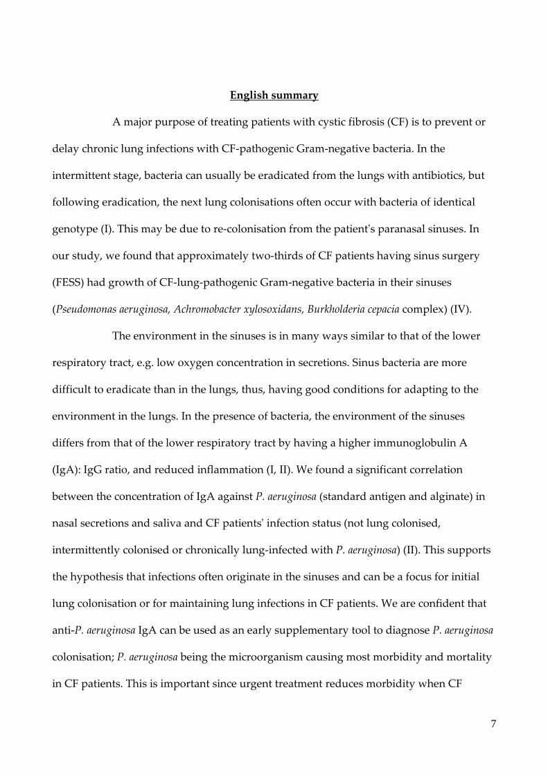
English summary
A major purpose of treating patients with cystic fibrosis (CF) is to prevent or
delay chronic lung infections with CF-pathogenic Gram-negative bacteria. In the
intermittent stage, bacteria can usually be eradicated from the lungs with antibiotics, but
following eradication, the next lung colonisations often occur with bacteria of identical
genotype (I). This may be due to re-colonisation from the patient's paranasal sinuses. In
our study, we found that approximately two-thirds of CF patients having sinus surgery
(FESS) had growth of CF-lung-pathogenic Gram-negative bacteria in their sinuses
(Pseudomonas aeruginosa, Achromobacter xylosoxidans, Burkholderia cepacia complex) (IV).
The environment in the sinuses is in many ways similar to that of the lower
respiratory tract, e.g. low oxygen concentration in secretions. Sinus bacteria are more
difficult to eradicate than in the lungs, thus, having good conditions for adapting to the
environment in the lungs. In the presence of bacteria, the environment of the sinuses
differs from that of the lower respiratory tract by having a higher immunoglobulin A
(IgA): IgG ratio, and reduced inflammation (I, II). We found a significant correlation
between the concentration of IgA against P. aeruginosa (standard antigen and alginate) in
nasal secretions and saliva and CF patients' infection status (not lung colonised,
intermittently colonised or chronically lung-infected with P. aeruginosa) (II). This supports
the hypothesis that infections often originate in the sinuses and can be a focus for initial
lung colonisation or for maintaining lung infections in CF patients. We are confident that
anti-P. aeruginosa IgA can be used as an early supplementary tool to diagnose P. aeruginosa
colonisation; P. aeruginosa being the microorganism causing most morbidity and mortality
in CF patients. This is important since urgent treatment reduces morbidity when CF
8
patients are early colonised with P. aeruginosa, however, there is a lack of diagnostic tools
for detecting the early colonisation in the lungs and in the sinuses.
We initiated a treatment strategy for CF patients to prevent sino-nasal bacteria
being seeded into the lower airways: we recommended extensive functional endoscopic
FESS with creation of sufficient drainage from all involved sinuses with subsequent IV
antibiotics and at least 6 months of twice daily nasal irrigation with saline and antibiotics.
By this strategy, sinus bacteria could be eradicated in a large proportion of patients (III).
Essentially, growth of CF-pathogenic bacteria from the lower respiratory tract was
decreased following the treatment. Furthermore, a number of patients have been free from
CF-pathogenic bacteria for more than one year after FESS, and thus re-classified as "not
lung colonised". We also corroborated that CF patients obtain an improved quality of life
and reduction in their symptoms of chronic rhinosinusitis after FESS (IV).
It is primarily intermittently lung colonised CF patients with CF-pathogenic
bacteria in their sinuses that seem to benefit from the treatment strategy. This is in
accordance with the fact that we did not see a significant increase in lung function and
only a small decrease in specific antibodies after FESS; a high systemic immune and
inflammatory response and a decreasing lung function is generally not present in patients
who primarily have sinus CF-pathogenic bacteria.
It is important that guidelines are created for how CF patients with CF-
pathogenic bacteria in the sinuses are to be treated, including criteria for who may likely
benefit from FESS, and who may be treated exclusively with conservative therapy, e.g.
saline and antibiotic irrigations.

9
Danish summary / Dansk resumé
Det vigtigste fokus-område inden for behandlingen af patienter med cystisk
fibrose (CF), er at prøve at forhindre eller udskyde tidspunktet for en kronisk lunge-
infektion med CF-patogene Gram-negative bakterier.
De første gange bakterierne dyrkers fra lungerne, kan de som regel udryddes
med antibiotika-terapi, men efterfølgende sker de næste koloniseringer ofte med bakterier
af samme genotype. Dette kan skyldes, at patienterne re-koloniseres fra egne bihuler, hvor
bakterierne er vanskeligere at bekæmpe end i lungerne. Vi fandt, at cirka to-tredjedele af
de CF patienter vi har bihule-opereret (FESS), havde CF-patogene Gram-negative
bakterier i bihulerne (Pseudomonas aeruginosa, Achromobacter xylosoxidans, Burkholderia
cepacia complex).
Miljøet i bihulerne minder om det, der findes i de nedre luftveje med f.eks. lav
ilt-koncentration i sekretet, hvorved bakterierne kan adaptere til miljøet i lungerne. Når
der er bakterier tilstede i bihulerne, ses en højere ratio af immunoglobulin A (IgA): IgG
antistoffer og mindre inflammation end i lungerne. Vi fandt en signifikant sammenhæng
mellem koncentrationen af IgA mod P. aeruginosa (standard antigen og alginate) i næse-
sekret og i spyt, med CF patienternes lunge-infektions-status (aldrig lunge-koloniseret,
intermitterende koloniseret eller kronisk inficeret med P. aeruginosa). Disse fund støtter
hypotesen om, at de alvorlige infektioner ofte starter i bihulerne, og kan være fokus for
den initiale eller vedvarende lunge-kolonisering af CF patienter.
Vi er overbeviste om, at vores nye metode til at bestemme koncentrationen af
anti-P. aeruginosa-IgA i fremtiden vil kunne bruges som en del af udredningen af de tidlige
bihule og lunge P. aeruginosa-kolonisering. Dette vil have stor betydning, da tidlig
10
behandling af P. aeruginosa koloniseringer forbedrer morbiditeten, men er svær, da der
mangler metoder til at diagnosticere tidlige P. aeruginosa kolonisationer i næse og lunger.
Vi har iværksat et behandlingsprogram for CF-patienter, hvis formål er at
modvirke at bakterierne fra bihulerne bliver transporteret ned til lungerne og giver
infektioner: vi anbefaler en ekstensiv FESS, hvor målet er at lave et godt afløb fra alle
bihuler, efterfulgt af intravenøs antibiotika, samt nasal skylning med saltvand og
antibiotika. Med denne behandling har vi vist, at en stor del af patienterne kan få fjernet
bakterier fra deres bihuler. Vi fandt også en reduktion i frekvensen af sekret-prøver fra de
nedre luftveje, som indeholdt bakterier der er farlige for CF patienter, og lige så vigtigt,
blev flere patienter fri for de CF-patogene bakterier i mere end et år efter operationen, og
kunne dermed re-kategoriseres som ‛ikke lunge-koloniseret‛. Ved den nævnte behandling
opnåede patienterne også en øget livskvalitet med færre symptomer på kronisk
bihulebetændelse.
Det var primært CF patienter, som er tidligt koloniseret i deres lunger, og
samtidig har CF-patogene bakterier i bihulerne, der så ud til at have gavn af behandlingen.
Dette passer med, at vi ikke så en forbedring af lungefunktionen eller et stort fald i
antistofferne efter FESS; de patienter, der primært har infektionen i øvre luftveje, har
generelt ikke dannet et systemisk immunrespons og fået fald i lungefunktionen.
Vi finder det vigtigt at der, i nær fremtid, bliver lavet retningsliner for hvilke
patienter med formodet CF-patogene bakterier i bihulerne, der skal tilbydes FESS, hvem
der ikke vil have gavn af det, og hvem der måske kan klare sig med konservativ
behandling, som f.eks. næseskylning og antibiotisk behandling.
11
1. Introduction
There has been little focus on the paranasal sinuses in patients with cystic
fibrosis (CF). However, our multidisciplinary group has contributed to creating awareness
about this important issue during recent years. Due to brevity, I have chosen to base this
PhD thesis on four studies, which all have been submitted to or accepted by peer-reviewed
journals. There is a common thread running through all four papers combining basic,
paraclinical and clinical research. The four papers are enclosed at the end of this thesis.
Other related CF projects that I have participated in will be discussed or cited when
appropriate.
1.1 Aims of thesis
I have focused on elucidating whether the paranasal sinuses can be a focus for
initial lung colonisations and whether it is plausible that they also may serve as a focus for
re-infections in CF-patients. In detail, I will:
a) Discuss the prevalence of bacteria found in the CF sinuses (I, II, III, IV).
b) Put the sinus mucosal inflammation into perspective (I, II).
c) Describe a potential new method of diagnosing CF pathogen sinusitis; this will be a
platform to discuss how CF sinusitis may be identified and treated (II, III).
d) Put forward a treatment strategy for CF patients with pathogen sinusitis and
discuss pros and cons for different treatments of CF sinuses (III, IV).
e) Present and discuss results on how our treatment addressing the sinuses influences
lung colonisations and re-infections and relate these to further studies (III, IV).
(The roman numbers refer to the papers in which the subject is dealt with)
12
2. Background
2.1 Cystic fibrosis (CF)
CF is a severe recessive genetic disease, which is common among the
Caucasian population. In Denmark, the incidence is 1:4,700 (1); the Faroe Islands having
one of the highest incidences in the world. Currently, approximately 450 patients with CF
are living in Denmark; one-third of the patients are followed at the Cystic Fibrosis Centre
in Århus while two-thirds are followed at the Cystic Fibrosis Centre in Copenhagen
Rigshospitalet.
The disease is caused by mutations in the cystic fibrosis transmembrane
conductance regulator protein (CFTR) located on chromosome 7 (2). The gene encodes the
cAMP-dependent chloride channel, and as a consequence of the defect, abnormal
transport of chloride and sodium across the cell epithelium is seen. Thus, all secretions
contain a higher concentration of salt, which more than doubles the viscosity compared
with non-CF individuals (3).
The main clinical characteristics of CF are increased salt loss in sweat,
malabsorption, diabetes, male infertility, chronic rhinosinusitis and increased fungal and
viral airway infections; most severe is the increased susceptibility to bacterial infections of
the lower airways.
2.2 Lower airways
Due to the viscous secretions, the mucocilliary clearance of inhaled microbes
is impaired making CF patients very susceptible to lower-airway infections (4;5). From
13
early childhood, the infections are mostly caused by Haemophilus influenzae and
Staphylococcus aureus. When older, the CF-pathogenic Gram-negative bacteria
Achromobacter xylosoxidans, Burkholderia cepacia complex and especially Pseudomonas
aeruginosa are more frequently seen. When reaching adulthood, the vast majority of
patients have been colonised or infected with one or more of the three mentioned CF-
pathogenic Gram-negative bacteria responsible for most of the morbidity and mortality in
CF (6).
The initial stage of ‚never been lung colonised‛ with CF-pathogenic Gram-
negative bacteria is often replaced by a stage of intermittent colonisation before entering
the final stage of chronic infection. In spite of a frequently and regularly intensive
antibiotic treatment (7), the bacteria are presumably constantly present in some
pulmonary segments when chronically infected. The chronic stage is paraclinically
characterised by constantly high serum levels of immunoglobulin G (IgG) antibodies and
numerous polymorphonuclear leukocytes (PMNs) in the lower airways. Elevated serum
levels of specific antibodies are also seen and are used as a supplementary diagnostic tool
(mentioned in 2.2.1 and 2.5.1). Likewise, increased numbers of PMNs are strongly
correlated to poor lung function; the imbalance between PMN-proteinases and their
inhibitors leads to impaired phagocytosis, T-cell and B-cell imbalance, and lung tissue
damage (8). Thus, the onset age of chronic lung infection with P.aeruginosa is correlated
with the life expectancy in CF patients (9).
14
Clinically, CF-patients with chronic bacterial lung infections tend to have
lower quality of life, lower body mass index (BMI) and declining lung function measured
by FEV1(forced expiratory volume in 1 sec. %-predicted) and FVC (forced vital capacity %-
predicted). The major purposes of treating patients with CF are to prevent or delay chronic
lung infections and keep the lung function at a steady state. This goal is difficult to
achieve, consequently, CF is the second largest group of lung-transplanted recipients in
Europe (10).
2.2.1 Grading of pulmonary infection
Years ago, there was no international consensus about the consequences of
Gram-negative chronic lung infections for the progression and prognosis of CF lung
disease. By an epidemiological study of the respiratory tract microbiology, the definition
of different infection categories were introduced (11). It was shown that high serum levels
of precipitating antibodies against P.aeruginosa was characteristic in chronically infected
patients and in patients harbouring mucoid strains (12), and that high and rapidly
increasing levels of antibodies correlated with poor prognosis (9). The antibody response
was shown to have high sensitivity and specificity for the early detection of chronic P.
aeruginosa lung infection and was included in the following definition of chronic infection:
persistent presence of bacteria in six consecutive months, or less when combined with the
presence of elevated precipitating antibodies (9;13).
Since 1974, our centre has used the Copenhagen criteria (9;12), which grades
pulmonary infection into three categories based on having 10–12 lower airway samples

15
cultured a year: 1) never colonised, 2) intermittently colonised, and 3) chronically infected.
These criteria cannot be applied in most centres, because the patients are not seen as
regularly as in Copenhagen and as only a few centres have access to the antibody tests.
Consequently, the Leeds criteria were developed (14) and where shown to correlate well
with the Copenhagen criteria (15-17). The advantage of using the Copenhagen criteria
when patients are seen on a monthly basis is that they allow an earlier initiation of
eradication or maintenance therapy, which improves lung function in both intermittently
colonised and chronically infected CF patients (16;18).
Based on the fact that most CF centres only see their patients every third
month, the following Leeds criteria are used:
1. Never infected: there has never been growth of any CF related Gram-negative
bacteria.
2. Non-infected: no growth of any CF related Gram-negative bacteria over 12 months.
3. Intermittent colonisation: growth in >0% and ≤ 50% of samples.
4. Chronic infection: growth in >50% of a patient’s monthly lower-airway samples.
2.3 Pseudomonas aeruginosa
P. aeruginosa is a Gram-negative rod-shaped bacterium frequently found in
soil, water and man-made environments (e.g. water pipes). It is an opportunistic pathogen
of immune-compromised individuals. It thrives not only in normal atmospheres, but can
adjust to hypoxic conditions as in the sputum and sinus secretions of CF patients (19;20).

16
Partly induced by oxygen radicals from the PMNs, some P. aeruginosa mutate during the
initial colonisation making them more suitable for a chronic infection. The most important
bacterial gene is mucA, which causes P. aeruginosa transition from a non-mucoid to a
mucoid-phenotype producing alginate and biofilms. Other important mutations or
changes of phenotypes cause: down-regulation of the cell-to-cell communication (quorum-
sensing; las and rhl genes); increase antibiotic resistance; change colony morphology;
reduce swimming, swarming and twitching motility; growth advantages; modify immune
system tolerance; and increased protease production (21;22).
Lung infections with P. aeruginosa cause inflammation resulting in a systemic
increase of IgG antibodies against polyvalent P. aeruginosa antigen (Standard Antigen (St-
Ag)) (23) and the mucoid exopolysaccharide alginate (a biofilm-matrix component), which
are highly characteristic of P. aeruginosa (24;25). In addition to serum, specific antibodies
are present in tears and in the upper airways as saliva and sputum; IgA being the
dominant antibody at mucosal surfaces (25;26). Prior to our studies (I, II), research on IgA
in nasal secretions from CF patients has, to our knowledge, never been investigated.
2.4 Achromobacter xylosoxidans, Burkholderia cepacia complex
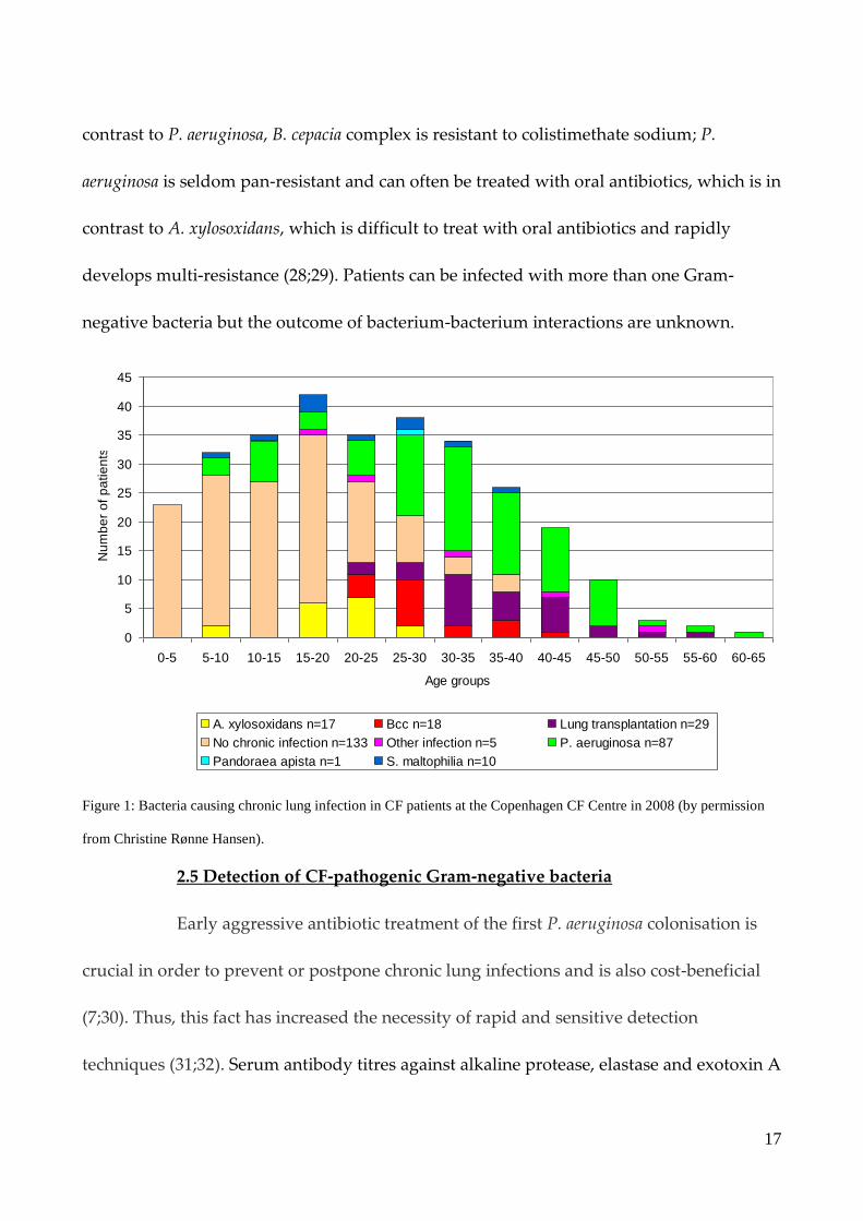
In CF, most research has been done on P. aeruginosa, being the bacteria
causing the majority of chronic infections (Figure 1). A. xylosoxidans and B. cepacia complex
are less prevalent but have similar negative impact on pulmonary disease progression.
They are expected to have similar adaptive mechanisms as P.aeruginosa causing similar
inflammation and lung destruction (27). These bacteria are also Gram-negative rods. In
17
contrast to P. aeruginosa, B. cepacia complex is resistant to colistimethate sodium; P.
aeruginosa is seldom pan-resistant and can often be treated with oral antibiotics, which is in
contrast to A. xylosoxidans, which is difficult to treat with oral antibiotics and rapidly
develops multi-resistance (28;29). Patients can be infected with more than one Gram-
negative bacteria but the outcome of bacterium-bacterium interactions are unknown.
0
5
10
15
20
25
30
35
40
45
0-5 5-10 10-15 15-20 20-25 25-30 30-35 35-40 40-45 45-50 50-55 55-60 60-65
Age groups
Nu
mb
er
of
pa
tie
nts
A. xylosoxidans n=17 Bcc n=18 Lung transplantation n=29
No chronic infection n=133 Other infection n=5 P. aeruginosa n=87
Pandoraea apista n=1 S. maltophilia n=10
Figure 1: Bacteria causing chronic lung infection in CF patients at the Copenhagen CF Centre in 2008 (by permission
from Christine Rønne Hansen).
2.5 Detection of CF-pathogenic Gram-negative bacteria
Early aggressive antibiotic treatment of the first P. aeruginosa colonisation is
crucial in order to prevent or postpone chronic lung infections and is also cost-beneficial
(7;30). Thus, this fact has increased the necessity of rapid and sensitive detection
techniques (31;32). Serum antibody titres against alkaline protease, elastase and exotoxin A
18
are on average low when P. aeruginosa is isolated from the respiratory tract for the first
time (33) and early diagnosis is challenging (34). In our clinic, specific IgG and
precipitating serum antibodies are used as a supplementary tool for monitoring lung
colonisations and infections. Clinical and paraclinical outcomes, e.g. pulmonary function
tests, are also used in the detection of pulmonary bacteria. Culturing lower airway
samples is the one of the most important tools in the detection. These samples are obtained
by coughed sputum, endolaryngeal suction in non-sputum producers, induced sputum
that increases the recovery rate of P. aeruginosa (35), or by bronchoalveolar lavage (BAL)
having a lower degree of upper respiratory tract contamination (36). Detection of CF-
pathogenic bacteria from the upper airways is discussed below.
2.5.1 Immune responses
Elevated levels of specific anti-Pseudomonas IgG antibodies, measured by
enzyme-linked immunosorbent assay (ELISA), is a risk-indicator for developing chronic P.
aeruginosa infection (37). Precipitating antibodies measured by crossed
immunoelectrophoresisis (CIE) is used as a supplementary tool for diagnosing and
predicting the outcome of lung infections (38). Precipitating antibodies remain within the
normal range (0–1) in most cases during intermittent lung colonisation but rise during
chronic infection. The antibody response has previously been shown to be helpful in
distinguishing between intermittently colonised and chronically infected patients using
the Copenhagen criteria mentioned in 2.2.1 (13;37;38).
19
2.6 Upper airways
2.6.1 Sinus anatomy
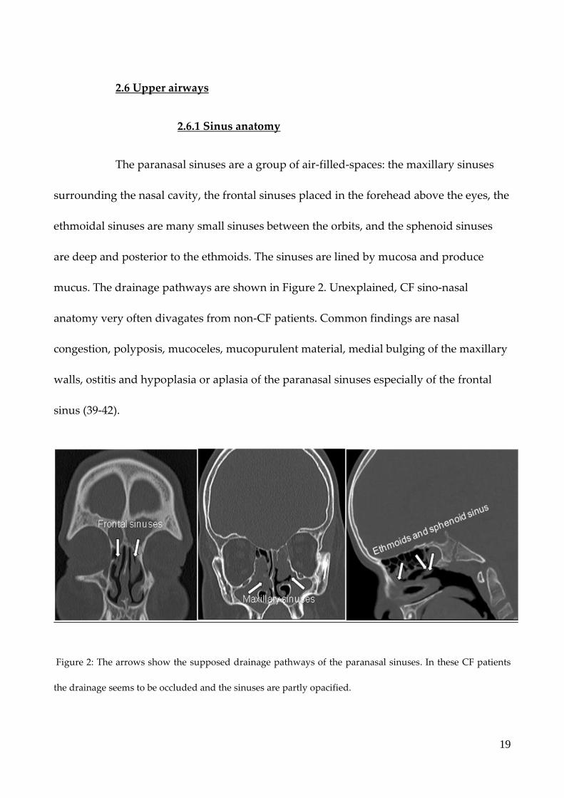
The paranasal sinuses are a group of air-filled-spaces: the maxillary sinuses
surrounding the nasal cavity, the frontal sinuses placed in the forehead above the eyes, the
ethmoidal sinuses are many small sinuses between the orbits, and the sphenoid sinuses
are deep and posterior to the ethmoids. The sinuses are lined by mucosa and produce
mucus. The drainage pathways are shown in Figure 2. Unexplained, CF sino-nasal
anatomy very often divagates from non-CF patients. Common findings are nasal
congestion, polyposis, mucoceles, mucopurulent material, medial bulging of the maxillary
walls, ostitis and hypoplasia or aplasia of the paranasal sinuses especially of the frontal
sinus (39-42).
Figure 2: The arrows show the supposed drainage pathways of the paranasal sinuses. In these CF patients
the drainage seems to be occluded and the sinuses are partly opacified.

20
2.6.2 Chronic rhinosinusitis
The hallmark of CF in the head and neck region is chronic rhinosinusitis (CRS)
and nasal polyps. There is no specific definition on CRS in CF patients, so they follow the
general definition stated in the European position paper on rhinosinusitis (EPOS) (43)
shown in Table 1. However, nasal and sinus mucosal disease is by definition present in
patients with CF because of defective CFTR-channels in the sinonasal mucosa, as found in
the lower CF airways (43). The inflamed tissue and viscous mucus results in a mechanical
obstruction of the sinus ostia (42;44). Further, the vast majority of CF patients have
radiologic evidence of sinus disease (39;42;45-50), and nasal polyposis becomes more
common with age that has been reported in varying prevalence with up to 50% of all CF
patients (45;49;51;52). There are inconsistent results on whether CF patients with nasal
polyps and symptoms of CRS can be correlated with a better lung function (53-56).
CF patients are likely to under-report their symptoms of CRS, giving a false
low share of CF patients with CRS by the definitions in Table 1; approximately two-thirds
of all CF patients have impaired olfactory function (57), and 81–86 % of CF patients fulfil
the EPOS criteria for CRS ((58) (unpublished material by Berkhout et al.), which is in
contrast to the low 10–15% who complain about CRS without specific questioning
(42;45;59-61). It is unknown whether the CF patients who do not complain about CRS
always were asymptomatic, if they have adapted to their symptoms, or if their CRS
symptoms are overshadowed by more troublesome symptoms from e.g. the lungs (42).
21
In general, non-CF patients with nasal polyposis being otherwise healthy have
been shown to score worse on quality of life than patients with chronic obstructive
pulmonary disease and patients with coronary artery disease (62;63). This ought to give
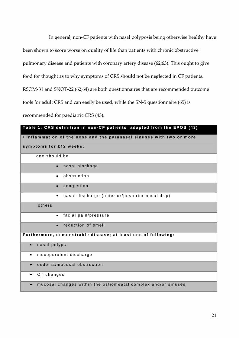
food for thought as to why symptoms of CRS should not be neglected in CF patients.
RSOM-31 and SNOT-22 (62;64) are both questionnaires that are recommended outcome
tools for adult CRS and can easily be used, while the SN-5 questionnaire (65) is
recommended for paediatric CRS (43).
T ab l e 1 : C R S d e f in i t i o n in no n - C F p at i en t s ad a pt e d f r o m th e EPO S ( 4 3 )
• I n f l a m m at i on o f t he n o s e an d t h e p a r an a s a l s i nu s e s w i th tw o o r m o r e
s ym p t o m s f o r ≥ 1 2 w e e k s ;
o n e s h o u ld b e
n a s a l b l o ck a g e
o b s t r uc t i o n
c o n g e s t i o n
n a s a l d i s c h a r g e ( a n te r i o r / p os t e r i o r n as a l d r i p )
o t h e rs
f ac i a l p a i n / p r es s u r e
r e d u c t i o n o f sm e l l
F u r th e r m o r e , d e mon s t r a b l e d i s e a s e; a t l e a s t on e o f f o l l ow in g :
n a s a l p o l yp s
m uc o p u r u l e n t d i s c h a r g e
o e d em a /m uc o s a l o bs t r u c t i o n
CT c h a n g es
m uc o s a l c h a n g e s w i t h i n t h e o s t i om e a t a l com p le x a n d / o r s i n use s
22
2.6.3 Bacteriology of the upper airways
In non-CF patients, the paranasal sinuses are regarded as sterile though they
may be frequently and transiently contaminated by bacteria from neighbouring surfaces
(66). CRS in otherwise healthy individuals predominantly have virus as a part of the
aetiology. When bacteria are involved, the following species are most frequently cultured:
Staphylococcus aureus, Streptococcus pneumoniae, Streptococcus pyogenes gr.A, Haemophilus
influenzae and Moraxella catarrhalis (67;68). In CF patients, the picture is somewhat
different. Sinus bacteria are more frequently present, P. aeruginosa being the most common
as in the lungs. Other frequently found bacteria are S. aureus, H. influenzae and coagulase-
negative staphylococci; anaerobes and other bacteria found in the lower airways such as
A. xylosoxidans and B. cepacia complex are also found in the CF sinuses (41;50;68-74).
Presence of sinus bacteria is reported in 44–95% (60;71;74). Two articles have described
fungal sinusitis among North American CF patients but disagree on the prevalence (0–
33%) (72;74).
Though it has not been addressed, it is likely that the sinus bacteria in CF
patients also produce biofilms that further increase antibiotic resistance in the same way as
in the non-CF patients (21;68;75;76). As in the lungs, the sinus bacteria develop phenotypes
that are resistant to the host immune response and antibiotic treatment.

23
2.7 United airways in CF patients
A marked association exists between upper and lower airway cultures in
patients with CF (21;43;69;73;74;77-85) due to the paranasal sinuses often being colonised
with concordant CF-lung-pathogenic Gram-negative bacteria of the same genotype
(21;50;77). Varying predictive values of CF-pathogenic bacteria in the upper airways have
been reported when diagnosing lower airway pathogens (60;69;73;82).
A CF patients’ initial lung colonisation with P. aeruginosa reflects the great
diversity of genotypes in the environment, being in contrast to some patients that, after
antibiotic eradication, subsequently are re-colonised with bacteria that are clonally related
(69;86-88). This indicates that the initial bacteria come from environmental sources rather
from transmission between patients (89) and an existence of a bacterial reservoir in the
patients’ close environment after the initial colonisation. This reservoir is likely to be the
sinuses where the bacteria can drain/migrate/be aspirated to the lower airways as seen
with viruses (21;90).
The environment of the sinuses and the lower airways are similar in many
ways (19;20;43), thus the sinuses may be colonised with bacteria before the lungs and be
an evolutionary ‘nest’ in early airway colonisations, where the bacteria are diversifying,
evolving antibiotic resistance and other phenotypes associated with adaptation to the CF
airways in general; from there, the bacteria intermittently migrate and colonise the lungs
and may ultimately cause chronic lung infections (20;21;69;74;77). When bacteria colonise
the lungs they are then pre-adapted to the environment and are therefore less virulent and

24
more resistant compared with environmental P. aeruginosa isolates (21). This also accounts
for lung transplanted (LTX) CF patients, where molecular epidemiology studies have
shown that CF lung-transplant recipients become re-colonised in their lung grafts with the
same bacterial clones as those cultured before transplantation (85).
One study has shown that CF-lung-pathogenic bacteria potentially can be
eradicated from the sinuses with extensive functional endonasal endoscopic sinus surgery
(FESS) and postoperative local antibiotic treatment (91). Nevertheless, large-scale
prospective studies investigating the effects of FESS on lung colonisation and infection in
CF are lacking (43), and data on surgical therapy for CF patients with CRS is primarily
based on level III evidence (Table 2) (43;92).
Table 2: Category of evidence (93)
Ia Evidence from meta-analysis of randomised controlled trials.
Ib Evidence from at least one randomised controlled trial.
IIa Evidence from at least one controlled study without randomisation.
IIb Evidence from at least one other type of quasi-experimental study.
III Evidence from non-experimental descriptive studies, such as comparative studies, correlation studies and case control studies.
IV Evidence from expert committees' reports or opinions and/or clinical experience of respected authorities.
25
Several studies have described the effect FESS on the lower airways using
various parameters and treatment modalities, thus showing inconsistent results (Table 3).
One prospective study has performed extensive FESS intending to eradicate sinus bacteria
in a group of 82 LTX patients, which is described in three papers (91;94;95), showing that
P. aeruginosa and A. xylosoxidans could be eradicated from the sinuses resulting in reduced
lung allograft infections. Shatz (96) found decreased antibiotic use, a lower hospitalisation
rate and an increase in FEV1 six months after FESS among 15 CF non-LTX patients.
Lewiston et al. (97) postoperatively installed tobramycin directly into the sinuses and
reported a lower rate of P. aeruginosa in the lungs among 11 LTX patients. The other
retrospective studies were all based on moderate sinus surgery, and did not focus on lung
infection status or a protocol for postoperative treatment. These studies found inconsistent
postoperative reduction in lung colonisation, lower hospitalisation rates, reduced use of
antibiotics and improvement of the pulmonary function tests (PFT). Table 3 shows an
overview of the published papers on sinus surgery in relation to the lungs; none of the
studies are level I evidence (Table 2).

26
Table 2: Review of studies correlating sinus surgery to lower airway conditions.
Authors No. of CF patients
Study design Extend of FESS Post-operative treatment
Outcome
Holzmann et al. (91;94;95)
82 LTX Prospective Fronto-spheno-ethmoidectomy and maxillary antrostomy
Nebulized colistin and irrigations; IV AB
Decrease in colonisation of the lower airways
Jarret et al. (98)
17 Non-LTX
Retrospective Ethmoidal and maxillary sinuses
Nasal saline irrigation; oral AB
PFT and BMI – non-significant
Leung et al. (99)
87 LTX Retrospective, case-control
Ethmoidal and maxillary sinuses
ND Lung re-colonisation– no significant.
Lewiston et al. (97)
11 LTX Retrospective Ethmoidal and maxillary sinuses
Tobramycin in the sinuses
Low hospitalization rate after surgery. Reduced PA in the lungs
Madonna et al. (100)
14 Non-LTX
Retrospective Ethmoidal and maxillary sinuses
IV AB PFT – non-significant
Osborn et al. (101)
41 Non-LTX
Retrospective Ethmoidal and maxillary sinuses
ND Sparse improvement in FVC non in FEV1. No effect on microbes
Rosbe et al. (102)
66 Mixed
Retrospective ND Some had IV AB Decrease in IHD. Steroid use and LFT –non-significant
Triglia et al. (103)
27 Non-LTX
Retrospective ND ND Decrease in AB treatment, non in LFT
Umetsu et al. (104)
4 (ND) Prospective Ethmoidal and maxillary sinuses ; AB flushing
IV AB IHD reduced postoperatively, non in PFT
Kempainen et al. (105)
32 Mixed
Retrospective Fronto-spheno-ethmoidectomy and maxillary antrostomy
ND PFT and IHD –non-significant
Shatz A (96) 15 Non-LTX
Retrospective Frontal, ethmoidal and maxillary sinuses
Nasal irrigations Decrease in AB and IHD. Increase in FEV1 after six months
Halvorson et al. (106)
8 Non-LTX
Retrospective, case-control
Ethmoidal and maxillary sinuses
ND Increase exercise tolerance/ increased PFT after 3 months
Kovell et al (107)
21 Non-LTX
Retrospective, case-control
ND ND Increase in PFT
LTX: Lung transplanted; PFT: Pulmonary Function Test; IHD: In-hospital-days; AB: antibiotics; BMI: Body Mass
Index; FEV1: Forced expiratory volume in one sec.; FVC: Forced Vital Capacity; ND: not described
27
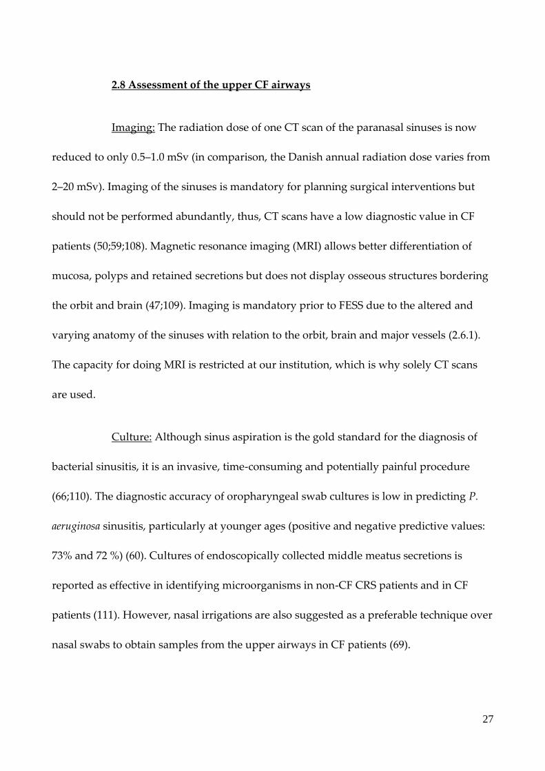
2.8 Assessment of the upper CF airways
Imaging: The radiation dose of one CT scan of the paranasal sinuses is now
reduced to only 0.5–1.0 mSv (in comparison, the Danish annual radiation dose varies from
2–20 mSv). Imaging of the sinuses is mandatory for planning surgical interventions but
should not be performed abundantly, thus, CT scans have a low diagnostic value in CF
patients (50;59;108). Magnetic resonance imaging (MRI) allows better differentiation of
mucosa, polyps and retained secretions but does not display osseous structures bordering
the orbit and brain (47;109). Imaging is mandatory prior to FESS due to the altered and
varying anatomy of the sinuses with relation to the orbit, brain and major vessels (2.6.1).
The capacity for doing MRI is restricted at our institution, which is why solely CT scans
are used.
Culture: Although sinus aspiration is the gold standard for the diagnosis of
bacterial sinusitis, it is an invasive, time-consuming and potentially painful procedure
(66;110). The diagnostic accuracy of oropharyngeal swab cultures is low in predicting P.
aeruginosa sinusitis, particularly at younger ages (positive and negative predictive values:
73% and 72 %) (60). Cultures of endoscopically collected middle meatus secretions is
reported as effective in identifying microorganisms in non-CF CRS patients and in CF
patients (111). However, nasal irrigations are also suggested as a preferable technique over
nasal swabs to obtain samples from the upper airways in CF patients (69).
28
Steroids: A Cochrane review states that oral corticosteroids appear to slow
progression of lung disease in CF (112). However, no research is published on oral
steroids’ effect on CRS symptoms in CF patients, while one study recommends intrapolyp
steroid injection (113). Another Cochrane review states that: ‚Overall, there is no clear
evidence for using topical steroids in people with CF and nasal polyposis.‛ This is due to
the neutrophilic domination in CF polyposis compared with the eosinophil domination in
non-CF patients (114). However, it should be mentioned that some studies report a
positive effect of using nasal steroids on CRS and nasal polyps in CF (115-117).
Surgery: In 2006, a Cochrane review concluded that more randomize
controlled trials comparing FESS with other treatments were required, thus it could not be
confirmed that CF patients with CRS symptoms could benefit from FESS (92). The newest
edition of EPOS 2012 and other recent studies state that symptoms of nasal airway
obstruction, nasal discharge, facial pain, snoring, olfactory dysfunction, frequency of sinus
infections and activity level are parameters that can significantly be improved after FESS
in CF patients (43;106;118-123). When evaluating the different studies, it is important to
note the criteria for FESS and what FESS and postoperative treatment comprise. In spite of
postoperative instrumental debridement and saline irrigations (120;124), it is accepted that
the effect of FESS on CRS symptoms, in general, last a shorter time in CF patients than in
non-CF patients (54;103;106;120;121;123), which is why a more extensive approach has
been suggested (81;125-129) combined with antibiotic sinus irrigations (70). As in any

29
other surgery, FESS involves risks. Though they are rare, situations where the optic nerve
or brain is damaged and extensive bleeding can occur. Nevertheless, when surgeons are
aware of the altered anatomy in CF patients (described in 2.6.1), reports show that FESS is
well tolerated and that the complication rate in CF patients is similar to that of the non-CF
population (43;103;130).
Local treatment: Nasal saline irrigations are well tolerated and the beneficial
effect appears to outweigh the minor side effects, thus they can be included as a treatment
adjunct for the symptoms of CRS in CF-patients (43;131). Hypertonic saline 7% may have
mucolytic effects and improve mucociliary clearance in the sinuses as seen in the lungs of
CF patients and may be used for nasal irrigations (132-134). Baby shampoo is also
introduced as a supplement to the saline (135;136), as is nasal inhalation of dornase alfa
used in the treatment of CRS in CF patients (137-139). Studies of nasal irrigations with
antibiotics (tobramycin or aminoglycosides) decrease bacterial colonisation and nasal
inflammation and show a positive effect on recurrence rate of CRS in non-CF patients
(140). However, there is low-level evidence for the use of topical anti-bacterials in CF
patients (43;141). Several devices including nebulizers have been developed for nasal
irrigations and distribution of medicine (138;142), which are all better than delivering it by
nasal spray (43). Finally, low-frequency ultrasound has recently been suggested as a
supplementary method for biofilm disruption in patients with CRS (143).

30
3. Material
3.1 Study population
In all studies in this thesis (I–IV), patients were recruited among the 300 CF
patients treated at the CF Centre in Copenhagen. The diagnosis of CF was based on
clinical characteristics, abnormal sweat electrolytes, and genotype. CF patients followed a
routine protocol with monthly medical examinations including lung function tests and
lower airway samples taken for microbiological culture. Additional lower airway samples
were taken whenever patients were hospitalised or when clinical and/or paraclinical
parameters indicated a risk of lung colonisation or infection. Approximately every third
month, blood samples were taken for analyses including specific antibodies against
relevant Gram-negative bacteria (38). LTX patients followed a different outpatient setting
with fewer routine samples taken.
All CF pathogens were treated with antibiotics regardless of clinical
symptoms according to the Copenhagen CF centre’s treatment protocols (7).
3.2 Usage of different grading of pulmonary infections
As mentioned in 2.2.1, there are at least two different ways of grading
pulmonary infections. In papers I and II, we applied our standard Copenhagen criteria for
defining lung infection status and LTX patients were categorised as chronically infected.
Modified Leeds criteria (14) were used for defining lung infection status in
paper III and IV; as the main outcome was the lung infection status in paper IV, it was
important to use simple criteria here that are known and can be used among other CF

31
centres. This facilitates an international comparison in the future. Secondly, the use of the
Leeds criteria allowed us to put our findings into perspective because intermittently
colonised patients could be re-classified as non-infected. Thirdly, a rise in antibodies was,
in some cases, a part of the reason for setting patients up for surgery (described in section
4.1.1), and so it would be a circular argument if antibodies were also used to define the
outcome of lung infection status.
4. Methods
4. 1. Functional endoscopic sinus surgery (FESS)
4.1.1 Criteria for FESS
In paper I, III, and IV FESS was a part of the study. CF patients were selected
for FESS based on following criteria:
1: Search for an infectious focus: Intermittently colonised patients with increasing
frequency of positive lower airway cultures or repeatedly declining lung function (> 10%),
despite intensive antibiotic chemotherapy. Patients with an unknown infectious focus and
increasing antibodies against P. aeruginosa, A. xylosoxidans or B. cepacia complex were given
the highest priority.
2: Patients who had recently been LTX. The ambition was to perform FESS within the first
postoperative year.
3: Patients with severe symptoms of chronic rhinosinusitis (CRS) according to the EPOS
(43).

32
4.1.2 FESS procedure
A BAL was performed under general anaesthetic. The subsequent FESS was to
ventilate and drain the paranasal sinuses and to make these accessible for postoperative
instrumental cleansing and irrigation with saline and topical antibiotics. Each patient was
evaluated for symptoms of chronic rhinosinusitis (43) followed by a clinical examination.
The extension of surgery (e.g., exploration of the frontal or sphenoid sinuses) was
undertaken based on the preoperative CT scan and perioperative findings. As a standard,
we applied FESS with an uncinectomy, an anterior ethmoidectomy and a medial
antrostomy, leaving a significantly enlarged maxillary ostium comprising more than half
the medial maxillary wall as recommended (43). Visible intramucosal abscess-like
structures (especially found in the maxillary sinuses) were resected along with other
inflamed mucosal tissue when accessible. Finally, the opened and now accessible sinuses
were irrigated with saline and colistimethate sodium.
To optimize culture results, no patients received IV antibiotics within two
weeks prior to FESS, and different anatomic sampling locations and multiple samples for
culture were prioritized during surgery, including: nasal secretions, pus, mucosa, polyps,
and bone (Figure 4–6). Samples taken for culture were collected with sharp instruments or
by suction tubes. The material obtained was immediately cultured at the Department of
Clinical Microbiology at Rigshospitalet.

33
34
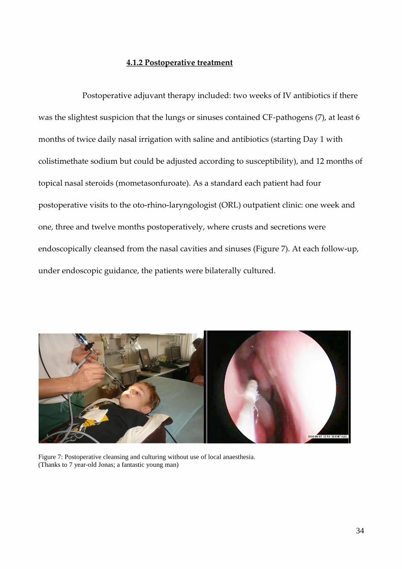
4.1.2 Postoperative treatment
Postoperative adjuvant therapy included: two weeks of IV antibiotics if there
was the slightest suspicion that the lungs or sinuses contained CF-pathogens (7), at least 6
months of twice daily nasal irrigation with saline and antibiotics (starting Day 1 with
colistimethate sodium but could be adjusted according to susceptibility), and 12 months of
topical nasal steroids (mometasonfuroate). As a standard each patient had four
postoperative visits to the oto-rhino-laryngologist (ORL) outpatient clinic: one week and
one, three and twelve months postoperatively, where crusts and secretions were
endoscopically cleansed from the nasal cavities and sinuses (Figure 7). At each follow-up,
under endoscopic guidance, the patients were bilaterally cultured.
Figure 7: Postoperative cleansing and culturing without use of local anaesthesia.
(Thanks to 7 year-old Jonas; a fantastic young man)
35
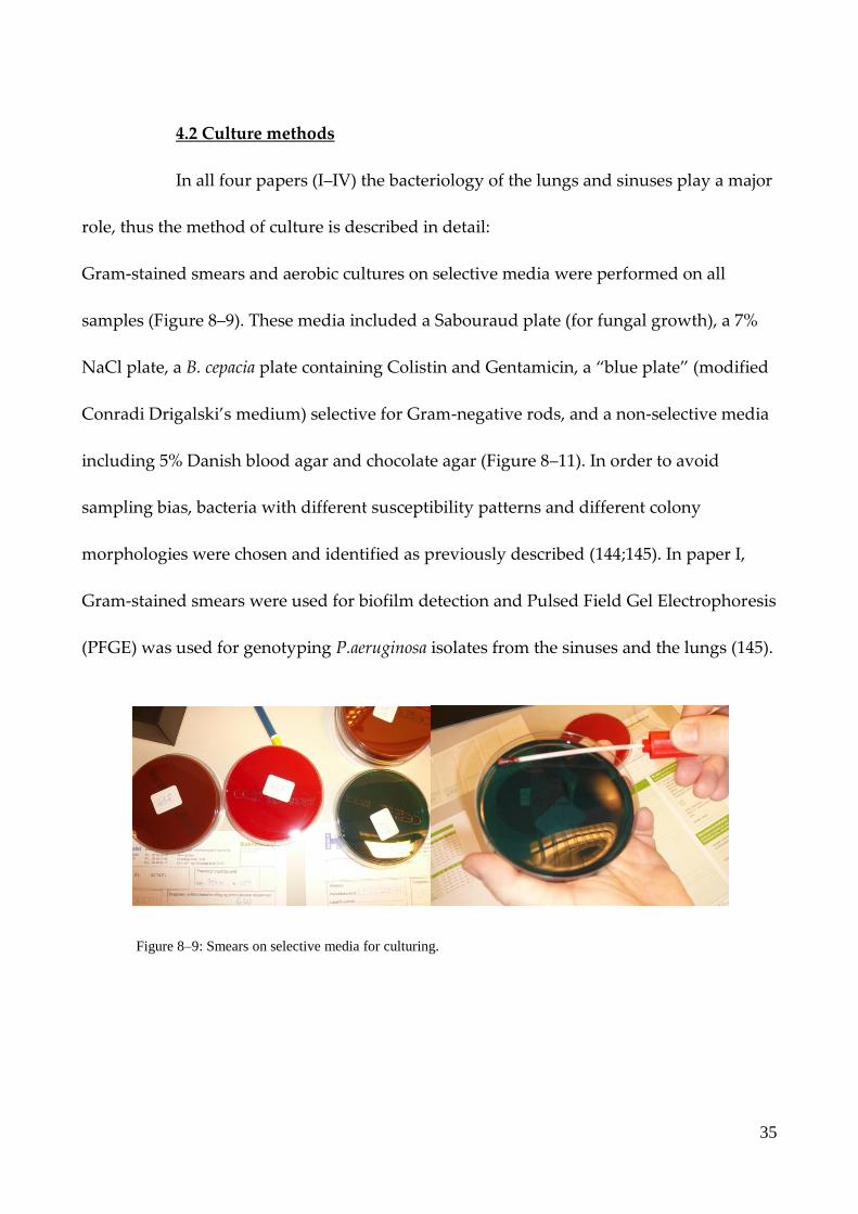
4.2 Culture methods
In all four papers (I–IV) the bacteriology of the lungs and sinuses play a major
role, thus the method of culture is described in detail:
Gram-stained smears and aerobic cultures on selective media were performed on all
samples (Figure 8–9). These media included a Sabouraud plate (for fungal growth), a 7%
NaCl plate, a B. cepacia plate containing Colistin and Gentamicin, a ‚blue plate‛ (modified
Conradi Drigalski’s medium) selective for Gram-negative rods, and a non-selective media
including 5% Danish blood agar and chocolate agar (Figure 8–11). In order to avoid
sampling bias, bacteria with different susceptibility patterns and different colony
morphologies were chosen and identified as previously described (144;145). In paper I,
Gram-stained smears were used for biofilm detection and Pulsed Field Gel Electrophoresis
(PFGE) was used for genotyping P.aeruginosa isolates from the sinuses and the lungs (145).
Figure 8–9: Smears on selective media for culturing.
36
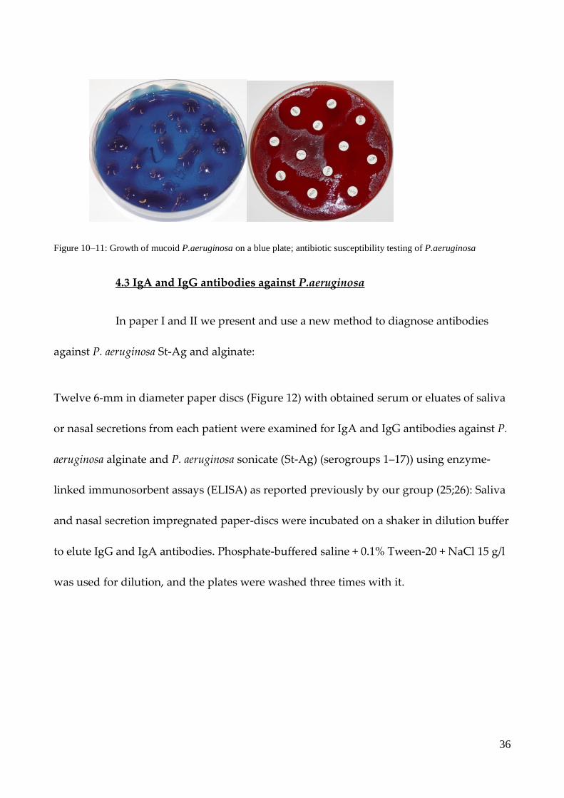
Figure 10–11: Growth of mucoid P.aeruginosa on a blue plate; antibiotic susceptibility testing of P.aeruginosa
4.3 IgA and IgG antibodies against P.aeruginosa
In paper I and II we present and use a new method to diagnose antibodies
against P. aeruginosa St-Ag and alginate:
Twelve 6-mm in diameter paper discs (Figure 12) with obtained serum or eluates of saliva
or nasal secretions from each patient were examined for IgA and IgG antibodies against P.
aeruginosa alginate and P. aeruginosa sonicate (St-Ag) (serogroups 1–17)) using enzyme-
linked immunosorbent assays (ELISA) as reported previously by our group (25;26): Saliva
and nasal secretion impregnated paper-discs were incubated on a shaker in dilution buffer
to elute IgG and IgA antibodies. Phosphate-buffered saline + 0.1% Tween-20 + NaCl 15 g/l
was used for dilution, and the plates were washed three times with it.

37
Figure 12: A paper disc to collect secretions inserted in the nasal cavity
4.3.1 Antibodies against P. aeruginosa alginate
Microtiter plates were coated with alginate purified from a mucoid CF P.
aeruginosa strain as previously reported by our group (146). The plates were coated and
blocked in dilution buffer. Diluted serum, saliva and nasal secretions (see above) were
added and allowed to react. After washing, horseradish peroxidase (HRP)-conjugated
rabbit anti-human IgA (P0216) and anti-human IgG (P0214) were added and reacted.
4.3.2 Antibodies against P. aeruginosa St-Ag
A sonicated cell extract of P. aeruginosa serogroups 1–17 was used as standard
St-Ag (25;26) and coated onto irrigated 96-well polystyrene plates. The plates were
incubated and blocked with dilution buffer. Serum, saliva, and nasal secretion were
diluted and allowed to react. After washing, horseradish peroxidase (HRP)-conjugated
rabbit anti-human IgA (P0216) and anti-human IgG were added and left to react.
38
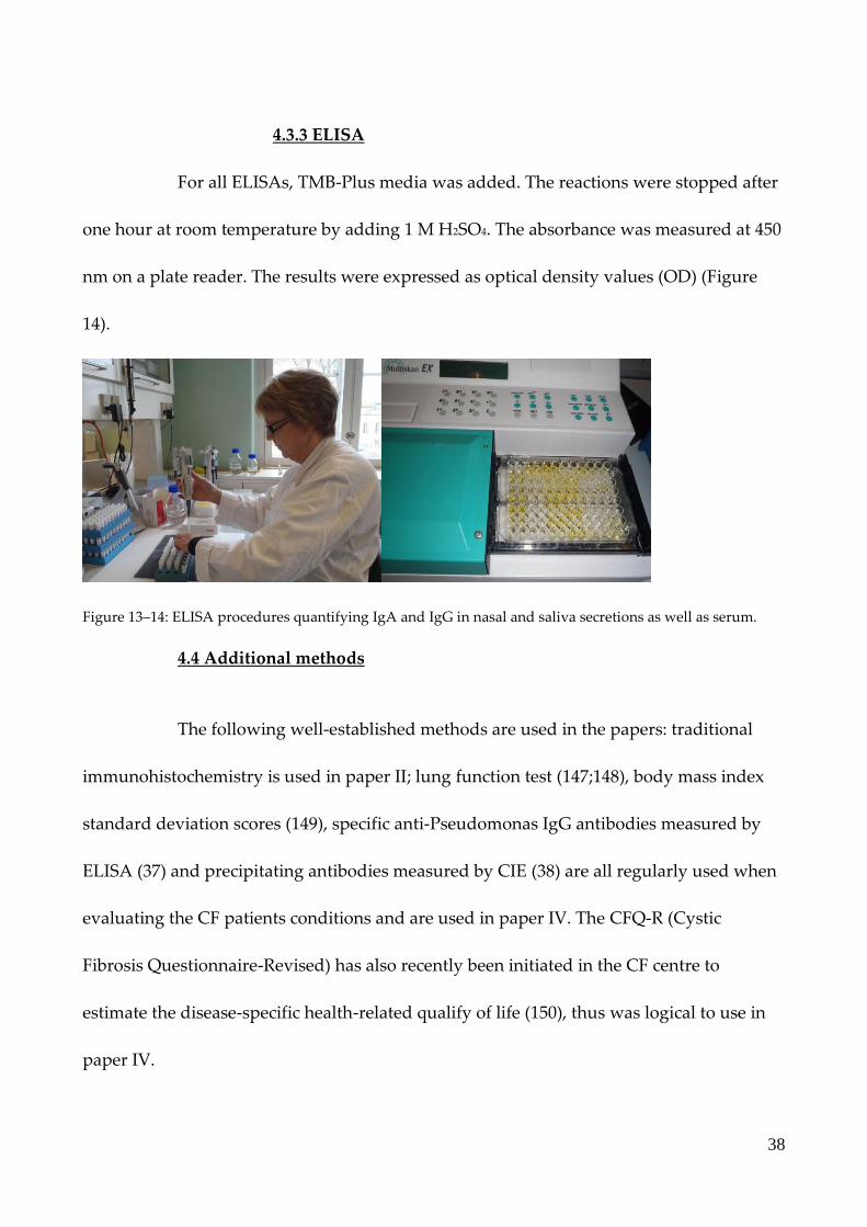
4.3.3 ELISA
For all ELISAs, TMB-Plus media was added. The reactions were stopped after
one hour at room temperature by adding 1 M H2SO4. The absorbance was measured at 450
nm on a plate reader. The results were expressed as optical density values (OD) (Figure
14).
Figure 13–14: ELISA procedures quantifying IgA and IgG in nasal and saliva secretions as well as serum.
4.4 Additional methods
The following well-established methods are used in the papers: traditional
immunohistochemistry is used in paper II; lung function test (147;148), body mass index
standard deviation scores (149), specific anti-Pseudomonas IgG antibodies measured by
ELISA (37) and precipitating antibodies measured by CIE (38) are all regularly used when
evaluating the CF patients conditions and are used in paper IV. The CFQ-R (Cystic
Fibrosis Questionnaire-Revised) has also recently been initiated in the CF centre to
estimate the disease-specific health-related qualify of life (150), thus was logical to use in
paper IV.

39
The sinonasal outcome test (SNOT-22) used in paper IV (inserted in section 10) deals with
sinonasal conditions (64), but also includes health-related questions that can be influenced
by other CF-related conditions, e.g. cough; the SNOT-22 questionnaire is used worldwide
when evaluating CRS.
4.5 Statistics
In all four papers (I–IV), we tested whether data were continuous and if the
comparisons fulfilled the criteria for normality and equal variance. The level of
significance was set to < 0.05 (two-tailed). SAS 9.1.3 was used for calculations.
In paper I, the non-parametric sign test was used to compare within patient
samples of antibodies, while the data of the antibodies in paper II were unpaired,
continuous and positively skewed distributed why Log10 transformations were made. The
transformed data had an approximately normal distribution justifying an unpaired two-
sample t-test for the means and a one way analysis of variance (ANOVA).
Receiver operating characteristic (ROC) curves was used to find the best cut-
off values between the three lung infection groups if IgA was to be used as a diagnostic
test (paper II). A Spearman rank coefficient test was used to correlate nasal secretions and
saliva in paper II as well.
A McNemar’s test was used to compare the nominal data of postoperative
frequencies of growth with the perioperative frequencies in paper III and to compare the
40
change in lung infection status after FESS (paper IV). In paper IV, a paired two-sample t-
test for the means and an ANOVA was used for the rest of the comparisons.
The biggest statistical challenge was in paper IV. When planning the study,
we received statistical advice from Professor Torben Martinussen at the Department of
Biostatistics in how to quantify the frequencies of positive cultures. The conclusion was
that every lower-airway sample was registered and given the same weight regardless of
the interval between the samples. Using a Spearman rank coefficient test, these results
were then compared to the results of lower-airways samples where each culture was given
weight according to the period until the next culture.
4.6 Ethics
The study was approved by the local ethics committee (H-A-2008-141), and all
patients gave informed consent. In patients <18 years of age, consent was also obtained
from their parents.
The inclusion for FESS was not a part of the study, solely the outcome. We
also obtained consent for doing additional analyses on the bacteria/material obtained
during FESS and BAL, the postoperative treatment and culturing, as well as for using
questionnaires and data from the patient files. In paper II, consent was used in order to
obtain and analyze secretions and blood and for culturing; no change in treatment
modality was made on behalf of these results.
41
5. Review of results
We found that the vast majority of CF patients have bacteria in their paranasal
sinuses (paper I–IV). They are often colonised with CF-lung pathogens, especially P.
aeruginosa, and there is a close correlation between the bacteriology of the sinuses and the
lungs, including identical genotypes in the sinuses and lungs (paper I, II, IV). Importantly,
the genotype remains unchanged over time. The chronically infected patients had the
same P. aeruginosa genotype in their lungs for a median of 15 years as found in their
sinuses, and up to 6 years in intermittently colonised patients, although the bacteria
apparently had been eradicated from the lungs (paper I).
Though the environment of the sinuses in many ways is similar to that of the
lower airways, including anaerobic niches and biofilm formation, it differs by excessive
presence of the non-phlogistic (does not induce inflammation) secretory-immunoglobulin
A (s-IgA) (paper I, II). Failure to eradicate CF-pathogens from the sinuses is probably a
result of an inefficient local immune response: locally produced specific s-IgA binds
Gram-negative bacteria on the mucosal surface, thereby reducing the inflammatory
response by preventing antigen presentation inhibiting complement activation, inhibiting
the recruitment of PMNs and thereby diminishing the oxidative burst (paper I). This was
visualized by immunohistochemistry showing excessive amounts of IgA-producing
plasma cells in the sino-nasal tissue and IgA in the excretory ducts. It was also visualized
by Gram-stained smears from the sinuses, where the bacterial biofilms were surrounded
42
by very few and scattered PMNs in marked contrast to the pulmonary findings (paper I,
II).
With this background information, our new method to quantify IgA and IgG
against P. aeruginosa antigen and against the P. aeruginosa-specific extracellular
polysaccharide alginate, was used to compare nasal, saliva and serum concentrations with
the patients lung infection status (described in 2.2.1) in a cross-sectional study (paper II). A
significant correlation (p<0.01) was found between the P. aeruginosa lung infection status
and the quantity of specific IgA in the nasal secretions and saliva; the intermittently
colonised patients had the higher IgA concentrations than the non-infected patients (Table
4). This test may then be used as a supplementary tool for detecting CF patients with early
lung colonisation. The theory background and our results indicate that the test actually
reveals a P. aeruginosa-sinusitis, which again is a surrogate marker for lung infections due
to the concordant bacteria in the upper and lower airways. In an upcoming prospective
study we hope that the usefulness of the IgA test as a marker of P.aeruginosa sinusitis can
be verified and that it will show a similar good sensitivity and negative predictive value as
was the case when related to the lung infection status (Table 5).

43
Table 4: Mean nasal and serum antibodies against P.aeruginosa. The Standard Deviations are shown in brackets. The
ratios were first calculated for each individual and following the mean values were calculated. The white marked
numbers refers to P.aeruginosa alginate; the grey marked numbers refers to P.aeruginosa St-Ag.
Table 5: combined nasal IgA
St-Ag and alginate used for
diagnostics
96 %
Sensitivity
81%
Specificity
79%
Positive predictive value
23 patients
True positive
6 patients
False positive
96%
Negative predictive value
1 patients
False negative
26 patients
True negative

44
Sinus infections with CF-pathogens do not seem to be eradicated by the
frequent oral and intravenous antibiotic therapies that CF patients receive. Conversely, in
a prospective follow-up study (paper III) P. aeruginosa, A. xylosoxidans and B. cepacia
complex could, in several cases, be eradicated from the sinuses or the quantity of colony-
forming units were at least reduced, so the bacteria could not be re-detected by thorough
sinus cultures for several months (Table 6). This was achieved by extensive sinus surgery
and postoperative treatment (described in 4.1.2). Achieving these results was a
prerequisite for doing the research in paper IV.
Lung status
at surgery
Perioperative One month Three
months
Six
months
Twelve
months
LTX
24 of 24
(100%)
8 of 24
(33%)
9 of 20
(45%)
11 of 22
(50%)
9 of 20
(45%)
Chronically
infected
25 of 26
(96%)
8 of 24
(33%)
12 of 24
(50%)
13 of 26
(50%)
8 of 18
(44%)
Intermittently
colonised
55 of 66
(83%)
5 of 60
(8%)
11 of 60
(18%)
8 of 50
(16%)
12 of 48
(25%)
Table 6: The table shows cultures from the left and right side of the middle meatus and maxillary sinus perioperative
and at follow-up. In conclusion, 21 patients had no re-growth at any time at any sinus during six months of follow-up
45
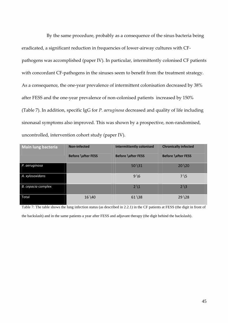
By the same procedure, probably as a consequence of the sinus bacteria being
eradicated, a significant reduction in frequencies of lower-airway cultures with CF-
pathogens was accomplished (paper IV). In particular, intermittently colonised CF patients
with concordant CF-pathogens in the sinuses seem to benefit from the treatment strategy.
As a consequence, the one-year prevalence of intermittent colonisation decreased by 38%
after FESS and the one-year prevalence of non-colonised patients increased by 150%
(Table 7). In addition, specific IgG for P. aeruginosa decreased and quality of life including
sinonasal symptoms also improved. This was shown by a prospective, non-randomised,
uncontrolled, intervention cohort study (paper IV).
Main lung bacteria Non-infected
Before \after FESS
Intermittently colonised
Before \after FESS
Chronically infected
Before \after FESS
P. aeruginosa 50 \31 20 \20
A. xylosoxidans 9 \6 7 \5
B. cepacia complex 2 \1 2 \3
Total 16 \40 61 \38 29 \28
Table 7: The table shows the lung infection status (as described in 2.2.1) in the CF patients at FESS (the digit in front of
the backslash) and in the same patients a year after FESS and adjuvant therapy (the digit behind the backslash).

46
6. Discussion
Bacterial sinusitis being a focus for lung colonisation and infection is
supported by our finding of nearly all CF patients having bacteria in their sinuses and by
frequent lower-airway cultures with CF-pathogens (P. aeruginosa, A. xylosoxidans, B. cepacia
complex) correlating with a high frequency of concordant bacteriology in the sinuses. In
our patients selected for FESS, 67% had concordant CF-pathogenic bacteria in the sinuses
and lungs and additionally 5% had CF-pathogens in the sinuses that were not found in the
lungs (paper IV). The high prevalence of bacterial sinusitis is despite massive intravenous
and oral antibiotic treatment. Additionally, we often did not get any positive lower-airway
cultures during BAL in intermittently lung-colonised patients even when we found CF-
pathogens in the sinuses; this indicates that the sinuses are a more permanent focus than
the lower airways. Our described prevalence of bacterial sinusitis is at the high end
compared with other studies describing CF-sinus bacteriology (described in section 2.6.3),
which is probably a result of our invasive and multiple-sample selection. Above all, we
consider the risk of false positive results very small; the only way that samples can be
cross-contaminated is via the anterior nasal cavity.
Few anaerobes and few fungus isolates were found in the sinuses. Anaerobes
were found when doing molecular studies (unpublished material) but not even here were
fungus frequently found. This is in contrast to our expectations, as CF-patients experience
pulmonary problems with fungus and one study cited by EPOS found a high prevalence
(43;72;151). However, our findings are in accordance with the general low prevalence of
47
fungus-sinusitis in Danish non-CF patients compared with the USA where the study was
carried out.
In the early stage of lung colonisation, migration of CF-pathogens mainly
occurs in a downward direction from the more permanent focus in the sinuses towards the
lungs (21). This migration occurs more frequently during the viral season where the nasal
secretions are more liquefied (90). The results in paper IV further support this theory, as it
may be concluded that if sinus surgery and adjuvant therapy reduce the frequencies of
lower-airway bacteria, the sinuses are bound to influence the lower airways by downward
migration of bacteria. Moreover, when evaluating the literature on this subject, including
the previous papers from our group, I find it unquestionable that the sinuses play an
important role in causing pulmonary colonisations and infections. It is more debateable
what can be done to eliminate this risk of colonisation.
There is empirical evidence that the persistent sinus bacteria are facilitated by
inflamed tissue obstructing the sinus ostia, lower antibiotic concentration than in the lungs
due to lower blood perfusion in the sinus mucosa, that the infection is localised as an
empyema in the sinuses and maybe also as intramucosal abscesses. Furthermore, our
previous research (21) has shown that the bacteria develop resistant genotypes and
phenotypes in the sinuses. In contrast to the nasal environment, where CF-polyps show
various patterns of neutrophil-dominated acute and chronic inflammation (152), we found
a reduced number of PMNs surrounding the biofilms on the sinus mucosa compared with
the lungs. All these points taken together with our results that the upper airways are

48
dominated by the non-phlogistic IgA (paper I, II), may explain the mechanism of why
sinus bacteria are more persistent than in the lungs. In essence, what is important for the
clearance of intermittent P. aeruginosa colonisation in the CF lungs is only partially
functional in the sinuses, providing opportunities for the bacteria to adapt through
evolution of resistance mechanisms.
Some non-infected CF patients were solely colonised with CF-pathogens in
the sinuses (IV). It is likely that this represented their initial colonisation. Nevertheless, we
cannot prove that these patients benefited from the treatment, and it is challenging to
determine the prevalence of how often the colonisations initiates in the sinuses. We are
confident that our prospective study on specific IgA in sputum and nasal secretions will
prove useful in diagnosing P. aeruginosa sinusitis and thereby come closer to a conclusion.
In fact, after we ended our study (paper II), two of the four patients from the non-infected
group with the highest IgA levels have now become intermittently lung-colonised with P.
aeruginosa, and so one might be led to think that they were already sinus-colonised at the
time of the study.
According to the Leeds criteria, CF-patients are cabable of having P. aeruginosa
sinusitis but being categorised as being free from P. aeruginosa (non-infected) (14). In my
opinion, it would be clinically relevant to characterise CF infections both according to their
sino-nasal bacteriology and according to the lower-airway colonisations/infections. This
will require more focus on treating the upper airways, a general collaboration with ORLs,
and that clinicians bear in mind that non-BAL lower-airway samples can be cross-

49
contaminated from the upper airways. Furthermore, in order to characterise CF infections
and select the right CF patients for FESS, it is essential to find a combination of tests that
can diagnose CF-pathogenic sinusitis with high sensitivity. Nasal lavage, as described by
Mainz (69) or in paper II, is a very easy way to obtain samples with little patient
discomfort. However, these samples also contain bacteria from the upper pharynx and
thereby do not solely represent sinus bacteria. It is also uncertain if saline from nasal
irrigations represent material from all sinuses. In unpublished data, we have found a
relatively low positive predictive value when doing middle meatus cultures in early
intermittently colonised patients not previously having sinus surgery, which makes us
conclude that this test cannot stand by itself. However, IgA can easily be obtained and
quantified by ELISA, and if this is combined with regularly obtained cultures from the
middle meatus, cultures from nasal irrigations and other paraclinical measures like serum
antibodies and pulmonary function, we believe it has a high diagnostic value.
It may also be clinically relevant to subdivide intermittently colonised patients
based on the colonisation pattern and bacteria genotypes as previously described (21): a)
patients with single or multiple events of short colonisation periods (<6 months) followed
by eradication; (b) intermittently colonised patients with multiple recurrent colonisation
events with the same genotype of bacteria and a low systemic immune response (77); (c)
patients with a rapid development of chronic lung infections with increasing precipitating
antibodies. Thus, it is most likely that intermittently colonised patients from group (b)
50
have an additional sinonasal infectious focus. This would help us to select patients for
upper-airway treatment by FESS and/or conservative treatment.
Though we have shown that nearly all chronically infected CF patients have
CF-pathogens in their sinuses, which in some cases could be eradicated (paper III, IV), we
did not expect that they would have a significant decrease in positive lower-airway
cultures (paper IV). In particular, four chronically infected patients had a pronounced
effect of the treatment and we put forward the theory that such patients could be false-
positive categorised if the lower-airway samples are cross-contaminated by the upper
airways. Thus, the result of true chronically infected patients having an effect of the
treatment is more uncertain. However, it is accepted that the lung damage in CF patients
with chronic infections characteristically is focal (153) leading to a focal loss of alveoles
and an annual decline of lung function of about 1–2% (76). In that way, true chronically
infected patients may benefit from having their sinus bacteria eradicated, as it may
prevent further spread of the infection by aspiration from the sinuses to new areas of the
lungs.
It can be argued that in paper III and IV we gave no answers as to whether the
same results could have been achieved by conservative treatment comprising nasal
irrigations and endoscopical cleansing. Studies on otherwise healthy patients with CRS
have shown that an ostial dimension should be >4 mm to ensure that irrigations penetrate
the maxillary sinus, and that the frontal sinuses are more difficult to irrigate (154;155). By
comparison, when using nebulizers the dimension requirements are thought to be smaller

51
(138;156). Literature addressing nasal irrigations in CF patients mainly focus on the
maxillary sinuses, but one should remember that CF patients may have frontal sinuses,
which contain CF-pathogens as often as the maxillary sinuses (paper III and unpublished
data). To ensure permanent drainage from the sinuses adequate extensive surgery may be
considered; this could comprise a modified endoscopic medial maxillectomy (127) or a
Draft III (129), the latter ensuring drainage from the frontal sinuses, which are the most
challenging sinuses to operate. We advocate that it is important to ensure permanent
access to the sinuses, both to reduce symptoms of CRS but also to facilitate postoperative
treatment preventing sinus infections and spread of bacteria to the lungs. We agree that
more extensive surgery is needed in CF patients than in patients without CF, but have to
await studies on FESS comparing surgical methods with postoperative clinical
examinations, symptoms, adverse effects and cultures.
In paper III and in section 2.8, the possibilities of using different or additional
ways to treat the upper airways are summarised but the most optimal combination is not
yet defined. In addition, a synergistic effect has been suggested when using tobramycin
and colistimethate sodium for inhalation, thus, this should also be considered when doing
research on which drugs to use for nasal irrigations/nebulizations (157). Especially when
aiming at eradicating A. xylosoxidans and B. cepacia complex, one must be aware of their
antibiotic susceptibility (described in 2.4). While others have described a good effect of
nebulizers such as the PARI sinus (138), the patients in our study have been able to choose
between two devices for nasal irrigations (Figure 15–16). We have no conflicts of interest

52
and find the device in Figure 15 creates a higher pressure than the one in Figure 16, thus
the saline being more likely to penetrate the sinuses.
Finally, based on our findings, I want to stress the importance of focussing on
upper airway bacteriology in CF patients, especially in the outpatient routine treatment
and the importance of guidelines for upper-airway treatment being established. This
requires collaboration between the CF physicians, microbiologists and ORLs.
Figure 15–16: Two methods of doing postoperatively nasal irrigations. (Nicely demonstrated by my 10-year-old son
Bertram).
6.1 Study strength and weaknesses
The major strength of our set-up is the establishment of a unique, effective,
collaboration focusing on CF; the microbiologists and CF physicians have had a strong
collaboration through many years. This project has allowed ORLs to be a part of this
collaboration making it multidisciplinary. We have a very large group of CF patients,
which are all seen on a monthly basis, which is very frequent compared with other CF
53
centres. This results in an abundance of data that can be evaluated for the benefit of the CF
patients. The patients seem very committed to the research, the adherence is high, and
only two patients did not wish to be enrolled in the IgA study (paper II) and only one
patient turned down the offer of FESS (paper IV). The willingness to attend the
postoperative controls and return the questionnaires was also high (80–99%).
A project is always strengthened by having one single committed coordinator;
this improves adherence and reduces bias. To maintain and develop our high quality of
treatment, I find it necessary that the ORLs are keep on seeing CF patients, evaluating
their CRS symptoms, doing endonasal endoscopy and sino-nasal cultures. Furthermore, it
is also important that the CF physicians on a standardized basis ask for CRS symptoms
and focus on possible upper airway infections.
The main outcome of all the studies (I–IV) is based on culture results and
antibody measurements. The Department of Clinical Microbiology has few, but very
dedicated and experienced, laboratory technicians who are responsible for doing the
bacterial and antibody CF analyses. Thus, the possibility of inter-observer errors is low.
In paper II, a weakness is that the study was not prospective; that is why our
hypothesis that high IgA actually reflects sinus colonisations cannot be finally proven. IgA
against alginate can, in small concentrations, be present in non-infected individuals and
IgA against St-Ag can cross-react with other Gram-negative bacteria and the test is
therefore not totally specific towards P. aeruginosa (16;158). Even if our theory is correct,

54
one should bear in mind that it only should be used as a supplementary test creating
awareness of possible colonisations.
In paper III, a drawback might be that due to ethical considerations, only a
minority of the postoperative samples were taken during general anaesthesia, which is
why false negative culture results from the sinuses cannot be excluded. However, I was
the only one who obtained the samples, the majority of patients had persistent opening to
their sinuses, and the follow-up procedure was standardized. Except when doing FESS,
the same point applies in all our studies where one can always discuss how representative
the material is. There are advantages and disadvantages in concern of doing regular
culture compared with molecular methods; these two methods will be compared in an
upcoming paper from our study group. In short, the true positive diagnostic value is high
using both methods. Though the molecular methods in a few cases did detect CF-
pathogens missed by regular methods, a case was also seen where the abundance of
different sinonasal bacteria resulted in a false-negative result of CF-pathogens by the
molecular methods. Sinus samples could in both cases become cross-contaminated by
bacteria from the anterior nasal cavity, but we find this fact clinically unimportant. What is
not unimportant is that cross-contamination of lower-airway samples from the upper
airways remains as a potential confounder. Hence, when the CF-pathogen bacteria were
eradicated from the upper airways, there would be a smaller risk of false-positive lower-
airway culture results.

55
Regardless of cross-contamination, CF-patients who do not show growth of
CF pathogens for a longer period will get their antibiotic treatment reduced.
Consequently, we could have analysed whether the use of antibiotics decreased as a
consequence of the decreased positive lower-airway cultures, or if unchanged, if the
higher rate of antibiotics compared with positive lower-airway cultures might have been a
confounder. The reason that this analysis was not included in paper IV was that we would
have had to differentiate between types of administration (oral, inhalation and
intravenous) and differentiate between prophylactic, eradication and maintenance
therapy. We found that this would have taken focus from our main outcome.
A general weakness concerning paper I, III and IV is that the FESS-project was
step-wise initiated. As a consequence, at the beginning of the study period, we were more
reluctant including patients for FESS, making FESS extensive, and encouraging patients to
be thorough with the postoperative treatment. As the existing research on FESS in CF
patients, including the extent and postoperative treatment, was very sparse, the FESS by
itself has not been a part of the research but solely the outcome of an established
treatment. As a consequence, some CF-patients with no symptoms of CRS have not been
offered FESS as early as we would now recommend and some not at all. Furthermore, we
have not done surgery so extensive and explored all sinuses if the symptoms were not
present. On the other hand, these facts ought not to influence the results in a positive way.
What may weaken the way our results can be interpreted is that we did not have a control
group to the FESS group. Instead we have to use the knowledge of the natural history of
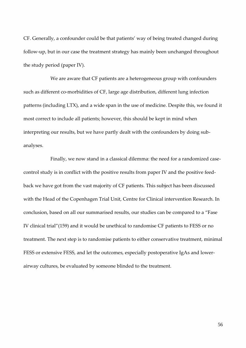
56
CF. Generally, a confounder could be that patients’ way of being treated changed during
follow-up, but in our case the treatment strategy has mainly been unchanged throughout
the study period (paper IV).
We are aware that CF patients are a heterogeneous group with confounders
such as different co-morbidities of CF, large age distribution, different lung infection
patterns (including LTX), and a wide span in the use of medicine. Despite this, we found it
most correct to include all patients; however, this should be kept in mind when
interpreting our results, but we have partly dealt with the confounders by doing sub-
analyses.
Finally, we now stand in a classical dilemma: the need for a randomized case-
control study is in conflict with the positive results from paper IV and the positive feed-
back we have got from the vast majority of CF patients. This subject has been discussed
with the Head of the Copenhagen Trial Unit, Centre for Clinical intervention Research. In
conclusion, based on all our summarised results, our studies can be compared to a ‚Fase
IV clinical trial‛(159) and it would be unethical to randomise CF patients to FESS or no
treatment. The next step is to randomise patients to either conservative treatment, minimal
FESS or extensive FESS, and let the outcomes, especially postoperative IgAs and lower-
airway cultures, be evaluated by someone blinded to the treatment.
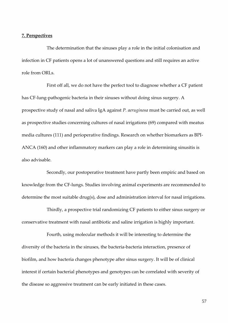
57
7. Perspectives
The determination that the sinuses play a role in the initial colonisation and
infection in CF patients opens a lot of unanswered questions and still requires an active
role from ORLs.
First off all, we do not have the perfect tool to diagnose whether a CF patient
has CF-lung-pathogenic bacteria in their sinuses without doing sinus surgery. A
prospective study of nasal and saliva IgA against P. aeruginosa must be carried out, as well
as prospective studies concerning cultures of nasal irrigations (69) compared with meatus
media cultures (111) and perioperative findings. Research on whether biomarkers as BPI-
ANCA (160) and other inflammatory markers can play a role in determining sinusitis is
also advisable.
Secondly, our postoperative treatment have partly been empiric and based on
knowledge from the CF-lungs. Studies involving animal experiments are recommended to
determine the most suitable drug(s), dose and administration interval for nasal irrigations.
Thirdly, a prospective trial randomizing CF patients to either sinus surgery or
conservative treatment with nasal antibiotic and saline irrigation is highly important.
Fourth, using molecular methods it will be interesting to determine the
diversity of the bacteria in the sinuses, the bacteria-bacteria interaction, presence of
biofilm, and how bacteria changes phenotype after sinus surgery. It will be of clinical
interest if certain bacterial phenotypes and genotypes can be correlated with severity of
the disease so aggressive treatment can be early initiated in these cases.

58
Finally, in the same way as CF patients, patients with primary ciliary
dyskinesia (PCD) may have sinusitis initiating colonisation and infection though the
mechanisms are different (161). In general, there is a lack on research on PCD, which is
why CF treatment often is used on this patient group (161). I recommend that a study be
done on extensive sinus surgery and adjuvant therapy in PCD patients.

59
8. Conclusion
As answers to the aims of this thesis (1.1):
a) There is a very high prevalence of CF pathogen sinusitis. The bacteria persist in
the sinuses for years and can be a focus for initial lung colonisation and maintenan the
infection.
b) In contrast to the lungs, the sinus inflammation is dominated by non-phlogistic
specific IgAs; this facilitates persistence of bacteria.
c) There is no single way of diagnosing CF-pathogens in the sinuses without being
invasive. Nasal IgA may be a surrogate-marker for P.aeruginosa in the lungs and
may be used as a supplementary diagnostic tool for P.aeruginosa-sinusitis.
d) We have treated our patients with extensive FESS and standardized
postoperative follow-up, IV antibiotics, prolonged nasal irrigation with saline and
antibiotics in addition to nasal steroids. Further studies are needed to find the most
effective treatment.
e) By this treatment strategy (d), quality of life was improved, bacterial sinus foci
could be eradicated and the frequency of pulmonary samples positive for CF pathogens
could be reduced. This indicates a reduced CF morbidity.
Altogether, the CF-upper airways should not be neglected and ORLs can
give a significant contribution to CF treatment.

60
9. Acknowledgements
The process of doing this thesis has taught me evidence-based medicine and
the ability to immerse into a subject. This work was made possible by many people. I
would like to thank everyone who have helped and supported me throughout the work.
In particular, I would like to express my gratitude to:
Christian von Buchwald, who offered me the opportunity to help with your project. It has
been a great pleasure having you as my principal supervisor and to benefit from your
ideas; though you are a busy man, you have given me all the support I could ask for.
Especially, thank you for teaching me to do FESS on the requested high and independent
level;
Helle Krogh Johansen, my other supervisor, a fantastic guide throughout microbiology
and the jungle of cystic fibrosis, lots of ideas; especially thank you for your loyalty, quick
but thorough feed-back, and your optimism towards me and starting new projects;
Niels Høiby, for your drive, ideas, input, and your ability to grasp cystic fibrosis. Thank
you for initiating the project together with Helle and Christian and allowing all the
samples to be analysed;
Tanja Pressler, Marianne Skov, Frederik Buchvald and Kim G. Nielsen, physicians at
the CF-centre: you all have a highly scientific level and your input has been priceless as
have your enthusiasm, cooperation and your benevolence to answer my novice question.
Other departments could learn a lot from our co-operation.; Thanks to Kim for teaching
me BAL; Thanks to Tanja for helping with this thesis;
Steen Seier Poulsen for helping me out with the immunohistochemistry;
Thomas Hjuler for help and always having a good way of viewing things;
Jacob Fisker for good ideas and an early introduction to CF;
Rene Kærgaard for supervising me in FESS;

61
Susse Kirkelund Hansen, Lars Jelsbak and Søren Molin for the cooperation on doing a
CF paper (21), which I have not found room for in this thesis;
Lars Fledelius Rickelt and Peter Østrup Jensen for the cooperation on doing a CF paper
(20), which I have not found room for in this thesis;
Jacob Rasmussen and Rikke Norling for the cooperation on doing a CF paper (50), which
I have not found room for in this thesis;
Niels Rasmussen, Mårten Segelmark, Ulrika Lindberg, Malin Carlsson and Jørgen
Wieslander for the cooperation on doing a CF paper (159), which I have not found room
for in this thesis;
Vibeke B. Rudkjøbing, Tine Yding Wolff and Trine Rolighed Thomsen for the
cooperation on doing the CF paper on ‚Microorganisms involved in sinus infection of
cystic fibrosis patients determined by culture and molecular based methods‛, which I have
not found room for in this thesis;
Secretaries and nurses at the CF-centre Copenhagen for their dedication to this project,
especially Ketty Vinding and Majbritt Presfeldt;
Thanks to Michelle, Simon, Christel, Kåre, Ramon, Emilie, David and Pernille for a
pleasant work environment;
The Danish Cystic Fibrosis association for economic support;
Pseudomonas-laboratory technicians at Department of Clinical Microbiology for their
tireless and countless analyses, especially Katja Bloksted, and Lene Nørregaard;
All the CF patients that participated in our trials;
Finally, I would like to thank Berit Street and the Candys Foundation; every day through
three years, I have highly appreciated your financial support. This has formed the basis for
all the productivity.

62
10. SNOT-22 questionnaire (64)
1. Need to blow nose 0 1 2 3 4 5
2. Sneezing 0 1 2 3 4 5
3. Runny nose 0 1 2 3 4 5
4. Cough 0 1 2 3 4 5
5. Postnasal discharge (dripping at the back of your throat) 0 1 2 3 4 5
6. Thick nasal discharge (snot) 0 1 2 3 4 5
7. Ear fullness 0 1 2 3 4 5
8. Dizziness 0 1 2 3 4 5
9. Ear pain 0 1 2 3 4 5
10. Facial pain/pressure 0 1 2 3 4 5
11. Nasal blokage 0 1 2 3 4 5
12. Loss of taste and or smell 0 1 2 3 4 5
13. Difficulty falling asleep 0 1 2 3 4 5
14. Waking up at night 0 1 2 3 4 5
15. Lack of a good night ’s sleep 0 1 2 3 4 5
16. Waking up tired 0 1 2 3 4 5
17. Fatigue 0 1 2 3 4 5
18. Reduced productivity 0 1 2 3 4 5
19. Reduced concentration 0 1 2 3 4 5
20. Frustrated/restless/irritable 0 1 2 3 4 5
21. Sad 0 1 2 3 4 5
22. Embarrassed 0 1 2 3 4 5 0= no problem; 1= very mild problem; 2= mild or slight problem; 3= moderate problem;
4=severe problem; 5= problem as bad as it can be.
Please mark the most important items affecting your health (maximum of 5 items)
63
11. References
(1) Nielsen OH, Thomsen BL, Green A, Andersen PK, Hauge M, Schiotz PO. Cystic fibrosis in Denmark 1945 to 1985. An analysis of incidence, mortality and influence of centralized treatment on survival. Acta Paediatr Scand 1988 Nov;77(6):836-41.
(2) Kerem B, Rommens JM, Buchanan JA, Markiewicz D, Cox TK, Chakravarti A, et al. Identification of the cystic fibrosis gene: genetic analysis. Science 1989 Sep 8;245(4922):1073-80.
(3) Doggett RG, Harrision GM, Wallis ES. Comparison of some properties of Pseudomonas aeruginosa isolated from infections in persons with and without cystic fibrosis. J Bacteriol 1964 Feb;87:427-31.
(4) Boucher RC. New concepts of the pathogenesis of cystic fibrosis lung disease. Eur Respir J 2004 Jan;23(1):146-58.
(5) Rubin BK. Mucus structure and properties in cystic fibrosis. Paediatr Respir Rev 2007 Mar;8(1):4-7.
(6) Cystic Fibrosis Foundation. Patient Registry 2007 Annual Report. 2009.
(7) Koch C, Hoiby N. Diagnosis and treatment of cystic fibrosis. Respiration 2000;67(3):239-47.
(8) Doring G. Host response to Pseudomonas aeruginosa. Acta Paediatr Scand Suppl 1989;363:37-9.
(9) Hoiby N, Flensborg EW, Beck B, Friis B, Jacobsen SV, Jacobsen L. Pseudomonas aeruginosa infection in cystic fibrosis. Diagnostic and prognostic significance of Pseudomonas aeruginosa precipitins determined by means of crossed immunoelectrophoresis. Scand J Respir Dis 1977 Apr;58(2):65-79.
(10) Bech B, Pressler T, Iversen M, Carlsen J, Milman N, Eliasen K, et al. Long-term outcome of lung transplantation for cystic fibrosis--Danish results. Eur J Cardiothorac Surg 2004 Dec;26(6):1180-6.
(11) Hoiby N. Epidemiological investigations of the respiratory tract bacteriology in patients with cystic fibrosis. Acta Pathol Microbiol Scand B Microbiol Immunol 1974 Aug;82(4):541-50.
64
(12) Hoiby N. Pseudomonas aeruginosa infection in cystic fibrosis. Relationship between mucoid strains of Pseudomonas aeruginosa and the humoral immune response. Acta Pathol Microbiol Scand B Microbiol Immunol 1974 Aug;82(4):551-8.
(13) Frederiksen B, Koch C, Hoiby N. Changing epidemiology of Pseudomonas aeruginosa infection in Danish cystic fibrosis patients (1974-1995). Pediatr Pulmonol 1999 Sep;28(3):159-66.
(14) Lee TW, Brownlee KG, Conway SP, Denton M, Littlewood JM. Evaluation of a new definition for chronic Pseudomonas aeruginosa infection in cystic fibrosis patients. J Cyst Fibros 2003 Mar;2(1):29-34.
(15) Brett MM, Ghoneim AT, Littlewood JM. Prediction and diagnosis of early Pseudomonas aeruginosa infection in cystic fibrosis: a follow-up study. J Clin Microbiol 1988 Aug;26(8):1565-70.
(16) Pressler T, Karpati F, Granstrom M, Knudsen PK, Lindblad A, Hjelte L, et al. Diagnostic significance of measurements of specific IgG antibodies to Pseudomonas aeruginosa by three different serological methods. J Cyst Fibros 2009 Jan;8(1):37-42.
(17) Proesmans M, Balinska-Miskiewicz W, Dupont L, Bossuyt X, Verhaegen J, Hoiby N, et al. Evaluating the "Leeds criteria" for Pseudomonas aeruginosa infection in a cystic fibrosis centre. Eur Respir J 2006 May;27(5):937-43.
(18) Hansen CR, Pressler T, Hoiby N. Early aggressive eradication therapy for intermittent Pseudomonas aeruginosa airway colonization in cystic fibrosis patients: 15 years experience. J Cyst Fibros 2008 Nov;7(6):523-30.
(19) Kolpen M, Hansen CR, Bjarnsholt T, Moser C, Christensen LD, van GM, et al. Polymorphonuclear leucocytes consume oxygen in sputum from chronic Pseudomonas aeruginosa pneumonia in cystic fibrosis. Thorax 2010 Jan;65(1):57-62.
(20) Aanaes K, Rickelt LF, Johansen HK, Buchwald C, Pressler T, Hoiby N, et al. Decreased mucosal oxygen tension in the maxillary sinuses in patients with cystic fibrosis. J Cyst Fibros 2011 Mar;10(2):114-20.
(21) Hansen SK, Rau MH, Johansen HK, Ciofu O, Jelsbak L, Yang L, et al. Evolution and diversification of Pseudomonas aeruginosa in the paranasal sinuses of cystic fibrosis children have implications for chronic lung infection. ISME J 2012 Jan;6(1):31-45.
(22) Yang L, Jelsbak L, Molin S. Microbial ecology and adaptation in cystic fibrosis airways. Environ Microbiol 2011 Jul;13(7):1682-9.
(23) Hoiby N. The serology of Pseudomonas aeruginosa analysed by means of quantitative immunoelectrophoretic methods. I. Comparison of thirteen O groups of Ps. aeruginosa, with a polyvalent Ps. aeruginosa antigen-antibody reference system
5. Acta Pathol Microbiol Scand B 1975 Aug;83(4):321-7.

65
(24) Bryan LE, Kureishi A, Rabin HR. Detection of antibodies to Pseudomonas aeruginosa alginate extracellular polysaccharide in animals and cystic fibrosis patients by enzyme-linked immunosorbent assay. J Clin Microbiol 1983 Aug;18(2):276-82.
(25) Pedersen SS, Moller H, Espersen F, Sorensen CH, Jensen T, Hoiby N. Mucosal immunity to Pseudomonas aeruginosa alginate in cystic fibrosis. APMIS 1992 Apr;100(4):326-34.
(26) Johansen HK, Hoiby N. Local IgA and IgG response to intratracheal immunization with Pseudomonas aeruginosa antigens. APMIS 1992 Jan;100(1):87-90.
(27) Hansen CR, Pressler T, Nielsen KG, Jensen PO, Bjarnsholt T, Hoiby N. Inflammation in Achromobacter xylosoxidans infected cystic fibrosis patients. J Cyst Fibros 2010 Jan;9(1):51-8.
(28) Magni A, Giordano A, Mancini C, Pecoraro C, Varesi P, Quattrucci S, et al. Emerging cystic fibrosis pathogens: incidence and antimicrobial resistance. New Microbiol 2007 Jan;30(1):59-62.
(29) Waters V. New treatments for emerging cystic fibrosis pathogens other than Pseudomonas. Curr Pharm Des 2012;18(5):696-725.
(30) Braccini G, Festini F, Boni V, Neri AS, Galici V, Campana S, et al. The costs of treatment of early and chronic Pseudomonas aeruginosa infection in cystic fibrosis patients. J Chemother 2009 Apr;21(2):188-92.
(31) Doring G, Hoiby N. Early intervention and prevention of lung disease in cystic fibrosis: a European consensus. J Cyst Fibros 2004 Jun;3(2):67-91.
(32) Frederiksen B, Koch C, Hoiby N. Antibiotic treatment of initial colonization with Pseudomonas aeruginosa postpones chronic infection and prevents deterioration of pulmonary function in cystic fibrosis. Pediatr Pulmonol 1997 May;23(5):330-5.
(33) Ratjen F, Walter H, Haug M, Meisner C, Grasemann H, Doring G. Diagnostic value of serum antibodies in early Pseudomonas aeruginosa infection in cystic fibrosis patients. Pediatr Pulmonol 2007 Mar;42(3):249-55.
(34) da Silva Filho LV, Tateno AF, Martins KM, zzuz Chernishev AC, Garcia DO, Haug M, et al. The combination of PCR and serology increases the diagnosis of Pseudomonas aeruginosa colonization/infection in cystic fibrosis. Pediatr Pulmonol 2007 Oct;42(10):938-44.
(35) De BK, Alifier M, Vandeputte S. Sputum induction in young cystic fibrosis patients. Eur Respir J 2000 Jul;16(1):91-4.
(36) Wood RE. The diagnostic effectiveness of the flexible bronchoscope in children. Pediatr Pulmonol 1985 Jul;1(4):188-92.
(37) Pressler T, Frederiksen B, Skov M, Garred P, Koch C, Hoiby N. Early rise of anti-pseudomonas antibodies and a mucoid phenotype of pseudomonas aeruginosa are risk

66
factors for development of chronic lung infection--a case control study. J Cyst Fibros 2006 Jan;5(1):9-15.
(38) Johansen HK, Norregaard L, Gotzsche PC, Pressler T, Koch C, Hoiby N. Antibody response to Pseudomonas aeruginosa in cystic fibrosis patients: a marker of therapeutic success?--A 30-year cohort study of survival in Danish CF patients after onset of chronic P. aeruginosa lung infection. Pediatr Pulmonol 2004 May;37(5):427-32.
(39) Eggesbo HB, Sovik S, Dolvik S, Kolmannskog F. CT characterization of inflammatory paranasal sinus disease in cystic fibrosis. Acta Radiol 2002 Jan;43(1):21-8.
(40) Mainz JG, Koitschev A. Management of chronic rhinosinusitis in CF. J Cyst Fibros 2009 Jun;8 Suppl 1:S10-S14.
(41) Mak GK, Henig NR. Sinus disease in cystic fibrosis. Clin Rev Allergy Immunol 2001 Aug;21(1):51-63.
(42) Robertson JM, Friedman EM, Rubin BK. Nasal and sinus disease in cystic fibrosis. Paediatr Respir Rev 2008 Sep;9(3):213-9.
(43) Fokkens WJ, Lund VJ, Mullol J, Bachert C, Alobid I, Baroody F, et al. EPOS 2012: European position paper on rhinosinusitis and nasal polyps 2012. A summary for otorhinolaryngologists. Rhinology 2012 Mar;50(1):1-12.
(44) Loebinger MR, Bilton D, Wilson R. Upper airway 2: Bronchiectasis, cystic fibrosis and sinusitis. Thorax 2009 Dec;64(12):1096-101.
(45) Cepero R, Smith RJ, Catlin FI, Bressler KL, Furuta GT, Shandera KC. Cystic fibrosis--an otolaryngologic perspective. Otolaryngol Head Neck Surg 1987 Oct;97(4):356-60.
(46) Eggesbo HB, Sovik S, Dolvik S, Eiklid K, Kolmannskog F. CT characterization of developmental variations of the paranasal sinuses in cystic fibrosis. Acta Radiol 2001 Sep;42(5):482-93.
(47) Eggesbo HB, Dolvik S, Stiris M, Sovik S, Storrosten OT, Kolmannskog F. Complementary role of MR imaging of ethmomaxillary sinus disease depicted at CT in cystic fibrosis. Acta Radiol 2001 Mar;42(2):144-50.
(48) Eggesbo HB, Sovik S, Dolvik S, Eiklid K, Kolmannskog F. Proposal of a CT scoring system of the paranasal sinuses in diagnosing cystic fibrosis. Eur Radiol 2003 Jun;13(6):1451-60.
(49) Gysin C, Alothman GA, Papsin BC. Sinonasal disease in cystic fibrosis: clinical characteristics, diagnosis, and management. Pediatr Pulmonol 2000 Dec;30(6):481-9.
(50) Rasmussen J, Aanaes K, Norling R, Nielsen KG, Johansen HK, Buchwald C. CT of the paranasal sinuses is not a valid indicator for sinus surgery in CF patients. J Cyst Fibros 2011 Oct 19.

67
(51) Halvorson DJ. Cystic fibrosis: an update for the otolaryngologist. Otolaryngol Head Neck Surg 1999 Apr;120(4):502-6.
(52) Stern RC, Boat TF, Wood RE, Matthews LW, Doershuk CF. Treatment and prognosis of nasal polyps in cystic fibrosis. Am J Dis Child 1982 Dec;136(12):1067-70.
(53) Friedman EM, Stewart M. An assessment of sinus quality of life and pulmonary function in children with cystic fibrosis. Am J Rhinol 2006 Nov;20(6):568-72.
(54) Henriksson G, Westrin KM, Karpati F, Wikstrom AC, Stierna P, Hjelte L. Nasal polyps in cystic fibrosis: clinical endoscopic study with nasal lavage fluid analysis. Chest 2002 Jan;121(1):40-7.
(55) Kingdom TT, Lee KC, FitzSimmons SC, Cropp GJ. Clinical characteristics and genotype analysis of patients with cystic fibrosis and nasal polyposis requiring surgery. Arch Otolaryngol Head Neck Surg 1996 Nov;122(11):1209-13.
(56) Feuillet-Fieux MN, Lenoir G, Sermet I, Elie C, Djadi-Prat J, Ferrec M, et al. Nasal polyposis and cystic fibrosis(CF): review of the literature. Rhinology 2011 Aug;49(3):347-55.
(57) Lindig J, Steger C, Beiersdorf N, Michl R, Beck JF, Hummel T, et al. Smell in cystic fibrosis. Eur Arch Otorhinolaryngol 2012 Aug 14.
(58) Coste A, Gilain L, Roger G, Sebbagh G, Lenoir G, Manach Y, et al. Endoscopic and CT-scan evaluation of rhinosinusitis in cystic fibrosis. Rhinology 1995 Sep;33(3):152-6.
(59) King VV. Upper respiratory disease, sinusitis, and polyposis. Clin Rev Allergy 1991;9(1-2):143-57.
(60) Bonestroo HJ, de Winter-de Groot KM, van der Ent CK, Arets HG. Upper and lower airway cultures in children with cystic fibrosis: do not neglect the upper airways. J Cyst Fibros 2010 Mar;9(2):130-4.
(61) Watelet JB, Van CP, Bachert C. Rhinological aspects of cystic fibrosis. Monaldi Arch Chest Dis 2000 Dec;55(6):475-7.
(62) Alobid I, Benitez P, Bernal-Sprekelsen M, Roca J, Alonso J, Picado C, et al. Nasal polyposis and its impact on quality of life: comparison between the effects of medical and surgical treatments. Allergy 2005 Apr;60(4):452-8.
(63) Sahlstrand-Johnson P, Ohlsson B, Buchwald C, Jannert M. A multi-centre study on quality of life and absenteeism in patients with CRS referred for endoscopic surgery. Rhinology 2011 Oct;49(4):420-8.
(64) Hopkins C, Gillett S, Slack R, Lund VJ, Browne JP. Psychometric validity of the 22-item Sinonasal Outcome Test. Clin Otolaryngol 2009 Oct;34(5):447-54.

68
(65) Kay DJ, Rosenfeld RM. Quality of life for children with persistent sinonasal symptoms. Otolaryngol Head Neck Surg 2003 Jan;128(1):17-26.
(66) Clinical practice guideline: management of sinusitis. Pediatrics 2001 Sep;108(3):798-808.
(67) Brook I. Microbiology of sinusitis. Proc Am Thorac Soc 2011 Mar;8(1):90-100.
(68) Digoy GP, Dunn JD, Stoner JA, Christie A, Jones DT. Bacteriology of the paranasal sinuses in pediatric cystic fibrosis patients. Int J Pediatr Otorhinolaryngol 2012 Jul;76(7):934-8.
(69) Mainz JG, Naehrlich L, Schien M, Kading M, Schiller I, Mayr S, et al. Concordant genotype of upper and lower airways P aeruginosa and S aureus isolates in cystic fibrosis. Thorax 2009 Jun;64(6):535-40.
(70) Moss RB, King VV. Management of sinusitis in cystic fibrosis by endoscopic surgery and serial antimicrobial lavage. Reduction in recurrence requiring surgery. Arch Otolaryngol Head Neck Surg 1995 May;121(5):566-72.
(71) Shapiro ED, Milmoe GJ, Wald ER, Rodnan JB, Bowen AD. Bacteriology of the maxillary sinuses in patients with cystic fibrosis. J Infect Dis 1982 Nov;146(5):589-93.
(72) Wise SK, Kingdom TT, McKean L, DelGaudio JM, Venkatraman G. Presence of fungus in sinus cultures of cystic fibrosis patients. Am J Rhinol 2005 Jan;19(1):47-51.
(73) Muhlebach MS, Miller MB, Moore C, Wedd JP, Drake AF, Leigh MW. Are lower airway or throat cultures predictive of sinus bacteriology in cystic fibrosis? Pediatr Pulmonol 2006 May;41(5):445-51.
(74) Roby BB, McNamara J, Finkelstein M, Sidman J. Sinus surgery in cystic fibrosis patients: comparison of sinus and lower airway cultures. Int J Pediatr Otorhinolaryngol 2008 Sep;72(9):1365-9.
(75) Suh JD, Cohen NA, Palmer JN. Biofilms in chronic rhinosinusitis. Curr Opin Otolaryngol Head Neck Surg 2010 Feb;18(1):27-31.
(76) Bjarnsholt T, Jensen PO, Fiandaca MJ, Pedersen J, Hansen CR, Andersen CB, et al. Pseudomonas aeruginosa biofilms in the respiratory tract of cystic fibrosis patients. Pediatr Pulmonol 2009 Jun;44(6):547-58.
(77) Johansen HK, Aanaes K, Pressler T, Nielsen KG, Fisker J, Skov M, et al. Colonisation and infection of the paranasal sinuses in cystic fibrosis patients is accompanied by a reduced PMN response. J Cyst Fibros 2012 May 15.
(78) Dosanjh A, Lakhani S, Elashoff D, Chin C, Hsu V, Hilman B. A comparison of microbiologic flora of the sinuses and airway among cystic fibrosis patients with maxillary antrostomies. Pediatr Transplant 2000 Aug;4(3):182-5.
69
(79) Taylor L, Corey M, Matlow A, Sweezey NB, Ratjen F. Comparison of throat swabs and nasopharyngeal suction specimens in non-sputum-producing patients with cystic fibrosis. Pediatr Pulmonol 2006 Sep;41(9):839-43.
(80) Godoy JM, Godoy AN, Ribalta G, Largo I. Bacterial pattern in chronic sinusitis and cystic fibrosis. Otolaryngol Head Neck Surg 2011 Oct;145(4):673-6.
(81) Davidson TM, Murphy C, Mitchell M, Smith C, Light M. Management of chronic sinusitis in cystic fibrosis. Laryngoscope 1995 Apr;105(4 Pt 1):354-8.
(82) Jung A, Kleinau I, Schonian G, Bauernfeind A, Chen C, Griese M, et al. Sequential genotyping of Pseudomonas aeruginosa from upper and lower airways of cystic fibrosis patients. Eur Respir J 2002 Dec;20(6):1457-63.
(83) Taylor-Robinson D, Whitehead M, Diderichsen F, Olesen HV, Pressler T, Smyth RL, et al. Understanding the natural progression in %FEV1 decline in patients with cystic fibrosis: a longitudinal study. Thorax 2012 May 3.
(84) Taylor RF, Morgan DW, Nicholson PS, Mackay IS, Hodson ME, Pitt TL. Extrapulmonary sites of Pseudomonas aeruginosa in adults with cystic fibrosis. Thorax 1992 Jun;47(6):426-8.
(85) Walter S, Gudowius P, Bosshammer J, Romling U, Weissbrodt H, Schurmann W, et al. Epidemiology of chronic Pseudomonas aeruginosa infections in the airways of lung transplant recipients with cystic fibrosis. Thorax 1997 Apr;52(4):318-21.
(86) Jelsbak L, Johansen HK, Frost AL, Thogersen R, Thomsen LE, Ciofu O, et al. Molecular epidemiology and dynamics of Pseudomonas aeruginosa populations in lungs of cystic fibrosis patients. Infect Immun 2007 May;75(5):2214-24.
(87) Munck A, Bonacorsi S, Mariani-Kurkdjian P, Lebourgeois M, Gerardin M, Brahimi N, et al. Genotypic characterization of Pseudomonas aeruginosa strains recovered from patients with cystic fibrosis after initial and subsequent colonization. Pediatr Pulmonol 2001 Oct;32(4):288-92.
(88) Taccetti G, Campana S, Festini F, Mascherini M, Doring G. Early eradication therapy against Pseudomonas aeruginosa in cystic fibrosis patients. Eur Respir J 2005 Sep;26(3):458-61.
(89) Hoiby N, Pedersen SS. Estimated risk of cross-infection with Pseudomonas aeruginosa in Danish cystic fibrosis patients. Acta Paediatr Scand 1989 May;78(3):395-404.
(90) Johansen HK, Hoiby N. Seasonal onset of initial colonisation and chronic infection with Pseudomonas aeruginosa in patients with cystic fibrosis in Denmark. Thorax 1992 Feb;47(2):109-11.
(91) Holzmann D, Speich R, Kaufmann T, Laube I, Russi EW, Simmen D, et al. Effects of sinus surgery in patients with cystic fibrosis after lung transplantation: a 10-year experience. Transplantation 2004 Jan 15;77(1):134-6.

70
(92) Khalil HS, Nunez DA. Functional endoscopic sinus surgery for chronic rhinosinusitis. Cochrane Database Syst Rev 2006;(3):CD004458.
(93) Shekelle PG, Woolf SH, Eccles M, Grimshaw J. Developing clinical guidelines. West J Med 1999 Jun;170(6):348-51.
(94) Vital D, Hofer M, Boehler A, Holzmann D. Posttransplant sinus surgery in lung transplant recipients with cystic fibrosis: a single institutional experience. Eur Arch Otorhinolaryngol 2012 Mar 30.
(95) Vital D, Hofer M, Benden C, Holzmann D, Boehler A. Impact of Sinus Surgery on Pseudomonal Airway Colonization, Bronchiolitis Obliterans Syndrome and Survival in Cystic Fibrosis Lung Transplant Recipients. Respiration 2012 Aug 22.
(96) Shatz A. Management of recurrent sinus disease in children with cystic fibrosis: a combined approach. Otolaryngol Head Neck Surg 2006 Aug;135(2):248-52.
(97) Lewiston N, King V, Umetsu D, Starnes V, Marshall S, Kramer M, et al. Cystic fibrosis patients who have undergone heart-lung transplantation benefit from maxillary sinus antrostomy and repeated sinus lavage. Transplant Proc 1991 Feb;23(1 Pt 2):1207-8.
(98) Jarrett WA, Militsakh O, Anstad M, Manaligod J. Endoscopic sinus surgery in cystic fibrosis: effects on pulmonary function and ideal body weight. Ear Nose Throat J 2004 Feb;83(2):118-21.
(99) Leung MK, Rachakonda L, Weill D, Hwang PH. Effects of sinus surgery on lung transplantation outcomes in cystic fibrosis. Am J Rhinol 2008 Mar;22(2):192-6.
(100) Madonna D, Isaacson G, Rosenfeld RM, Panitch H. Effect of sinus surgery on pulmonary function in patients with cystic fibrosis. Laryngoscope 1997 Mar;107(3):328-31.
(101) Osborn AJ, Leung R, Ratjen F, James AL. Effect of endoscopic sinus surgery on pulmonary function and microbial pathogens in a pediatric population with cystic fibrosis. Arch Otolaryngol Head Neck Surg 2011 Jun;137(6):542-7.
(102) Rosbe KW, Jones DT, Rahbar R, Lahiri T, Auerbach AD. Endoscopic sinus surgery in cystic fibrosis: do patients benefit from surgery? Int J Pediatr Otorhinolaryngol 2001 Nov 1;61(2):113-9.
(103) Triglia JM, Nicollas R. Nasal and sinus polyposis in children. Laryngoscope 1997 Jul;107(7):963-6.
(104) Umetsu DT, Moss RB, King VV, Lewiston NJ. Sinus disease in patients with severe cystic fibrosis: relation to pulmonary exacerbation. Lancet 1990 May 5;335(8697):1077-8.
(105) Kempainen RR, Sajan JA, Pylkas AM, Dunitz JM, Rimell FL, Milla CE. Effect of Endoscopic Sinus Surgery on Pulmonary Status of Adults with Cystic Fibrosis. Otolaryngol Head Neck Surg 2012 Apr 18.
71
(106) Halvorson DJ, Dupree JR, Porubsky ES. Management of chronic sinusitis in the adult cystic fibrosis patient. Ann Otol Rhinol Laryngol 1998 Nov;107(11 Pt 1):946-52.
(107) Kovell LC, Wang J, Ishman SL, Zeitlin PL, Boss EF. Cystic fibrosis and sinusitis in children: outcomes and socioeconomic status. Otolaryngol Head Neck Surg 2011 Jul;145(1):146-53.
(108) McMurphy AB, Morriss C, Roberts DB, Friedman EM. The usefulness of computed tomography scans in cystic fibrosis patients with chronic sinusitis. Am J Rhinol 2007 Nov;21(6):706-10.
(109) Graham SM, Launspach JL, Welsh MJ, Zabner J. Sequential magnetic resonance imaging analysis of the maxillary sinuses: implications for a model of gene therapy in cystic fibrosis. J Laryngol Otol 1999 Apr;113(4):329-35.
(110) Gwaltney JM, Jr. Acute community-acquired sinusitis. Clin Infect Dis 1996 Dec;23(6):1209-23.
(111) Araujo E, Palombini BC, Cantarelli V, Pereira A, Mariante A. Microbiology of middle meatus in chronic rhinosinusitis. Am J Rhinol 2003 Jan;17(1):9-15.
(112) Cheng K, Ashby D, Smyth RL. Oral steroids for long-term use in cystic fibrosis. Cochrane Database Syst Rev 2011;(10):CD000407.
(113) Becker SS, Rasamny JK, Han JK, Patrie J, Gross CW. Steroid injection for sinonasal polyps: the University of Virginia experience. Am J Rhinol 2007 Jan;21(1):64-9.
(114) Sorensen H, Mygind N, Tygstrup I, Winge FE. Histology of nasal polyps of different etiology. Rhinology 1977 Sep;15(3):121-8.
(115) Costantini D, Di CM, Giunta A, Amabile G. Nasal polyposis in cystic fibrosis treated by beclomethasone dipropionate. Acta Univ Carol Med (Praha) 1990;36(1-4):220-1.
(116) Hadfield PJ, Rowe-Jones JM, Mackay IS. A prospective treatment trial of nasal polyps in adults with cystic fibrosis. Rhinology 2000 Jun;38(2):63-5.
(117) Weber SA, Ferrari GF. Incidence and evolution of nasal polyps in children and adolescents with cystic fibrosis. Braz J Otorhinolaryngol 2008 Jan;74(1):16-20.
(118) Cuyler JP. Follow-up of endoscopic sinus surgery on children with cystic fibrosis. Arch Otolaryngol Head Neck Surg 1992 May;118(5):505-6.
(119) Nishioka GJ, Barbero GJ, Konig P, Parsons DS, Cook PR, Davis WE. Symptom outcome after functional endoscopic sinus surgery in patients with cystic fibrosis: a prospective study. Otolaryngol Head Neck Surg 1995 Oct;113(4):440-5.
(120) Rowe-Jones JM, Mackay IS. Endoscopic sinus surgery in the treatment of cystic fibrosis with nasal polyposis. Laryngoscope 1996 Dec;106(12 Pt 1):1540-4.
72
(121) Keck T, Rozsasi A. Medium-term symptom outcomes after paranasal sinus surgery in children and young adults with cystic fibrosis. Laryngoscope 2007 Mar;117(3):475-9.
(122) Jones JW, Parsons DS, Cuyler JP. The results of functional endoscopic sinus (FES) surgery on the symptoms of patients with cystic fibrosis. Int J Pediatr Otorhinolaryngol 1993 Dec;28(1):25-32.
(123) Yung MW, Gould J, Upton GJ. Nasal polyposis in children with cystic fibrosis: a long-term follow-up study. Ann Otol Rhinol Laryngol 2002 Dec;111(12 Pt 1):1081-6.
(124) Bugten V, Nordgard S, Steinsvag S. The effects of debridement after endoscopic sinus surgery. Laryngoscope 2006 Nov;116(11):2037-43.
(125) Cho DY, Hwang PH. Results of endoscopic maxillary mega-antrostomy in recalcitrant maxillary sinusitis. Am J Rhinol 2008 Nov;22(6):658-62.
(126) Fuchsmann C, Ayari S, Reix P, Colreavy M, Bellon G, Durieux I, et al. Contribution of CT-assisted navigation and microdebriders to endoscopic sinus surgery in cystic fibrosis. Int J Pediatr Otorhinolaryngol 2008 Mar;72(3):343-9.
(127) Virgin FW, Rowe SM, Wade MB, Gaggar A, Leon KJ, Young KR, et al. Extensive surgical and comprehensive postoperative medical management for cystic fibrosis chronic rhinosinusitis. Am J Rhinol Allergy 2012 Jan;26(1):70-5.
(128) Crockett DM, McGill TJ, Healy GB, Friedman EM, Salkeld LJ. Nasal and paranasal sinus surgery in children with cystic fibrosis. Ann Otol Rhinol Laryngol 1987 Jul;96(4):367-72.
(129) Georgalas C, Hansen F, Videler WJ, Fokkens WJ. Long terms results of Draf type III (modified endoscopic Lothrop) frontal sinus drainage procedure in 122 patients: a single centre experience. Rhinology 2011 Jun;49(2):195-201.
(130) Lamberty JM, Rubin BK. The management of anaesthesia for patients with cystic fibrosis. Anaesthesia 1985 May;40(5):448-59.
(131) Harvey R, Hannan SA, Badia L, Scadding G. Nasal saline irrigations for the symptoms of chronic rhinosinusitis. Cochrane Database Syst Rev 2007;(3):CD006394.
(132) Boek WM, Keles N, Graamans K, Huizing EH. Physiologic and hypertonic saline solutions impair ciliary activity in vitro. Laryngoscope 1999 Mar;109(3):396-9.
(133) Wark PA, McDonald V, Jones AP. Nebulised hypertonic saline for cystic fibrosis. Cochrane Database Syst Rev 2005;(3):CD001506.
(134) Pynnonen MA, Mukerji SS, Kim HM, Adams ME, Terrell JE. Nasal saline for chronic sinonasal symptoms: a randomized controlled trial. Arch Otolaryngol Head Neck Surg 2007 Nov;133(11):1115-20.

73
(135) Chiu AG, Palmer JN, Woodworth BA, Doghramji L, Cohen MB, Prince A, et al. Baby shampoo nasal irrigations for the symptomatic post-functional endoscopic sinus surgery patient. Am J Rhinol 2008 Jan;22(1):34-7.
(136) Isaacs S, Fakhri S, Luong A, Whited C, Citardi MJ. The effect of dilute baby shampoo on nasal mucociliary clearance in healthy subjects. Am J Rhinol Allergy 2011 Jan;25(1):e27-e29.
(137) Cimmino M, Nardone M, Cavaliere M, Plantulli A, Sepe A, Esposito V, et al. Dornase alfa as postoperative therapy in cystic fibrosis sinonasal disease. Arch Otolaryngol Head Neck Surg 2005 Dec;131(12):1097-101.
(138) Mainz JG, Schiller I, Ritschel C, Mentzel HJ, Riethmuller J, Koitschev A, et al. Sinonasal inhalation of dornase alfa in CF: A double-blind placebo-controlled cross-over pilot trial. Auris Nasus Larynx 2011 Apr;38(2):220-7.
(139) Raynor EM, Butler A, Guill M, Bent JP, III. Nasally inhaled dornase alfa in the postoperative management of chronic sinusitis due to cystic fibrosis. Arch Otolaryngol Head Neck Surg 2000 May;126(5):581-3.
(140) Kobayashi T, Baba S. Topical use of antibiotics for paranasal sinusitis. Rhinol Suppl 1992;14:77-81.
(141) Adappa ND, Wei CC, Palmer JN. Nasal irrigation with or without drugs: the evidence. Curr Opin Otolaryngol Head Neck Surg 2012 Feb;20(1):53-7.
(142) Hansen FS, Djupesland PG, Fokkens WJ. Preliminary efficacy of fluticasone delivered by a novel device in recalcitrant chronic rhinosinusitis. Rhinology 2010 Sep;48(3):292-9.
(143) Karosi T, Sziklai I, Csomor P. Low-frequency ultrasound for biofilm disruption in chronic rhinosinusitis with nasal polyposis: In vitro pilot study. Laryngoscope 2012 Aug 14.
(144) Høiby N, Frederiksen B. Microbiology of cystic fibrosis. In: Hodson ME GD, editor. Cystic Fibrosis. 2nd edition ed. Arnold; London; 2000. p. 83-107.
(145) Johansen HK, Moskowitz SM, Ciofu O, Pressler T, Hoiby N. Spread of colistin resistant non-mucoid Pseudomonas aeruginosa among chronically infected Danish cystic fibrosis patients. J Cyst Fibros 2008 Sep;7(5):391-7.
(146) Pedersen SS, Espersen F, Hoiby N, Jensen T. Imunoglobulin A and immunoglobulin G antibody responses to alginates from Pseudomonas aeruginosa in patients with cystic fibrosis. J Clin Microbiol 1990 Apr;28(4):747-55.
(147) Hankinson JL, Odencrantz JR, Fedan KB. Spirometric reference values from a sample of the general U.S. population. Am J Respir Crit Care Med 1999 Jan;159(1):179-87.
(148) Wang X, Dockery DW, Wypij D, Fay ME, Ferris BG, Jr. Pulmonary function between 6 and 18 years of age. Pediatr Pulmonol 1993 Feb;15(2):75-88.

74
(149) Nysom K, Molgaard C, Hutchings B, Michaelsen KF. Body mass index of 0 to 45-y-old Danes: reference values and comparison with published European reference values. Int J Obes Relat Metab Disord 2001 Feb;25(2):177-84.
(150) Quittner AL, Buu A, Messer MA, Modi AC, Watrous M. Development and validation of The Cystic Fibrosis Questionnaire in the United States: a health-related quality-of-life measure for cystic fibrosis. Chest 2005 Oct;128(4):2347-54.
(151) Knutsen AP, Slavin RG. Allergic bronchopulmonary aspergillosis in asthma and cystic fibrosis. Clin Dev Immunol 2011;2011:843763.
(152) Rozsasi A, Keck T. Lipopolysaccharide induction of cyclooxygenase-2 in long-term nasal epithelial cultures in cystic fibrosis. Arch Otolaryngol Head Neck Surg 2010 Apr;136(4):335-9.
(153) Tiddens HA. Detecting early structural lung damage in cystic fibrosis. Pediatr Pulmonol 2002 Sep;34(3):228-31.
(154) Grobler A, Weitzel EK, Buele A, Jardeleza C, Cheong YC, Field J, et al. Pre- and postoperative sinus penetration of nasal irrigation. Laryngoscope 2008 Nov;118(11):2078-81.
(155) Singhal D, Weitzel EK, Lin E, Feldt B, Kriete B, McMains KC, et al. Effect of head position and surgical dissection on sinus irrigant penetration in cadavers. Laryngoscope 2010 Dec;120(12):2528-31.
(156) Mainz JG, Michl R, Pfister W, Beck JF. Cystic fibrosis upper airways primary colonization with Pseudomonas aeruginosa: eradicated by sinonasal antibiotic inhalation. Am J Respir Crit Care Med 2011 Nov 1;184(9):1089-90.
(157) Herrmann G, Yang L, Wu H, Song Z, Wang H, Hoiby N, et al. Colistin-tobramycin combinations are superior to monotherapy concerning the killing of biofilm Pseudomonas aeruginosa. J Infect Dis 2010 Nov 15;202(10):1585-92.
(158) Pedersen SS. Lung infection with alginate-producing, mucoid Pseudomonas aeruginosa in
cystic fibrosis. APMIS 1000 Suppl. 28, 1-79. 1992. Ref Type: Journal (Full)
(159) Robey RR. A five-phase model for clinical-outcome research. J Commun Disord 2004
Sep;37(5):401-11.
(160) Aanaes K, Rasmussen N, Pressler T, Segelmark M, Johansen HK, Lindberg U, et al. Extensive endoscopic image-guided sinus surgery decreases BPI-ANCA in patients with cystic fibrosis. Scand J Immunol 2012 Sep 5.
(161) Campbell R. Managing upper respiratory tract complications of primary ciliary dyskinesia in children. Curr Opin Allergy Clin Immunol 2012 Feb;12(1):32-8.

Paper I
Journal of Cystic Fibrosis xx (2012) xxx–xxx
JCF-00791; No of Pages 7
www.elsevier.com/locate/jcf
Original Article
Colonisation and infection of the paranasal sinuses in cystic fibrosis patientsis accompanied by a reduced PMN response☆
Helle Krogh Johansen a,⁎, Kasper Aanaes b, Tania Pressler c, Kim Gjerrum Nielsen c,Jacob Fisker b, Marianne Skov c, Niels Høiby a, d, Christian von Buchwald c
a Department of Clinical Microbiology, afsnit 9301, Rigshospitalet, Juliane Maries Vej 22, DK-2100 Copenhagen Ø, Denmarkb Department of Otolaryngology, Head and Neck Surgery, afsnit 2072, Rigshospitalet, Blegdamsvej 9, DK-2100 Copenhagen Ø, Denmark
c CF Centre Copenhagen and Paediatric Pulmonary Service, Department of Paediatrics and Adolescent Medicine, Rigshospitalet, Blegdamsvej 9,DK-2100 Copenhagen Ø, Denmark
d Institute of International Health, Immunology and Microbiology, Panum Institute, University of Copenhagen, Blegdamsvej 3B, 2200 Copenhagen N, Denmark
Received 11 February 2012; received in revised form 12 April 2012; accepted 26 April 2012
Abstract
Background: We studied whether the sinuses might be foci for Pseudomonas aeruginosa lung infection.Methods: Endoscopic Sinus Surgery was performed in 78 CF patients; PFGE was used for bacterial genotyping. Material from sinuses and lungswere Gram-stained to detect biofilms. Immunoglobulins were measured in serum and saliva.Results: When P. aeruginosa was cultured simultaneously from the sinuses and the lungs they were genetically identical in 38 of the 40 patients(95%). In the sinuses, P. aeruginosa formed biofilms with minimal cellular inflammation, probably because of a significantly higher localproduction of secretory IgA compared with IgG (pb0.001).Conclusions: We have shown that P. aeruginosa form biofilm in the sinuses, which constitute an important bacterial reservoir for subsequent lunginfection. The high amount of IgA in the upper airways probably protects P. aeruginosa from the inflammatory immune system, and they canproceed unnoticed into a permanent infectious focus that cannot be eradicated with antibiotics.© 2012 European Cystic Fibrosis Society. Published by Elsevier B.V. All rights reserved.
Keywords: Rhinosinusitis; P. aeruginosa; Cystic fibrosis; Foci; Biofilm
☆ Part of this work has been presented as a poster at the American Society forMicrobiology, 108th General Meeting Boston, MA, USA June 1–5, 2008, “Theparanasal sinuses: a possible focus for bacterial lung infections in cystic fibrosispatients?”, as a poster at the 31st European Cystic Fibrosis Conference, June11–14, Prague, Czech republic, 2008 “The paranasal sinuses are focus forcolonisation and chronic biofilm lung infections in CF patients”. J Cyst Fibros2008;suppl 2:S38, at The 2008 North American Cystic Fibrosis Conference,Orlando, Florida, USA, October 23–25, 2008 as a poster “The paranasal sinusesare focus for colonisation and chronic biofilm lung infection in CF patients” andas a poster at the 32nd European Cystic Fibrosis Conference, June 10–13, Brest,France, 2009 “Paranasal sinuses are a focus for P. aeruginosa (PA) lunginfection in CF. J Cyst Fibros 2009;suppl 2:S36.⁎ Corresponding author. Tel.: +45 3545 4973; fax: +45 3545 6412.E-mail address: [email protected] (H.K. Johansen).
1569-1993/$ -see front matter © 2012 European Cystic Fibrosis Society. Publisheddoi:10.1016/j.jcf.2012.04.011
Please cite this article as: Johansen HK, et al, Colonisation and infection of the paresponse, J Cyst Fibros (2012), doi:10.1016/j.jcf.2012.04.011
1. Introduction
The most common manifestations of cystic fibrosis (CF) arepulmonary disease, radio-opaque sinuses, nasal polyposis andexocrine pancreatic insufficiency. Symptoms of chronic rhino-sinusitis have been reported in 11–94% of CF patients [1].Sinusitis is seldom recognised by the patients but nearly all CFpatients have radiographic and clinical signs of infection [1,2].
Molecular epidemiology studies have shown that CF lungtransplant recipients become re-colonised in their lung grafts withthe same clones as those cultured before transplantation [3]. It islikely that Pseudomonas aeruginosa is drained into the lungallografts from a bacterial reservoir in the paranasal sinuses viathe airways [3,4]. In lung transplanted CF patients sinus surgeryaiming at eradication of bacteria, mostly P. aeruginosa, has
by Elsevier B.V. All rights reserved.
ranasal sinuses in cystic fibrosis patients is accompanied by a reduced PMN
2 H.K. Johansen et al. / Journal of Cystic Fibrosis xx (2012) xxx–xxx
lowered the incidence of bacterial colonisation and infection aftertransplantation [5].
We have previously genotyped initial and subsequentP. aeruginosa lung isolates from CF patients by using ArrayTube biochip analysis (Clondiag®), a method based on singlenucleotide polymorphisms (SNPs) [6] and by Pulsed Field GelElectrophoresis (PFGE) [6]. We found that the initial colonisingP. aeruginosa strains had different genotypes; i.e. every patienthad a genotype different from all other patients. This suggests thatthe initial colonisation comes from environmental sources ratherthan from transmission between patients [7]. We also found thatnew lung colonising isolates had the same genotype as the initialcolonising strain after antibiotic eradication of P. aeruginosa,suggesting a bacterial reservoir either in the patient or in thepatient's close environment [6].
In the present prospective study, we examined whether theparanasal sinuses serve as a bacterial reservoir for CF pathogens,especially P. aeruginosa, leading to reinfections with the samegenotype as the initial infection. We cultured and genotypedhistorical isolates as well as paired P. aeruginosa isolates fromsputum and sinuses from children and adults with CF whounderwent Functional Endoscopic Sinus Surgery (FESS) to see ifbacteria from the two anatomical locations were identical.
2. Materials and methods
2.1. General care
300 CF patients are followed at the Danish CF centre,Copenhagen [8,9] and treated according to fixed guidelines [8].
Intermittent colonisation: Patients with at least one isolate ofP. aeruginosa, but normal levels of precipitating antibodiesagainst P. aeruginosa ( 0–1 precipitins) [8].
Chronic P. aeruginosa lung infection: Growth of P. aeruginosain six consecutive monthly sputum samples, or less if there are twoor more precipitating antibodies against P. aeruginosa [8].
2.2. Bacteria
Since 1973, we have collected and stored (−80 °C)P. aeruginosa sputum isolates sequentially [8,9].
2.3. Included patients
Seventy-eight CF patients N6 years (median 19 years, range6–50), (29 males, 49 females) were treated with FESS betweenJanuary 27, 2007 and April 1, 2010.
There are no guidelines for sinus surgery in CF patients [10].Patients were selected based on one or more of the followingcriteria: 1) intermittent colonisation with declining lungfunction despite intensive antibiotic therapy and/or increasingantibodies against Gram-negative bacteria e.g. P. aeruginosa,Achromobacter xylosoxidans or Burkholderia cepacia complex,2) lung transplantation within the last year, and 3) severesymptoms of rhinosinusitis according to the European PositionPaper guidelines (EPOS) [10]. The majority fulfilled the firstand third criterion.
Please cite this article as: Johansen HK, et al, Colonisation and infection of the presponse, J Cyst Fibros (2012), doi:10.1016/j.jcf.2012.04.011
2.4. Functional Endoscopic Sinus Surgery (FESS)
FESS created ventilation and drainage pathways to and fromthe sinuses, making the paranasal sinuses accessible forirrigations [11].
An average of six tissue or pus samples (range 1–14) wastaken from each patient. The FESS was finalised by sinusirrigation applying 3 MIE polymyxin E (colistin).
A sputum sample was obtained on the same day as thesurgery to compare bacteriology in the lungs with the sinuses.
After surgery, the majority of patients did nasal irrigationswith colistin twice daily for at least 6 months. An ENT specialistperformed postoperative follow-up five times during the first yearafter surgery.
2.5. Bacteriology and typing methods (PFGE)
Gram-stained smears for biofilm detection, and aerobic andanaerobic cultures at 37 °C on standard agar media for 5–7 days,were carried out on all tissue and pus samples [9,12]. PFGE wasused for genotyping P. aeruginosa isolates from the sinuses andthe lungs [9].
2.6. Serum and saliva
Serum and saliva were collected from 25 randomly chosenFESS patients who were either intermittently colonised orchronically infected with P. aeruginosa. Saliva was obtainedby using four sterile 6 mm diameter paper discs (AntibioticaTestblættchen, Struers, Denmark) that were placed on themouth mucosa for 30 sec. The saliva-soaked discs were storedat −80 °C until analysed.
2.7. IgG and IgA antibodies against P. aeruginosa in salivaand serum
Specific antibodies were measured using ELISA [13,14].The volume of all reagents for the serum ELISA was 100 μl,but 50 μl for the saliva ELISA. Phosphate-buffered saline (PBSpH 7.2)+0.1% Tween-20 (Sigma)+NaCl 15 g/l (= dilutionbuffer) were used for all washing steps and the plates werewashed three times.
Saliva-impregnated paper-discs were incubated on a shakerfor one hour at 35 °C in 175 μl dilution buffer to elute IgG andIgA antibodies.
Antibodies against alginate, 96-well microtiter plates (Mikro-well, BiotechLine A/S, Denmark) were coated with alginate(10 μg/ml) purified from a mucoid CF P. aeruginosa strain(6680NH). The plates were coated over night at 35 °C andblocked for one hour at 35 °C in dilution buffer. Serum wasdiluted 1:4,000 and saliva was already diluted 1:8 in the elutionprocedure and used without further dilution for detection of IgAand IgG. After washing, horseradish peroxidase (HRP)-conjugate(DakoA/S, Glostrup, Denmark) detecting IgG and IgA antibodieswere added for one hour. The detecting antibodies were rabbitanti-human IgG (γ-chain-specific) diluted 1:10,000 or rabbit anti-human IgA (α-chain-specific) diluted 1:10,000.
aranasal sinuses in cystic fibrosis patients is accompanied by a reduced PMN

3H.K. Johansen et al. / Journal of Cystic Fibrosis xx (2012) xxx–xxx
Antibodies against P. aeruginosa standard antigens (a soni-cated cell extract of P. aeruginosa serogroups 1–17) were usedas standard antigen (protein concentration 16 mg/ml). The antigenwas coated onto irrigated 96-well polysterene plates (Maxisorb,BiotechLine A/S, Denmark) at a dilution of 1:2,000. The plateswere incubated for one hour at 35 °C and blocked overnight withdilution buffer at 4 °C. Serumwas diluted 1:100 and saliva 1:8 fordetection of IgG and IgA and allowed to react for one hour at35 °C. After washing, horseradish peroxidase (HRP)-conjugatediluted 1:20,000 (Dako A/S, Glostrup, Denmark) detecting IgG(P0214) and IgA (P0216) antibodies in serum and saliva wereadded for one hour. The peroxidase-conjugated second antibodieswere as described above.
TMB Plus was added (KemEnTec Diagnostics). The reactionwas stopped after one hour by 1 M H2SO4. The absorbancewas measured at 450 nm on a plate reader (Multiscan EX, Bie& Berntsen, Denmark). The results were expressed as opticaldensity values (OD).
2.8. Ethics
The study was approved by the local ethics committee,Region Hovedstaden, (H-A-141) and all patients gave informedconsent.
2.9. Statistics
Within-patients samples of serum and saliva antibodies werecompared using the sign test.
3. Results
Thirty-two of the 78 patients were chronically infected; 21with P. aeruginosa (median age 31 years, range 12–50 years),5 with A. xylosoxidans, 4 with B. multivorans complex and 2with Stenotrophomonas maltophilia. The remaining 46 patientswere intermittently colonised; 31 with P. aeruginosa (medianage 14 years, range 7–29 years), 6 with A. xylosoxidans and 9with other CF pathogens such as Haemophilus influenzae,Staphylococcus aureus or Streptococcus pneumoniae in theirlungs.
Of the 21 patients chronically infected with P. aeruginosa,18 (86%) had simultaneous growth of P. aeruginosa in theirsinuses and lower airways; this was also the case for all 5patients with growth of A. xylosoxidans, all 4 with growth ofB. multivorans complex, whereas none of the 2 patients withchronic S. maltophilia infection in their lungs had growth ofthis bacterium in their sinuses.
In 22 of the 31 (71%) intermittently colonised patients, wecultured P. aeruginosa from both the sinuses and lungs.
We did not detect any anaerobic microorganisms in any ofthe sinuses although the cultures were performed less than onehour after the sinus samples were taken and incubation wasperformed for 7 days.
PFGE patterns were used to compare the relatednessof sequentially collected P. aeruginosa lung isolates withP. aeruginosa cultured from the lungs and sinuses after FESS.
Please cite this article as: Johansen HK, et al, Colonisation and infection of the paresponse, J Cyst Fibros (2012), doi:10.1016/j.jcf.2012.04.011
In 18 of the 18 patients (100%) with chronic P. aeruginosa lunginfection and from whom we had simultaneous growth ofP. aeruginosa in their sinuses and lungs, P. aeruginosa wasgenetically identical. We found that chronically infectedpatients had the same P. aeruginosa genotype in the lungs fora median of 15 years similar to the P. aeruginosa that werecultured in the sinuses (range 1–29 years). We found that 5 ofthe 18 patients had identical genotypes in their lungs for morethan 20 years and this was the same genotype as cultured fromthe sinuses after FESS.
Twenty of the 22 (91%) intermittently colonised patientswith P. aeruginosa in their sinuses had PFGE identical isolatesin their lungs, whereas a different genotype was found in twopatients. Patients who had identical P. aeruginosa in their lungsand sinuses at the time of surgery had a similar genotype intheir lungs for a median of 3.5 years (range 1–6 years).
Gram-stained smears from the sinuses showed that allpatients chronically infected with P. aeruginosa had theirbacteria organised in biofilm-like structures similar to what isseen in the lungs of the patients [15,16] (Fig. 1 a–f). Whenmicroscopically analysing Gram-stained smears from the lungand sinuses obtained on the same day in patients chronicallyinfected with P. aeruginosa, we found that bacterial biofilms inthe lung were surrounded by a large number of inflammatorycells, predominantly polymorphonuclear cells (PMNs), whereasonly very few and scattered PMNs were seen in the surroundingsof the sinus biofilms (Fig. 1 b, d, f). In addition, we found thatpatients with chronic A. xylosoxidans and B. cepacia complexinfection also had their bacteria organised in aggregates in thesinuses [17].
None of the 21 patients chronically infected with P. aeruginosahad the bacteria eradicated from the lungs following FESS. Inintermittently colonised patients (N=31) the median time fromsinus surgery to regrowth of P. aeruginosa in sputum was7.3 months. In patients who had P. aeruginosa cultured from theirsinuses (N=22), P. aeruginosa was cultured again from the lungs5.3 months after FESS.
Saliva IgA against P. aeruginosa sonicate (median opticaldensity (OD) 83) and alginate (median OD 186) was 15 and 39times higher than serum IgA (median OD 5 in both cases)(pb0.001 in both cases), indicating a local production of IgA inthe upper airways. The IgA antibody levels in saliva againstP. aeruginosa sonicate and alginate were also significantlyhigher than the saliva IgG production (median OD 2 and 0,respectively) (pb0.001 in both cases). The IgG levels in serumagainst P. aeruginosa sonicate and alginate were also low(median OD 3 in both cases).
4. Discussion
We have performed the largest published study till now ofinvasive sinus surgery investigating the possible role of thesinuses as a reservoir for bacterial adaptation and repeatedcolonisation and infection of the lungs [3,18,19]. The group ofintermittently colonised patients, which we included in this study,is a selected subgroup among our children population since mostof them have been intermittently colonised in their lungs with the
ranasal sinuses in cystic fibrosis patients is accompanied by a reduced PMN
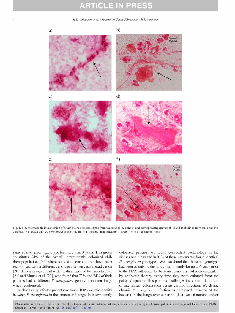
a) b)
c) d)
e) f)
Fig. 1. a–f. Microscopic investigation of Gram-stained smears of pus from the sinuses (a, c and e) and corresponding sputum (b, d and f) obtained from three patientschronically infected with P. aeruginosa at the time of sinus surgery, magnification ×1000. Arrows indicate biofilms.
4 H.K. Johansen et al. / Journal of Cystic Fibrosis xx (2012) xxx–xxx
same P. aeruginosa genotype for more than 3 years. This groupconstitutes 24% of the overall intermittently colonised chil-dren population [20] whereas most of our children have beenrecolonised with a different genotype after successful eradication[20]. This is in agreement with the data reported by Taccetti et al.[21] and Munck et al. [22], who found that 73% and 74% of theirpatients had a different P. aeruginosa genotype in their lungswhen recolonised.
In chronically infected patients we found 100%genetic identitybetween P. aeruginosa in the sinuses and lungs. In intermittently
Please cite this article as: Johansen HK, et al, Colonisation and infection of the presponse, J Cyst Fibros (2012), doi:10.1016/j.jcf.2012.04.011
colonised patients, we found concordant bacteriology in thesinuses and lungs and in 91% of these patients we found identicalP. aeruginosa genotypes. We also found that the same genotypehad been colonising the lungs intermittently for up to 6 years priorto the FESS, although the bacteria apparently had been eradicatedby antibiotic therapy every time they were cultured from thepatients' sputum. This paradox challenges the current definitionof intermittent colonisation versus chronic infection. We definechronic P. aeruginosa infection as continued presence of thebacteria in the lungs over a period of at least 6 months and/or
aranasal sinuses in cystic fibrosis patients is accompanied by a reduced PMN

5H.K. Johansen et al. / Journal of Cystic Fibrosis xx (2012) xxx–xxx
serum content of 2 or more precipitating antibodies againstP. aeruginosa, whereas intermittent colonisation is defined asisolation of P. aeruginosa in a patient with no measurableantibodies in the blood (normal: 0–1 precipitins) and no clinicalsymptoms [8]. The intermittently colonised patients that have hadsinus surgery in this article have been colonised with the samegenotype for a median of 3.5 years with no measurable antibodyresponses and therefore need to be classified into a new group ofchronically colonised patients. If the same clone of P. aeruginosapersists for such long periods of time it should be consideredchronic despite the lack of precipitating antibodies. We thereforeneed to modify our operational definition of chronic infection.
All patients chronically infected with P. aeruginosa in theirlungs and from whom we cultured P. aeruginosa in the sinusesharbored identical clones. A small subgroup of patients whounderwent FESS had the same genotype in their sinuses andlungs for more than two decades. This is in accordance with thefindings byMainz et al. [23] who in a cross-sectional study foundthat 95% of the P. aeruginosa isolates from 24 patients hadidentical SNP-genotypes in both compartments, indicating thatthe upper airways play a role as a reservoir of P. aeruginosa inCF, and by Muhlenbach et al. [24] who demonstrated identicalgenotypes in the upper and lower airways in 83% of samplesobtained from twelve patients.
Our study shows that the adaptation of P. aeruginosa to thesinuses is different from the adaptation in the lungs. In the lungsthere is a PMN dominated inflammation with release of oxygenradicals [25], probably because the lungs, especially the respi-ratory zone, contain high levels of IgG against P. aeruginosa,which promotes an inflammation dominated by PMNs. In thesinuses, we did not find a similar PMN-dominated inflammationbut a high level of IgA compared to IgG, and IgA has non-inflammatory properties and inhibit the PMN recruitment [26].The immunoglobulin distributions fit well with a recent study bySchraven et al. showing that the number of plasma cells in CFpatients with chronic polypoid sinusitis was significantly elevatedcompared to non-CF patients and that the number of neutrophilswere low in both groups [27].We have previously found that thereis a common mucosal secretory IgA response to P. aeruginosa inCF patients, whereas the systemic response is dominated by IgG[28]. The secretory IgA response, both to the biofilm matrix-component alginate and to the P. aeruginosa sonicate, includingproteins and LPS, implies that the P. aeruginosa sinusitis is rathersilent because of the lack of PMNs compared with the chroniclung infection [16] and that P. aeruginosa may adapt to thechronic life-style and be well suited for causing lung infectionsespecially if adaptation and biofilm formation has begun in thesinuses [11,20]. Although we cannot totally exclude that specificsubpopulations of bacteria from the lungs are transmitted to thesinuses from time to time our previous results suggest that thedirection of migration is mainly downwards at the early stages ofinfection [20]. None of the intermittently colonised patients hadelevated precipitating antibodies against P. aeruginosa beforesurgery, which supports our hypothesis that the bacteria in thesinuses are not being recognised by the systemic immune system(IgG antibodies) but by the mucosal immune system (secretoryIgA) [28]. In chronically infected patients we found that
Please cite this article as: Johansen HK, et al, Colonisation and infection of the paresponse, J Cyst Fibros (2012), doi:10.1016/j.jcf.2012.04.011
P. aeruginosa also forms biofilms in the sinuses similar to thebiofilms in the lungs. However, there is an important difference.There are only very few PMNs around the biofilm in the sinuses incontrast to the significant amount of PMNs surrounding thebiofilms in the lungs and this is, probably due to the non-inflammatory secretory IgA response (Fig. 1a–f) [16].
We have previously demonstrated that many CF patients inour clinic contracted their initial P. aeruginosa colonisation andchronic lung infection during the viral season in the wintermonths (October to March) [29]. When patients have a viralinfection, the excessive secretions formed in the upper respiratorytract become liquid, and will easily find its way into the deepestportion of the lungs within minutes [30]. When P. aeruginosacontaining secretions in the sinuses of CF patients becomeliquefied, they may easily enter the lungs by aspiration andbecome difficult to clear. Accumulation of bacteria-laden liquidin the supralaryngeal portion during viral infection may byaspiration overwhelm the mucociliary defense mechanisms of thesub-laryngeal portion, and the secretions are aspirated especiallyduring sleep [30]. When bacteria from the sinuses are aspiratedinto the lungs they might be pre-adapted and therefore lessvirulent compared with environmental P. aeruginosa isolates,since they have had the opportunity to evolve to the lifestyle inthe lungs [20,31]. The sinuses can be seen as an “evolutionarynest” where bacteria are diversifying, develop antibiotic resis-tance and other phenotypes associated with adaptation to the CFairways in general [20].
In intermittently colonised patients the overall duration untilregrowth of P. aeruginosa in the sputum after FESS was7 months whereas the median time to recurrence ofP. aeruginosain sputum in patients from whom we cultured P. aeruginosa intheir sinuses was less than half a year. We believe that the FESSprocedure itself cannot keep the children free of P. aeruginosa fora prolonged period of time but has to be carried out together withother postoperative treatment such as nasal irrigation with colistinand inhaled intravenous antibiotics. This is in agreement with arecent trial in CF patients assessing 0.9% NaCl versus dornasealpha delivered by vibrating aerosols into the paranasal sinuses[32]. Dornase alpha inhalation was associated with a significantlyimproved quality of life score. In the future such therapeuticapproaches with simultaneously dornase alpha and sinonasalinhalation of antibiotics might benefit patients where FESS is notan option.
There are some limitations in our study that should beconsidered when interpreting the results. We could not cultureany anaerobic microorganisms in the sinuses although this wasexpected based on our previous findings [11]. It could be due tothe intensive antibiotic treatment of these patients or insufficienthandling of the samples, although they were cultured within onehour after surgery [8,12]. In addition, we expected to find an evenhigher number of patients with matching P. aeruginosa in theirsinuses and lungs, but there might have been too few bacteriain our samples. Molecular based methods may have beenappropriate, but this would not have given any information ongenotypes.
In conclusion, we have shown that the sinuses are bacterial focifor P. aeruginosa in CF patients. In the sinuses, P. aeruginosa
ranasal sinuses in cystic fibrosis patients is accompanied by a reduced PMN
6 H.K. Johansen et al. / Journal of Cystic Fibrosis xx (2012) xxx–xxx
grow in biofilms that are similar to the biofilms seen in the lungsof chronically infected patients. In contrast to the situation in thelungs, a high concentration of non-inflammatory secretory IgA inthe sinuses probably impedes the PMNs from being recruited,which may prevent local and systemic inflammation andrecognition of microorganisms, contributing to adaptation andpersistence of P. aeruginosa ready to colonise and infect thelungs.
Funding
KA was funded by Candys Foundation.
Competing interests
None.
Financial disclosures
None.
Contributors
HKJ was principal investigator. HKJ contributed to theconception and study design together with KA, NH and CvB.TP, KGN and MS included the patients and KA, JF and CvBdid the surgery. HKJ and NH performed the microbiologicalanalyses including genotyping of P. aeruginosa strains. HKJ,KA, NH and CvB contributed to the analysis and interpretationof data. HKJ drafted the manuscript and all authors commentedon the manuscript prior to submission and approved thesubmitted version.
Acknowledgements
We thank Katja Bloksted, Ulla Rydahl Johansen, LenaNørregaard, Helle Nordbjerg and Pia Poss for excellenttechnical assistance; Majbritt Prefeldt and Ketty Vinding atthe CF-clinic for collaboration and recruitment of the patients;and Peter C Gøtzsche and Søren Molin for comments on themanuscript.
References
[1] Robertson JM, Friedman EM, Rubin BK. Nasal and sinus disease in cysticfibrosis. Paediatr Respir Rev 2008;9:213–9.
[2] Loebinger MR, Bilton D, Wilson R. Upper airway 2: bronchiectasis,cystic fibrosis and sinusitis. Thorax 2009;64:1096–101.
[3] Walter S, Gudowius P, Bosshammer J, Römling U, Weissbrodt H,Schurmann W, et al. Epidemiology of chronic Pseudomonas aeruginosainfections in the airways of lung transplant recipients with cystic fibrosis.Thorax 1997;52:318–21.
[4] Leung MK, Rachakonda L, Weill D, Hwang PH. Effects of sinus surgeryon lung transplantation outcomes in cystic fibrosis. Am J Rhinol 2008;22:192–6.
[5] Holzmann DR, Speich R, Kaufmann T, Laube I, Russi EW, Simmen D,et al. Effects of sinus surgery in patients with cystic fibrosis after lungtransplantation: a 10-year experience. Transplantation 2004;77:134–6.
Please cite this article as: Johansen HK, et al, Colonisation and infection of the presponse, J Cyst Fibros (2012), doi:10.1016/j.jcf.2012.04.011
[6] Jelsbak L, Johansen HK, Frost AL, Thøgersen R, Thomsen LE, Ciofu O,et al. Molecular epidemiology and dynamics of Pseudomonas aeruginosapopulations in lungs of cystic fibrosis patients. Infect Immun 2007;75:2214–24.
[7] Høiby N, Pedersen SS. Estimated risk of cross-infection with Pseudomo-nas aeruginosa in Danish cystic fibrosis patients. Acta Paediatr Scand1989;78:395–404.
[8] Johansen HK, Nørregaard L, Gøtzsche PC, Pressler T, Koch C, Høiby N.Antibody response to Pseudomonas aeruginosa in cystic fibrosis patients:a marker of therapeutic success?—a 30-year cohort study of survival inDanish CF patients after onset of chronic P. aeruginosa lung infection.Pediatr Pulmonol 2004;37:427–32.
[9] Johansen HK, Moskowitz SM, Ciofu O, Pressler T, Høiby N. Spread ofcolistin resistant non-mucoid Pseudomonas aeruginosa among chroni-cally infected Danish cystic fibrosis patients. J Cyst Fibros 2008;7:391–7.
[10] Thomas M, Yawn BP, Price D, Lund V, Mullol J, Fokkens W. EPOSprimary care guidelines: European position paper on the primary carediagnosis and management of rhinosinusitis and nasal polyps 2. PrimCare Respir J 2008;17:79–89.
[11] Aanaes K, Rickelt LF, Johansen HK, von Buchwald C, Pressler T, HøibyN, et al. Decreased mucosal oxygen tension in the maxillary sinuses inpatients with cystic fibrosis. J Cyst Fibros 2011;10:114–20.
[12] Høiby N, Frederiksen B. Microbiology of cystic fibrosis. In: Hodson ME,Geddes DM, editors. Cystic Fibrosis. 2nd edition. London: Arnold; 2000.p. 83–107.
[13] Pedersen SS, Møller H, Espersen F, Sørensen CH, Jensen T, Høiby N.Mucosal immunity to Pseudomonas aeruginosa alginate in cystic fibrosis.APMIS 1992;100:326–34.
[14] Johansen HK, Høiby N. Local IgA and IgG response to intratrachealimmunization with Pseudomonas aeruginosa antigens. APMIS 1992;100:87–90.
[15] Kirketerp-Møller K, Jensen PØ, Fazli M, Madsen KG, Pedersen J, Moser C,et al. Distribution, organization, and ecology of bacteria in chronic wounds. JClin Microbiol 2008;46:2717–22.
[16] Bjarnsholt T, Jensen PØ, Fiandaca MJ, Pedersen J, Hansen CR, AndersenCB, et al. Pseudomonas aeruginosa biofilms in the respiratory tract ofcystic fibrosis patients. Pediatr Pulmonol 2009;44:547–58.
[17] Hansen CR, Pressler T, Nielsen KG, Jensen PØ, Bjarnsholt T, HøibyN, et al. Inflammation in Achromobacter xylosoxidans infected cysticfibrosis patients. J Cyst Fibros 2010;9:51–8.
[18] Bonestroo HJ, de Winter-de Groot KM, van der Ent CK, Arets HG. Upperand lower airway cultures in children with cystic fibrosis: do not neglectthe upper airways. J Cyst Fibros 2010;9:130–4.
[19] Roby BB, McNamara J, Finkelstein M, Sidman J. Sinus surgery in cysticfibrosis patients: comparison of sinus and lower airway cultures. Int JPediatr Otorhinolaryngol 2008;72:1365–9.
[20] Hansen SK, Rau MH, Johansen HK, Ciofu O, Jelsbak L, Yang L, et al.Evolution and diversification of Pseudomonas aeruginosa in the paranasalsinuses of cystic fibrosis children have implications for chronic lunginfection. ISME J 2012;6:31–45.
[21] Taccetti G, Campana S, Festini F, Mascherini M, Döring G. Earlyeradication therapy against Pseudomonas aeruginosa in cystic fibrosispatients. Eur Respir J 2005;26:458–61.
[22] Munck A, Bonacorsi S, Mariani-Kurkdjian P, Lebourgeois M, GérardinM, Brahimi N, et al. Genotypic characterization of Pseudomonasaeruginosa strains recovered from patients with cystic fibrosis after initialand subsequent colonization. Pediatr Pulmonol 2001;32:288–92.
[23] Mainz JG, Naehrlich L, Schien M, Kading M, Schiller I, Mayr S, et al.Concordant genotype of upper and lower airways P. aeruginosa and S.aureus isolates in cystic fibrosis. Thorax 2009;64:535–40.
[24] Muhlebach MS, Miller MB, Moore C, Wedd JP, Drake AF, Leigh MW.Are lower airway or throat cultures predictive of sinus bacteriology incystic fibrosis? Pediatr Pulmonol 2006;41:445–51.
[25] Kolpen M, Hansen CR, Bjarnsholt T, Moser C, Christensen LD, vanGinnep M, et al. Polymorphonuclear leucocytes consume oxygen insputum from chronic Pseudomonas aeruginosa pneumonia in cysticfibrosis. Thorax 2010;65:57–62.
aranasal sinuses in cystic fibrosis patients is accompanied by a reduced PMN
7H.K. Johansen et al. / Journal of Cystic Fibrosis xx (2012) xxx–xxx
[26] Kilian M, Russell MW. Microbial evasion of IgA functions. In: Ogra PL,Mestecky J, Lamm ME, editors. Mucosal immunology. 2nd ed. AcademicPress; 1999. p. 225–40.
[27] Schraven SP, Wehrmann M, Wagner W, Blumenstock G, Koitschev A.Prevalence and histopathology of chronic polypoid sinusitis in pediatricpatients with cystic fibrosis. J Cyst Fibros 2011;10:181–6.
[28] Pedersen SS, Møller H, Espersen F, Sørensen CH, Jensen T, Høiby N.Mucosal immunity to Pseudomonas aeruginosa alginate in cystic fibrosis.APMIS 1992;100:326–34.
[29] Johansen HK, Høiby N. Seasonal onset of initial colonisation and chronicinfection with Pseudomonas aeruginosa in patients with cystic fibrosis inDenmark. Thorax 1992;47:109–11.
Please cite this article as: Johansen HK, et al, Colonisation and infection of the paresponse, J Cyst Fibros (2012), doi:10.1016/j.jcf.2012.04.011
[30] Wright GW. Structure and function of respiratory tract in relation toinfection. Bacteriol Rev 1961;25:219–27.
[31] Yang L, Haagensen JA, Jelsbak L, Johansen HK, Sternberg C, Høiby N,et al. In situ growth rates and biofilm development of Pseudomonasaeruginosa populations in chronic lung infections. J Bacteriol 2008;190:2767–76.
[32] Mainz JG, Schiller I, Ritschel C, Mentzel HJ, Riethmüller J, Koitschev A,et al. Sinonasal inhalation of dornase alfa in CF: a double-blind placebo-controlled cross-over trial. Auris Nasus Larynx 2011;38:220–7.
ranasal sinuses in cystic fibrosis patients is accompanied by a reduced PMN

Paper II
Journal of Cystic Fibrosis xx (2012) xxx–xxx
JCF-00812; No of Pages 7
www.elsevier.com/locate/jcf
Secretory IgA as a diagnostic tool for Pseudomonas aeruginosarespiratory colonization
Kasper Aanaes a,⁎, Helle Krogh Johansen b, Steen Seier Poulsen c, Tacjana Pressler d,Christian Buchwald a, Niels Høiby b, e
a Department of Otolaryngology – Head & Neck Surgery and Audiology, Rigshospitalet, Copenhagen University Hospital, Copenhagen, Blegdamsvej 9,2100 Copenhagen, Denmark
b Department of Clinical Microbiology 9301, Rigshospitalet, Copenhagen University Hospital, Copenhagen, Juliane Maries Vej 22, 2100 Copenhagen, Denmarkc Endocrinology Research Section, Department of Biomedical Sciences, The Faculty of Health Sciences, The University of Copenhagen. Blegdamsvej 3B,
2200 Copenhagen, Denmarkd Copenhagen CF Centre, Rigshospitalet, Copenhagen University Hospital, Copenhagen. Blegdamsvej 9, 2100 Copenhagen, Denmark
e Department of International Health, Immunology and Microbiology, The Faculty of Health Sciences, Panum Institute, The University of Copenhagen,Blegdamsvej 3B, 2200 Copenhagen, Denmark
24 April 2012; 28 June 2012; 2 July 2012Available online xxxx
Abstract
Background: Pseudomonas aeruginosa sinusitis may be the focus for intermittent lung colonization in patients with cystic fibrosis (CF). Thesinusitis may induce elevated IgA levels in nasal secretion and saliva against P. aeruginosa.Methods: 120 CF patients chronically infected, intermittently colonized or without P. aeruginosa in the lungs participated in this cross-sectionalstudy. IgA and IgG against P. aeruginosa sonicate and alginate were measured in nasal secretions, saliva, and in serum by ELISA.Results: The intermittently colonized patients had significantly higher IgA levels in nasal secretions and saliva than those without P. aeruginosa inthe lungs, indicating that P. aeruginosa sinusitis may precede intermittent colonization and chronic infection of the lungs.Conclusions: Specific IgA against P. aeruginosa in nasal secretions and saliva can contribute to differentiation between patients chronicallyinfected, intermittently colonized, and without P. aeruginosa in the lungs. The diagnostic value of the IgA ELISA awaits a prospective study.© 2012 European Cystic Fibrosis Society. Published by Elsevier B.V. All rights reserved.
Keywords: Pseudomonas aeruginosa; Cystic fibrosis; Colonization; IgA antibodies; Diagnosis; Sinusitis
1. Introduction
Patients with Cystic Fibrosis (CF) have increased susceptibil-ity to infections, and Pseudomonas aeruginosa is the cause ofmost of the morbidity and mortality in these patients [1]. Lunginfections with P. aeruginosa cause inflammation resulting ingradually declining lung function and a systemic increase ofantibodies against a polyvalent P. aeruginosa antigen (Standard
⁎ Corresponding author at: Department of Otolaryngology – Head & NeckSurgery and Audiology, Rigshospitalet, Copenhagen. Blegdamsvej 9, 2100KBH Ø, Denmark. Tel.: +45 35 45 86 91.
E-mail address: [email protected] (K. Aanaes).
1569-1993/$ -see front matter © 2012 European Cystic Fibrosis Society. Pubdoi:10.1016/j.jcf.2012.07.001
Please cite this article as: Aanaes K, et al, Secretory IgA as a diagnosticdoi:10.1016/j.jcf.2012.07.001
lished
tool f
Antigen (St-Ag)) [2] and the mucoid extracellular polysaccharidealginate [3,4]. In addition to serum, specific antibodies are presentin tears and upper airways such as saliva and sputum; secretoryimmunoglobulin A being the dominant antibody on mucosalsurfaces [4,5].
The immune system of the upper airways differs from that ofthe lower airways, by generating an early local response with ahigh plasma cell production of IgA when infected with P.aeruginosa [6,7]. The non-phlogistic IgA, binds P. aeruginosaon the mucosal surface, reducing the inflammatory response,inhibiting complement activation and the recruitment ofpolymorphonuclear leucocytes (PMN), and diminishing theiroxidative burst by preventing fagocytosis [6,8]. P.aeruginosa
by Elsevier B.V. All rights reserved.
or Pseudomonas aeruginosa respiratory colonization, J Cyst Fibros (2012),

2 K. Aanaes et al. / Journal of Cystic Fibrosis xx (2012) xxx–xxx
probably colonizes and adapts to the paranasal sinuses acting as areservoir, even before intermittently colonizing and chronicallyinfecting the lungs [9]. The reduced inflammation contributes tothis rather ‘silent’ adaptation, allowing P. aeruginosa to evolve tothe chronic biofilm phenotype in the paranasal sinuses [9,10].
When CF patients become intermittently colonized in the lungswith P. aeruginosa, elevated levels of specific IgG against P.aeruginosa, measured by ELISA, can be detected 1–3 yearsbefore onset of the chronic lung infection, maybe reflecting a‘hidden’ focus for subsequent intermittent lung colonization andchronic infection [8]. The ‘hidden focus’ may be the paranasalsinuses, which in CF patients frequently contain P. aeruginosa andare filled with mucus and pus [6,10]. In fact, re-infection ofthe lungs of transplanted CF patients often originates fromthe paranasal sinuses [11–14]. Chronic lung infection can bepostponed by early antibiotic treatment of intermittent colonization[15]. Serological assays (IgG) have high sensitivity and specificityin distinguishing between CF patients chronically and notchronically P. aeruginosa infected in the lungs [16,17]. However,these methods have not been useful to distinguish between CFpatients with intermittent colonization and patients withoutP. aeruginosa in the lungs [16–19], but the possible presence ofP. aeruginosa sinusitis as a focus for subsequent lung colonizationand chronic infection was not considered at the time of thesepublications. A diagnostic antibody assay for early detection ofP. aeruginosa infection in the paranasal sinuses would therefore beuseful [20]. As secretory IgA antibodies are detectable in CFpatients with chronic P. aeruginosa lung infection [4], wehypothesized that P. aeruginosa sinusitis may cause a local risein the specific IgA in nasal secretions and saliva, which could beused to diagnose P.aeruginosa sinusitis in addition to detectionof P. aeruginosa by cultures from the sinuses. The clinicalconsequence could, hopefully, be successful attempts toeradicate P. aeruginosa from the paranasal sinuses and therebyprevent subsequent intermittent colonization and chronicinfection of the lungs.
2. Material and methods
2.1. Patients
All CF patients above the age of seven, who were treated at theCF centre Copenhagen, were eligible for this cross-sectional study.The CF-diagnoses were based on characteristic clinical features,abnormal sweat electrolytes and genotypes. The CF patients arefollowed in the out-patient clinic every month, and the examina-tions are composed of sputum samples taken for microbiologicalexaminations and regularly blood samples are analyzed foranti-bacterial antibodies [8]. In total 120 CF patients (60/60 male/female, mean age 22 years) participated in the study; saliva andnasal secretions were obtained from 73 patients, 24 patients hadonly saliva samples obtained and 23 patients had only nasalsecretions obtained. Nasal secretions and new blood samples wereobtained approximately six months later than the saliva samples.All patients had blood samples analyzed. Patients who both hadsputum and nasal secretions obtained had two separate bloodsamples analyzed.
Please cite this article as: Aanaes K, et al, Secretory IgA as a diagnostic tool fdoi:10.1016/j.jcf.2012.07.001
Twelve healthy employees from the Department of ClinicalMicrobiology Rigshospitalet participated as healthy controls.
2.2. Infection status
As reported previously, we defined chronicP. aeruginosa lunginfection (CF+P (c)) as growth of this bacteria in six consecutivemonthly samples taken from lower respiratory tract secretions, ora shorter period if there were two or more precipitatingantibodies against P. aeruginosa [16,19]. Lung transplantedpatients were categorized according to their infection statusbefore transplantation.
Intermittent lung colonization was defined as growth of P.aeruginosa for less than 6 months, but normal levels ofprecipitating antibodies (0–1) against P. aeruginosa. If themonthly samples had never contained P. aeruginosa, patientswere classified as without P. aeruginosa.
Based on these criteria the patients were divided into fourgroups: 1: CF patients without P. aeruginosa in the lungs (CF-P) 2:intermittently colonized with P. aeruginosa (CF+P(i)) in thelungs, 3: chronically infected in the lungs with P. aeruginosa(CF+P(c)) or 4: colonized/infected in the lungs with other CFpathogenic Gram-negative bacteria (Stenotrophomonas maltho-philia, Achromobacter xylosoxidans or Burkholderia cepaciacomplex)(CF+GNB).
2.3. Collection of serum, saliva, and nasal secretions
The blood samples and secretions were obtained simultaneous-ly. Mixed saliva was collected by using four sterile 6 mm diameterpaper discs (Antibiotica Testblættchen, Struers, Copenhagen,Denmark) which were placed on the oral mucosa for 30 s asreported previously [5].
Nasal secretions were obtained by using four sterile 6 mmpaper discs as mentioned above (Fig. 1B). With forceps, thediscs where gently stroked against the mucosa for five seconds,each absorbing nasal secretions. Depending on the patients'anatomy and co-operation the anterior part of the medialmeatus could be reached, being the anatomical target for thediscs (Fig. 1B). No decongestion was used. The patientsdecided if the samples were taken from the left, right or bothsides. The saliva/nasal secretion-containing paper discs werestored at room temperature until analyzed.
2.4. IgA and IgG against P. aeruginosa
Serum and eluates of saliva and nasal secretions from thepaper discs were examined for IgA and IgG antibodies againstP. aeruginosa alginate and P. aeruginosa sonicate ((St-Ag)(serogroups 1–17)) using enzyme-linked immunosorbent assays(ELISA) as reported previously [4,5]. Saliva- and nasal secretionimpregnated paper-discs (mean: 25 μl/disc as reported previous-ly [5]) were incubated on a shaker for one hour at 35 °C in 175 μldilution buffer to elute IgG and IgA antibodies (1:8 dilution).Serum was diluted 1:100 for the St-Ag ELISAs and 1:4000 forthe alginate ELISAs. The volume of diluted serum samples forthe ELISA was 100 μl, and 50 μl for the saliva and nasal
or Pseudomonas aeruginosa respiratory colonization, J Cyst Fibros (2012),

Fig. 1. A–F. A: A structure we thought was an abscess, but it turned out to be a sterile sinus-associated-lymphoid-tissue with plasma cells and IgA. B: Blotting paperwithin the right middle meatus absorbing nasal secretions. C: 09-33224 IgA x 40. Immunoreaction visualizing IgA immunoreactive (brown) plasma cells (arrows) inthe lamina propria close to the pseudostratified respiratory surface epithelium. D: 09-32541 IgA x 10. An accumulation of IgA containing plasma cells (brown, largearrow) in the lamina propria close to the mucosal surface and also a strong IgA immunoreaction in the luminal mucosal layer (brown, smaller arrows). E and F:01-586 IgA x 25 IgA immunoreactive plasma cells (brown) concentrated around secretory acini of the glands of the sinus tissue and also positive immunoreaction insecretions in the excretory ducts (arrows). (For interpretation of the references to color in this figure legend, the reader is referred to the web version of this article).
3K. Aanaes et al. / Journal of Cystic Fibrosis xx (2012) xxx–xxx
secretion ELISAs. Phosphate-buffered saline (PBS pH 7.2)+0.1% Tween-20 (Sigma) (washing buffer)+NaCl 15 g/l (= di-lution buffer) was used and the plates were washed three times.
2.4.1. Antibodies against alginate (IgA-alginate, IgG-alginate)Ninety-six-well microtiter plates (Mikrowell, BiotechLine A/S,
Denmark) were coated with alginate (10 μg/ml) purified from amucoid CF P. aeruginosa strain (6680NH) as previously reported[7]. The plates were coated overnight at 35 °C and blocked for onehour at 35 °C in dilution buffer. Diluted serum, saliva and nasalsecretions (see above) were added and allowed to react for onehour at 35 °C. After washing, horseradish peroxidase (HRP)-conjugated rabbit anti-human IgA (P0216) and anti-human IgG
Please cite this article as: Aanaes K, et al, Secretory IgA as a diagnostic tool fdoi:10.1016/j.jcf.2012.07.001
(P0214) (Dako A/S, Glostrup, Denmark) both diluted 1:10,000were added and left to react for one hour at 35 °C.
2.4.2. Antibodies against P. aeruginosa standard antigen (IgA-St-Ag, IgG-St-Ag)
A sonicated cell extract of P. aeruginosa serogroups 1–17 wasused as standard antigen (St-Ag, protein concentration 16 mg/ml)[4,5] and coated onto irrigated 96-well polystyrene plates(Maxisorb, BiotechLine A/S, Denmark) at a dilution of 1:2000.The plates were incubated for one hour at room temperature andblocked overnight with dilution buffer at 4 °C. Serum was diluted1:100, saliva and nasal secretion 1:8 and allowed to react for onehour at room temperature. After washing, horseradish peroxidase(HRP)-conjugated rabbit anti-human IgA (P0216) and anti-human
or Pseudomonas aeruginosa respiratory colonization, J Cyst Fibros (2012),
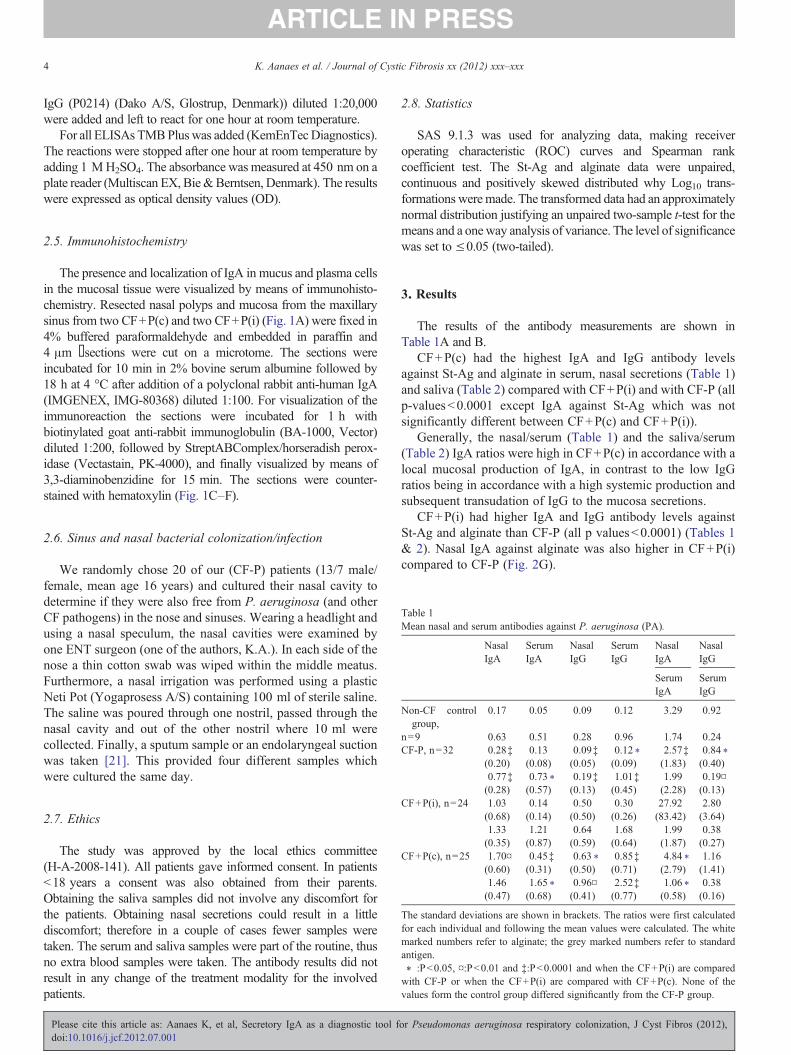
4 K. Aanaes et al. / Journal of Cystic Fibrosis xx (2012) xxx–xxx
IgG (P0214) (Dako A/S, Glostrup, Denmark)) diluted 1:20,000were added and left to react for one hour at room temperature.
For all ELISAs TMBPlus was added (KemEnTecDiagnostics).The reactions were stopped after one hour at room temperature byadding 1 MH2SO4. The absorbance was measured at 450 nm on aplate reader (Multiscan EX, Bie&Berntsen, Denmark). The resultswere expressed as optical density values (OD).
2.5. Immunohistochemistry
The presence and localization of IgA in mucus and plasma cellsin the mucosal tissue were visualized by means of immunohisto-chemistry. Resected nasal polyps and mucosa from the maxillarysinus from two CF+P(c) and two CF+P(i) (Fig. 1A) were fixed in4% buffered paraformaldehyde and embedded in paraffin and4 μm sections were cut on a microtome. The sections wereincubated for 10 min in 2% bovine serum albumine followed by18 h at 4 °C after addition of a polyclonal rabbit anti-human IgA(IMGENEX, IMG-80368) diluted 1:100. For visualization of theimmunoreaction the sections were incubated for 1 h withbiotinylated goat anti-rabbit immunoglobulin (BA-1000, Vector)diluted 1:200, followed by StreptABComplex/horseradish perox-idase (Vectastain, PK-4000), and finally visualized by means of3,3-diaminobenzidine for 15 min. The sections were counter-stained with hematoxylin (Fig. 1C–F).
Table 1Mean nasal and serum antibodies against P. aeruginosa (PA).
NasalIgA
SerumIgA
NasalIgG
SerumIgG
NasalIgA
NasalIgG
SerumIgA
SerumIgG
Non-CF controlgroup,
0.17 0.05 0.09 0.12 3.29 0.92
n=9 0.63 0.51 0.28 0.96 1.74 0.24CF-P, n=32 0.28‡ 0.13 0.09‡ 0.12 ⁎ 2.57‡ 0.84 ⁎
(0.20) (0.08) (0.05) (0.09) (1.83) (0.40)0.77‡ 0.73 ⁎ 0.19‡ 1.01‡ 1.99 0.19¤
2.6. Sinus and nasal bacterial colonization/infection
We randomly chose 20 of our (CF-P) patients (13/7 male/female, mean age 16 years) and cultured their nasal cavity todetermine if they were also free from P. aeruginosa (and otherCF pathogens) in the nose and sinuses. Wearing a headlight andusing a nasal speculum, the nasal cavities were examined byone ENT surgeon (one of the authors, K.A.). In each side of thenose a thin cotton swab was wiped within the middle meatus.Furthermore, a nasal irrigation was performed using a plasticNeti Pot (Yogaprosess A/S) containing 100 ml of sterile saline.The saline was poured through one nostril, passed through thenasal cavity and out of the other nostril where 10 ml werecollected. Finally, a sputum sample or an endolaryngeal suctionwas taken [21]. This provided four different samples whichwere cultured the same day.
(0.28) (0.57) (0.13) (0.45) (2.28) (0.13)CF+P(i), n=24 1.03 0.14 0.50 0.30 27.92 2.80
(0.68) (0.14) (0.50) (0.26) (83.42) (3.64)1.33 1.21 0.64 1.68 1.99 0.38(0.35) (0.87) (0.59) (0.64) (1.87) (0.27)
CF+P(c), n=25 1.70¤ 0.45‡ 0.63 ⁎ 0.85‡ 4.84 ⁎ 1.16(0.60) (0.31) (0.50) (0.71) (2.79) (1.41)1.46 1.65 ⁎ 0.96¤ 2.52‡ 1.06 ⁎ 0.38(0.47) (0.68) (0.41) (0.77) (0.58) (0.16)
The standard deviations are shown in brackets. The ratios were first calculatedfor each individual and following the mean values were calculated. The whitemarked numbers refer to alginate; the grey marked numbers refer to standardantigen.⁎ :Pb0.05, ¤:Pb0.01 and ‡:Pb0.0001 and when the CF+P(i) are comparedwith CF-P or when the CF+P(i) are compared with CF+P(c). None of thevalues form the control group differed significantly from the CF-P group.
2.7. Ethics
The study was approved by the local ethics committee(H-A-2008-141). All patients gave informed consent. In patientsb18 years a consent was also obtained from their parents.Obtaining the saliva samples did not involve any discomfort forthe patients. Obtaining nasal secretions could result in a littlediscomfort; therefore in a couple of cases fewer samples weretaken. The serum and saliva samples were part of the routine, thusno extra blood samples were taken. The antibody results did notresult in any change of the treatment modality for the involvedpatients.
Please cite this article as: Aanaes K, et al, Secretory IgA as a diagnostic tool fdoi:10.1016/j.jcf.2012.07.001
2.8. Statistics
SAS 9.1.3 was used for analyzing data, making receiveroperating characteristic (ROC) curves and Spearman rankcoefficient test. The St-Ag and alginate data were unpaired,continuous and positively skewed distributed why Log10 trans-formations weremade. The transformed data had an approximatelynormal distribution justifying an unpaired two-sample t-test for themeans and a oneway analysis of variance. The level of significancewas set to ≤0.05 (two-tailed).
3. Results
The results of the antibody measurements are shown inTable 1A and B.
CF+P(c) had the highest IgA and IgG antibody levelsagainst St-Ag and alginate in serum, nasal secretions (Table 1)and saliva (Table 2) compared with CF+P(i) and with CF-P (allp-valuesb0.0001 except IgA against St-Ag which was notsignificantly different between CF+P(c) and CF+P(i)).
Generally, the nasal/serum (Table 1) and the saliva/serum(Table 2) IgA ratios were high in CF+P(c) in accordance with alocal mucosal production of IgA, in contrast to the low IgGratios being in accordance with a high systemic production andsubsequent transudation of IgG to the mucosa secretions.
CF+P(i) had higher IgA and IgG antibody levels againstSt-Ag and alginate than CF-P (all p valuesb0.0001) (Tables 1& 2). Nasal IgA against alginate was also higher in CF+P(i)compared to CF-P (Fig. 2G).
or Pseudomonas aeruginosa respiratory colonization, J Cyst Fibros (2012),
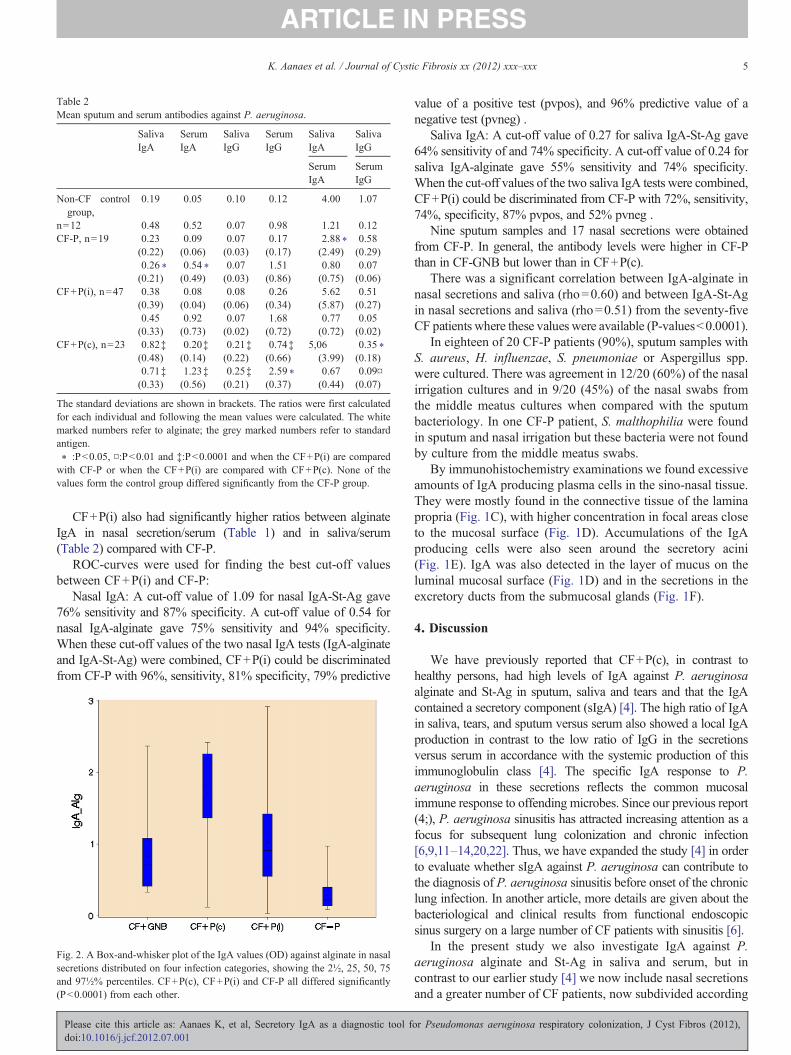
Table 2Mean sputum and serum antibodies against P. aeruginosa.
SalivaIgA
SerumIgA
SalivaIgG
SerumIgG
SalivaIgA
SalivaIgG
SerumIgA
SerumIgG
Non-CF controlgroup,
0.19 0.05 0.10 0.12 4.00 1.07
n=12 0.48 0.52 0.07 0.98 1.21 0.12CF-P, n=19 0.23 0.09 0.07 0.17 2.88 ⁎ 0.58
(0.22) (0.06) (0.03) (0.17) (2.49) (0.29)0.26 ⁎ 0.54 ⁎ 0.07 1.51 0.80 0.07(0.21) (0.49) (0.03) (0.86) (0.75) (0.06)
CF+P(i), n=47 0.38 0.08 0.08 0.26 5.62 0.51(0.39) (0.04) (0.06) (0.34) (5.87) (0.27)0.45 0.92 0.07 1.68 0.77 0.05(0.33) (0.73) (0.02) (0.72) (0.72) (0.02)
CF+P(c), n=23 0.82‡ 0.20‡ 0.21‡ 0.74‡ 5,06 0.35 ⁎(0.48) (0.14) (0.22) (0.66) (3.99) (0.18)0.71‡ 1.23‡ 0.25‡ 2.59 ⁎ 0.67 0.09¤(0.33) (0.56) (0.21) (0.37) (0.44) (0.07)
The standard deviations are shown in brackets. The ratios were first calculatedfor each individual and following the mean values were calculated. The whitemarked numbers refer to alginate; the grey marked numbers refer to standardantigen.⁎ :Pb0.05, ¤:Pb0.01 and ‡:Pb0.0001 and when the CF+P(i) are comparedwith CF-P or when the CF+P(i) are compared with CF+P(c). None of thevalues form the control group differed significantly from the CF-P group.
5K. Aanaes et al. / Journal of Cystic Fibrosis xx (2012) xxx–xxx
CF+P(i) also had significantly higher ratios between alginateIgA in nasal secretion/serum (Table 1) and in saliva/serum(Table 2) compared with CF-P.
ROC-curves were used for finding the best cut-off valuesbetween CF+P(i) and CF-P:
Nasal IgA: A cut-off value of 1.09 for nasal IgA-St-Ag gave76% sensitivity and 87% specificity. A cut-off value of 0.54 fornasal IgA-alginate gave 75% sensitivity and 94% specificity.When these cut-off values of the two nasal IgA tests (IgA-alginateand IgA-St-Ag) were combined, CF+P(i) could be discriminatedfrom CF-P with 96%, sensitivity, 81% specificity, 79% predictive
Fig. 2. A Box-and-whisker plot of the IgA values (OD) against alginate in nasalsecretions distributed on four infection categories, showing the 2½, 25, 50, 75and 97½% percentiles. CF+P(c), CF+P(i) and CF-P all differed significantly(Pb0.0001) from each other.
Please cite this article as: Aanaes K, et al, Secretory IgA as a diagnostic tool fdoi:10.1016/j.jcf.2012.07.001
value of a positive test (pvpos), and 96% predictive value of anegative test (pvneg) .
Saliva IgA: A cut-off value of 0.27 for saliva IgA-St-Ag gave64% sensitivity of and 74% specificity. A cut-off value of 0.24 forsaliva IgA-alginate gave 55% sensitivity and 74% specificity.When the cut-off values of the two saliva IgA tests were combined,CF+P(i) could be discriminated from CF-P with 72%, sensitivity,74%, specificity, 87% pvpos, and 52% pvneg .
Nine sputum samples and 17 nasal secretions were obtainedfrom CF-P. In general, the antibody levels were higher in CF-Pthan in CF-GNB but lower than in CF+P(c).
There was a significant correlation between IgA-alginate innasal secretions and saliva (rho=0.60) and between IgA-St-Agin nasal secretions and saliva (rho=0.51) from the seventy-fiveCF patients where these values were available (P-valuesb0.0001).
In eighteen of 20 CF-P patients (90%), sputum samples withS. aureus, H. influenzae, S. pneumoniae or Aspergillus spp.were cultured. There was agreement in 12/20 (60%) of the nasalirrigation cultures and in 9/20 (45%) of the nasal swabs fromthe middle meatus cultures when compared with the sputumbacteriology. In one CF-P patient, S. malthophilia were foundin sputum and nasal irrigation but these bacteria were not foundby culture from the middle meatus swabs.
By immunohistochemistry examinations we found excessiveamounts of IgA producing plasma cells in the sino-nasal tissue.They were mostly found in the connective tissue of the laminapropria (Fig. 1C), with higher concentration in focal areas closeto the mucosal surface (Fig. 1D). Accumulations of the IgAproducing cells were also seen around the secretory acini(Fig. 1E). IgA was also detected in the layer of mucus on theluminal mucosal surface (Fig. 1D) and in the secretions in theexcretory ducts from the submucosal glands (Fig. 1F).
4. Discussion
We have previously reported that CF+P(c), in contrast tohealthy persons, had high levels of IgA against P. aeruginosaalginate and St-Ag in sputum, saliva and tears and that the IgAcontained a secretory component (sIgA) [4]. The high ratio of IgAin saliva, tears, and sputum versus serum also showed a local IgAproduction in contrast to the low ratio of IgG in the secretionsversus serum in accordance with the systemic production of thisimmunoglobulin class [4]. The specific IgA response to P.aeruginosa in these secretions reflects the common mucosalimmune response to offending microbes. Since our previous report(4;), P. aeruginosa sinusitis has attracted increasing attention as afocus for subsequent lung colonization and chronic infection[6,9,11–14,20,22]. Thus, we have expanded the study [4] in orderto evaluate whether sIgA against P. aeruginosa can contribute tothe diagnosis of P. aeruginosa sinusitis before onset of the chroniclung infection. In another article, more details are given about thebacteriological and clinical results from functional endoscopicsinus surgery on a large number of CF patients with sinusitis [6].
In the present study we also investigate IgA against P.aeruginosa alginate and St-Ag in saliva and serum, but incontrast to our earlier study [4] we now include nasal secretionsand a greater number of CF patients, now subdivided according
or Pseudomonas aeruginosa respiratory colonization, J Cyst Fibros (2012),
6 K. Aanaes et al. / Journal of Cystic Fibrosis xx (2012) xxx–xxx
to the lung infection status. Significant differences of IgA levelsto the P. aeruginosa antigens were seen between bothCF+P(c), CF+P(i) and CF-P. Accordingly, we also found anabundance of local IgA producing plasma cells in the nasalmucosa of CF+P(c) CF+P(i). Many plasma cells havepreviously been found in sinonasal tissue from CF patients[23]. Importantly, our results allow a differentiation betweenCF+P(i) and CF-P. Nevertheless, the test is limited by thepossibility of cross-reaction with other pathogenic Gram-negative bacteria (e.g. S. malthophilia, A. xylosoxidans orB. cepacia complex) [15]. We conclude that the sensitivity,specificity, pvpos, and pvneg of each of the results of the IgAELISAs or combined ELISA results are high enough to beevaluated as a diagnostic tool for P. aeruginosa sinusitis in aprospective study in our CF centre.
IgA concentration is in general elevated as a result of thelocal mucosal antibody response, whereas IgG is elevated as aconsequence of the systemic inflammatory response [4,7]. Ourresults indicate an early, local production of IgA in the sinuseswhen these are infected with P. aeruginosa. This assumption isfortified, as we did not find P.aeruginosa in the nasal irrigationor middle meatus cultures from CF-P, which indicates that thesinuses are also free from P.aeruginosa [22,24]. This result canbe compared with our previous findings of pathogenic bacteriain the majority of the sinuses in CF+P(i) and CF+P(c) [6].
Our results are therefore in accordance with our previousfindings of high IgA levels in saliva, tears and sputum [4] andadditionally show that nasal secretions contain even higher levels.This is in agreement with the increasing evidence of P. aeruginosasinusitis being a focus for subsequently intermittent lungcolonization and infection [6,9,11–14,22]. When P. aeruginosasettles in the paranasal sinuses, a local increase of mucosal IgAresponse is initiated. However, the inflammatory response isreduced compared with that of chronic lung infection since thesecretory IgA is non-phlogistic in contrast to IgG which formsimmune complexes with P. aeruginosa antigens and activatecomplement and attract PMNs in the respiratory zone of the lungs[1,6,10]. The result may be that IgA keeps P. aeruginosa at adistance from the cells of mucosal membranes [25] and therebydiminishing the clinical symptoms of sinusitis [10]. The samemechanism may be working in the conductive airways ofCF+P(c), where most P. aeruginosa are located inside sputumtogether with the highly active PMNs [1,26,27], probably also dueto IgA in mucosa and mucus in the conductive airways [4].Keeping the bacteria at a distance from the mucosal membranecells may be a common strategy in the respiratory and intestinaltracts [25] by means of the mucus layer and the anti-bacterialmolecules such as IgA which prevent inflammation. P. aeruginosasinusitis in CF may therefore resemble secretory otitis media witheffusion where bacterial biofilms and IgA have also been found[28–30].
There are no standardized guidelines for detection of upperairway P. aeruginosa colonization or how or when to eradicateCF-pathogenic bacteria from the CF sinuses by sinus surgery and/or with antibiotic treatment [24,31]. The potential of surgical and/or conservative antibiotic treatment for eradication of P.aeruginosasinusitis requires further prospective evaluation; for now an early
Please cite this article as: Aanaes K, et al, Secretory IgA as a diagnostic tool fdoi:10.1016/j.jcf.2012.07.001
stage attempt of eradicating P. aeruginosa may be consideredwhen detected in the sinus [6,9,11,12,22,32].
A prospective study is planned to examine if the IgA responsein nasal secretion or saliva is helpful for early diagnosis ofP.aeruginosa sinusitis. It would also be relevant to investigatewhether IgA in nasal secretions, collected through a gentle nasallavage, is an easier way to collect IgA or if the method iscomplicated by dilution problems. Nasal lavage is already used asa supplementary diagnostic tool for CF infections [22], and as aresearch tool for rhinitis [33].
5. Conclusions
Both CF+P(c) and CF+P(i) have significantly higher IgAantibody levels in nasal secretions and saliva against P.aeruginosa alginate and St-Ag compared to CF-P. In CF+P(i)the elevated IgA probably reflect P. aeruginosa sinusitis wherethe bacteria adapt to the environment of CF airways CFpatients; sinusitis may then be the focus for subsequentintermittent P. aeruginosa colonization and chronic infectionof the lungs. The diagnostic value of IgA in saliva and nasalsecretions for sinusitis will be studied prospectively.
Funding
The work was supported by The Candys Foundation, anon-profit organization.
Acknowledgements
We would like to thank the nurses at the CF centre,laboratory technicians Lena Nørregaard, Heidi Paulsen andLise Strange for their dedication to this project.
References
[1] Bjarnsholt T, Jensen PO, Fiandaca MJ, Pedersen J, Hansen CR, AndersenCB, et al. Pseudomonas aeruginosa biofilms in the respiratory tract ofcystic fibrosis patients. Pediatr Pulmonol Jun 2009;44(6):547–58.
[2] Johansen HK, Norregaard L, Gotzsche PC, Pressler T, Koch C, Høiby N.Antibody response to Pseudomonas aeruginosa in cystic fibrosis patients:a marker of therapeutic success?–A 30-year cohort study of survival inDanish CF patients after onset of chronic P. aeruginosa lung infection.Pediatr Pulmonol May 2004;37(5):427–32.
[3] Bryan LE, Kureishi A, Rabin HR. Detection of antibodies to Pseudomo-nas aeruginosa alginate extracellular polysaccharide in animals and cysticfibrosis patients by enzyme-linked immunosorbent assay. J Clin MicrobiolAug 1983;18(2):276–82.
[4] Pedersen SS, Moller H, Espersen F, Sørensen CH, Jensen T, Hoiby N.Mucosal immunity to Pseudomonas aeruginosa alginate in cystic fibrosis.APMIS Apr 1992;100(4):326–34.
[5] Johansen HK, Høiby N. Local IgA and IgG response to intratrachealimmunization with Pseudomonas aeruginosa antigens. APMIS Jan1992;100(1):87–90.
[6] Johansen HK, Aanaes K, Pressler T, Nielsen KG, Fisker J, Skov M, et al.Colonisation and infection of the paranasal sinuses in cystic fibrosispatients is accompanied by a reduced PMN response. J Cyst Fibros May15, 2012 [Epub ahead of print].
[7] Pedersen SS, Espersen F, Høiby N, Jensen T. Immunoglobulin A andimmunoglobulin G antibody responses to alginates from Pseudomonas
or Pseudomonas aeruginosa respiratory colonization, J Cyst Fibros (2012),
7K. Aanaes et al. / Journal of Cystic Fibrosis xx (2012) xxx–xxx
aeruginosa in patients with cystic fibrosis. J Clin Microbiol Apr1990;28(4):747–55.
[8] Pressler T, Frederiksen B, Skov M, Garred P, Koch C, Høiby N. Early riseof anti-pseudomonas antibodies and a mucoid phenotype of Pseudomonasaeruginosa are risk factors for development of chronic lung infection–acase control study. J Cyst Fibros Jan 2006;5(1):9–15.
[9] Hansen SK, Rau MH, Johansen HK, Ciofu O, Jelsbak L, Yang L, et al.Evolution and diversification of Pseudomonas aeruginosa in the paranasalsinuses of cystic fibrosis children have implications for chronic lunginfection. ISME Jan 2012;6(1):31–45.
[10] Rasmussen J, Aanaes K, Norling R, Nielsen KG, Johansen HK, BuchwaldC. CT of the paranasal sinuses is not a valid indicator for sinus surgery inCF patients. J Cyst Fibros Mar 2011;11(2):93–9.
[11] Holzmann D, Speich R, Kaufmann T, Laube I, Russi EW, Simmen D,et al. Effects of sinus surgery in patients with cystic fibrosis after lungtransplantation: a 10-year experience. Transplantation Jan 15, 2004;77(1):134–6.
[12] Vital D, Hofer M, Boehler A, Holzmann D. Posttransplant sinus surgery inlung transplant recipients with cystic fibrosis: a single institutionalexperience. Eur Arch Otorhinolaryngol Mar 30, 2012 [Epub ahead of print].
[13] Walter S, Gudowius P, Bosshammer J, Romling U, Weissbrodt H,Schurmann W, et al. Epidemiology of chronic Pseudomonas aeruginosainfections in the airways of lung transplant recipients with cystic fibrosis.Thorax Apr 1997;52(4):318–21.
[14] Mainz JG, Hentschel J, Schien C, Cramer N, Pfister W, Beck JF, et al.Sinonasal persistence of Pseudomonas aeruginosa after lung transplanta-tion. J Cyst Fibros Mar 2012;11(2):158–61.
[15] Frederiksen B, Koch C, Høiby N. Antibiotic treatment of initialcolonization with Pseudomonas aeruginosa postpones chronic infectionand prevents deterioration of pulmonary function in cystic fibrosis. PediatrPulmonol May 1997;23(5):330–5.
[16] Pressler T, Karpati F, Granstrom M, Knudsen PK, Lindblad A, Hjelte L,et al. Diagnostic significance of measurements of specific IgG antibodiesto Pseudomonas aeruginosa by three different serological methods. J CystFibros Jan 2009;8(1):37–42.
[17] Ratjen F, Walter H, Haug M, Meisner C, Grasemann H, Döring G.Diagnostic value of serum antibodies in early Pseudomonas aeruginosainfection in cystic fibrosis patients. Pediatr Pulmonol Mar 2007;42(3):249–55.
[18] da Silva Filho LV, Tateno AF, Martins KM, Azzuz Chernishev AC,Garcia DO, Haug M, et al. The combination of PCR and serologyincreases the diagnosis of Pseudomonas aeruginosa colonization/infectionin cystic fibrosis. Pediatr Pulmonol Oct 2007;42(10):938–44.
[19] Hansen CR, Pressler T, Høiby N. Early aggressive eradication therapy forintermittent Pseudomonas aeruginosa airway colonization in cysticfibrosis patients: 15 years experience. J Cyst Fibros Nov 2008;7(6):523–30.
Please cite this article as: Aanaes K, et al, Secretory IgA as a diagnostic tool fdoi:10.1016/j.jcf.2012.07.001
[20] Bonestroo HJ, de Winter-de Groot KM, van der Ent CK, Arets HG. Upperand lower airway cultures in children with cystic fibrosis: do not neglectthe upper airways. J Cyst Fibros Mar 2010;9(2):130–4.
[21] Høiby N, Frederiksen B, Pressler T. Eradication of early Pseudomonasaeruginosa infection. J Cyst Fibros Aug 2005;4(Suppl. 2):49–54.
[22] Mainz JG, Naehrlich L, Schien M, Kading M, Schiller I, Mayr S, et al.Concordant genotype of upper and lower airways P aeruginosa and Saureus isolates in cystic fibrosis. Thorax Jun 2009;64(6):535–40.
[23] Schraven SP, Wehrmann M, Wagner W, Blumenstock G, Koitschev A.Prevalence and histopathology of chronic polypoid sinusitis in pediatricpatients with cystic fibrosis. J Cyst Fibros May 2011;10(3):181–6.
[24] Thomas M, Yawn BP, Price D, Lund V, Mullol J, Fokkens W. EPOSprimary care guidelines: European position paper on the primary carediagnosis and management of rhinosinusitis and nasal polyps. Prim CareRespir J Jun 2008;17(2):79–89.
[25] Johansson ME, Hansson GC. Microbiology. Keeping bacteria at adistance. Science Oct 14, 2011;334(6053):182–3.
[26] Kolpen M, Hansen CR, Bjarnsholt T, Moser C, Christensen LD, van GM,et al. Polymorphonuclear leucocytes consume oxygen in sputum fromchronic Pseudomonas aeruginosa pneumonia in cystic fibrosis. ThoraxJan 2010;65(1):57–62.
[27] Worlitzsch D, Tarran R, Ulrich M, Schwab U, Cekici A, Meyer KC, et al.Effects of reduced mucus oxygen concentration in airway Pseudomonasinfections of cystic fibrosis patients. J Clin Invest Feb 2002;109(3):317–25.
[28] Dingman JR, Rayner MG, Mishra S, Zhang Y, Ehrlich MD, Post JC, et al.Correlation between presence of viable bacteria and presence of endotoxinin middle-ear effusions. J Clin Microbiol Nov 1998;36(11):3417–9.
[29] Rayner MG, Zhang Y, Gorry MC, Chen Y, Post JC, Ehrlich GD. Evidenceof bacterial metabolic activity in culture-negative otitis media witheffusion. JAMA Jan 28, 1998;279(4):296–9.
[30] Sørensen CH. Quantitative aspects of the mucosal immunity andbacteriology of the nasopharynx and middle ear cavity. Studies onchildren with clinically different forms of otitis media. APMIS Suppl1990;16:1–41.
[31] Kerem E, Conway S, Elborn S, Heijerman H. Standards of care forpatients with cystic fibrosis: a European consensus. J Cyst Fibros Mar2005;4(1):7–26.
[32] Mainz JG, Michl R, Pfister W, Beck JF. Cystic fibrosis upper airwaysprimary colonization with Pseudomonas aeruginosa: eradicated bysinonasal antibiotic inhalation. Am J Respir Crit Care Med Nov 1,2011;184(9):1089–90.
[33] Howarth PH, Persson CG, Meltzer EO, Jacobson MR, Durham SR,Silkoff PE. Objective monitoring of nasal airway inflammation in rhinitis.J Allergy Clin Immunol Mar 2005;115(3 Suppl 1):S414–41.
or Pseudomonas aeruginosa respiratory colonization, J Cyst Fibros (2012),

Paper III
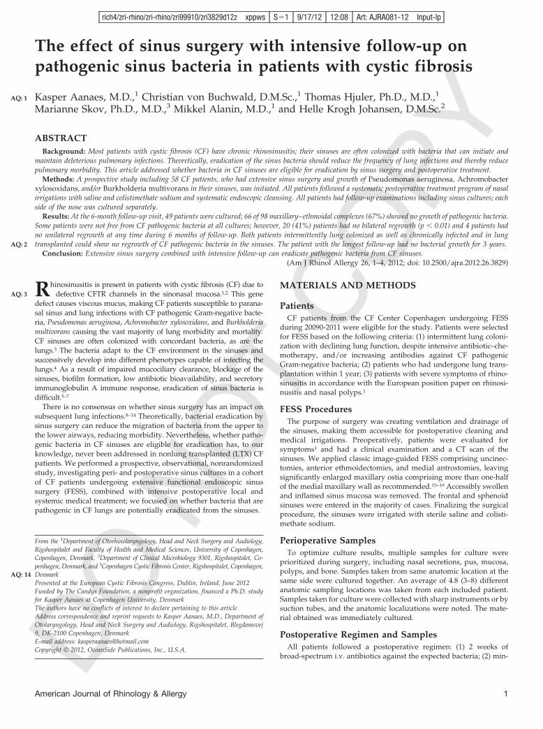
The effect of sinus surgery with intensive follow-up onpathogenic sinus bacteria in patients with cystic fibrosis
Kasper Aanaes, M.D.,1 Christian von Buchwald, D.M.Sc.,1 Thomas Hjuler, Ph.D., M.D.,1
Marianne Skov, Ph.D., M.D.,3 Mikkel Alanin, M.D.,1 and Helle Krogh Johansen, D.M.Sc.2
ABSTRACTBackground: Most patients with cystic fibrosis (CF) have chronic rhinosinusitis; their sinuses are often colonized with bacteria that can initiate and
maintain deleterious pulmonary infections. Theoretically, eradication of the sinus bacteria should reduce the frequency of lung infections and thereby reducepulmonary morbidity. This article addressed whether bacteria in CF sinuses are eligible for eradication by sinus surgery and postoperative treatment.
Methods: A prospective study including 58 CF patients, who had extensive sinus surgery and growth of Pseudomonas aeruginosa, Achromobacterxylosoxidans, and/or Burkholderia multivorans in their sinuses, was initiated. All patients followed a systematic postoperative treatment program of nasalirrigations with saline and colistimethate sodium and systematic endoscopic cleansing. All patients had follow-up examinations including sinus cultures; eachside of the nose was cultured separately.
Results: At the 6-month follow-up visit, 49 patients were cultured; 66 of 98 maxillary–ethmoidal complexes (67%) showed no growth of pathogenic bacteria.Some patients were not free from CF pathogenic bacteria at all cultures; however, 20 (41%) patients had no bilateral regrowth (p � 0.01) and 4 patients hadno unilateral regrowth at any time during 6 months of follow-up. Both patients intermittently lung colonized as well as chronically infected and in lungtransplanted could show no regrowth of CF pathogenic bacteria in the sinuses. The patient with the longest follow-up had no bacterial growth for 3 years.
Conclusion: Extensive sinus surgery combined with intensive follow-up can eradicate pathogenic bacteria from CF sinuses.(Am J Rhinol Allergy 26, 1–4, 2012; doi: 10.2500/ajra.2012.26.3829)
Rhinosinusitis is present in patients with cystic fibrosis (CF) due todefective CFTR channels in the sinonasal mucosa.1,2 This gene
defect causes viscous mucus, making CF patients susceptible to parana-sal sinus and lung infections with CF pathogenic Gram-negative bacte-ria, Pseudomonas aeruginosa, Achromobacter xylosoxidans, and Burkholderiamultivorans causing the vast majority of lung morbidity and mortality.CF sinuses are often colonized with concordant bacteria, as are thelungs.3 The bacteria adapt to the CF environment in the sinuses andsuccessively develop into different phenotypes capable of infecting thelungs.4 As a result of impaired mucociliary clearance, blockage of thesinuses, biofilm formation, low antibiotic bioavailability, and secretoryimmunoglobulin A immune response, eradication of sinus bacteria isdifficult.5–7
There is no consensus on whether sinus surgery has an impact onsubsequent lung infections.8–14 Theoretically, bacterial eradication bysinus surgery can reduce the migration of bacteria from the upper tothe lower airways, reducing morbidity. Nevertheless, whether patho-genic bacteria in CF sinuses are eligible for eradication has, to ourknowledge, never been addressed in nonlung transplanted (LTX) CFpatients. We performed a prospective, observational, nonrandomizedstudy, investigating peri- and postoperative sinus cultures in a cohortof CF patients undergoing extensive functional endoscopic sinussurgery (FESS), combined with intensive postoperative local andsystemic medical treatment; we focused on whether bacteria that arepathogenic in CF lungs are potentially eradicated from the sinuses.
MATERIALS AND METHODS
PatientsCF patients from the CF Center Copenhagen undergoing FESS
during 20090-2011 were eligible for the study. Patients were selectedfor FESS based on the following criteria: (1) intermittent lung coloni-zation with declining lung function, despite intensive antibiotic–che-motherapy, and/or increasing antibodies against CF pathogenicGram-negative bacteria; (2) patients who had undergone lung trans-plantation within 1 year; (3) patients with severe symptoms of rhino-sinusitis in accordance with the European position paper on rhinosi-nusitis and nasal polyps.1
FESS ProceduresThe purpose of surgery was creating ventilation and drainage of
the sinuses, making them accessible for postoperative cleaning andmedical irrigations. Preoperatively, patients were evaluated forsymptoms1 and had a clinical examination and a CT scan of thesinuses. We applied classic image-guided FESS comprising uncinec-tomies, anterior ethmoidectomies, and medial antrostomies, leavingsignificantly enlarged maxillary ostia comprising more than one-halfof the medial maxillary wall as recommended.15–19 Accessibly swollenand inflamed sinus mucosa was removed. The frontal and sphenoidsinuses were entered in the majority of cases. Finalizing the surgicalprocedure, the sinuses were irrigated with sterile saline and colisti-methate sodium.
Perioperative SamplesTo optimize culture results, multiple samples for culture were
prioritized during surgery, including nasal secretions, pus, mucosa,polyps, and bone. Samples taken from same anatomic location at thesame side were cultured together. An average of 4.8 (3–8) differentanatomic sampling locations was taken from each included patient.Samples taken for culture were collected with sharp instruments or bysuction tubes, and the anatomic localizations were noted. The mate-rial obtained was immediately cultured.
Postoperative Regimen and SamplesAll patients followed a postoperative regimen: (1) 2 weeks of
broad-spectrum i.v. antibiotics against the expected bacteria; (2) min-
From the 1Department of Otorhinolaryngology, Head and Neck Surgery and Audiology,Rigshospitalet and Faculty of Health and Medical Sciences, University of Copenhagen,Copenhagen, Denmark, 2Department of Clinical Microbiology 9301, Rigshospitalet, Co-penhagen, Denmark, and 3Copenhagen Cystic Fibrosis Center, Rigshospitalet, Copenhagen,DenmarkPresented at the European Cystic Fibrosis Congress, Dublin, Ireland, June 2012Funded by The Candys Foundation, a nonprofit organization, financed a Ph.D. studyfor Kasper Aanaes at Copenhagen University, DenmarkThe authors have no conflicts of interest to declare pertaining to this articleAddress correspondence and reprint requests to Kasper Aanaes, M.D., Department ofOtolaryngology, Head and Neck Surgery and Audiology, Rigshospitalet, Blegdamsvej9, DK-2100 Copenhagen, DenmarkE-mail address: [email protected] © 2012, OceanSide Publications, Inc., U.S.A.
AQ: 1
AQ: 2
AQ: 3
AQ: 14
American Journal of Rhinology & Allergy 1
rich4/zri-rhino/zri-rhino/zri99910/zri3829d12z xppws S�1 9/17/12 12:08 Art: AJRA081-12 Input-lp

imum 6 months of topical nasal steroids (mometasone furoate); (3)minimum of 6 months of daily nasal irrigations with saline andantibiotics (colistimethate sodium when susceptible bacteria werecultured); and (4) visits to the ear, nose, and throat outpatient clinic,1 week and 1, 3 and 12 months postoperatively, where crusts andsecretions were endoscopically cleansed from the nose and sinuses.At each follow-up the patients were bilaterally cultured using endo-scopic guidance. No local anesthetics were used. In the majority ofpatients, a 2- or 3-mm curved suction tube was used to enter andcollect secretions from the maxillary sinuses, ethmoids, and/or fron-tal recesses. The left- and right-side samples taken for culture weretreated separately. A cotton swab was wiped within each middlemeatus in cases where the maxillary sinuses were not accessible as aconsequence of mucosal swelling. The samples were immediatelycultured. In some cases the samples were taken while the patient wasunder general anesthesia for other reasons. The strategy of antibiotictreatment was postoperatively unchanged and performed accordingto guidelines and susceptibility testing.20
Microbiological SamplesSinus samples were cultured aerobically and anaerobically at 37°C
on standard agar media for 5–7 days as previously described.21
EthicsThe study was approved by the local ethics committee (H-A-2008-
141). All patients gave informed consent. In patients �18 years of age,informed consent was obtained from their parents.
StatisticsA McNemar’s test was used in SAS 9.1.3 to compare the postoper-
ative frequencies of growth with the perioperative frequencies.
RESULTSA total of 81 consecutive patients had FESS during 2009–2011. All
had bacteria in their sinuses; 60 patients had growth of P. aeruginosa,A. xylosoxidans, and/or B. multivorans in their perioperative sinussamples. One patient was lost to follow-up and one patient only hadpathogenic bacteria in the sphenoid sinuses and was excluded, be-cause the sphenoid sinuses do not drain into the middle meatus.Consequently, 58 patients were included in our prospective study
(Tables 1 and 2). They had the following lung infection status at timeof surgery: 12 LTX; 13 chronically lung infected; and 33 intermittentlylung colonized with P. aeruginosa, A. xylosoxidans, and/or B. multiv-orans.22 In one patient, the bacteria found in the sinuses (A. xylosoxi-dans) were different from the pulmonary bacteria (P. aeruginosa); inthe remaining patients the sinus bacteria were in accordance with thepulmonary bacteria the year before surgery or before LTX being asfollows: 42 with P. aeruginosa, 13 with A. xylosoxidans, and 2 with B.multivorans. The majority of patients fulfilled criterion 1 or 2 forsurgery, but 45 patients also fulfilled criterion 3. Except for onepatient who only had pathogenic bacteria in one of her frontal si-nuses, the 57 remaining patients had growth of pathogenic bacteria intheir maxillary sinuses; in 12 patients the growth was only unilateral.
All patients attended at least two postoperative follow-up visits.The adherence to participation in the follow-up visits was high; atevery postoperative follow-up, �90% of the patients attended (Tables1 and 2). In addition, 14 patients were additionally cultured outsidethe planned schedule (9–36 months postoperatively).
Peri- and Postoperative Culture ResultsOverall, 430 postoperative sinus samples were obtained and cul-
tured; 123 of these had growth of CF pathogenic bacteria (P. aerugi-nosa, A. xylosoxidans, and/or B. multivorans). The results of the sinuscultures are shown in Tables 1 and 2 and described in the followingparagraphs.
Initially, the 12 LTX patients (6 men and 6 women aged 21–45 years[mean, 33 years]) had bilateral CF pathogenic bacterial growth in theirsinuses, in accordance with their lung bacteriology before transplan-tation. Five of these patients (eight sinuses) already had regrowth ofCF pathogenic bacteria at the first postoperative culture after 1 month.At the 6-month follow-up, five patients only had unilateral regrowth,and three patients showed no bilateral regrowth. This was a signifi-cant decrease when compared with the perioperative results (p �0.01). One of these patients had the longest follow-up period of 36months where no CF pathogenic regrowth was seen.
The 13 patients with chronic lung infections (10 men and 3 womenaged 19–42 years [mean, 29 years]) initially consisted of 24 left andright sinus sides with pathogenic bacteria present at surgery. At the6-month follow-up, 13 sinus complexes showed no regrowth of CFpathogenic bacteria, where 6 of these had no regrowth at any timeduring the 6–12 months of follow-up (p � 0.01).
Table 1 Number of unilateral sinus cultures with CF pathogenic growth according to time after FESS
Lung Status at Surgery Perioperative 1 mo 3 mo 6 mo 12 mo
LTX 24 of 24 (100%) 8 of 24 (33%) 9 of 20 (45%) 11 of 22 (50%) 9 of 20 (45%)Chronically infected 25 of 26 (96%) 8 of 24 (33%) 12 of 24 (50%) 13 of 26 (50%) 8 of 18 (44%)Intermittently colonized* 55 of 66 (83%) 5 of 60 (8%) 11 of 60 (18%) 8 of 50 (16%) 12 of 48 (25%)
Fifty-eight patients (116 sinus complexes) with CF underwent FESS and follow-up during 2009–2012; each patient had separate sinus cultures taken fromthe left and right side. The postoperative frequencies all differed significantly from the perioperative ones (p � 0.01). *Intermittently colonized is defined asgrowth of CF pathogenic bacteria (Pseudomonas aeruginosa, Achromobacter xylosoxidans, and/or Burkholderia multivorans) in 1 to �50% of themonthly samples taken from the lower airways with in the last year.CF � cystic fibrosis; FESS � functional endoscopic sinus surgery; LTX � nonlung transplanted.
Table 2 Number of patients with no growth and unilateral and bilateral CF pathogenic growth in cultures taken from the sinusesaccording to time after FESS among 58 patients with CF undergoing FESS and follow-up
Perioperative 1 mo 3 mo 6 mo 12 mo
No. of patients with no growth 0 (0%) 40 (69%) 29 (50%) 25 (47%) 23 (50%)No. of patients with unilateral growth 12 (21%) 7 (12%) 14 (24%) 16 (30%) 11 (24%)No. of patients with bilateral growth 46 (79%) 7 (12%) 9 (16%) 8 (15%) 9 (20%)Did not show for follow-up — 4 (7%) 6 (10%) 4 (8%) 3 (7%)
CF � cystic fibrosis; FESS � functional endoscopic sinus surgery.
AQ: 4
AQ: 5
T1-T2
AQ: 6
AQ: 7
2
rich4/zri-rhino/zri-rhino/zri99910/zri3829d12z xppws S�1 9/17/12 12:08 Art: AJRA081-12 Input-lp
All 33 patients with intermittent lung colonization (18 male and 15female patients aged 6–43 years [mean, 17 years]), except for 1, hadconcordant bacteriology in their sinuses and lungs at surgery; thesingle exception was a girl intermittently lung colonized with P.aeruginosa but harbored A. xylosoxidans in several of her sinuses. Inaddition, two patients who were intermittently lung colonized with P.aeruginosa also harbored A. xylosoxidans in their sinuses together withP. aeruginosa. At the 1-month follow-up, only four patients had one ormore sinus samples that showed regrowth. Of the patients who werefollowed for 6 months, 52 of 60 sinus complexes had no CF patho-genic growth (p � 0.01); 37 sinus complexes showed no pathogenicgrowth at any time during the 6 months (p � 0.01). Among theintermittently colonized patients, the longest follow-up without anyregrowth was 28 months.
At the 6-month follow-up, 49 patients were cultured; 66 of 98maxillary–ethmoidal complexes (67%) showed no growth of patho-genic bacteria. Some patients were not free from CF pathogenicbacteria at all cultures; however, 20 (41%) patients had no bilateralregrowth (p � 0.01) and 4 patients had no unilateral regrowth at anytime during 6 months of follow-up.
Sampling AccuracyPostoperative samples were obtained for culture eight times during
a second general anesthesia because of revision surgery or otherprocedures; in these cases the quality of the sampling could be com-pared with the one during surgery. Eleven of 16 sinus complexes hadno regrowth of pathogenic Gram-negative bacteria according to thisthorough sampling. Six months after FESS, 84% of the maxillarysinuses were accessible and cultured. In the remaining 14%, sampleswere collected from the middle meatus and anterior ethmoidal cells.We did not compare our results with sinus cultures from nonoperatedCF patients because such samples would have required general an-esthesia.
DISCUSSIONWe present the largest prospective cohort study, to date, of CF
patients undergoing FESS. We showed that extensive FESS withpostoperative treatment eradicated the CF pathogenic bacteria fromthe paranasal sinuses in many CF patients, in several patients for �1year. Bacteria were most successfully eradicated in patients withintermittent lung colonization. In some cases, the bacteria were onlyunilaterally eradicated, giving weight to the proposition that the leftand right sinuses should be regarded as two separate compartments.4
Our results show that sinus bacteria can be eradicated independent oflung infection status. Otolaryngologists have an important role in CFmanagement in focusing on the upper part of the airways; we suggestthat this include regular endoscopic guided sinoscopy, and obtainingof representative material for culture in operated as well as in non-operated patients.
No standardized guidelines comprising criteria for FESS or post-operative treatment exist for patients with CF.1,23 We offer FESS as asupplementary diagnostic tool and a method to attempt eradication ofbacterial sinus infections. This methodology has been introduced as aconsequence of the general recognition of bacterial sinusitis as a focusfor intermittent colonization of the lungs.3–5The present study showsthat extensive FESS with postoperative treatment can eradicate CFsinus bacteria; nevertheless, we stress that intensive postoperativetreatment is mandatory to obtain this achievement. It is evident thatsaline nasal irrigations alleviate symptoms of chronic rhinosinusitisand is broadly used postoperatively together with endoscopic re-moval of nasal crusts as we did in this study.1,11,19,24,25 Other studieshave reported use of nasal irrigations with antibiotics or suggest it tohave efficacy in chronic rhinosinusitis treatment.8,24–26 Colistimethatesodium is broadly used as inhaled therapy in CF patients with P.aeruginosa or A. xylosoxidans lung infections. Thus, we used this drugfor nasal irrigations when the bacteria were susceptible. Nasal irrita-
tion was the only side effect reported. Because this treatment strategyhas a low level of evidence, the dosage, administration intervals, anddrug(s) of choice should be considered when making postoperativeguidelines for CF patients. Overall, there is no evidence for usingtopical steroids in patients with CF and nasal polyposis.27 However,we recommend topical steroids as a part of the postoperative treat-ment, because they may reduce inflammation and the size of polypsand have limited side effects.27 Adding baby shampoo to the sa-line,28,29 using nebulizers for irrigations, and use of mucolytics30–32 areall modalities that have been suggested by others as postoperativetreatments.
The impact of FESS on CF lung bacteriology has been previouslyaddressed.8–14 Apart from our study, Holzmann et al.8 and Lewistonet al.,11 who included only LTX patients, are the only groups conduct-ing prospective studies aimed at the implementation of FESS foreradication of bacteria, thus making the surgery and postoperativecare more extensive. Consequently, these two studies showed themost promising results. As in a study by Holzmann et al.,8 we ex-plored the majority of sinuses during FESS, including those withminimal opacification, because CF sinuses may harbor CF pathogenicbacteria independent of the CT findings and the clinical examination.6
We found a striking concordance between sinus and lung bacteria,which is in agreement with studies showing coherence between bac-teria genotypes and phenotypes in the sinuses and lungs in CFpatients.3,4 Nevertheless, only one other study,8 focused solely on LTXpatients, has addressed the possibility of eradicating CF sinus bacte-ria. They found that 54% of the LTX patients had three or fewerpositive sinus cultures during follow-up after FESS, which stronglycorrelated with the absence of tracheobronchitis. This finding is inaccordance with our results among CF LTX patients. By showing thatbacteria can potentially be eradicated from the paranasal sinuses, weshow that extensive FESS with postoperative treatment may reducelung morbidity in patients intermittently lung colonized, by prevent-ing bacteria from the sinuses infecting the lungs. Whether sinusbacteria can be used as a surrogate marker for the positive effect ofFESS still needs to be elucidated. Covariates as pulmonary functiontest, quality of life, antibody levels, and frequency of lung coloniza-tions should be taken into account.
Our study is strengthened by having a prospective design, a largeadherent cohort, a minimum of dropouts, and the same FESS surgeonperforming the follow-up visits, as well as having a standardizedprogram for a thorough sampling for culture during surgery. How-ever, there were two limitations in our study. Our study was obser-vational, because we found it unethical to include a control groupwho would not receive any treatment. Second, although some of thepostoperative samples were obtained during cleansing under generalanesthesia, and some of these did not show any bacterial regrowth,the majority of samples were obtained for culture in our outpatientclinic. Although being thorough, the value of a negative culturesample obtained by suction tube versus a biopsy during generalanesthesia will always be questionable; it is uncertain whether cul-tures of secretions will reveal presence of bacteria, especially whenpatients are doing nasal irrigations with antibiotics or if the bacteriaonly were present embedded in submucosal abscesses or biofilm.Thus, when stating that we eradicated the bacteria from the paranasalsinuses, we at least reduced the quantity of colony-forming units, sothe bacteria could not be redetected by sinus cultures for longerperiods.
CONCLUSIONOur study shows that extensive FESS combined with postoperative
treatment in several cases can eradicate pathogenic bacteria from thesinuses, especially in CF patients with intermittent lung colonization.This supports the hypothesis that FESS reduces the frequency ofbacterial lung colonization.
American Journal of Rhinology & Allergy 3
rich4/zri-rhino/zri-rhino/zri99910/zri3829d12z xppws S�1 9/17/12 12:08 Art: AJRA081-12 Input-lp

ACKNOWLEDGMENTSThe authors thank Majbritt Presfeldt and the Pseudomonas labora-
tory technicians at the Department of Clinical Microbiology for theirdedication, and Niels Høiby for proofreading.
REFERENCES1. Thomas M, Yawn BP, Price D, et al. EPOS Primary care guidelines:
European position paper on the primary care diagnosis and manage-ment of rhinosinusitis and nasal polyps. Prim Care Respir J 17:79–89,2008.
2. Robertson JM, Friedman EM, and Rubin BK. Nasal and sinus diseasein cystic fibrosis. Paediatr Respir Rev 9:213–219, 2008.
3. Mainz JG, Naehrlich L, Schien M, et al. Concordant genotype ofupper and lower airways P. aeruginosa and S. aureus isolates in cysticfibrosis. Thorax 64:535–540, 2009.
4. Hansen SK, Rau MH, Johansen HK, et al. Evolution and diversifica-tion of Pseudomonas aeruginosa in the paranasal sinuses of cysticfibrosis children have implications for chronic lung infection. ISME J❚:❚–❚, 2011.
5. Johansen HK, Aanaes K, Pressler T, et al. Colonisation and infectionof the paranasal sinuses in cystic fibrosis patients is accompanied bya reduced PMN response. J Cyst Fibros ❚:❚–❚, 2012.
6. Rasmussen J, Aanaes K, Norling R, et al. CT of the paranasal sinusesis not a valid indicator for sinus surgery in CF patients. J Cyst Fibros11:93–99, 2012.
7. Suh JD, Cohen NA, and Palmer JN. Biofilms in chronic rhinosinusitis.Curr Opin Otolaryngol Head Neck Surg 18:27–31, 2010.
8. Holzmann D, Speich R, Kaufmann T, et al. Effects of sinus surgery inpatients with cystic fibrosis after lung transplantation: A 10-yearexperience. Transplantation 77:134–136, 2004.
9. Jarrett WA, Militsakh O, Anstad M, and Manaligod J. Endoscopicsinus surgery in cystic fibrosis: Effects on pulmonary function andideal body weight. Ear Nose Throat J 83:118–121, 2004.
10. Leung MK, Rachakonda L, Weill D, and Hwang PH. Effects of sinussurgery on lung transplantation outcomes in cystic fibrosis. Am JRhinol 22:192–196, 2008.
11. Lewiston N, King V, Umetsu D, et al. Cystic fibrosis patients whohave undergone heart-lung transplantation benefit from maxillarysinus antrostomy and repeated sinus lavage. Transplant Proc 23:1207–1208, 1991.
12. Madonna D, Isaacson G, Rosenfeld RM, and Panitch H. Effect ofsinus surgery on pulmonary function in patients with cystic fibrosis.Laryngoscope 107:328–331, 1997.
13. Osborn AJ, Leung R, Ratjen F, and James AL. Effect of endoscopicsinus surgery on pulmonary function and microbial pathogens in apediatric population with cystic fibrosis. Arch Otolaryngol HeadNeck Surg 137:542–547, 2011.
14. Rosbe KW, Jones DT, Rahbar R, et al. Endoscopic sinus surgery incystic fibrosis: Do patients benefit from surgery? Int J Pediatr Oto-rhinolaryngol 61:113–119, 2001.
15. Cho DY, and Hwang PH. Results of endoscopic maxillary mega-antrostomy in recalcitrant maxillary sinusitis. Am J Rhinol 22:658–662, 2008.
16. Fuchsmann C, Ayari S, Reix P, et al. Contribution of CT-assistednavigation and microdebriders to endoscopic sinus surgery in cysticfibrosis. Int J Pediatr Otorhinolaryngol 72:343–349, 2008.
17. Ramadan HH. Revision endoscopic sinus surgery in children: Surgi-cal causes of failure. Laryngoscope 119:1214–1217, 2009.
18. Singhal D, Weitzel EK, Lin E, et al. Effect of head position andsurgical dissection on sinus irrigant penetration in cadavers. Laryn-goscope 120:2528–2531, 2010.
19. Virgin FW, Rowe SM, Wade MB, et al. Extensive surgical and com-prehensive postoperative medical management for cystic fibrosischronic rhinosinusitis. Am J Rhinol Allergy 26:70–75, 2012.
20. Hoiby N, Frederiksen B, and Pressler T. Eradication of early Pseu-domonas aeruginosa infection. J Cyst Fibros 4(suppl 2):49–54, 2005.
21. Høiby N, and Frederiksen B. Microbiology of cystic fibrosis. In CysticFibrosi, 2nd ed. Hodson GD, and Arnold ME (Eds). ❚, ❚: ❚, 83–107,2000.
22. Lee TW, Brownlee KG, Conway SP, et al. Evaluation of a newdefinition for chronic Pseudomonas aeruginosa infection in cystic fibro-sis patients. J Cyst Fibros 2:29–34, 2003.
23. Kerem E, Conway S, Elborn S, et al. Standards of care for patientswith cystic fibrosis: A European consensus. J Cyst Fibros 4:7–26, 2005.
24. Adappa ND, Wei CC, and Palmer JN. Nasal irrigation with or with-out drugs: the evidence. Curr Opin Otolaryngol Head Neck Surg20:53–57, 2012.
25. Moss RB, and King VV. Management of sinusitis in cystic fibrosis byendoscopic surgery and serial antimicrobial lavage. Reduction inrecurrence requiring surgery. Arch Otolaryngol Head Neck Surg121:566–572, 1995.
26. Schulte DL, and Kasperbauer JL. Safety of paranasal sinus surgery inpatients with cystic fibrosis. Laryngoscope 108:1813–1815, 1998.
27. Beer H, Southern KW, and Swift AC. Topical nasal steroids fortreating nasal polyposis in people with cystic fibrosis. CochraneDatabase Syst Rev ❚:CD008253, 2011.
28. Chiu AG, Palmer JN, Woodworth BA, et al. Baby shampoo nasalirrigations for the symptomatic post-functional endoscopic sinus sur-gery patient. Am J Rhinol 22:34–37, 2008.
29. Isaacs S, Fakhri S, Luong A, et al. The effect of dilute baby shampooon nasal mucociliary clearance in healthy subjects. Am J RhinolAllergy 25:e27–e29, 2011.
30. Cimmino M, Nardone M, Cavaliere M, et al. Dornase alfa as postop-erative therapy in cystic fibrosis sinonasal disease. Arch OtolaryngolHead Neck Surg 131:1097–1101, 2005.
31. Mainz JG, Schiller I, Ritschel C, et al. Sinonasal inhalation of dornasealfa in CF: A double-blind placebo-controlled cross-over pilot trial.Auris Nasus Larynx 38:220–227, 2011.
32. Raynor EM, Butler A, Guill M, and Bent JP 3rd. Nasally inhaleddornase alfa in the postoperative management of chronic sinusitisdue to cystic fibrosis. Arch Otolaryngol Head Neck Surg 126:581–583,2000. e
AQ: 8
AQ: 9
AQ: 10
AQ: 11
AQ: 12
AQ: 13
4
rich4/zri-rhino/zri-rhino/zri99910/zri3829d12z xppws S�1 9/17/12 12:08 Art: AJRA081-12 Input-lp

Paper IV
1
Title page:
Clinical Effects of Sinus Surgery and Adjuvant Therapy in Cystic Fibrosis Patients — Can Chronic Lung Infections be postponed?
Short title: Sinus surgery can reduce lower airway pathogens
Kasper Aanaes MD1, Helle Krogh Johansen DMSc
2, Marianne Skov PhD MD
3&4, Frederik F.
Buchvald PhD MD 3&4
,Thomas Hjuler PhD MD1, Tacjana Pressler, DMSc.
3&4, Niels Høiby
DMSc2&5
, Kim G. Nielsen, DMSc3&4
, Christian von Buchwald DMSc1
1: Department of Otorhinolaryngology, Head & Neck Surgery and Audiology,
Rigshospitalet and Faculty of Health and Medical Sciences, University of Copenhagen
2: Department of Clinical Microbiology 9301, Rigshospitalet, Juliane Maries Vej 22, DK-2100
Copenhagen, Denmark.
3: Danish PCD & chILD Centre, CF Centre Copenhagen, Paediatric Pulmonary Service
Department of Paediatrics and Adolescent Medicine, Copenhagen University Hospital,
Rigshospitalet, Denmark.
4: Copenhagen CF Centre, Rigshospitalet, Blegdamsvej 9, DK-2100 Copenhagen, Denmark.
5: Department of International Health, Immunology and Microbiology, The Faculty of Health
Sciences, Panum Institute, The University of Copenhagen, Blegdamsvej 3B, 2200 Copenhagen,
Denmark.
Corresponding author:
Kasper Aanaes

2
Department of Otolaryngology, Head & Neck Surgery and Audiology, Rigshospitalet, Blegdamsvej
9, DK-2100 Copenhagen, Denmark.
E-mail address: [email protected]
Phone: +45 35 45 86 91, Fax: +45 35 45 26 29
Keywords:
Cystic fibrosis; paranasal sinus surgery; lower airway cultures; Pseudomonas aeruginosa; quality of
life; pulmonary function
3
ABSTRACT
Background: In patients with cystic fibrosis (CF), the paranasal sinuses are often colonised by CF
pathogens (Pseudomonas aeruginosa, Achromobacter xylosoxidans, Burkholderia cepacia
complex). The sinuses may be a bacterial reservoir for pulmonary infections.
Objectives: In this Prospective, non-randomised, uncontrolled, intervention cohort study, the aim
was to assess the clinical effect of extensive endoscopic sinus surgery followed by two weeks‟
intravenous antibiotics, 6 months‟ antibiotic nasal irrigations, and 12 months‟ topical steroids.
Methods: 106 CF patients underwent extensive sinus surgery. Their data were pre- and
postoperatively compared regarding: frequency of lower airway cultures positive for CF pathogens,
spirometry, BMI, levels of specific serum IgG antibodies, symptoms of chronic rhinosinusitis,
disease-specific health-related quality of life, and surgical complications.
Results: One year after sinus surgery, the prevalence of intermittently colonised patients had
decreased by 38%, while the prevalence of non-colonised patients had increased by 150% (p<0.01).
The frequency of pulmonary samples with CF pathogens was reduced (38% before vs. 30% after;
p<0.01). Specific IgG against P. aeruginosa decreased after six months (p<0.05). No effects were
seen on BMI or lung function, which remained stable. Additionally, the self reported symptoms of
chronic rhinosinusitis and quality of life improved (p<0.05). No irreversible surgical complications
were seen.
Conclusions: Combined extensive sinus surgery and postoperative systemic and topical antibiotic
treatment significantly reduced the frequency of pulmonary samples positive for CF pathogens in
the first year after sinus surgery. Moreover, we substantiate that sinus surgery relieves symptoms of
chronic rhinosinusitis in CF patients and improves quality of life.

4
INTRODUCTION
A marked association exists between upper and lower airway cultures in patients with
cystic fibrosis (CF)[1-3] due to the paranasal sinuses often being colonised by CF-lung-pathogenic
Gram-negative bacteria [2;3]. The environment in the sinuses and the lower airways is similar,
[1;4;5] albeit the sinus colonisations are dominated by the non-inflammatory IgA antibodies and
lack of polymorphonuclear leukocytes [3;6]. The sinuses can therefore be an evolutionary „nest‟ in
early airway colonisations, where the bacteria diversify, evolving antibiotic resistance and other
phenotypes associated with adaptation to the CF airways in general. Subsequently the bacteria
migrate and intermittently colonise the lungs and may ultimately cause chronic lung infections [2-
4;7]. Likewise, the sinuses in lung transplanted (LTX) CF patients are regarded as a bacterial
reservoir, causing lung allograft infections and rejections [8].
CF pathogens can potentially be eradicated with extensive functional endoscopic sinus
surgery (FESS) and postoperative antibiotic treatment [9;10]. It has been hypothesized that FESS
can reduce the frequency of lung colonisations and may postpone chronic lung infections [2;3].
Nevertheless, large-scale prospective studies on the effects of FESS on lung colonisation and
infection in CF are lacking [1], and data on surgical therapy for CF patients with chronic
rhinosinusitis (CRS) are primarily level III evidence [1;11]. Thus, the aim of this prospective cohort
study was to assess various clinical effects of extensive FESS followed by topical steroids and
intravenous and nasal antibiotics.

5
MATERIAL AND METHODS
Study population
Patients were recruited among the 300 CF patients treated at the CF Centre in Copenhagen. The
diagnosis of CF was based on clinical characteristics, abnormal sweat electrolytes, and genotype.
Patients were recruited among the 300 CF patients treated at the CF Centre in Copenhagen. The
diagnosis of CF was based on clinical characteristics, abnormal sweat electrolytes, and genotype.
CF patients followed a routine protocol with monthly medical examinations including lung function
tests and lower airway samples taken for microbiological culture. Additional lower airway samples
were taken whenever patients were hospitalised or when clinical and/or paraclinical parameters
indicated a risk of lung colonisation or infection. Approximately every third month, blood samples
were taken for analyses including specific antibodies against relevant Gram-negative bacteria [12].
LTX patients followed a different outpatient setting with fewer routine samples taken.
All CF pathogens were treated with antibiotics regardless of clinical symptoms according to the
Copenhagen CF centre‟s treatment protocols [13].
Grading of pulmonary infection
Modified Leeds criteria[14] were employed:
1. Non-infected: no growth of CF pathogens (Pseudomonas aeruginosa, Achromobacter
xylosoxidans or Burkholderia cepacia complex) over 12 months (CF-).
2. Intermittently colonised: growth in >0% and ≤ 50% of samples (CF+(i)).
3. Chronically infected: growth in >50% of a patient‟s monthly lower airway samples
(CF+(c)).
6
Design
A prospective, non-randomized, uncontrolled, intervention study was conducted over 3½ years
including CF patients aged more than six years with FESS from July 2007 to January 2012. Those
included had approximately one year of follow-up with postoperative treatment, lower airway
samples cultured, spirometry, body weight, serum samples, and questionnaires (Figure 1). These
data were compared with preoperative data from the same patients extracted from our CF database.
In patients having revision surgery, only the outcome of the primary extensive FESS was included
in the calculations, except for four cases where no CF pathogens were detected in the lungs the
postoperative year; in these cases revision surgery performed more than a year later was included
instead.
During the study period, the protocol was supplemented by additional clinical outcome measures:
the sinonasal outcome test (SNOT-22) was included from May 2009, the Cystic Fibrosis
Questionnaire-Revised (CFQ-R) questionnaire from November 2009, and bronchoalveolar lavage
(BAL) during FESS from December 2009.
Inclusion criteria
The patients were selected for FESS based on the following criteria:
1: Search for an infectious focus: (CF+(i)) patients with increasing frequency of positive lower
airway cultures or repeatedly declining lung function (> 10%), despite intensive antibiotic
chemotherapy. Patients with an unknown infectious focus and increasing antibodies against P.
aeruginosa, A. xylosoxidans or B. cepacia complex were given the highest priority.
2: Patients who had recently been LTX.
3: Patients with severe symptoms of chronic rhinosinusitis (CRS) according to the European
Position Paper on Rhinosinusitis [1].

7
Perioperative BAL and functional endoscopic sinus surgery (FESS)
A BAL was performed under general anaesthetic. The subsequent FESS was to ventilate and drain
the paranasal sinuses and to make these accessible for postoperative instrumental cleansing and
irrigation with saline and topical antibiotics. Each patient was evaluated for symptoms of chronic
rhinosinusitis[1] followed by a clinical examination. The extension of surgery (e.g., exploration of
the frontal or sphenoid sinuses) was undertaken based on the preoperative CT scan and
perioperative findings. As a standard, we applied FESS with an uncinectomy, an anterior
ethmoidectomy and a medial antrostomy, leaving a significantly enlarged maxillary ostium
comprising more than half the medial maxillary wall as recommended [1]. Visible intramucosal
abscess-like structures (especially found in the maxillary sinuses) were resected along with other
inflamed mucosal tissue when accessible. Following the surgical procedure, the opened and now
accessible sinuses were irrigated with saline and colistimethate sodium.
Bacteriology
To optimise sinus culture results, multiple perioperative samples were obtained including nasal
secretions, pus, mucosal tissue, polyps, and bone. Samples were collected with sharp instruments or
by suction. Sinus and lower airway samples were cultured aerobically and anaerobically at 37 °C on
standard agar media for 5–7 days as previously described [15].
Postoperative treatment
Postoperative adjuvant therapy included: two weeks of IV antibiotics [13], at least 6 months of
twice daily nasal irrigation with saline and antibiotics (starting Day 1 with colistimethate sodium
but could be adjusted according to susceptibility), and 12 months of topical nasal steroids
(mometasonfuroate). As a standard each patient had four postoperative visits to the ENT outpatient
clinic where crusts and secretions were endoscopically cleansed (Figure 1).
8
Lower airway cultures
The percentage of the lower airway samples taken monthly and positive for CF pathogens (P.
aeruginosa, A. xylosoxidans or B. cepacia complex) 365–0 days before FESS was compared with
cultures taken 1–365 days after FESS. Additionally, the grade of pulmonary infection (CF-, CF+(i),
CF+(c)) at surgery was compared with the grade one year postoperatively [14].
In patients who underwent LTX the year before FESS, only the days after the LTX were evaluated.
If more than one CF pathogen was cultured, only the most frequently cultured bacterium was
evaluated. We did not differentiate between how the lower airway samples were obtained
(expectoration, endolaryngeal suction or BAL).
Spirometry
Spirometry was performed at every clinical visit. The average of all measurements expressed as
percent of predicted values [16;17] and trend slopes of FVC and FEV1 were calculated 6 and 12
months pre-and postoperatively in 104 patients (two LTX patients had no postoperative values).
Body mass index
Body mass index standard deviation scores (BMI z-scores) [18] were calculated at every clinical
visit. The average percentages and z-BMI trend slopes were calculated 6 and 12 months pre-and
postoperatively.
Serum antibodies
Elevated levels of specific anti-Pseudomonas IgG antibodies measured by ELISA is a risk factor
for developing chronic P. aeruginosa infection [19]. Averages of anti-Pseudomonas IgG values
measured 6 and 12 months pre-and postoperatively were compared in patients with no chronic
infection.
9
SNOT-22
The SNOT-22 questionnaire focuses on sinonasal conditions [20]. It contains 22 items graded from
0 (no problem) to 5 (problem as bad as it can be). Some items are specific while others are more
general, for example cough and fatigue. We initiated a back and forth translation from English to
Danish according to accepted international criteria.
Health-related qualify of life (HRQOL)
The Cystic Fibrosis Questionnaire-Revised (CFQ-R) measures disease-specific HRQOL [21]. The
questionnaires and domains can be viewed at: http://www.psy.miami.edu/cfq_QLab. Two
questionnaires were used: one for patients aged more than 13 years (maximum score of 201), and
one for those aged 6–13 years (maximum score of 177). As we were interested in the changes over
time and not the actual score, the scores of the two questionnaires were analysed together.
Ethics
In consultation with the local ethics committee, it was determined that only approval of the
additionally BAL was needed. The study was approved by the local ethics committee (H-A-2008-
141). All patients gave informed consent, for patients <18 years of age, consent was also obtained
from their parents.
Statistics
SAS 9.1.3 was used for statistical analyses. The data were continuous and in all cases the
differences between the pre- and postoperative data had an approximately normal distribution. The
data fulfilled the criteria for normality and equal variance. A two-sample paired t-test or an
ANOVA test was used to compare the pre- and postoperative data.

10
When comparing the change in lung infection status, the nominal data were analysed by
McNemar‟s test (two categories were merged).
RESULTS
Study population characteristics
Demographic data on the 106 included patients are shown in Table 1 and their criteria for FESS
appear in Figure 2; in the study period, 21 patients had surgery twice and 4 had surgery three times;
4 patients had revision surgery during the first postoperative year because of a satisfactory but
transient effect of the primary surgery.
The average time between LTX and FESS was 3.5 years (½–11 years). None of the patients had
LTX during the year after FESS.
Upper airway culture results
All but one patient had bacterial growth from at least one sinus in their perioperatively obtained
samples, but only 8 patients showed fungus isolates (Aspergillus, Candida species, and unidentified
yeast). In 71 patients (67%) the same type of CF pathogen was perioperatively cultured from the
sinuses and in the lungs the previous year. In 35 patients (33%) there was no correlation between
lung and sinus bacteriology; these also included one (CF+(i)) patient with A. xylosoxidans in the
sinuses but P. aeruginosa in the lungs, one (CF+(c)) patient without CF pathogens in the sinuses,
and 4 (CF-) patients (2 LTX) where P. aeruginosa was found only in the sinuses.
Lower airway culture results
The one-year prevalence of CF+(i) patients decreased by 38% (CI: 24%–51%) after FESS (58% at
surgery compared with 36% one year postoperatively; p<0.01). The one-year prevalence of (CF-)
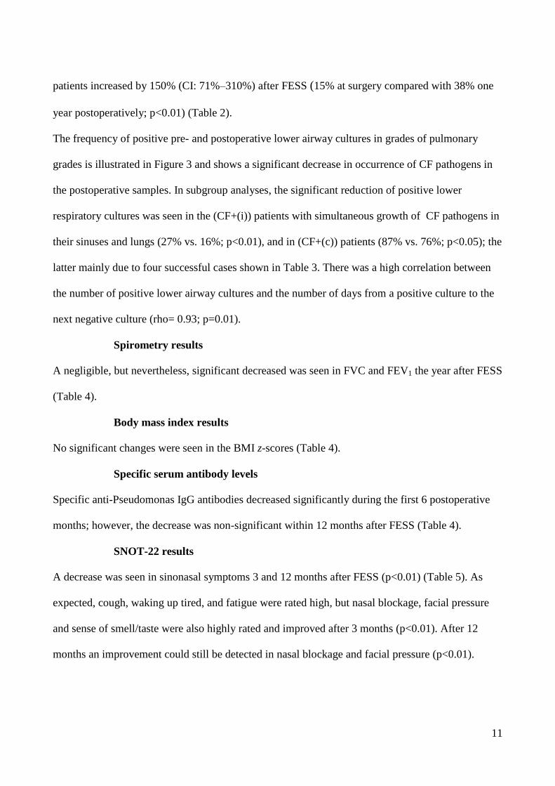
11
patients increased by 150% (CI: 71%–310%) after FESS (15% at surgery compared with 38% one
year postoperatively; p<0.01) (Table 2).
The frequency of positive pre- and postoperative lower airway cultures in grades of pulmonary
grades is illustrated in Figure 3 and shows a significant decrease in occurrence of CF pathogens in
the postoperative samples. In subgroup analyses, the significant reduction of positive lower
respiratory cultures was seen in the (CF+(i)) patients with simultaneous growth of CF pathogens in
their sinuses and lungs (27% vs. 16%; p<0.01), and in (CF+(c)) patients (87% vs. 76%; p<0.05); the
latter mainly due to four successful cases shown in Table 3. There was a high correlation between
the number of positive lower airway cultures and the number of days from a positive culture to the
next negative culture (rho= 0.93; p=0.01).
Spirometry results
A negligible, but nevertheless, significant decreased was seen in FVC and FEV1 the year after FESS
(Table 4).
Body mass index results
No significant changes were seen in the BMI z-scores (Table 4).
Specific serum antibody levels
Specific anti-Pseudomonas IgG antibodies decreased significantly during the first 6 postoperative
months; however, the decrease was non-significant within 12 months after FESS (Table 4).
SNOT-22 results
A decrease was seen in sinonasal symptoms 3 and 12 months after FESS (p<0.01) (Table 5). As
expected, cough, waking up tired, and fatigue were rated high, but nasal blockage, facial pressure
and sense of smell/taste were also highly rated and improved after 3 months (p<0.01). After 12
months an improvement could still be detected in nasal blockage and facial pressure (p<0.01).

12
Quality of life results
An increase in HRQOL was seen 6 months postoperatively (p<0.05). However, at 12 months
postoperatively no significant differences in HRQOL were observed (Table 5).
Surgical complications
No perioperative major blood loss was observed. One patient was hospitalized three days after
surgery because of epistaxis, one patient experienced a month of paresthesia of the skin below the
eye and discomfort when looking to the side (the orbit was not entered during surgery), one patient
had a watery eye requiring intervention, and one patient had an accidental penetration of the orbital
lamina (with unaffected vision). Other than one patient who experienced decreased sense of smell
(surgically unexplained), no persistent sequelae were noted.
DISCUSSION
In this prospective cohort study, we present data from a large cohort of CF patients undergoing
extensive FESS and adjuvant therapy. A significant reduction in the frequency of lower airway
cultures with CF-pathogens accomplished after FESS is shown for the first time. Subsequently, the
proportion of intermittently colonised (CF+(i)) patients decreased as a consequence of the increased
number of non-infected (CF-) patients. Specific IgG against P. aeruginosa decreased and quality of
life including sinonasal symptoms was improved. Only a small decrease in lung function was seen
after a year, which is in agreement with an annual average decline of 1–2% in CF patients [22].
Other studies have addressed the effect of sinus surgery on CF lungs using varying parameters and
showing inconsistent results [10;23-32]. Comparable to our study, one prospective study performed
extensive sinus surgery intending to eradicate sinus bacteria in a group of 82 LTX patients [10],
showing that P. aeruginosa and B. cepacia complex could be eradicated from the sinuses, resulting

13
in reduced lung allograft infections. Shatz [31] found decreased antibiotic use, a lower
hospitalisation rate, and an increase in FEV1 six months after FESS among 15 non-LTX patients.
Lewiston et al. [27] postoperatively installed Tobramycin in the sinuses and reported a lower rate of
P.aeruginosa in the lungs of 11 LTX patients. Other studies differ in several ways: as they were
retrospective, most did less extensive surgery compared with our procedures, none focused on the
grading of pulmonary infection and only few included intensive follow-up with use of nasally
applied antibiotics. These studies found no postoperative reduction in lung colonisation; however,
some found a slight improvement of lung function, lower hospitalisation rates, and reduced use of
antibiotics [23;24;26;28;29;32;33].
In the present study, (CF+(i)) patients simultaneously sinus and pulmonary colonised experienced
the greatest benefit from our treatment protocol; it is likely that the treatment protected the lungs in
this group of „risk‟ patients from subsequent recurrent lung colonisation and chronic infection. The
challenge is to detect these specific patients and to estimate when they may benefit from FESS. Our
results support previously published studies showing a that LTX patients benefits from FESS
[27;34], and further indicate that patients at the early stage of chronic lung infection with well-
preserved lung function and low levels of specific antibodies, such as the patients in Table 3, may
benefit from FESS. We put forward the possibility that these patients may be false positively
categorized as (CF+(c)) since lower airway samples can be cross-contaminated by secretions from
the upper airways; in these cases the antibody response has shown to be helpful in distinguishing
(CF+(i)) patients from (CF+(c)) [12;19].
The SNOT-22 questionnaire is suitable as a disease-specific instrument in sinonasal research and is
widely used in non CF patients [20]. We found that the questionnaire is a good tool for evaluating
CRS symptoms; however, the risk of CF patients underreporting symptoms of CRS should be taken

14
into account [35]. The majority of patients reported decreased nasal discharge and decreased nasal
blockage after FESS despite very low preoperative scoring of these two items.
The CFQ-R has undergone validity testing [36] and is a widely used HRQOL measure for CF. CRS
affects quality of life [37] but the CFQ-R contains only one question on CRS. Nevertheless, in our
population, CFQ-R improved six months after FESS.
All our results, both positive and negative, highlight the need for guidelines for when to offer CF
patients sinus surgery and/or nasal care.
Although we did not address the need for revision surgery, our prospective study gives weight to
the conclusions of the recently published European position paper on rhinosinusitis and fulfils the
requirements for new studies [1]: “Future prospective studies are needed to further elucidate the role
of medical and surgical therapy in CF patients with chronic rhinosinusitis (CRS), but the data on
surgical therapy support the safety and efficacy of endoscopic sinus surgery”.
The strengths of our study are the large cohort, its prospective design taking multiple parameters
into consideration, the extensive surgery done to eradicate CF pathogens, and, in particular, the
unchanged CF treatment protocol throughout the intervention [13]. Longer follow-up times and
larger cohorts with the possibility of randomising patients into different treatment groups are
needed to further elucidate the role of medical and surgical therapy among CF patients. With this
study design, the effect of FESS on pulmonary infections could not be sufficiently evaluated in
(CF-) patients who had CF pathogens only in the sinuses and not in the lungs.
Our study is limited by the lack of a control group and the tardy initiation of the questionnaires.
Further, we were initially reluctant to explore all sinuses. Nevertheless, for each FESS procedure,
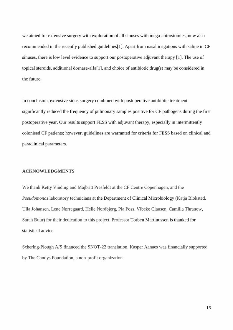
15
we aimed for extensive surgery with exploration of all sinuses with mega-antrostomies, now also
recommended in the recently published guidelines[1]. Apart from nasal irrigations with saline in CF
sinuses, there is low level evidence to support our postoperative adjuvant therapy [1]. The use of
topical steroids, additional dornase-alfa[1], and choice of antibiotic drug(s) may be considered in
the future.
In conclusion, extensive sinus surgery combined with postoperative antibiotic treatment
significantly reduced the frequency of pulmonary samples positive for CF pathogens during the first
postoperative year. Our results support FESS with adjuvant therapy, especially in intermittently
colonised CF patients; however, guidelines are warranted for criteria for FESS based on clinical and
paraclinical parameters.
ACKNOWLEDGMENTS
We thank Ketty Vinding and Majbritt Presfeldt at the CF Centre Copenhagen, and the
Pseudomonas laboratory technicians at the Department of Clinical Microbiology (Katja Bloksted,
Ulla Johansen, Lene Nørregaard, Helle Nordbjerg, Pia Poss, Vibeke Clausen, Camilla Thranow,
Sarah Buur) for their dedication to this project. Professor Torben Martinussen is thanked for
statistical advice.
Schering-Plough A/S financed the SNOT-22 translation. Kasper Aanaes was financially supported
by The Candys Foundation, a non-profit organization.

16
Reference List
1 Fokkens WJ, Lund VJ, Mullol J, Bachert C, Alobid I, Baroody F, Cohen N, Cervin A,
Douglas R, Gevaerts P, Georgalas C, Goossens H, Harvey R, Hellings P, Hopkins C, Jones
N, Joos G, Kalogjera L, Kern B, Kowalski M, Price D, Riechelmann H, Schlosser R, Senior
B, Thomas M, Toskala E, Voegels R, Wang DY, Wormald PJ: EPOS 2012: European
position paper on rhinosinusitis and nasal polyps 2012. A summary for
otorhinolaryngologists. Rhinology 2012;50:1-12.
2 Hansen SK, Rau MH, Johansen HK, Ciofu O, Jelsbak L, Yang L, Folkesson A, Jarmer
HO, Aanaes K, Buchwald C, Hoiby N, Molin S: Evolution and diversification of
Pseudomonas aeruginosa in the paranasal sinuses of cystic fibrosis children have
implications for chronic lung infection. ISME J 2011.
3 Johansen HK, Aanaes K, Pressler T, Nielsen KG, Fisker J, Skov M, Hoiby N,
Buchwald C: Colonisation and infection of the paranasal sinuses in cystic fibrosis patients is
accompanied by a reduced PMN response. J Cyst Fibros 2012.
4 Aanaes K, Rickelt LF, Johansen HK, Buchwald C, Pressler T, Hoiby N, Jensen PO:
Decreased mucosal oxygen tension in the maxillary sinuses in patients with cystic fibrosis. J
Cyst Fibros 2011;10:114-120.
5 Kolpen M, Hansen CR, Bjarnsholt T, Moser C, Christensen LD, van GM, Ciofu O,
Mandsberg L, Kharazmi A, Doring G, Givskov M, Hoiby N, Jensen PO:
Polymorphonuclear leucocytes consume oxygen in sputum from chronic Pseudomonas
aeruginosa pneumonia in cystic fibrosis. Thorax 2010;65:57-62.
6 Aanaes K, Johansen HK, Poulsen SS, Pressler T, Buchwald C, Hoiby N: Secretory
IgA as a diagnostic tool for Pseudomonas aeruginosa respiratory colonization. J Cyst Fibros
2012.
7 Mainz JG, Naehrlich L, Schien M, Kading M, Schiller I, Mayr S, Schneider G,
Wiedemann B, Wiehlmann L, Cramer N, Pfister W, Kahl BC, Beck JF, Tummler B:
Concordant genotype of upper and lower airways P aeruginosa and S aureus isolates in
cystic fibrosis. Thorax 2009;64:535-540.
8 Nunley DR, Grgurich W, Iacono AT, Yousem S, Ohori NP, Keenan RJ, Dauber JH:
Allograft colonization and infections with pseudomonas in cystic fibrosis lung transplant
recipients. Chest 1998;113:1235-1243.
9 Aanaes K, Johansen HK, Hjuler T, Skov M, Buchwald C: Extensive sinus surgery and
follow-up can eradicate pathogenic bacterial sinusitis in patients with Cystic Fibrosis (CF);
2012, p S53.
10 Vital D, Hofer M, Benden C, Holzmann D, Boehler A: Impact of Sinus Surgery on
Pseudomonal Airway Colonization, Bronchiolitis Obliterans Syndrome and Survival in
Cystic Fibrosis Lung Transplant Recipients. Respiration 2012.
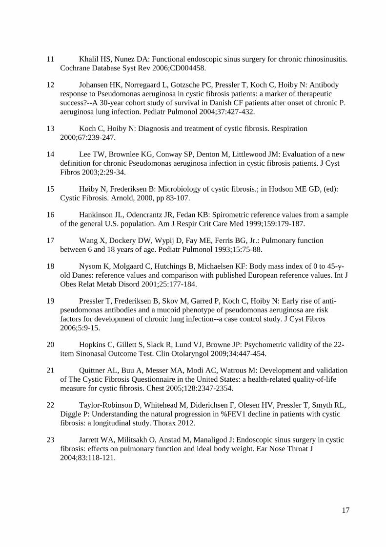
17
11 Khalil HS, Nunez DA: Functional endoscopic sinus surgery for chronic rhinosinusitis.
Cochrane Database Syst Rev 2006;CD004458.
12 Johansen HK, Norregaard L, Gotzsche PC, Pressler T, Koch C, Hoiby N: Antibody
response to Pseudomonas aeruginosa in cystic fibrosis patients: a marker of therapeutic
success?--A 30-year cohort study of survival in Danish CF patients after onset of chronic P.
aeruginosa lung infection. Pediatr Pulmonol 2004;37:427-432.
13 Koch C, Hoiby N: Diagnosis and treatment of cystic fibrosis. Respiration
2000;67:239-247.
14 Lee TW, Brownlee KG, Conway SP, Denton M, Littlewood JM: Evaluation of a new
definition for chronic Pseudomonas aeruginosa infection in cystic fibrosis patients. J Cyst
Fibros 2003;2:29-34.
15 Høiby N, Frederiksen B: Microbiology of cystic fibrosis.; in Hodson ME GD, (ed):
Cystic Fibrosis. Arnold, 2000, pp 83-107.
16 Hankinson JL, Odencrantz JR, Fedan KB: Spirometric reference values from a sample
of the general U.S. population. Am J Respir Crit Care Med 1999;159:179-187.
17 Wang X, Dockery DW, Wypij D, Fay ME, Ferris BG, Jr.: Pulmonary function
between 6 and 18 years of age. Pediatr Pulmonol 1993;15:75-88.
18 Nysom K, Molgaard C, Hutchings B, Michaelsen KF: Body mass index of 0 to 45-y-
old Danes: reference values and comparison with published European reference values. Int J
Obes Relat Metab Disord 2001;25:177-184.
19 Pressler T, Frederiksen B, Skov M, Garred P, Koch C, Hoiby N: Early rise of anti-
pseudomonas antibodies and a mucoid phenotype of pseudomonas aeruginosa are risk
factors for development of chronic lung infection--a case control study. J Cyst Fibros
2006;5:9-15.
20 Hopkins C, Gillett S, Slack R, Lund VJ, Browne JP: Psychometric validity of the 22-
item Sinonasal Outcome Test. Clin Otolaryngol 2009;34:447-454.
21 Quittner AL, Buu A, Messer MA, Modi AC, Watrous M: Development and validation
of The Cystic Fibrosis Questionnaire in the United States: a health-related quality-of-life
measure for cystic fibrosis. Chest 2005;128:2347-2354.
22 Taylor-Robinson D, Whitehead M, Diderichsen F, Olesen HV, Pressler T, Smyth RL,
Diggle P: Understanding the natural progression in %FEV1 decline in patients with cystic
fibrosis: a longitudinal study. Thorax 2012.
23 Jarrett WA, Militsakh O, Anstad M, Manaligod J: Endoscopic sinus surgery in cystic
fibrosis: effects on pulmonary function and ideal body weight. Ear Nose Throat J
2004;83:118-121.
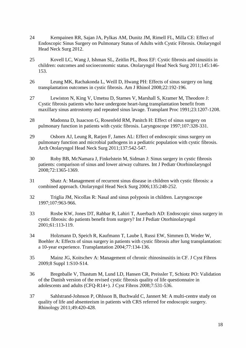
18
24 Kempainen RR, Sajan JA, Pylkas AM, Dunitz JM, Rimell FL, Milla CE: Effect of
Endoscopic Sinus Surgery on Pulmonary Status of Adults with Cystic Fibrosis. Otolaryngol
Head Neck Surg 2012.
25 Kovell LC, Wang J, Ishman SL, Zeitlin PL, Boss EF: Cystic fibrosis and sinusitis in
children: outcomes and socioeconomic status. Otolaryngol Head Neck Surg 2011;145:146-
153.
26 Leung MK, Rachakonda L, Weill D, Hwang PH: Effects of sinus surgery on lung
transplantation outcomes in cystic fibrosis. Am J Rhinol 2008;22:192-196.
27 Lewiston N, King V, Umetsu D, Starnes V, Marshall S, Kramer M, Theodore J:
Cystic fibrosis patients who have undergone heart-lung transplantation benefit from
maxillary sinus antrostomy and repeated sinus lavage. Transplant Proc 1991;23:1207-1208.
28 Madonna D, Isaacson G, Rosenfeld RM, Panitch H: Effect of sinus surgery on
pulmonary function in patients with cystic fibrosis. Laryngoscope 1997;107:328-331.
29 Osborn AJ, Leung R, Ratjen F, James AL: Effect of endoscopic sinus surgery on
pulmonary function and microbial pathogens in a pediatric population with cystic fibrosis.
Arch Otolaryngol Head Neck Surg 2011;137:542-547.
30 Roby BB, McNamara J, Finkelstein M, Sidman J: Sinus surgery in cystic fibrosis
patients: comparison of sinus and lower airway cultures. Int J Pediatr Otorhinolaryngol
2008;72:1365-1369.
31 Shatz A: Management of recurrent sinus disease in children with cystic fibrosis: a
combined approach. Otolaryngol Head Neck Surg 2006;135:248-252.
32 Triglia JM, Nicollas R: Nasal and sinus polyposis in children. Laryngoscope
1997;107:963-966.
33 Rosbe KW, Jones DT, Rahbar R, Lahiri T, Auerbach AD: Endoscopic sinus surgery in
cystic fibrosis: do patients benefit from surgery? Int J Pediatr Otorhinolaryngol
2001;61:113-119.
34 Holzmann D, Speich R, Kaufmann T, Laube I, Russi EW, Simmen D, Weder W,
Boehler A: Effects of sinus surgery in patients with cystic fibrosis after lung transplantation:
a 10-year experience. Transplantation 2004;77:134-136.
35 Mainz JG, Koitschev A: Management of chronic rhinosinusitis in CF. J Cyst Fibros
2009;8 Suppl 1:S10-S14.
36 Bregnballe V, Thastum M, Lund LD, Hansen CR, Preissler T, Schiotz PO: Validation
of the Danish version of the revised cystic fibrosis quality of life questionnaire in
adolescents and adults (CFQ-R14+). J Cyst Fibros 2008;7:531-536.
37 Sahlstrand-Johnson P, Ohlsson B, Buchwald C, Jannert M: A multi-centre study on
quality of life and absenteeism in patients with CRS referred for endoscopic surgery.
Rhinology 2011;49:420-428.
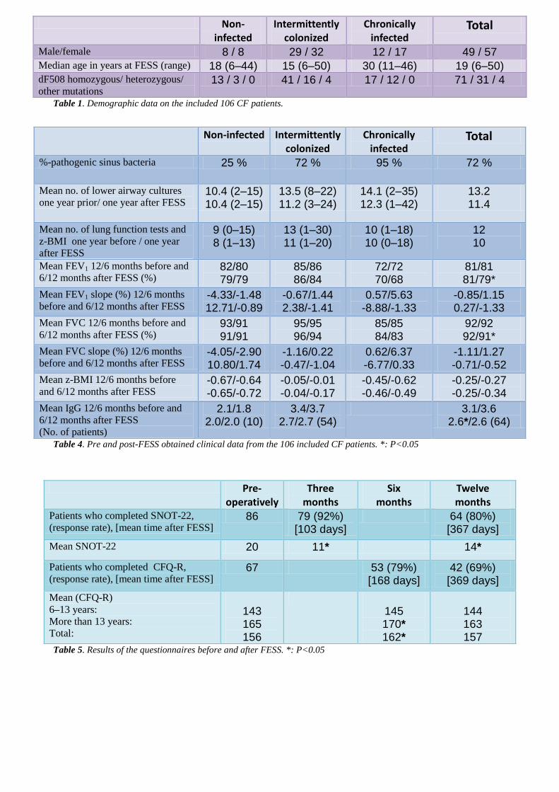
Table 1. Demographic data on the included 106 CF patients.
Table 4. Pre and post-FESS obtained clinical data from the 106 included CF patients. *: P<0.05
Table 5. Results of the questionnaires before and after FESS. *: P<0.05
Non-infected Intermittently colonized
Chronically infected
Total
%-pathogenic sinus bacteria 25 % 72 % 95 % 72 %
Mean no. of lower airway cultures
one year prior/ one year after FESS 10.4 (2–15) 10.4 (2–15)
13.5 (8–22) 11.2 (3–24)
14.1 (2–35) 12.3 (1–42)
13.2 11.4
Mean no. of lung function tests and
z-BMI one year before / one year
after FESS
9 (0–15) 8 (1–13)
13 (1–30) 11 (1–20)
10 (1–18) 10 (0–18)
12 10
Mean FEV1 12/6 months before and
6/12 months after FESS (%) 82/80 79/79
85/86 86/84
72/72 70/68
81/81 81/79*
Mean FEV1 slope (%) 12/6 months
before and 6/12 months after FESS -4.33/-1.48 12.71/-0.89
-0.67/1.44 2.38/-1.41
0.57/5.63 -8.88/-1.33
-0.85/1.15 0.27/-1.33
Mean FVC 12/6 months before and
6/12 months after FESS (%) 93/91 91/91
95/95 96/94
85/85 84/83
92/92 92/91*
Mean FVC slope (%) 12/6 months
before and 6/12 months after FESS -4.05/-2.90 10.80/1.74
-1.16/0.22 -0.47/-1.04
0.62/6.37 -6.77/0.33
-1.11/1.27 -0.71/-0.52
Mean z-BMI 12/6 months before
and 6/12 months after FESS -0.67/-0.64 -0.65/-0.72
-0.05/-0.01 -0.04/-0.17
-0.45/-0.62 -0.46/-0.49
-0.25/-0.27 -0.25/-0.34
Mean IgG 12/6 months before and
6/12 months after FESS
(No. of patients)
2.1/1.8 2.0/2.0 (10)
3.4/3.7 2.7/2.7 (54)
3.1/3.6 2.6*/2.6 (64)
Non-infected
Intermittently colonized
Chronically infected
Total
Male/female 8 / 8 29 / 32 12 / 17 49 / 57 Median age in years at FESS (range) 18 (6–44) 15 (6–50) 30 (11–46) 19 (6–50) dF508 homozygous/ heterozygous/
other mutations 13 / 3 / 0 41 / 16 / 4 17 / 12 / 0 71 / 31 / 4
Pre-operatively
Three months
Six months
Twelve months
Patients who completed SNOT-22,
(response rate), [mean time after FESS] 86 79 (92%)
[103 days] 64 (80%)
[367 days]
Mean SNOT-22 20 11* 14*
Patients who completed CFQ-R,
(response rate), [mean time after FESS] 67 53 (79%)
[168 days] 42 (69%) [369 days]
Mean (CFQ-R)
6–13 years:
More than 13 years:
Total:
143 165 156
145 170* 162*
144 163 157
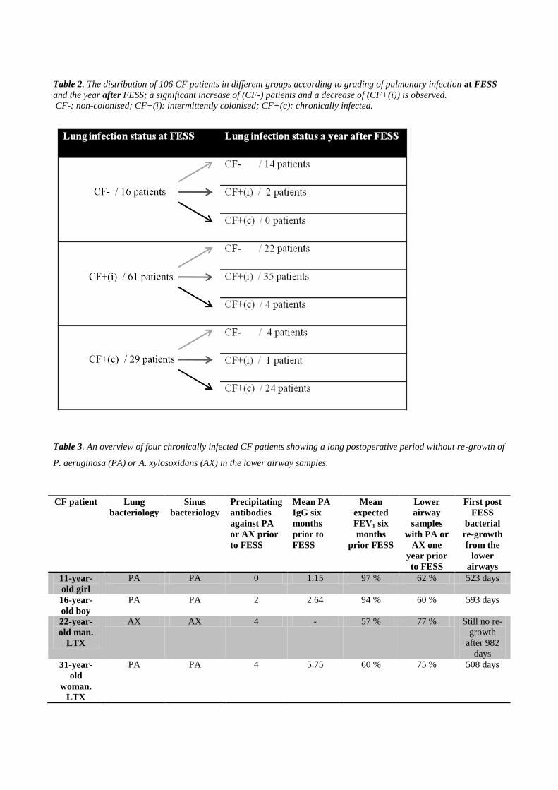
Table 2. The distribution of 106 CF patients in different groups according to grading of pulmonary infection at FESS
and the year after FESS; a significant increase of (CF-) patients and a decrease of (CF+(i)) is observed.
CF-: non-colonised; CF+(i): intermittently colonised; CF+(c): chronically infected.
Table 3. An overview of four chronically infected CF patients showing a long postoperative period without re-growth of
P. aeruginosa (PA) or A. xylosoxidans (AX) in the lower airway samples.
CF patient Lung
bacteriology
Sinus
bacteriology
Precipitating
antibodies
against PA
or AX prior
to FESS
Mean PA
IgG six
months
prior to
FESS
Mean
expected
FEV1 six
months
prior FESS
Lower
airway
samples
with PA or
AX one
year prior
to FESS
First post
FESS
bacterial
re-growth
from the
lower
airways
11-year-
old girl
PA PA 0 1.15 97 % 62 % 523 days
16-year-
old boy
PA PA 2 2.64 94 % 60 % 593 days
22-year-
old man.
LTX
AX AX 4 - 57 % 77 % Still no re-
growth
after 982
days
31-year-
old
woman.
LTX
PA PA 4 5.75 60 % 75 % 508 days

Figure 1. Time-line showing the interventions in the study.
Figure 2. The criteria fulfilled for FESS in 106 CF patients undergoing surgery. (CRS: chronic rhinosinusitis; LTX:
lung transplantation).

Figure 3. Frequencies of lower airway cultures with growth of CF-pathogenic Gram-negative bacteria a year before
and after FESS, distributed on lung infection status and on the perioperatively bacterial findings in the sinuses.