Molecular Staging Estimates Occult Tumor Burden in Colorectal Cancer
Upload
independentCategory
view
0download
0
Cancer Cell
Article
B55b-Associated PP2A Complex ControlsPDK1-Directed Myc Signaling and ModulatesRapamycin Sensitivity in Colorectal CancerJing Tan,1,2 Puay Leng Lee,1 Zhimei Li,1 Xia Jiang,1 Yaw Chyn Lim,2 Shing Chuan Hooi,2 and Qiang Yu1,2,3,*1Cancer Biology and Pharmacology, Genome Institute of Singapore, Agency for Science, Technology and Research, Biopolis, Singapore2Department of Physiology, Yong Loo Lin School of Medicine, National University of Singapore, Singapore3Cancer and Stem Cell Biology, DUKE-NUS Graduate Medical School of Singapore, Singapore*Correspondence: [email protected]
DOI 10.1016/j.ccr.2010.10.021
SUMMARY
The PP2A serine/threonine protein phosphatase serves as a critical cellular regulator of cell growth, prolifer-ation, and survival. However, how this pathway is altered in human cancer to confer growth advantage islargely unknown. Here, we show that PPP2R2B, encoding the B55b regulatory subunit of the PP2A complex,is epigenetically inactivated by DNA hypermethylation in colorectal cancer. B55b-associated PP2A interactswith PDK1 and modulates its activity toward Myc phosphorylation. On loss of PPP2R2B, mTORC1 inhibitorrapamycin triggers a compensatory Myc phosphorylation in PDK1-dependent, but PI3K and AKT-indepen-dent manner, resulting in resistance. Reexpression of PPP2R2B, genetic ablation of PDK1 or pharmacologicinhibition of PDK1 abrogates the rapamycin-induced Myc phosphorylation, leading to rapamycin sensitiza-tion. Thus, PP2A-B55b antagonizes PDK1-Myc signaling and modulates rapamycin sensitivity.
INTRODUCTION
Protein phosphatase 2A (PP2A) functions as a multimetric
enzyme that contains the catalytic C subunit, a scaffolding
A-subunit and one of a large array of regulatory B-subunits. Eu-
karyotic cells contain over 200 biochemical distinct PP2A
complexes derived from differential combinations of A, B, C,
and other subunits. The regulatory subunits are expressed in
a tissue-specific manner, leading to the presence of different
PP2A complexes in different mammalian tissues (Virshup and
Shenolikar, 2009). Moreover, it is these regulators, rather than
the catalytic subunit, that provides the substrate specificity
and catalyzes distinct dephosphorylation events that result in
specific functional outcomes.
Although a tumor suppressor role of PP2A has been previously
shown in a variety of immortalized human cell types (Chen et al.,
2004; Eichhorn et al., 2009; Janssens and Goris, 2001; Rangar-
ajan et al., 2004; Sablina et al., 2007; Zhao et al., 2003), the
Significance
Clinical evidence pointing to a wide occurrence of PP2A tumoHere, we show the occurrence of epigenetic silencing of PP2Ahuman colorectal cancer. Loss of PPP2R2B results in PDK1-dylation in response to mTOR inhibitor rapamycin, resulting in raPDK1 abrogates rapamycin-induced Myc phosphorylation, remechanism underlying rapamycin resistance, which is indepebiomarker to predict rapamycin sensitivity. Further, we showprovide a treatment strategy for colorectal cancer.
Can
genetic and/or the epigenetic evidence pointing to a prevalent
inactivation of PP2A in human malignancy have not been re-
ported. Somaticmutations in the A subunit of the PP2A complex,
which can result in the loss of B subunit binding (Ruediger et al.,
2001), were found in only up to 15% of some human cancers
(Calin et al., 2000; Ruediger et al., 2001; Takagi et al., 2000; Tam-
aki et al., 2004;Wang et al., 1998;Westermarck andHahn, 2008),
and the reduced expression of PP2A subunit B56g has been re-
ported only in some cancer cell lines (Chen et al., 2004; Zhao
et al., 2003). In general, the genetic or epigenetic changes of
PP2A complexes in human cancer remains to be defined, as is
its impact on cancer signaling and therapeutic responses to tar-
geted therapy.
One of the PP2A regulated cancer signaling pathways is the
mTOR pathway, a key component of PI3K pathway that many
cancer cells are ‘‘addicted’’ to for growth advantage (Guertin
and Sabatini, 2007; Sabatini, 2006). Although small molecule
mTORC1 inhibitors, such as rapamycin and its analogs have
r suppressor inactivation has not been previously provided.regulatory B55b subunit, encoded by PPP2R2B, in > 90%
ependent, but PI3K-independnt induction of Myc phosphor-pamycin resistance. Restoration of PPP2R2B or inhibition ofsensitizing rapamycin. Our data demonstrate an alternatendent of PI3K-AKT. Thus, PPP2R2B is likely to be a usefulthat a combination of PDK1 inhibitor with rapamycin might
cer Cell 18, 459–471, November 16, 2010 ª2010 Elsevier Inc. 459

E G
A
PPP2R1A
PPP2R2A
PPP2CA
PPP2R5A
PPP2R4
PPP2R2D
PPP2R5E
PPP2R5B
PPP2R3B
PPP2R5C
PPP2R3B
PPP2R5D
PPP2R1B
PPP2R2C
PPP2CB
PPP2CB
PPP2R3A
PPP2R2B
Normal tissue Primary tumor
N 1
N 2
N 3
N 4
N 5
N 7
N 9
N 11
N 12
N 13
N 14
N 15
N 16
N 17
N 18
N 19
N 20
N 21
N 22
N 24
N 25
N 26
N 27
N 28
T 1 T 2 T 3 T 4 T 5 T 7 T 9 T 11
T 12
T 13
T 14
T 15
T 16
T 17
T 18
T 19
T 20
T 21
T 22
T 24
T 25
T 26
T 27
T 28
GAPDH
PPP2R2B
Col
on ti
ssue
HC
T116
RKO
SW48
0
HT2
9
HT1
5
DLD
1
CRC lines
PPP2R2A
PPP2R5C
F H
C
D
Nor
mal
HC
T116
DKO
BGS
1
-350 +29
PPP2R2B
CpG island
100bp
Ctrl Aza
DLD1
HC
T116
DKO
GAPDH
PPP2R2B
M U M U M U M U M U M U M U M U
2T 4T 12T 16T 19T 21T 22T20TT22 N22
M U M U
PPP2R2B
B
N T N T N T N T N T N T N T N T
2 4 12 16 19 20 21 22
-actin
PPP2R2B
PPP2R2A
PPP2R5C
HCT116 RKO SW480 DLD1 HT29 HT15
PPP2R2B
M U M U M U M U M U M U M U
Normal Colon
HCT116 DKO
M U M U
PPP2R2B
I
N(24) T(24)
PPP2R2B
** p<0.001
-7.5
-5.0
-2.5
0.0
2.5
5.0
7.5
Nor
mal
ized
exp
ress
ion
(log2
)
#
Figure 1. Loss of PPP2R2B Expression by Promoter DNA Hypermethylation in CRC(A) Box-plot showing the differential expression of PPP2R2BmRNA levels in 24 pairs of patient-derived CRC and matched normal colon mucosa as determined
by Illumina array analysis. P value for difference between tumor and normal is indicated.
(B) Hierarchical clustering of expression levels of PP2A subunits in 24 pairs of colorectal tumors (T) and matched normal mucosa (N).
(C) RT-PCR analysis of PPP2R2B, PPP2R2A, and PPP2R5C expression from eight randomly selected pairs of human CRC (T) and matched mucosa (N).
(D) RT-PCR analysis of PPP2R2B, PPP2R2A, and PPP2R5C in a panel of CRC cell lines compared to the normal colon tissue as well as nontransformed cell lines.
(E) Methylation specific PCR (MSP) analysis of PPP2R2B promoter in CRC cell lines and nontransformed cells. M: methylated; U: unmethylated.
(F) MSP analysis of PPP2R2B promoter in eight tumor and normal controls. M: methylated; U: unmethylated.
(G) RT-PCR analysis of PPP2R2B in HCT116 and DKO or DLD1 cells treated with or without 5-AzaC (5 mM) for 3 days.
(H) MSP analysis of PPP2R2B promoter in HCT116 and DKO cells. M: methylated; U: unmethylated.
(I) Methylation analysis of PPP2R2B promoter by bisulfite-sequencing analysis (BGS). The region analyzed is indicated. The arrow indicates the transcriptional
start site. Open circles represent unmethylated CpGs; closed circles denote methylated CpGs. See also Figure S1 and Tables S1 and S2.
Cancer Cell
PP2A-B55b Modulates PDK1-Myc Signaling
shown promise as cancer therapeutics and have been approved
for clinical application (Guertin and Sabatini, 2007; Hudes et al.,
2007), they have had only limited successes and clinical
outcomes are mostly unpredictable. Although a known mecha-
nism of rapamycin resistance is linked to its feedback activation
of AKT phosphorylation through PI3K and mTORC2 (O’Reilly
et al., 2006; Sarbassov et al., 2006), a deeper understanding of
the resistance mechanisms and the identification of biomarkers
that help predict therapeutic responses have become important
topics (Mao et al., 2008; Scott et al., 2009; Thomas et al., 2006).
RESULTS
Loss of PPP2R2B Expression by DNA Hypermethylationin Colorectal CancerGiven the low frequency of mutations found in PP2A family
members in human malignancy, including colorectal cancer
(CRC), we sought to determine whether they are epigenetically
inactivated in CRC. Through interrogating expression data of
a series of CRC cell lines we published previously (Jiang et al.,
2008), we found that PPP2R2B, encoding B55b, is the only
subunit that is consistently downregulated or silenced in all
examined CRC cell lines, but not in the normal colon mucosa
samples (see Table S1 available online). The significant downre-
gulation of PPP2R2B was further validated using gene expres-
sion array data of 24 pairs of patient-derived CRC tumors and
matched normal mucosa controls (p < 0.01) (Figure 1A), and
460 Cancer Cell 18, 459–471, November 16, 2010 ª2010 Elsevier Inc
this appeared to occur in >90% of CRC samples (Figure 1B;
Table S2). Semiquantitative and quantitative RT-PCR analysis
confirmed the PPP2R2B downregulation in eight randomly
selected CRC compared to the matched controls, as well as in
a series of CRC cells lines (Figures 1C and 1D; Figure S1A),
but not in the nontransformed epithelial cells (Figure S1B). By
contrast, PPP2R2A (B55a) and PPP2R5C (B56g), which has
been previously reported to be downregulated in lung cancer
(Chen et al., 2004), were not downregulated in CRC (Figures
1C and 1D).
We next determined whether the loss of PPP2R2B expression
in CRC is associated with promoter DNA hypermethylation.
Methylation-specific PCR (MSP) analysis revealed a hyperme-
thylated PPP2R2B promoter in CRC cell lines, as well as in all
eight CRC tumor samples examined, but not in their matched
normal controls (Figures 1E and 1F), nor in nontransformed
epithelial cells (Figure S1C). Furthermore, HCT116 cells deleted
of both DNMT1 and DNMT3B (DKO) or DLD1 cells treated with
5-aza-dC reexpressed PPP2R2B (Figure 1G), correlated with
a demethylated PPP2R2B promoter in DKO, as demonstrated
by both MSP and bisulfate genomic sequencing (Figures 1H
and 1I). Taken together, these results demonstrate that
PPP2R2B is epigenetically silenced in CRC through promoter
DNA hypermethylation. Moreover, Oncomine data mining
reveals that PPP2R2B is also significantly downregulated in
other human cancers, such as bladder, brain and esophagus
carcinomas (Figure S1D).
.
A B DC
E
0
0.2
0.4
0.6
0.8
1.0
1.2
siNC
siPPP
2R2B
#1
siPPP
2R2B
#SP
Rel
ativ
e m
RN
A le
vel
Mean
tumo
r volu
me (m
m3)
Days of treatment0 1 3 5 6 9 10 13 15 16 17 20 21 23
0100
200300400500600700800900
1000
**
PPP2R2B+DoxPPP2R2B+PBSVector+DoxVector+PBS
Exogenous B55β
Dox:
B55β-myc
β-actin
Anti-myc
Anti-B55
- +
DLD1-PPP2R2B
Endogenous B55
Vector PPP2R2B
0
10
20
30
40
50
60 CtrlDox
BrdU
inco
rpor
atio
n (%
)
*p<0.001
G
-Dox
+Dox
SA-β-gal
Vector PPP2R2B
siPPP2R2B#SP
siNC SV40-ST
siPPP2R2B#1
0
50
100
150
Col
onie
sin
soft
agar
si NC
siPPP
2R2B
#1
siPPP
2R2B
#SP
SV40
-ST -
-Dox
+Dox
HCT116DLD1
Vector PPP2R2B Vector PPP2R2B
- -F
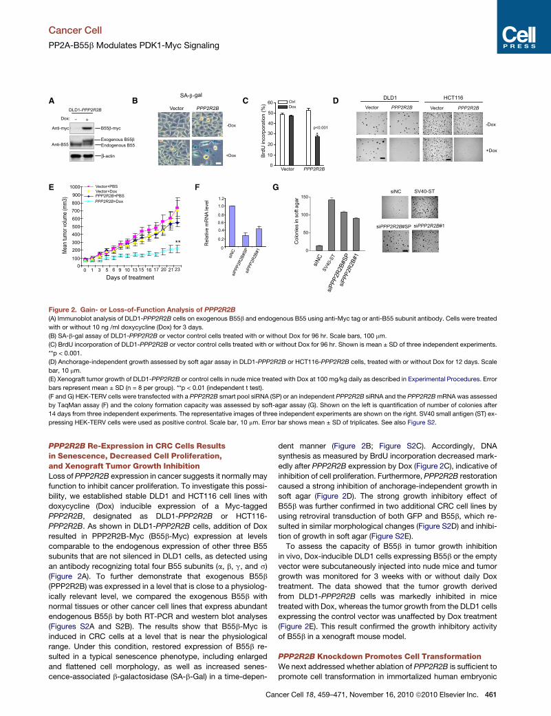
Figure 2. Gain- or Loss-of-Function Analysis of PPP2R2B
(A) Immunoblot analysis of DLD1-PPP2R2B cells on exogenous B55b and endogenous B55 using anti-Myc tag or anti-B55 subunit antibody. Cells were treated
with or without 10 ng /ml doxycycline (Dox) for 3 days.
(B) SA-b-gal assay of DLD1-PPP2R2B or vector control cells treated with or without Dox for 96 hr. Scale bars, 100 mm.
(C) BrdU incorporation of DLD1-PPP2R2B or vector control cells treated with or without Dox for 96 hr. Shown is mean ± SD of three independent experiments.
**p < 0.001.
(D) Anchorage-independent growth assessed by soft agar assay in DLD1-PPP2R2B or HCT116-PPP2R2B cells, treated with or without Dox for 12 days. Scale
bar, 10 mm.
(E) Xenograft tumor growth of DLD1-PPP2R2B or control cells in nude mice treated with Dox at 100 mg/kg daily as described in Experimental Procedures. Error
bars represent mean ± SD (n = 8 per group). **p < 0.01 (independent t test).
(F and G) HEK-TERV cells were transfected with a PPP2R2B smart pool siRNA (SP) or an independent PPP2R2B siRNA and the PPP2R2BmRNA was assessed
by TaqMan assay (F) and the colony formation capacity was assessed by soft-agar assay (G). Shown on the left is quantification of number of colonies after
14 days from three independent experiments. The representative images of three independent experiments are shown on the right. SV40 small antigen (ST) ex-
pressing HEK-TERV cells were used as positive control. Scale bar, 10 mm. Error bar shows mean ± SD of triplicates. See also Figure S2.
Cancer Cell
PP2A-B55b Modulates PDK1-Myc Signaling
PPP2R2B Re-Expression in CRC Cells Resultsin Senescence, Decreased Cell Proliferation,and Xenograft Tumor Growth InhibitionLoss of PPP2R2B expression in cancer suggests it normally may
function to inhibit cancer proliferation. To investigate this possi-
bility, we established stable DLD1 and HCT116 cell lines with
doxycycline (Dox) inducible expression of a Myc-tagged
PPP2R2B, designated as DLD1-PPP2R2B or HCT116-
PPP2R2B. As shown in DLD1-PPP2R2B cells, addition of Dox
resulted in PPP2R2B-Myc (B55b-Myc) expression at levels
comparable to the endogenous expression of other three B55
subunits that are not silenced in DLD1 cells, as detected using
an antibody recognizing total four B55 subunits (a, b, g, and s)
(Figure 2A). To further demonstrate that exogenous B55b
(PPP2R2B) was expressed in a level that is close to a physiolog-
ically relevant level, we compared the exogenous B55b with
normal tissues or other cancer cell lines that express abundant
endogenous B55b by both RT-PCR and western blot analyses
(Figures S2A and S2B). The results show that B55b-Myc is
induced in CRC cells at a level that is near the physiological
range. Under this condition, restored expression of B55b re-
sulted in a typical senescence phenotype, including enlarged
and flattened cell morphology, as well as increased senes-
cence-associated b-galactosidase (SA-b-Gal) in a time-depen-
Can
dent manner (Figure 2B; Figure S2C). Accordingly, DNA
synthesis as measured by BrdU incorporation decreased mark-
edly after PPP2R2B expression by Dox (Figure 2C), indicative of
inhibition of cell proliferation. Furthermore, PPP2R2B restoration
caused a strong inhibition of anchorage-independent growth in
soft agar (Figure 2D). The strong growth inhibitory effect of
B55b was further confirmed in two additional CRC cell lines by
using retroviral transduction of both GFP and B55b, which re-
sulted in similar morphological changes (Figure S2D) and inhibi-
tion of growth in soft agar (Figure S2E).
To assess the capacity of B55b in tumor growth inhibition
in vivo, Dox-inducible DLD1 cells expressing B55b or the empty
vector were subcutaneously injected into nude mice and tumor
growth was monitored for 3 weeks with or without daily Dox
treatment. The data showed that the tumor growth derived
from DLD1-PPP2R2B cells was markedly inhibited in mice
treated with Dox, whereas the tumor growth from the DLD1 cells
expressing the control vector was unaffected by Dox treatment
(Figure 2E). This result confirmed the growth inhibitory activity
of B55b in a xenograft mouse model.
PPP2R2B Knockdown Promotes Cell TransformationWe next addressed whether ablation of PPP2R2B is sufficient to
promote cell transformation in immortalized human embryonic
cer Cell 18, 459–471, November 16, 2010 ª2010 Elsevier Inc. 461

A B C
D E
F
HG
Figure 3. Restoration of PPP2R2B-PP2A Complex Results in Inhibition of p70S6K and Myc Phosphorylation
(A) Immunoblot analysis of HCT116-PPP2R2B or the vector control cells for indicated proteins in the presence or absence of Dox for 48 hr.
(B and C) Immunoblot analysis of DLD1-PPP2R2B or the vector control cells treated with or without Dox for the indicated length of times.
(D) Coimmunoprecipitation of PP2A scaffolding A subunit and catalytic C subunit with PPP2R2B. DLD1-PPP2R2B or vector control cells were treated with Dox
and PPP2R2B was immunoprecipitated with anti-Myc tag antibody. WCL, whole cell lysates.
(E) Serine/threonine phosphatase activity for DLD1-PPP2R2B or vector control cells. Protein phosphatase activity of the immunoprecipitates of PPP2R2B-Myc
was measured in triplicates from three independent experiments.
(F) Immunoblot analysis of DLD1-PPP2R2B cells for Myc, p-Myc (S62), and p70S6K, treated PPP2R1A siRNA or a negative control siRNA, with or without Dox
treatment.
(G) Growth curve of HCT116 and DLD1 cells treated with Myc, p70S6K siRNA, or negative control siRNA for indicated days.
(H) Growth inhibition of DLD1 cells induced by Myc knockdown, PPP2R2B expression or both. Data in (E), (G), and (H) represent mean ± SD of triplicates or three
independent experiments. See also Figure S3.
Cancer Cell
PP2A-B55b Modulates PDK1-Myc Signaling
kidney (HEK) fibroblasts expressing hTERT and oncogenic Ras
(HEK-TERV). In this previously well-characterized transformation
model, inhibition of PP2A by overexpression of small T antigen
results in efficient transformation (Chen et al., 2004; Hahn
et al., 2002; Sontag et al., 1993). Small interfering RNA
(siRNA)-directed knockdown by targeting two different regions
of PPP2R2B mRNA also resulted in an enormous increase in
anchorage-independent growth of HEK-TERV cells (Figure 2F
&G). A similar result was also obtained in HEK-TERV cells ex-
pressing a stable shRNA targeting PPP2R2B (Figure S2F). On
the basis of both the gain- and loss-of-function data, we propose
that loss of PPP2R2B facilitates oncogenic transformation.
PPP2R2B-Associated PP2A Complex ModulatesPhosphorylation of Myc and p70S6K in CRC CellsThe PP2A B regulatory subunits confer substrate specificity for
dephosphorylation events in a cell- and context-dependent
manner (Virshup and Shenolikar, 2009). Several oncogenic
proteins, including AKT, p70S6K, mTOR, b-catenin, and Myc,
462 Cancer Cell 18, 459–471, November 16, 2010 ª2010 Elsevier Inc
have been previously identified to be the substrates of PP2A in
various cell systems (Andrabi et al., 2007; Arnold and Sears,
2008; Peterson et al., 1999; Seeling et al., 1999; Yeh et al.,
2004). To dissect out the possible mechanisms responsible for
the growth inhibitory effect of B55b, we began with the
HCT116-PPP2R2B cells to probe several oncogenic signaling
pathways known to be important in CRC that might be affected
by PP2A (Figure 3A). The analysis led to the identification of two
phosphorylation events that are inhibited byPPP2R2B reexpres-
sion after Dox treatment. First, using phosphorylation specific
antibodies, we show that the phosphorylation of p70S6K at
T421/S424 was markedly reduced on PPP2R2B re-expression,
whereas the phosphorylation at T389 appeared to be unaf-
fected, suggesting a site-specific modulation of p70S6K by
PPP2R2B. Second, c-Myc phosphorylation, detected by using
a phospho-Myc antibody specific for S62, was also downregu-
lated on PPP2R2B reexpression. Other oncogenic signals,
such as p-AKT, p-ERK, b-catenin, and p-p38 were not notice-
ably affected by PPP2R2B re-expression (Figure 3A).
.

Cancer Cell
PP2A-B55b Modulates PDK1-Myc Signaling
The effects of B55b on p70S6K and c-Myc (thereafter
referred to as Myc) were further confirmed in DLD1 cells in
a time-course analysis (Figure 3B). The data showed that Dox
induction of PPP2R2B expression resulted in a rapid dephos-
phorylation of Myc and p70S6K (T421/S424, but not T389) rela-
tive to total p70S6K and Myc protein levels, suggesting that
these changes are the early effects of B55b and unlikely to be
the secondary effect of growth inhibition. The decreased Myc
phosphorylation eventually resulted in less protein accumula-
tion by 48 hr of Dox treatment (Figure 3C), which is consistent
with the previous finding that increased Myc phosphorylation
at S62 correlates with Myc protein accumulation (Arnold and
Sears, 2006; Junttila et al., 2007; Yeh et al., 2004). Thus,
p70S6K and Myc are two downstream signals affected by
B55b-PP2A complex, whereas no marked differences were
observed in the expression levels nor phosphorylation status
of other oncogenic signaling pathways known to be important
in colorectal cancer.
Of note, PPP2R2B re-expression had no effect on AKT T308
phosphorylation, which is known to be targeted by PP2A/B55a
or B56b complex in NIH 3T3 cells (Kuo et al., 2008; Padmanab-
han et al., 2009), neither on b-catenin phosphorylation that is
a target of PP2Aa in CRC (Su et al., 2008). In addition, in human
mammary epithelial cell cells, SV40 small t antigen(ST)-mediated
PP2A inhibition is associated with increased AKT S473 phos-
phorylation and the mTOR-mediated p70S6K T389 phosphory-
lation (Andrabi et al., 2007; Chen et al., 2005; Zhao et al.,
2003), but these changes were not observed here, in this cellular
context. These findings are consistent with the substrate specific
functions of different PP2A/B subunits in tissue specific contexts
and show that B55b-associated PP2A complex is distinguished
from the other PP2A complexes by affecting different down-
stream substrates.
To demonstrate that ectopic B55b in fact interacts with other
PP2A subunits to form an active PP2A complex, we performed
the coimmunoprecipitation assays to show that the B55b coim-
munoprecipitates with both PP2A structural (A) and catalytic (C)
subunits (Figure 3D). Furthermore, in an in vitro PP2A assay
using a synthetic phosphothreonine peptide RRA(pT)VA as
a substrate (Chen et al., 2004), the immunoprecipitates of
B55b from Dox-treated DLD1-PPP2R2B cells clearly displayed
increased PP2A activity compared to the controls (Figure 3E),
validating that PPP2R2B re-expression restored the loss of the
associated PP2A activity in CRC cells. To confirm that dephos-
phorylation of p70S6K (T421/424) or Myc on PPP2R2B re-
expression requires PP2A activity, we depleted the A subunit
of PP2A complex by siRNA in DLD1-PPP2R2B cells and showed
that this manipulation clearly prevented the dephosphorylation
of Myc and p70S6K by PPP2R2B (Figure 3F). Together, these
experiments provided evidence demonstrating a functional role
of PP2A-B55b complex in modulating p70S6K and Myc phos-
phorylation. In addition, we have detected the p70S6K in
PP2A-B55b immunoprecipitates (Figures S3A and S3B), but
we were unable to detect the physical interaction between
Myc with B55b. Although another PP2A subunit B56a has
been previously shown to interact and dephosphorylate Myc in
HEK293 cells (Arnold and Sears, 2006; Junttila et al., 2007),
B55b-PP2A complex may modulate Myc phosphorylation indi-
rectly in CRC cells.
Can
Finally, we evaluated the functional significance of Myc and
p70S6K downregulation in CRC. We found that Myc knockdown
strongly suppressed DLD1 cell viability, whereas p70S6K knock-
down did not yield a significant effect (Figure 3G). Moreover,
PPP2R2B re-expression did not induce further growth inhibition
when Myc was depleted in the cells (Figure 3H). Thus, this data
indicate a functional contribution of Myc inhibition, to the growth
inhibitory effect of B55b. This is consistent with the established
role of Myc in colorectal tumorigenesis (Korinek et al., 1997;
Morin et al., 1997; Sansom et al., 2007).
PPP2R2B Re-Expression Sensitizes mTORC1 InhibitorRapamycinThe mTOR kinase inhibitor rapamycin has a sporadic anticancer
activity and its effect onmTOR downstream substrate p70S6K is
often used as a surrogate marker to evaluate rapamycin
response (Sawyers, 2008). The plausible connection between
B55b and p70S6K signaling prompted us to investigate the
possibility that PPP2R2B expression status may affect the
cellular sensitivity to rapamycin.
We thus compared the effect of rapamycin on cell viability of
DLD1-PPP2R2B cells in the presence or absence of Dox. The
data showed that rapamycin-induced growth inhibition was
much more effective when PPP2R2B was reexpressed after
Dox treatment, as measured by either the cell viability assay
for 5 days or the colony formation assay for 14 days (Figures
4A and 4B). In addition, cell cycle analysis by flow cytometry
shows that Dox induction of PPP2R2B expression resulted in
cell cycle G2 arrest, which is further markedly augmented by
adding rapamycin, indicating that B55b and rapamycin synergis-
tically induced cell cycle arrest (Figure 4C).
To verify the in vitro results in vivo, we studied the effects of ra-
pamycin on the xenograft tumor growth in nude mice using
DLD1-PPP2R2B with or without co-treatment with Dox.
Although the rapamycin or Dox treatment alone only moderately
attenuated the tumor growth, their combination gave rise to
a strong tumor growth inhibition in DLD1-PPP2R2B cells (Fig-
ure 4D). Collectively, these results obtained from both in vitro
and in vivo experiments established that the PPP2R2B re-
expression in CRC cells led to improved therapeutic effect of
rapamycin. Thus, the data suggested that epigenetic loss
of PPP2R2B may be a molecular event affecting the sensitivity
of CRC to mTOR inhibitors.
Rapamycin Induces Myc Phosphorylation and ProteinAccumulation in CRC Cells, which Is Overriddenby PPP2R2B Re-ExpressionA feedback mechanism leading to PI3K activation and AKT S473
phosphorylation in mTORC2-depenent manner has been linked
to rapamycin resistance in cancer (O’Reilly et al., 2006; Sarbas-
sov et al., 2006). Indeed, rapamycin treatment of CRC cells
resulted in induction of AKT S473 phosphorylation, but this
phosphorylation seemed to be unaffected on PPP2R2B re-
expression (Figure 5A). On the other hand, both p70S6K and
S6 phosphorylation was effectively abolished by rapamycin
treatment in both vector control and PPP2R2B expressing cells
(Figure 5A), thus excluding the possibility that PPP2R2B-
induced rapamycin sensitization is associated with AKT S473
or p70S6K. On the contrary, and intriguingly, we found that
cer Cell 18, 459–471, November 16, 2010 ª2010 Elsevier Inc. 463

A B
DC
Figure 4. PPP2R2B Re-Expression in CRC Sensitizes Rapamycin Both In Vitro and In Vivo and Overrides Rapamycin-Induced Myc
Phosphorylation
(A) Proliferation of DLD1-vector and DLD1-PPP2R2B cells treated with 10 ng/ml Dox (Dox) or 10 nM rapamycin or both (R+D) for indicated days. Error bar shows
the mean ± SD of triplicates.
(B) Dense foci formation on a monolayer of DLD1-vector or DLD1-PPP2R2B cells treated with 10 nM rapamycin, with or without 10 ng/ml Dox treatment for
14 days.
(C) Cell cycle G2/M arrest in DLD1-vector or DLD1-PPP2R2B cells treated with Dox or rapamycin or both for 48 hr. Error bar shows the mean ± SD of three inde-
pendent experiments.
(D) Xenograft tumor growth of DLD1-PPP2R2B cells in nudemice treated with Dox at 100mg/kg, or rapamycin at 4mg/kg or both, every other day as described in
Experimental Procedures. Error bars represent ± SEM (n = 8 per group). ***p < 0.01(independent t test).
Cancer Cell
PP2A-B55b Modulates PDK1-Myc Signaling
rapamycin treatment resulted in a strong induction of Myc phos-
phorylation and protein accumulation, which was nearly
completely abolished on PPP2R2B re-expression (Figure 5A).
This induction of Myc protein was not due to increased Myc
mRNA (Figure 5B). A time course analysis indicates that the
Myc response occurred as early as 4 hr, in parallel with the
induction of AKT S473 phosphorylation, revealing an additional
compensation event in response to mTOR inhibition (Figure 5C).
We further confirmed that the rapamycin-induced Myc phos-
phorylation and accumulation is indeed the result of mTORC1
inhibition, as knockdown of mTOR, or raptor, an essential
component of mTORC1, but not mTORC2 component rictor, re-
sulted in a similar induction of Myc phosphorylation (Figure 5D).
Given the important role of Myc in CRC tumorigenesis, this
observation immediately suggests a possible mechanism under-
lying the PPP2R2B-mediated sensitization to rapamycin.
To substantiate the association of PPP2R2B expression with
Myc response and rapamycin sensitivity, we compared the
HCT116 cells with DNMTs-deficient HCT116 (DKO) cells in
which PPP2R2B becomes re-expressed as a result of promoter
demethylation (see Figure 1I). As in DLD1 cells, Myc phosphory-
lation was strongly induced by rapamycin in HCT116 cells,
whereas in DKO cells, consistent with PPP2R2B re-expression,
Myc protein was expressed in a low basal level, and did not
respond to rapamycin (Figure S4A). Of note, AKT S473 phos-
phorylation, however, was similarly induced by rapamycin in
both cell lines, regardless of PPP2R2B expression status (Fig-
ure S4A). Accordingly, DKO cells that displayed no Myc induc-
tion were more sensitive to rapamycin treatment as compared
464 Cancer Cell 18, 459–471, November 16, 2010 ª2010 Elsevier Inc
to the parental HCT116 cells (Figure S4B). Taken together, the
effect of B55b on Myc correlated well with the rapamycin
response and support a role of B55b in inhibiting Myc phosphor-
ylation and protein level and thus rapamycin sensitivity.
In contrast to colorectal cancer, the Oncomine database (Rho-
des et al., 2004) revealed increased expression of PPP2R2B in
several other human malignancies, including renal, liver, and
ovarian cancers (Figure S4C). Real-time TaqMan assay validated
the abundant expression of PPP2R2B in a series of cell lines
derived from above tumors, including HepG2 and Hep3B cells
form hepatoma; 786-O and Caki2 cells from renal carcinoma;
OVCAR3, OVCAR5, and SK-OV-3 cells from ovarian carcinoma;
as well as U2OS cells from osteosarcoma, as opposed to the
silenced expression of PPP2R2B in all CRC lines (Figure S4D).
Consistently, no promoter methylation was detected in
PPP2R2B-expressing cancer cells (Figure S4E). Corresponding
to the consistent silencing of PPP2R2B in CRC, all the CRC cell
lines we have examined exhibited a marked induction of Myc
phosphorylation on rapamycin treatment (Figure 5E). By
contrast, none of the PPP2R2B-expressing cancer cell lines
showed a similar Myc induction by rapamycin. Consistently, all
these CRC cell lines were in general resistant to rapamycin,
showing a growth inhibition of <10% after 5 days treatment
with 10 nM rapamycin (Figure 5F), a concentration that results
in strong growth inhibition in other cancer cell lines expressing
PPP2R2B (Figure 5F). Of notice, rapamycin can induce AKT
S473 phosphorylation in both sensitive and resistant cell lines.
These results together have provided evidence to show: (1) the
PPP2R2B silence or expression in cancer cells correlates with
.

B C D
EF
A
Figure 5. Rapamycin Induces Myc Phosphorylation in CRC Cell Lines
(A) DLD1-PPP2R2B or DLD1 vector control cells were treated with 10 nM rapamycin in the presence or absence of Dox for 48 hr. The immunoblot analysis shows
that rapamycin induces Myc S62 phosphorylation, which is abrogated on PPP2R2B expression.
(B) TaqMan assay of Myc mRNA change in response to rapamycin treatment. Bar shows the mean ± SD of triplicates.
(C) Immunoblot analysis of indicated proteins in DLD1 cells treated with 10 nM rapamycin for the indicated times.
(D) Immunoblot analysis of Myc in DLD1 cells treated with shRNAs targeting mTOR, raptor, or rictor.
(E) Immunoblot analysis of indicated proteins in CRC and non-CRC cell lines that express PPP2R2B, treated with or without rapamycin.
(F) Growth inhibition induced by rapamycin in CRC cell lines and non-CRC cancer cell lines as assessed by CellTiter-Glo Luminescent Cell Viability Assay and
normalized to untreated cells. Bar shows the mean ± SD of triplicates. See also Figure S4.
Cancer Cell
PP2A-B55b Modulates PDK1-Myc Signaling
the sensitivity or resistance to rapamycin; and (2) this correlation
is associated with, at least in part, the ability of rapamycin to
induce Myc phosphorylation.
Rapamycin-Induced Myc Phosphorylation Is PDK1Dependent but PIK3CA-AKT IndependentmTORC2-dependent AKT S473 phosphorylation depends on
receptor tyrosine kinase (RTK) signaling on growth factor stimu-
lation, and PIK3CA (encoding p110a) is required for this process
(Guertin and Sabatini, 2007; Sekulic et al., 2000, Jia et al., 2008;
Knight et al., 2006; Zhao et al., 2006). We found that serum star-
vation abolished rapamycin-induced AKT S473 phosphorylation
but had no significant effect on rapamycin-induced Myc phos-
phorylation (Figure 6A). In addition, PIK-103, a dual P1K3CA
and mTORC1 inhibitor (Fan et al., 2006), reduced AKT S473
phosphorylation, but enhanced Myc phosphorylation (Fig-
ure 6B).These findings suggest that rapamycin induces Myc
phosphorylation through a distinct mechanism that does not
depend on P1K3CA.
To further elucidate the upstream signals leading to rapamycin-
induced Myc phosphorylation, we knocked down several major
components in the PI3K and mTOR pathway. The results show
that PDK1 knockdown effectively inhibited rapamycin-induced
Myc phosphorylation, whereas knockdown of PIK3CA, PIK3CB,
or AKT1 had no such an effect (Figure 6C). Further experiments
with two additional PDK1 siRNAs and a PDK1 shRNA in DLD1
and SW480 cells confirmed its effect on Myc phosphorylation
(Figures 6D and 6E); of note, PDK1 knockdown had no discernible
Can
effect on AKT-S473 phosphorylation, which, however, can be
clearly abolished by PIK3CA knockdown (Figure 6C). To substan-
tiate this finding,wemadeuseofa specificsmallmolecule inhibitor
of PDK1, BX912 (Feldman et al., 2005) and a specific p110a inhib-
itor PIK90 (Fan et al., 2006; Knight et al., 2006). Consistent with
PDK1 knockdown, BX912 treatment abolished rapamycin-
induced Myc phosphorylation (Figure 6F), but had nomuch effect
on AKT S473. Conversely, PIK90was unable to inhibit rapamycin-
induced Myc phosphorylation; but effectively abolished the AKT
S473 phosphorylation (Figure 6F). Taken together, these results
provided convincing evidence to show that rapamycin induces
a separate PDK1-dependent Myc phosphorylation, in addition to
PIK3CA-sensitive AKT S473 phosphorylation.
Furthermore, we investigated whether ectopic expression of
PDK1 can lead to Myc phosphorylation. Cotransfection of PDK1
and Myc in 293T cells resulted in a huge induction of Myc phos-
phorylation and protein accumulation (Figure 6G), and this induc-
tion of Myc was effectively abolished by PDK1 inhibitor BX912,
but not by p110a inhibitor PIK90 (Figure 6H). Ectopic PDK1 can
also induced endogenous Myc phosphorylation in immortalized
mammary epithelial MCF10A and HEK-TERV cells (Figure 6I).
Collectively, these results demonstrate a role of PDK1 in Myc
regulation.
B55b Binds to and Inhibits PDK1 Recruitment to CellMembrane for ActivationWe next tested the possibility that B55b may directly interact
with PDK1. Cotransfection of PPP2R2B and PDK1 plasmids in
cer Cell 18, 459–471, November 16, 2010 ª2010 Elsevier Inc. 465

A B C D
EF G H I
Figure 6. Rapamycin-Induced Myc Phosphorylation Requires PDK1 but not PI3K-AKT
(A) Immunoblot analysis of DLD1 cells for AKT andMyc. DLD1 cells were serum starved for 48 hr and followed by treatment with 10 nM rapamycin for the indicated
times.
(B) Immunoblot analysis of DLD1 cells for AKT and Myc in response to a dual mTORC1 and P110a inhibitor PI-103 (0.5 mM) for the indicated times.
(C) Immunoblot analysis of DLD1 cells for Myc, AKT, PI3K, or PDK1. Cells were transfected with siRNAs targeting the indicated genes or a negative control siRNA
for 48 hr, followed by 10 nM rapamycin treatment for 24 hr.
(D) Immunoblot analysis of PDK1,Myc and AKT in DLD1 cells transfectedwith two different PDK1 siRNAs or a negative control siRNA and then treated with 10 nM
rapamycin for 24 hr.
(E) Immunoblot analysis of PDK1, Myc, and AKT in SW480 cells infected with a retroviral PDK1 shRNA.
(F) Immunoblot analysis of Myc, AKT, and S6K in DLD1 cells treated with PDK1 inhibitor BX912 (2.5 mM), or p110a inhibitor PIK90 (5 mM), rapamycin (10 nM) or
indicated combinations for 48 hr.
(G) Immunoblot analysis of Myc in 293T cells transfected with Myc, PDK1, or both.
(H) Immunoblot analysis of Myc in 293T cells transfected with Myc, PDK1, or both, treated with or without PDK1 inhibitor BX912, or PIK3CA inhibitor PIK90.
(I) Immunoblot analysis of endogenous Myc in MCF10A and HEKTERV cells expressing exogenous PDK1.
Cancer Cell
PP2A-B55b Modulates PDK1-Myc Signaling
293T cells showed that ectopic PDK1 coimmunoprecipitated
with the B55b-Myc and vice versa (Figure 7A). This was further
confirmed in DLD1-PPP2R2B cells in which Dox-induced B55b
coimmunoprecipitated with endogenous PDK1 (Figure 7B). To
show the interaction between the endogenous PDK1 and
PPP2R2B proteins, we took the advantage of HEK-TERV and
HEK-TERV-PPP2R2B (B55b) shRNA cells and conducted the
coimmunoprecipitation experiments using PDK1 and B55 anti-
body, respectively. The result shows that the endogenous
PDK1 clearly interacted with total B55 and this interaction was
largely diminished on B55b depletion (Figure 7C). This result indi-
cates that out of four B55 subunits B55b is a major one that
interacts with PDK1. It is known that on activation PDK1 is phos-
phorylated and recruited from the cytosol to the plasma
membrane for activation (Kikani et al., 2005). Both B55b and
PDK1 are predominately located in the cytoplasm (Figures S5A
and S5B), and on PPP2R2B re-expression, PDK1 was found to
be downregulated in the plasma membrane (Figure 7D). More-
over, in serum-starved DLD1-PPP2R2B cells, ectopic PDK1-HA
was mainly detected in the cytosol, but expressed in both
cytosol and cell membrane on serum stimulation, which was
abolished when cells were treated with Dox to induce PPP2R2B
expression (Figure 7E). As a consequence of this inhibitory effect
466 Cancer Cell 18, 459–471, November 16, 2010 ª2010 Elsevier Inc
on PDK1 membrane localization, we show that p-PKCz(T410),
a known PDK1 substrate was downregulated by PPP2R2B re-
expression or PDK1 knockdown, although other examined
PDK1 substrates did not seem to be significantly regulated by
PDK1 in DLD1 cells and were thus not affected by PPP2R2B
re-expression (Figure 7F). Taken together, the results demon-
strate that B55b-PP2A complex binds to and inhibits PDK1
membrane recruitment, resulting in inhibition of its activity
toward its downstream substrates. Because Myc is accumu-
lated mainly in the nucleus in response to rapamycin (Fig-
ure S5C), the effect of cytoplasmic B55b -PDK1 on Myc is
most likely to be indirect and may route through PDK1 down-
stream kinase substrates.
Finally, Immunohistochemistry (IHC) staining with p-PDK1
(S241) and p-PDK1-(S410) showed a clear differential staining
betweenmalignant and nonmalignant mucosal tissues. Although
membrane expression of PDK-1 S241 is detected in 36.1% (35
of 97) in tumor tissues, it is only 2.7% (2 of 74) in normal cases
(Figure 7F). In addition, 80.2% (77 of 96 cases) of the tumor
samples exhibit intense cytoplasmic expression of PDK-1
S410, whereas it was detected mainly in the nucleus (81.9%,
59 of 72) of normal cells (Figure 7G). These findings are consis-
tent with an active role of PDK1 toward its substrate in the
.

A B
E
D
C
FG
Figure 7. PPP2R2B Interacts with PDK1 and Inhibits Its Membrane Localization
(A) Coimmunoprecipitation assay in 293T cells transfected with B55b -Myc, PDK1-HA, or both.
(B) Coimmunoprecipitation assays in DLD1-PPP2R2B cells. Cells were treated with or without Dox for 24 hr and B55b-Myc or PDK1 were pulled down and sub-
jected to immunoblot analysis.
(C) Coimmunoprecipitation assays in HEKTERV and HEKTERV-shPPP2R2B cells using B55 antibody and PDK1 antibody, respectively. The endogenous inter-
action between PDK1 and B55b was markedly reduced in HEKTERV expressing shPPP2R2B.
(D) Immunoblotting analysis of the membrane fractions on PDK1 prepared from DLD- PPP2R2B cells treated with or without Dox for 48 hr.
(E) Immunofluorescence for PDK1-HA in DLD1-PPP2R2B with or without serum and Dox treatment. Scale bar, 10 mM.
(F) Immunoblotting analysis of known PDK1 substrates on PPP2R2B re-expression or PDK1 knockdown in DLD1 cells.
(G) Representative images of immunohistochemical (IHC) analysis of phosphorylated PDK1 in human colon and normal mucosa. Dark brown color represents
positive signal of phospho-PDK1 at S241 (upper panel) and S410 (lower panel), and blue color represents the nuclear staining. See also Figure S5.
Cancer Cell
PP2A-B55b Modulates PDK1-Myc Signaling
cytoplasm/membrane, whereas its nuclear localization may
restrict its function in cell proliferation (Lim et al., 2003). We
thus conclude that PDK1 activity is differentially regulated in
majority of colon tumors as compared to the normal tissues.
Although the mutations of PIK3CA and PTEN, which occurs in
50% of CRC, contribute to the activation of PDK1, the loss of
PPP2R2B in >90% of CRC may provide additional mechanism
leading to PDK1 activation.
Inhibition of PDK1 and Myc, but Not PIK3CA and AKT,Sensitizes Therapeutic Response of RapamycinWe next evaluated how the two distinct signaling pathways
contribute to rapamycin resistance. PDK1 or Myc knockdown
resulted in markedly increased sensitivity of rapamycin in
Can
HCT116 cells, whereas PIK3CA or AKT knockdown did not
give rise to a similar effect (Figure 8A). The effect of PDK1 abla-
tion on Myc and rapamycin sensitivity was further validated in
SW480 cells expressing a retroviral PDK1 shRNA (Figure 8B).
By contrast, PDK1 knockdown did not sensitize rapamycin in
PPP2R2B expressing U2OS, Hep3B, and SKOV3 cells (Fig-
ure S6A). These results support a causal relationship between
PDK1-Myc induction and rapamycin resistance in CRC cells.
Moreover, the data argue for amore important role of PDK1-Myc
signaling, as compared with p110a-AKT signaling, in rapamycin
resistance in CRC cells.
Identification of B55b-regulated PDK1 suggests a practical
approach for overcoming rapamycin resistance, which may be
achieved through pharmacological inhibition of PDK1. We found
cer Cell 18, 459–471, November 16, 2010 ª2010 Elsevier Inc. 467

A B
D E
F
C
Figure 8. Inhibition of PDK1-Myc Signaling Overcomes Rapamycin Resistance
(A) HCT116 cells were transfected with siRNAs targetingMyc, PDK1, AKT1, or PIK3CA for 48 hr, and then treated with 100 nM rapamycin for 5 days. The graph
bars show the rapamycin-induced growth inhibition relative to nontreated cells.
(B) Rapamycin-induced growth inhibition in SW480 cells expressing PDK1 shRNA or a negative control shRNA.
(C) G2/M phase arrest in SW480 and DLD1 cells induced by rapamycin (10 nM), BX912 (2.5 mM), PIK90 (5 mM), single or in combinations, assessed by PI staining
and FACS analysis. Data are presented as mean ± SD of the percentages of cells arrested in G2/M.
(D) Cell viability of DLD1 and SW480 cells treated with BX912 (2.5 mM), rapamycin (10 nM), or both for indicated days.
(E) Dense foci formation for 14 days on a monolayer of DLD1 and SW480 cells treated as (D).
(F) A model indicating a role of B55b-regulated PDK1-Myc pathway in modulating rapamycin response. Loss of PPP2R2B expression in CRC results in induction
of PDK1-dependent Myc phosphorylation by rapamycin, conferring rapamycin resistance. Data in (A–D) represent the mean ± SD of three independent exper-
iments per panel. See also Figure S6.
Cancer Cell
PP2A-B55b Modulates PDK1-Myc Signaling
that PDK1 shRNA or BX912, but not PIK90, induced a morpho-
logical change similar to that seen on PPP2R2B re-expression
(Figure S6B). Moreover, BX912, but not PIK90, synergized with
rapamycin to induce strong G2/M arrest in CRC cells (Figure 8C;
Figure S6C), which is again remarkably reminiscent of the syner-
gistic effect between PP2R2B restoration and rapamycin on
G2/M induction (see earlier Figure 4C). Thus, pharmacologic
inhibition of PDK1, but not p110a, has phenocopied the effect
of PPP2R2B re-expression, further supporting the genetic inter-
action between B55b and PDK1. As such, we observed a syner-
gistic loss of cell viability between BX912 and rapamycin in CRC
cells in both a 5 day cell viability assay (Figure 8D) and a 2 week
colony formation assay (Figure 8E). These findings support
a model in which the loss of PPP2R2B in CRC results in activa-
tion of PDK1-dependent Myc phosphorylation in response to
rapamycin treatment, leading to rapamycin resistance in
a p110a-AKT independent manner (Figure 8F). Pharmacologic
inhibition of PDK1 can overcome rapamycin resistance by pre-
venting Myc phosphorylation, pointing to a potential combina-
tion strategy for CRC treatment.
468 Cancer Cell 18, 459–471, November 16, 2010 ª2010 Elsevier Inc
DISCUSSION
We have demonstrated here that PPP2R2B, encoding PP2A
regulatory B55b subunit, is epigenetically inactivated by DNA hy-
permethylation in colorectal cancer. Despite the fact that the
tumor suppressor function of PP2A had beenwell-demonstrated
in transformed model systems (Chen et al., 2004, 2005; Sablina
et al., 2007; Zhao et al., 2003), PP2A subunits have been found to
be mutated or deleted only in 8%–15% of human cancers. Our
study now identifies that the epigenetic loss of PPP2R2B occurs
in >90% of colorectal tumor samples. In addition to colorectal
cancer, PPP2R2B may also be downregulated in other cancers
such as bladder, brain and esophagus carcinoma, as revealed
by Oncomine database. Thus, considering the low frequency
of PP2A mutations among various subunits (<15%), the epige-
netic mechanism leading to PP2A inactivation may play a more
dominant role in human cancer like CRC.
Due to the substrate diversity of PP2A regulatory subunits, it
is not surprising to see numerous oncogenic signaling path-
ways affected in various tissues or cellular contexts. We
.

Cancer Cell
PP2A-B55b Modulates PDK1-Myc Signaling
hypothesize that the loss of PPP2R2B associated specific
PP2A complex may promote the deregulation of certain onco-
gene signaling pathways required for CRC cell survival and
proliferation. Myc appears to be a crucial downstream target
of PP2A-B55b and inactivation of Myc by PP2A-B55b is consis-
tent with the strong growth inhibition effect of B55b on CRC
cells. Although a distinct PP2A subunit B56a has been shown
to associate with Myc and regulates its stability (Arnold and
Sears, 2006; Junttila et al., 2007), we show that B55b-PP2A
routes through PDK1 to regulate Myc. Thus, loss of PPP2R2B
provides an additional mechanism leading to deregulation of
PDK1 and Myc in CRC.
Our study reveals a previously undescribed mechanism
leading to rapamycin resistance. The PDK1-Myc signaling is
independent of p110a (encoded by PIK3CA-AKT) and may
constitute an alternative feedback mechanism leading to rapa-
mycin resistance. Although p110a and mTORC2-dependent
AKT S473 phosphorylation has been suggested to be a crucial
mechanism accounting for rapamycin resistance in certain
contexts (O’Reilly et al., 2006; Sarbassov et al., 2006), this
pathway in CRC may not be as critical as rapamycin-induced
Myc phosphorylation for rapamycin resistance, as knockdown
of PIK3CA or AKT, does not sensitize rapamycin. As such, we
propose a PI3K/AKT-independent signaling module comprised
of PDK1 and Myc contributing to rapamycin resistance in CRC.
This notion seems to be consistent with a recent finding that
AKT is often not required for proliferation of cancer cells with acti-
vated PI3K pathway (Vasudevan et al., 2009). This study widens
our understanding of cancer cell growth control and rapamycin
resistance and emphasizes the importance of epigenetic mech-
anisms in regulating oncogenic signaling and therapeutic
response. However, although we demonstrate Myc as a down-
stream target of PDK1, PDK1 may not regulate Myc directly. It
is possible that PDK1 may route through other downstream
kinases to affect Myc phosphorylation. The precise feedback
mechanism leading to the induction of Myc phosphorylation by
rapamycin remains unclear and warrants further investigation.
Our findings may have implications for clinical application of
rapamycin derivatives in human cancer. Several rapamycin
analogs have been under the clinical development (Easton and
Houghton, 2006; Faivre et al., 2006; Granville et al., 2006), but
the therapeutic response to rapamycin is highly variable, indi-
cating a strong need for biomarkers that are capable of predict-
ing the therapeutic effect of rapamycin. Our data suggest that
PPP2R2Bmay serve as one of the predictive markers for patient
selection,whereasMycphosphorylation canbeuseda surrogate
marker to evaluate the drug response.
Finally, our data support PDK1 as a therapeutic target in
CRC, as removal of PDK1 reduces Myc signaling and alleviates
rapamycin resistance. This notion was further illustrated using
a small molecule PDK1 inhibitor BX912, which is able to abolish
rapamycin-induced Myc phosphorylation and thus synergizes
with rapamycin in CRC. Notably, the PDK1 inhibitor as an anti-
cancer agent has been shown to be effective in vitro and in vivo
in cancer (Maurer et al., 2009; Peifer and Alessi, 2008) and is
currently under clinical development. Therefore, uncovering
a PDK1-Myc pathway allows us to propose that targeting of
PDK1 may become a useful treatment strategy for Myc-driven
tumors.
Can
EXPERIMENTAL PROCEDURES
Clinical Samples, Cell Lines, and Drugs
Human tissue samples were obtained from Singapore Tissue Network and
National University of Singapore using protocols approved by the Institutional
Review Board of the National University of Singapore (NUS-IRB); informed
consent was obtained from each individual who provided the tissues. The
cancer cell lines used in this study were purchased from the American Type
Culture Collection (Manassas, VA). HCT116 cells with genetic disruption of
DNMT1 and DNMT3B (HCT116 DKO) were kindly provided by Dr. Bert Vogel-
stein (Johns Hopkins University, MD).HEK-TERV cells were a generous gift
from Dr. W.C. Hahn at Dana-Farber Cancer Institute. 5-AzaC and Doxycycline
were purchased from Sigma. Rapamycin and PI-103 were purchased from
Alexis (San Diego, CA). PDK1 inhibitor BX912 and the PIK3CA inhibitor
PIK90 were obtained from Axon Medchem (Groningen, The Netherlands).
Plasmids and Stable Cell Lines
Information for PPP2R2B plasmid and stable cell lines construct are provided
in Supplemental Experimental Procedures.
Illuminar Gene Expression, Semiquantitative RT-PCR, and TaqMan
Assay
Illuminar gene expression data of human CRC and matched normal controls
have been described previously (Jiang et al., 2008) and can be found at the
Gene Expression Omnibus public database (accession number GSE10972).
Details for RT-PCR and TaqMan assay are provided in Supplemental Experi-
mental Procedures.
DNA Methylation Analysis
Bisulfite modification of DNA was carried out by using the EZ DNA methyla-
tion-Gold kit (ZYMO Research) according the manufacture’s instructions.
The CpG island DNA methylation status was determined by methylation-
specific PCR (MSP) and bisulfite genomic sequencing (BGS) as previously
described (Jiang et al., 2008). Details are provided in Supplemental Experi-
mental Procedures.
Antibodies
The following antibodies were used: Myc, p70-S6K, p-p70S6K(T421/S424),
p-p70S6K(T389), p-p70S6K(S371), p-AKT (S473), p-AKT (T308), AKT, p-RSK2
(S227), p-PKCz(T410), PKCz, mTOR, Raptor, Rictor, PDK1(S241), S6, p-S6
(S235/236), p-MEK1/2(S217/221), p-ERK1/2(T202/Y204), p-p38MAPK(T180/
Y182), PP2A A Subunit(81G5), p110a, and p110b (Cell Signaling Technology).
p-PDK1(S410) (Abcam), b-catenin and PDK1 (BD Biosciences), anti-PP2A B
subunit (B55) (Upstate Biotechnology), p-Myc(S62) (BioAcademia), HA
(SC-805) and b-Actin (Santa Cruz Biotech), Myc (9E10) (Sigma-Aldrich), and
p-S6K(T229) (R&D Systems).
RNA Interference
The SMARTpool siRNA targeting PPP2R2B and the nontargeting control were
purchased from Dharmacon (Lafayette, CO). A separate PPP2R2B siRNA tar-
geting the following sequence: 50-GCUUACUUUCUUCUGUCUA-30 was
obtained from Sigma-Proligo. Cells were transfected with 100 nM final
concentration of siRNA duplexes using Lipofectamine RNAiMAX (Invitrogen)
following the manufacturer’s instructions. To generate PPP2R2B shRNA
stable cell, the targeted sequence (GCUUACUUUCUUCUGUCUA-30) was
cloned into the pSIREN-RetroQ-ZsGreen retroviral expression vector (BD
Bioscience). The pSIREN-RetroQ-Neg vector was used as negative control
shRN (BD Bioscience), and cells were sorted with GFP for further analysis.
Details for other siRNAs and shRNA used in this study are provided in Supple-
mental Experimental Procedures.
Coimmunoprecipitation
For immunoprecipitation analysis, 293T cells were transiently transfected with
HA-PDK1 and PPP2R2B-Myc by using Fugen HD (Roche). At 48 hr
posttranfection, the cells were lysed and immunoprecipitated with antibodies
for HA-tag (SC-805, Santa, Cruz) and Myc-tag (9E10, Roche). To study direct
interaction between B55b-Myc and endogenous PDK1, DLD1-PPP2R2B cells
cer Cell 18, 459–471, November 16, 2010 ª2010 Elsevier Inc. 469

Cancer Cell
PP2A-B55b Modulates PDK1-Myc Signaling
were treated with Dox for 24 hr and cells were lysed and immunoprecipitated
with anti-PDK1 and anti-Myc-tag.
Tumor Xenografts
All animal studies were conducted in compliance with animal protocols
approved by the ASTAR-Biopolis Institutional Animal Care andUseCommittee
(IACUC) of Singapore. Details are provided in Supplemental Experimental
Procedures.
Soft Agar and Cell Proliferation Assay
Methods are described in Supplemental Experimental Procedures.
Confocal and Immunohistochemistry
Confocal and immunohistochemistry for cell line and human samples were
carried out as described in Supplemental Experimental Procedures.
In Vitro Phosphatase Assay
For phosphatase assays, DLD1-PPP2R2B or DLD1 control cells were sus-
pended in lysis buffer (50 mM Tris-HCl, pH 7.4, 7.5% Glycerol, 1 mM EDTA,
150 mM NaCl, 0.5% NP-40, 1 mM Na3VO4, Complete Protease Inhibitor),
cleared from debris by centrifugation, incubated with c-Myc (9E10, Roche)
followed by incubation with Anti-Mouse IgG Beads (Roche). The beads were
resuspended in PP2A phosphatase reaction buffer and analyzed for PP2A
activity using Serine/Threonine Phosphatase Assay kit (Upstate) according
to themanufacturer’s specifications. Fluorescencewasmeasured using a fluo-
rescence microplate reader (Sunrise, Tecan).
Statistical Analysis
All values from in vitro assays are expressed as mean ± SD or SEM of at least
three independent experiments or replicates. P values were calculated with
the two-tailed Student’s t test. A p value <�0.05 is considered statistically
significant.
SUPPLEMENTAL INFORMATION
Supplemental Information includes Supplemental Experimental Procedures,
Supplemental References, six figures, and two tables and can be found with
this article online at doi:10.1016/j.ccr.2010.10.021.
ACKNOWLEDGMENTS
We thank Dr. William C. Hahn for the HEK-TERV cells and Dr. Bert Vogelstein
for the HCT116DKO cells.We thank Dr. David Virshup for the critical reading of
the manuscript. We thank Aau Meiyee for assistance with some TaqMan
assay. The tissue microarray was a kind gift of Dr. Manuel Salto-Tellez. This
work was supported by the Agency for Science, Technology and Research
of Singapore. We thank the Singapore Tissue Network for providing the human
samples.
Received: February 18, 2010
Revised: June 12, 2010
Accepted: September 13, 2010
Published: November 15, 2010
REFERENCES
Andrabi, S., Gjoerup, O.V., Kean, J.A., Roberts, T.M., and Schaffhausen, B.
(2007). Protein phosphatase 2A regulates life and death decisions via Akt in
a context-dependent manner. Proc. Natl. Acad. Sci. USA 104, 19011–19016.
Arnold, H.K., and Sears, R.C. (2006). Protein phosphatase 2A regulatory
subunit B56alpha associates with c-myc and negatively regulates c-myc
accumulation. Mol. Cell. Biol. 26, 2832–2844.
Arnold, H.K., and Sears, R.C. (2008). A tumor suppressor role for
PP2A-B56alpha through negative regulation of c-Myc and other key oncopro-
teins. Cancer Metastasis Rev. 27, 147–158.
Calin, G.A., di Iasio, M.G., Caprini, E., Vorechovsky, I., Natali, P.G., Sozzi, G.,
Croce, C.M., Barbanti-Brodano, G., Russo, G., and Negrini, M. (2000). Low
470 Cancer Cell 18, 459–471, November 16, 2010 ª2010 Elsevier Inc
frequency of alterations of the alpha (PPP2R1A) and beta (PPP2R1B) isoforms
of the subunit A of the serine-threonine phosphatase 2A in human neoplasms.
Oncogene 19, 1191–1195.
Chen, W., Possemato, R., Campbell, K.T., Plattner, C.A., Pallas, D.C., and
Hahn, W.C. (2004). Identification of specific PP2A complexes involved in
human cell transformation. Cancer Cell 5, 127–136.
Chen,W., Arroyo, J.D., Timmons, J.C., Possemato, R., and Hahn,W.C. (2005).
Cancer-associated PP2A Alpha subunits induce functional haploinsufficiency
and tumorigenicity. Cancer Res. 65, 8183–8192.
Easton, J.B., and Houghton, P.J. (2006). mTOR and cancer therapy.
Oncogene 25, 6436–6446.
Eichhorn, P.J., Creyghton, M.P., and Bernards, R. (2009). Protein phosphatase
2A regulatory subunits and cancer. Biochim. Biophys. Acta 1795, 1–15.
Faivre, S., Kroemer, G., and Raymond, E. (2006). Current development of
mTOR inhibitors as anticancer agents. Nat. Rev. Drug Discov. 5, 671–688.
Fan, Q.W., Knight, Z.A., Goldenberg, D.D., Yu, W., Mostov, K.E., Stokoe, D.,
Shokat, K.M., and Weiss, W.A. (2006). A dual PI3 kinase/mTOR inhibitor
reveals emergent efficacy in glioma. Cancer Cell 9, 341–349.
Feldman, R.I., Wu, J.M., Polokoff, M.A., Kochanny, M.J., Dinter, H., Zhu, D.,
Biroc, S.L., Alicke, B., Bryant, J., Yuan, S., et al. (2005). Novel small molecule
inhibitors of 3-phosphoinositide-dependent kinase-1. J. Biol. Chem. 280,
19867–19874.
Granville, C.A., Memmott, R.M., Gills, J.J., and Dennis, P.A. (2006).
Handicapping the race to develop inhibitors of the phosphoinositide
3-kinase/Akt/mammalian target of rapamycin pathway. Clin. Cancer Res. 12,
679–689.
Guertin, D.A., and Sabatini, D.M. (2007). Defining the role of mTOR in cancer.
Cancer Cell 12, 9–22.
Hahn, W.C., Dessain, S.K., Brooks, M.W., King, J.E., Elenbaas, B., Sabatini,
D.M., DeCaprio, J.A., and Weinberg, R.A. (2002). Enumeration of the simian
virus 40 early region elements necessary for human cell transformation. Mol.
Cell. Biol. 22, 2111–2123.
Hudes, G., Carducci, M., Tomczak, P., Dutcher, J., Figlin, R., Kapoor, A.,
Staroslawska, E., Sosman, J., McDermott, D., Bodrogi, I., et al. (2007).
Temsirolimus, interferon alfa, or both for advanced renal-cell carcinoma.
N. Engl. J. Med. 356, 2271–2281.
Janssens, V., and Goris, J. (2001). Protein phosphatase 2A: a highly regulated
family of serine/threonine phosphatases implicated in cell growth and signal-
ling. Biochem. J. 353, 417–439.
Jia, S., Liu, Z., Zhang, S., Liu, P., Zhang, L., Lee, S.H., Zhang, J., Signoretti, S.,
Loda, M., Roberts, T.M., et al. (2008). Essential roles of PI(3)K-p110beta in cell
growth, metabolism and tumorigenesis. Nature 454, 776–779.
Jiang, X., Tan, J., Li, J., Kivimae, S., Yang, X., Zhuang, L., Lee, P.L., Chan,M.T.,
Stanton, L.W., Liu, E.T., et al. (2008). DACT3 is an epigenetic regulator of Wnt/
beta-catenin signaling in colorectal cancer and is a therapeutic target of
histone modifications. Cancer Cell 13, 529–541.
Junttila, M.R., Puustinen, P., Niemela, M., Ahola, R., Arnold, H., Bottzauw, T.,
Ala-aho, R., Nielsen, C., Ivaska, J., Taya, Y., et al. (2007). CIP2A inhibits PP2A
in human malignancies. Cell 130, 51–62.
Kikani, C.K., Dong, L.Q., and Liu, F. (2005). ‘‘New’’-clear functions of PDK1:
beyond a master kinase in the cytosol? J. Cell. Biochem. 96, 1157–1162.
Knight, Z.A., Gonzalez, B., Feldman, M.E., Zunder, E.R., Goldenberg, D.D.,
Williams, O., Loewith, R., Stokoe, D., Balla, A., Toth, B., et al. (2006). A phar-
macological map of the PI3-K family defines a role for p110alpha in insulin
signaling. Cell 125, 733–747.
Korinek, V., Barker, N., Morin, P.J., van Wichen, D., de Weger, R., Kinzler,
K.W., Vogelstein, B., and Clevers, H. (1997). Constitutive transcriptional acti-
vation by a beta-catenin-Tcf complex in APC�/� colon carcinoma. Science
275, 1784–1787.
Kuo, Y.C., Huang, K.Y., Yang, C.H., Yang, Y.S., Lee, W.Y., and Chiang, C.W.
(2008). Regulation of phosphorylation of Thr-308 of Akt, cell proliferation, and
survival by the B55alpha regulatory subunit targeting of the protein phospha-
tase 2A holoenzyme to Akt. J. Biol. Chem. 283, 1882–1892.
.

Cancer Cell
PP2A-B55b Modulates PDK1-Myc Signaling
Lim, M.A., Kikani, C.K., Wick, M.J., and Dong, L.Q. (2003). Nuclear transloca-
tion of 30-phosphoinositide-dependent protein kinase 1 (PDK-1): a potential
regulatory mechanism for PDK-1 function. Proc. Natl. Acad. Sci. USA 100,
14006–14011.
Mao, J.H., Kim, I.J., Wu, D., Climent, J., Kang, H.C., DelRosario, R., and
Balmain, A. (2008). FBXW7 targets mTOR for degradation and cooperates
with PTEN in tumor suppression. Science 321, 1499–1502.
Maurer, M., Su, T., Saal, L.H., Koujak, S., Hopkins, B.D., Barkley, C.R., Wu, J.,
Nandula, S., Dutta, B., Xie, Y., et al. (2009). 3-Phosphoinositide-dependent
kinase 1 potentiates upstream lesions on the phosphatidylinositol 3-kinase
pathway in breast carcinoma. Cancer Res. 69, 6299–6306.
Morin, P.J., Sparks, A.B., Korinek, V., Barker, N., Clevers, H., Vogelstein, B.,
and Kinzler, K.W. (1997). Activation of beta-catenin-Tcf signaling in colorectal
cancer by mutations in beta-catenin or APC. Science 275, 1787–1790.
O’Reilly, K.E., Rojo, F., She, Q.B., Solit, D., Mills, G.B., Smith, D., Lane, H.,
Hofmann, F., Hicklin, D.J., Ludwig, D.L., et al. (2006). mTOR inhibition induces
upstream receptor tyrosine kinase signaling and activates Akt. Cancer Res.
66, 1500–1508.
Padmanabhan, S., Mukhopadhyay, A., Narasimhan, S.D., Tesz, G., Czech,
M.P., and Tissenbaum, H.A. (2009). A PP2A regulatory subunit regulates C. el-
egans insulin/IGF-1 signaling by modulating AKT-1 phosphorylation. Cell 136,
939–951.
Peifer, C., and Alessi, D.R. (2008). Small-molecule inhibitors of PDK1.
ChemMedChem 3, 1810–1838.
Peterson, R.T., Desai, B.N., Hardwick, J.S., and Schreiber, S.L. (1999). Protein
phosphatase 2A interacts with the 70-kDa S6 kinase and is activated by inhi-
bition of FKBP12-rapamycinassociated protein. Proc. Natl. Acad. Sci. USA 96,
4438–4442.
Rangarajan, A., Hong, S.J., Gifford, A., and Weinberg, R.A. (2004). Species-
and cell type-specific requirements for cellular transformation. Cancer Cell
6, 171–183.
Rhodes, D.R., Yu, J., Shanker, K., Deshpande, N., Varambally, R., Ghosh, D.,
Barrette, T., Pandey, A., and Chinnaiyan, A.M. (2004). ONCOMINE: a cancer
microarray database and integrated data-mining platform. Neoplasia 6, 1–6.
Ruediger, R., Pham, H.T., and Walter, G. (2001). Alterations in protein phos-
phatase 2A subunit interaction in human carcinomas of the lung and colon
with mutations in the A beta subunit gene. Oncogene 20, 1892–1899.
Sabatini, D.M. (2006). mTOR and cancer: insights into a complex relationship.
Nat. Rev. Cancer 6, 729–734.
Sablina, A.A., Chen, W., Arroyo, J.D., Corral, L., Hector, M., Bulmer, S.E.,
DeCaprio, J.A., and Hahn, W.C. (2007). The tumor suppressor PP2A Abeta
regulates the RalA GTPase. Cell 129, 969–982.
Sansom, O.J., Meniel, V.S., Muncan, V., Phesse, T.J., Wilkins, J.A., Reed,
K.R., Vass, J.K., Athineos, D., Clevers, H., and Clarke, A.R. (2007). Myc dele-
tion rescues Apc deficiency in the small intestine. Nature 446, 676–679.
Sarbassov, D.D., Ali, S.M., Sengupta, S., Sheen, J.H., Hsu, P.P., Bagley, A.F.,
Markhard, A.L., and Sabatini, D.M. (2006). Prolonged rapamycin treatment
inhibits mTORC2 assembly and Akt/PKB. Mol. Cell 22, 159–168.
Sawyers, C.L. (2008). The cancer biomarker problem. Nature 452, 548–552.
Can
Scott, K.L., Kabbarah, O., Liang, M.C., Ivanova, E., Anagnostou, V., Wu, J.,
Dhakal, S., Wu, M., Chen, S., Feinberg, T., et al. (2009). GOLPH3 modulates
mTOR signalling and rapamycin sensitivity in cancer. Nature 459, 1085–1090.
Seeling, J.M., Miller, J.R., Gil, R., Moon, R.T., White, R., and Virshup, D.M.
(1999). Regulation of beta-catenin signaling by the B56 subunit of protein
phosphatase 2A. Science 283, 2089–2091.
Sekulic, A., Hudson, C.C., Homme, J.L., Yin, P., Otterness, D.M., Karnitz, L.M.,
and Abraham, R.T. (2000). A direct linkage between the phosphoinositide
3-kinase-AKT signaling pathway and the mammalian target of rapamycin in
mitogen-stimulated and transformed cells. Cancer Res. 60, 3504–3513.
Sontag, E., Fedorov, S., Kamibayashi, C., Robbins, D., Cobb, M., and Mumby,
M. (1993). The interaction of SV40 small tumor antigen with protein phospha-
tase 2A stimulates the map kinase pathway and induces cell proliferation. Cell
75, 887–897.
Su, Y., Fu, C., Ishikawa, S., Stella, A., Kojima, M., Shitoh, K., Schreiber, E.M.,
Day, B.W., and Liu, B. (2008). APC is essential for targeting phosphorylated
beta-catenin to the SCFbeta-TrCP ubiquitin ligase. Mol. Cell 32, 652–661.
Takagi, Y., Futamura, M., Yamaguchi, K., Aoki, S., Takahashi, T., and Saji, S.
(2000). Alterations of the PPP2R1B gene located at 11q23 in human colorectal
cancers. Gut 47, 268–271.
Tamaki, M., Goi, T., Hirono, Y., Katayama, K., and Yamaguchi, A. (2004).
PPP2R1B gene alterations inhibit interaction of PP2A-Abeta and PP2A-C
proteins in colorectal cancers. Oncol. Rep. 11, 655–659.
Thomas, G.V., Tran, C., Mellinghoff, I.K., Welsbie, D.S., Chan, E., Fueger, B.,
Czernin, J., and Sawyers, C.L. (2006). Hypoxia-inducible factor determines
sensitivity to inhibitors of mTOR in kidney cancer. Nat. Med. 12, 122–127.
Vasudevan, K.M., Barbie, D.A., Davies, M.A., Rabinovsky, R., McNear, C.J.,
Kim, J.J., Hennessy, B.T., Tseng, H., Pochanard, P., Kim, S.Y., et al. (2009).
AKT-independent signaling downstream of oncogenic PIK3CA mutations in
human cancer. Cancer Cell 16, 21–32.
Virshup, D.M., and Shenolikar, S. (2009). From promiscuity to precision:
protein phosphatases get a makeover. Mol. Cell 33, 537–545.
Wang, S.S., Esplin, E.D., Li, J.L., Huang, L., Gazdar, A., Minna, J., and Evans,
G.A. (1998). Alterations of the PPP2R1B gene in human lung and colorectal
cancer. Science 282, 284–287.
Westermarck, J., and Hahn, W.C. (2008). Multiple pathways regulated by the
tumor suppressor PP2A in transformation. Trends Mol. Med. 14, 152–160.
Yeh, E., Cunningham,M., Arnold, H., Chasse, D., Monteith, T., Ivaldi, G., Hahn,
W.C., Stukenberg, P.T., Shenolikar, S., Uchida, T., et al. (2004). A signalling
pathway controlling c-Myc degradation that impacts oncogenic transforma-
tion of human cells. Nat. Cell Biol. 6, 308–318.
Zhao, J.J., Gjoerup, O.V., Subramanian, R.R., Cheng, Y., Chen, W., Roberts,
T.M., and Hahn, W.C. (2003). Human mammary epithelial cell transformation
through the activation of phosphatidylinositol 3-kinase. Cancer Cell 3,
483–495.
Zhao, J.J., Cheng, H., Jia, S., Wang, L., Gjoerup, O.V., Mikami, A., and
Roberts, T.M. (2006). The p110alpha isoform of PI3K is essential for proper
growth factor signaling and oncogenic transformation. Proc. Natl. Acad. Sci.
USA 103, 16296–16300.
cer Cell 18, 459–471, November 16, 2010 ª2010 Elsevier Inc. 471
Copyright © 2022 FDOKUMEN


![[Colorectal Carcinoma with Suspected Lynch Syndrome: A Multidisciplinary Algorithm.]](https://static.fdokumen.com/doc/165x107/6335f98064d291d2a302b343/colorectal-carcinoma-with-suspected-lynch-syndrome-a-multidisciplinary-algorithm.jpg)