Alternative Fixation Method Improves Flow Cytometry-Assisted Phospho-Detection Competence
RESEARCH ARTICLE
Avoiding False Positive Antigen Detection byFlow Cytometry on Blood Cell DerivedMicroparticles: The Importance of anAppropriate Negative ControlEmerence Crompot1*, Michael Van Damme1, Hugues Duvillier2, Karlien Pieters1,Marjorie Vermeesch3, David Perez-Morga3, Nathalie Meuleman2, Philippe Mineur4,Dominique Bron2, Laurence Lagneaux1, Basile Stamatopoulos1*
1 Laboratory of Clinical Cell Therapy, Université Libre de Bruxelles (ULB), Jules Bordet Institute, Brussels,Belgium, 2 Hematology Department, Jules Bordet Institute, Brussels, Belgium, 3 Laboratory of MolecularParasitology, IBMM, Université Libre de Bruxelles (ULB), Gosselies, Belgium, 4 Department of Hemato-Oncology, Grand Hôpital de Charleroi, Gilly, Belgium
* [email protected] (BS); [email protected] (EC)
Abstract
Background
Microparticles (MPs), also called microvesicles (MVs) are plasma membrane-derived frag-
ments with sizes ranging from 0.1 to 1μm. Characterization of these MPs is often performed
by flow cytometry but there is no consensus on the appropriate negative control to use that
can lead to false positive results.
Materials and Methods
We analyzed MPs from platelets, B-cells, T-cells, NK-cells, monocytes, and chronic lym-
phocytic leukemia (CLL) B-cells. Cells were purified by positive magnetic-separation and
cultured for 48h. Cells and MPs were characterized using the following monoclonal antibod-
ies (CD19,20 for B-cells, CD3,8,5,27 for T-cells, CD16,56 for NK-cells, CD14,11c for mono-
cytes, CD41,61 for platelets). Isolated MPs were stained with annexin-V-FITC and gated
between 300nm and 900nm. The latex bead technique was then performed for easy detec-
tion of MPs. Samples were analyzed by Transmission (TEM) and Scanning Electron mi-
croscopy (SEM).
Results
Annexin-V positive events within a gate of 300-900nm were detected and defined as MPs.
Our results confirmed that the characteristic antigens CD41/CD61 were found on platelet-
derived-MPs validating our technique. However, for MPs derived from other cell types, we
were unable to detect any antigen, although they were clearly expressed on the MP-produc-
ing cells in the contrary of several data published in the literature. Using the latex bead tech-
nique, we confirmed detection of CD41,61. However, the apparent expression of other
PLOS ONE | DOI:10.1371/journal.pone.0127209 May 15, 2015 1 / 17
OPEN ACCESS
Citation: Crompot E, Van Damme M, Duvillier H,Pieters K, Vermeesch M, Perez-Morga D, et al.(2015) Avoiding False Positive Antigen Detection byFlow Cytometry on Blood Cell Derived Microparticles:The Importance of an Appropriate Negative Control.PLoS ONE 10(5): e0127209. doi:10.1371/journal.pone.0127209
Academic Editor: Alain Haziot, INSERM, FRANCE
Received: September 2, 2014
Accepted: April 13, 2015
Published: May 15, 2015
Copyright: © 2015 Crompot et al. This is an openaccess article distributed under the terms of theCreative Commons Attribution License, which permitsunrestricted use, distribution, and reproduction in anymedium, provided the original author and source arecredited.
Data Availability Statement: All relevant data arewithin the paper and its Supporting Information files.
Funding: This research was supported by a Télévieand a FRSM (Fonds de la Recherche ScientifiqueMédicale) grant, both affiliated to the “Fonds Nationalde la Recherche Scientifique – FNRS” (F.R.S-F.N.R.S.).
Competing Interests: The authors have declaredthat no competing interests exist.
antigens (already deemed positive in several studies) was determined to be false positive,
indicated by negative controls (same labeling was used on MPs from different origins).
Conclusion
We observed that mother cell antigens were not always detected on corresponding MPs by
direct flow cytometry or latex bead cytometry. Our data highlighted that false positive results
could be generated due to antibody aspecificity and that phenotypic characterization of
MPs is a difficult field requiring the use of several negative controls.
IntroductionIn recent years, a large number of publications have established that cells are able to produce‘‘extracellular vesicles” (EVs), which are important mediators of physiological processes in nor-mal and pathological cells (e.g., cell growth, activation, proliferation, apoptosis, senescence)[1;2]. EVs principally include three populations distinguishable by size, composition and bio-genesis: exosomes (50–100 nm in diameter), microparticles (100 nm to 1 μm) and apoptoticbodies (AB; 1 μm to 4 μm) [3]. In this study, we focused on microparticles (MPs), also calledmicrovesicles (MVs) by some authors. These particles are released into the extracellular spaceby outward budding and fission of the plasma membrane [4–6]. The release of vesicles is effi-ciently induced upon cellular activation or apoptosis and the subsequent increase of intracellu-lar Ca2+. These MPs contain proteins and nucleic acids, including cytoplasmic and membraneproteins [7], mRNAs [8;9], microRNAs (miRNAs) [10–12], non-coding RNAs (ncRNAs) [13],and DNA [14–17]. All of these elements can be delivered to other cells by different mechanisms[4;18]. MPs normally feature antigens from parental cells and phosphatidylserine (PS), whichcan be detected by annexin-V staining [19;20]. However, some observations also suggest theexistence of MPs without PS externalization [21–25].
The characterization of MPs is most often performed by flow cytometry, which is consid-ered the gold standard technique used in 75% of MP publications. Lacroix et al defined an ac-curate MP gate between 0.3 and 1 μm as the best compromise between good resolution and alevel of background noise that does not impede cytometer performance [26]. Over the years,other techniques have been applied to improve the study of MPs such as electron microscopy,ELISA, nanoparticle tracking analysis, and atomic force microscopy [27].
The field of MP study is rapidly expanding. It has been already shown that MPs in body flu-ids could be used as prognostic markers for pathologies that include cardiovascular diseases,inflammation, sepsis, lupus, HIV, and several cancers [28–31]. MPs also have significant po-tential for clinical applications, especially in brain cancer, where EVs have been used as deliveryvehicle to transport therapeutic molecules [32–34].
However, some discrepancies exist in literature concerning phenotypic characterization ofMPs. Ghosh et al [35] and Macey et al [36] were able to detect some CD19+ B lymphocyte-de-rived MPs, Blanchard et al [37] showed CD3+ T lymphocyte-derived MVs while Miguet et al[38] demonstrated by proteomic study that these antigens were not found in vesicles. Blan-chard et al highlighted also that CD28, CD40L and CD45 were not found on MVs derivedfrom T lymphocytes despite these antigens were clearly detected in the original cells [37]. Inaddition, since MP analysis by flow cytometry is quite difficult due to their small size, severalauthors [37;39;40] used technique based on latex beads with different protocols. These beadscan generate non-specific staining depending on the choice of antibody, or saturation methods
Avoiding False Positive Detection on MPs
PLOSONE | DOI:10.1371/journal.pone.0127209 May 15, 2015 2 / 17
and thus false positive results. In the present paper, we demonstrated that several results pub-lished in the literature are more than probably wrong due to the use of inappropriate controls.The purpose of this study was thus to clarify antigen detection on MPs from blood cells and topropose new negative control in MP analysis by flow cytometry to avoid false positive results.
Materials and Methods
Ethics statementThis study has been approved by the Bordet Institute Ethics Committee and conducted accord-ing to the principles expressed in the Declaration of Helsinki. All samples were collected afterwritten informed consent.
Biological samples and cell culturePeripheral blood mononuclear cells (PBMC) were obtained from healthy donor buffy coat(provided by the “service francophone du sang de la croix rouge de Belgique”). PBMC were iso-lated by Ficoll-Hypaque gradient centrifugation as previously described [41]. B-cells, T-cells,NK-cells, and monocytes were purified by positive selection, using CD19, CD3, CD56, andCD14 microbeads, respectively (Macs Miltenyi Biotec, Leiden, the Netherlands). The purity ofcells was from 97.46 to 99% after immune magnetic enrichment (data not shown). Plateletswere obtained from expired single-donor platelet apheresis units. They were collected by the“service francophone du sang de la croix rouge de Belgique”. Chronic Lymphocytic Leukemia(CLL) samples were obtained from CLL patients with informed written consent. The normalpurified cells and CLL B-cells were cultured at densities between 4–70 x 106 cells/ml (accordingto cell type) in 0.2 μm filtered RPMI 1640 (Lonza, Basel, Switzerland). A serum-free mediumwas used to avoid contamination by Fetal Bovine Serum (FBS)-derived vesicles [42]. We alsoused bone marrow mesenchymal stromal cells (BM-MSCs) to include a non-hematopoieticnegative control. BM-MSCs were harvested from the sternum or iliac crest of healthy volun-teers and were isolated by the classical adhesion method, as previously described [43].BM-MSCs were plated at 1000 cells/cm2 and cultures were used at a subconfluent state (2 x 10⁶
cells). BM-MSCs were cultured in a T-175 flask with serum-free DMEM (Dulbecco’s modifiedEagle medium-low glucose) filtered through a 0.2 μm filter. Cells were incubated at 37°C in a5% CO2 humidified atmosphere. With the exception of platelets, all samples were cultured for2 days prior to MP isolation. Cell viability was analyzed by Trypan blue exclusion assay andconfirmed by Annexin V/7AAD labeling (S1 Table).
Microparticle isolationMPs were prepared from the supernatant of several cell cultures (healthy purified cells, CLLcells, and BM-MSCs). Although a standardized centrifugation protocol has not been estab-lished, a majority of authors have used a 20,000 x g centrifugation for MP recovery, while a100,000 to 200,000 x g centrifugation is generally necessary to isolate exosomes [26;44–46].Cell-free supernatant (1ml of blood cell culture and 50ml of MSC culture) was obtained by 2successive centrifugations at 300 x g for 10 minutes (for platelet samples, centrifugation at 450x g for 15 minutes was used to remove a majority of platelets). The supernatants were then sub-jected to 20,000 x g centrifugation for 1 h at 4°C (Ultracentrifuge MX 120+, Swinging Bucketrotor S50-ST, k-factor 77). The MP pellet was washed using 0.2 μm filtered phosphate bufferedsaline (PBS) and MPs were again centrifuged for 1 h at 20, 000 x g. The MP pellet was finally re-constituted in 150 μl of PBS and stored at -80°C. To ensure that the freezing step do not affect
Avoiding False Positive Detection on MPs
PLOSONE | DOI:10.1371/journal.pone.0127209 May 15, 2015 3 / 17
the labeling results, we performed a staining comparison between the samples, before and afterthe congelation (data not shown).
Antibodies and other reagentsBecause unfiltered buffers and antibodies have been shown to contain interfering elements thatcan give false positive signals in cytometry [47–51], all products used here were filtered through0.2 μm filters (VWR, Leuven, Belgium). The following monoclonal antibodies were used:CD19-PE (LT19), CD3-PE(BW264/56), and CD14-PE(TÜK4), CD61-PE(Y2/51) (Macs Milte-nyi). CD19-PC5(HIB19), CD20-PE(2H7), CD11c-PE(B-ly6) (BD Pharmingen, Erembodegen,Belgium), CD16-PE(3G8), CD41-PE(P2), CD56-PC5(HLDA6), CD5-PE(BL1a)(BeckmanCoulter, Marseille, France), CD8-PC5(DK25) (Dako, Heverlee, Belgium) and CD27-Percp(0323) (Biolegend, San Diego, CA, USA). Annexin-V (Invitrogen) and 7-AAD (BD Pharmin-gen) were applied to detect apoptotic bodies. Appropriate PE, PE-Cy5, APC isotypes were usedas negative controls, all from BD Pharmingen. All the concentrations/clones/origins were re-ported in S2 Table. Each couple of antibodies were tested in PBS alone to prove their perfectmatching, the geometric means were similar between specific antibodies and their isotype con-trol, for all the tested antibodies (data not shown).
Cell and MP flow cytometry analysisAll analyses were performed on a Navios cytometer (Beckman Coulter). The MP gate was es-tablished based on light scattering and size properties (Forward scatter- FSCW2), using Mega-mix Plus-FSC beads of 0.1, 0.3, 0.5, and 0.9 μm (Biocytex, Marseille, France) and defining MPsas events<1 μm. The lower detection limit was defined as a threshold above the electronicnoise of the flow cytometer (0.3 μm). Annexin-V-FITC was used as a general marker for MPs[25;52]. Both annexin-V positive and negative MPs were analyzed in all experiments. Cells andMPs were characterized for the expression of following antigens: CD19 and 20 for B-cells,CD3, 8, 5, and 27 for T-cells, CD16 and 56 for NK-cells, CD14 and 11c for monocytes, CD41and 61 for platelets. A total of 10 μl of isolated MPs were stained for 15 minutes at room tem-perature in the dark with 10 μl of annexin-V-FITC and 10 μl of specific antibody. The sampleswere previously diluted with 20 μl of PBS and 50 μl specific buffer for annexin-V binding (Invi-trogen). We performed serial dilutions of antibodies to determine the optimal antibody dilu-tion and to optimize the separation of positive and negative signals. We also used differentfluorochromes and isotype controls to confirm our previous results (S3 and S4 Tables). TheCD3 titration on T-cells and T-cell derived MPs and CD41 on platelet derived MPs are shownon S1 and S2 Figs.
Events were acquired during 2 minutes of medium flow. Lysis with 0.05% triton was used tomonitor false positive signals caused by protein complex (PC); MPs, but not PCs, have beenshown to be lysed by triton [53]. To ensure that we analyzed MPs only, we labeled sampleswith annexin-V and 7-AAD to detect the possible presence of AB. The same labeling was real-ized without Ca2+ binding and without 7AAD as a negative control.
For cell immunophenotypic analysis, all cells were washed in PBS, suspended in 100 μl andlabeled with a cell-specific antibody (10 μl) for 15 minutes in the dark. Cells were washed withPBS and a minimum of 20,000 cells were acquired. Data collected from all experiments wereanalyzed using FSC 3.0 (De novo analysis software, Los Angeles, CA, USA).
Characterization of MPs by the latex bead techniqueDifferent protocols utilizing latex beads can be found in the literature [37;54;55]. We first usedthe method described by Wu et al and Mokarizadeh et al [39;40], but we obtained false positive
Avoiding False Positive Detection on MPs
PLOSONE | DOI:10.1371/journal.pone.0127209 May 15, 2015 4 / 17
results when MP-coated beads were incubated with antibodies despite the use of a blocking so-lution such as Bovine Serum Albumin (BSA), Fetal Calf Serum (FCS) or glycine solution, con-firming the observations made by Oksvold et al. [56]. Protein concentration was determinedwith a Nanodrop (Thermo scientific, Nanodrop 2000c)[57]. A total of 15 μg (60 μg for plate-lets) of MPs in 100 μl of PBS were incubated with 10 μl of antibody for 15 minutes in the dark.Samples were washed with PBS and centrifuged at 20,000 x g for 1 hour at 4°C. Labeled MPswere resuspended in 1 ml of PBS and 1 μl of beads (aldehyde sulfate latex beads 4 μm-Invitro-gen) was added. Samples were incubated overnight at 4°C under gentle agitation, followed by awash step with centrifugation at 300 x g for 10 minutes. Beads were also incubated with the an-tibody ‘‘washed at 20,000 x g” (without MPs) as the negative control. Labeling of MPs with anegative marker (a marker that is not present on the cells from which the MPs were derived;for example, CD19 for CD3-derived MPs) before incubation with beads was used as a secondnegative control. The coated beads were resuspended in PBS before reading with a MACS-Quant analyzer (Miltenyi Biotec). All data were analyzed using FSC 3.0 (De novo analysis soft-ware, Los Angeles, CA, USA).
Transmission and scanning electron microscopyElectron microscopy was used to study the morphology of MPs. The MP pellets obtained bycentrifugation were submitted for TEM (transmission electron microscopy) and SEM (scan-ning electron microscopy). For TEM, 40 μl of vesicle suspension were placed on a carbon-coat-ed EM grid, and 0.4 μl of 25% glutaraldehyde was added. Vesicles were then allowed to settleonto the grid overnight at 4°C. Grids were then blotted on filter paper and stained for 30 sec-onds with 2% uranyl acetate. After further blotting and drying, samples were directly observedon a Tecnai 10 TEM (FEI). Images were captured with a Veleta camera and processed withiTEM and Adobe Photoshop software. For SEM, samples were fixed overnight at 4°C in 2.5%glutaraldehyde, 0.1 M cacodylate buffer (pH 7.2); placed on a glass coverslip; and incubatedovernight at 4°C to allow the vesicles to settle. Samples were then rinsed and post-fixed in 2%OsO4 for 1 h. After serial dehydration in ethanol, the samples were critical-point dried andcoated with platinum according to standard procedures. Imaging was carried out on a TecnaiFEG ESEM Quanta 200 (FEI), and images were processed with iTEM and Adobe Photoshop.We also performed immunogold labeling to confirm our data with a more sensitive techniqueto detect antigen on MPs. For this assays, purified MPs were fixed in paraformaldehyde 2% aspreviously described by C. Thery et al. [58]. Grids containing the samples were blocked withPBS with 10% FBS (previously ultracentrifuged during 18h [59]). Each antibody was diluted inPBS with 5% FBS to obtain a concentration of 5μg/ml. Antibody (CD5) was incubated withMPs during 30 minutes before 3 washing. The grids were then incubated with gold-labeled sec-ondary antibodies (goat secondary antibody to mouse IgG 10nm gold /Abcam, Cambridge, En-gland) diluted in PBS with 2% FBS for 30 min, and then washed 3 times. The grids were thenobserved under electron microscope. These data were added in S3 Fig.
Results
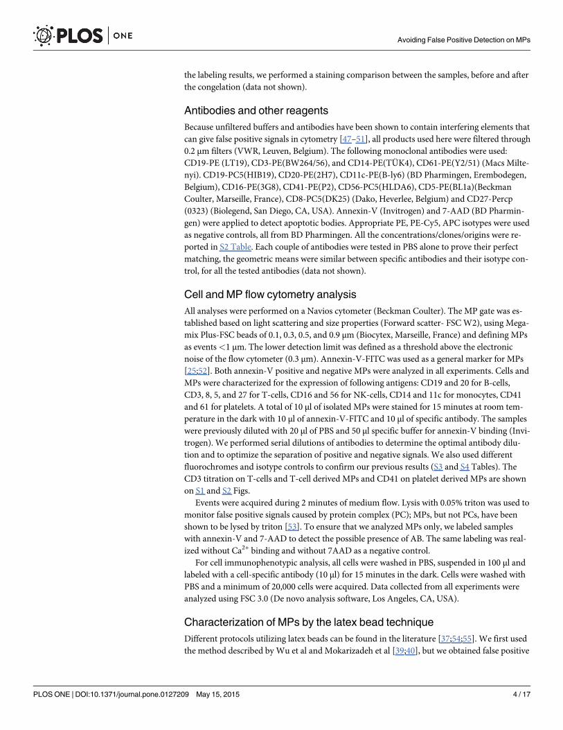
Detection of MPs by flow cytometry and electron microscopyTo characterize MPs, a Navios cytometer (Beckman Coulter, Marseille, France), an FSC opti-mized instrument, was used. As shown in Fig 1, the cytometer was calibrated using a standard-ized protocol from Biocytex. Briefly, 0.1 μm, 0.3 μm, 0.5 μm, and 0.9 μmMegamix beads-FCSplus were detected in channel FL1 (Fig 1A). With the forward scattering resolution of this cy-tometer, we were able to accurately distinguish the different bead sizes (Fig 1B), which allowedto define a standardized size-related gate for MP analysis (Fig 1C). Fig 1D shows a
Avoiding False Positive Detection on MPs
PLOSONE | DOI:10.1371/journal.pone.0127209 May 15, 2015 5 / 17
representative scatter plot of an MP population from PBMC. We also observed that the popula-tion of smaller MPs was more significant than the population of larger MPs. Double labelingwith annexin-V-FITC and CD41-PE allowed targeting of platelet derived-MPs (Fig 1E). Al-though apoptotic bodies (AB) were excluded by size criteria, we monitored for their possiblepresence by double staining with annexin V and 7AAD. As shown in Fig 1F, AB were not de-tected in our samples. The presence of protein complex (PC) can also interfere with MP analy-sis; therefore, we used lysis with 0.05% triton to evaluate the proportion of PC, which we foundwas<2% in all analyzed samples (Fig 1G). Finally, MPs were analyzed by electron microscopy.
Fig 1. MP analysis by flow cytometry and electronmicroscopy. Calibration of Navios cytometer using 0.1, 0.3, 0.5, and 0.9 μm beads (Biocytex). Thecytometer is able to differentiate the 4 different populations of beads (A-B). We can delimit the gate in accordance with size to study MPs (0.3–0.9 μm) (C).Characteristic elongated shape of MPs repartition limited in a gate of 0.3–0.9 μm. (D). Labeling of platelet derived-MPs with CD41 and annexin-V revealed adouble positive population for these markers (E). Example of 7-AAD/annexin V labeling to detect the presence of apoptotic bodies (AB). (F). Absence ofcontamination with AB. To monitor the presence of protein complexes (PC), lysis with 0.05% triton was used. After lysis, the positive signal disappeared (G).Scanning Electron Microscopy (SEM) shows the production of MPs by B-lymphocytes (white arrows) (H). Transmission electron microscopy (TEM) showsthe structure of one MP with a characteristic lipid bilayer (white arrows) (I).
doi:10.1371/journal.pone.0127209.g001
Avoiding False Positive Detection on MPs
PLOSONE | DOI:10.1371/journal.pone.0127209 May 15, 2015 6 / 17
Fig 1H shows the production of MPs by B-lymphocytes by scanning electron microscopy. Fur-thermore, the presence of MPs in our culture supernatant was demonstrated using TEM,which revealed the characteristic spherical vesicle with a lipid bilayer (Fig 1I).
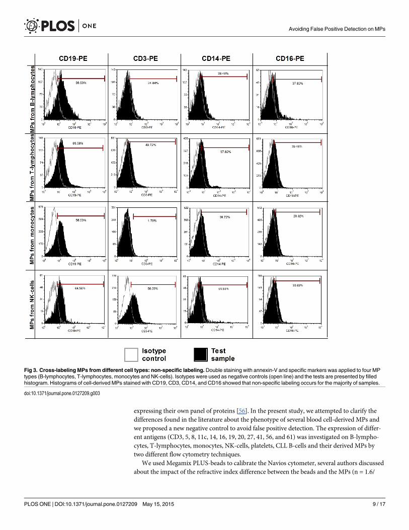
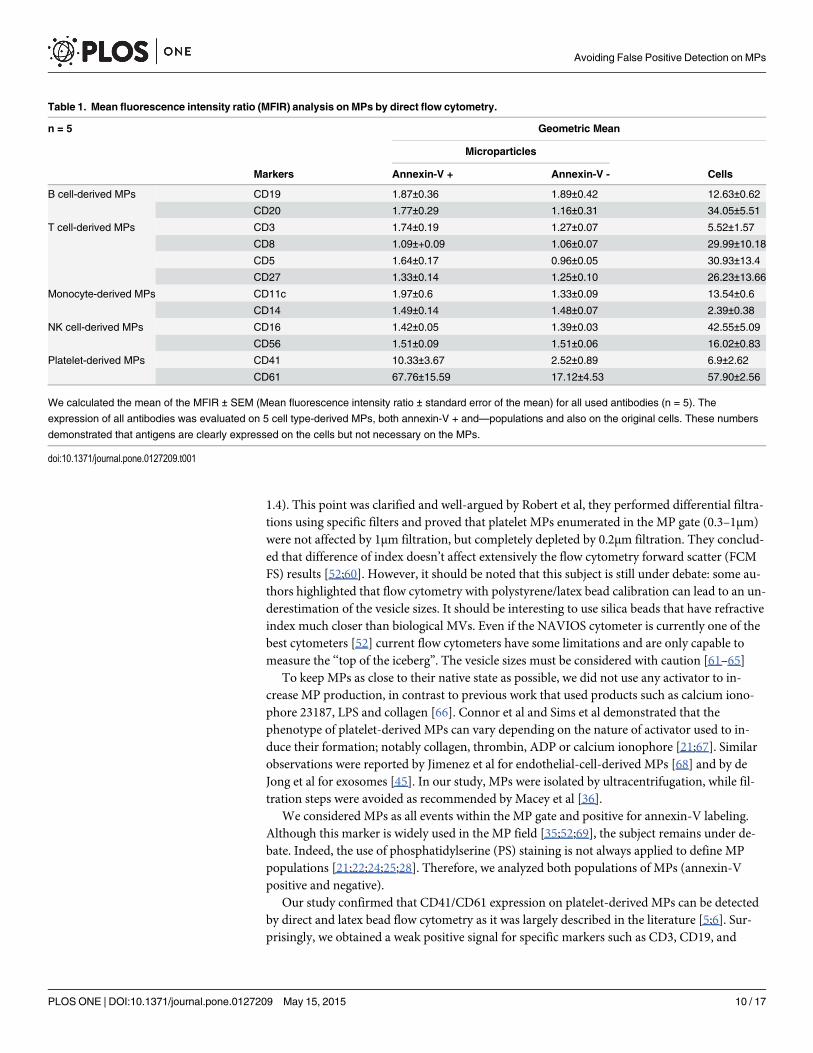
Immunophenotypic characterization of blood cells and blood cell-derivedMPs by direct flow cytometryWe first investigated the expression of several antigens on the cell surface and cell-derived MPsby direct flow cytometry (n = 5). Signals from platelet markers (CD41 and CD61) were clearlypositive on both platelet-derived MPs and platelets (Fig 2A–2C). These initial results indicatedthat we were able to detect annexin V+/marker+ MPs. These first 2 antigens were largely de-scribed in the MP literature and were used as positive controls to initialize the setup of our ex-periments. However, for all other cell-derived MPs (from B-cells, T-cells, NK cells, monocytes,and CLL B-cells), the markers used were not detected, despite the fact that these antigens werehighly expressed by the source cells. A representative example of some of these markers is pro-vided in Fig 2. For CD56, CD3, and CD19 staining, a weak positive signal was observed com-pared to the isotopic control (Fig 2J, 2M and 2Q). However, this signal was a false positive,shown by using the same antibodies to label MPs from different origins (Fig 3). It should benoted that CD14 labeling on monocytes was quite weak, most likely due to the positive selec-tion with anti-CD14 beads (Fig 2I). Heterogeneous expression of phosphatidylserine depen-dent on the cellular source of MPs was also observed. Double staining with annexin-V-FITCand specific markers was applied on four MP types (B-lymphocytes, T-lymphocytes, mono-cytes, NK-cells) with cross-labeling different MPs to detect false positive expression (Fig 3).Histograms of these cell derived-MPs stained with CD19-PE, CD3-PE, CD14-PE, andCD16-PE showed that non-specific labeling occurred in a majority of samples. Means of theMean Fluorescence Intensity Ratio (MFIR) for all antibodies (studied on both annexin-V posi-tive and negative MPs, as well as cell populations) are provided in Table 1. This study demon-strated that all antigens were clearly detected on cells but not on their corresponding MPs, withthe exceptions of CD41 and CD61 for platelet-derived MPs. We confirmed the detection ofCD41 on platelet derived MPs and the ‘‘non detection ‘‘ of CD5 on CLL B-cell derived MPs byimmunogold labeling (S3 Fig).
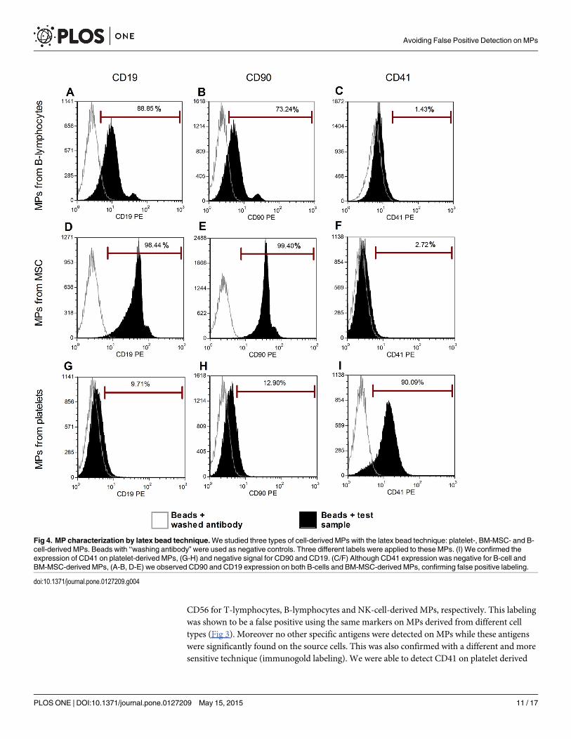
Immunophenotypic characterization by latex bead flow cytometryTo confirm the results obtained by direct flow cytometry, we studied MPs derived from threecell types (platelets, BM-MSC, and B-cells) with an optimized latex bead technique. Three dif-ferent labels were applied to these MPs (Fig 4). Using this method, as previously described, weconfirmed the expression of CD41, a platelet marker, on platelet-derived MPs (Fig 4I). This an-tigen was not found on the 2 other populations of MPs (Fig 4C and 4F), validating our protocol.Regarding the direct flow cytometry, positive signals were observed for some markers(CD19-PE on B cell-derived MPs, CD90 on MSC-derived MPs). However, when the differentMPs were cross-labeled for these markers (CD19 on MSC-derived MPs and CD90 on B-cell-de-rived MPs), a positive signal was also observed, indicating that the signals were false positives.
DiscussionMicrovesicles play a crucial role in cellular interactions and have been considered a novelmechanism of cross-talk between normal and malignant cells [4]. Even if the phenotypic char-acterization of extracellular vesicles is well defined in the literature, there are some discrepan-cies concerning antigen detection. Besides, Oksvold et al. described that there is an importantdifference in subpopulations of exosomes derived from the same B-cell lymphoma cell line
Avoiding False Positive Detection on MPs
PLOSONE | DOI:10.1371/journal.pone.0127209 May 15, 2015 7 / 17
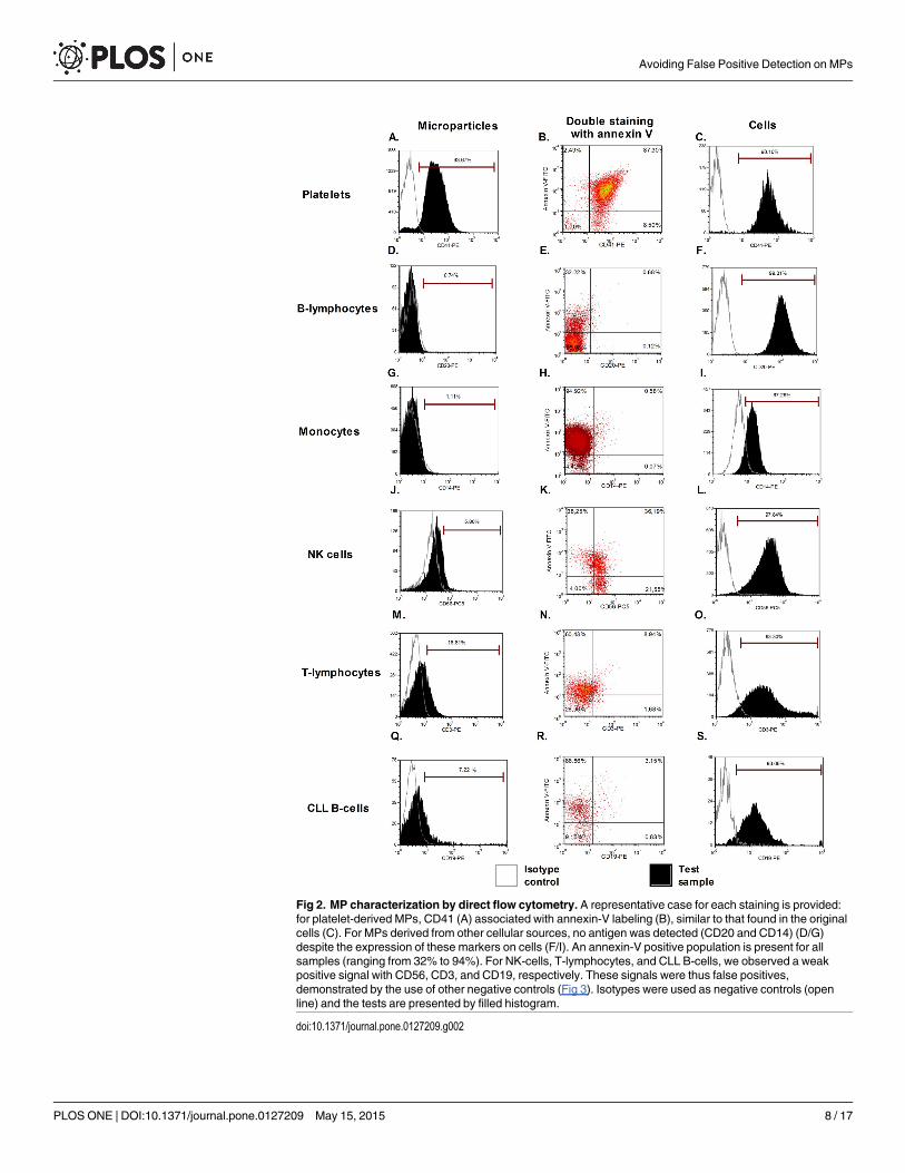
Fig 2. MP characterization by direct flow cytometry. A representative case for each staining is provided:for platelet-derived MPs, CD41 (A) associated with annexin-V labeling (B), similar to that found in the originalcells (C). For MPs derived from other cellular sources, no antigen was detected (CD20 and CD14) (D/G)despite the expression of these markers on cells (F/I). An annexin-V positive population is present for allsamples (ranging from 32% to 94%). For NK-cells, T-lymphocytes, and CLL B-cells, we observed a weakpositive signal with CD56, CD3, and CD19, respectively. These signals were thus false positives,demonstrated by the use of other negative controls (Fig 3). Isotypes were used as negative controls (openline) and the tests are presented by filled histogram.
doi:10.1371/journal.pone.0127209.g002
Avoiding False Positive Detection on MPs
PLOSONE | DOI:10.1371/journal.pone.0127209 May 15, 2015 8 / 17
expressing their own panel of proteins [56]. In the present study, we attempted to clarify thedifferences found in the literature about the phenotype of several blood cell-derived MPs andwe proposed a new negative control to avoid false positive detection. The expression of differ-ent antigens (CD3, 5, 8, 11c, 14, 16, 19, 20, 27, 41, 56, and 61) was investigated on B-lympho-cytes, T-lymphocytes, monocytes, NK-cells, platelets, CLL B-cells and their derived MPs bytwo different flow cytometry techniques.
We used Megamix PLUS-beads to calibrate the Navios cytometer, several authors discussedabout the impact of the refractive index difference between the beads and the MPs (n = 1.6/
Fig 3. Cross-labeling MPs from different cell types: non-specific labeling. Double staining with annexin-V and specific markers was applied to four MPtypes (B-lymphocytes, T-lymphocytes, monocytes and NK-cells). Isotypes were used as negative controls (open line) and the tests are presented by filledhistogram. Histograms of cell-derived MPs stained with CD19, CD3, CD14, and CD16 showed that non-specific labeling occurs for the majority of samples.
doi:10.1371/journal.pone.0127209.g003
Avoiding False Positive Detection on MPs
PLOSONE | DOI:10.1371/journal.pone.0127209 May 15, 2015 9 / 17
1.4). This point was clarified and well-argued by Robert et al, they performed differential filtra-tions using specific filters and proved that platelet MPs enumerated in the MP gate (0.3–1μm)were not affected by 1μm filtration, but completely depleted by 0.2μm filtration. They conclud-ed that difference of index doesn’t affect extensively the flow cytometry forward scatter (FCMFS) results [52;60]. However, it should be noted that this subject is still under debate: some au-thors highlighted that flow cytometry with polystyrene/latex bead calibration can lead to an un-derestimation of the vesicle sizes. It should be interesting to use silica beads that have refractiveindex much closer than biological MVs. Even if the NAVIOS cytometer is currently one of thebest cytometers [52] current flow cytometers have some limitations and are only capable tomeasure the ‘‘top of the iceberg”. The vesicle sizes must be considered with caution [61–65]
To keep MPs as close to their native state as possible, we did not use any activator to in-crease MP production, in contrast to previous work that used products such as calcium iono-phore 23187, LPS and collagen [66]. Connor et al and Sims et al demonstrated that thephenotype of platelet-derived MPs can vary depending on the nature of activator used to in-duce their formation; notably collagen, thrombin, ADP or calcium ionophore [21;67]. Similarobservations were reported by Jimenez et al for endothelial-cell-derived MPs [68] and by deJong et al for exosomes [45]. In our study, MPs were isolated by ultracentrifugation, while fil-tration steps were avoided as recommended by Macey et al [36].
We considered MPs as all events within the MP gate and positive for annexin-V labeling.Although this marker is widely used in the MP field [35;52;69], the subject remains under de-bate. Indeed, the use of phosphatidylserine (PS) staining is not always applied to define MPpopulations [21;22;24;25;28]. Therefore, we analyzed both populations of MPs (annexin-Vpositive and negative).
Our study confirmed that CD41/CD61 expression on platelet-derived MPs can be detectedby direct and latex bead flow cytometry as it was largely described in the literature [5;6]. Sur-prisingly, we obtained a weak positive signal for specific markers such as CD3, CD19, and
Table 1. Mean fluorescence intensity ratio (MFIR) analysis on MPs by direct flow cytometry.
n = 5 Geometric Mean
Microparticles
Markers Annexin-V + Annexin-V - Cells
B cell-derived MPs CD19 1.87±0.36 1.89±0.42 12.63±0.62
CD20 1.77±0.29 1.16±0.31 34.05±5.51
T cell-derived MPs CD3 1.74±0.19 1.27±0.07 5.52±1.57
CD8 1.09±+0.09 1.06±0.07 29.99±10.18
CD5 1.64±0.17 0.96±0.05 30.93±13.4
CD27 1.33±0.14 1.25±0.10 26.23±13.66
Monocyte-derived MPs CD11c 1.97±0.6 1.33±0.09 13.54±0.6
CD14 1.49±0.14 1.48±0.07 2.39±0.38
NK cell-derived MPs CD16 1.42±0.05 1.39±0.03 42.55±5.09
CD56 1.51±0.09 1.51±0.06 16.02±0.83
Platelet-derived MPs CD41 10.33±3.67 2.52±0.89 6.9±2.62
CD61 67.76±15.59 17.12±4.53 57.90±2.56
We calculated the mean of the MFIR ± SEM (Mean fluorescence intensity ratio ± standard error of the mean) for all used antibodies (n = 5). The
expression of all antibodies was evaluated on 5 cell type-derived MPs, both annexin-V + and—populations and also on the original cells. These numbers
demonstrated that antigens are clearly expressed on the cells but not necessary on the MPs.
doi:10.1371/journal.pone.0127209.t001
Avoiding False Positive Detection on MPs
PLOSONE | DOI:10.1371/journal.pone.0127209 May 15, 2015 10 / 17
CD56 for T-lymphocytes, B-lymphocytes and NK-cell-derived MPs, respectively. This labelingwas shown to be a false positive using the same markers on MPs derived from different celltypes (Fig 3). Moreover no other specific antigens were detected on MPs while these antigenswere significantly found on the source cells. This was also confirmed with a different and moresensitive technique (immunogold labeling). We were able to detect CD41 on platelet derived
Fig 4. MP characterization by latex bead technique.We studied three types of cell-derived MPs with the latex bead technique: platelet-, BM-MSC- and B-cell-derived MPs. Beads with ‘‘washing antibody” were used as negative controls. Three different labels were applied to these MPs. (I) We confirmed theexpression of CD41 on platelet-derived MPs, (G-H) and negative signal for CD90 and CD19. (C/F) Although CD41 expression was negative for B-cell andBM-MSC-derived MPs, (A-B, D-E) we observed CD90 and CD19 expression on both B-cells and BM-MSC-derived MPs, confirming false positive labeling.
doi:10.1371/journal.pone.0127209.g004
Avoiding False Positive Detection on MPs
PLOSONE | DOI:10.1371/journal.pone.0127209 May 15, 2015 11 / 17

MPs but no CD19/CD5 on CLL B-cells derived MPs, confirming our cytometry results (S3Fig). Interestingly, our study showed that the expression profiles of CD41/CD61 are clearly dif-ferent between annexin-V positive and negative populations. This observation was also madefor other antigens by Nielsen et al and Connor et al in 2010 [21;70]. Further investigation isneeded to better understand this difference.
A recent report suggested that there is a major decrease in antigen expression between cellsand EVs [71]. Moreover, it has already been shown that MPs are derived from lipid rafts andwe can thus hypothesize that these parts of the membrane can be characterized by differentialpartitioning of antigens [72–75]. Miguet et al observed by a proteomic study that while someantigens are detected on MPs, CD3, 5, and 8 for T-cells and CD19 for B-cells are not found onMPs despite being highly expressed on the original cells [76]. Our results conflict with those ofauthors who report detection by direct flow cytometry of CD105, CD90, and CXCR4 on MPsfrom mesenchymal stem cells [77] and CD3, CD14 and CD19 on MPs from the plasma ofpolymyositis/dermatomyositis patients [78]. In addition, Ghosh et al and Macey et al detectedannexin-V-CD19+ [35] and CD15+ MPs [36], respectively. These discrepancies with our studycould be explained by several factors: speed of centrifugation, choice of specific negative con-trols and antibodies, MP origin and the presence of specific activators in culture. Indeed, theseauthors analyzed MPs from cell culture or directly from plasma and they either applied centri-fugation forces of 16,000 x g to 19,800 x g for 10 min to 1 h or they did not use centrifugationat all. More importantly, all of these groups used different negative controls. Baka et al used un-stained samples and Kim et al, Ghosh et al, Macey et al used isotype controls. Trummer andcolleagues demonstrated that using isotype negative controls can induce errors: they concludedthat using flow cytometry to discriminate between positive and negative populations of MPs la-beled with several antibodies could be an obstacle for their characterization [79]. All of thesedifferences could explain the variation in results between several groups. The study of MPs iscomplicated by their limited surface area and relatively small number of proteins available forantibody binding. Thus, the latex bead technique has been used by several groups to facilitatethe characterization of MPs by flow cytometry. Different protocols have been published[37;54;55], but we adapted the protocols of Wu et al and Mokarizadeh et al with minor modifi-cations [39;40]. Since no significant difference was observed by using blocking reagent, we re-moved this step, as previously described by Oksvold et al [56]. Three different negativecontrols were used: beads alone without any staining, beads with MPs and isotype control andbeads with MPs and a negative marker (one not present on the original cells). Isotype controlswere removed from our study because they resulted in some false positive results (data notshown). Szczepanski et al, Wu et al and Mokarizadeh et al presented positive results from thefirst protocol, notably for CD9, CD33, CD63, CD73, and CD90 [39;40;54]. However, in ourstudy, we demonstrated that this protocol is associated with non-specific labeling due to anti-body affinity for latex beads. Indeed incubating beads with antibody in the absence of MPs re-sulted in an increase of background noise (data not shown). Thereafter, we designed our ownprotocol, by directly incubating the beads with previously labeled MPs. In this way, we avoidedan increase in background signals due to non-specific binding of antibodies to beads. Usingthis protocol we were able to detect specific markers on platelet-derived MPs. Together, our re-sults indicated that several previously published data sets using the latex bead technique in-clude false positive results due to the use of inappropriate protocols or negative controls.
The goal of this study was to emphasize on the inconsistencies in literature concerning thecharacterization of MPs by flow cytometry and the importance of the negative control choice.We confirmed the data of Trummer and colleagues concerning the failure of classical negativecontrol (isotype) in the MP study. Since, some antigens could be expressed by different cell
Avoiding False Positive Detection on MPs
PLOSONE | DOI:10.1371/journal.pone.0127209 May 15, 2015 12 / 17
types (i.e. CD5 could be found on T or B cells), several negative controls should be tested for aspecific MV staining.
In conclusion, the characterization of MPs is still a challenging field requiring lot of precau-tions in the interpretation of the fluorescence signal. We demonstrated that isotype controlsand unstained samples are not suitable for MP characterization. Therefore, we proposed usingother negative markers (marker not found on the original cell) by cross-staining MPs to dem-onstrate the true positive labeling. We want to underline the fact that this method is applicableon cell culture supernatants specifically but not on body fluids (like urine or plasma), whichare more complex samples. By this manner, non-specific antibody labeling could be subtractedout, and reliable results could be obtained.
Supporting InformationS1 Fig. CD3 titration on T cells and T cell derived MPs.(DOCX)
S2 Fig. CD41 titration on platelet derived MPs.(DOCX)
S3 Fig. Immunogold labeling on CLL B-cell derived and platelet derived MPs with CD5and CD41 antibodies.(DOCX)
S1 Table. Viability of cells by Trypan blue assay.(DOCX)
S2 Table. All the Concentrations/Clones/Origins of antibodies used in experiments.(DOCX)
S3 Table. Mean Fluorescence Intensity (MFI) and MFI Ratio (MFIR) of the CD3 labelingon T cells. These numbers demonstrated that CD3 antigen is clearly expressed on T-cells.(DOCX)
S4 Table. Mean Fluorescence Intensity (MFI) and MFI Ratio (MFIR) of the CD3 labelingon T cell derived MPs. These numbers demonstrated that CD3 antigen is not necessary ex-pressed on T cell derived MPs.(DOCX)
Author ContributionsConceived and designed the experiments: EC LL BS. Performed the experiments: EC HD KPMVD. Analyzed the data: EC MVD BS. Contributed reagents/materials/analysis tools: EC HDKPMV DP NM PMDB LL BS. Wrote the paper: EC MVD LL BS.
References1. Gyorgy B, Szabo TG, Pasztoi M, Pal Z, Misjak P, Aradi B et al. Membrane vesicles, current state-of-
the-art: emerging role of extracellular vesicles. Cell Mol Life Sci 2011; 68(16):2667–2688. doi: 10.1007/s00018-011-0689-3 PMID: 21560073
2. Raposo G, Stoorvogel W. Extracellular vesicles: exosomes, microvesicles, and friends. J Cell Biol2013; 200(4):373–383. doi: 10.1083/jcb.201211138 PMID: 23420871
3. Crescitelli R, Lasser C, Szabo TG, Kittel A, Eldh M, Dianzani I et al. Distinct RNA profiles in subpopula-tions of extracellular vesicles: apoptotic bodies, microvesicles and exosomes. J Extracell Vesicles2013; 2.
Avoiding False Positive Detection on MPs
PLOSONE | DOI:10.1371/journal.pone.0127209 May 15, 2015 13 / 17
4. Cocucci E, Racchetti G, Meldolesi J. Shedding microvesicles: artefacts no more. Trends Cell Biol 2009;19(2):43–51. doi: 10.1016/j.tcb.2008.11.003 PMID: 19144520
5. Piccin A, MurphyWG, Smith OP. Circulating microparticles: pathophysiology and clinical implications.Blood Rev 2007; 21(3):157–171. PMID: 17118501
6. Burnier L, Fontana P, Kwak BR, Angelillo-Scherrer A. Cell-derived microparticles in haemostasis andvascular medicine. Thromb Haemost 2009; 101(3):439–451. PMID: 19277403
7. Choi DS, Kim DK, Kim YK, Gho YS. Proteomics, transcriptomics and lipidomics of exosomes and ecto-somes. Proteomics 2013; 13(10–11):1554–1571. doi: 10.1002/pmic.201300131 PMID: 24150872
8. Laffont B, Corduan A, Ple H, Duchez AC, Cloutier N, Boilard E et al. Activated platelets can delivermRNA regulatory Ago2*microRNA complexes to endothelial cells via microparticles. Blood 2013; 122(2):253–261. doi: 10.1182/blood-2013-03-492801 PMID: 23652806
9. Patz S, Trattnig C, Grunbacher G, Ebner B, Gully C, Novak A et al. More than cell dust: microparticlesisolated from cerebrospinal fluid of brain injured patients are messengers carrying mRNAs, miRNAs,and proteins. J Neurotrauma 2013; 30(14):1232–1242. doi: 10.1089/neu.2012.2596 PMID: 23360174
10. Hunter MP, Ismail N, Zhang X, Aguda BD, Lee EJ, Yu L et al. Detection of microRNA expression inhuman peripheral blood microvesicles. PLoS ONE 2008; 3(11):e3694. doi: 10.1371/journal.pone.0003694 PMID: 19002258
11. Fonsato V, Collino F, Herrera MB, Cavallari C, Deregibus MC, Cisterna B et al. Human liver stem cell-derived microvesicles inhibit hepatoma growth in SCID mice by delivering antitumor microRNAs. StemCells 2012; 30(9):1985–1998. doi: 10.1002/stem.1161 PMID: 22736596
12. Moldovan L, Batte K, Wang Y, Wisler J, Piper M. Analyzing the circulating microRNAs in exosomes/ex-tracellular vesicles from serum or plasma by qRT-PCR. Methods Mol Biol 2013; 1024:129–145. doi:10.1007/978-1-62703-453-1_10 PMID: 23719947
13. Lee Y, El Andaloussi S, Wood MJA. Exosomes and microvesicles: extracellular vesicles for genetic in-formation transfer and gene therapy. HumMol Genet 2012; 21(R1):R125–R134. PMID: 22872698
14. Pisetsky DS, Gauley J, Ullal AJ. Microparticles as a source of extracellular DNA. Immunol Res 2011;49(1–3):227–234. doi: 10.1007/s12026-010-8177-7 PMID: 21170741
15. Balaj L, Lessard R, Dai L, Cho YJ, Pomeroy SL, Breakefield XO et al. Tumour microvesicles contain ret-rotransposon elements and amplified oncogene sequences. Nat Commun 2011; 2:180. doi: 10.1038/ncomms1180 PMID: 21285958
16. Waldenstrom A, Genneback N, Hellman U, Ronquist G. Cardiomyocyte microvesicles contain DNA/RNA and convey biological messages to target cells. PLoS ONE 2012; 7(4):e34653. doi: 10.1371/journal.pone.0034653 PMID: 22506041
17. D'Souza-Schorey C, Clancy JW. Tumor-derived microvesicles: shedding light on novel microenviron-ment modulators and prospective cancer biomarkers. Genes Dev 2012; 26(12):1287–1299. doi: 10.1101/gad.192351.112 PMID: 22713869
18. Mause SF, Weber C. Microparticles: protagonists of a novel communication network for intercellular in-formation exchange. Circ Res 2010; 107(9):1047–1057. doi: 10.1161/CIRCRESAHA.110.226456PMID: 21030722
19. Heijnen HF, Schiel AE, Fijnheer R, Geuze HJ, Sixma JJ. Activated platelets release two types of mem-brane vesicles: microvesicles by surface shedding and exosomes derived from exocytosis of multivesi-cular bodies and alpha-granules. Blood 1999; 94(11):3791–3799. PMID: 10572093
20. van Doormaal FF, Kleinjan A, Di Nisio M, Buller HR, Nieuwland R. Cell-derived microvesicles and can-cer. Neth J Med 2009; 67(7):266–273. PMID: 19687520
21. Connor DE, Exner T, Ma DDF, Joseph JE. The majority of circulating platelet-derived microparticles failto bind annexin V, lack phospholipid-dependent procoagulant activity and demonstrate greater expres-sion of glycoprotein Ib. Thromb Haemost 2010; 103(5):1044–1052. doi: 10.1160/TH09-09-0644 PMID:20390225
22. Ayers L, Kohler M, Harrison P, Sargent I, Dragovic R, Schaap M et al. Measurement of circulating cell-derived microparticles by flow cytometry: sources of variability within the assay. Thromb Res 2011;127(4):370–377. doi: 10.1016/j.thromres.2010.12.014 PMID: 21257195
23. Boulanger CM, Amabile N, Tedgui A. Circulating microparticles: a potential prognostic marker for ath-erosclerotic vascular disease. Hypertension 2006; 48(2):180–186. PMID: 16801490
24. Amabile N, Guerin AP, Leroyer A, Mallat Z, Nguyen C, Boddaert J et al. Circulating endothelial micro-particles are associated with vascular dysfunction in patients with end-stage renal failure. J Am SocNephrol 2005; 16(11):3381–3388. PMID: 16192427
25. Budoni M, Fierabracci A, Luciano R, Petrini S, Di Ciommo V, Muraca M. The immunosuppressive effectof mesenchymal stromal cells on B lymphocytes is mediated by membrane vesicles. Cell Transplant2013; 22(2):369–379. doi: 10.3727/096368911X582769 PMID: 23433427
Avoiding False Positive Detection on MPs
PLOSONE | DOI:10.1371/journal.pone.0127209 May 15, 2015 14 / 17
26. Lacroix R, Plawinski L, Robert S, Doeuvre L, Sabatier F, Martinez de Lizarrondo S et al. Leukocyte-and endothelial-derived microparticles: a circulating source for fibrinolysis. Haematologica 2012; 97(12):1864–1872. doi: 10.3324/haematol.2012.066167 PMID: 22733025
27. Tissot JD, Rubin O, Canellini G. Analysis and clinical relevance of microparticles from red blood cells.Curr Opin Hematol 2010; 17(6):571–577. PMID: 20960973
28. Boulanger CM, Amabile N, Tedgui A. Circulating microparticles: a potential prognostic marker for ath-erosclerotic vascular disease. Hypertension 2006; 48(2):180–186. PMID: 16801490
29. Soriano AO, Jy W, Chirinos JA, Valdivia MA, Velasquez HS, Jimenez JJ et al. Levels of endothelial andplatelet microparticles and their interactions with leukocytes negatively correlate with organ dysfunctionand predict mortality in severe sepsis. Crit Care Med 2005; 33(11):2540–2546. PMID: 16276178
30. Galindo-Hernandez O, Villegas-Comonfort S, Candanedo F, Gonzalez-Vazquez MC, Chavez-OcanaS, Jimenez-Villanueva X et al. Elevated concentration of microvesicles isolated from peripheral blood inbreast cancer patients. Arch Med Res 2013; 44(3):208–214. doi: 10.1016/j.arcmed.2013.03.002PMID: 23506723
31. Nielsen CT. Circulating microparticles in systemic Lupus Erythematosus. Dan Med J 2012; 59(11):B4548. PMID: 23171755
32. Munoz JL, Bliss SA, Greco SJ, Ramkissoon SH, Ligon KL, Rameshwar P. Delivery of Functional Anti-miR-9 by Mesenchymal Stem Cell-derived Exosomes to GlioblastomaMultiforme Cells Conferred Che-mosensitivity. Mol Ther Nucleic Acids 2013; 2:e126. doi: 10.1038/mtna.2013.60 PMID: 24084846
33. Nakamizo A, Marini F, Amano T, Khan A, Studeny M, Gumin J et al. Human bone marrow-derived mes-enchymal stem cells in the treatment of gliomas. Cancer Res 2005; 65(8):3307–3318. PMID:15833864
34. Akao Y, Iio A, Itoh T, Noguchi S, Itoh Y, Ohtsuki Y et al. Microvesicle-mediated RNAmolecule deliverysystem using monocytes/macrophages. Mol Ther 2011; 19(2):395–399. doi: 10.1038/mt.2010.254PMID: 21102562
35. Ghosh AK, Secreto CR, Knox TR, Ding W, Mukhopadhyay D, Kay NE. Circulating microvesicles in B-cell chronic lymphocytic leukemia can stimulate marrow stromal cells: implications for disease progres-sion. Blood 2010; 115(9):1755–1764. doi: 10.1182/blood-2009-09-242719 PMID: 20018914
36. Macey MG, Enniks N, Bevan S. Flow cytometric analysis of microparticle phenotype and their role inthrombin generation. Cytometry B Clin Cytom 2011; 80(1):57–63. doi: 10.1002/cyto.b.20551 PMID:20632415
37. Blanchard N, Lankar D, Faure F, Regnault A, Dumont C, Raposo G et al. TCR activation of human Tcells induces the production of exosomes bearing the TCR/CD3/zeta complex. J Immunol 2002; 168(7):3235–3241. PMID: 11907077
38. Miguet L, Bechade G, Fornecker L, Zink E, Felden C, Gervais C et al. Proteomic analysis of malignantB-cell derived microparticles reveals CD148 as a potentially useful antigenic biomarker for mantle celllymphoma diagnosis. J Proteome Res 2009; 8(7):3346–3354. doi: 10.1021/pr801102c PMID:19413345
39. Mokarizadeh A, Delirezh N, Morshedi A, Mosayebi G, Farshid AA, Mardani K. Microvesicles derivedfrommesenchymal stem cells: potent organelles for induction of tolerogenic signaling. Immunol Lett2012; 147(1–2):47–54. doi: 10.1016/j.imlet.2012.07.003 PMID: 22841963
40. Wu S, Ju GQ, Du T, Zhu YJ, Liu GH. Microvesicles derived from human umbilical cord Wharton's jellymesenchymal stem cells attenuate bladder tumor cell growth in vitro and in vivo. PLoS ONE 2013; 8(4):e61366. doi: 10.1371/journal.pone.0061366 PMID: 23593475
41. Van DammeM, Crompot E, Meuleman N, Mineur P, Bron D, Lagneaux L et al. HDAC isoenzyme ex-pression is deregulated in chronic lymphocytic leukemia B-cells and has a complex prognostic signifi-cance. Epigenetics 2012; 7(12):1403–1412. doi: 10.4161/epi.22674 PMID: 23108383
42. Witwer KW, Buzas EI, Bemis LT, Bora A, Lasser C, Lotvall J et al. Standardization of sample collection,isolation and analysis methods in extracellular vesicle research. J Extracell Vesicles 2013; 2.
43. Najar M, Rouas R, Raicevic G, Boufker HI, Lewalle P, Meuleman N et al. Mesenchymal stromal cellspromote or suppress the proliferation of T lymphocytes from cord blood and peripheral blood: the impor-tance of low cell ratio and role of interleukin-6. Cytotherapy 2009; 11(5):570–583. doi: 10.1080/14653240903079377 PMID: 19565371
44. Mahaweni NM, Kaijen-Lambers MEH, Dekkers J, Aerts JGJV, Hegmans JPJJ. Tumour-derived exo-somes as antigen delivery carriers in dendritic cell-based immunotherapy for malignant mesothelioma.J Extracell Vesicles 2013; 2.
45. de Jong OG, Verhaar MC, Chen Y, Vader P, Gremmels H, Posthuma G et al. Cellular stress conditionsare reflected in the protein and RNA content of endothelial cell-derived exosomes. J Extracell Vesicles2012; 1. doi: 10.3402/jev.v1i0.19179 PMID: 24009887
Avoiding False Positive Detection on MPs
PLOSONE | DOI:10.1371/journal.pone.0127209 May 15, 2015 15 / 17
46. Sabatier F, Roux V, Anfosso F, Camoin L, Sampol J, Dignat-George F. Interaction of endothelial micro-particles with monocytic cells in vitro induces tissue factor-dependent procoagulant activity. Blood2002; 99(11):3962–3970. PMID: 12010795
47. Jayachandran M, Miller VM, Heit JA, OwenWG. Methodology for isolation, identification and character-ization of microvesicles in peripheral blood. J Immunol Methods 2012; 375(1–2):207–214. doi: 10.1016/j.jim.2011.10.010 PMID: 22301270
48. Dey-Hazra E, Hertel B, Kirsch T, Woywodt A, Lovric S, Haller H et al. Detection of circulating microparti-cles by flow cytometry: influence of centrifugation, filtration of buffer, and freezing. Vasc Health RiskManag 2010; 6:1125–1133. doi: 10.2147/VHRM.S13236 PMID: 21191433
49. Gyorgy B, Modos K, Pallinger E, Paloczi K, Pasztoi M, Misjak P et al. Detection and isolation of cell-de-rived microparticles are compromised by protein complexes resulting from shared biophysical parame-ters. Blood 2011; 117(4):e39–e48. doi: 10.1182/blood-2010-09-307595 PMID: 21041717
50. Gyorgy B, Szabo TG, Turiak L, Wright M, Herczeg P, Ledeczi Z et al. Improved flow cytometric assess-ment reveals distinct microvesicle (cell-derived microparticle) signatures in joint diseases. PLoS ONE2012; 7(11):e49726. doi: 10.1371/journal.pone.0049726 PMID: 23185418
51. Aass HC, Ovstebo R, Troseid AM, Kierulf P, Berg JP, Henriksson CE. Fluorescent particles in the anti-body solution result in false TF- and CD14-positive microparticles in flow cytometric analysis. Cytome-try A 2011; 79(12):990–999. doi: 10.1002/cyto.a.21147 PMID: 21990118
52. Robert S, Lacroix R, Poncelet P, Harhouri K, Bouriche T, Judicone C et al. High-sensitivity flow cytome-try provides access to standardized measurement of small-size microparticles—brief report. Arterios-cler Thromb Vasc Biol 2012; 32(4):1054–1058. doi: 10.1161/ATVBAHA.111.244616 PMID: 22328775
53. Gyorgy B, Szabo TG, Turiak L, Wright M, Herczeg P, Ledeczi Z et al. Improved flow cytometric assess-ment reveals distinct microvesicle (cell-derived microparticle) signatures in joint diseases. PLoS ONE2012; 7(11):e49726. doi: 10.1371/journal.pone.0049726 PMID: 23185418
54. Szczepanski MJ, Szajnik M, Welsh A, Whiteside TL, Boyiadzis M. Blast-derived microvesicles in serafrom patients with acute myeloid leukemia suppress natural killer cell function via membrane-associat-ed transforming growth factor-beta1. Haematologica 2011; 96(9):1302–1309. doi: 10.3324/haematol.2010.039743 PMID: 21606166
55. Caby MP, Lankar D, Vincendeau-Scherrer C, Raposo G, Bonnerot C. Exosomal-like vesicles are pres-ent in human blood plasma. Int Immunol 2005; 17(7):879–887. PMID: 15908444
56. Oksvold MP, Kullmann A, Forfang L, Kierulf B, Li M, Brech A et al. Expression of B-cell surface antigensin subpopulations of exosomes released from B-cell lymphoma cells. Clin Ther 2014; 36(6):847–862.doi: 10.1016/j.clinthera.2014.05.010 PMID: 24952935
57. Shefler I, Salamon P, Reshef T, Mor A, Mekori YA. T cell-induced mast cell activation: a role for micro-particles released from activated T cells. J Immunol 2010; 185(7):4206–4212. doi: 10.4049/jimmunol.1000409 PMID: 20810987
58. Thery C, Amigorena S, Raposo G, Clayton A. Isolation and characterization of exosomes from cell cul-ture supernatants and biological fluids. Curr Protoc Cell Biol 2006; Chapter 3:Unit. doi: 10.1002/0471143030.cb0324s31 PMID: 18228492
59. Shelke GV, Lasser C, Gho YS, Lotvall J. Importance of exosome depletion protocols to eliminate func-tional and RNA-containing extracellular vesicles from fetal bovine serum. J Extracell Vesicles 2014; 3.
60. Robert S, Poncelet P, Lacroix R, Raoult D, Dignat-George F. More on: calibration for the measurementof microparticles: value of calibrated polystyrene beads for flow cytometry-based sizing of biological mi-croparticles. J Thromb Haemost 2011; 9(8):1676–1678. doi: 10.1111/j.1538-7836.2011.04387.xPMID: 21645234
61. Chandler WL, YeungW, Tait JF. A newmicroparticle size calibration standard for use in measuringsmaller microparticles using a new flow cytometer. J Thromb Haemost 2011; 9(6):1216–1224. doi: 10.1111/j.1538-7836.2011.04283.x PMID: 21481178
62. Ayers L, Harrison P, Kohler M, Ferry B. Procoagulant and platelet-derived microvesicle absolute countsdetermined by flow cytometry correlates with a measurement of their functional capacity. J ExtracellVesicles 2014; 3.
63. Dragovic RA, Gardiner C, Brooks AS, Tannetta DS, Ferguson DJP, Hole P et al. Sizing and phenotyp-ing of cellular vesicles using Nanoparticle Tracking Analysis. Nanomedicine 2011; 7(6):780–788. doi:10.1016/j.nano.2011.04.003 PMID: 21601655
64. van der Pol E, Coumans F, Varga Z, Krumrey M, Nieuwland R. Innovation in detection of microparticlesand exosomes. J Thromb Haemost 2013; 11 Suppl 1:36–45. doi: 10.1111/jth.12254 PMID: 23809109
65. van der Pol E, Coumans FAW, Sturk A, Nieuwland R, van Leeuwen TG. Refractive index determinationof nanoparticles in suspension using nanoparticle tracking analysis. Nano Lett 2014; 14(11):6195–6201. doi: 10.1021/nl503371p PMID: 25256919
Avoiding False Positive Detection on MPs
PLOSONE | DOI:10.1371/journal.pone.0127209 May 15, 2015 16 / 17
66. Burger D, Schock S, Thompson CS, Montezano AC, Hakim AM, Touyz RM. Microparticles: biomarkersand beyond. Clin Sci (Lond) 2013; 124(7):423–441. doi: 10.1042/CS20120309 PMID: 23249271
67. Sims PJ, Wiedmer T, Esmon CT, Weiss HJ, Shattil SJ. Assembly of the platelet prothrombinase com-plex is linked to vesiculation of the platelet plasmamembrane. Studies in Scott syndrome: an isolateddefect in platelet procoagulant activity. J Biol Chem 1989; 264(29):17049–17057. PMID: 2793843
68. Jimenez JJ, Jy W, Mauro LM, Soderland C, Horstman LL, Ahn YS. Endothelial cells release phenotypi-cally and quantitatively distinct microparticles in activation and apoptosis. Thromb Res 2003; 109(4):175–180. PMID: 12757771
69. Liu R, Klich I, Ratajczak J, Ratajczak MZ, Zuba-Surma EK. Erythrocyte-derived microvesicles maytransfer phosphatidylserine to the surface of nucleated cells and falsely 'mark' them as apoptotic. Eur JHaematol 2009; 83(3):220–229. doi: 10.1111/j.1600-0609.2009.01271.x PMID: 19456851
70. Nielsen MH, Beck-Nielsen H, AndersenMN, Handberg A. A flow cytometric method for characterizationof circulating cell-derived microparticles in plasma. J Extracell Vesicles 2014; 3.
71. Gheldof D, Hardij J, Cecchet F, Chatelain B, Dogne JM, Mullier F. Thrombin generation assay andtransmission electron microscopy: a useful combination to study tissue factor-bearing microvesicles. JExtracell Vesicles 2013; 2.
72. Davizon P, Munday AD, Lopez JA. Tissue factor, lipid rafts, and microparticles. Semin Thromb Hemost2010; 36(8):857–864. doi: 10.1055/s-0030-1267039 PMID: 21049386
73. Del Conde I, Shrimpton CN, Thiagarajan P, Lopez JA. Tissue-factor-bearing microvesicles arise fromlipid rafts and fuse with activated platelets to initiate coagulation. Blood 2005; 106(5):1604–1611.PMID: 15741221
74. Biro E, Akkerman JWN, Hoek FJ, Gorter G, Pronk LM, Sturk A et al. The phospholipid composition andcholesterol content of platelet-derived microparticles: a comparison with platelet membrane fractions. JThromb Haemost 2005; 3(12):2754–2763. PMID: 16359513
75. Tan SS, Yin Y, Lee T, Lai RC, Yeo RWY, Zhang B et al. Therapeutic MSC exosomes are derived fromlipid raft microdomains in the plasmamembrane. J Extracell Vesicles 2013; 2.
76. Miguet L, Pacaud K, Felden C, Hugel B, Martinez MC, Freyssinet JM et al. Proteomic analysis of malig-nant lymphocyte membrane microparticles using double ionization coverage optimization. Proteomics2006; 6(1):153–171. PMID: 16342139
77. Kim SJ, Moon GJ, Cho YH, Kang HY, Hyung NK, Kim D et al. Circulating mesenchymal stem cells mi-croparticles in patients with cerebrovascular disease. PLoS ONE 2012; 7(5):e37036. doi: 10.1371/journal.pone.0037036 PMID: 22615882
78. Baka Z, Senolt L, Vencovsky J, Mann H, Simon PS, Kittel A et al. Increased serum concentration of im-mune cell derived microparticles in polymyositis/dermatomyositis. Immunol Lett 2010; 128(2):124–130. doi: 10.1016/j.imlet.2009.12.018 PMID: 20043950
79. Trummer A, De Rop C, Tiede A, Ganser A, Eisert R. Isotype controls in phenotyping and quantificationof microparticles: a major source of error and how to evade it. Thromb Res 2008; 122(5):691–700. doi:10.1016/j.thromres.2008.01.005 PMID: 18304614
Avoiding False Positive Detection on MPs
PLOSONE | DOI:10.1371/journal.pone.0127209 May 15, 2015 17 / 17
Copyright © 2022 FDOKUMEN