Available Online through Sweety Kumari*et al Int J Pharm Bio Sci NEPHROTOXICITY INDUCED BY PAN...
8
Available Online through www.ijpbs.com (or) www.ijpbsonline.com IJPBS |Volume 3| Issue 1 |JAN-MAR |2013|231-238 Research Article Pharmaceutical Sciences International Journal of Pharmacy and Biological Sciences (e-ISSN: 2230-7605) Sweety Kumari*et al Int J Pharm Bio Sci www.ijpbs.com or www.ijpbsonline.com Page231 NEPHROTOXICITY INDUCED BY PAN MASALA IN SWISS MICE AND ITS PROTECTION BY ELETTARIA CARDAMOMUM (L.) MATON Sweety Kumari 1 *, Abhijit Dutta 1 , Saabiya Farooqui 1 , Musarrat Naaz 1 , Swati Soren 1 , Sunita Dutta 2 , Abha Prasad 2 , Neeta Lal 3 , Anjali Smita 4 1 Department of Zoology, Ranchi University, Ranchi, India 2 Department of Zoology, Women’s College, Ranchi, India 3 Department of Zoology, RLSY College, Ranchi, India 4 Department of Zoology, Nirmala College, Ranchi, India *Corresponding Author Email: [email protected] ABSTRACT This study was designed to investigate the protective effect of cardamom on renal tissue damage caused by pan masala. Experimental animals were divided into 3 groups, control, pan masala treated and cardamom along with pan masala treated. They were exposed till 12 months to observe changes histologically and ultrastructurally. Male Swiss mice were given orally pan masala at a dose of 2% of the feed which caused acute tubular necrosis along with dilation and atrophied glomerulus seen in its light microscopic structures, whereas ultrastructural changes showed pyknotic nucleus, swollen mitochondria and loss of membrane integrity. When cardamom was given at a dose of 0.2% along with pan masala or alone, damages were less showing normal glomerulus with less inflammation, and normal nucleus. It is, therefore, concluded that cardamom may be beneficial in preventing pan masala induced renal tissue damage and shows potential for clinical use. KEY WORDS Cardamom, kidney, necrosis, pan masala INTRODUCTION The mammalian kidney is a target organ for a wide variety of toxic agents due to its prime function as a blood filter during the excretory process and is also sensitive to drug induced injury 1 . In this experiment, kidney is studied to assess the nephrotoxicity of pan masala and the protective properties of cardamom against it. Pan masala is a dry mixture of tobacco, areca nuts, slaked lime, catechu, and flavouring agents such as menthol, camphor, sugar, rosewater, aniseed, mint, or other spices are sometimes added in different regions. These have been found to promote excessive and harmful use and also lead to dependence 2 . It is sold in small pouches in India, Pakistan, Srilanka, Bangladesh and in migrant populations from these regions in other countries 3 . It has become a very serious health hazard and its increasing trend has given rise to a number of human health problems like cancer, reproductive and developmental defects, cardiovascular problem etc. Various organs have been studied but the data on kidney is scarce. Thus, the present study was designed to evaluate the damages imposed by pan masala on kidney and its protection by cardamom. MATERIALS AND METHODS The experiment was cleared by the Ethical committee, Ranchi University, Ranchi, for
-
Upload
ranchiuniversity -
Category
Documents
-
view
0 -
download
0
Transcript of Available Online through Sweety Kumari*et al Int J Pharm Bio Sci NEPHROTOXICITY INDUCED BY PAN...
Available Online through
www.ijpbs.com (or) www.ijpbsonline.com IJPBS |Volume 3| Issue 1 |JAN-MAR |2013|231-238
Research Article
Pharmaceutical Sciences
International Journal of Pharmacy and Biological Sciences (e-ISSN: 2230-7605)
Sweety Kumari*et al Int J Pharm Bio Sci www.ijpbs.com or www.ijpbsonline.com
Pag
e23
1
NEPHROTOXICITY INDUCED BY PAN MASALA IN SWISS MICE AND ITS PROTECTION
BY ELETTARIA CARDAMOMUM (L.) MATON
Sweety Kumari 1*, Abhijit Dutta1, Saabiya Farooqui1, Musarrat Naaz1, Swati Soren1, Sunita Dutta2,
Abha Prasad2, Neeta Lal3, Anjali Smita4
1Department of Zoology, Ranchi University, Ranchi, India 2Department of Zoology, Women’s College, Ranchi, India
3Department of Zoology, RLSY College, Ranchi, India
4Department of Zoology, Nirmala College, Ranchi, India
*Corresponding Author Email: [email protected]
ABSTRACT This study was designed to investigate the protective effect of cardamom on renal tissue damage caused by pan
masala. Experimental animals were divided into 3 groups, control, pan masala treated and cardamom along with
pan masala treated. They were exposed till 12 months to observe changes histologically and ultrastructurally.
Male Swiss mice were given orally pan masala at a dose of 2% of the feed which caused acute tubular necrosis
along with dilation and atrophied glomerulus seen in its light microscopic structures, whereas ultrastructural
changes showed pyknotic nucleus, swollen mitochondria and loss of membrane integrity. When cardamom was
given at a dose of 0.2% along with pan masala or alone, damages were less showing normal glomerulus with less
inflammation, and normal nucleus. It is, therefore, concluded that cardamom may be beneficial in preventing pan
masala induced renal tissue damage and shows potential for clinical use.
KEY WORDS Cardamom, kidney, necrosis, pan masala
INTRODUCTION
The mammalian kidney is a target organ for a
wide variety of toxic agents due to its prime
function as a blood filter during the excretory
process and is also sensitive to drug induced
injury1. In this experiment, kidney is studied to
assess the nephrotoxicity of pan masala and the
protective properties of cardamom against it.
Pan masala is a dry mixture of tobacco, areca
nuts, slaked lime, catechu, and flavouring agents
such as menthol, camphor, sugar, rosewater,
aniseed, mint, or other spices are sometimes
added in different regions. These have been
found to promote excessive and harmful use and
also lead to dependence2. It is sold in small
pouches in India, Pakistan, Srilanka, Bangladesh
and in migrant populations from these regions in
other countries3. It has become a very serious
health hazard and its increasing trend has given
rise to a number of human health problems like
cancer, reproductive and developmental defects,
cardiovascular problem etc. Various organs have
been studied but the data on kidney is scarce.
Thus, the present study was designed to
evaluate the damages imposed by pan masala on
kidney and its protection by cardamom.
MATERIALS AND METHODS
The experiment was cleared by the Ethical
committee, Ranchi University, Ranchi, for
Available Online through
www.ijpbs.com (or) www.ijpbsonline.com IJPBS |Volume 3| Issue 1 |JAN-MAR |2013|231-238
International Journal of Pharmacy and Biological Sciences (e-ISSN: 2230-7605)
Sweety Kumari*et al Int J Pharm Bio Sci www.ijpbs.com or www.ijpbsonline.com
Pag
e23
2
conducting research on Swiss mice. Thirty male
Swiss albino mice weighing 22± 5g acquired from
B. N. Ghosh and Company, CIT Road, Kolkata,
were housed in the laboratory under natural
condition and allowed water ad-lib. Animals
were randomly divided into three groups:
control (fed with formulated feed), PMT (fed
with formulated diet along with pan masala; 2%
of the feed) and PMCT (formulated diet mixed
with fixed combination of pan masala and
cardamom, 2% and 0.2% of the feed) and were
kept for 9 months4. Afterwards, mice from the
group PMT and PMCT were continued with only
cardamom along with the feed for another 3
months to check its ameliorating property.
At the end of the experiment, mice from all
groups were sacrificed by cervical dislocation
under anaesthesia, and kidney was excised. The
fragments from harvested tissue were fixed in
Bouin’s fixative, embedded in paraffin, stained
and observed under light microscope.
Another portion was fixed with a mixture of 2%
paraformaldehyde and 2.5% glutaraldehyde in
0.1 M phosphate buffer and processed to
observe its ultrastructure by Philips CM-10
transmission electron microscope (Netherland)
at AIIMS, New Delhi.
RESULTS
Under the light microscope, the kidney of male
mice in the control and sham control group
showed a typical cortex represented by vascular
glomerulus and convoluted tubules, which are
lined by cylindrical epithelial cells (figure not
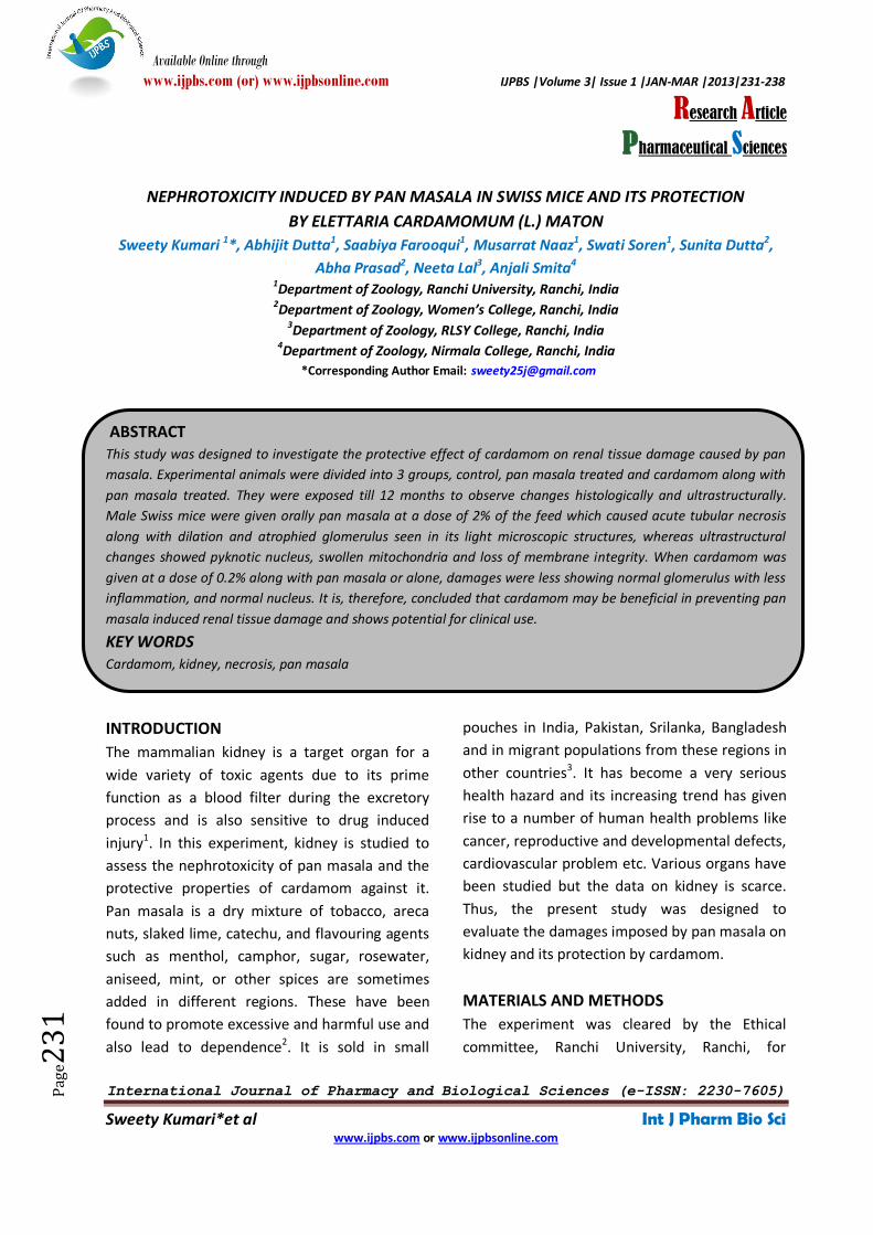
shown). However, vascular changes with
interstitial edema, severe inflammation and
shrunken glomerulus were visible in PMT mice
along with tubular dilation (Fig.1).
Figure 1: Photomicrograph of PMT mice showing severe inflammation (*), also shown in the insat (40x).
Dilation of tubules (arrow) and shrunken glomeruli (bounded by square). (9 months; H&E, 10x)
Cortical renal tubules show various degenerative
changes with focal tubular necrosis, atrophied
and fragmented glomerulus (Fig. 2).
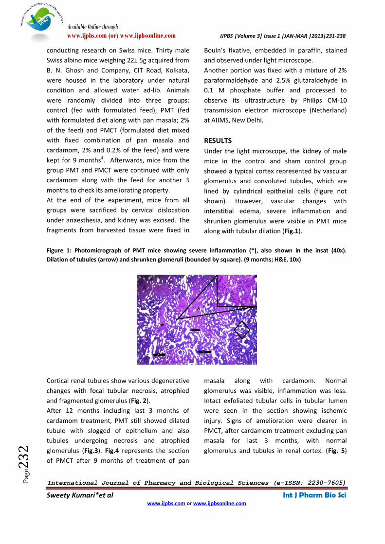
After 12 months including last 3 months of
cardamom treatment, PMT still showed dilated
tubule with slogged of epithelium and also
tubules undergoing necrosis and atrophied
glomerulus (Fig.3). Fig.4 represents the section
of PMCT after 9 months of treatment of pan
masala along with cardamom. Normal
glomerulus was visible, inflammation was less.
Intact exfoliated tubular cells in tubular lumen
were seen in the section showing ischemic
injury. Signs of amelioration were clearer in
PMCT, after cardamom treatment excluding pan
masala for last 3 months, with normal
glomerulus and tubules in renal cortex. (Fig. 5)
Available Online through
www.ijpbs.com (or) www.ijpbsonline.com IJPBS |Volume 3| Issue 1 |JAN-MAR |2013|231-238
International Journal of Pharmacy and Biological Sciences (e-ISSN: 2230-7605)
Sweety Kumari*et al Int J Pharm Bio Sci www.ijpbs.com or www.ijpbsonline.com
Pag
e23
3
Figure 2: Photomicrograph of PMT mice showing necrosis (*) and atrophied glomerulus (large arrow) alongwith
fragmented glomerulus (small arrow). (9 months; Mallory’s triple stain, 40x)
Figure 3: Photomicrograph of PMT mice showing dilated Proximal Convoluted Tubule (T) and atrophied
glomerulus (G). (12 months; Mallory’s triple stain, 40x)
Figure 4: Photomicrograph of PMCT mice showing normal glomerulus (G) along with ischemic injury (*). (9
months; H&E, 40x)
Available Online through
www.ijpbs.com (or) www.ijpbsonline.com IJPBS |Volume 3| Issue 1 |JAN-MAR |2013|231-238
International Journal of Pharmacy and Biological Sciences (e-ISSN: 2230-7605)
Sweety Kumari*et al Int J Pharm Bio Sci www.ijpbs.com or www.ijpbsonline.com
Pag
e23
4
Figure 5: Photomicrograph of PMCT mice showing normal glomerulus (arrow) and necrotic tubules (*) in one
part of the section showed. (12 months; Crossman stain, 10x)
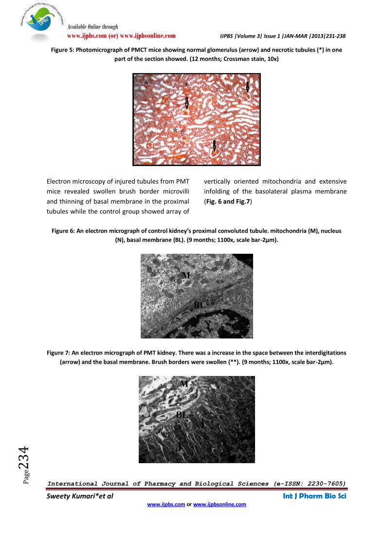
Electron microscopy of injured tubules from PMT
mice revealed swollen brush border microvilli
and thinning of basal membrane in the proximal
tubules while the control group showed array of
vertically oriented mitochondria and extensive
infolding of the basolateral plasma membrane
(Fig. 6 and Fig.7)
Figure 6: An electron micrograph of control kidney’s proximal convoluted tubule. mitochondria (M), nucleus
(N), basal membrane (BL). (9 months; 1100x, scale bar-2µm).
Figure 7: An electron micrograph of PMT kidney. There was a increase in the space between the interdigitations
(arrow) and the basal membrane. Brush borders were swollen (**). (9 months; 1100x, scale bar-2µm).
Available Online through
www.ijpbs.com (or) www.ijpbsonline.com IJPBS |Volume 3| Issue 1 |JAN-MAR |2013|231-238
International Journal of Pharmacy and Biological Sciences (e-ISSN: 2230-7605)
Sweety Kumari*et al Int J Pharm Bio Sci www.ijpbs.com or www.ijpbsonline.com
Pag
e23
5
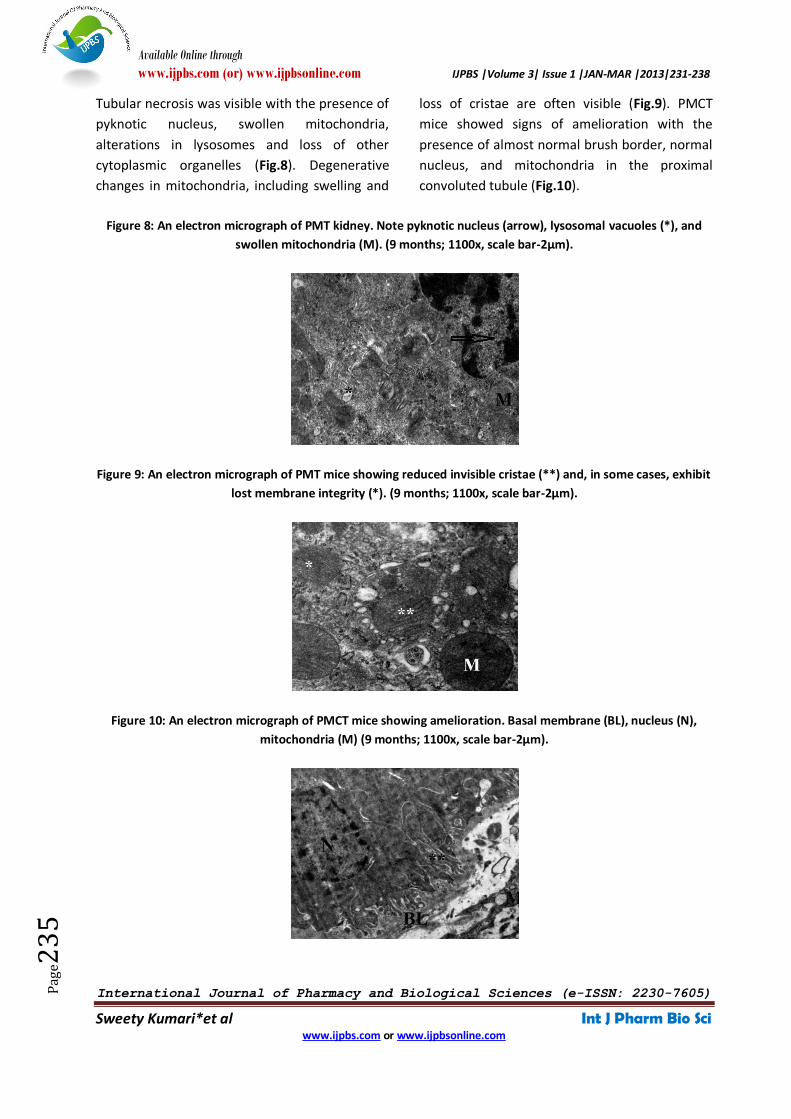
Tubular necrosis was visible with the presence of
pyknotic nucleus, swollen mitochondria,
alterations in lysosomes and loss of other
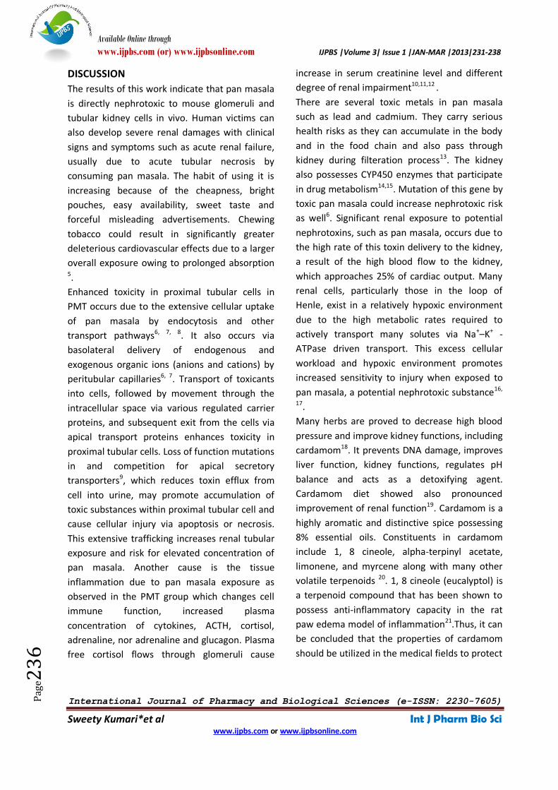
cytoplasmic organelles (Fig.8). Degenerative
changes in mitochondria, including swelling and
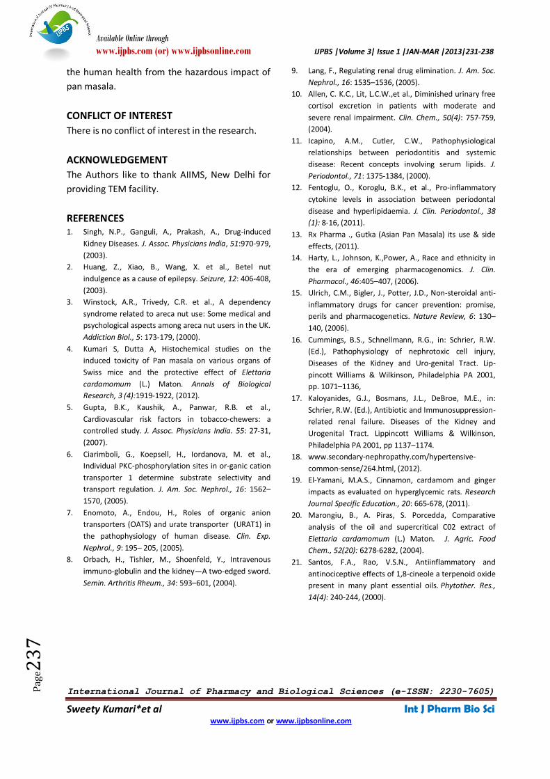
loss of cristae are often visible (Fig.9). PMCT
mice showed signs of amelioration with the
presence of almost normal brush border, normal
nucleus, and mitochondria in the proximal
convoluted tubule (Fig.10).
Figure 8: An electron micrograph of PMT kidney. Note pyknotic nucleus (arrow), lysosomal vacuoles (*), and
swollen mitochondria (M). (9 months; 1100x, scale bar-2µm).
Figure 9: An electron micrograph of PMT mice showing reduced invisible cristae (**) and, in some cases, exhibit
lost membrane integrity (*). (9 months; 1100x, scale bar-2µm).
Figure 10: An electron micrograph of PMCT mice showing amelioration. Basal membrane (BL), nucleus (N),
mitochondria (M) (9 months; 1100x, scale bar-2µm).
Available Online through
www.ijpbs.com (or) www.ijpbsonline.com IJPBS |Volume 3| Issue 1 |JAN-MAR |2013|231-238
International Journal of Pharmacy and Biological Sciences (e-ISSN: 2230-7605)
Sweety Kumari*et al Int J Pharm Bio Sci www.ijpbs.com or www.ijpbsonline.com
Pag
e23
6
DISCUSSION
The results of this work indicate that pan masala
is directly nephrotoxic to mouse glomeruli and
tubular kidney cells in vivo. Human victims can
also develop severe renal damages with clinical
signs and symptoms such as acute renal failure,
usually due to acute tubular necrosis by
consuming pan masala. The habit of using it is
increasing because of the cheapness, bright
pouches, easy availability, sweet taste and
forceful misleading advertisements. Chewing
tobacco could result in significantly greater
deleterious cardiovascular effects due to a larger
overall exposure owing to prolonged absorption 5.
Enhanced toxicity in proximal tubular cells in
PMT occurs due to the extensive cellular uptake
of pan masala by endocytosis and other
transport pathways6, 7, 8. It also occurs via
basolateral delivery of endogenous and
exogenous organic ions (anions and cations) by
peritubular capillaries6, 7. Transport of toxicants
into cells, followed by movement through the
intracellular space via various regulated carrier
proteins, and subsequent exit from the cells via
apical transport proteins enhances toxicity in
proximal tubular cells. Loss of function mutations
in and competition for apical secretory
transporters9, which reduces toxin efflux from
cell into urine, may promote accumulation of
toxic substances within proximal tubular cell and
cause cellular injury via apoptosis or necrosis.
This extensive trafficking increases renal tubular
exposure and risk for elevated concentration of
pan masala. Another cause is the tissue
inflammation due to pan masala exposure as
observed in the PMT group which changes cell
immune function, increased plasma
concentration of cytokines, ACTH, cortisol,
adrenaline, nor adrenaline and glucagon. Plasma
free cortisol flows through glomeruli cause
increase in serum creatinine level and different
degree of renal impairment10,11,12 .
There are several toxic metals in pan masala
such as lead and cadmium. They carry serious
health risks as they can accumulate in the body
and in the food chain and also pass through
kidney during filteration process13. The kidney
also possesses CYP450 enzymes that participate
in drug metabolism14,15. Mutation of this gene by
toxic pan masala could increase nephrotoxic risk
as well6. Significant renal exposure to potential
nephrotoxins, such as pan masala, occurs due to
the high rate of this toxin delivery to the kidney,
a result of the high blood flow to the kidney,
which approaches 25% of cardiac output. Many
renal cells, particularly those in the loop of
Henle, exist in a relatively hypoxic environment
due to the high metabolic rates required to
actively transport many solutes via Na+–K+ -
ATPase driven transport. This excess cellular
workload and hypoxic environment promotes
increased sensitivity to injury when exposed to
pan masala, a potential nephrotoxic substance16,
17.
Many herbs are proved to decrease high blood
pressure and improve kidney functions, including
cardamom18. It prevents DNA damage, improves
liver function, kidney functions, regulates pH
balance and acts as a detoxifying agent.
Cardamom diet showed also pronounced
improvement of renal function19. Cardamom is a
highly aromatic and distinctive spice possessing
8% essential oils. Constituents in cardamom
include 1, 8 cineole, alpha-terpinyl acetate,
limonene, and myrcene along with many other
volatile terpenoids 20. 1, 8 cineole (eucalyptol) is
a terpenoid compound that has been shown to
possess anti-inflammatory capacity in the rat
paw edema model of inflammation21.Thus, it can
be concluded that the properties of cardamom
should be utilized in the medical fields to protect
Available Online through
www.ijpbs.com (or) www.ijpbsonline.com IJPBS |Volume 3| Issue 1 |JAN-MAR |2013|231-238
International Journal of Pharmacy and Biological Sciences (e-ISSN: 2230-7605)
Sweety Kumari*et al Int J Pharm Bio Sci www.ijpbs.com or www.ijpbsonline.com
Pag
e23
7
the human health from the hazardous impact of
pan masala.
CONFLICT OF INTEREST
There is no conflict of interest in the research.
ACKNOWLEDGEMENT
The Authors like to thank AIIMS, New Delhi for
providing TEM facility.
REFERENCES 1. Singh, N.P., Ganguli, A., Prakash, A., Drug-induced
Kidney Diseases. J. Assoc. Physicians India, 51:970-979,
(2003).
2. Huang, Z., Xiao, B., Wang, X. et al., Betel nut
indulgence as a cause of epilepsy. Seizure, 12: 406-408,
(2003).
3. Winstock, A.R., Trivedy, C.R. et al., A dependency
syndrome related to areca nut use: Some medical and
psychological aspects among areca nut users in the UK.
Addiction Biol., 5: 173-179, (2000).
4. Kumari S, Dutta A, Histochemical studies on the
induced toxicity of Pan masala on various organs of
Swiss mice and the protective effect of Elettaria
cardamomum (L.) Maton. Annals of Biological
Research, 3 (4):1919-1922, (2012).
5. Gupta, B.K., Kaushik, A., Panwar, R.B. et al.,
Cardiovascular risk factors in tobacco-chewers: a
controlled study. J. Assoc. Physicians India. 55: 27-31,
(2007).
6. Ciarimboli, G., Koepsell, H., Iordanova, M. et al.,
Individual PKC-phosphorylation sites in or-ganic cation
transporter 1 determine substrate selectivity and
transport regulation. J. Am. Soc. Nephrol., 16: 1562–
1570, (2005).
7. Enomoto, A., Endou, H., Roles of organic anion
transporters (OATS) and urate transporter (URAT1) in
the pathophysiology of human disease. Clin. Exp.
Nephrol., 9: 195– 205, (2005).
8. Orbach, H., Tishler, M., Shoenfeld, Y., Intravenous
immuno-globulin and the kidney—A two-edged sword.
Semin. Arthritis Rheum., 34: 593–601, (2004).
9. Lang, F., Regulating renal drug elimination. J. Am. Soc.
Nephrol., 16: 1535–1536, (2005).
10. Allen, C. K.C., Lit, L.C.W.,et al., Diminished urinary free
cortisol excretion in patients with moderate and
severe renal impairment. Clin. Chem., 50(4): 757-759,
(2004).
11. Icapino, A.M., Cutler, C.W., Pathophysiological
relationships between periodontitis and systemic
disease: Recent concepts involving serum lipids. J.
Periodontol., 71: 1375-1384, (2000).
12. Fentoglu, O., Koroglu, B.K., et al., Pro-inflammatory
cytokine levels in association between periodontal
disease and hyperlipidaemia. J. Clin. Periodontol., 38
(1): 8-16, (2011).
13. Rx Pharma ., Gutka (Asian Pan Masala) its use & side
effects, (2011).
14. Harty, L., Johnson, K.,Power, A., Race and ethnicity in
the era of emerging pharmacogenomics. J. Clin.
Pharmacol., 46:405–407, (2006).
15. Ulrich, C.M., Bigler, J., Potter, J.D., Non-steroidal anti-
inflammatory drugs for cancer prevention: promise,
perils and pharmacogenetics. Nature Review, 6: 130–
140, (2006).
16. Cummings, B.S., Schnellmann, R.G., in: Schrier, R.W.
(Ed.), Pathophysiology of nephrotoxic cell injury,
Diseases of the Kidney and Uro-genital Tract. Lip-
pincott Williams & Wilkinson, Philadelphia PA 2001,
pp. 1071–1136,
17. Kaloyanides, G.J., Bosmans, J.L., DeBroe, M.E., in:
Schrier, R.W. (Ed.), Antibiotic and Immunosuppression-
related renal failure. Diseases of the Kidney and
Urogenital Tract. Lippincott Williams & Wilkinson,
Philadelphia PA 2001, pp 1137–1174.
18. www.secondary-nephropathy.com/hypertensive-
common-sense/264.html, (2012).
19. El-Yamani, M.A.S., Cinnamon, cardamom and ginger
impacts as evaluated on hyperglycemic rats. Research
Journal Specific Education., 20: 665-678, (2011).
20. Marongiu, B., A. Piras, S. Porcedda, Comparative
analysis of the oil and supercritical C02 extract of
Elettaria cardamomum (L.) Maton. J. Agric. Food
Chem., 52(20): 6278-6282, (2004).
21. Santos, F.A., Rao, V.S.N., Antiinflammatory and
antinociceptive effects of 1,8-cineole a terpenoid oxide
present in many plant essential oils. Phytother. Res.,
14(4): 240-244, (2000).
Available Online through
www.ijpbs.com (or) www.ijpbsonline.com IJPBS |Volume 3| Issue 1 |JAN-MAR |2013|231-238
International Journal of Pharmacy and Biological Sciences (e-ISSN: 2230-7605)
Sweety Kumari*et al Int J Pharm Bio Sci www.ijpbs.com or www.ijpbsonline.com
Pag
e23
8
*Corresponding Author: Sweety Kumari* Department of Zoology, Ranchi University, Ranchi-834008, India