Hematopoietic Stem Cell Transplantation Leukemia Patients ...
Upload
umcutrechtCategory
view
0download
0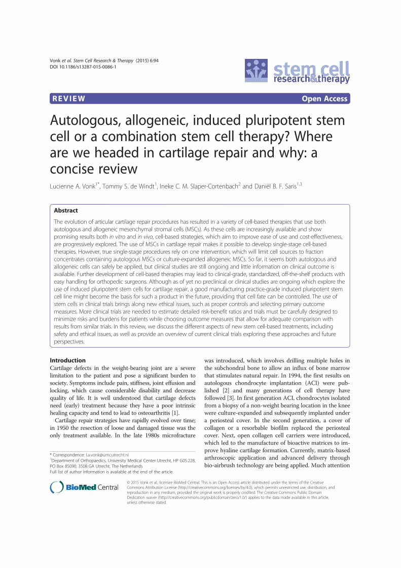
REVIEW Open Access
Autologous, allogeneic, induced pluripotent stemcell or a combination stem cell therapy? Whereare we headed in cartilage repair and why: aconcise reviewLucienne A. Vonk1*, Tommy S. de Windt1, Ineke C. M. Slaper-Cortenbach2 and Daniël B. F. Saris1,3
Abstract
The evolution of articular cartilage repair procedures has resulted in a variety of cell-based therapies that use bothautologous and allogeneic mesenchymal stromal cells (MSCs). As these cells are increasingly available and showpromising results both in vitro and in vivo, cell-based strategies, which aim to improve ease of use and cost-effectiveness,are progressively explored. The use of MSCs in cartilage repair makes it possible to develop single-stage cell-basedtherapies. However, true single-stage procedures rely on one intervention, which will limit cell sources to fractionconcentrates containing autologous MSCs or culture-expanded allogeneic MSCs. So far, it seems both autologous andallogeneic cells can safely be applied, but clinical studies are still ongoing and little information on clinical outcome isavailable. Further development of cell-based therapies may lead to clinical-grade, standardized, off-the-shelf products witheasy handling for orthopedic surgeons. Although as of yet no preclinical or clinical studies are ongoing which explore theuse of induced pluripotent stem cells for cartilage repair, a good manufacturing practice-grade induced pluripotent stemcell line might become the basis for such a product in the future, providing that cell fate can be controlled. The use ofstem cells in clinical trials brings along new ethical issues, such as proper controls and selecting primary outcomemeasures. More clinical trials are needed to estimate detailed risk-benefit ratios and trials must be carefully designed tominimize risks and burdens for patients while choosing outcome measures that allow for adequate comparison withresults from similar trials. In this review, we discuss the different aspects of new stem cell-based treatments, includingsafety and ethical issues, as well as provide an overview of current clinical trials exploring these approaches and futureperspectives.
IntroductionCartilage defects in the weight-bearing joint are a severelimitation to the patient and pose a significant burden tosociety. Symptoms include pain, stiffness, joint effusion andlocking, which cause considerable disability and decreasequality of life. It is well understood that cartilage defectsneed (early) treatment because they have a poor intrinsichealing capacity and tend to lead to osteoarthritis [1].Cartilage repair strategies have rapidly evolved over time;
in 1950 the resection of loose and damaged tissue was theonly treatment available. In the late 1980s microfracture
was introduced, which involves drilling multiple holes inthe subchondral bone to allow an influx of bone marrowthat stimulates natural repair. In 1994, the first results onautologous chondrocyte implantation (ACI) were pub-lished [2] and many generations of cell therapy havefollowed [3]. In first generation ACI, chondrocytes isolatedfrom a biopsy of a non-weight bearing location in the kneewere culture-expanded and subsequently implanted undera periosteal cover. In the second generation, a cover ofcollagen or a resorbable biofilm replaced the periostealcover. Next, open collagen cell carriers were introduced,which led to the manufacture of bioactive matrices to im-prove hyaline cartilage formation. Currently, matrix-basedarthroscopic application and advanced delivery throughbio-airbrush technology are being applied. Much attention
* Correspondence: [email protected] of Orthopaedics, University Medical Center Utrecht, HP G05.228,PO Box 85090, 3508 GA Utrecht, The NetherlandsFull list of author information is available at the end of the article
© 2015 Vonk et al.; licensee BioMed Central. This is an Open Access article distributed under the terms of the CreativeCommons Attribution License (http://creativecommons.org/licenses/by/4.0), which permits unrestricted use, distribution, andreproduction in any medium, provided the original work is properly credited. The Creative Commons Public DomainDedication waiver (http://creativecommons.org/publicdomain/zero/1.0/) applies to the data made available in this article,unless otherwise stated.
Vonk et al. Stem Cell Research & Therapy (2015) 6:94 DOI 10.1186/s13287-015-0086-1
was also given to the culture-expansion phase resulting inthe introduction of characterized cells that show the mostchondrogenic potential and establishing release criteriaand production guidelines.The mid- to long-term results of ACI have been en-
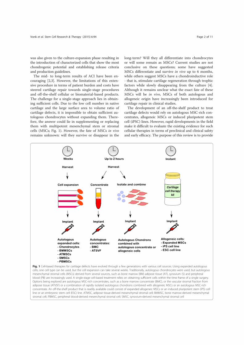
couraging [2,3]. However, the limitations of this exten-sive procedure in terms of patient burden and costs havesteered cartilage repair towards single-stage proceduresand off-the-shelf cellular or biomaterial-based products.The challenge for a single-stage approach lies in obtain-ing sufficient cells. Due to the low cell number in nativecartilage and the large surface area to volume ratio ofcartilage defects, it is impossible to obtain sufficient au-tologous chondrocytes without expanding them. There-fore, the answer could lie in supplementing or replacingthem with multipotent mesenchymal stem or stromalcells (MSCs; Fig. 1). However, the fate of MSCs in vivoremains unknown: will they survive or disappear in the
long-term? Will they all differentiate into chondrocytesor will some remain as MSCs? Current studies are notconclusive on these questions; some have suggestedMSCs differentiate and survive in vivo up to 6 months,while others suggest MSCs have a chondroinductive role- that is, stimulate cartilage regeneration through trophicfactors while slowly disappearing from the culture [4].Although it remains unclear what the exact fate of theseMSCs will be in vivo, MSCs of both autologous andallogeneic origin have increasingly been introduced forcartilage repair in clinical studies.The development of an off-the-shelf product to treat
cartilage defects would rely on autologous MSC-rich con-centrates, allogeneic MSCs or induced pluripotent stemcell (iPSC) lines. However, rapid developments in the fieldmake it difficult to evaluate the existing evidence for suchcellular therapies in terms of preclinical and clinical safetyand early efficacy. The purpose of this review is to provide
Fig. 1 Cell-based therapies for cartilage defects have evolved through a few generations with various cell sources. Using expanded autologouscells, one cell type can be used, but the cell expansion can take several weeks. Traditionally, autologous chondrocytes were used, but autologousmesenchymal stromal cells (MSCs) derived from several sources, such as bone marrow (BM) adipose tissue (AT), synovium (S) and peripheralblood (PB) are increasingly used. A single-stage cell-based treatment relies on obtaining sufficient cells within the time frame of a single surgery.Options being explored are autologous MSC-rich concentrates, such as a bone marrow concentrate (BMC), or the vascular stromal fraction fromadipose tissue (ATVSF) or a combination of rapidly isolated autologous chondrons combined with allogeneic MSCs or an autologous MSC-richconcentrate. An off-the-shelf product that is readily available could consist of expanded allogeneic MSCs or an induced pluripotent stem (iPS) cellline or an embryonic stem cell (ESC) line. ATMSC, adipose tissue-derived mesenchymal stromal cell; BMMSC, bone marrow-derived mesenchymalstromal cell; PBMSC, peripheral blood-derived mesenchymal stromal cell; SMSC, synovium-derived mesenchymal stromal cell
Vonk et al. Stem Cell Research & Therapy (2015) 6:94 Page 2 of 11

a concise overview of the available literature on autolo-gous and allogeneic MSCs for cartilage repair of focal de-fects. Besides clinical studies, the sources of MSCs, safetyand ethical issues with respect to allogeneic MSCs, theuse of iPSCs and future perspectives are discussed.
Sources for mesenchymal stromal cellsMinimal criteria to define expanded multipotent humanMSCs, as defined by the International Society for CellularTherapy, include that they must be plastic-adherent whenmaintained in standard culture conditions, express CD105,CD73 and CD90, and lack expression of CD45, CD34,CD14 or CD11b, CD79α or CD19 and HLA-DR surfacemolecules, and they must be capable of differentiating intoosteoblasts, adipocytes and chondroblasts in vitro [5]. MSCscan be isolated and expanded from a variety of sources,such as bone marrow, adipose tissue, synovial membrane,synovial fluid, umbilical cord blood, peripheral blood, der-mis, trabecular bone, infrapatellar fat pad, dermis, perios-teum and muscle. The phenotypic characteristics of MSCsderived from different sources are similar, but the numberof MSCs and their proliferation and differentiation poten-tials can differ [6]. Bone marrow is often used as a sourcefor MSCs (BMMSCs). Although only a small percentage ofits mononuclear fraction consists of BMMSCs, they arerelatively easy to isolate and expand and they have a highpotential for differentiation [7]. The stromal vascular frac-tion of adipose tissue contains more MSCs (ATMSCs)compared with bone marrow (as measured in a colony-forming unit-fibroblasts (CFU-F) assay) and harvestingadipose tissue is less invasive [8]. ATMSCs show enhancedrates of proliferation and they can undergo more popula-tion doublings before senescence [8,9]. However, thein vitro chondrogenic potential of ATMSCs is lowercompared with BMMSCs in vitro, especially when pelletcultures are stimulated with transforming growth factor(TGF)-beta. [9]. The tissue formed by ATMSCs chondro-genically differentiated with TGF-beta contained less type IIcollagen and proteoglycans compared with tissue formedby chondrogenically differentiated BMMSCs from the samedonors. The exact reason is unknown, but it is suggestedthere might be less chondroprogenitor cells present in theATMSC population or that the expansion favors clonalexpansion of cells with higher proliferative rates, albeit withless differentiation potential [9]. However, other studieshave shown a good chondrogenic potential of ATMSCswhen bone morphogenetic protein (BMP)-6 was used,which may be explained by an altered TGF-beta receptorand BMP profile of ATMSCs compared with BMMSCs[10,11].MSCs derived from the synovial membrane (SMSCs) can
be harvested through an arthroscopic procedure or fromsynovial fluid. The amount of SMSCs in synovial fluid is verylow; only about 14 cells per milliliter of synovial fluid from
healthy donors can form CFU-F colonies. Parts of thesecolony-forming cells are considered SMSCs as they candifferentiate into the adipogenic, osteogenic and chondro-genic lineages. Compared with BMMSCs and ATMSCs, theyhave a higher rate of proliferation [12,13]. Sakaguchi andcolleagues showed superior chondrogenic differentiation ofSMSCs compared with donor-matched BMMSCs, ATMSCsand MSCs from periosteum and skeletal muscle in vitro [14].SMSCs have also shown potential in in vitro generation ofhyaline cartilage tissue-engineered constructs [15]. Implant-ation of these in vitro-generated constructs showed good re-pair of cartilage defects in a pig model with SMSCs isolatedfrom both immature and mature pigs [16,17].MSCs can also be isolated from peripheral blood
(PBMSCs) [18]. MSC isolation from blood provides lowcell numbers, but peripheral blood can be easily obtainedin a non-invasive way. Although there is a large variationin the success rates of isolation of MSCs from umbilicalcord blood (UMSCs), they have good chondrogenic po-tential [19]. The accessibility of UMSCs along with theirefficient expanding characteristics have made allogeneicUMSCs the only off-the-shelf cell product for cartilage re-pair [20]. MSCs can also be isolated from the periosteum,but the limited availability and complex tissue harvest pro-cedure forms a barrier for their use. Currently, isolatedBMMSCs and bone marrow concentrates (BMCs) aremost commonly used for treatment of cartilage defects ina clinical trial setting (Table 1).One of the concerns with using MSCs for cartilage re-
pair is that if they do differentiate into the chondrogeniclineage and engraft the new cartilage, they might undergoterminal differentiation and become hypertrophic, as thedefault route of chondrogenic differentiation is terminaldifferentiation [21]. This concern is not limited to MSCsonly, as chondrocytes can also undergo hypertrophicdifferentiation, which has been found in ACI [22].Articular cartilage itself, especially the superficial layers,
is also a reservoir for progenitor cells with multilineagepotential [23,24]. Cartilage-derived progenitors even havea decreased potential for osteogenic and hypertrophic dif-ferentiation. Although the research on cartilage-derivedprogenitor cells is still very limited, a goat study hasproven their ability to repair chondral defects. Williamsand colleagues [24] suggested that about 0.7% of all cellsin cartilage are progenitor cells.
Clinical studies using autologous mesenchymalstromal cellsSince Wakitani and colleagues [25] performed the firsttreatment of full-thickness cartilage defects with autolo-gous MSCs in 2004, autologous MSCs and MSC-richconcentrates are increasingly used for cartilage repair(overview provided in Table 1). Most published resultsare obtained from low level (IV or V) evidence studies
Vonk et al. Stem Cell Research & Therapy (2015) 6:94 Page 3 of 11

Table1Ove
rview
ofclinical
stud
iesap
plying
autologo
usmesen
chym
alstromal
cells
toacartila
gede
fect
Celltype
Stud
ytype
Num
bero
fpatients
Follow-up
Results
Reference
BMMSC
Case
repo
rt2
5years
Clinicalim
provem
entandde
fect
fillw
ithfib
rocartilage
[25]
BMMSC
Case
repo
rt1
1year
Bone
andcartilage
repair
[26]
BMMSC
Case
repo
rt3
17-27mon
ths
Clinicalim
provem
entandde
fect
fillw
ithfib
rocartilage
[27]
BMMSC
Case
repo
rt1
12mon
ths
Clinicalim
provem
entandde
fect
fillw
ithhyalinetissue
[28]
BMMSC
Case
series
512
mon
ths
Clinicalim
provem
entandde
fect
fill
[29]
BMMSC
Case
repo
rt2
31mon
ths
Clinicalim
provem
entandde
fect
fill
[30]
BMC
Case
series
484years
Clinicalim
provem
entandde
fect
fill
[31,32]
BMC
Case
series
2024
mon
ths
Bone
andcartilage
repair
[33]
BMC
Case
series
512
mon
ths
Defectfillw
ithhyalineto
fibrocartilagino
ustissue
[34]
BMC
Case
series
545years
Clinicalim
provem
entandgo
odintegrationof
repairtissue
[35]
BMC
Case
series
152years
Clinicalim
provem
entandde
fect
fillw
ithhyalinetissue
[36]
BMCandAC
yCa
seseries
40Interim
results
1year
Clinicalim
provem
entandde
fect
fillw
ithhyalinetissue
[37]
NCT
01041885
SMSC
[16,17]
BMMSC
Comparativestud
y36
(total72)
24mon
ths
Clinicalim
provem
ent,de
fect
fillw
ithhyalinetissue
[38]
BMC
Comparativestud
y25
(total81)
36mon
ths
Clinicalim
provem
entandde
fect
fillw
ithhyalinetissue
[39]
PBMSC
orBM
CCo
mparativestud
y25
PBMSC
,21BM
C5years
Clinicalim
provem
entandde
fect
fillfor
both
grou
ps[40]
BMMSC
Case
series
2512
mon
ths
NCT
00891501
BMMSC
Case
series
612
mon
ths
NCT
00850187
BMC
Case
series
140
36mon
ths
NCT
02005861
BMC
Case
series
5012
mon
ths
NCT
01159899
ATSVF
Comparativestud
y40
24mon
ths
NCT
02090140
ATMSC
Comparativestud
y30
18mon
ths
NCT
01399749
BMMSC
Comparativestud
y50
intotal
5years
NCT
00885729
ACyau
tologo
uschon
drocyte,
ATMSC
adiposetissue-de
rived
mesen
chym
alstromal
cell,AT
SVFad
iposetissuestromal
vascular
fractio
n,BM
Cbo
nemarrow
concen
trate,
BMMSC
bone
marrow-derived
mesen
chym
alstromal
cell,PB
MSC
perip
heralb
lood
-derived
mesen
chym
alstromal
cell,SM
SCsyno
vium
-derived
mesen
chym
alstromal
cell
Vonk et al. Stem Cell Research & Therapy (2015) 6:94 Page 4 of 11
[25-37], and few comparative studies are available [38-40].Nejadnik and colleagues [38] compared the implantationof BMMSCs (36 patients) with first-generation ACI (36matched patients) in a cohort study (evidence level III).Based on clinical and subjective improvement up to 2 yearspostoperatively, it was concluded that BMMSCs are aseffective as chondrocytes for articular cartilage repair.Histological evaluation of biopsies taken from a fewpatients (four BMMSC, three ACI) showed hyaline-likecartilage tissue and no abnormal calcification or necrosis.Interestingly, patients younger than 45 years scored betterthan patients aged over 45 years in the ACI group, whileage did not make a difference in the BMMSC group. Fol-lowing several case series, Giannini and colleagues [31-33]reported on a one-step approach to treat osteochondraltalar dome defects and compared a MSC-rich BMC (25patients) with ACI (10 patients) and an arthroscopic ACI(46 patients) (evidence level IV) [39]. As in the previouslydescribed study, similar clinical improvement was ob-served, and magnetic resonance imaging (MRI) and histo-logical evaluation showed complete defect fill withhyaline-like cartilage tissue in the majority of patients.Only one study compared the use of two MSC-based
treatments for cartilage repair [40]. In this study, 21 pa-tients were treated with BMCs and 25 with PBMSCs. Clin-ical improvement was found in a total of 40 patients, inwhich the patients treated with PBMSCs showed superiorresults compared with the patients treated with BMCs.Poor results were found for four patients in the BMC groupand two patients in the PBMSC group. Although MRI wasalso performed in this study, no MRI results were reported.Although only two studies directly compared MSC-based
treatments with ACI [38,39], the conclusions from thesestudies do suggest that MSCs are a promising cell sourcefor cartilage repair. This is supported by the findings in thelevel IV and V evidence studies that used BMMSCs orBMC for cartilage repair; all have reported clinical improve-ment with a follow-up period ranging from 1 year to 5 years[25,27-32,35,36,38-40]. The studies that included MRI ana-lysis in their outcome measures reported complete defectfill [27-29,31-33,35,36] and mostly congruity with the nativecartilage [29,35]. Histological evaluation of biopsies showedthe reparative tissue was hyaline-like cartilage [28,33,35,36,38,39], fibrocartilage [25,27,31,32], or a mixture of both[26,34].Several other studies using autologous MSCs or concen-
trates are still ongoing, including two studies using ATMSCsto treat cartilage defects (Table 1; NCT01399749 andNCT02090140). So far, ATMSCs have only entered thepreclinical phase in cartilage repair. In clinical use, con-centrated ATMSCs have been injected intra-articularly fortreatment of osteoarthritis [41,42]. SMSCs have been usedin preclinical studies, which gave promising results[16,17]. The tissue-engineered construct made by SMSCs
as described in those preclinical studies is currently beingexplored in an investigator-driven phase I/II clinical trialin a small cohort in Japan.Thus, only clinical results using expanded undifferenti-
ated BMMSCs, PBMSCs or BMCs (bone-marrow derivedbuffy coat or the mononuclear fraction of bone marrow)are reported. Pre-differentiated MSCs have not been usedas yet. Although MSCs and MSC-rich concentrates arepromising for cartilage repair, a lack of comparative stud-ies confines a prediction to what the optimal cell sourcefor MSC-based cartilage repair would be. Moreover, MSCsand BMCs have been implanted using various cell carriers,passages and doses (sometimes even not reported; Tables 2and 3), so much remains to be investigated and learned.
Safety considerations using allogeneicmesenchymal stromal cellsIt took until 2010 before the first clinical study exploringthe use of allogeneic MSCs for cartilage repair started[20], probably due to the unknown risk of an immune re-sponse to allogeneic cells. MSCs have been shown to havelow immunogenicity based on the lack of expression ofmarkers such as CD45 and CD34 and HLA-DR surfacemolecules [43]. In addition, they are known to interactwith immune cell populations and modulate the host im-mune responses [43]. Because of the immunosuppressiveproperties of MSCs, allogeneic MSCs are currently infusedintravenously for the treatment of steroid-resistant graft-versus-host disease, acute respiratory distress syndromeand Crohn’s disease in clinical trials. However, as itremains unclear what the exact fate of these MSCs isin vivo, it cannot be excluded that the MSCs differentiate,leading to a loss in their immune-modulating propertyand a change in their immunogenicity [44]. Several pre-clinical studies in rabbits, pigs and goats showed effectivecartilage repair after implantation of allogeneic MSCs incartilage defects without any adverse events or rejection[17,45,46]. Moreover, no adverse events were reportedwhen fully differentiated allogeneic chondrocytes or allo-geneic cartilage pieces were transplanted in several animaland human clinical trials [47,48], possibly due to the im-mune privileged character of cartilage as it is avascularand has no lymphatic system. It has to be noted thatcartilage defects are often debrided, which can cause pene-tration of the subchondral bone, allowing an influx ofbone marrow. This might become a concern for usingpre-differentiated allogeneic cells or allogeneic iPSCs.
Clinical studies using allogeneic mesenchymalstromal cellsOnly a few clinical trials have been initiated using allo-geneic MSCs for cartilage repair (Table 4). In Korea, aphase III clinical trial comparing allogeneic UMSCs withsodium hyaluronate (CARTISTEM®, MEDIPOST, Korea)
Vonk et al. Stem Cell Research & Therapy (2015) 6:94 Page 5 of 11

Table2Details
onmesen
chym
alstromal
cells
used
inclinical
stud
ies
Celltype
Cellcarrier
Celldo
seExpansionmed
ium
Passage
Characterization
Reference
BMMSC
Collage
nge
l5×10
6 /ml
αMEM
,15%
ASSing
lepassaged
CD73,C
D90,C
D105+
,CD
14,C
D34,H
LA-DR-
[25,27,28]
BMMSC
Hydroxyapatite
ceramic
1.15
×10
6in
total
DMEM
,15%
ASSing
lepassaged
[26]
BMMSC
Fibrin
glue
with
perio
steum
2×10
6cells/cm
2DMEM
,10%
FBS,50
μg/m
lAA2
P,1%
antib
iotic-antim
ycotic
Sing
lepassaged
CD90,C
D105+
,CD14,C
D34-
[38]
BMMSC
Platelet-rich
fibrin
gel
2×10
6cells/cm
2DMEM
,10%
FBS,1%
pen/strep
Sing
lepassaged
CD73+,C
D34,C
D45-
[29]
BMMSC
Collage
nscaffold
DMEM
,10%
,FBS
1%pe
n/strep
Passaged
[30]
BMMSC
Mem
brane
NCT
00885729
BMMSC
Perio
steum
Passaged
NCT
00891501
BMMSC
Type
Icollage
nscaffold
Passaged
NCT
00850187
PBMSC
Collage
nmem
brane
1.25-5.2×10
6N/A
Unp
assage
d[40]
ATMSC
Perio
steum
Passaged
NCT
01399749
SMSC
SMSC
TEC
[16,17]
UMSC
Sodium
hyaluron
ate
Passaged
[20]
NCT
01041001
BMMSC
andAC
Fibrin
glue
2×10
6cells/cm
2αM
EM,5%
platelet
lysate
Passage3
CD73,C
D90,C
D105+
,CD
45,C
D34,C
D11b,
CD14,
CD31,C
D79,C
D19,H
LA-DR-
[3,46]
NCT
02037204
αMEM
alph
aminim
alessentialm
edium,A
A2PL-ascorbicacid
2-ph
osph
atesesquimag
nesium
salthy
drate,
ACau
tologo
uschon
dron
s,AS
autologo
usserum,A
TMSC
adiposetissue-de
rived
mesen
chym
alstromal
cell,
BMMSC
bone
marrow-derived
mesen
chym
alstromal
cell,DMEM
Dulbe
cco’smod
ified
Eaglemed
ium,FBS
fetalb
ovineserum,N
/Ano
tap
plicab
le,P
BMSC
perip
heralb
lood
-derived
mesen
chym
alstromal
cell,Pen/strep
penicillin/streptom
ycin,SMSC
syno
vium
-derived
mesen
chym
alstromal
cell,TECtissueen
gine
ered
construct,UMSC
umbilical
cord
bloo
d-de
rived
mesen
chym
alstromal
cell
Vonk et al. Stem Cell Research & Therapy (2015) 6:94 Page 6 of 11

to microfracture treatment has recently finished. About100 patients with articular cartilage defects were in-cluded in this study to assess the safety and efficacy witha follow-up of 48 weeks (NCT01041001). The safety ofusing allogeneic UMSCs was confirmed and histologicalanalyses showed repair with hyaline-like tissue [20].Currently, the study is expanded with a follow-up timeof 60 months (NCT01626677). CARTISTEM® has re-cently been introduced in a phase I/II clinical trial inthe USA (NCT01733186).A clinical trial using an allograft SMSC-based tissue
engineered construct is under review by the Pharma-ceutical and Medical Devices Agency of Japan for pos-sible commercialization.In the Netherlands, we have started an investigator-driven
phase I/II clinical trial (IMPACT) using a mixture of rapidlyisolated autologous chondrocytes with their pericellularmatrix (chondrons) combined with allogeneic BMMSCs infibrin glue [3,46] (NCT02037204). The inclusion of thetargeted 35 patients has recently been completed and notreatment-related adverse events have been observed (thepatients are currently at a follow-up ranging from 7 monthsto 1 year after surgery). Preliminary safety monitoring hasnot shown any immunological concerns while clinicaloutcome and structural outcome as measured by MRI and
second-look arthroscopies have demonstrated encouraginginitial results.
Induced pluripotent stem cellsThe ability to generate iPSCs from somatic cells has cre-ated new opportunities for the field of cartilage repair.Just like human embryonic stem cells (hESCs), theyshow unlimited self-renewal and they can differentiateinto all three germ layers (ectoderm, endoderm andmesoderm), but without having the ethical concernsassociated with hESCs. However, there are some differ-ences reported in the efficiency to differentiate towardsseveral lineages, such as neural, cardiovascular andhemangioblastic lineages. iPSCs can be generated byoverexpressing transcription factors associated withpluripotency, such as Oct3/4, Klf4, c-myc and Sox2. Thegenetic reprogramming to induce pluripotency is a lim-iting factor for clinical use as the most efficient viraltransductions lead to integration of viral DNA into thechromosome. Reprogramming without causing geneticchange has gained recent interest and several non-viralmethods using microRNA, synthetic messenger RNAand proteins have been developed.In vitro studies showed chondrogenic differentiation
and cartilage formation by iPSCs derived from human
Table 4 Clinical studies applying allogeneic mesenchymal stromal cells to a cartilage defect for repairCell type Number of patients Follow-up Results Reference
UMSC 104 48 weeks Safe application and repair with hyaline tissue [20] NCT01041001
UMSC 103 60 months NCT01626677
UMSC 12 24 months NCT01733186
SMSC [16,17]
BMMSC with AC 35 1 year Preliminary: safe application and repair with hyaline tissue [3,46] NCT02037204
AC autologous chondrons, BMMSC bone marrow-derived mesenchymal stromal cell, SMSC synovium-derived mesenchymal stromal cell, UMSC umbilical cordblood-derived mesenchymal stromal cell
Table 3 Details on bone marrow concentrates used in clinical studiesType Cell carrier Harvest location Amount
harvestedAmount used Reference
BMC Collagen membrane Ilium 27 ml 0.5-2.7 × 10⁶ cells [40]
BMC Collagen powder or hyaluronicacid membrane and platelet gel
Posterior iliac crest 60 ml 2 ml 10 × concentrated BM [31-33,39] NCT02005861
BMC Type I collagen scaffold Iliac crest 60 ml [34]
BMC Collagen membrane Ipsilateral iliac crest 60 ml 4-6 × concentrated BM,CFU-F/ml 2,000-5,700
[35]
BMC Collagen I/III membrane Ilium 30 ml [36]
BMC Protein matrix in collagenhydroxyapatite scaffold
NCT01159899
BMC and ACy INSTRUCT scaffold [37] NCT01041885
ATSVF Collagen scaffold Infrapatellarfat pad
5 cc NCT02090140
ACy autologous chondrocyte, ATSVF adipose tissue stromal vascular fraction, BM bone marrow, BMC bone marrow concentrate, CFU-F colony-forming unit-fibroblasts
Vonk et al. Stem Cell Research & Therapy (2015) 6:94 Page 7 of 11

fetal neural stem cells [49] and human osteoarthriticchondrocytes [50]. One study showed that overexpres-sion of Oct4 and Klf4 (two-factor reprogramming) wassuccessful in generating iPSCs from murine neural stemcells, which were capable of differentiating into thechondrogenic lineage [51]. Differentiation of iPSCs tothe chondrogenic lineage was efficient if they were firstdifferentiated towards an MSC-like intermediate pheno-type [52,53].Chondrogenic cells were also generated directly from
somatic cells by reprogramming with c-Myc, Klf4 and thechondrogenic transcription factor Sox9. The cells werenon-tumorigenic and had stable karyotypes, and theyformed homogeneous hyaline cartilage [54,55].Diekman and colleagues [56] generated iPSCs from
murine fibroblasts and purified the type II collagen-drivengreen fluorescent protein-expressing cells upon chondro-genic differentiation to obtain a uniformly differentiatedcell population. This cell population was subsequentlysuccessfully used to fill a defect in an in vitro chondral de-fect model. As it was reported that iPSCs can differentiateeasier along the lineages related to the cell type oforigin, iPSCs derived from several chondrocyte donorswere investigated for their chondrogenic potential [57].Indeed, these reprogrammed chondrocytes could bedifferentiated into cartilage-producing chondrocytesmore easily than fibroblast-derived iPSCs. However, oneof the chondrocyte-derived iPSC lines showed higheraggrecan gene expression level compared with the othergenerated iPSC cell lines, while no differences were ob-served in gene expression levels of other chondrogenicmarkers. So even the chondrogenic potential of iPSCsdiffers between different iPSC lines.Although safety precautions and new iPSC generation
techniques have been introduced, it remains to be shownthat cell fate and phenotype can be controlled withouthaving the risk of teratoma formation. Thus, before pre-clinical and clinical tests can be done, there is a need forreliable control of the cell fate.
Ethical considerations in stem cell-basedtreatmentsThe design and initiation of clinical trials using stemcells for cartilage repair is ethically challenging [58].Only a limited number of case reports and clinical trialsusing a stem cell-based treatment have been reported.Moreover, the end product that is used is often poorlydescribed - critical information on culture methods (ifapplicable), cell characterization, source, concentration,and carrier are often missing. All these factors have apronounced influence on the behavior of cells andcould, therefore, also affect clinical outcomes of stemcell-based treatments. In the case of BMCs it shouldbe reported how much bone marrow was initially
harvested, how much concentrate is used for the treat-ment and what the CFU/ml is, such as providedby Gobbi and colleagues [35]. The limited number ofstudies and the lacking information make it hard to ac-curately predict the risks and clinical outcomes of MSC-based treatments. There are risks associated with theintervention and the harvesting procedures of MSCs,while the invasiveness of both procedures may varydepending on the MSC source and treatment strategy.A risk-benefit ratio should be assessed, as the risk toparticipants must be proportional to the anticipatedbenefits. In the relatively new field of MSC-based treat-ment for cartilage defects, it is hard to predict clinicaloutcomes and thus benefits for the first individual pa-tients in a clinical study, while the scientific and societalrelevance is increased. To be able to assess accuraterisk-benefit ratios, negative results should also be pub-lished. Moreover, including all data in the Europeangroup for Blood and Marrow Transplantation databasewill enable the risk-benefit assessment for cellulartherapy products [59].Uniform use of outcome parameters facilitates the
comparison of treatments used in various clinical stud-ies. There is still an ongoing discussion whether struc-tural cartilage regeneration, clinical improvement or acombination should be the main outcome measure.Clinical improvement is undoubtedly an important out-come measure, but placebo and nonspecific effects canaffect the patient’s perspective and it has been suggestedthat clinical improvement does not necessarily correlatewith cartilage tissue regeneration. A second-look arth-roscopy and histological evaluation of a biopsy is thegolden standard to evaluate structural parameters ofcartilage regeneration, but is relatively invasive for pa-tients. A less invasive, but also less detailed and inform-ative, measure is MRI. However, there is only a weakcorrelation between clinical and MRI outcomes, so thechallenge remains to determine how clinical and struc-tural results may correlate [60].Another important ethical consideration is the selection
of an appropriate control group. For a double-blinded ran-domized controlled trial, the use of a placebo, or in thecase of cartilage repair a sham intervention, could be ne-cessary. In the case of MSC-based cartilage repair, the useof a sham group is unacceptable as there is an alternativetreatment that provides medical advantage (ACI) and therisks and invasiveness of sham procedures are dispropor-tionate to the social value. ACI can serve as a control.However, it is impossible to compare the two-stage ACItreatment to a single-stage procedure without introducinga sham intervention. It is also unacceptable to test safety,tolerability, pharmacokinetics and pharmacodynamics ofMSC-based cell products on healthy volunteers, as therisks and burdens of the intervention are too high.
Vonk et al. Stem Cell Research & Therapy (2015) 6:94 Page 8 of 11

Considerations and future perspectivesWith respect to the technovolution of articular cartilagerepair strategies, it is expected that more single-stage proce-dures will emerge that utilize a stem cell-based approach aswell as procedures using instructive biomaterials that mayfacilitate the differentiation of MSCs into the chondrogeniclineage. Single-stage cell-based cartilage repair reduces theburden on patients and eliminates a costly cell-expansionphase. As a true one-stage strategy requires only one surgi-cal intervention, additional biopsies apart from the surgeryof any kind to isolate chondrocytes or MSCs should beavoided. This suggests that cells should either be isolatedduring the time frame of one surgery or allogeneic cellsshould be used.It is common to select MSCs from a heterogeneous
starting population based on their ability to attach and ex-pand on plastic. During culture, they overgrow the othercell types, leading to a culture-expansion-driven isolationof MSCs. For a single-stage strategy this would not bepossible if autologous cells were to be used. MSCs couldalso be isolated by fluorescence-activated cell sorting(FACS) based on their cell surface markers. Antibodiesused for the FACS sorting should comply with goodmanufacturing practice (GMP) regulations for clinical use,which at present is quite expensive. Moreover, as theamount of MSCs is relatively low in adult tissues, it is un-likely sufficient MSCs could be isolated in this way for asingle-stage approach. What is more, relatively little infor-mation is available on freshly FACS-isolated MSCs withrespect to their behavior and chondrogenic capacity. Thismight differ from expanded MSCs as expansion can favorcertain clones. To overcome this problem, autologousbone marrow concentrate (containing the mononuclearcell fraction) and the stromal vascular fraction of adiposetissue are being investigated. Just like the cartilage repaircapacities of MSCs from different tissue types are not yetcompared in clinical studies, there is no real comparativeclinical study on concentrated cell fractions versus MSCs.However, several studies confirmed fibrocartilaginous tohyaline-like repair tissue in cartilage defects treated withBMC [31-37,39,40]. Thus, it might be valuable to investi-gate the outcomes of concentrated cell fractions comparedwith expanded MSCs, as allogeneic MSCs are also a viableoption for cartilage repair.Allogeneic MSCs have safely been used in clinical studies.
The applicability of allogeneic MSCs opens up the possibilityto generate an off-the-shelf cell product for cartilage repair.A clinical grade standardized off-the-shelf product with easyhandling for orthopedic surgeons would create a consider-able advantage. Critical steps in the development of such aproduct would be to choose the origin of the cells and thecell carrier, as both factors have a pronounced effecton chondrogenesis and cartilage formation. Besides thesefactors, such a product should contain cells with proper
potency from a single cell line to avoid differences in clinicaloutcomes due to batch variation. Finally, the productionprocess should be performed in a GMP-licensed cell therapyfacility with easy access to the treating hospitals. Althoughas of yet no preclinical or clinical studies are ongoingwhich explore the use of iPSCs for cartilage repair, aGMP-grade iPSC cell line might become the basis forsuch a product in the future, providing that cell fate canbe controlled. A hESC cell line would also still possessthis therapeutic potential, but would bring along someethical concerns. Thus far, both autologous MSC-richconcentrates such as BMC and the vascular stromalfraction from adipose tissue, and allogeneic MSCs seempromising cell sources that are currently being used forsingle-stage treatments of cartilage defects in clinics.
ConclusionImplantation of MSCs is a realistic and promising approachfor the treatment of cartilage defects, which is increasinglybeing introduced in early clinical trials. To make optimaluse of these different cell types, considerable work remainsto be done in terms of finding the optimal cell source, celldose and carrier along with understanding the (long-term)cell fate and new ethical issues these cell types bring along.
AbbreviationsACI: Autologous chondrocyte implantation; ATMSC: Adipose tissue-derivedmesenchymal stromal cell; BMC: Bone marrow concentrate; BMMSC: Bonemarrow-derived mesenchymal stromal cell; BMP: Bone morphogenetic protein;CFU-F: Colony-forming unit-fibroblasts; FACS: Fluorescence-activated cell sorting;GMP: Good manufacturing practice; hESC: Human embryonic stem cell;iPSC: Induced pluripotent stem cell; MRI: Magnetic resonance imaging;MSC: Mesenchymal stromal cell; PBMSC: Peripheral blood-derived mesenchymalstromal cell; SMSC: Synovium-derived mesenchymal stromal cell; TGF: Transforminggrowth factor; UMSC: Umbilical cord blood-derived mesenchymal stromal cell.
Competing interestsThe authors declare that they have no competing interests.
AcknowledgementsThe authors’ research is supported by ZonMw (The Netherlands). DBFS receivestrial support from Sanofi/Genzyme and consultancy and teaching fees fromTigenix and Smith & Nephew.
Author details1Department of Orthopaedics, University Medical Center Utrecht, HP G05.228,PO Box 85090, 3508 GA Utrecht, The Netherlands. 2Cell Therapy Facility,Department of Clinical Pharmacy, University Medical Center Utrecht, HPF03.821, PO Box 85090, 3508 GA Utrecht, The Netherlands. 3MIRA institute,University Twente, ME125, PO Box 217, 7500 AE Enschede, The Netherlands.
Note: This article is part of a thematic series on Biology
and clinical applications of stem cells for autoimmune and
musculoskeletal disorders, edited by Christian Jorgensen and
Anthony Hollander. Other articles in this series can be found at
http://www.biomedcentral.com/series/MSC.
Vonk et al. Stem Cell Research & Therapy (2015) 6:94 Page 9 of 11

References1. Gelber AC, Hochberg MC, Mead LA, Wang NY, Wigley FM, Klag MJ. Joint
injury in young adults and risk for subsequent knee and hip osteoarthritis.Ann Intern Med. 2000;133:321–8.
2. Brittberg M, Lindahl A, Nilsson A, Ohlsson C, Isaksson O, Peterson L.Treatment of deep cartilage defects in the knee with autologouschondrocyte transplantation. N Engl J Med. 1994;331:889–95.
3. Mastbergen SC, Saris DBF, Lafeber FP. Functional articular cartilage repair:here, near, or is the best approach not yet clear? Nat Rev Rheumatol.2013;9:277–90.
4. De Windt TS, Hendriks JA, Zhao X, Vonk LA, Creemers LB, Dhert WJ, et al.Concise review: unraveling stem cell cocultures in regenerative medicine:which cell interactions steer cartilage regeneration and how. Stem CellsTransl Med. 2014;3:723–33.
5. Dominici M, Le Blanc K, Mueller I, Slaper-Cortenbach I, Marini F, Krause D, et al.Minimal criteria for defining multipotent mesenchymal stromal cells. TheInternational Society for Cellular Therapy position statement. Cytotherapy.2006;8:315–7.
6. Bourin P, Bunnell BA, Casteilla L, Dominici M, Katz AJ, March KL, et al. Stromal cellsfrom the adipose tissue-derived stromal vascular fraction and culture expandedadipose tissue-derived stromal/stem cells: a joint statement of the InternationalFederation for Adipose Therapeutics and Science (IFATS) and the InternationalSociety for Cellular Therapy (ISCT). Cytotherapy. 2013;15:641–8.
7. Pittenger MF, Mackay AM, Beck SC, Jaiswal RK, Douglas R, Mosca JD, et al.Multilineage potential of adult human mesenchymal stem cells. Science.1999;284:143–7.
8. Peng L, Jia Z, Yin X, Zhang X, Liu Y, Chen P, et al. Comparative analysis ofmesenchymal stem cells from bone marrow, cartilage, and adipose tissue.Stem Cells Dev. 2008;17:761–73.
9. Huang JI, Kazmi N, Durbhakula MM, Hering TM, Yoo JU, Johnstone B.Chondrogenic potential of progenitor cells derived from human bonemarrow and adipose tissue: a patient-matched comparison. J Orthop Res.2005;23:1383–9.
10. Estes BT, Wu AW, Guilak F. Potent induction of chondrocytic differentiationof human adipose-derived adult stem cells by bone morphogenetic protein6. Arthritis Rheum. 2006;54:1222–32.
11. Hennig T, Lorenz H, Thiel A, Goetzke K, Dickut A, Geiger F, et al. Reducedchondrogenic potential of adipose tissue derived stromal cells correlatedwith an altered TGFbeta receptor and BMP profile and is overcome byBMP-6. J Cell Physiol. 2007;211:682–91.
12. Jones EA, Crawford A, English A, Henshaw K, Mundy J, Corscadden D, et al.Synovial fluid mesenchymal stem cells in healthy and early osteoarthritis:detection and functional evaluation at the single-cell level. Arthritis Rheum.2008;58:1731–40.
13. De Bari C, Dell’Accio F, Tylzanowski P, Luyten FP. Multipotent mesenchymalstem cells from adult human synovial membrane. Arthritis Rheum.2001;44:1928–42.
14. Sakaguchi Y, Sekiya I, Yagishita K, Muneta T. Comparison of human stemcells derived from various mesenchymal tissues: superiority of synovium asa cell source. Arthritis Rheum. 2005;52:2521–9.
15. Ando W, Tateishi K, Katakai D, Hart DA, Higuchi C, Nakata K, et al. In vitrogeneration of a scaffold-free tissue-engineered construct (TEC) derived fromhuman synovial mesenchymal stem cells: biological and mechanical propertiesand further chondrogenic potential. Tissue Eng Part A. 2008;14:2041–9.
16. Ando W, Tateishi K, Hart DA, Katakai D, Tanaka Y, Nakata K, et al. Cartilagerepair using an in vitro generated scaffold-free tissue-engineered constructderived from porcine synovial mesenchymal stem cells. Biomaterials.2007;28:5462–70.
17. Shimomura K, Ando W, Tateishi K, Nansai R, Fujie H, Hart DA, et al. The influenceof skeletal maturity on allogeneic synovial mesenchymal stem cell-based repair ofcartilage in a large animal model. Biomaterials. 2010;31:8004–11.
18. Chong PP, Selvaratnam L, Abbas AA, Kamarul T. Human peripheral bloodderived mesenchymal stem cells demonstrate similar characteristics andchondrogenic differentiation potential to bone marrow derivedmesenchymal stem cells. J Orthop Res. 2012;30:634–42.
19. Pievani A, Scagliotti V, Russo FM, Azario I, Ramaldi B, Sacchetti B, et al.Comparative analysis of multilineage properties of mesenchymal stromalcells derived from fetal sources shows an advantage of mesenchymal
stromal cells isolated from cord blood in chondrogenic differentiationpotential. Cytotherapy. 2014;16:893–905.
20. Cartistem. http://www.medi-post.com/cartistem. Accessed 28 Mar 2015.21. Pelttari K, Winter A, Steck E, Goetzke K, Hennig T, Ochs BG, et al. Premature
induction of hypertrophy during in vitro chondrogenesis of humanmesenchymal stem cells correlates with calcification and vascular invasionafter ectopic transplantation in SCID mice. Arthritis Rheum. 2006;54:3254–66.
22. Harris JD, Siston RA, Brophy RH, Lattermann C, Carey JL, Flanigan DC. Failures,re-operations, and complications after autologous chondrocyte implantation - asystematic review. Osteoarthritis Cartilage. 2011;19:779–91.
23. Dowthwaite GP, Bishop JC, Redman SN, Khan IM, Rooney P, Evans DJ, et al.The surface of articular cartilage contains a progenitor cell population. J CellSci. 2004;117:889–97.
24. Williams R, Khan IM, Richardson K, Nelson L, McCarthy HE, Analbelsi T, et al.Identification and clonal characterisation of a progenitor cell sub-populationin normal human articular cartilage. PLoS One. 2010;5:e13246.
25. Wakitani S, Mitsuoka T, Nakamura N, Toritsuka Y, Nakamura Y, Horibe S.Autologous bone marrow stromal cell transplantation for repair offull-thickness articular cartilage defects in human patellae: two case reports.Cell Transplant. 2004;13:595–600.
26. Adachi N, Ochi M, Deie M, Ito Y. Transplant of mesenchymal stem cells andhydroxyapatite ceramics to treat severe osteochondral damage after septicarthritis of the knee. J Rheumatol. 2005;32:1615–8.
27. Wakitani S, Nawata M, Tensho K, Okabe T, Machida H, Ohgushi H. Repair ofarticular cartilage defects in the patello-femoral joint with autologous bonemarrow mesenchymal cell transplantation: three case reports involving ninedefects in five knees. J Tissue Eng Regen Med. 2007;1:74–9.
28. Kuroda R, Ishida K, Matsumoto T, Akisue T, Fujioka H, Mizuno K, et al. Treatment ofa full-thickness articular cartilage defect in the femoral condyle of an athlete withautologous bone-marrow stromal cells. Osteoarthritis Cartilage. 2007;15:226–31.
29. Haleem AM, Singergy AA, Sabry D, Atta HM, Rashed LA, Chu CR, et al. The clinicaluse of human culture-expanded autologous bone marrow mesenchymal stemcells transplanted on platelet-rich fibrin glue in the treatment of articular cartilagedefects: a pilot study and preliminary results. Cartilage. 2010;1:253–61.
30. Kasemkijwattana C, Hongeng S, Kesprayura S, Rungsinaporn V, Chaipinyo K,Chansiri K. Autologous bone marrow mesenchymal stem cells implantationfor cartilage defects: two cases report. J Med Assoc Thai. 2011;94:395–400.
31. Giannini S, Buda R, Vannini F, Cavallo M, Grigolo B. One-step bone marrow-derived cell transplantation in talar osteochondral lesions. Clin Orthop RelatRes. 2009;467:3307–20.
32. Giannini S, Buda R, Battaglia M, Cavallo M, Ruffilli A, Ramponi L, et al. One-step repair in talar osteochondral lesions: 4-year clinical results and t2-mapping capability in outcome prediction. Am J Sports Med. 2013;41:511–8.
33. Buda R, Vannini F, Cavallo M, Grigolo B, Cenacchi A, Giannini S.Osteochondral lesions of the knee: a new one-step repair technique withbone-marrow-derived cells. J Bone Joint Surg Am. 2010;92:2–11.
34. Gigante A, Calcagno S, Cecconi S, Ramazzotti D, Manzotti S, Enea D. Use ofcollagen scaffold and autologous bone marrow concentrate as a one-stepcartilage repair in the knee: histological results of second-look biopsies at1 year follow-up. Int J Immunopathol Pharmacol. 2011;24 Suppl 2:69–72.
35. Gobbi A, Karnatzikos G, Scotti C, Mahajan V, Mazzucco L, Grigolo B. One-stepcartilage repair with bone marrow aspirate concentrated cells and collagenmatrix in full-thickness knee cartilage lesions: results at 2-year follow-up.Cartilage. 2011;2:286–99.
36. Skowronski J, Skowronski R, Rutka M. Large cartilage lesions of the kneetreated with bone marrow concentrate and collagen membrane - results.Ortop Traumatol Rehabil. 2013;15:69–76.
37. First clinical experience with INSTRUCT - a single surgery, autologous cellbased technology for cartilage repair. http://www.cellcotec.com/content/P187%20first%20clinical%20experience%20with%20INSTRUCT%20%20final2.pdf.
38. Nejadnik H, Hui JH, Feng Choong EP, Tai BC, Lee EH. Autologous bonemarrow-derived mesenchymal stem cells versus autologous chondrocyteimplantation: an observational cohort study. Am J Sports Med. 2010;38:1110–6.
39. Giannini S, Buda R, Cavallo M, Ruffilli A, Cenacchi A, Cavallo C, et al.Cartilage repair evolution in post-traumatic osteochondral lesions of thetalus: from open field autologous chondrocyte to bone-marrow-derivedcells transplantation. Injury. 2010;41:1196–203.
40. Skowronski J, Rutka M. Osteochondral lesions of the knee reconstructedwith mesenchymal stem cells - results. Ortop Traumatol Rehabil.2013;15:195–204.
Vonk et al. Stem Cell Research & Therapy (2015) 6:94 Page 10 of 11

41. Pak J. Regeneration of human bones in hip osteonecrosis and humancartilage in knee osteoarthritis with autologous adipose-tissue-derived stemcells: a case series. J Med Case Rep. 2011;5:296.
42. Koh YG, Choi YJ. Infrapatellar fat pad-derived mesenchymal stem celltherapy for knee osteoarthritis. Knee. 2012;19:902–7.
43. Nauta AJ, Fibbe WE. Immunomodulatory properties of mesenchymalstromal cells. Blood. 2007;110:3499–506.
44. Ryan AE, Lohan P, O’Flynn L, Treacy O, Chen X, Coleman C, et al.Chondrogenic differentiation increase antidonor immune response toallogeneic mesenchymal stem cell transplantation. Mol Ther.2014;22:655–67.
45. Li WJ, Chiang H, Kuo TF, Lee HS, Jiang CC, Tuan RS. Evaluation of articularcartilage repair using biodegradable nanofibrous scaffolds in a swine model:a pilot study. J Tissue Eng Regen Med. 2009;3:1–10.
46. Bekkers JE, Tsuchida AI, van Rijen MH, Vonk LA, Dhert WJ, Creemers LB,et al. Single-stage cell-based cartilage regeneration using a combination ofchondrons and mesenchymal stromal cells: comparison with microfracture.Am J Sports Med. 2013;41:2158–66.
47. Williams SK, Amiel D, Ball ST, Allen RT, Tontz Jr WL, Emmerson BC, et al.Analysis of cartilage tissue on a cellular level in fresh osteochondral allograftretrievals. Am J Sports Med. 2007;35:2022–32.
48. HDhollander AA, Verdonk PC, Lambrecht S, Verdonk R, Elewaut D,Verbruggen G, et al. Midterm results of the treatment of cartilage defects inthe knee using alginate beads containing human mature allogenicchondrocytes. Am J Sports Med. 2012;40:75–82.
49. Medvedev SP, Grigor’eva EV, Shevchenko AI, Malakhova AA, DementyevaEV, Shilov AA, et al. Human induced pluripotent stem cells derived fromfetal neural stem cells successfully undergo directed differentiation intocartilage. Stem Cells Dev. 2011;20:1099–112.
50. Wei Y, Zeng W, Wan R, Wang J, Zhou Q, Qiu S, et al. Chondrogenicdifferentiation of induced pluripotent stem cells from osteoarthriticchondrocytes in alginate matrix. Eur Cells Mater. 2012;23:1–12.
51. Kuboth S, Kramer J, Rohwedel J. Chondrogenic differentiation in vitro ofmurine two-factor induced pluripotent stem cells is comparable to murineembryonic stem cells. Cells Tissues Organs. 2012;196:481–9.
52. Guzzo RM, Gibson J, Xu RH, Lee FY, Drissi H. Efficient differentiation ofhuman iPSC-derived mesenchymal stem cells to chondroprogenitor cells.J Cellular Biochem. 2013;114:480–90.
53. Koyama N, Miura M, Nakao K, Kondo E, Fuji T, et al. Human inducedpluripotent stem cells differentiated into chondrogenic lineage via generationof mesenchymal progenitor cells. Stem Cells Dev. 2013;22:102–13.
54. Hiramatsu K, Sasagawa S, Outani H, Nakagawa K, Yoshikawa H, Tsumaki N.Generation of hyaline cartilaginous tissue from mouse adult dermalfibroblast culture by defined factors. J Clin Invest. 2011;121:640–57.
55. Outani H, Okada M, Yamashita A, Nakagawa K, Yoshikawa H, Tsumaki N.Direct induction of chondrogenic cells from human dermal fibroblastculture by defined factors. PLoS One. 2013;8:e77365.
56. Diekman BO, Christoforou N, Willard VP, Sun H, Sanchez-Adams J, LeongKW, et al. Cartilage tissue engineering using differentiated and purifiedinduced pluripotent stem cells. Proc Natl Acad Sci U S A. 2012;109:19172–7.
57. Borestrom C, Simonsson S, Enochson L, Bigdeli N, Brantsing C, Ellerstrom C,et al. Footprint-free human induced pluripotent stem cells from articularcartilage with redifferentiation capacity: a first step towards a clinical-gradecell source. Stem Cells Transl Med. 2014;3:433–47.
58. Niemansburg SL, Teraa M, Hesam H, van Delden JJ, Verhaar MC, BredenoordAL. Stem cell trials for cardiovascular medicine: ethical rationale. Tissue EngPart A. 2014;20:2567–74.
59. Martin I, Baldomero H, Bocelli-Tyndall C, Passweg J, Saris D, Tyndall A. Thesurvey on cellular and engineered tissue therapies in Europe in 2010.Tissue Eng Part A. 2012;18:2268–79.
60. de Windt TS, Welsch GH, Brittberg M, Vonk LA, Marlovits S, Trattnig S, et al.Is magnetic resonance imaging reliable in predicting clinical outcome afterarticular cartilage repair of the knee? A systematic review and meta-analysis.Am J Sports Med. 2013;41:1695–702.
Vonk et al. Stem Cell Research & Therapy (2015) 6:94 Page 11 of 11
Copyright © 2022 FDOKUMEN