Assessment of Tremor Activity in the Parkinson’s Disease Using a Set of Wearable Sensors
10
478 IEEE TRANSACTIONS ON INFORMATION TECHNOLOGY IN BIOMEDICINE, VOL. 16, NO. 3, MAY2012 Assessment of Tremor Activity in the Parkinson’s Disease Using a Set of Wearable Sensors George Rigas, Alexandros T. Tzallas, Member, IEEE, Markos G. Tsipouras, Member, IEEE, Panagiota Bougia, Evanthia E. Tripoliti, Member, IEEE, Dina Baga, Dimitrios I. Fotiadis, Senior Member, IEEE, Sofia G. Tsouli, and Spyridon Konitsiotis Abstract—Tremor is the most common motor disorder of Parkinson’s disease (PD) and consequently its detection plays a crucial role in the management and treatment of PD patients. The current diagnosis procedure is based on subject-dependent clini- cal assessment, which has a difficulty in capturing subtle tremor features. In this paper, an automated method for both resting and action/postural tremor assessment is proposed using a set of ac- celerometers mounted on different patient’s body segments. The estimation of tremor type (resting/action postural) and severity is based on features extracted from the acquired signals and hidden Markov models. The method is evaluated using data collected from 23 subjects (18 PD patients and 5 control subjects). The obtained results verified that the proposed method successfully: 1) quantifies tremor severity with 87% accuracy, 2) discriminates resting from postural tremor, and 3) discriminates tremor from other Parkin- sonian motor symptoms during daily activities. Index Terms—Hidden Markov models (HMMs), Levodopa- induced dyskinesia (LID), Parkinson’s disease (PD), posture recog- nition, tremor. I. INTRODUCTION T REMOR is defined as a rapid back-and-forth movement of a body segment [1]. It is one of the most common movement disorders encountered in clinical practice and is a readily apparent motor phenomenon in most instances. Tremors are classified either based on behavioral (rest, postural, and kinetic tremor) or etiological factors (physiological, enhanced physiological, essential, dystonia) [2]. Parkinson’s disease (PD) tremor is mainly present at rest and tends to disappear during posture or movement [3]. In the later stages of the disease, how- ever, rest tremor may remain present, in some patients, during hand posture or movement [4]. Resting tremor is a common Manuscript received April 19, 2011; revised August 1, 2011; accepted December 23, 2011. Date of publication January 2, 2012; date of current version May 4, 2012. G. Rigas, A. T. Tzallas, M. G. Tsipouras, P. Bougia, E. E. Tripoliti, and D. Baga, are with the Unit of Medical Technology and Intelligent Information Systems, Department of Material Sciences and Engineering, University of Ioan- nina, Ioannina GR 45110, Greece (e-mail: [email protected]; [email protected]; [email protected]; [email protected]; [email protected]; [email protected]). D. I. Fotiadis the Unit of Medical Technology and Intelligent Informa- tion Systems, Department of Material Sciences and Engineering, and the Biomedical Research Institute, Foundation for Research and Technology- Hellas (FORTH), University of Ioannina, Ioannina, GR 45110, Greece (e-mail: [email protected]). S. G. Tsouli and S. Konitsiotis are with the Department of Neurology, Medical School, University of Ioannina, Ioannina GR 45110, Greece (e-mail: [email protected]; [email protected]). Digital Object Identifier 10.1109/TITB.2011.2182616 symptom of PD and occurs in a body segment while this body segment is maintained at rest. Resting tremor typically ranges from 3.5 to 7.5 Hz [5]. The presence and severity of resting tremor can change during the day, and as such, detection, as- sessment, and followup of the changes of these signs during daily activities are of great interest [5]. Postural tremor occurs in body segments during the maintenance of a posture, such as holding a cup. It is triggered by maintaining a position against gravity and often causes a significant disability. The frequency of postural tremor is usually from 4 to 12 Hz [1]–[3]. Resting and postural tremors are very hard to distinguish, examining only their frequency print and thus, their discrimination pre- requisites, the recognition of body position. There are several works which use body fixed sensors (BFS), such as accelerom- eters, to recognize posture or specific activities [6]–[10]. Most of these methods use signal features to classify body’s position. A similar approach is used here to classify resting and posture state. Clinical tremor assessment is mainly based on subject- dependent methods such as clinical scales (Unified PD Rating Scale (UPDRS) [5], [11], Schwab and England Activities of Daily Living Scale, Hoehn and Yahr scale, and Webster scale) and assessment of hand-writing or drawing of an Archimedes spiral [5]. Although administered under clinician observations, these scales lack validation against actual tremor amplitude and the coarse resolution of the ratings is insufficient for assessing minute changes in tremor severity. Moreover, the extent of in- terclinician and intersubject rating variability is unknown [5], [11]. Several research groups have proposed objective methods to detect and quantify tremor [1], [5], [11]–[28]. More specifi- cally, detection and quantification of tremor has been achieved by computational methods such as time-domain analysis [12], spectral analysis [13], time-frequency analysis [14], and nonlin- ear analysis [13]. Recently, there has been a growing interest in applications of body-BFS [15]–[17] and in particular kinematic sensors for long-term monitoring of PD patients [1], [18]. Sev- eral researchers have used accelerometers [15], [16], [18], [19], [25]–[28], gyroscopes [17], [25], electromyography (EMG) [1], handwriting and drawing samples [13], [20], actigraphy [21], electromagnetic tracking [22], [23], and a laser system for trans- ducing velocity [24] to objectively detect and assess tremor activity. However, the aforementioned methods present several limitations such as: 1) mainly focus on tremor detection and not on tremor sever- ity assessment [1], [12], [14]–[17], [21]–[24]; 2) use simulated data [13]; 3) mainly present qualitative results [19], [21]; 1089-7771/$31.00 © 2012 IEEE
-
Upload
independent -
Category
Documents
-
view
0 -
download
0
Transcript of Assessment of Tremor Activity in the Parkinson’s Disease Using a Set of Wearable Sensors
478 IEEE TRANSACTIONS ON INFORMATION TECHNOLOGY IN BIOMEDICINE, VOL. 16, NO. 3, MAY 2012
Assessment of Tremor Activity in the Parkinson’sDisease Using a Set of Wearable Sensors
George Rigas, Alexandros T. Tzallas, Member, IEEE, Markos G. Tsipouras, Member, IEEE, Panagiota Bougia,Evanthia E. Tripoliti, Member, IEEE, Dina Baga, Dimitrios I. Fotiadis, Senior Member, IEEE,
Sofia G. Tsouli, and Spyridon Konitsiotis
Abstract—Tremor is the most common motor disorder ofParkinson’s disease (PD) and consequently its detection plays acrucial role in the management and treatment of PD patients. Thecurrent diagnosis procedure is based on subject-dependent clini-cal assessment, which has a difficulty in capturing subtle tremorfeatures. In this paper, an automated method for both resting andaction/postural tremor assessment is proposed using a set of ac-celerometers mounted on different patient’s body segments. Theestimation of tremor type (resting/action postural) and severity isbased on features extracted from the acquired signals and hiddenMarkov models. The method is evaluated using data collected from23 subjects (18 PD patients and 5 control subjects). The obtainedresults verified that the proposed method successfully: 1) quantifiestremor severity with 87% accuracy, 2) discriminates resting frompostural tremor, and 3) discriminates tremor from other Parkin-sonian motor symptoms during daily activities.
Index Terms—Hidden Markov models (HMMs), Levodopa-induced dyskinesia (LID), Parkinson’s disease (PD), posture recog-nition, tremor.
I. INTRODUCTION
TREMOR is defined as a rapid back-and-forth movementof a body segment [1]. It is one of the most common
movement disorders encountered in clinical practice and is areadily apparent motor phenomenon in most instances. Tremorsare classified either based on behavioral (rest, postural, andkinetic tremor) or etiological factors (physiological, enhancedphysiological, essential, dystonia) [2]. Parkinson’s disease (PD)tremor is mainly present at rest and tends to disappear duringposture or movement [3]. In the later stages of the disease, how-ever, rest tremor may remain present, in some patients, duringhand posture or movement [4]. Resting tremor is a common
Manuscript received April 19, 2011; revised August 1, 2011; acceptedDecember 23, 2011. Date of publication January 2, 2012; date of current versionMay 4, 2012.
G. Rigas, A. T. Tzallas, M. G. Tsipouras, P. Bougia, E. E. Tripoliti, andD. Baga, are with the Unit of Medical Technology and Intelligent InformationSystems, Department of Material Sciences and Engineering, University of Ioan-nina, Ioannina GR 45110, Greece (e-mail: [email protected]; [email protected];[email protected]; [email protected]; [email protected]; [email protected]).
D. I. Fotiadis the Unit of Medical Technology and Intelligent Informa-tion Systems, Department of Material Sciences and Engineering, and theBiomedical Research Institute, Foundation for Research and Technology-Hellas (FORTH), University of Ioannina, Ioannina, GR 45110, Greece (e-mail:[email protected]).
S. G. Tsouli and S. Konitsiotis are with the Department of Neurology,Medical School, University of Ioannina, Ioannina GR 45110, Greece (e-mail:[email protected]; [email protected]).
Digital Object Identifier 10.1109/TITB.2011.2182616
symptom of PD and occurs in a body segment while this bodysegment is maintained at rest. Resting tremor typically rangesfrom 3.5 to 7.5 Hz [5]. The presence and severity of restingtremor can change during the day, and as such, detection, as-sessment, and followup of the changes of these signs duringdaily activities are of great interest [5]. Postural tremor occursin body segments during the maintenance of a posture, such asholding a cup. It is triggered by maintaining a position againstgravity and often causes a significant disability. The frequencyof postural tremor is usually from 4 to 12 Hz [1]–[3]. Restingand postural tremors are very hard to distinguish, examiningonly their frequency print and thus, their discrimination pre-requisites, the recognition of body position. There are severalworks which use body fixed sensors (BFS), such as accelerom-eters, to recognize posture or specific activities [6]–[10]. Mostof these methods use signal features to classify body’s position.A similar approach is used here to classify resting and posturestate. Clinical tremor assessment is mainly based on subject-dependent methods such as clinical scales (Unified PD RatingScale (UPDRS) [5], [11], Schwab and England Activities ofDaily Living Scale, Hoehn and Yahr scale, and Webster scale)and assessment of hand-writing or drawing of an Archimedesspiral [5]. Although administered under clinician observations,these scales lack validation against actual tremor amplitude andthe coarse resolution of the ratings is insufficient for assessingminute changes in tremor severity. Moreover, the extent of in-terclinician and intersubject rating variability is unknown [5],[11]. Several research groups have proposed objective methodsto detect and quantify tremor [1], [5], [11]–[28]. More specifi-cally, detection and quantification of tremor has been achievedby computational methods such as time-domain analysis [12],spectral analysis [13], time-frequency analysis [14], and nonlin-ear analysis [13]. Recently, there has been a growing interest inapplications of body-BFS [15]–[17] and in particular kinematicsensors for long-term monitoring of PD patients [1], [18]. Sev-eral researchers have used accelerometers [15], [16], [18], [19],[25]–[28], gyroscopes [17], [25], electromyography (EMG) [1],handwriting and drawing samples [13], [20], actigraphy [21],electromagnetic tracking [22], [23], and a laser system for trans-ducing velocity [24] to objectively detect and assess tremoractivity. However, the aforementioned methods present severallimitations such as:
1) mainly focus on tremor detection and not on tremor sever-ity assessment [1], [12], [14]–[17], [21]–[24];
2) use simulated data [13];3) mainly present qualitative results [19], [21];
1089-7771/$31.00 © 2012 IEEE
RIGAS et al.: ASSESSMENT OF TREMOR ACTIVITY IN THE PARKINSON’S DISEASE USING A SET OF WEARABLE SENSORS 479
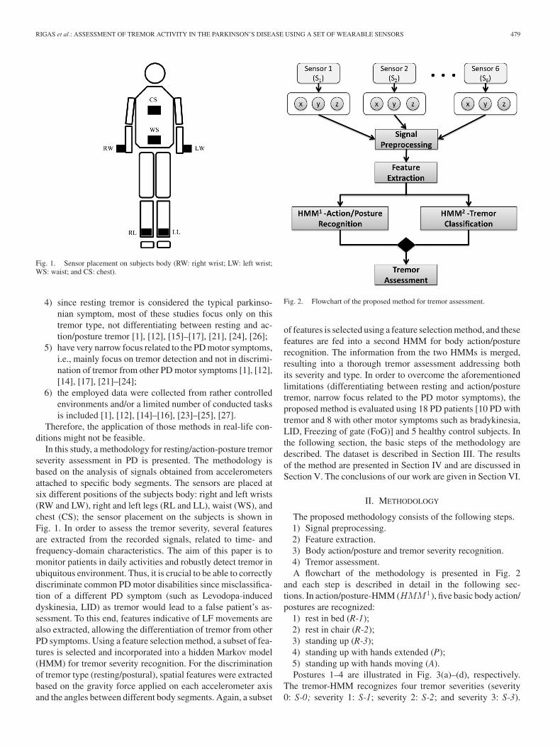
Fig. 1. Sensor placement on subjects body (RW: right wrist; LW: left wrist;WS: waist; and CS: chest).
4) since resting tremor is considered the typical parkinso-nian symptom, most of these studies focus only on thistremor type, not differentiating between resting and ac-tion/posture tremor [1], [12], [15]–[17], [21], [24], [26];
5) have very narrow focus related to the PD motor symptoms,i.e., mainly focus on tremor detection and not in discrimi-nation of tremor from other PD motor symptoms [1], [12],[14], [17], [21]–[24];
6) the employed data were collected from rather controlledenvironments and/or a limited number of conducted tasksis included [1], [12], [14]–[16], [23]–[25], [27].
Therefore, the application of those methods in real-life con-ditions might not be feasible.
In this study, a methodology for resting/action-posture tremorseverity assessment in PD is presented. The methodology isbased on the analysis of signals obtained from accelerometersattached to specific body segments. The sensors are placed atsix different positions of the subjects body: right and left wrists(RW and LW), right and left legs (RL and LL), waist (WS), andchest (CS); the sensor placement on the subjects is shown inFig. 1. In order to assess the tremor severity, several featuresare extracted from the recorded signals, related to time- andfrequency-domain characteristics. The aim of this paper is tomonitor patients in daily activities and robustly detect tremor inubiquitous environment. Thus, it is crucial to be able to correctlydiscriminate common PD motor disabilities since misclassifica-tion of a different PD symptom (such as Levodopa-induceddyskinesia, LID) as tremor would lead to a false patient’s as-sessment. To this end, features indicative of LF movements arealso extracted, allowing the differentiation of tremor from otherPD symptoms. Using a feature selection method, a subset of fea-tures is selected and incorporated into a hidden Markov model(HMM) for tremor severity recognition. For the discriminationof tremor type (resting/postural), spatial features were extractedbased on the gravity force applied on each accelerometer axisand the angles between different body segments. Again, a subset
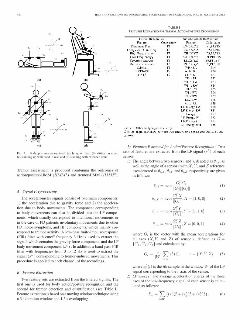
Fig. 2. Flowchart of the proposed method for tremor assessment.
of features is selected using a feature selection method, and thesefeatures are fed into a second HMM for body action/posturerecognition. The information from the two HMMs is merged,resulting into a thorough tremor assessment addressing bothits severity and type. In order to overcome the aforementionedlimitations (differentiating between resting and action/posturetremor, narrow focus related to the PD motor symptoms), theproposed method is evaluated using 18 PD patients [10 PD withtremor and 8 with other motor symptoms such as bradykinesia,LID, Freezing of gate (FoG)] and 5 healthy control subjects. Inthe following section, the basic steps of the methodology aredescribed. The dataset is described in Section III. The resultsof the method are presented in Section IV and are discussed inSection V. The conclusions of our work are given in Section VI.
II. METHODOLOGY
The proposed methodology consists of the following steps.1) Signal preprocessing.2) Feature extraction.3) Body action/posture and tremor severity recognition.4) Tremor assessment.A flowchart of the methodology is presented in Fig. 2
and each step is described in detail in the following sec-tions. In action/posture-HMM (HMM 1), five basic body action/postures are recognized:
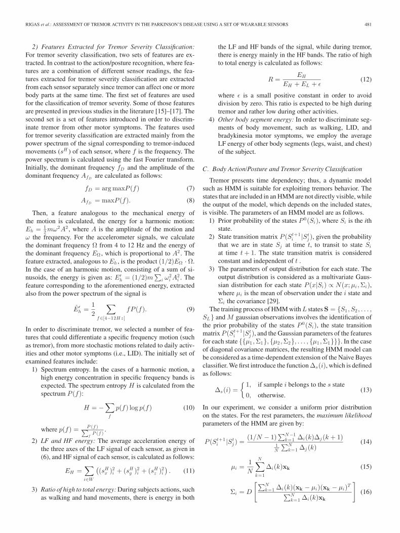
1) rest in bed (R-1);2) rest in chair (R-2);3) standing up (R-3);4) standing up with hands extended (P);5) standing up with hands moving (A).Postures 1–4 are illustrated in Fig. 3(a)–(d), respectively.
The tremor-HMM recognizes four tremor severities (severity0: S-0; severity 1: S-1; severity 2: S-2; and severity 3: S-3).
480 IEEE TRANSACTIONS ON INFORMATION TECHNOLOGY IN BIOMEDICINE, VOL. 16, NO. 3, MAY 2012
Fig. 3. Body postures recognized: (a) lying on bed, (b) sitting on chair,(c) standing up with hand in rest, and (d) standing with extended arms.
Tremor assessment is produced combining the outcomes ofaction/posture-HMM (HMM 1) and tremor-HMM (HMM 2).
A. Signal Preprocessing
The accelerometer signals consist of two main components:1) the acceleration due to gravity force and 2) the accelera-tion due to body movements. The component correspondingto body movements can also be divided into the LF compo-nents, which usually correspond to intentional movements orin the case of PD patients involuntary movements due to otherPD motor symptoms, and HF components, which mainly cor-respond to tremor activity. A low-pass finite-impulse-response(FIR) filter with cutoff frequency 3 Hz is used to extract thesignal, which contains the gravity force components and the LFbody movement component (sL ) . In addition, a band-pass FIRfilter with frequencies from 3 to 12 Hz is used to extract thesignal (sH ) corresponding to tremor-induced movements. Thisprocedure is applied to each channel of the recordings.
B. Feature Extraction
Two feature sets are extracted from the filtered signals. Thefirst one is used for body action/posture recognition and thesecond for tremor detection and quantification (see Table I).Feature extraction is based on a moving window technique usinga 3 s duration window and 1.5 s overlapping.
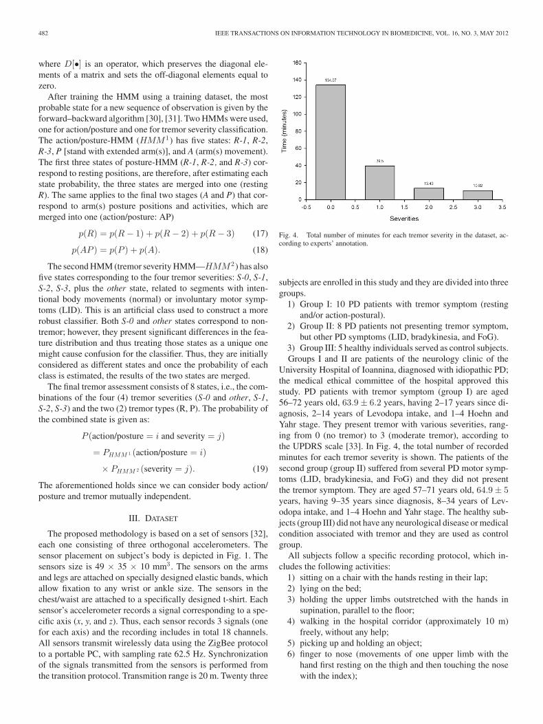
TABLE IFEATURES EXTRACTED FOR TREMOR ACTION/POSTURE RECOGNITION
1) Features Extracted for Action/Posture Recognition: Twosets of features are extracted from the LF signal (sL ) of eachsensor.
1) The angle between two sensors i and j, denoted as θi,j , aswell as the angle of a sensor i with X , Y , and Z referenceaxes denoted as θi,X , θi,Y and θi,Z , respectively, are givenas follows:
θi,j = acosGT
i Gj
‖Gi‖‖Gj‖(1)
θi,X = acosGT
i X
‖Gi‖,X = [1, 0, 0] (2)
θi,Y = acosGT
i Y
‖Gi‖, Y = [0, 1, 0] (3)
θi,Z = acosGT
i Z
‖Gi‖, Z = [0, 0, 1] (4)
where Gi is the vector with the mean accelerations forall axes (X,Y, and Z) of sensor i, defined as G ={Gx,Gy ,Gz} and calculated by:
Gr =1
|W |∑
i∈W
sLr (i), r = {X,Y,Z} (5)
where sLr (i) is the ith sample in the window W of the LF
signal corresponding to the r axis of the sensor.2) LF energy: The average acceleration energy of the three
axes of the low-frequency signal of each sensor is calcu-lated as follows:
EL =∑
i∈W
((sL
x )2i + (sL
y )2i + (sL
z )2i
). (6)
RIGAS et al.: ASSESSMENT OF TREMOR ACTIVITY IN THE PARKINSON’S DISEASE USING A SET OF WEARABLE SENSORS 481
2) Features Extracted for Tremor Severity Classification:For tremor severity classification, two sets of features are ex-tracted. In contrast to the action/posture recognition, where fea-tures are a combination of different sensor readings, the fea-tures extracted for tremor severity classification are extractedfrom each sensor separately since tremor can affect one or morebody parts at the same time. The first set of features are usedfor the classification of tremor severity. Some of those featuresare presented in previous studies in the literature [15]–[17]. Thesecond set is a set of features introduced in order to discrim-inate tremor from other motor symptoms. The features usedfor tremor severity classification are extracted mainly from thepower spectrum of the signal corresponding to tremor-inducedmovements (sH ) of each sensor, where f is the frequency. Thepower spectrum is calculated using the fast Fourier transform.Initially, the dominant frequency fD and the amplitude of thedominant frequency AfD
are calculated as follows:
fD = arg maxP (f) (7)
AfD= maxP (f). (8)
Then, a feature analogous to the mechanical energy ofthe motion is calculated, the energy for a harmonic motion:Eh = 1
2 mω2A2 , where A is the amplitude of the motion andω the frequency. For the accelerometer signals, we calculatethe dominant frequency Ω from 4 to 12 Hz and the energy ofthe dominant frequency EΩ , which is proportional to A2 . Thefeature extracted, analogous to Eh , is the product (1/2)EΩ · Ω.In the case of an harmonic motion, consisting of a sum of si-nusoids, the energy is given as: E′
h = (1/2)m∑
i ω2i A2
i . Thefeature corresponding to the aforementioned energy, extractedalso from the power spectrum of the signal is
E ′h =
12
∑
f∈[4−12H z ]
fP (f). (9)
In order to discriminate tremor, we selected a number of fea-tures that could differentiate a specific frequency motion (suchas tremor), from more stochastic motions related to daily activ-ities and other motor symptoms (i.e., LID). The initially set ofexamined features include:
1) Spectrum entropy. In the cases of a harmonic motion, ahigh energy concentration in specific frequency bands isexpected. The spectrum entropy H is calculated from thespectrum P (f):
H = −∑
f
p(f) log p(f) (10)
where p(f) = P (f )∑f
P (f ).
2) LF and HF energy: The average acceleration energy ofthe three axes of the LF signal of each sensor, as given in(6), and HF signal of each sensor, is calculated as follows:
EH =∑
i∈W
((sH
x )2i + (sH
y )2i + (sH
z )2i
). (11)
3) Ratio of high to total energy: During subjects actions, suchas walking and hand movements, there is energy in both
the LF and HF bands of the signal, while during tremor,there is energy mainly in the HF bands. The ratio of highto total energy is calculated as follows:
R =EH
EH + EL + ε(12)
where ε is a small positive constant in order to avoiddivision by zero. This ratio is expected to be high duringtremor and rather low during other activities.
4) Other body segment energy: In order to discriminate seg-ments of body movement, such as walking, LID, andbradykinesia motor symptoms, we employ the averageLF energy of other body segments (legs, waist, and chest)of the subject.
C. Body Action/Posture and Tremor Severity Classification
Tremor presents time dependency; thus, a dynamic modelsuch as HMM is suitable for exploiting tremors behavior. Thestates that are included in an HMM are not directly visible, whilethe output of the model, which depends on the included states,is visible. The parameters of an HMM model are as follows.
1) Prior probability of the states P 0(Si), where Si is the ithstate.
2) State transition matrix P (St+1i |St
j ), given the probabilitythat we are in state Sj at time t, to transit to state Si
at time t + 1. The state transition matrix is consideredconstant and independent of t .
3) The parameters of output distribution for each state. Theoutput distribution is considered as a multivariate Gaus-sian distribution for each state P (x|Si) ∝ N(x;μi,Σi),where μi is the mean of observation under the i state andΣi the covariance [29].
The training process of HMM with L states S = {S1 , S2 , . . . ,SL} and M gaussian observations involves the identification ofthe prior probability of the states P 0(Si), the state transitionmatrix P (St+1
i |Stj ), and the Gaussian parameters of the features
for each state {{μ1 ,Σ1}, {μ2 ,Σ2}, . . . , {μ1 ,Σ1}}}. In the caseof diagonal covariance matrices, the resulting HMM model canbe considered as a time-dependent extension of the Naive Bayesclassifier. We first introduce the function Δs(i), which is definedas follows:
Δs(i) ={
1, if sample i belongs to the s state
0, otherwise.(13)
In our experiment, we consider a uniform prior distributionon the states. For the rest parameters, the maximum likelihoodparameters of the HMM are given by:
P (St+1i |St
j ) =(1/N − 1)
∑N −1k=1 Δi(k)Δj (k + 1)
1N
∑Nk=1 Δj (k)
(14)
μi =1N
N∑
k=1
Δi(k)xk (15)
Σi = D
[∑Nk=1 Δi(k)(xk − μi)(xk − μi)T
∑Nk=1 Δi(k)xk
](16)
482 IEEE TRANSACTIONS ON INFORMATION TECHNOLOGY IN BIOMEDICINE, VOL. 16, NO. 3, MAY 2012
where D[•] is an operator, which preserves the diagonal ele-ments of a matrix and sets the off-diagonal elements equal tozero.
After training the HMM using a training dataset, the mostprobable state for a new sequence of observation is given by theforward–backward algorithm [30], [31]. Two HMMs were used,one for action/posture and one for tremor severity classification.The action/posture-HMM (HMM 1) has five states: R-1, R-2,R-3, P [stand with extended arm(s)], and A (arm(s) movement).The first three states of posture-HMM (R-1, R-2, and R-3) cor-respond to resting positions, are therefore, after estimating eachstate probability, the three states are merged into one (restingR). The same applies to the final two stages (A and P) that cor-respond to arm(s) posture positions and activities, which aremerged into one (action/posture: AP)
p(R) = p(R − 1) + p(R − 2) + p(R − 3) (17)
p(AP ) = p(P ) + p(A). (18)
The second HMM (tremor severity HMM—HMM 2 ) has alsofive states corresponding to the four tremor severities: S-0, S-1,S-2, S-3, plus the other state, related to segments with inten-tional body movements (normal) or involuntary motor symp-toms (LID). This is an artificial class used to construct a morerobust classifier. Both S-0 and other states correspond to non-tremor; however, they present significant differences in the fea-ture distribution and thus treating those states as a unique onemight cause confusion for the classifier. Thus, they are initiallyconsidered as different states and once the probability of eachclass is estimated, the results of the two states are merged.
The final tremor assessment consists of 8 states, i.e., the com-binations of the four (4) tremor severities (S-0 and other, S-1,S-2, S-3) and the two (2) tremor types (R, P). The probability ofthe combined state is given as:
P (action/posture = i and severity = j)
= PHMM 1 (action/posture = i)
× PHMM 2 (severity = j). (19)
The aforementioned holds since we can consider body action/posture and tremor mutually independent.
III. DATASET
The proposed methodology is based on a set of sensors [32],each one consisting of three orthogonal accelerometers. Thesensor placement on subject’s body is depicted in Fig. 1. Thesensors size is 49 × 35 × 10 mm3 . The sensors on the armsand legs are attached on specially designed elastic bands, whichallow fixation to any wrist or ankle size. The sensors in thechest/waist are attached to a specifically designed t-shirt. Eachsensor’s accelerometer records a signal corresponding to a spe-cific axis (x, y, and z). Thus, each sensor records 3 signals (onefor each axis) and the recording includes in total 18 channels.All sensors transmit wirelessly data using the ZigBee protocolto a portable PC, with sampling rate 62.5 Hz. Synchronizationof the signals transmitted from the sensors is performed fromthe transition protocol. Transmition range is 20 m. Twenty three
Fig. 4. Total number of minutes for each tremor severity in the dataset, ac-cording to experts’ annotation.
subjects are enrolled in this study and they are divided into threegroups.
1) Group I: 10 PD patients with tremor symptom (restingand/or action-postural).
2) Group II: 8 PD patients not presenting tremor symptom,but other PD symptoms (LID, bradykinesia, and FoG).
3) Group III: 5 healthy individuals served as control subjects.Groups I and II are patients of the neurology clinic of the
University Hospital of Ioannina, diagnosed with idiopathic PD;the medical ethical committee of the hospital approved thisstudy. PD patients with tremor symptom (group I) are aged56–72 years old, 63.9 ± 6.2 years, having 2–17 years since di-agnosis, 2–14 years of Levodopa intake, and 1–4 Hoehn andYahr stage. They present tremor with various severities, rang-ing from 0 (no tremor) to 3 (moderate tremor), according tothe UPDRS scale [33]. In Fig. 4, the total number of recordedminutes for each tremor severity is shown. The patients of thesecond group (group II) suffered from several PD motor symp-toms (LID, bradykinesia, and FoG) and they did not presentthe tremor symptom. They are aged 57–71 years old, 64.9 ± 5years, having 9–35 years since diagnosis, 8–34 years of Lev-odopa intake, and 1–4 Hoehn and Yahr stage. The healthy sub-jects (group III) did not have any neurological disease or medicalcondition associated with tremor and they are used as controlgroup.
All subjects follow a specific recording protocol, which in-cludes the following activities:
1) sitting on a chair with the hands resting in their lap;2) lying on the bed;3) holding the upper limbs outstretched with the hands in
supination, parallel to the floor;4) walking in the hospital corridor (approximately 10 m)
freely, without any help;5) picking up and holding an object;6) finger to nose (movements of one upper limb with the
hand first resting on the thigh and then touching the nosewith the index);
RIGAS et al.: ASSESSMENT OF TREMOR ACTIVITY IN THE PARKINSON’S DISEASE USING A SET OF WEARABLE SENSORS 483
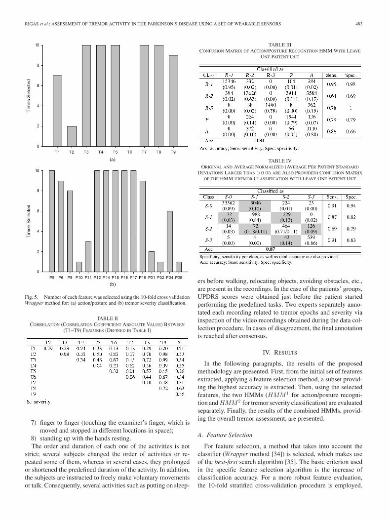
Fig. 5. Number of each feature was selected using the 10-fold cross validationWrapper method for: (a) action/posture and (b) tremor severity classification.
TABLE IICORRELATION (CORRELATION COEFFICIENT ABSOLUTE VALUE) BETWEEN
(T1–T9) FEATURES (DEFINED IN TABLE I)
7) finger to finger (touching the examiner’s finger, which ismoved and stopped in different locations in space);
8) standing up with the hands resting.The order and duration of each one of the activities is not
strict; several subjects changed the order of activities or re-peated some of them, whereas in several cases, they prolongedor shortened the predefined duration of the activity. In addition,the subjects are instructed to freely make voluntary movementsor talk. Consequently, several activities such as putting on sleep-
TABLE IIICONFUSION MATRIX OF ACTION/POSTURE RECOGNITION HMM WITH LEAVE
ONE PATIENT OUT
TABLE IVORIGINAL AND AVERAGE NORMALIZED (AVERAGE PER PATIENT STANDARD
DEVIATIONS LARGER THAN >0.05 ARE ALSO PROVIDED) CONFUSION MATRIX
OF THE HMM TREMOR CLASSIFICATION WITH LEAVE ONE PATIENT OUT
ers before walking, relocating objects, avoiding obstacles, etc.,are present in the recordings. In the case of the patients’ groups,UPDRS scores were obtained just before the patient startedperforming the predefined tasks. Two experts separately anno-tated each recording related to tremor epochs and severity viainspection of the video recordings obtained during the data col-lection procedure. In cases of disagreement, the final annotationis reached after consensus.
IV. RESULTS
In the following paragraphs, the results of the proposedmethodology are presented. First, from the initial set of featuresextracted, applying a feature selection method, a subset provid-ing the highest accuracy is extracted. Then, using the selectedfeatures, the two HMMs (HMM 1 for action/posture recogni-tion and HMM 2 for tremor severity classification) are evaluatedseparately. Finally, the results of the combined HMMs, provid-ing the overall tremor assessment, are presented.
A. Feature Selection
For feature selection, a method that takes into account theclassifier (Wrapper method [34]) is selected, which makes useof the best-first search algorithm [35]. The basic criterion usedin the specific feature selection algorithm is the increase ofclassification accuracy. For a more robust feature evaluation,the 10-fold stratified cross-validation procedure is employed.
484 IEEE TRANSACTIONS ON INFORMATION TECHNOLOGY IN BIOMEDICINE, VOL. 16, NO. 3, MAY 2012
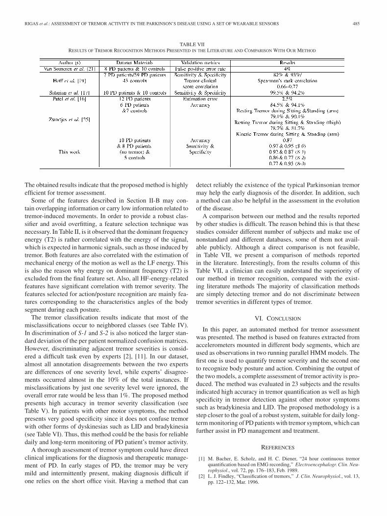
TABLE VNORMALIZED CONFUSION MATRIX OF THE TREMOR ASSESSMENT
Wrapper is performed using the Naive Bayes classifier, whichcan be considered as a single time-step HMM with Gaussianobservations. In order to remove possible bias in feature selec-tion, we applied the aforementioned procedure on a balanceddataset (equally sampled classed). This dataset was obtained af-ter resampling original dataset (from all three subject groups).In Fig. 5(a), we present the outcome of the Wrapper method interms of the action/posture recognition features. Features, whichare selected more than five times (out of 10) by the method, areincorporated in the action/posture HMM. In Table II, the corre-lation between the extracted features and the correlation of thefeatures with the class are presented. The correlations betweenfeatures and severities presented in Table II are indicative ofthe features selected by the Wrapper feature selection method,which are presented in Fig. 5(b). The least selected features arethe dominant frequency (T2) and the mechanic energy (T6);these two features are not included in the tremor-HMM.
B. Action/Posture Recognition
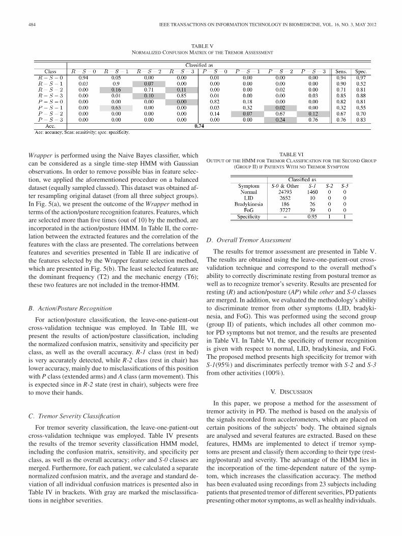
For action/posture classification, the leave-one-patient-outcross-validation technique was employed. In Table III, wepresent the results of action/posture classification, includingthe normalized confusion matrix, sensitivity and specificity perclass, as well as the overall accuracy. R-1 class (rest in bed)is very accurately detected, while R-2 class (rest in chair) haslower accuracy, mainly due to misclassifications of this positionwith P class (extended arms) and A class (arm movement). Thisis expected since in R-2 state (rest in chair), subjects were freeto move their hands.
C. Tremor Severity Classification
For tremor severity classification, the leave-one-patient-outcross-validation technique was employed. Table IV presentsthe results of the tremor severity classification HMM model,including the confusion matrix, sensitivity, and specificity perclass, as well as the overall accuracy; other and S-0 classes aremerged. Furthermore, for each patient, we calculated a separatenormalized confusion matrix, and the average and standard de-viation of all individual confusion matrices is presented also inTable IV in brackets. With gray are marked the misclassifica-tions in neighbor severities.
TABLE VIOUTPUT OF THE HMM FOR TREMOR CLASSIFICATION FOR THE SECOND GROUP
(GROUP II) IF PATIENTS WITH NO TREMOR SYMPTOM
D. Overall Tremor Assessment
The results for tremor assessment are presented in Table V.The results are obtained using the leave-one-patient-out cross-validation technique and correspond to the overall method’sability to correctly discriminate resting from postural tremor aswell as to recognize tremor’s severity. Results are presented forresting (R) and action/posture (AP) while other and S-0 classesare merged. In addition, we evaluated the methodology’s abilityto discriminate tremor from other symptoms (LID, bradyki-nesia, and FoG). This was performed using the second group(group II) of patients, which includes all other common mo-tor PD symptoms but not tremor, and the results are presentedin Table VI. In Table VI, the specificity of tremor recognitionis given with respect to normal, LID, bradykinesia, and FoG.The proposed method presents high specificity for tremor withS-1(95%) and discriminates perfectly tremor with S-2 and S-3from other activities (100%).
V. DISCUSSION
In this paper, we propose a method for the assessment oftremor activity in PD. The method is based on the analysis ofthe signals recorded from accelerometers, which are placed oncertain positions of the subjects’ body. The obtained signalsare analysed and several features are extracted. Based on thesefeatures, HMMs are implemented to detect if tremor symp-toms are present and classify them according to their type (rest-ing/postural) and severity. The advantage of the HMM lies inthe incorporation of the time-dependent nature of the symp-tom, which increases the classification accuracy. The methodhas been evaluated using recordings from 23 subjects includingpatients that presented tremor of different severities, PD patientspresenting other motor symptoms, as well as healthy individuals.
RIGAS et al.: ASSESSMENT OF TREMOR ACTIVITY IN THE PARKINSON’S DISEASE USING A SET OF WEARABLE SENSORS 485
TABLE VIIRESULTS OF TREMOR RECOGNITION METHODS PRESENTED IN THE LITERATURE AND COMPARISON WITH OUR METHOD
The obtained results indicate that the proposed method is highlyefficient for tremor assessment.
Some of the features described in Section II-B may con-tain overlapping information or carry low information related totremor-induced movements. In order to provide a robust clas-sifier and avoid overfitting, a feature selection technique wasnecessary. In Table II, is it observed that the dominant frequencyenergy (T2) is rather correlated with the energy of the signal,which is expected in harmonic signals, such as those induced bytremor. Both features are also correlated with the estimation ofmechanical energy of the motion as well as the LF energy. Thisis also the reason why energy on dominant frequency (T2) isexcluded from the final feature set. Also, all HF-energy-relatedfeatures have significant correlation with tremor severity. Thefeatures selected for action/posture recognition are mainly fea-tures corresponding to the characteristics angles of the bodysegment during each posture.
The tremor classification results indicate that most of themisclassifications occur to neighbored classes (see Table IV).In discrimination of S-1 and S-2 is also noticed the larger stan-dard deviation of the per patient normalized confusion matrices.However, discriminating adjacent tremor severities is consid-ered a difficult task even by experts [2], [11]. In our dataset,almost all annotation disagreements between the two expertsare differences of one severity level, while experts’ disagree-ments occurred almost in the 10% of the total instances. Ifmisclassifications by just one severity level were ignored, theoverall error rate would be less than 1%. The proposed methodpresents high accuracy in tremor severity classification (seeTable V). In patients with other motor symptoms, the methodpresents very good specificity since it does not confuse tremorwith other forms of dyskinesias such as LID and bradykinesia(see Table VI). Thus, this method could be the basis for reliabledaily and long-term monitoring of PD patient’s tremor activity.
A thorough assessment of tremor symptom could have directclinical implications for the diagnosis and therapeutic manage-ment of PD. In early stages of PD, the tremor may be verymild and intermittently present, making diagnosis difficult ifone relies on the short office visit. Having a method that can
detect reliably the existence of the typical Parkinsonian tremormay help the early diagnosis of the disorder. In addition, sucha method can also be helpful in the assessment in the evolutionof the disease.
A comparison between our method and the results reportedby other studies is difficult. The reason behind this is that thesestudies consider different number of subjects and make use ofnonstandard and different databases, some of them not avail-able publicly. Although a direct comparison is not feasible,in Table VII, we present a comparison of methods reportedin the literature. Interestingly, from the results column of thisTable VII, a clinician can easily understand the superiority ofour method in tremor recognition, compared with the exist-ing literature methods The majority of classification methodsare simply detecting tremor and do not discriminate betweentremor severities in different types of tremor.
VI. CONCLUSION
In this paper, an automated method for tremor assessmentwas presented. The method is based on features extracted fromaccelerometers mounted in different body segments, which areused as observations in two running parallel HMM models. Thefirst one is used to quantify tremor severity and the second oneto recognize body posture and action. Combining the output ofthe two models, a complete assessment of tremor activity is pro-duced. The method was evaluated in 23 subjects and the resultsindicated high accuracy in tremor quantification as well as highspecificity in tremor detection against other motor symptomssuch as bradykinesia and LID. The proposed methodology is astep closer to the goal of a robust system, suitable for daily long-term monitoring of PD patients with tremor symptom, which canfurther assist in PD management and treatment.
REFERENCES
[1] M. Bacher, E. Scholz, and H. C. Diener, “24 hour continuous tremorquantification based on EMG recording,” Electroencephalogr. Clin. Neu-rophysiol., vol. 72, pp. 176–183, Feb. 1989.
[2] L. J. Findley, “Classification of tremors,” J. Clin. Neurophysiol., vol. 13,pp. 122–132, Mar. 1996.
486 IEEE TRANSACTIONS ON INFORMATION TECHNOLOGY IN BIOMEDICINE, VOL. 16, NO. 3, MAY 2012
[3] G. Deuschl, P. Krack, M. Lauk, and J. Timmer, “Clinical neurophysiologyof tremor,” J. Clin. Neurophysiol., vol. 13, pp. 110–121, Mar. 1996.
[4] R. Wenzelburger, J. Raethjen, K. Lffler, H. Stolze, M. Illert, andG. Deuschl, “Kinetic tremor in a reach-to-grasp movement in Parkinson’sdisease,” Mov. Disord., vol. 15, pp. 1084–1094, 2000.
[5] A. Budzianowska and K. Honczarenko, “Assessment of rest tremor inParkinson’s disease,” Polish J. Neurol. Neurosurg., vol. 42, pp. 12–21,2008.
[6] J. A. Ward, P. Lukowicz, G. Troster, and T. E. Starner, “Activity recogni-tion of assembly tasks using body-worn microphones and accelerometers,”IEEE Trans. Pattern Anal. Mach. Intell., vol. 28, no. 10, pp. 1553–1567,Oct. 2006.
[7] K. R. Westerterp, “Assessment of physical activity: A critical appraisal,”Eur. J. Appl. Physiol., vol. 105, pp. 823–828, Apr. 2009.
[8] K. Aminian and B. Najafi, “Capturing human motion using body-fixedsensors: Outdoor measurement and clinical applications,” Comput. Ani-mat. Virtual Worlds, vol. 15, pp. 79–94, 2004.
[9] Y. Oshima, K. Kawaguchi, S. Tanaka, K. Ohkawara, Y. Hikihara,K. Ishikawa-Takata, and I. Tabata, “Classifying household and locomotiveactivities using a triaxial accelerometer,” Gait Post., vol. 31, pp. 370–374,2010.
[10] F. Foerster, M. Smeja, and J. Fahrenberg, “Detection of posture and motionby accelerometry: A validation study in ambulatory monitoring,” Comp.Human Behav., vol. 15, pp. 571–583, 1999.
[11] J. Jankovic, “Parkinson’s disease: Clinical features and diagnosis,” J.Neurol. Neurosurg. Psychiatry, vol. 79, pp. 368–376, 2008.
[12] J. Timmer, C. Gantert, G. Deuschl, and J. Honerkamp, “Characteristics ofhand tremor time series,” Biol. Cybern., vol. 70, pp. 75–80, 1993.
[13] C. N. Riviere, S. G. Reich, and N. V. Thakor, “Adaptive fourier modelingfor quantification of tremor,” J. Neurosci. Methods, vol. 74, pp. 77–87,Jun. 1997.
[14] P. E. O’Suilleabhain and J. Y. Matsumoto, “Time–frequency analysis oftremors,” Brain, vol. 121, pp. 2127–2134, Nov. 1998.
[15] S. Patel, R. Hughes, N. Huggins, D. Standaert, J. Growdon, J. Dy, andP. Bonato, “Using wearable sensors to predict the severity of symptomsand motor complications in late stage Parkinson’s Disease,” in Proc. 30thIEEE Annu. Int. Conf. Eng. Med. Biol. Soc., Vancouver, BC, Canada,2008, pp. 3686–3689.
[16] S. Patel, K. Lorincz, R. Hughes, N. Huggins, J. Growdon, D. Standaert,M. Akay, J. Dy, M. Welsh, and P. Bonato, “Monitoring motor fluctuationsin patients with Parkinson’s disease using wearable sensors,” IEEE Trans.Info. Tech. Biomed., vol. 13, no. 6, pp. 864–873, Nov. 2009.
[17] A. Salarian, H. Russmann, C. Wider, P. R. Burkhard, F. J. Vingerhoets, andK. Aminian, “Quantification of tremor and bradykinesia inParkinson’s disease using a novel quantification of tremor and bradyki-nesia in Parkinson’s disease using a novel ambulatory monitoring systemambulatory monitoring system,” IEEE Trans. Biomed. Eng., vol. 54, no. 2,pp. 313–322, Feb. 2007.
[18] M. Smeja, F. Foerster, G. Fuchs, D. Emmans, A. Hornig, and J. Fahrenberg,“24–h assessment of tremor activity and posture in Parkinson’s disease bymulti-channel accelerometry,” J. Psychophysiol., vol. 13, pp. 245–256,1999.
[19] J. I. Hoff, E. A. Wagemans, and B. J. van Hilten, “Ambulatory objectiveassessment of tremor in Parkinson’s disease,” Clin. Neuropharmacol.,vol. 24, pp. 280–283, Sep./Oct. 2001.
[20] M. Rudzinska, A. Izworski, K. Banaszkiewicz, S. Bukowczan, M. Marona,and A. Szczudlik, “Quantitative tremor measurement with the computer-ized analysis of spiral drawing,” Neurol. Neurochir. Pol., vol. 41, pp. 510–516, Nov./Dec. 2007.
[21] E. J. van Someren, B. F. Vonk, W. A. Thijssen, J. D. Speelman, P. R.Schuurman, M. Mirmiran, and D. F. Swaab, “A new actigraph for long-term registration of the duration and intensity of tremor and movement,”IEEE Trans. Biomed. Eng., vol. 45, no. 3, pp. 386–395, Mar. 1998.
[22] P. E. O’Suilleabhain and R. B. Dewey, Jr., “Validation for tremor quan-tification of an electromagnetic tracking device,” Mov. Disord., vol. 16,pp. 265–271, Mar. 2001.
[23] V. Rajaraman, D. Jack, S. V. Adamovich, W. Hening, J. Sage, andH. Poizner, “A novel quantitative method for 3D measurement of Parkin-sonian tremor,” Clin. Neurophysiol., vol. 111, pp. 338–343, Feb. 2000.
[24] K. E. Norman, R. Edwards, and A. Beuter, “The measurement of tremorusing a velocity transducer: Comparison to simultaneous recordings us-ing transducers of displacement, acceleration and muscle activity,” J.Neurosci. Methods, vol. 92, pp. 41–54, 1999.
[25] D. Zwartjes, T. Heida, J. van Vugt, J. Geelen, and P. Veltink, “Developmentof a system for measurement and analysis of tremor using a three-axisaccelerometer,” IEEE Trans. Biomed. Eng., vol. 57, pp. 2779–2786, 2010.
[26] N. Mamorita, T. Iizuka, A. Takeuchi, M. Shirataka, and N. Ikeda, “Ambula-tory monitoring of activities and motor symptoms in Parkinson’s disease,”Methods Inf. Med., vol. 48, pp. 589–594, 2009.
[27] I. Schlesinger, O. Benyakov, I. Erikh, S. Suraiya, and Y. Schiller, “Parkin-son’s disease tremor is diminished with relaxation guided imagery,” Mov.Disorders, vol. 24, pp. 2059–2062, 2009.
[28] R. A. Joundi, J.-S. Brittain, N. Jenkinson, A. L. Green, and T. Aziz, “Rapidtremor frequency assessment with the iPhone accelerometer,” Parkinson-ism Related Disorders, vol. 17, pp. 288–290, 2011.
[29] L. R. Rabiner, “A tutorial on hidden Markov models and selected appli-cations in speech recognition,” Proc. IEEE, vol. 77, no. 2, pp. 257–286,Feb. 1989.
[30] L. R. Rabiner and B. Juang, “An introduction to hidden Markov models,”IEEE–ASSP Mag., vol. 3, no. 1, pp. 4–16, Jan. 1986.
[31] K. Murphy, “The Bayes Net Toolbox for MATLAB,” Comput. Sci. Statist.,vol. 33, pp. 1786–1789, 2001.
[32] ANCO. (2010). [Online]. Available: http://www.anco.gr/[33] B. Post, M. P. Merkus, R. M. de Bie, R. J. de Haan, and J. D. Speelman,
“Unified Parkinson’s disease rating scale motor examination: Are ratingsof nurses, residents in neurology, and movement disorders specialistsinterchangeable?” Mov. Disord., vol. 20, pp. 1577–1584, Dec. 2005.
[34] R. Kohavi and G. John, “Wrappers for feature subset selection,” Artif.Intell., vol. 97, pp. 273–324, 1997.
[35] S. Russell and P. Norvig, Artificial Intelligence: A Modern Approach,2nd ed. Englewood Cliffs, NJ: Prentice-Hall, 2002.
George Rigas was born in Pella, Greece, in 1981. Hereceived the Bachelors, M.Sc., and Ph.D. degrees incomputer science from the Department of ComputerScience, University of Ioannina, Ioannina, Greece, in2003, 2005, and 2009, respectively
Since 2010, he has been a Postdoctoral Researcherin the Unit of Medical Technology and IntelligentInformation Systems,University of Ioannina. His re-search interests include signal processing, machinelearning, and pattern recognition.
Alexandros T. Tzallas (M’07) was born in Athens,Greece, in 1977. He received the Diploma degree inphysics and the Ph.D. degree in medical physics infrom the University of Ioannina, Ioannina, Greece, in2001 and 2009, respectively.
He is currently a Postdoctoral Researcher in theDepartment of Materials Science and Engineering,University of Ioannina. He has been involved in sev-eral research and development European and nationalprograms as a Software Engineer, Researcher, sem-inar Instructor, and Postdoctoral Researcher. He has
three years of teaching experience in universities and technological educationalinstitutes. He is the author or coauthor of 12 papers in scientific journals, 18papers in peer-reviewed conference proceedings, and 1 chapter in book.
Dr. Tzallas is a reviewer in several scientific journals and conferences.
Markos G. Tsipouras (M’07) was born in Athens,Greece, in 1977. He received the Diploma, M.Sc.,and Ph.D. degrees in computer science from the Uni-versity of Ioannina, Ioannina, Greece, in 1999, 2002,and 2009, respectively.
He is currently a Postdoctoral Researcher in theDepartment of Materials Science and Engineering,University of Ioannina. He is the author or coauthorof more than 15 papers in scientific journals, 30 pa-pers in peer-reviewed conference proceedings, and 6chapters in books. His work has received more than
120 citations. His research interests include biomedical engineering, decisionsupport and medical expert systems, and biomedical applications.
RIGAS et al.: ASSESSMENT OF TREMOR ACTIVITY IN THE PARKINSON’S DISEASE USING A SET OF WEARABLE SENSORS 487
Panagiota Bougia was born in Ioannina, Greece, in1980. She received the Diploma degree in electri-cal and computer engineering from the Polytech-nic School, Aristotle University of Thessaloniki,Thessaloniki, Greece, in 2003, and the M.Sc. degreein telecommunications applications from the Depart-ment of Physics, University of Ioannina, Ioannina,Greece, in 2008.
Since 2004, she has been a Software Engineerin the Unit of Medical Technology and Intelligent In-formation Systems, University of Ioannina. She is the
coauthor of several published scientific articles in related conferences, journals,and books.
Ms. Bougia has participated in a large number of European Commissionfunded projects as a Technical Manager.
Evanthia E. Tripoliti (M’08) was born onSeptember 12, 1978. She received the Diploma andM.Sc. degrees from the Department of ComputerScience, School of Natural Sciences, University ofIoannina, Ioannina, Greece, in 2001 and 2003, re-spectively, where she is currently working toward thePh.D. degree. Her Ph.D. degree focuses on the de-velopment of a method for the automated analysis ofbrain function of patients with Alzheimer’s diseaseusing functional magnetic resonance imaging.
Since 1999, she has been a member of Unit ofMedical Technology and Intelligent Information Systems, Department of Ma-terials Science and Engineering, University of Ioannina.
Dina Baga was born in Greece, in 1976. She re-ceived the Diploma degree in computer science fromthe University of Ioannina, Ioannina, Greece, in 1997.
Until 2009, she was the head of the Software En-gineering Team of the Unit of Medical Technologyand Intelligent Information Systems, University ofIoannina, and a Technical or Scientific Manager forseveral projects. She is now collaborating with theunit as a Software Engineer specialized on health in-formatics and intelligent decision support systems.She is the coauthor of nine published scientific arti-
cles in related conferences and journals.Ms. Baga has participated at the eHealth Awards (Cork, May 2004) with the
USBone project, as an award finalist.
Dimitrios I. Fotiadis (M’01–SM’07) was born inIoannina, Greece, in 1961. He received the Diplomadegree in chemical engineering from the NationalTechnical University of Athens, Athens, Greece, in1985, and the Ph.D. degree in chemical engineeringfrom the University of Minnesota, Minneapolis, in1990.
From 1995 to early 2008, he was with the Depart-ment of Computer Science, University of Ioannina,Ioannina, Greece, where he is currently a Professorin the Department of Materials Science and Technol-
ogy, the Director of the Unit of Medical Technology and Intelligent InformationSystems, and the President of the Science and Technology Park of Epirus. Heis also with the Biomedical Research Institute, Foundation for Research andTechnology-Hellas (FORTH), Ioannina. His current research interests includebiomedical technology, biomechanics, scientific computing, and intelligent in-formation systems.
Sofia G. Tsouli was born in Ioannina, Greece, in1977. She received the M.D. degree in medicine fromIoannina Medical School, Ioannina, Greece.
She is currently a Consultant Neurologist at theUniversity Hospital of Ioannina, Department of Neu-rology, University of Ioannina, Ioannina, Greece. Hercurrent research interests include the movement dis-orders in neurology.
Spyridon Konitsiotis was born in Corfu, Greece, in1963. He received the M.D. degree from the Univer-sity of Ioannina Medical School, Ioannina, Greece,in 1987, and the Ph.D. degree from the National In-stitutes of Health, Bethesda, MD, in 1990. His Ph.D.thesis focused on the neuropharmacology of basalganglia.
Since 2000, he has been with the University ofIoannina Medical School, where he is currently anAssistant Professor of Neurology. His research in-terests include movement disorders, Parkinson’s dis-
ease, neuropharmacology, and drug-induced movement disorders.