MDCT imaging of post interventional liver: a pictorial essay

Eur Radiol (2010) 20: 458–468DOI 10.1007/s00330-009-1571-7 MUSCULOSKELETAL
Ahi S. IsseverThomas M. LinkMarie KentenichPatrik RogallaAndrew J. BurghardtGalateia J. KazakiaSharmila MajumdarGerd Diederichs
Received: 18 January 2009Revised: 24 June 2009Accepted: 5 July 2009Published online: 27 August 2009# The Author(s) 2009. This article ispublished with open access atSpringerlink.com
Assessment of trabecular bone structure using
MDCT: comparison of 64- and 320-slice CT
using HR-pQCT as the reference standard
Abstract Objectives: The aim of ourstudy was to perform trabecular bonestructure analysis with images from64- and 320-slice multidetector com-puted tomography (MDCT) and tocompare these with high-resolutionperipheral computed tomography(HR-pQCT). Materials andmethods: Twenty human cadaverdistal forearm specimens were imagedon a 64- and 320-slice MDCT systemat 120 kVp, 200 mA and 135 kVp,400 mA (in-plane pixel size 234 µm;slice thickness 500 µm). HR-pQCT
imaging was performed at an isotro-pic voxel size of 41 µm. Bone volumefraction (BV/TV), trabecular number(Tb.N), thickness (Tb.Th) and sepa-ration (Tb.Sp) were computed.Results: MDCT-derived BV/TVand Tb.Sp were highly correlated(r=0.92–0.96, p<0.0001) with thecorresponding HR-pQCT parameters.Tb.Th was the only structure measurethat did not yield any significantcorrelation. Conclusion: The 64-and 320-slice MDCT systems bothperform equally well in depictingtrabecular bone architecture. Howev-er, because of constrained resolutionsaccurate derivation of trabecular bonemeasures is limited to only a subset ofmicroarchitectural parameters.
Keywords 320-slice MDCT .HR-pQCT . Osteoporosis .Structure analysis . Trabecular bone
Introduction
Osteoporosis is defined as a systemic skeletal diseasecharacterised by a loss of bone mass and a deterioration ofbone microarchitecture, subsequently leading to increasedsusceptibility to fracture [1]. Numerous studies have shownthat bone mineral density (BMD) is a fundamentaldeterminant of bone strength [2–7]. Hence, the establishedmethod in current osteoporosis diagnosis is the assessmentof BMD using either dual-energy X-ray absorptiometry(DXA) or quantitative computed tomography (QCT).However, clinicians and researchers have learned in recentyears that BMD only accounts for approximately 60–70%
of bone strength [1, 8], and sufficient discriminationbetween patients with and without increased fracture risk isnot provided by BMD measurements alone [9–11]. TheNational Institutes of Health Consensus Conference onOsteoporosis introduced the concept of “bone quality” in2001, referring to all characteristics of bone—apart fromBMD—responsible for its strength [1]. Among thesecharacteristics trabecular bone structure has been identifiedas a major contributor to bone strength [12–14].
With average trabecular dimensions ranging from 50 to200 µm, depiction and analysis of trabecular bone requirehigh-resolution imaging techniques. Micro-computedtomography (µCT), which is capable of approximately 3-
A. S. Issever . M. Kentenich .P. Rogalla . G. DiederichsDepartment of Radiology, CharitéCampus Mitte, Universitaetsmedizin,Berlin, Germany
A. S. Issever . T. M. Link .M. Kentenich . A. J. Burghardt .G. J. Kazakia . S. Majumdar .G. DiederichsDepartment of Radiology andBiomedical Imaging, Musculoskeletaland Quantitative Imaging ResearchGroup, University of California,San Francisco, CA, USA
A. S. Issever (*)Department of Radiology,Charité Campus Mitte,Universitaetsmedizin Berlin,Charitéplatz 1,10117 Berlin, Germanye-mail: [email protected].: +49-30-627043Fax: +49-30-527910

µm isotropic resolutions, may currently be considered themost established high-resolution imaging technique fortrabecular bone depiction. However, the application ofµCT imaging is restricted to either in vitro samples orinvasive biopsy imaging and is therefore not suitable fordaily clinical use. In the field of in vivo trabecular boneimaging, multidetector computed tomography (MDCT)and high-resolution magnetic resonance imaging (HR-MRI) have both been investigated in detail, but with in-plane pixel sizes of approximately 100–300 µm and slicethicknesses of 300–500 µm resolution constraints remain[15–18].
Recently, a high-resolution peripheral quantitative com-puted tomography (HR-pQCT) device was introduced thatpermits in vivo imaging of distal sites at a resolution of lessthan 100 µm [19]. MacNeil and Boyd tested the accuracy ofthis HR-pQCT device—at an isotropic 82-µm voxel size—on ten distal radii specimens using µCT (isotropic 19-µmvoxels) as the standard [20]. Highly significant correlationsfor Tb.Nwere found (R2=0.95). HR-pQCTmeasures of Tb.Th showed the lowest correlation (R2=0.59) with µCT inthis study. This suggests that HR-pQCT may be used as astandard of reference for in vivo trabecular bone imaging ofthe distal radius and the distal tibia.
However, as the availability of HR-pQCT systems islimited to only a few specialised facilities, implementationinto research and clinical practice may be consideredrestricted and strategies involving more accessible techni-ques such as MDCT and HR-MRI for in vivo trabecularbone imaging will remain essential in future osteoporosismanagement. The current study was performed to evaluatethe feasibility of MDCT-derived trabecular bone structureanalysis on intact human cadaver forearm specimens. Theaim of this study was to compare MDCT-derived apparentstructure measures with HR-pQCT-derived structure mea-sures as the standard of reference, and to evaluate possibledifferences in the derivation of apparent structure measuresusing two different MDCT devices (64-slice vs. 320-slice)with two different protocols (120 kVp and 200 mA vs.135 kVp and 400 mA).
Materials and methods
Specimens
Twenty human cadaver forearms fixed in 4% paraformal-dehyde were obtained from ten body donors (sevenwomen, three men; mean age 82 years, range 61–91 years). The specimens were obtained from theDepartment of Anatomy, Charité Campus Mitte, Universi-taetsmedizin Berlin, Germany. Before death, all donors hadgiven signed consent to dedicate their bodies to research.The forearms were removed from the remaining upperextremity distal from the elbow joint. Criteria for exclusionfrom the study were evidence of previous fracture,
osteolytic or osteoblastic bone lesions, and high-gradeosteoarthritis.
Multidetector computed tomography (MDCT)imaging
MDCT examinations of the whole specimens wereperformed using 64-slice (Toshiba Aquilion 64, Otawara,Japan) and 320-slice MDCT systems (Toshiba AquillionONE, Otawara, Japan) with the following protocols: (A)standard dose protocol with a tube current of 120 kVp and200 mA (rotation time 0.5 s), and (B) high-dose protocolwith 135 kVp and 400 mA (rotation time 1.0 s). Theacquisition mode on the 64-slice MDCT device was ahelical CT mode, whereas a volume CT mode was used forthe 320-slice MDCT device. A slice thickness of 0.5 mmwas obtained in both protocols and devices. The recon-struction interval was set to 0.3 mm on the 64-slice CTsystem and to 0.25 mm on the 320-slice CT system. Imageswere reconstructed with a field of view (FOV) of 120 mmon a standard image matrix size of 512×512 pixels yieldingan in-plane pixel size of 0.234 mm (Table 1). The imagingrange extended from the metacarpophalangeal joints to themid third of the radius. A high-resolution kernel (FC 81)was used for postprocessing of the 64-slice MDCT images.As this kernel option was not available for the 320-sliceMDCTa standard bone kernel (FC 30) in combination withan edge-enhancing filter was used on the 320-slice MDCTimages. For each scan protocol the manufacturer’s mod-ulation transfer function (MTF) values for the 64- and 320-slice MDCT devices are given in Table 1. Consistent withthe standard clinical patient scanning protocol (subjectpositioned prone with the arm extended above the head) thespecimens were placed supine (palmar side down) andalong the axis of the forearm on the CT table. In addition toassess BMD a standard hydroxyapatite (HA)-based calibra-tion phantom—consisting of five tubular inserts with varyingconcentrations (0, 50, 100, 150, 200 mg HA/cm3)—wasplaced underneath the specimens.
High-resolution peripheral quantitative computedtomography (HR-pQCT)
After MDCT imaging all specimens underwent imagingwith a clinical HR-pQCT system (XtremeCT, ScancoMedical AG, Bassersdorf, Switzerland). To ensure optimalimage co-registration the specimens were positioned in thesame manner as they were positioned for the MDCT(supine and along the axis of the forearm). A modifiedstandard in vivo protocol at the distal end of the radius wasused. The X-ray source potential was set to 60 kVp with acurrent of 900 µA. The 126-mm field of view (FOV) wasreconstructed across a 3,072×3,072 matrix, yielding 41-µm isotropic voxels.
459

Based on the two-dimensional X-ray scout view, theimage was defined to begin 9.5 mm proximal to the midlineof the distal radius endplate (Fig. 1). A three-dimensional(3D) stack of 220 high-resolution CT slices over a 9.02-mm-thick section was acquired. The overall imaging timewas 7.4 min per specimen. The calibration of thereconstructed linear attenuation values was carried outusing a separate density measurement performed on ahydroxyapatite phantom before imaging.
ROIs and image registration
The regions of interest (ROI) for the evaluation ofstructural parameters were placed in the trabecular com-partment of the distal radius, along the cortical and
trabecular bone interface, with an internal, endosteal offsetof at least 1 mm. Visual registration was used to evaluateimages of the same anatomical site on the HR-pQCT, 320-slice MDCT and 64-slice MDCT images. On the basis ofthe standardised specimen positioning during each proce-dure, similar axial slices were identified in each of the threeimaging datasets and defined as starting slices for furtherevaluation. Characteristics of the trabecular bone networkand the surrounding cortex served as confirmation (Fig. 2a, c and e). To cover the same imaging range of 9.0 mm asused in HR-pQCT imaging, 30 images were evaluated inthe 64-slice MDCT datasets (in increments of 300 µm) and36 axial images in the 320-slice MDCT datasets (inincrements of 250 µm).
Trabecular structure analysis
Structural analysis necessitates segmentation of grey-levelimages into binarised images consisting only of a bonephase and a bone marrow phase (Fig. 2 b, d and f).However, as the histogram characteristics of HR-pQCT(bimodal) and MDCT (monomodal) vary, different thresh-olding techniques have to be applied. Furthermore, becauseof differences in image characteristics and spatial resolu-tion (41×41×41 µm3 isotropic voxels in HR-pQCT vs.234×234×300/250 µm3 anisotropic voxels in MDCT)different algorithms to compute structural parameters haveto be used for each technique.
HR-pQCT Structure analysis of the HR-pQCT images wasperformed using the software provided by the manufac-turer. Image binarisation was performed within the definedtrabecular ROI after applying a global threshold. Theglobal threshold was calculated for each specimen using a
Fig. 1 HR-pQCT scout view. The imaging procedure was definedto begin 9.5 mm proximal to the midline of the distal radius endplate(white line). The imaging range covered 9.0 mm (distance betweenthe dotted lines)
Table 1 Imaging protocols and specifics applied with regard to 64- and 320-slice MDCT
MDCT (slice) 320 64
CT mode Volume Helical
Protocol A B A B
kVp 120 135 120 135
mA 200 400 200 400
Imaging time (s) 0.5 1.0 0.5 1.0
Radiation dose (mSV) < 0.1 < 0.1 < 0.1 < 0.1
Kernel FC 30 (bone) + edge enhancing FC 81 (high-resolution)
MTF 50% 9.41 7.46 11.33 9.00
MTF 10% 11.76 9.86 13.27 12.52
In-plane pixel size (µm) 234 234 234 234
Slice thickness (µm) 500 500 500 500
Slice increment (µm) 250 250 300 300
Concerning the radiation dose please note that the radiation dose for the standard HR-pQCT scans, which are performed at an isotropicresolution of 82 µm3, is below 0.3 µSV
460

common adaptive iterative method upon a specific spec-imen-based histogram analysis [21]. The underlyingassumption with regard to this method is that thehistogram intensity distribution is bimodal, expressing abone and background peak. The midpoint between the twopeaks was used as the threshold value. After imagebinarisation bone volume/total volume (BV/TV) wasderived via simple voxel counting. Calculation of trabec-ular number (Tb.N), trabecular thickness (Tb.Th) andtrabecular separation (Tb.Sp) was performed withoutmodel assumptions as direct measures [22]. Based onthe assumption that compact bone has an HA densityvalue of 1,200 mg/cm3, BMD of the trabecular compart-ment was obtained after calibration against a phantomcomposed of five cylinders of HA–resin mixtures with arange of mineral concentrations (0, 100, 200, 400,800 mg HA/cm3). Overall HR-pQCT measures served asthe standard of reference in this study.
MDCT Structure analysis of the MDCT images wasperformed using in-house-developed image analysis soft-ware programmed in IDL (RSI, Boulder, CO, USA). Forthreshold definition purposes six different thresholdsbased on six different HA density values (50, 100, 150,200, 250 and 300 mg HA/cm3) were applied to eachMDCT dataset. The HA density values were reconvertedto Hounsfield units (HU) via back-calibration using themanufacturer’s phantom on which the specimens wereplaced during the examination. The inverse calculation(HU to HA) was used to measure the BMD of the
trabecular compartment. Structural measures obtained foreach MDCT dataset with each of the six thresholds werecompared with the standard structure measures as assessedwith the HR-pQCT. The overall global threshold wasdefined to be at the HA density level that computed anoptimal combination of correlations, as described later.Following image binarisation apparent trabecular structuremeasures were derived, as previously described in detailby Majumdar and Genant [23]. Structural parametersanalogous to those obtained with standard histomorpho-metry techniques were assessed, but defined to be apparent(app.) structure measures, as standard histomorphometryanalysis is performed at much higher spatial resolutions(voxel sizes, approximately 5 μm). The followingapparent parameters were obtained: app. BV/TV, app.Tb.N, app. Tb.Th and app. Tb.Sp. Briefly, the totalnumber of bright pixels contributing to the bone phase inthe binarised image relative to the total number of pixels inthe ROI was used to compute app. BV/TV. The totalnumber of black and white pixel edges that cross a set ofparallel rays at a given angle θ through the image werecounted, then a measure of the mean intercept length wascomputed as the ratio between the total area of the brightpixels and half the number of edges. The mean value ofthe intercept length for all angles provided the width of thebright pixels and was defined as apparent trabecularthickness. From these measurements of app. BV/TV andapp. Tb.Th, two other morphological parameters weredetermined: app. Tb.N (area fraction of bright pixels/app.Tb.Th) and app. Tb.Sp ([1/app. Tb.N]−app. Tb.Th).
Fig. 2 Cross-sectional HR-pQCT image at an isotropic 41-µm resolution (a). MatchedMDCT images (protocol B)after visual image co-registra-tion acquired with the 64-slice(c) and 320-slice (e) MDCTsystems. Subfigures b, d and fshow the corresponding bi-narised images
461

Statistics
Mean values and standard deviations were calculated forHR-pQCT-derived BV/TV, Tb.N, Tb.Th, Tb.Sp and BMDmeasures and for MDCT-derived app. BV/TV, app. Tb.N,app. Tb.Th, app. Tb.Sp and BMD measures. Furthermoretwo-sided 95% confidence intervals were computed. One-way analysis of variance (ANOVA) tests were applied tocalculate differences in the mean values. Using linearregression analysis, Spearman rank’s correlation coeffi-cient r and the coefficient of determination R2 wereobtained to compare parameters. The level of significancewas set at p<0.05, and the level of high significance wasset at p<0.0001. All calculations were performed with JMPsoftware (SAS Institute, Cary, NC, USA).
Results
HR-pQCT vs. MDCT density measures
Highly significant correlations with r=0.97 to r=0.99 (p<0.0001) were found among BMD values of the trabecularcompartment as assessed by HR-pQCT- and all MDCT-derived BMD values. Student’s t test showed that thedensity measures showed no statistically relevant differ-ence (p>0.05) in the values acquired. The mean densityvalue as assessed with all techniques and imaging protocolswas 125.63±58.48 mg HA/cm3.
Highly significant (p<0.0001) correlations were foundfor HR-pQCT BMD measures vs. BV/TV (r=0.96), Tb.Sp(r=−0.89), and Tb.N (r=0.87). Tb.Th was the onlymeasure that showed no significant correlation with BMD.
Threshold definition in the MDCT datasets
To define the optimal global threshold, significant (p<0.05) Spearman correlation coefficient r values (MDCT vs.HR-pQCT)—for all structure measures—were evaluated asa function of the applied threshold level (50, 100, 150, 200,
250 and 300 mg HA/cm3). Overall, the highest and mostsignificant correlation was found for BV/TV, but withincreasing threshold—especially for measures acquiredusing protocol A—a gradual decrease in the correlationwas observed (e.g. 64-slice MDCTprotocol A fromr50 mg HA=cm3 =0.98 to r300 mg HA=cm3 =0.77). A decreasingcorrelation with increasing threshold was also observed forTb.Sp (64-slice MDCTprotocol A from r50 mg HA=cm3 =0.95to r200 mg HA=cm3 =0.47). Depending on the device type andprotocol the correlations for Tb.Sp lost significance (p>0.05) after applying a threshold of 150 to 250 mg HA/cm3.Inversely for correlations of Tb.N, significance was onlyachieved above a threshold level of 150 mg HA/cm3,whereas for Tb.Th significance was only found within thethreshold level range of at least 200 to 300 mg HA/cm3.Thus—as shown in Fig. 3 (320-slice MDCTprotocol A), forexample—the optimal combination of correlations wasfound—for each protocol, and each device—to be the150 mg HA/cm3 density level, which was then defined asthe global threshold for further evaluation.
HR-pQCT vs. MDCT structure measures
Table 2 shows HR-pQCT- and MDCT-derived mean andstandard deviation values for BV/TV, Tb.N, Tb.Th andTb.Sp.Compared with HR-pQCT-derived structure measures,MDCT-derived measures yielded two- to threefold highervalues for app. Tb.Th, up to twofold higher values for app. Tb.Sp, and increased app. BV/TV measures, thus overallresulting in a smaller amount of trabeculae detected, as seenby nearly 50% decreased app. Tb.N values.
ANOVA showed no device- (64-slice vs. 320-slice) orprotocol-dependent difference (120 kVp and 200 mA vs.135 kVp and 400 mA) in the primary apparent structuremeasures (app. BV/TV and app. Tb.Th) with p valuesabove 0.2. When comparing HR-pQCT-derived structuremeasures with MDCT-derived apparent structure measures(Table 3) highly significant correlations (p<0.0001) werefound for BV/TV and Tb.Sp with r values of 0.95 to 0.96
Fig. 3 Variations in the corre-lations between structure mea-sures derived from 320-sliceMDCT using protocol A com-pared with HR-pQCT-derivedstructure measures are presentedas a function of the appliedthreshold. The optimal combi-nation of correlations is found atthe level of 150 mg HA/cm3
462

and 0.92 to 0.93, respectively. Tb.N showed weakercorrelations; nonetheless, significant r values of 0.55 tohighly significant r values of 0.82 were obtained for the320-slice MDCT with protocol B. Neither of the MDCTdevices (64 vs. 320) nor protocols (A vs. B) producedsignificant correlations for Tb.Th values. When comparingthe MDCT-derived apparent structure measures amongeach other (320 vs. 64 and protocol A vs. B) highly (p<0.0001) significant correlations are found as shown inFigs. 4 and 5, with the lowest protocol-dependent R2 of0.84 for app. Tb.N as assessed with the 320-slice MDCTdevice.
Discussion
In this study trabecular bone structure measures acquiredwith two different MDCT devices and two differentprotocols were compared with HR-pQCT. The resultsshow that most MDCT-derived structure measures yieldhighly significant correlations to HR-pQCT. Furthermore,no MDCT device- (320-slice vs. 64-slice) or protocol-dependent (120 kVp and 200 mAvs. 135 kVp and 400 mA)differences were identified for primary trabecular structuremeasures.
The current, clinically established method of diagnosingosteoporosis is the assessment of BMD. BMD accounts forapproximately 60–70% of bone strength [1, 8]; however,
for the purposes of fracture risk discrimination theassessment of BMD alone is insufficient. There is asubstantial overlap of patients with manifest osteoporoticfractures and normal BMD values vs. patients withoutfractures and osteoporotic BMD values [10, 25].
The term “bone quality” was introduced by the NationalInstitutes of Health Consensus Conference on Osteoporosisin 2001, encompassing all characteristics and features ofbone responsible for its strength, such as tissue composi-tion, geometry, turnover, cortical and trabecular micro-architecture, pre-existing damage accumulation etc. [1]. Asbone turnover processes are highly surface-sensitive,trabecular bone microarchitecture is apt to reflect physio-logical, pathophysiological and drug-induced changes.Thus, many studies have focused on the quantitativeanalysis of trabecular bone. Hildebrand et al. found that acombination of BMD and trabecular structure measuresmay explain 94% of trabecular bone strength comparedwith 64% if only BMD measures are used [22]. Thetrabecular bone structure of specimens from patients withfractures, as compared with those without fractures, ischaracterised by a significantly higher degree of anisotro-py, with proportionately fewer trabecular elements trans-verse to the primary load axis [26]. In a histomorphometricstudy of transiliac bone biopsies of 152 men it was revealedthat the number of clinical risk factors for osteoporosis isstrongly related to microarchitectural alterations of trabec-ular bone [27]. The assessment and analysis of trabecular
Table 3 Correlation coefficients (Spearman) of MDCT-derived apparent trabecular structure measures compared with HR-pQCT-derivedtrabecular structure measures
Structural parameters MDCT vs. HR-pQCT
64 – A 64 – B 320 – A 320 – B
BV/TV 0.96* 0.95* 0.96* 0.95*
Tb.N 0.76** 0.65** 0.55** 0.82*
Tb.Th NS. NS NS NS
Tb.Sp 0.93* 0.93* 0.93* 0.92*
NS nonsignificant correlations are not listed*p<0.0001; **p<0.05
Table 2 HR-pQCT-derived trabecular structure measures and MDCT-derived apparent trabecular structure measures acquired with the 64-slice (64) and the 320-slice (320) MDCT device
HR-pQCT MDCT
64 – A 320 – A 64 – B 320 – B
BV/TV (%) 24±6 [22; 27] 39±19 [30; 48] 46±17 [37; 54] 40±21 [29; 50] 36±19 [27; 45]
Tb.N (1/mm) 1.38±0.30 [1.24; 1.52] 0.56±0.15 [0.49; 0.63] 0.68±0.12 [0.63; 0.73] 0.47±0.14 [0.41; 0.54] 0.50±0.16 [0.42; 0.58]
Tb.Th (mm) 0.25±0.02 [0.24; 0.26] 0.67±0.22 [0.56; 0.78] 0.65±0.20 [0.56; 0.75] 0.79±0.31 [0.64; 0.94] 0.68±0.22 [0.58; 0.78]
Tb.Sp (mm) 0.77±0.31 [0.63; 0.92] 1.31±0.87 [0.90; 1.73] 0.87±0.46 [0.65; 1.09] 1.62±1.20 [1.04; 2.19] 1.66±1.21 [1.09; 2.23]
Data are expressed in mean ± standard deviation, 95% confidence interval [lower bound, upper bound]A 120 kVp, 200 mA; B 135 kVp, 400 mA
463

microarchitecture, in combination with BMD measures,may thus provide substantial advancement in the field ofosteoporosis diagnosis and therapy monitoring. Therefore,in daily clinical practice noninvasive—ideally high-reso-lution—imaging techniques, capable of depicting trabec-ular bone, are needed.
The first in vivo studies investigating trabecular bonestructure assessment were performed nearly 20 years ago,and since then there have been significant technicaladvances improving the spatial resolution of imaging [28,29]. The first peripheral quantitative computed tomographydevices were limited to nominal isotropic resolutions of170 µm [30], but with the HR-pQCT systems, in vivoimaging at a nominal isotropic resolution of 82 µm is nowapplicable at distal sites [19]. A number of studies havealready been published with HR-pQCT as the primary
imaging technique for trabecular bone structure analysis[19, 24, 31, 32].
The underlying motive of the current study was toevaluate the in vivo feasibility of trabecular bone structureanalysis at the distal radius, a commonly affected osteo-porosis fracture site. The lifetime risk of women at the ageof 50 of sustaining a wrist fracture ranges from 13.3% inAustralia to 20.8% in Sweden [33]. A recently publishedstudy has identified that previous wrist fractures stronglypredict the 3-year risk of any future osteoporotic fracture inpostmenopausal women, independent of BMD [34].Among the few published HR-pQCT studies, Kazakia etal. and Krug et al. compared HR-MRI- and HR-pQCT-derived in vivo structure measures of the distal radius [35–37]. They found highly significant intertechnique correla-tions; however, it was also observed that there are
Fig. 4 Comparison of apparent trabecular structure measures (BV/TV (a), Tb.N (b), Tb.Sp (c), Tb.Th (d)) derived using 320-slice vs.64-slice MDCT as assessed with protocol A (120 kVp and 200 mA,
black dots) and protocol B (135 kVp and 400 mA, red dots).Coefficient of determination R2 is given
464
significant differences in absolute structure values. Asidefrom HR-MRI, high-resolution MDCT is an alternativeimaging technique that has been used for in vivo trabecularbone imaging, but mainly for central sites such as the spine[17, 18, 28]. The aim of this study was to compare HR-pQCT trabecular bone structure measures of the distal radius(at an isotropic resolution of 41 µm) with MDCT-derivedstructure measures. The study design used is unique in twoaspects: to simulate a clinical, in vivo-like imaging setup,paraformaldehyde-fixed human cadaver distal forearms—with intact surrounding soft tissue—were used. Further-more, in this study the assessment of trabecular bonestructure was evaluated using a 320-slice MDCTsystem andcompared with a 64-slice MDCT system.
The “volume scan mode” of the 320-slice MDCT systemoffers a single rotation acquisition of 16 cm in the z-direction, thus avoiding interslice stitching artefacts and the
necessity of data interpolations, as required for standardhelicalCTacquisitions.Moreover, a novel algorithm enablingimage reconstruction with a slice increment of 250 µm (vs.300 µm) is implemented in the 320-slice device. Thus, whencovering an imaging range of 9 mm, as in our study, the 320-slice device computes 36 axial images, compared with 30axial images from the 64-slice device. Regardless of theseminor technical variations the yielded voxel sizes—for bothdevices—remain above the dimension of individual trabec-ulae. Thus—in line with published literature [38–40]—ourdata validate the anticipated result that both device types areequally well suited for the derivation of primary apparentstructure measures. No quantifiable difference with regard totrabecular bone depiction in the z-direction was foundbetween the 64- and 320-slice systems.
Our study protocol consisted of two different doseapplications: the standard dose protocol A used in our
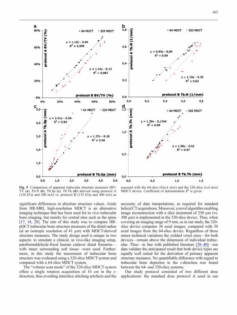
Fig. 5 Comparison of apparent trabecular structure measures (BV/TV (a), Tb.N (b), Tb.Sp (c), Tb.Th (d)) derived using protocol A(120 kVp and 200 mA) vs. protocol B (135 kVp and 400 mA) as
assessed with the 64-slice (black dots) and the 320-slice (red dots)MDCT device. Coefficient of determination R2 is given
465

clinic for peripheral bone imaging (120 kVp and200 mA), and a high-dose protocol B (135 kVp and400 mA). The aim was to evaluate whether high-doselevels improve trabecular bone structure depiction.However, our results show no statistically significantdifferences between primary measures acquired usingprotocol A and B. In a study on trabecular bone structureanalysis of the calcaneus performed by Patel et al. [41]four different imaging protocols were applied. Theauthors report that structure measures assessed with120 kVp and 150 mA did not differ substantially fromthose obtained with 120 kVp and 300 mA. Although ourprotocols ranged within different dose levels, one mayoverall hypothesize that beyond a given dose, an increaseof the same does not improve the assessment oftrabecular bone structure using MDCT. The identificationof this dose threshold for different skeletal regions maybe of interest for future research.
As possible MDCT device- and protocol-dependentalterations have been addressed, comparisons of MDCT-with HR-pQCT-derived structure measures are to bediscussed next. Overall, our results show highly significant(p<0.0001) correlations between MDCT and HR-pQCT,especially for BV/TV (r=0.95–0.96) and Tb.Sp (r=0.92–0.93). However, both measures show a substantial over-estimation when assessed with MDCT. These resultscorrespond well with previously published data. Diederichset al. [42] performed a study in which HR-pQCT andMDCT structure measures of proximal femur specimenswere compared. In this study, BV/TV and Tb.Sp were alsothe two structure measures that correlated best with HR-pQCT. However, the level of significance reached (p<0.01) and the correlation coefficients (BV/TV rPearson=0.87; Tb.Sp rPearson=0.66) were both lower than those inour study. This is most likely because of soft-tissue-induced constraints (central vs. peripheral sites) in trabec-ular bone assessment, as investigated by Bauer et al. [43].The average MDCT-derived app. Tb.Th in our studyyielded two- to threefold higher mean values than thecorresponding HR-pQCT measure. An overestimation oftrabecular dimensions based on partial volume effects maybe the reason for this. However, considering that only about50% of the trabeculae were detected with MDCT, one mayalso draw the more obvious conclusion that only thickertrabeculae were depicted, and thinner trabeculae weremissed. Overall, no significant correlation was found forMDCT- and HR-pQCT-derived Tb.Th measures. As aresult of MDCT resolutions (234×234×500 µm3) beyondthe average trabecular size, this is not unexpected and hasalready been observed by other investigators [38, 42–44].At this point, it needs to be carefully considered that givenpixel and voxel sizes are not equivalent to the true spatialresolution of an MDCT device. The spatial resolution is ameasure that quantifies the ability of an imaging system todifferentiate two objects of varying density from eachother, whereas the voxel size reflects the display resolution.
The modulation transfer function (MTF) is commonly usedto describe the spatial resolution in CT imaging. Amongothers it depends on the used kernel, contrast resolutionand dosage level. Thus, although it is possible to shrinkthe FOV to 6 cm and decrease the in-plane pixel size to117×117 µm2 for radius imaging, substantial benefits forthe derivation of structure measures remain hypothetical.Besides, full coverage of the density calibration phantomwould have not been assured, consecutively resulting inan inaccurate threshold definition. Thresholding may beconsidered as the first crucial postprocessing step intrabecular bone structure analysis. Careful identificationof the threshold level and subsequent image binarisationneed to ensure that dense bones do not only consist of“on” pixels and osteoporotic bones do not only consist of“off” pixels. Different methods have been proposed forMDCT image binarisation, among which visual optimi-sation-based approaches have frequently been used [17,41, 43]. However, in this study—to avoid operator-dependent bias—a correlation-based threshold definition[42], with HR-pQCT structure measures as a reference,has been applied.
One limitation of our study is the used sample size,which may be regarded small as the 20 specimens wereobtained from only ten body donors, resulting in arestricted distribution of BMD and trabecular structure.However, it needs to be considered that the availability ofhuman cadaver specimens with intact surrounding softtissue is generally constrained.
Another limitation of this study—in terms of itscomparability with daily clinical practice—is that byusing human cadaver specimens, no motion-inducedartefacts compromised image quality. For the standard 3-min HR-pQCT scans, it is known that patient movement isa significant source of error, commonly necessitatingreimaging or study exclusion. In this context, the fasterdata acquisition for MDCT systems (with an imaging timeframe of at most 1 s) would be expected to considerablyreduce the occurrence of motion artefacts.
In summary, this study evaluated the assessment oftrabecular structure measures from a 320-slice MDCTdevice and compared those with structure measuresobtained from a 64-slice MDCT device. Using intacthuman cadaver forearm specimens an in vivo-like studydesign for distal radius imaging was introduced. HR-pQCT at an isotropic resolution of 41 µm was used asthe standard of reference. No device- (320- vs. 64-slice)or protocol-dependent (120 kVp and 200 mA vs.135 kVp and 400 mA) differences were found. Althoughsubstantial differences in absolute values were observed,overall, most MDCT-derived structure measures corre-lated highly significantly with HR-pQCT measures.Thus, even though accurate depiction of individualtrabeculae is not given, these findings still confirm thatMDCT is capable of quantifying characteristics of thetrabecular bone network in the radius.
466

Acknowledgements This work was supported by funding from anElsbeth Bonhoff Grant (Charité 898 32 232).
Open Access This article is distributed under the terms of theCreative Commons Attribution Noncommercial License whichpermits any noncommercial use, distribution, and reproduction inany medium, provided the original author(s) and source are credited.
References
1. NIH Consensus Development Panel onOsteoporosis Prevention, Diagnosis,and Therapy (2001) Osteoporosis pre-vention, diagnosis, and therapy. JAMA285:785–795
2. Beck TJ, Ruff CB, Warden KE, ScottWW Jr, Rao GU (1990) Predictingfemoral neck strength from bone min-eral data. A structural approach. InvestRadiol 25:6–18
3. Bell GH, Cuthbertson DP, Orr J (1941)Strength and size of bone in relation tocalcium intake. J Physiol 100:299–317
4. Hansson T, Roos B, Nachemson A(1980) The bone mineral content andultimate compressive strength of lum-bar vertebrae. Spine 5:46–55
5. McBroom RJ, Hayes WC, EdwardsWT, Goldberg RP, White AA 3rd(1985) Prediction of vertebral bodycompressive fracture using quantitativecomputed tomography. J Bone JointSurg Am 67:1206–1214
6. Mosekilde L, Bentzen SM, Ortoft G,Jorgensen J (1989) The predictive valueof quantitative computed tomographyfor vertebral body compressive strengthand ash density. Bone 10:465–470
7. Mosekilde L, Mosekilde L, DanielsenCC (1987) Biomechanical competenceof vertebral trabecular bone in relationto ash density and age in normalindividuals. Bone 8:79–85
8. Ammann P, Rizzoli R (2003) Bonestrength and its determinants. Osteo-poros Int 14(Suppl 3):S13–S18
9. Kanis JA, Borgstrom F, De Laet C,Johansson H, Johnell O, Jonsson B,Oden A, Zethraeus N, Pfleger B,Khaltaev N (2005) Assessment offracture risk. Osteoporos Int 16:581–589
10. Schuit SC, van der Klift M, Weel AE,de Laet CE, Burger H, Seeman E,Hofman A, Uitterlinden AG, vanLeeuwen JP, Pols HA (2004) Fractureincidence and association with bonemineral density in elderly men andwomen: the Rotterdam study. Bone34:195–202
11. Taylor BC, Schreiner PJ, Stone KL,Fink HA, Cummings SR, Nevitt MC,Bowman PJ, Ensrud KE (2004) Long-term prediction of incident hip fracturerisk in elderly white women: study ofosteoporotic fractures. J Am GeriatrSoc 52:1479–1486
12. Kleerekoper M, Villanueva AR,Stanciu J, Rao DS, Parfitt AM (1985)The role of three-dimensional trabecu-lar microstructure in the pathogenesisof vertebral compression fractures.Calcif Tissue Int 37:594–597
13. Ulrich D, van Rietbergen B, Laib A,Ruegsegger P (1999) The ability ofthree-dimensional structural indices toreflect mechanical aspects of trabecularbone. Bone 25:55–60
14. van der Linden JC, Weinans H (2007)Effects of microarchitecture on bonestrength. Curr Osteoporos Rep 5:56–61
15. Alberich-Bayarri A, Marti-Bonmati L,Sanz-Requena R, Belloch E, Moratal D(2008) In vivo trabecular bone mor-phologic and mechanical relationshipusing high-resolution 3-T MRI. AJRAm J Roentgenol 191:721–726
16. Chang G, Pakin SK, Schweitzer ME,Saha PK, Regatte RR (2008) Adapta-tions in trabecular bone microarchitec-ture in Olympic athletes determined by7T MRI. J Magn Reson Imaging27:1089–1095
17. Graeff C, Timm W, Nickelsen TN,Farrerons J, Marin F, Barker C, GluerCC (2007) Monitoring teriparatide-as-sociated changes in vertebral micro-structure by high-resolution CT in vivo:results from the EUROFORS study. JBone Miner Res 22:1426–1433
18. Ito M, Ikeda K, Nishiguchi M, ShindoH, Uetani M, Hosoi T, Orimo H (2005)Multi-detector row CT imaging ofvertebral microstructure for evaluationof fracture risk. J Bone Miner Res20:1828–1836
19. Boutroy S, Bouxsein ML, Munoz F,Delmas PD (2005) In vivo assessmentof trabecular bone microarchitecture byhigh-resolution peripheral quantitativecomputed tomography. J Clin Endocri-nol Metab 90:6508–6515
20. MacNeil JA, Boyd SK (2007) Accura-cy of high-resolution peripheral quan-titative computed tomography formeasurement of bone quality. Med EngPhys 29:1096–1105
21. Ridler TW (1978) Picture thresholdingusing an iterative selection method.IEEE Trans Syst Man Cybern 8:630–632
22. Hildebrand T, Laib A, Muller R,Dequeker J, Ruegsegger P (1999) Di-rect three-dimensional morphometricanalysis of human cancellous bone:microstructural data from spine, femur,iliac crest, and calcaneus. J Bone MinerRes 14:1167–1174
23. Majumdar S, Genant HK (1997) As-sessment of trabecular structure usinghigh resolution magnetic resonanceimaging. Stud Health Technol Inform40:81–96
24. Chavassieux P, Asser Karsdal M,Segovia-Silvestre T, Neutzsky-WulffAV, Chapurlat R, Boivin G, Delmas PD(2008) Mechanisms of the anaboliceffects of teriparatide on bone: insightfrom the treatment of a patient withpycnodysostosis. J Bone Miner Res23:1076–1083
25. Beck TJ, Looker AC, Ruff CB,Sievanen H, Wahner HW (2000)Structural trends in the aging femoralneck and proximal shaft: analysis of theThird National Health and NutritionExamination Survey dual-energy X-rayabsorptiometry data. J Bone Miner Res15:2297–2304
26. Ciarelli TE, Fyhrie DP, Schaffler MB,Goldstein SA (2000) Variations inthree-dimensional cancellous bone ar-chitecture of the proximal femur infemale hip fractures and in controls. JBone Miner Res 15:32–40
27. Legrand E, Audran M, Guggenbuhl P,Levasseur R, Chales G, Basle MF,Chappard D (2007) Trabecular bonemicroarchitecture is related to thenumber of risk factors and etiology inosteoporotic men. Microsc Res Tech70:952–959
28. Chevalier F, Laval-Jeantet AM, Laval-Jeantet M, Bergot C (1992) CT imageanalysis of the vertebral trabecularnetwork in vivo. Calcif Tissue Int 51:8–13
29. Durand EP, Ruegsegger P (1991)Cancellous bone structure: analysis ofhigh-resolution CT images with therun-length method. J Comput AssistTomogr 15:133–139
467

30. Muller R, Hildebrand T, Ruegsegger P(1994) Non-invasive bone biopsy: anew method to analyse and display thethree-dimensional structure of trabecu-lar bone. Phys Med Biol 39:145–164
31. Sornay-Rendu E, Boutroy S, Munoz F,Delmas PD (2007) Alterations of cor-tical and trabecular architecture areassociated with fractures in postmeno-pausal women, partially independent ofdecreased BMD measured by DXA: theOFELY study. J Bone Miner Res22:425–433
32. Vico L, Zouch M, Amirouche A, FrereD, Laroche N, Koller B, Laib A,Thomas T, Alexandre C (2008) High-resolution pQCT analysis at the distalradius and tibia discriminates patientswith recent wrist and femoral neckfractures. J Bone Miner Res 23:1741–1750
33. Johnell O, Kanis J (2005) Epidemiolo-gy of osteoporotic fractures. Osteo-poros Int 16(Suppl 2):S3–S7
34. Barrett-Connor E, Sajjan SG, Siris ES,Miller PD, Chen YT, Markson LE(2008) Wrist fracture as a predictor offuture fractures in younger versus olderpostmenopausal women: results fromthe National Osteoporosis Risk As-sessment (NORA). Osteoporos Int19:607–613
35. Kazakia GJ, Hyun B, Burghardt AJ,Krug R, Newitt DC, de Papp AE, LinkTM, Majumdar S (2008) In vivodetermination of bone structure inpostmenopausal women: a comparisonof HR-pQCT and high-field MR imag-ing. J Bone Miner Res 23:463–474
36. Krug R, Carballido-Gamio J, BanerjeeS, Burghardt AJ, Link TM, MajumdarS (2008) In vivo ultra-high-field mag-netic resonance imaging of trabecularbone microarchitecture at 7 T. J MagnReson Imaging 27:854–859
37. Krug R, Carballido-Gamio J, BurghardtAJ, Kazakia G, Hyun BH, Jobke B,Banerjee S, Huber M, Link TM,Majumdar S (2008) Assessment oftrabecular bone structure comparingmagnetic resonance imaging at 3 Teslawith high-resolution peripheral quanti-tative computed tomography ex vivoand in vivo. Osteoporos Int 19:653–661
38. Kothari M, Keaveny TM, Lin JC,Newitt DC, Genant HK, Majumdar S(1998) Impact of spatial resolution onthe prediction of trabecular architectureparameters. Bone 22:437–443
39. Muller R, Koller B, Hildebrand T, LaibA, Gianolini S, Ruegsegger P (1996)Resolution dependency of microstruc-tural properties of cancellous bonebased on three-dimensional mu-tomo-graphy. Technol Health Care 4:113–119
40. Sode M, Burghardt AJ, Nissenson RA,Majumdar S (2008) Resolution depen-dence of the non-metric trabecularstructure indices. Bone 42:728–736
41. Patel PV, Prevrhal S, Bauer JS, Phan C,Eckstein F, Lochmuller EM, MajumdarS, Link TM (2005) Trabecular bonestructure obtained from multislice spiralcomputed tomography of the calcaneuspredicts osteoporotic vertebral defor-mities. J Comput Assist Tomogr29:246–253
42. Diederichs G, Link T, Marie K, HuberM, Rogalla P, Burghardt A, MajumdarS, Issever A (2008) Feasibility ofmeasuring trabecular bone structure ofthe proximal femur using 64-slicemultidetector computed tomography ina clinical setting. Calcif Tissue Int 83(5):332–41
43. Bauer JS, Link TM, Burghardt A,Henning TD, Mueller D, Majumdar S,Prevrhal S (2007) Analysis of trabecu-lar bone structure with multidetectorspiral computed tomography in a si-mulated soft-tissue environment. CalcifTissue Int 80:366–373
44. Bauer JS, Issever AS, Fischbeck M,Burghardt A, Eckstein F, Rummeny EJ,Majumdar S, Link TM (2004) Multi-slice-CT for structure analysis of tra-becular bone—a comparison withmicro-CT and biomechanical strength.Rofo 176:709–718
468
Copyright © 2022 FDOKUMEN