Line segment detection using weighted mean shift procedures on a 2D slice sampling strategy
Upload
independentCategory
view
5download
0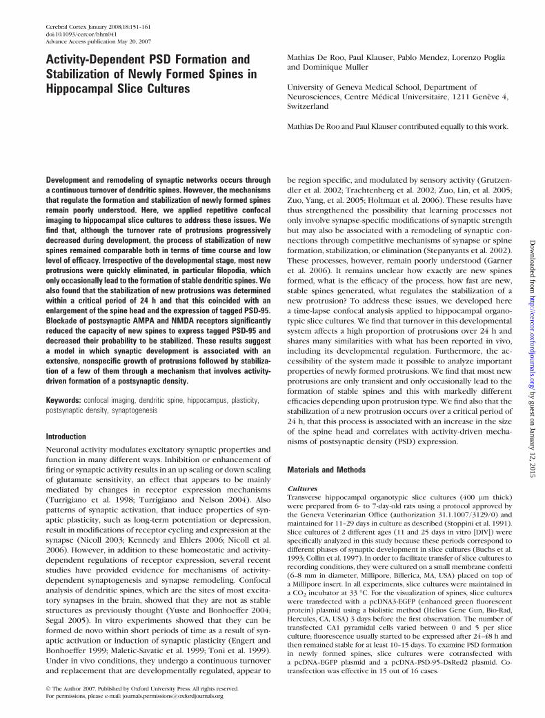
Activity-Dependent PSD Formation andStabilization of Newly Formed Spines inHippocampal Slice Cultures
Mathias De Roo, Paul Klauser, Pablo Mendez, Lorenzo Poglia
and Dominique Muller
University of Geneva Medical School, Department of
Neurosciences, Centre Medical Universitaire, 1211 Geneve 4,
Switzerland
Mathias De Roo and Paul Klauser contributed equally to thiswork.
Development and remodeling of synaptic networks occurs througha continuous turnover of dendritic spines. However, the mechanismsthat regulate the formation and stabilization of newly formed spinesremain poorly understood. Here, we applied repetitive confocalimaging to hippocampal slice cultures to address these issues. Wefind that, although the turnover rate of protrusions progressivelydecreased during development, the process of stabilization of newspines remained comparable both in terms of time course and lowlevel of efficacy. Irrespective of the developmental stage, most newprotrusions were quickly eliminated, in particular filopodia, whichonly occasionally lead to the formation of stable dendritic spines. Wealso found that the stabilization of new protrusions was determinedwithin a critical period of 24 h and that this coincided with anenlargement of the spine head and the expression of tagged PSD-95.Blockade of postsynaptic AMPA and NMDA receptors significantlyreduced the capacity of new spines to express tagged PSD-95 anddecreased their probability to be stabilized. These results suggesta model in which synaptic development is associated with anextensive, nonspecific growth of protrusions followed by stabiliza-tion of a few of them through a mechanism that involves activity-driven formation of a postsynaptic density.
Keywords: confocal imaging, dendritic spine, hippocampus, plasticity,postsynaptic density, synaptogenesis
Introduction
Neuronal activity modulates excitatory synaptic properties and
function in many different ways. Inhibition or enhancement of
firing or synaptic activity results in an up scaling or down scaling
of glutamate sensitivity, an effect that appears to be mainly
mediated by changes in receptor expression mechanisms
(Turrigiano et al. 1998; Turrigiano and Nelson 2004). Also
patterns of synaptic activation, that induce properties of syn-
aptic plasticity, such as long-term potentiation or depression,
result in modifications of receptor cycling and expression at the
synapse (Nicoll 2003; Kennedy and Ehlers 2006; Nicoll et al.
2006). However, in addition to these homeostatic and activity-
dependent regulations of receptor expression, several recent
studies have provided evidence for mechanisms of activity-
dependent synaptogenesis and synapse remodeling. Confocal
analysis of dendritic spines, which are the sites of most excita-
tory synapses in the brain, showed that they are not as stable
structures as previously thought (Yuste and Bonhoeffer 2004;
Segal 2005). In vitro experiments showed that they can be
formed de novo within short periods of time as a result of syn-
aptic activation or induction of synaptic plasticity (Engert and
Bonhoeffer 1999; Maletic-Savatic et al. 1999; Toni et al. 1999).
Under in vivo conditions, they undergo a continuous turnover
and replacement that are developmentally regulated, appear to
be region specific, and modulated by sensory activity (Grutzen-
dler et al. 2002; Trachtenberg et al. 2002; Zuo, Lin, et al. 2005;
Zuo, Yang, et al. 2005; Holtmaat et al. 2006). These results have
thus strengthened the possibility that learning processes not
only involve synapse-specific modifications of synaptic strength
but may also be associated with a remodeling of synaptic con-
nections through competitive mechanisms of synapse or spine
formation, stabilization, or elimination (Stepanyants et al. 2002).
These processes, however, remain poorly understood (Garner
et al. 2006). It remains unclear how exactly are new spines
formed, what is the efficacy of the process, how fast are new,
stable spines generated, what regulates the stabilization of a
new protrusion? To address these issues, we developed here
a time-lapse confocal analysis applied to hippocampal organo-
typic slice cultures. We find that turnover in this developmental
system affects a high proportion of protrusions over 24 h and
shares many similarities with what has been reported in vivo,
including its developmental regulation. Furthermore, the ac-
cessibility of the system made it possible to analyze important
properties of newly formed protrusions. We find that most new
protrusions are only transient and only occasionally lead to the
formation of stable spines and this with markedly different
efficacies depending upon protrusion type. We find also that the
stabilization of a new protrusion occurs over a critical period of
24 h, that this process is associated with an increase in the size
of the spine head and correlates with activity-driven mecha-
nisms of postsynaptic density (PSD) expression.
Materials and Methods
CulturesTransverse hippocampal organotypic slice cultures (400 lm thick)
were prepared from 6- to 7-day-old rats using a protocol approved by
the Geneva Veterinarian Office (authorization 31.1.1007/3129/0) and
maintained for 11--29 days in culture as described (Stoppini et al. 1991).
Slice cultures of 2 different ages (11 and 25 days in vitro [DIV]) were
specifically analyzed in this study because these periods correspond to
different phases of synaptic development in slice cultures (Buchs et al.
1993; Collin et al. 1997). In order to facilitate transfer of slice cultures to
recording conditions, they were cultured on a small membrane confetti
(6--8 mm in diameter, Millipore, Billerica, MA, USA) placed on top of
a Millipore insert. In all experiments, slice cultures were maintained in
a CO2 incubator at 33 �C. For the visualization of spines, slice cultures
were transfected with a pcDNA3-EGFP (enhanced green fluorescent
protein) plasmid using a biolistic method (Helios Gene Gun, Bio-Rad,
Hercules, CA, USA) 3 days before the first observation. The number of
transfected CA1 pyramidal cells varied between 0 and 5 per slice
culture; fluorescence usually started to be expressed after 24--48 h and
then remained stable for at least 10--15 days. To examine PSD formation
in newly formed spines, slice cultures were cotransfected with
a pcDNA--EGFP plasmid and a pcDNA--PSD-95--DsRed2 plasmid. Co-
transfection was effective in 15 out of 16 cases.
Cerebral Cortex January 2008;18:151--161
doi:10.1093/cercor/bhm041
Advance Access publication May 20, 2007
� The Author 2007. Published by Oxford University Press. All rights reserved.
For permissions, please e-mail: [email protected]
by guest on January 12, 2015http://cercor.oxfordjournals.org/
Dow
nloaded from
Confocal Imaging and AnalysisLaser scanning microscopy was realized with an Olympus Fluoview 300
system using a 488-nm Argon laser or a 2-photon laser set at 920 nm
(Coherent, Santa Clara, CA, USA). Transfected slice cultures were main-
tained in a recording chamber under immersion conditions with culture
serum for the time of observation (10 min, 25 �C) and then transferred
back to the incubator. In each experiment, a complete image of the slice
was initially obtained with a 53 objective to allow precise recognition
and localization of the dendritic segment under analysis (one segment
per slice culture). Also an image of the entire CA1 pyramidal neuron
with steps of 3 lm between scans was realized each day using the 403
objective to check for the general morphology and health of the
selected neuron. All analyses were carried out on dendritic segments of
about 35 lm in length and located between 150 and 300 lm from the
soma and imaged with a 403 objective using a 103 additional zoom
(final definition: 25 pixels per micron; steps between scans: 0.25--0.5
lm). Control experiments showed that this procedure could be applied
up to 10 consecutive times without deleterious effects on cell viability,
as indicated by absence of cell death, dendritic beadings, or propidium
iodide staining.
PSD-95--DsRed2 was imaged in all experiments using a spinning-disk
confocal system (Visitron Systems, Puchheim, Germany) and an excita-
tion laser set at 568 nm, and imaging was only carried out at the end of the
experiment to avoid possibilities of phototoxicity. Images were obtained
with a 403 objective using z-steps of 0.4 microns with Metamorph soft-
ware. Maximal intensity projections of the z-stacks were used to analyze
the presence or absence of PSD-95--DsRed2 staining. The average inten-
sity of the spine head area (determined in the EGFP image) was measured
in the red channel (PSD-95--DsRed2). Positive spines were in average 17%
above the threshold level. Spines were considered positive for PSD-95
staining if this average intensity was more than 5% higher than the back-
ground determined in an adjacent area. The minimal average intensity for
a positive spine was 11% and the maximal 30%.
All analyses were carried out blind by at least 2 independent observers
on z-stacks of raw images using a plugin specifically developed for
OsiriX software (http://homepage.mac.com/rossetantoine/osirix/).
Protrusions were classified as filopodia (protrusions without enlarge-
ment at the tip), stubby spines (short protrusions without neck and in
direct continuity with the dendritic surface), and mushroom spines
(protrusions with a neck and an enlargement at the tip). Determination
of spine behavior over time was done by systematic analysis of individual
z-stacked images. Also quantifications of spines width were carried out
by measuring the largest diameter of the spine head observed on any of
the z-stacked confocal images. All statistics are given with the standard
error of the mean. Standard t-tests were performed to compare Gaussian
distributions, and Mann--Whitney tests were used for non-Gaussian
distributions. For all tests, a was set to 5%.
Results
Spine Turnover Rate in Hippocampal Slice Cultures
To visualize dendritic spines, CA1 pyramidal neurons in slice
cultures were transfected with EGFP at either 8 or 22 DIV and
imaged repetitively over the next 3--7 days using confocal
microscopy. Control experiments showed that repetitive imag-
ing of the same cells and/or dendritic segments for up to 10
times under the conditions used resulted in no obvious signs
of toxicity such as beadings or cell death and no staining
Figure 1. Developmental regulation of protrusion turnover in hippocampal slice cultures. (A) Illustration of a typical CA1 pyramidal neuron imaged 3 (D1) and 8 (D5) days aftertransfection in an 11 DIV hippocampal organotypic culture. (B) Illustration of protrusion turnover obtained by daily confocal imaging of the same dendritic segment. The arrowheadindicates one stable spine; plus and minus signs are examples of new and lost spines, respectively. (C) Bar graph showing the percent of unchanged, stable spines (white bars),stable spines with changes in morphology (dashed bars), newly formed spines (black bars), and lost spines (gray bars) detected over a 24-h period in 11 DIV and 25 DIVhippocampal cultures. Data are mean ± standard error of the mean of analyses made on 10 dendritic segments from 10 pyramidal cells per group; P\ 0.05. (D) Changesin protrusion density expressed in each experiment as percents of initial values in 11 (open squares) and 25 (black circles) DIV cultures (n5 10; P\ 0.05; scale bars: A 100 lm;B 1 lm).
152 PSD and New Spine Stabilization d Roo et al.
by guest on January 12, 2015http://cercor.oxfordjournals.org/
Dow
nloaded from
for propidium iodide. Figure 1A illustrates one CA1 pyramidal
neuron imaged on days 1 and 5, following a daily analysis of the
spine changes occurring on one of its dendritic segments (Fig.
1B). As apparent in this example, numerous changes could
be detected over a 24-h period, including transformation or
modifications of the shape of spines, appearance of filopodia,
formation of new spines, and disappearance of preexisting
protrusions. Analyses were carried out on 20 different cells from
either 11 or 25 DIV hippocampal slice cultures with daily
observations made for 5 consecutive days on a total of 978
protrusions. The data, summarized in Figure 1C, indicate that
the changes detected over a 24-h period were considerably
larger in all aspects examined in 11 as compared with 25 DIV
slice cultures, thus clearly pointing to a developmental regula-
tion of spine turnover and plasticity in slice cultures. The
proportion of new protrusions averaged 19 ± 2% in 11 DIV slice
cultures, but only 8 ± 2% in 25 DIV tissue (n = 10 per group; P <
0.05), whereas the proportion of lost protrusions decreased
from 19 ± 3% to 12 ± 2% (n = 10; P < 0.05). Accordingly, the
proportion of stable protrusions increased from 81 ± 3% to 88 ±2% (n = 10; P < 0.05). Overall, the turnover rate calculated as
half of the sum of the ratios of new and lost protrusions
observed over 24 h decreased from 19 ± 2% to 10 ± 1% between
11 and 25 DIV (n = 10; P < 0.05).
Interestingly, in addition to new and lost protrusions, we also
observed a significant proportion of transformations or changes
in spine shape. These included not only transformations of
filopodia into stubby or mushroom spines but also transforma-
tions of stubby into mushroom spines (Fig. 3). The proportion of
these shape changes also considerably decreased with de-
velopmental maturation of slice cultures between DIV 11 and
25 (from 16 ± 2% to 8 ± 2%; n = 10; P < 0.05). Finally, as indicatedin Figure 1D, all these modifications occurred without detect-
able changes in protrusion density that remained constant both
during the periods of observation (Fig. 1D) and by comparison
of 11 and 25 DIV slice cultures (1.02 ± 0.11 vs. 1.06 ± 0.11
protrusions/1 lm, respectively; n = 10). All together, these data
indicated that dendritic spines in hippocampal slice cultures
exhibit a high degree of turnover that is developmentally
regulated.
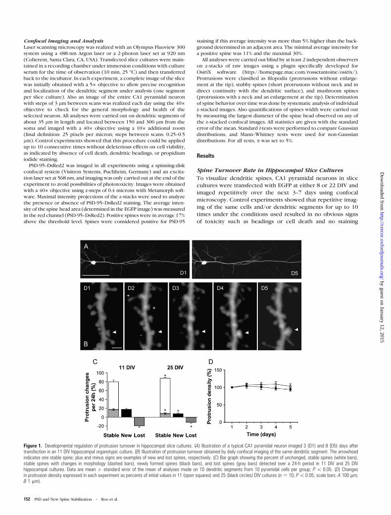
Characteristics of Protrusion Changes
In order to more precisely define the characteristics of new
protrusions, we classified them into 3 main categories: filopodia,
defined as long protrusions without head or widening at the
tip, stubby spines, defined as protrusions without neck, and
mushroom spines, protrusions with a neck and a widening
at the tip (Fig. 2A). Overall, the proportion between these
different protrusions did not vary greatly with development, as
the ratios were comparable in 11 and 25 DIV slice cultures,
although, as shown in Figure 2B, the proportion of filopodia was
slightly but not significantly smaller at 25 DIV (4 ± 2% vs. 8 ±3%). To verify this further and test the accuracy of the confocal
analyses, we also carried out an electron microscopic determi-
nation of the proportion of these different protrusions. For this,
Figure 2. Categorization of spine types in hippocampal cultures. (A) Typical examples of a mushroom spine (left), a stubby spine (center), and a filopodium (right) imaged withconfocal microscopy (left panel) and electron microscopy (right panel). (B) Proportion of protrusion types (mushroom spines: black columns; stubby spines: gray columns; filopodia:white columns) detected on dendritic segments imaged with confocal microscopy in 11 and 25 DIV cultures (n5 10 cells; 720 protrusions analyzed) and with electron microscopyin 22 DIV cultures (n5 3 cultures; 99 protrusions analyzed). (C) Proportion of the different types of new protrusions (mushroom spines: black columns; stubby spines: gray columns;filopodia: white columns) detected using different observation intervals in 11 DIV cultures. Data were obtained through analysis of 9 different cells (25--66 new protrusionsanalyzed). Note the nonlinearity of results indicating low stability of new protrusions and the high rates of formation of both new spines and filopodia with the shortest interval (scalebar: A 0.5 lm).
Cerebral Cortex January 2008, V 18 N 1 153
by guest on January 12, 2015http://cercor.oxfordjournals.org/
Dow
nloaded from

dendritic segments from 3 hippocampal slice cultures of 22 DIV
were analyzed from serial sections (Fig. 2A, right panel) and the
proportion of filopodia, stubby, and mushroom spines deter-
mined. As illustrated in Figure 2B, the ratios so obtained nicely
correlated with the data obtained by confocal microscopy.
Next, we used these categories to analyze the mechanisms
through which new spines were generated. For this, we
classified all new protrusions detected over different periods
of time. As shown on Figure 2C, the number of new protrusions
detected by repetitive imaging showed some variability, but
clearly depended upon the period of observation, indicating
that some protrusions were likely to have a rather short lifetime.
For this reason, we also carried out analyses with a short time
interval (2.5 h). These data indicated that new protrusions
mainly appeared as new mushroom spines (50%) or as filopodia
(40%) and that the rates of formation for both types of
protrusions were in the range of 2% of existing protrusions
per hour, which is rather high, but consistent with previous
Figure 3. Fate and stability of new protrusions. (A--D) Illustration of new protrusions fate. (A) New spine that remained stable for [24 h (arrowhead). (B) Filopodium thattransformed into a mushroom spine (arrowhead). (C) Transient filopodium. (D) Stubby spine that transformed into a mushroom spine. (E) Quantitative analysis of the fate of a pool of33 new filopodia analyzed in 7 experiments. Data are expressed as percent of the number of initial filopodia still present as filopodia (open bars) or mushroom spines (black bars) onthe next 4 consecutive days. (F) Same, but for a pool of 29 new stubby spines (gray bars) and their transformations in mushroom spines (black bars). (G) Same, but for a pool of 59new mushroom spines (black bars), among which some could transform back into stubby spines (gray bars). (H) Proportion of protrusion types (F: filopodia; S: stubby spines; M:mushroom spines) resulting in the formation of stable dendritic spines exhibiting a minimum 48 h stability in 11 DIV cultures. Note the low efficiency of filopodia in generating stablespines (scale bar: 0.5 lm).
154 PSD and New Spine Stabilization d Roo et al.
by guest on January 12, 2015http://cercor.oxfordjournals.org/
Dow
nloaded from
results obtained through continuous imaging over shorter
periods of time (Engert and Bonhoeffer 1999; Jourdain et al.
2003; Nagerl et al. 2004).
Fate and Stability of New Protrusions
In order to analyze the behavior, half-life, and efficacy with
which these different new protrusions generated stable synap-
tic contacts, we next followed the new events detected after
the first 2.5 h over 5 consecutive days. A total of 121 new
protrusions (33 filopodia, 29 stubby spines, and 59 new
mushroom spines) were analyzed together with their trans-
formations. Figure 3A--D illustrates some examples of these,
namely, a new mushroom spine that remained stable for 2 days
(Fig. 3A), a new filopodium that transformed into a mushroom
spine (Fig. 3B), a new filopodium that disappeared on the next
day (Fig. 3C), and a stubby spine that transformed into
a mushroom spine (Fig. 3D). These changes were then analyzed
quantitatively and summarized in Figure 3E--G. As illustrated in
Figure 3E, new filopodia were very labile structures as 33% of
them had already disappeared after 2.5 h (data not shown) and
only 13% were still present as filopodia after 24 h, 2% after 48 h,
and none after 72 h. Considering an exponential rate of
disappearance, one can calculate that the half-life of filopodia
was approximately 6 h. Although, in most cases (64%), filopodia
simply disappeared within 24 h, in 13% of cases, they trans-
formed into mushroom spines (Fig. 3E, black columns). These
transformations, however, were usually unstable as well, as most
of them (94%) also disappeared within the next 2 days. Overall,
only 1% of all filopodia resulted in the formation of mushroom
spines stable for over 2 days.
As indicated in Figure 3F, stubby spines were also rather
unstable protrusions, with only 17% and 9% of them being still
present after 1 and 2 days, respectively. This suggests a half-life
of about 15 h. A significant proportion of stubby spines (17%),
comparable to that of filopodia, was also transformed over 24 h
into mushroom spines. However, the stability of these mush-
room spines was not better than for filopodia as only 23% of
them persisted for the next 24 h. Overall, only 4% of all new
stubby spines resulted in the formation of mushroom spines
stable for at least 2 days.
Curiously, new mushroom spines were also markedly un-
stable. Most of them (56%) also disappeared within 24 h,
whereas 36% were still present and 8% transformed into stubby
spines. Their half-life calculated from their rate of disappear-
ance (Fig. 3G) was approximately 20 h. Overall, only 24% of all
newmushroom spines persisted for at least 2 days, indicating an
important rate of elimination. On a quantitative basis, these
results indicate that only 16% of all newly grown protrusions
detected over a 2.5-h period resulted in the formation of
dendritic spines stable for at least 2 days in 11 DIV slice
cultures. Interestingly, this very high initial instability was not
related to the developmental stage of the cultures. Similar
experiments carried out in 25 DIV slice cultures showed that,
although the turnover rate and rate of protrusion formation
decreased by a factor of 2 (Fig. 1C), the proportion of new
protrusions that succeeded in becoming stable remained very
low, with about 90% of them being still eliminated within the
first 48 h (n = 10). Furthermore, considering the relative
proportion of the different types of protrusions, it appears
that filopodia, although quite numerous, contributed to only
11% of all new mushroom spines exhibiting a minimum 48 h
stability; stubby spines contributed to 7%, whereas 82% of all
new stable spines were formed directly as new mushroom
spines (Fig. 3H).
Stability of Preexisting Protrusions
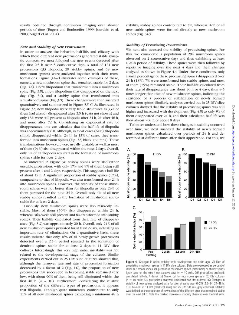
We next also assessed the stability of preexisting spines. For
this, we considered a population of 294 mushroom spines
observed on 2 consecutive days and thus exhibiting at least
a 24-h period of stability. These spines were then followed by
repetitive imaging over the next 4 days and their changes
analyzed as shown in Figure 4A. Under these conditions, only
a small percentage of these preexisting spines disappeared over
24 h (18%), 7% were transformed into stubby spines, and most
of them (75%) remained stable. Their half-life calculated from
their rate of disappearance was about 90 h or 4 days, thus 4--5
times longer than that of new mushroom spines, indicating the
existence of a process of stabilization of newly formed
mushroom spines. Similarly, analyses carried out in 25 DIV slice
cultures showed that the stability of preexisting spines was still
higher and increased with development (Fig. 4B) as only 9% of
them disappeared over 24 h, and their calculated half-life was
then almost 200 h or about 8 days.
To better understand how these changes in stability occurred
over time, we next analyzed the stability of newly formed
mushroom spines calculated over periods of 24 h and de-
termined at different times after their appearance. For this, we
Figure 4. Changes in spine stability with development and spine age. (A) Fate ofpreexisting mushroom spines in 11 DIV slice cultures. Data are expressed as percent ofinitial mushroom spines still present as mushroom spines (black bars) or stubby spines(gray bars) on the next 4 consecutive days (n 5 10 cells; 294 protrusions analyzed;calculated half-life: 4 days). (B) Same, but for mushroom spines in 25 DIV cultures(n 5 10 cells; 239 protrusions analyzed; calculated half-life: 8 days). (C) Changes instability of new spines analyzed as a function of spine age (0--2.5, 2.5--24, 24--48 h:n 5 14--408) in 11 DIV (black columns) and 25 DIV cultures (gray columns). Stabilitywas defined as the proportion of new spines of the different ages that remained stableover the next 24 h. Note the marked increase in stability observed over the first 24 h.
Cerebral Cortex January 2008, V 18 N 1 155
by guest on January 12, 2015http://cercor.oxfordjournals.org/
Dow
nloaded from

repetitively imaged mushroom spines at time 0, 2.5, 24, 48, and
72 h on dendritic segments obtained from 3 to 7 different cells
in 11 and 25 DIV cultures. We then calculated a stability index
for newly formed spines of different ages (ages: 0--2.5, 2.5--24,
24--48 h) by measuring the proportion of them that were still
present 24 h later. As shown in Figure 4C, in 11 DIV cultures,
only 36% of the new mushroom spines formed during the 0- to
2.5-h interval (n = 11) remained after the first 24 h, indicating
a very limited initial stability of very young, newly formed
spines. We next considered the pool of new spines generated
over the period 2.5--24 h (n = 54). From these, 59% were still
present at 48 h, whereas for new spines, aged between 24 and
48 h, 75% of them persisted for the next 24 h (Fig. 4C), a value
that is similar to that of preexisting spines. Thus, newly formed
spines acquired the same stability as preexisting spines within
a rather short period of time of about 24 h. Interestingly, very
similar results were also obtained with 25 DIV slice cultures,
despite the overall greater stability of preexisting spines at this
developmental stage. Initially, newly formed spines aged 0--2.5 h
showed a very high instability as only 42% of them persisted for
the next 24 h, but their stability then rapidly increased over the
next 24 h to reach the overall stability of preexisting spines (Fig.
4C). This result therefore indicates the existence of a stabiliza-
tion process that progressively increased the stability of newly
formed spines over the first 24--48 h.
Spine Stability and Spine Head Size
We next investigated mechanisms that might contribute to this
stabilization process by analyzing the possible role of the spine
head size, which has previously been proposed to correlate with
spine stability (Trachtenberg et al. 2002; Kasai et al. 2003;
Holtmaat et al. 2006). In a first group of experiments, we simply
measured the width of newly formed spines (age < 24 h) and
compared it to the values obtained for 3 days stable spines. As
shown in Figure 5B, newly formed mushroom spines were
characterized by a significantly smaller spine head than stable
ones (0.463 ± 0.011 vs. 0.539 ± 0.009 lm; n = 190--379; P <
0.001), indicating that spine head enlargement is indeed part of
a maturation process.
Figure 5. Stabilization of new spines is associated with an increase in spine head diameter. (A) Illustration of a newly formed mushroom spine showing an increase in headdiameter over the first 24 h (scale bar: 0.5 lm). (B) Newly formed spines (age\24 h) have smaller head width than stable spines (age[72 h). Data are mean ± standard error ofthe mean of measurements made on 190 new and 379 stable spines obtained from 16 cells (P\ 0.05). (C) Stability of spines expressed as a function of the spine head diameter(n5 6 cells; 178 spines analyzed; P\0.001). Stability was defined as the proportion of spines stable over 72 h. (D) Changes in spine head diameter measured over time in 6 newlyformed mushrooms spines (filled circles) that showed a 24-h stability and 9 preexisting, stable spines located on the same dendritic segments (open circles).
156 PSD and New Spine Stabilization d Roo et al.
by guest on January 12, 2015http://cercor.oxfordjournals.org/
Dow
nloaded from
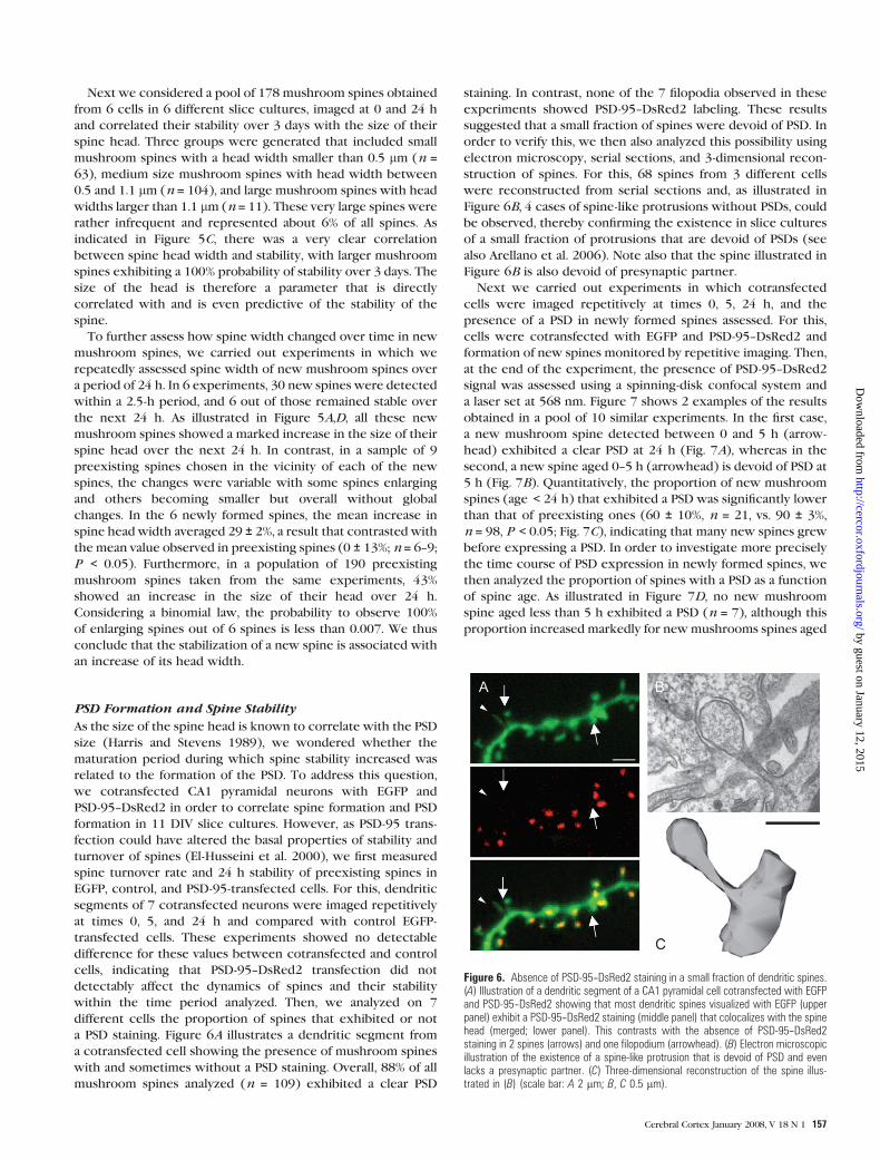
Next we considered a pool of 178 mushroom spines obtained
from 6 cells in 6 different slice cultures, imaged at 0 and 24 h
and correlated their stability over 3 days with the size of their
spine head. Three groups were generated that included small
mushroom spines with a head width smaller than 0.5 lm (n =63), medium size mushroom spines with head width between
0.5 and 1.1 lm (n = 104), and large mushroom spines with head
widths larger than 1.1 lm (n = 11). These very large spines were
rather infrequent and represented about 6% of all spines. As
indicated in Figure 5C, there was a very clear correlation
between spine head width and stability, with larger mushroom
spines exhibiting a 100% probability of stability over 3 days. The
size of the head is therefore a parameter that is directly
correlated with and is even predictive of the stability of the
spine.
To further assess how spine width changed over time in new
mushroom spines, we carried out experiments in which we
repeatedly assessed spine width of new mushroom spines over
a period of 24 h. In 6 experiments, 30 new spines were detected
within a 2.5-h period, and 6 out of those remained stable over
the next 24 h. As illustrated in Figure 5A,D, all these new
mushroom spines showed a marked increase in the size of their
spine head over the next 24 h. In contrast, in a sample of 9
preexisting spines chosen in the vicinity of each of the new
spines, the changes were variable with some spines enlarging
and others becoming smaller but overall without global
changes. In the 6 newly formed spines, the mean increase in
spine head width averaged 29 ± 2%, a result that contrasted with
the mean value observed in preexisting spines (0 ± 13%; n = 6--9;
P < 0.05). Furthermore, in a population of 190 preexisting
mushroom spines taken from the same experiments, 43%
showed an increase in the size of their head over 24 h.
Considering a binomial law, the probability to observe 100%
of enlarging spines out of 6 spines is less than 0.007. We thus
conclude that the stabilization of a new spine is associated with
an increase of its head width.
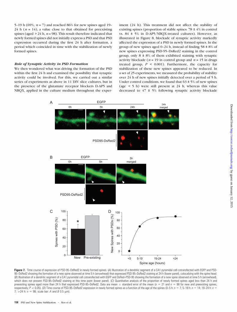
PSD Formation and Spine Stability
As the size of the spine head is known to correlate with the PSD
size (Harris and Stevens 1989), we wondered whether the
maturation period during which spine stability increased was
related to the formation of the PSD. To address this question,
we cotransfected CA1 pyramidal neurons with EGFP and
PSD-95--DsRed2 in order to correlate spine formation and PSD
formation in 11 DIV slice cultures. However, as PSD-95 trans-
fection could have altered the basal properties of stability and
turnover of spines (El-Husseini et al. 2000), we first measured
spine turnover rate and 24 h stability of preexisting spines in
EGFP, control, and PSD-95-transfected cells. For this, dendritic
segments of 7 cotransfected neurons were imaged repetitively
at times 0, 5, and 24 h and compared with control EGFP-
transfected cells. These experiments showed no detectable
difference for these values between cotransfected and control
cells, indicating that PSD-95--DsRed2 transfection did not
detectably affect the dynamics of spines and their stability
within the time period analyzed. Then, we analyzed on 7
different cells the proportion of spines that exhibited or not
a PSD staining. Figure 6A illustrates a dendritic segment from
a cotransfected cell showing the presence of mushroom spines
with and sometimes without a PSD staining. Overall, 88% of all
mushroom spines analyzed (n = 109) exhibited a clear PSD
staining. In contrast, none of the 7 filopodia observed in these
experiments showed PSD-95--DsRed2 labeling. These results
suggested that a small fraction of spines were devoid of PSD. In
order to verify this, we then also analyzed this possibility using
electron microscopy, serial sections, and 3-dimensional recon-
struction of spines. For this, 68 spines from 3 different cells
were reconstructed from serial sections and, as illustrated in
Figure 6B, 4 cases of spine-like protrusions without PSDs, could
be observed, thereby confirming the existence in slice cultures
of a small fraction of protrusions that are devoid of PSDs (see
also Arellano et al. 2006). Note also that the spine illustrated in
Figure 6B is also devoid of presynaptic partner.
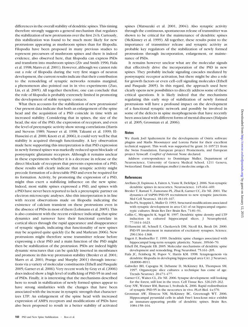
Next we carried out experiments in which cotransfected
cells were imaged repetitively at times 0, 5, 24 h, and the
presence of a PSD in newly formed spines assessed. For this,
cells were cotransfected with EGFP and PSD-95--DsRed2 and
formation of new spines monitored by repetitive imaging. Then,
at the end of the experiment, the presence of PSD-95--DsRed2
signal was assessed using a spinning-disk confocal system and
a laser set at 568 nm. Figure 7 shows 2 examples of the results
obtained in a pool of 10 similar experiments. In the first case,
a new mushroom spine detected between 0 and 5 h (arrow-
head) exhibited a clear PSD at 24 h (Fig. 7A), whereas in the
second, a new spine aged 0--5 h (arrowhead) is devoid of PSD at
5 h (Fig. 7B). Quantitatively, the proportion of new mushroom
spines (age < 24 h) that exhibited a PSD was significantly lower
than that of preexisting ones (60 ± 10%, n = 21, vs. 90 ± 3%,
n = 98, P < 0.05; Fig. 7C), indicating that many new spines grew
before expressing a PSD. In order to investigate more precisely
the time course of PSD expression in newly formed spines, we
then analyzed the proportion of spines with a PSD as a function
of spine age. As illustrated in Figure 7D, no new mushroom
spine aged less than 5 h exhibited a PSD (n = 7), although this
proportion increased markedly for newmushrooms spines aged
Figure 6. Absence of PSD-95--DsRed2 staining in a small fraction of dendritic spines.(A) Illustration of a dendritic segment of a CA1 pyramidal cell cotransfected with EGFPand PSD-95--DsRed2 showing that most dendritic spines visualized with EGFP (upperpanel) exhibit a PSD-95--DsRed2 staining (middle panel) that colocalizes with the spinehead (merged; lower panel). This contrasts with the absence of PSD-95--DsRed2staining in 2 spines (arrows) and one filopodium (arrowhead). (B) Electron microscopicillustration of the existence of a spine-like protrusion that is devoid of PSD and evenlacks a presynaptic partner. (C) Three-dimensional reconstruction of the spine illus-trated in (B) (scale bar: A 2 lm; B, C 0.5 lm).
Cerebral Cortex January 2008, V 18 N 1 157
by guest on January 12, 2015http://cercor.oxfordjournals.org/
Dow
nloaded from
5--19 h (69%, n = 7) and reached 86% for new spines aged 19--
24 h (n = 14), a value close to that obtained for preexisting
spines (aged > 24 h, n = 98). This result therefore indicated that
newly formed spines did not initially express a PSD and that PSD
expression occurred during the first 24 h after formation, a
period which coincided in time with the stabilization of newly
formed spines.
Role of Synaptic Activity in PSD Formation
We then wondered what was driving the formation of the PSD
within the first 24 h and examined the possibility that synaptic
activity could be involved. For this, we carried out a similar
series of experiments as above in 11 DIV slice cultures, but in
the presence of the glutamate receptor blockers D-AP5 and
NBQX, applied in the culture medium throughout the exper-
iment (24 h). This treatment did not affect the stability of
existing spines (proportion of stable spines: 78 ± 4% in control
vs. 84 ± 5% in D-AP5/NBQX-treated cultures). However, as
illustrated in Figure 8, blockade of synaptic activity markedly
affected the expression of a PSD in newly formed spines. In the
group of new spines aged 0--24 h, instead of finding 58 ± 8% of
new spines expressing PSD-95--DsRed2 staining in the control
group, only 8 ± 8% of them exhibited staining with synaptic
activity blockade (n = 19 in control group and n = 15 in drugs
treated group; P < 0.001). Furthermore, the capacity for
stabilization of these new spines appeared to be reduced. In
a set of 25 experiments, we measured the probability of stability
over 24 h of new spines initially detected over a period of 5 h.
Under control conditions, we found that 63 ± 5% of new spines
(age < 5 h) were still present at 24 h, whereas this value
decreased to 47 ± 5% following synaptic activity blockade
Figure 7. Time course of expression of PSD-95--DsRed2 in newly formed spines. (A) Illustration of a dendritic segment of a CA1 pyramidal cell cotransfected with EGFP and PSD-95--DsRed2 showing the formation of a new spine observed at time 5 h (arrowhead) that expressed PSD-95--DsRed2 staining at 24 h (lower panel), colocalizing with the spine head.(B) Illustration of a dendritic segment of a CA1 pyramidal cell cotransfected with EGFP and DsRed--PSD-95 showing the formation of a new spine observed at time 5 h (arrowhead),which does not present PSD-95--DsRed2 staining at this time point (lower panel). (C) Quantitative analysis of the proportion of newly formed spines aged less than 24 h andpreexisting spines aged more than 24 h that expressed PSD-95--DsRed2. Data are mean ± standard error of the mean (n 5 21 and n 5 98 for new and preexisting spines,respectively; P\0.05). (D) Time course of PSD-95--DsRed2 expression in newly formed spines as a function of the age of the spines (0--5 h: n5 7; 5--19 h: n5 14; 19--24 h: n57;[24 h: n 5 98; scale bar: A and B 0.5 lm).
158 PSD and New Spine Stabilization d Roo et al.
by guest on January 12, 2015http://cercor.oxfordjournals.org/
Dow
nloaded from

(n = 23--32; P < 0.05). These results therefore suggest a role
for synaptic activity in regulating expression of the PSD and
stabilization of newly formed spines.
Discussion
By analyzing dendritic spine turnover in developing hippocam-
pal organotypic slice cultures, we provide here important new
insights into the mechanisms that contribute to the formation
and stabilization of new protrusions. First, we find that the high
rate of protrusion formation that underlies synaptic develop-
ment in slice cultures is surprisingly poorly effective, as most
new protrusions (86--90%) are only transient and do not result
in the formation of stable dendritic spines. This is particularly
the case for filopodia, which, although numerous, almost always
failed to generate stable mushroom spines. Second, this early
elimination of newly formed spines appears to be characteristic
of the spine formation process, independently of the develop-
mental stage of the tissue, and to occur during a critical
stabilization period of about 24 h. Interestingly, we find that
this stabilization is directly correlated in its time course with an
enlargement of the spine head and the expression of a PSD, and
furthermore that expression of the PSD is regulated by synaptic
activity. All together these results are consistent with a model
where synaptic development proceeds through a marked, non-
specific growth of dendritic protrusions, followed by the
stabilization of a small number of spines through a process
that involves the expression of a PSD through a mechanism that
is driven by synaptic activity (Fig. 9).
This interpretation is in agreement with a number of existing
observations regarding spine properties or plasticity. First, the
turnover rate observed here in hippocampal slice cultures not
only compares well with the results reported in previous,
shorter time-lapse experiments (Engert and Bonhoeffer 1999;
Jourdain et al. 2003; Nagerl et al. 2004) but also falls within the
variability reported in the cortex of living mice (Grutzendler
et al. 2002; Trachtenberg et al. 2002; Holtmaat et al. 2005;
Majewska et al. 2006). In particular, the values obtained in 3- to
4-week-old cultures appear to be in range quite similar to that
reported in the barrel cortex of young, adult mice (Grutzendler
et al. 2002). Furthermore, the stability of spines in 25 DIV slice
cultures is consistent with what is reported in young mice
(Grutzendler et al. 2002; Trachtenberg et al. 2002). Also, we
find that this turnover rate is strongly developmentally regu-
lated in slice cultures as was reported in vivo (Holtmaat et al.
2005; Zuo, Lin, et al. 2005). These similarities add therefore
to the important correlations found otherwise in terms of
spine density, spine type distribution, spine shape, or synaptic
properties and function reported between hippocampal organo-
typic slice cultures and hippocampal or cortical tissue (Gahwiler
et al. 1997; Toni et al. 2001), strengthening the likely physio-
logical relevance of the present results.
Although the existence of a developmental regulation of
protrusion formation in slice cultures is of interest, a probably
more surprising result is the high proportion of these new
protrusions that are rapidly eliminated. This process seems to
differ from the usual concept of pruning that involves a com-
petitive elimination of existing spines or synapses (Segal 2005;
Zuo, Lin, et al. 2005). Here protrusions appear to be eliminated
even before they have a chance to form a synaptic contact and
express PSD-95. This conclusion is based both on confocal
analyses of the lack of expression of tagged PSD-95 in very
young, newly formed spines and electron microscopic obser-
vations of spine-like protrusions that are devoid of PSDs or
sometimes even lack a presynaptic partner in their immediate
vicinity (Fig. 6; Arellano et al. 2006). It would seem therefore
that the growth of these new protrusions initially occurs
without presynaptic involvement, a conclusion that is in
agreement with and extend previous and recent in vivo
observations (Marrs et al. 2001; Knott et al. 2006). As shown in
this study, the decision between elimination and stabilization
appears to be made within the first 24 h, a time course that does
not seem to vary with development, because the phenomenon
was similar in 11 and 25 DIV cultures, despite important
Figure 9. Schematic diagram summarizing the mechanisms of synaptic development likely to occur in hippocampal slice cultures. Spine turnover involves the growth of many newprotrusions extending in a nonspecific manner (middle panel). Among these, most of them will be rapidly eliminated within 24 h, whereas the few of them that will experiencesynaptic activity will be stabilized through expression of a PSD (right panel).
Figure 8. Synaptic activity regulates the expression of PSD-95--DsRed2 staining andthe stabilization of a new spine. (A) Proportion of newly formed spines, aged less than24 h, and stable, preexisting spines that expressed PSD-95--DsRed2 under controlconditions (gray columns) and in the presence of D-AP5 (50 lM) and NBQX (10 lM;black columns; *P\ 0.001). (B) Stability of newly formed spines, measured as theprobability of new spines aged 0--5 h to be still present 24 h later under controlconditions (gray column) and in the presence of D-AP5 and NBQX (n 5 23--32;*P\ 0.05).
Cerebral Cortex January 2008, V 18 N 1 159
by guest on January 12, 2015http://cercor.oxfordjournals.org/
Dow
nloaded from
differences in the overall stability of dendritic spines. This timing
therefore strongly suggests a general mechanism that regulates
the stabilization of new protrusions over the first 24 h. Curiously,
this stabilization was found to be much more likely for new
protrusions appearing as mushroom spines than for filopodia.
Filopodia have been proposed in many previous studies to
represent precursors of mushroom spines and there is indeed
evidence, also observed here, that filopodia can express PSDs
and transform into mushroom spines (Ziv and Smith 1996; Fiala
et al. 1998; Marrs et al. 2001). However, although we cannot rule
out a role of filopodia during the very first stages of neuron
development, the current results indicate that their contribution
to the remodeling of synaptic networks remains marginal,
a phenomenon also pointed out in in vivo experiments (Zuo,
Lin, et al. 2005). All together therefore, one can conclude that
the role of filopodia is probably extremely limited in promoting
the development of stable synaptic contacts.
What then accounts for the stabilization of new protrusions?
Our present data indicate that both an enlargement of the spine
head and the acquisition of a PSD coincide in time with an
increased stability. Considering that in spines, the size of the
head, the size of the PSD, the expression of receptors, and even
the level of presynaptic activity show strong correlations (Harris
and Stevens 1989; Nusser et al. 1998; Takumi et al. 1999; El-
Husseini et al. 2000; Knott et al. 2006), it could very well be that
stability is acquired through functionality. A key observation
made here supporting this interpretation is that PSD expression
in newly formed spines was markedly reduced upon blockade of
postsynaptic glutamate receptors. Although it remains unclear
in these experiments whether it is a decrease in release or the
direct blockade of receptors that prevents expression of a PSD,
these results still clearly indicate that synaptic activity might
precede formation of a detectable PSD and even be required for
its formation. Activity, by promoting the expression of a PSD,
might thus exert a stabilizing influence on the new spine.
Indeed, most stable spines expressed a PSD, and spines with
a PSD have never been reported to lack a presynaptic partner on
electron microscopic analyses. Also this interpretation is in line
with recent observations made on filopodia indicating the
existence of calcium transient on these protrusions even in
the absence of PSDs in most of them (Lohmann et al. 2005). This
is also consistent with the recent evidence indicating that spine
dynamics and turnover have their functional correlate in
cortical slices through the rapid appearance and disappearance
of synaptic signals, indicating that functionality of new spines
may be acquired quite quickly (Le Be and Markram 2006). New
protrusions might therefore sense transmitter release before
expressing a clear PSD and a main function of the PSD might
then be stabilization of the protrusion. PSDs are indeed highly
dynamic structures that can be quickly inserted in membranes
and promote in this way protrusion stability (Bresler et al. 2001;
Marrs et al. 2001; Prange and Murphy 2001) through interac-
tions via a variety of molecular constituents (Ethell and Pasquale
2005; Garner et al. 2006). Very recent work by Gray et al. (2006)
does indeed show a high level of trafficking of PSD-95 in and out
of PSDs. Finally, it is interesting that the mechanisms described
here to result in stabilization of newly formed spines appear to
have strong similarities with the changes that have been
associated with the increase in synaptic strength that character-
izes LTP. An enlargement of the spine head with increased
expression of AMPA receptors and modifications of PSDs have
also been proposed to result in a better stability of activated
spines (Matsuzaki et al. 2001, 2004). Also synaptic activity
through the continuous, spontaneous release of transmitter was
shown to be critical for the maintenance of dendritic spines
(McKinney et al. 1999). All together, these results point to the
importance of transmitter release and synaptic activity as
probable key regulators of the stabilization of newly formed
protrusions through incorporation, enlargement, or mainte-
nance of PSDs.
It remains however unclear what are the molecular signals
that effectively drive the incorporation of the PSD in new
spines. They probably include signaling cascades mediated by
postsynaptic receptor activation, but there might be also a role
for growth factors or even cell--cell signaling molecules (Ethell
and Pasquale 2005). In this regard, the approach used here
clearly opens new possibilities to directly address some of these
critical questions. It is likely indeed that the mechanisms
regulating this early step of stabilization of newly formed
protrusions will have a profound impact on the development
of a functional synaptic network and possibly be involved in
some of the defects in spine morphogenesis that have recently
been associated with different forms of mental diseases (Halpain
et al. 2005; Grossman et al. 2006).
Notes
We thank Joel Spaltenstein for the developments of Osirix software
plugins and Marlis Moosmayer and Lorena Parisi for their excellent
technical support. This work was supported by grant 31-105721 from
the Swiss Foundation, European project Promemoria, and European
project Synscaff. Conflict of Interest: None declared.
Address correspondence to Dominique Muller, Department of
Neuroscience, University of Geneva Medical School, 1211 Geneva,
Switzerland. Email: [email protected].
References
Arellano JI, Espinosa A, Fairen A, Yuste R, Defelipe J. 2006. Non-synaptic
dendritic spines in neocortex. Neuroscience. 145:464--469.
Bresler T, Ramati Y, Zamorano PL, Zhai R, Garner CC, Ziv NE. 2001. The
dynamics of SAP90/PSD-95 recruitment to new synaptic junctions.
Mol Cell Neurosci. 18:149--167.
Buchs PA, Stoppini L, Muller D. 1993. Structural modifications associated
with synaptic development in area CA1 of rat hippocampal organo-
typic cultures. Brain Res Dev Brain Res. 71:81--91.
Collin C, Miyaguchi K, Segal M. 1997. Dendritic spine density and LTP
induction in cultured hippocampal slices. J Neurophysiol.
77:1614--1623.
El-Husseini AE, Schnell E, Chetkovich DM, Nicoll RA, Bredt DS. 2000.
PSD-95 involvement in maturation of excitatory synapses. Science.
290:1364--1368.
Engert F, Bonhoeffer T. 1999. Dendritic spine changes associated with
hippocampal long-term synaptic plasticity. Nature. 399:66--70.
Ethell IM, Pasquale EB. 2005. Molecular mechanisms of dendritic spine
development and remodeling. Prog Neurobiol. 75:161--205.
Fiala JC, Feinberg M, Popov V, Harris KM. 1998. Synaptogenesis via
dendritic filopodia in developing hippocampal area CA1. J Neurosci.
18:8900--8911.
Gahwiler BH, Capogna M, Debanne D, McKinney RA, Thompson SM.
1997. Organotypic slice cultures: a technique has come of age.
Trends Neurosci. 20:471--477.
Garner CC, Waites CL, Ziv NE. 2006. Synapse development: still looking
for the forest, still lost in the trees. Cell Tissue Res. 326:249--262.
Gray NW, Weimer RM, Bureau I, Svoboda K. 2006. Rapid redistribution
of synaptic PSD-95 in the neocortex in vivo. PLoS Biol. 4:e370.
Grossman AW, Elisseou NM, McKinney BC, Greenough WT. 2006.
Hippocampal pyramidal cells in adult Fmr1 knockout mice exhibit
an immature-appearing profile of dendritic spines. Brain Res.
1084:158--164.
160 PSD and New Spine Stabilization d Roo et al.
by guest on January 12, 2015http://cercor.oxfordjournals.org/
Dow
nloaded from
Grutzendler J, Kasthuri N, Gan WB. 2002. Long-term dendritic spine
stability in the adult cortex. Nature. 420:812--816.
Halpain S, Spencer K, Graber S. 2005. Dynamics and pathology of
dendritic spines. Prog Brain Res. 147:29--37.
Harris KM, Stevens JK. 1989. Dendritic spines of CA 1 pyramidal cells in
the rat hippocampus: serial electron microscopy with reference to
their biophysical characteristics. J Neurosci. 9:2982--2997.
Holtmaat A, Wilbrecht L, Knott GW, Welker E, Svoboda K. 2006.
Experience-dependent and cell-type-specific spine growth in the
neocortex. Nature. 441:979--983.
Holtmaat AJ, Trachtenberg JT, Wilbrecht L, Shepherd GM, Zhang X,
Knott GW, Svoboda K. 2005. Transient and persistent dendritic
spines in the neocortex in vivo. Neuron. 45:279--291.
Jourdain P, Fukunaga K, Muller D. 2003. Calcium/calmodulin-dependent
protein kinase II contributes to activity-dependent filopodia growth
and spine formation. J Neurosci. 23:10645--10649.
Kasai H, Matsuzaki M, Noguchi J, Yasumatsu N, Nakahara H. 2003.
Structure-stability-function relationships of dendritic spines. Trends
Neurosci. 26:360--368.
Kennedy MJ, Ehlers MD. 2006. Organelles and trafficking machinery for
postsynaptic plasticity. Annu Rev Neurosci. 29:325--362.
Knott GW, Holtmaat A, Wilbrecht L, Welker E, Svoboda K. 2006. Spine
growth precedes synapse formation in the adult neocortex in vivo.
Nat Neurosci. 9:1117--1124.
Le Be JV, MarkramH. 2006. Spontaneous and evoked synaptic rewiring in
the neonatal neocortex. Proc Natl Acad Sci USA. 103:13214--13219.
Lohmann C, Finski A, Bonhoeffer T. 2005. Local calcium transients
regulate the spontaneous motility of dendritic filopodia. Nat Neuro-
sci. 8:305--312.
Majewska AK, Newton JR, Sur M. 2006. Remodeling of synaptic structure
in sensory cortical areas in vivo. J Neurosci. 26:3021--3029.
Maletic-Savatic M, Malinow R, Svoboda K. 1999. Rapid dendritic
morphogenesis in CA1 hippocampal dendrites induced by synaptic
activity. Science. 283:1923--1927.
Marrs GS, Green SH, Dailey ME. 2001. Rapid formation and remodeling of
postsynaptic densities in developing dendrites. Nat Neurosci. 4:
1006--1013.
Matsuzaki M, Ellis-Davies GC, Nemoto T, Miyashita Y, Iino M, Kasai H.
2001. Dendritic spine geometry is critical for AMPA receptor
expression in hippocampal CA1 pyramidal neurons. Nat Neurosci.
4:1086--1092.
Matsuzaki M, Honkura N, Ellis-Davies GC, Kasai H. 2004. Structural basis
of long-term potentiation in single dendritic spines. Nature. 429:
761--766.
McKinney RA, Capogna M, Durr R, Gahwiler BH, Thompson SM. 1999.
Miniature synaptic events maintain dendritic spines via AMPA
receptor activation. Nat Neurosci. 2:44--49.
Nagerl UV, Eberhorn N, Cambridge SB, Bonhoeffer T. 2004. Bidirectional
activity-dependent morphological plasticity in hippocampal neu-
rons. Neuron. 44:759--767.
Nicoll RA. 2003. Expression mechanisms underlying long-term poten-
tiation: a postsynaptic view. Philos Trans R Soc Lond B Biol Sci.
358:721--726.
Nicoll RA, Tomita S, Bredt DS. 2006. Auxiliary subunits assist AMPA-type
glutamate receptors. Science. 311:1253--1256.
Nusser Z, Lujan R, Laube G, Roberts JD, Molnar E, Somogyi P. 1998. Cell
type and pathway dependence of synaptic AMPA receptor number
and variability in the hippocampus. Neuron. 21:545--559.
Prange O, Murphy TH. 2001. Modular transport of postsynaptic density-
95 clusters and association with stable spine precursors during early
development of cortical neurons. J Neurosci. 21:9325--9333.
Segal M. 2005. Dendritic spines and long-term plasticity. Nat Rev
Neurosci. 6:277--284.
Stepanyants A, Hof PR, Chklovskii DB. 2002. Geometry and structural
plasticity of synaptic connectivity. Neuron. 34:275--288.
Stoppini L, Buchs PA, Muller D. 1991. A simple method for organotypic
cultures of nervous tissue. J Neurosci Methods. 37:173--182.
Takumi Y, Ramirez-Leon V, Laake P, Rinvik E, Ottersen OP. 1999.
Different modes of expression of AMPA and NMDA receptors in
hippocampal synapses. Nat Neurosci. 2:618--624.
Toni N, Buchs PA, Nikonenko I, Bron CR, Muller D. 1999. LTP promotes
formation of multiple spine synapses between a single axon terminal
and a dendrite. Nature. 402:421--425.
Toni N, Buchs PA, Nikonenko I, Povilaitite P, Parisi L, Muller D. 2001.
Remodeling of synaptic membranes after induction of long-term
potentiation. J Neurosci. 21:6245--6251.
Trachtenberg JT, Chen BE, Knott GW, Feng G, Sanes JR, Welker E,
Svoboda K. 2002. Long-term in vivo imaging of experience-
dependent synaptic plasticity in adult cortex. Nature. 420:788--794.
Turrigiano GG, Leslie KR, Desai NS, Rutherford LC, Nelson SB. 1998.
Activity-dependent scaling of quantal amplitude in neocortical
neurons. Nature. 391:892--896.
Turrigiano GG, Nelson SB. 2004. Homeostatic plasticity in the de-
veloping nervous system. Nat Rev Neurosci. 5:97--107.
Yuste R, Bonhoeffer T. 2004. Genesis of dendritic spines: insights from
ultrastructural and imaging studies. Nat Rev Neurosci. 5:24--34.
Ziv NE, Smith SJ. 1996. Evidence for a role of dendritic filopodia in
synaptogenesis and spine formation. Neuron. 17:91--102.
Zuo Y, Lin A, Chang P, Gan WB. 2005. Development of long-term
dendritic spine stability in diverse regions of cerebral cortex.
Neuron. 46:181--189.
Zuo Y, Yang G, Kwon E, Gan WB. 2005. Long-term sensory deprivation
prevents dendritic spine loss in primary somatosensory cortex.
Nature. 436:261--265.
Cerebral Cortex January 2008, V 18 N 1 161
by guest on January 12, 2015http://cercor.oxfordjournals.org/
Dow
nloaded from
Copyright © 2022 FDOKUMEN