Article wjpps 1410169597 2014
28
www.wjpps.com Vol 3, Issue 9, 2014. 1586 Saxena et al. World Journal of Pharmacy and Pharmaceutical Sciences EMERGING ROLE OF FUNGI IN NANOPARTICLE SYNTHESIS AND THEIR APPLICATIONS Juhi Saxena a,b* , Madan Mohan Sharma a , Sarika Gupta b and Abhijeet Singh a a Department of Biosciences, Manipal University Jaipur, b Dr. B. Lal Institute of Biotechnology, Malviya Industrial Area Jaipur, India *Manipal University Jaipur, Dehmi Kalan, Near GVK Toll Plaza, Jaipur-Ajmer Express Highway, Jaipur, Rajasthan. ABSTRACT Nanotechnology research represents a cutting edge technology due to its diverse applications. The synthesis of nanoparticles with high monodispersity, specific composition and size is one of the challenging issues in nanotechnology. In view of this, biosynthesis of nanoparticle is of considerable importance due to its less toxicity. Among different biological systems used for synthesis, fungi are better biogenic agent due to its diversity and better growth control. Fungi can synthesize nanoparticles both extra and intracellularly. Mycosynthesized nanoparticles found its vast application in pathogen detection and control, wound healing, food preservation textile fabrics and many more. The present review describes fungi as potent nanofactories, mechanism for synthesis of nanoparticles, characterization as well as their applications. Keywords: Fungi, Nanoparticles, Synthesis, Applications. INTRODUCTION Nanotechnology, a multidisciplinary science, covers a diverse area of research and technology in physics, chemistry and biology. [1] Since its introduction and definition by Professor Norio Taniguchi in 1974, [2] more than 90,000 research papers have been documented in pubmed (till to date). Researchers are immensely interested in nanoparticles synthesis by physical or chemical means said as engineered nanoparticles (ENPs). Moreover, WORLD JOURNAL OF PHARMACY AND PHARMACEUTICAL SCIENCES SJIF Impact Factor 2.786 Volume 3, Issue 9, 1586-1613. Review Article ISSN 2278 – 4357 Article Received on 13 July 2014, Revised on 09 August 2014, Accepted on 30 August 2014 *Correspondence for Author Juhi Saxena Manipal University Jaipur, Dehmi Kalan, Near GVK Toll Plaza, Jaipur-Ajmer Express Highway, Jaipur, Rajasthan
Transcript of Article wjpps 1410169597 2014

www.wjpps.com Vol 3, Issue 9, 2014.
1586
Saxena et al. World Journal of Pharmacy and Pharmaceutical Sciences
EMERGING ROLE OF FUNGI IN NANOPARTICLE SYNTHESIS AND
THEIR APPLICATIONS
Juhi Saxenaa,b*
, Madan Mohan Sharmaa, Sarika Gupta
b and Abhijeet Singh
a
aDepartment of Biosciences, Manipal University Jaipur,
bDr. B. Lal Institute of
Biotechnology, Malviya Industrial Area Jaipur, India
*Manipal University Jaipur, Dehmi Kalan, Near GVK Toll Plaza, Jaipur-Ajmer Express
Highway, Jaipur, Rajasthan.
ABSTRACT
Nanotechnology research represents a cutting edge technology due to
its diverse applications. The synthesis of nanoparticles with high
monodispersity, specific composition and size is one of the
challenging issues in nanotechnology. In view of this, biosynthesis of
nanoparticle is of considerable importance due to its less toxicity.
Among different biological systems used for synthesis, fungi are
better biogenic agent due to its diversity and better growth control.
Fungi can synthesize nanoparticles both extra and intracellularly.
Mycosynthesized nanoparticles found its vast application in pathogen
detection and control, wound healing, food preservation textile fabrics
and many more. The present review describes fungi as potent
nanofactories, mechanism for synthesis of nanoparticles, characterization as well as their
applications.
Keywords: Fungi, Nanoparticles, Synthesis, Applications.
INTRODUCTION
Nanotechnology, a multidisciplinary science, covers a diverse area of research and
technology in physics, chemistry and biology.[1]
Since its introduction and definition by
Professor Norio Taniguchi in 1974,[2]
more than 90,000 research papers have been
documented in pubmed (till to date). Researchers are immensely interested in nanoparticles
synthesis by physical or chemical means said as engineered nanoparticles (ENPs). Moreover,
WWOORRLLDD JJOOUURRNNAALL OOFF PPHHAARRMMAACCYY AANNDD PPHHAARRMMAACCEEUUTTIICCAALL SSCCIIEENNCCEESS
SSJJIIFF IImmppaacctt FFaaccttoorr 22..778866
VVoolluummee 33,, IIssssuuee 99,, 11558866--11661133.. RReevviieeww AArrttiiccllee IISSSSNN 2278 – 4357
Article Received on
13 July 2014,
Revised on 09 August
2014,
Accepted on 30 August 2014
*Correspondence for
Author
Juhi Saxena
Manipal University Jaipur,
Dehmi Kalan, Near GVK
Toll Plaza, Jaipur-Ajmer
Express Highway, Jaipur,
Rajasthan

www.wjpps.com Vol 3, Issue 9, 2014.
1587
Saxena et al. World Journal of Pharmacy and Pharmaceutical Sciences
vast applications and rapid utilization of ENPs would inevitably lead to the release of these
materials into the environment and different ecosystems including aquatic and food chain and
hence adversely affect algae, fungi and plants. ENPs penetrate the leaves and enter inside the
cells. Inside cells, ENPs induce the alterations of membranes and other cell structures and
molecules, as well as other protective mechanisms possibly by generating free radicals. In the
past few years, interest in biogenic synthesis of nanoparticles by plant, fungi and bacteria has
been increased as biosynthesized nanoparticles show good polydispersity, dimensions and
stability.[3]
A large number of micro organisms apart from plants have been found to
synthesize nanoparticles, either intra or extracellularly. Nanoparticles having size range
between 0.1-100nm can be attributed to applications in green energy, medicine and
diagnostics, optics, electronics, water treatment systems and even many more in the recent
years.
In this review, we have discussed biogenic approaches for the synthesis of nanoparticles
involving the use of microorganisms such as bacteria, yeast, fungi and plants. Furthermore,
brief overview on potential applications of nanotechnology and nanoparticles in different
fields have been discussed.
Biological synthesis
The biological synthesis of nanoparticles is preferred over physical and chemical means
because of rapid synthesis, better control over size and shape characteristics, less toxicity,
cost-effectiveness and eco-friendly approach.
Nanoparticle synthesis by bacteria: Among microorganisms, the synthesis of nanoparticles
using bacteria has drawn the most attention. Escherichia coli have been used to synthesize
silver nanoparticles (AgNPs).[4]
The synthesized AgNPs were uniformly distributed with an
average size of 50 nm. Furthermore, Gurunathan et al (2009) have altered the parameters
like temperature, pH and concentration of AgNO3 and concluded that nanoparticle size could
be controlled by altering these parameters.[4]
Juibari et al, (2011) explored the potential of
the extremophilic Ureibacillus thermosphaericus in AgNPs synthesis at elevated
temperatures and high silver ion concentrations. Maximum synthesis of AgNPs was achieved
using 0.01 M AgNO3 at 800C.
[5] Pathogenic Gram positive bacteria Staphylococcus aureus
has been used to synthesize AgNPs.[6]
Interestingly, the synthesized AgNPs showed anti-
bacterial activity against methicillin-resistant S. aureus, methicillin-resistant Staphylococcus
epidermidis and Streptococcus pyogenes, whereas only intermediate antimicrobial activity

www.wjpps.com Vol 3, Issue 9, 2014.
1588
Saxena et al. World Journal of Pharmacy and Pharmaceutical Sciences
was seen against Salmonella typhi and Klebsiella pneumoniae. Biosynthesis of AgNPs by
Bacillus thuringiensis,[7]
Corynebacterium strain SH09,[8]
Bacillus cereus [9]
have been
reported.
In another study, lactic acid bacteria, Lactobacillus spp., Pediococcus pentosaceus,
Enterococcus faecium, and Lactococcus garvieae, were used to produce AgNPs.[10]
These
bacteria were able to produce AgNPs non-enzymatically through the interaction of silver ions
and organic compounds present on the bacterial cell. Better silver recovery on high pH has
also been shown which depicts rapid synthesis of AgNPs by Lactobacillus spp. Sweeney et
al (2004) have used E. coli to synthesize cadmium sulfide (CdS) nanocrystals
intracellularly.[11]
Nanoparticle synthesis by yeast: Silver tolerant yeast strain MKY3 has been used to
synthesize AgNPs which is producing in nanoparticles in large quantities, with simple
downstream processing.[12]
AgNPs of size range between 2-5 nm was synthesized when
challenged with 1 mM soluble silver in the log phase of MKY3 growth. The biosynthesis of
AgNPs and AuNPs by using an extremophilic yeast strain isolated from acid mine drainage
has been reported.[13]
The synthesized AgNPs of average diameter 20nm and AuNPs of
diameter 20 to 100 nm were well dispersed and capped by proteins secreted by yeast.
Biosynthesis of cadmium nanoparticles by using Candida glabrata and Schizosaccharomyce
pombe was reported by Dameron et al (1989).[14]
Biosynthesis of lead sulfide nanoparticles
by the lead resistant marine yeast, Rhodosporidium diobovatum has been reported by
Seshadri et al (2011).[15]
Synthesis of magneto-sensitive nanoparticles by using
Saccharomyces cerevisiae and Cryptococcus humicola has been reported recently.[16]
Nanoparticle synthesis by plant extracts: Green synthesis of nanoparticles by plants is
gaining importance nowadays because of single step biosynthesis process, absence of
toxicants and occurrence of natural capping agents. Singh et al (2013) have reported the cost
effective and environment friendly approach for green synthesis of AuNPs through the
extract of chickpea leaf that act as a reducing agent as well as capping agent.[17]
Synthesis of
Au, Ag, and bimetallic Au core-Ag shell nanoparticles using neem (Azadirachta indica) leaf
broth has been reported by Shankar et al (2004).[18]
The extracellularly synthesized silver
and gold nanoparticles are polydisperse and exhibiting flat, plate like morphology. Rate of
synthesis of Au, Ag, and bimetallic Au core-Ag shell nanoparticles using neem leaf extract
was much faster than those observed using fungi. In another study, rapid, cheap and

www.wjpps.com Vol 3, Issue 9, 2014.
1589
Saxena et al. World Journal of Pharmacy and Pharmaceutical Sciences
convenient methods to prepare AuNPs from aqueous extract of Mirabilis jalapa flowers has
been reported.[19]
Curry leaf (Murraya koenigii), a well known and potent antioxidant, has been used to
synthesize AgNPs extracellularly.[20]
The synthesis of AgNPs have been shown to be rapid
and particles are of fairly uniform size 10-25 nm and spherical in shape. Further, it was
observed that the increased broth concentration leads augment the rate of reduction, diminish
the particle size and their agglomeration. Synthesis of AgNPs from Cardiospermum
helicacabum leaf extracts has been reported.[21]
They studied the role of extraction
temperature on formation of nanoparticles and found that improved AgNPs synthesis
occurred at 950C (5min) as compared to 65
0C (5min, 10min, 15 min).
Reductant (plant extract) concentration and precursor solution (silver nitrate) has wide impact
on the morphology and the reaction kinetics of nanoparticle synthesis as reported by Khan et
al (2013).[22]
They synthesized AgNPs using an aqueous extract of Pulicaria glutinosa.
Recently, synthesis and characterization of iron oxide nanoparticles from aqueous extracts of
Hordeum vulgare (monocotyledonous) and Rumex acetosa (dicotyledonous) plants has been
documented.[23]
In addition to that, green synthesis of platinum nanoparticles using Diopyros
kaki leaf extract, [24]
AuNPs using Gnidia glauca flower extract,[25]
AuNPs and AgNPs with
clove extract [26]
and Aloe vera plant extract [27]
have been reported.
Nanoparticle synthesis by fungi: Role of fungi as effective nanofactories is catching
attention from the researchers worldwide. Mycogenic route for nanoparticles synthesis has
been well recognized because this totipotent eukaryotic microorganism has several
remarkable features which have been well documented. Fungi can be used as excellent source
of various extracellular enzymes which influences nanoparticle synthesis. There are various
reasons onto which fungi can be chosen as better nano factories over bacteria and plants.
Excellent secretor of protein - Fungi produce large amounts of extracellular enzymes which
catalyse the heavy metal ions and produce nanoparticles. Due to which fungi can produce
Nanoparticle at faster rate than chemical synthesis. [28]
Easy to isolate and culture - Fungi are easy to isolate and subculture as they have simple
nutritional requirements. Serial dilutions, plating and hyphal extraction are the simple

www.wjpps.com Vol 3, Issue 9, 2014.
1590
Saxena et al. World Journal of Pharmacy and Pharmaceutical Sciences
methods required to isolate fungi. Fungi are totipotent and therefore hyphae or spores can be
used to grow fungus and can be sub-cultured to obtain pure isolate. [28]
Extracellular synthesis of nanoparticle: Fungi can produce nanoparticle extracellularly
which is suitable for easier downstream processing and handling of biomass. Extracellular
synthesis of AgNPs by Aspergillus sp. has been reported. [29]
Better manipulation and growth control: These are additional advantages of fungi to be
used as nanofactories also enzymes secreted by fungi can be used to synthesize nanoparticles
of defined size and shape. Fungi are able to sustain under high agitation and flow pressure as
compared to bacteria and plants. [30]
Strategies for Nanoparticle synthesis by fungi: Top Down: This includes formation of
nanosize material from massive substrate. It involves cutting, etching, grinding by
mechanical, chemical or electrochemical methods (Fig. 1) depending upon the nature of basal
matter.[31]
This may be due to lots of impurities and structural defects in synthesized
nanoparticles by lithography.[ 32]
Fig. 1: Top down approach
Bottom Up: Opposite to top down, bottom approach involves construction of structures by
self or positional assembly into crystals or tubes followed by particle synthesis with
nanoscale dimension (Fig. 2). It is mediated by congregation of substrate to atoms/molecules
and assembly into nanostructures likes nanorods, nanotubes, nanowires or quantum dots. [33]
The key point in mycosynthesis of nanoparticles is the secretory enzymes having reducing

www.wjpps.com Vol 3, Issue 9, 2014.
1591
Saxena et al. World Journal of Pharmacy and Pharmaceutical Sciences
power that are responsible for reduction of metal compounds into respective nanoparticle. [34]
Bottom up approach for synthesis offers diverse range nanoparticles with more uniformity
and less defects. [35]
The main reason behind this is reduction of Gibbs free energy that results
in synthesis of nanoparticles which are closed to thermodynamic equilibrium. [32]
Fig. 2: Bottom up approach
Mechanism of mycosynthesis of nanoparticle
Fungi can produce nanoparticles both extracellularly as well as intracellularly however the
exact mechanism is not understood completely. Putative mechanisms during intracellular
synthesis include heavy metal binding to fungal cell wall by proteins or enzymes present on it
via electrostatic interactions. Furthermore, the metal ions are reduced by enzymes present in
cell wall. This leads to aggregation of metal ions and formation of nanoparticles. [36]
Extracellular synthesis assumed interaction of metal ions and release of enzyme mainly
reductase with subsequent formation of nanoparticles in solution. [36]
Extracellular synthesis
of nanoparticles has advantages as it does not require lysis of fungal cell, downstream
processing for recovery and purification of nanoparticles [29]
whereas, in case of intracellular
synthesis recovery and purification of nanoparticles from fungi biomass is tedious task and
hence analytical equipments and long processing techniques are required.
The specific role of enzyme alpha NADPH dependent nitrate reductase in AgNPs synthesis
was demonstrated by Kumar et al (2007) [37]
(Fig. 3). The Ag+ ions were reduced by nitrate
reductase leads to formation of silver nanoparticles having 10-20 mm in diameter and
characterized by XRD, TEM, UV-Vis absorbtion.

www.wjpps.com Vol 3, Issue 9, 2014.
1592
Saxena et al. World Journal of Pharmacy and Pharmaceutical Sciences
Fig. 3: Mechanism of NP synthesis
Further, Srivastava et al (2013) synthesized AgNPs from Halococcus salifodiane and
confirmed the role of intracellular nitrate reductase in it. The synthesized nanoparticles were
50 nm in size as characterized by TEM and XRD analysis. [38]
In addition to that Trichoderma
virens mediated silver nanoparticles production also involves nitrate reductase mediated
silver ion reduction. [39]
Manivasagan et al, 2013 have reported the possible role of nitrate
reductase in silver nanoparticle synthesis as well. They reduced silver ion in culture
supernatant of Nocardiopsis sp. and found extracellular secretion of nitrate reductase
confirmed by FTIR analysis. [40]
The presence of nitrate reductase in cell free extract of
Neuraspora intermedia for AgNPs synthesis has been reported by Hamedi et al, 2014. [41]
The synthesized AgNPs characterized by XRD, FTIR, UV-Visible spectroscopy showed
significant antibacterial activity. In recent times, AgNPs have been synthesized in the
presence of purified enzyme nitrate reductase. [42]
They isolated enzyme from Fusarium
oxysporum on selective medium and then purified by ion exchange chromatography and
ultrafiltration technique. The nitrate reductase mediated synthesis of silver nanoparticle was
dependent on NADPH using gelatin as a capping agent.
Improved surface properties of nanoparticles: Green synthesis is an eco-friendly approach
for generation of nanoparticles with improved surface enhanced Raman scattering (SERS)
properties as reported by Quester et al, 2013. [43]
Authors have conducted experiments to
confirm that biosynthesized metal nanoparticles showed enhance Raman scattering and has
molecules absorbed on metal surfaces. Min et al 2009 reported that silver nanoparticles have
high fraction of surface atoms which shows more antimicrobial effect compared to bulk

www.wjpps.com Vol 3, Issue 9, 2014.
1593
Saxena et al. World Journal of Pharmacy and Pharmaceutical Sciences
silver. [44]
With similar findings, Das et al, 2009 also reported that gold nanoparticles
synthesized from Rhizopus oryzae have strong adsorption capacity. [45]
Mycosynthesis of different metallic nanoparticles
There have been several reports on extra and intracellular synthesis of nanoparticles by
different fungi using different metals. The recent research on synthesizing nanoparticles using
various fungi is enlisted in table 1.
Silver (Ag): Afreen et al, 2011 have reported extracellular synthesis of monodispersed
AgNPs by Rhizopus stolonifer which is cost effective as well as eco-friendly and
characterized by UV-Vis, SEM, TEM, FTIR and AFM. Further they have also extended their
studies on determining the antibacterial activity against multidrug resistant Psuedomonas
aeruginosa isolated from burnt patients. [46]
Role of Phoma glomerata in the synthesis of
AgNPs has been investigated. [47]
Characterization by UV-Vis, SEM and FTIR characterized
Nanoparticle confirmed capping over AgNPs. In this study they have also suggested that
capping by biomolecules could serve as better candidate for drug delivery system.
Furthermore, they have also investigated the enhanced antibacterial efficacy of synthesized
silver nanoparticles against resistant E. coli, P. aeruginosa, and S. aureus.
Table 1 - Fungi in the synthesis of nanoparticles
Nanoparticle Fungi Size
(nm) Morphology References
Ag R. stolonifer 5–50 Spherical 46
P. glomerata 60–80 Spherical 47
F. oxysporum 50 Spherical 49
F. solani 16.23 Spherical 51
F. solani 3–8 Spherical 52
P. sajor caju 5–50 Spherical 53
A. alternata 20–60 Spherical 55
F. acuminatum 4–50 Spherical 56
P. fellutanum 5–25 Spherical 57
Penicillium
brevicompactum
58.35 ±
17.88 Spherical 58
A. clavatus 10–25 Spherical,
hexagonal 59
A. flavus 17 ± 5.9 Spherical 61
F. oxysporum 20–70 Multishaped 62
V. volvacea 20–150 Spherical,
hexagonal 63
Pestalotia sp. 10–40 Spherical 64
A. clavatus 10–25 Spherical, 59
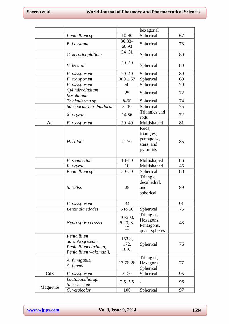
www.wjpps.com Vol 3, Issue 9, 2014.
1594
Saxena et al. World Journal of Pharmacy and Pharmaceutical Sciences
hexagonal
Penicillium sp. 10-40 Spherical 67
B. bassiana 36.88–
60.93 Spherical 73
C. keratinophilium 24–51
Spherical 80
V. lecanii 20–50
Spherical 80
F. oxysporum 20–40 Spherical 80
F. oxysporum 300 ± 57 Spherical 69
F. oxysporum 50 Spherical 70
Cylindrocladium
floridanum 25 Spherical 72
Trichoderma sp. 8-60 Spherical 74
Saccharomyces boulardii 3–10 Spherical 75
X. oryzae 14.86 Triangles and
rods 72
Au F. oxysporum 20–40 Multishaped 81
H. solani 2–70
Rods,
triangles,
pentagons,
stars, and
pyramids
85
F. semitectum 18–80 Multishaped 86
R. oryzae 10 Multishaped 45
Penicillium sp. 30–50 Spherical 88
S. rolfsii 25
Triangle,
decahedral,
and
spherical
89
F. oxysporum 34 91
Lentinula edodes 5 to 50 Spherical 75
Neurospora crassa
10-200,
6-23, 3-
12
Triangles,
Hexagons,
Pentagons,
quasi-spheres
43
Penicillium
aurantiogriseum,
Penicillium citrinum,
Penicillium waksmanii,
153.3,
172,
160.1
Spherical 76
A. fumigatus,
A. flavus 17.76-26
Triangles,
Hexagons,
Spherical
77
CdS F. oxysporum 5–20 Spherical 95
Magnetite
Lactobacillus sp.
S. cerevisiae 2.5–5.5 - 96
C. versicolor 100 Spherical 97

www.wjpps.com Vol 3, Issue 9, 2014.
1595
Saxena et al. World Journal of Pharmacy and Pharmaceutical Sciences
S. pombe 1-1.5 Hexagonal
lattice 3
F. oxysporum 20–50 Quasi-
spherical 99
CdSe F. oxysporum 9–15 Spherical 100
Se A. alternata 30 ± 5 Spherical 101
SrCO3 F. oxysporum - Needle
shaped 102
Si F. oxysporum 5–15 Quasi-
Spherical 103
Ti
F. oxysporum 2−6 Quasi-
Spherical 104
F. oxysporum 5–15 Quasi-
Spherical 103
BaTiO3
Lactobacillus sp. S.
cerevisiae 8–35 - 105
F. oxysporum 4–5 Quasi-
Spherical 104
Bi2O3 F. oxysporum 5–8 Quasi-
Spherical 107
Pt F. oxysporum 20–60 Triangle 108
F. oxysporum 10–50
Triangle,
hexagons,
square,
rectangles
109
F. oxysporum 5-30 - 78
N. crassa 20-110 Spherical 80
Several species of fungi like F. oxysporum, [48-50]
Fusarium solani, [51,52]
Pleurotus sajorcaju,
[53] Fusarium semitectum,
[54] Alternaria alternata,
[55] Fusarium acuminatum,
[56] Penicillium
fellutanum, [57]
Penicillium brevicompactum, [58]
Aspergillus clavatus, [59]
Aspergillus flavus
[60] have been known to synthesize AgNPs. Jain et al, 2010 confirmed the presence of
extracellular protein of molecular weight 32 kDa during synthesis of silver nanoparticle using
cell filterate of A. flavus. [61]
Furthermore, AgNPs were synthesized by exploiting the fungus
F. oxysporum through biotransformation. [62]
Extracellular synthesis of AgNP using extract of
edible mushroom, Volvaniella volvacea as reducing and protecting agent has also been
documented. [63]
Endophytic fungi, Pestalotia sp. isolated from leaves of Syzgium cumini has
been used to produce spherical and polydispersed AgNP having average size of 12.4 nm. [64]
They have reported this silver nanoparticle as better antimicrobial agent by evaluating its
antibacterial activity against S. aureus and S. typhi. Sanghi et al 2009, synthesized protein
capped AgNP using fungus proteins of Coriolus versicolor. [65]
The amino group of protein
was found to be bound on Ag NP as determined by FTIR. The reaction rate was much faster
during NP synthesis under alkaline conditions. Hexagonal and spherical shaped AgNPs were

www.wjpps.com Vol 3, Issue 9, 2014.
1596
Saxena et al. World Journal of Pharmacy and Pharmaceutical Sciences
synthesized by using A. clavatus isolated from stem tissues of Azardirachta indica. [59]
Trichoderma reesei an eco-friendly fungus known for its large amount of extracellular
enzyme production has been used in the AgNP synthesis as well. [66]
This fungus can be used
for scale up production of AgNP due to its capacity to produce large amount of extracellular
enzyme.
Endophytic fungi living in symbiotic association with plants are also involved in AgNPs
synthesis. The endophytic fungi, Penicillium sp. isolated from Curcuma longa leaves were
found to be excellent producer of silver nanoparticles as reported recently. [67]
Furthermore,
these silver nanoparticles were also found to be effective antimicrobial against E. coli and S.
aureus. In another study Qian et al 2013, synthesized AgNps from an endophytic fungi
Epicoccum nigrum isolated from cambium of Phellodendron amurense. The synthesized
AgNP was found to be highly stable even at varied pH and temperature. [68]
Gold (Au): Being more toxic to the fungus than silver, F. oxysporum mediated AuNP
showed more aggregation and irregularity in shape and size. [81]
Intracellular synthesis of
AuNPs by using Penicillium sp. has been reported by Zhang 2009. [82]
Variation in the
temperature was found to control the size of biosynthesized gold nanoparticles. [82,83]
Shankar et al 2003 synthesised AuNPs using endophytic fungi. Colletotrichum sp. isolated
from leaves of Pelarogonium graveolus as determined by TEM analysis. [84]
Several reports
on mycosynthesis of AuNPs using Helminthosporium solani, [85]
F. semitectum, [86]
Tricothecium sp., [87]
R. oryzae have been well documented. [45]
Rapid extracellular synthesis
of AuNP in cell filterate and intracellular synthesis in fungal biomass by Penicillim sp. has
been performed by Du et al 2011. [88]
They synthesized gold nanoparticle in one minute via
extracellular and intracellular route. Mechanistically role of NADPH dependent enzyme in
AuNPs production has been investigated by Narayanan, 2010. [89]
The Sclerotium rolfsii
mediated gold nanoparticles were found to be spherical and anisotropic that is of variable
shapes as triangle, hexagonal rod and decahedral in shape. Size shape and state of
aggregation of nanoparticle is determined by various factors including various concentrations
of precursor salts, different cellular fractions of culture. Deepa et al 2014 synthesized AuNPs
from culture filterate of F. oxysporum and found diverse shape and size of AuNPs in the
presence of different cellular fractions. Specificity and sensitivity of assay determines the
pathogen detection in less time with more accuracy. [90]
F. oxysporum mediated synthesized
AuNPs and AgNPs conjugated with Candida sp. DNA allowed its rapid detection in modified

www.wjpps.com Vol 3, Issue 9, 2014.
1597
Saxena et al. World Journal of Pharmacy and Pharmaceutical Sciences
PCR assay. [91]
Synthesis of AuNPs by edible mushroom Pleurotus florida has been
documented. [92]
The synthesized AuNPs showed anticancer activity against human lung
carcinoma (A-549), Human chronic myelogenous leukemia (K562), Human cervix (HeLa)
and human adenocarcinoma mammary gland (MDA-MB) under invitro conditions. Synthesis
of catalytically active AuNPs by P. florida has been reported. [93]
The glucan content of
mushroom was responsible for stability of synthesized AuNPs.
Cadmium (CdTe/ CdS): Synthesis of CdTe quantum dots by using F. oxysporum has been
reported by Syed et al 2013 [94]
and Ahmad 2002. [95]
The nanoparticles synthesized via
biological route shows enhanced antibacterial activity against Gram positive and Gram
negative bacteria. Prasad 2010 has reported synthesis of CdS Nanoparticle using S. cerevisae
as rapid and low cost green method. [96]
Involvement of white rot fungus C. versicolor has
also been well reported. [97]
Other nanoparticles from fungi: In addition to the above, several other metallic
nanoparticles were synthesized using fungi for instances, synthesis of nanosized magnetite by
Mucor javanicus, [98]
F. oxysporum and Verticellum sp., [99]
CdSe quantum dots by F.
oxysporum, [100]
Selenium nanoparticle by A. alternata, [101]
Strontium carbonate crystals by
F. oxysporum, [102]
Silica nanoparticle by F. oxysporum, [103,104]
Titanium nanoparticle by F.
oxysporum, [103]
S. cereviseae, [105]
A. flavus, [106]
Barium titanate nanoparticle by F.
oxysporum, [104]
Bi2O3 nanoparticle by F. oxysporum [107]
and Platinum nanoparticle by F.
oxysporum. [108,109]
Applications of mycosynthesized nanoparticles
Catalysis: Nanoparticles synthesized from fungi have remarkable biocatalytic properties.
Mishra et al, 2014 reported the catalytic power of biosynthesized gold nanoparticles from
Trichoderma. viridae. [110]
This gold nanoparticle in presence of NaBH4 reduced nitrophenol
to 4 aminophenol and also shows antimicrobial property. Further, metal nanoparticles
obtained from fungi can be used in enzyme immobilization for improved enzymatic activity.
Vector control: Role of fungi as antimosquito is well established. Recently Banu et al 2014
synthesised AgNPs from entomopathogenic fungus Beauveria bassiana and found it effective
against dengu vector Aedes aegypti. [72]
In another study Soni et al 2012 evaluated the
adulticidal effect of AgNPs synthesized from Chrysosporium keratinophilium, Verticillium
lecanii and F. oxysporum against filiariasis vector Culex quinquefasciatus. [79]

www.wjpps.com Vol 3, Issue 9, 2014.
1598
Saxena et al. World Journal of Pharmacy and Pharmaceutical Sciences
Wound healing – Sundaramoorthi et al (2009) have explored the wound healing property
of AgNPs synthesized from Aspergillus niger in experimental rat model (Excision wound
model and thermal wound model). [111]
Their findings suggested AgNPs as better wound
healing property as demonstrated by measuring percentage of wound contraction and period
of epithelialization in dose and time dependent manner. In another study, Phytophthora
infestans synthesized AgNPs was assessed for its wound healing activity. [112]
They found
0.125% (w/w) AgNPs ointments having better wound healing property as compared to
standard silver sulphadiazine ointment.
Textile fabrics – AgNPs synthesized by Lecanicillium lecanii, when incorporated within
cotton fabrics has shown to inhibit the growth of S. aureus and E. coli. [113]
This cotton fabric
cloth can be used to prevent bacterial infections in hospitals. Duran et al (2007) have
demonstrated the incorporation of AgNPs synthesized from F. oxysporum into cotton fabrics
followed by its antibacterial activity against antibacterial activity against S. aureus. [114]
El-
Rafie et al (2009) have shown the reduction in antibacterial activity of AgNPs loaded cotton
fabric against S. aureus and E. coli when washed in laundry after 20 cycles. [115]
Incorporation of a binder in the finishing formulation has retained its antibacterial activity.
Vegetable and Food preservation – Fayaz et al (2009) demonstrated the AgNPs
synthesized from T. viride when incorporated into sodium alginate thin film showed
antibacterial activity and increases the shelf life of carrot and pear as compared to control in
terms of weight loss and soluble protein content. [116]
Molecular Detection: The new PCR assay has been developed by Bansod et al 2013 for
rapid detection of pathogenic fungi Candida sp. from low concentrated DNA. [91]
They have
synthesized gold and silver nanoparticles by F. oxysporum and conjugate these particles with
master mix and DNA sample of Candida sp. This bioconjugate nano PCR assay have shown
high specificity and sensitivity as compared to conventional method.
Anti-bacterial: The number of multi-resistant bacterial strains has been increasing at an
alarming rate and represents a major threat to modern medicine. Emergence of antibiotic
resistance is the consequence of a complex interaction of factors involved in the evolution
and spread of resistance mechanisms. [117]
The sharp increase in antibiotic resistance is caused
by the extensive and improper use of antibiotics in human and animal medicine and in
agriculture. During the past decade, a great potential in nanomedicine has been realized due

www.wjpps.com Vol 3, Issue 9, 2014.
1599
Saxena et al. World Journal of Pharmacy and Pharmaceutical Sciences
to effectiveness of various nanoconjugates against pathogenic microbes, but they also show
severe toxicity among humans. [118]
The development of nontoxic methods of synthesizing
nanoparticles would be a major step in nanotechnology to allow their application in
nanomedicine. AgNPs have the ability to penetrate the bacterial cell wall and subsequently
damage the cell membrane that leads to death of the cell. Matsumura et al (2003) have
documented the generation of reactive oxygen species (ROS) when AgNPs interact with
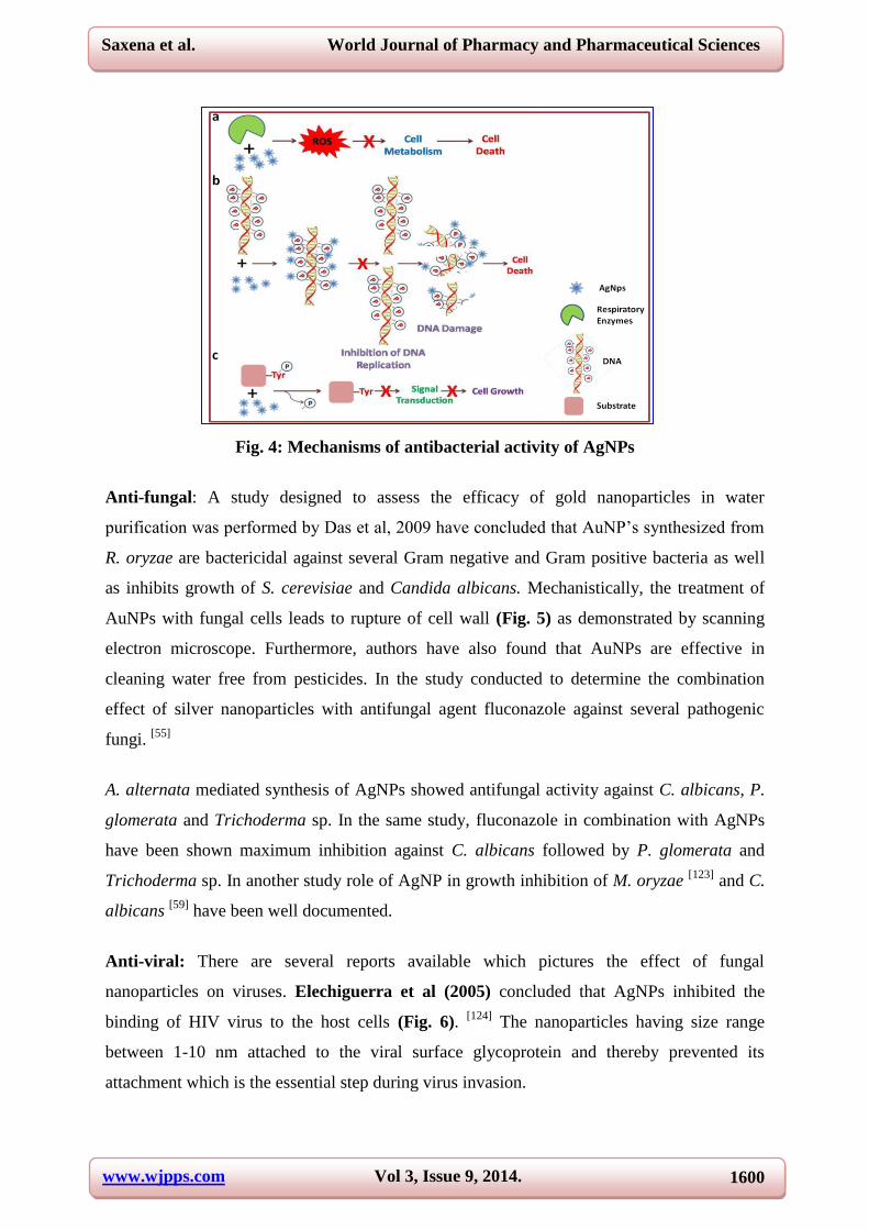
respiratory enzymes after entering to the bacterial cells (Fig. 4a). [119]
The generated ROS is
lethal to the cell as it oxidizes all the macromolecules ultimately leads to the cell death. Being
positively charged, Ag+ ions have great affinity towards negative charge present on
phosphate group of DNA (Fig. 4b). Interaction of Ag+ phosphate of DNA inhibits DNA
replication and causes DNA damage that leads to bacterial cell death. [120]
AgNPs have also
shown to modulate the signaling pathway required for cell growth. [121]
AgNPs
dephosphorylate the substrates on tyrosine residues, which lead to inhibition of signal
cascade and thus stops the cell growth (Fig. 4c).
Silver nanoparticles which are spherical in shape synthesized by R. stolonifer have been
shown antibacterial activity against MDR strains of P. aeruginosa isolated from burnt
patients. [46]
The authors have also performed the synergistic effect of silver nanoparticles
with standard antibiotics and found it more effective. In another study P. glomerata mediated
synthesized AgNPs showed remarkable antibacterial activity against E. coli, P. aeruginosa
and S. aureus [47]
and in combination with antibiotics. Pathogens showing resistance to
antibiotics like ampicillin, vancomycin, streptomycin showed susceptibility with silver
nanoparticles alone and when given in combination with antibiotics. Concentration dependent
studies of silver nanoparticles synthesized from endophytic fungi isolated from Curcuma
longa was found to be bactericidal against multi drug resistance E. coli and S. aureus. [67]
In
another study the potentiality of saprophytic fungi, Nigrospora oryzae for synthesis of silver
nanoparticle and its antimicrobial effect was evaluated. [122]
AgNPs were screened against six
bacterial starins namely E. coli, B. cerus, Proteus vulgaris, P. aeruginosa and Micrococus
luteus by well diffusion method and found it bactericidal activity at concentration of
100ug/ml. Several studies including antibacterial effect of mycosynthesised AgNPs against
pathogenic bacteria like S. aureus, S. typhi, E. coli, [56]
Psuedomonas fluorescens, E. coli, [59]
S. aureus, E. coli, [113]
P. aeruginosa, E. coli, S. aureus, [53]
S. aureus, S. typhi. [64]
E. coli,
Agrobacterium tumifaciens, Magnaporthe oryzae [123]
has been reported.
www.wjpps.com Vol 3, Issue 9, 2014.
1600
Saxena et al. World Journal of Pharmacy and Pharmaceutical Sciences
Fig. 4: Mechanisms of antibacterial activity of AgNPs
Anti-fungal: A study designed to assess the efficacy of gold nanoparticles in water
purification was performed by Das et al, 2009 have concluded that AuNP’s synthesized from
R. oryzae are bactericidal against several Gram negative and Gram positive bacteria as well
as inhibits growth of S. cerevisiae and Candida albicans. Mechanistically, the treatment of
AuNPs with fungal cells leads to rupture of cell wall (Fig. 5) as demonstrated by scanning
electron microscope. Furthermore, authors have also found that AuNPs are effective in
cleaning water free from pesticides. In the study conducted to determine the combination
effect of silver nanoparticles with antifungal agent fluconazole against several pathogenic
fungi. [55]
A. alternata mediated synthesis of AgNPs showed antifungal activity against C. albicans, P.
glomerata and Trichoderma sp. In the same study, fluconazole in combination with AgNPs
have been shown maximum inhibition against C. albicans followed by P. glomerata and
Trichoderma sp. In another study role of AgNP in growth inhibition of M. oryzae [123]
and C.
albicans [59]
have been well documented.
Anti-viral: There are several reports available which pictures the effect of fungal
nanoparticles on viruses. Elechiguerra et al (2005) concluded that AgNPs inhibited the
binding of HIV virus to the host cells (Fig. 6). [124]
The nanoparticles having size range
between 1-10 nm attached to the viral surface glycoprotein and thereby prevented its
attachment which is the essential step during virus invasion.

www.wjpps.com Vol 3, Issue 9, 2014.
1601
Saxena et al. World Journal of Pharmacy and Pharmaceutical Sciences
Fig. 5: Mechanism of antifungal activity of AuNPs
Fig. 6: Mechanism of anti-viral activities of AgNPs
Conclusion and Future Directions
A brief description about the fungi in biosynthesis of metallic nanoparticles has been
illustrated. Furthermore, mechanism of synthesis and its diverse applications has been
discussed. Fungi have an upper edge over other biological systems due to its wide diversity,
easy to culture methods, time and cost-effectiveness as well as eco-friendly approach for
nanoparticle synthesis. Myconanotechnology is relatively new development, the future lies in
the optimization of biochemical reactions for producing nanoparticles with improved

www.wjpps.com Vol 3, Issue 9, 2014.
1602
Saxena et al. World Journal of Pharmacy and Pharmaceutical Sciences
composition, size, shape and monodispersity. Genetic engineering technique can be
employed to improve the particle properties in near future.
REFERENCES
1. Vuk U. Nanomaterials and nanotechnologies: approaching the crest of this big wave.
Current Nanoscience, 2008; 4(2): 119–129.
2. Taniguchi N. On the basic concept of nano-technology, in Production Engineering, Proc.
Tokyo, Part II, Japan Society of Precision Engineering, Tokyo: 1974.
3. Kowshik M, Deshmukh N, Vogel W, Urban J, Kulkarni SK, Paknikar KM. Microbial
synthesis of semiconductor CdS nanoparticles, their characterization, and their use in
fabrication of an ideal diode. Biotechnology and Bioengineering, 2002; 78(5): 583–588.
4. Gurunathan S, Kalishwaralal K, Vaidyanathan R, Venkataraman D, Pandian SR,
Muniyandi J, Hariharan N, Eom SH. Biosynthesis, purification and characterization of
silver nanoparticles using Escherichia coli. Colloids and Surfaces B: Biointerfaces, 2009;
74(1): 328-335.
5. Juibari MM, Abbasalizadehb S, Jouzanib GS, Noruzic M. Intensified biosynthesis of
silver nanoparticles using a native extremophilic Ureibacillus thermosphaericus strain.
Materials Letters, 2011; 65(6): 1014–1017.
6. Nanda A, Saravanan M. Biosynthesis of silver nanoparticles from Staphylococcus aureus
and its antimicrobial activity against MRSA and MRSE. Nanomedicine, 2009; 5(4): 452-
456.
7. Jain D, Kachhwaha S, Jain R, Srivastava G, Kothari SL. Novel microbial route to
synthesize silver nanoparticles using spore crystal mixture of Bacillus thuringiensis.
Indian J Exp Biol, 2010; 48(11): 1152-1156.
8. Zhang H, Li Q, Lu Y, Sun D, Lin X, Deng X, He N, Zheng S, Biosorption and
bioreduction of diamine silver complex by Corynebacterium. Journal of Chemical
Technology and Biotechnology, 2005; 80(3): 285–290.
9. Babu GMM, Gunasekaran P. Production and structural characterization of crystalline
silver nanoparticles from Bacillus cereus isolate. Colloids Surf B Biointerfaces, 2009;
74(1): 191-195s.
10. Sintubin L, De Windt W, Dick J, Mast J, van der Ha D, Verstraete W, Boon N. Lactic
acid bacteria as reducing and capping agent for the fast and efficient production of silver
nanoparticles. Appl Microbiol Biotechnol, 2009; 84(4): 741-749.

www.wjpps.com Vol 3, Issue 9, 2014.
1603
Saxena et al. World Journal of Pharmacy and Pharmaceutical Sciences
11. Sweeney RY, Mao C, Gao X, Burt JL, Belcher AM, Georgiou G, Iverson BL. Bacterial
biosynthesis of cadmium sulfide nanocrystals. Chemistry & Biology, 2004; 11(11): 1553–
1559.
12. Kowshik M, Ashtaputre S, Kharrazi S, Vogel W, Urban J, Kulkarni SK, Paknikar KM.
Extracellular synthesis of silver nanoparticles by a silver-tolerant yeast strain MKY3.
Nanotechnology, 2003; 14(1): 95–100.
13. Mourato A, Gadanho M, Lino AR, Tenreiro R. Biosynthesis of Crystalline Silver and
Gold Nanoparticles by Extremophilic Yeasts. Bioinorganic Chemistry and Applications,
2011: 546074.
14. Dameron CT, Reese RN, Mehra RK, Kortan AR, Carroll PJ , Steigerwald ML , Brus
LE, Winge DR. Biosynthesis of cadmium sulphide quantum semiconductor crystallites.
Nature, 1989; 338: 596 – 597.
15. Seshadri S, Saranya K, Kowshik M. Green synthesis of lead sulfide nanoparticles by the
lead resistant marine yeast, Rhodosporidium diobovatum. Biotechnol Prog, 2011; 27(5):
1464-1469.
16. Vainshtein M, Belova N, Kulakovskaya T, Suzina N, Sorokin V. Synthesis of magneto-
sensitive iron-containing nanoparticles by yeasts. J Ind Microbiol Biotechnol, 2014;
41(4): 657-663.
17. Singh A, Sharma MM, Batra A. Synthesis of gold nanoparticles using chick pea leaf
extract using green chemistry. Journal of Optoelectronics and Biomedical Materials,
2013; 5(2): 27–32.
18. Shankar SS, Rai A, Ahmad A, Sastry M. Rapid synthesis of Au, Ag, and bimetallic Au
core-Ag shell nanoparticles using Neem (Azadirachta indica) leaf broth. J Colloid
Interface Sci, 2004; 275(2): 496-502.
19. Vankar PS, Bajpai D. Preparation of gold nanoparticles from Mirabilis jalapa flowers.
Indian J Biochem Biophys, 2010; 47(3): 157-160.
20. Christensen L, Vivekanandhan S, Misra M, Mohanty AK. Biosynthesis of silver
nanoparticles using Murraya koenigii (curry leaf): An investigation on the effect of broth
concentration in reduction mechanism and particle size. Adv. Mat. Lett, 2011; 2(6): 429-
434.
21. Mitra B, Vishnudas D, Sant SB, Annamalai A. Green-synthesis and characterization of
silver nanoparticles by aqueous leaf extracts of Cardiospermum helicacabum leaves.
Drug Invention Today, 2012; 4(2): 340-344.

www.wjpps.com Vol 3, Issue 9, 2014.
1604
Saxena et al. World Journal of Pharmacy and Pharmaceutical Sciences
22. Khan M, Khan M, Adil SF, Tahir MN, Tremel W, Alkhathlan HZ, Al-Warthan A,
Siddiqui MR, Green synthesis of silver nanoparticles mediated by Pulicaria glutinosa
extract. Int J Nanomedicine, 2013; 8: 1507-1516.
23. Makarov VV, Makarova SS, Love AJ, Sinitsyna OV, Dudnik AO, Yaminsky IV,
Taliansky ME, Kalinina NO. Biosynthesis of stable iron oxide nanoparticles in aqueous
extracts of Hordeum vulgare and Rumex acetosa plants. Langmuir, 2014; 30(20): 5982-
5988.
24. Song JY, Kwon EY, Kim BS, Biological synthesis of platinum nanoparticles using
Diopyros kaki leaf extract. Bioprocess Biosyst Eng, 2010; 33(1): 159-164.
25. Ghosh S, Patil S, Ahire M, Kitture R, Gurav DD, Jabgunde AM, Kale S, Pardesi K,
Shinde V, Bellare J, Dhavale DD, Chopade BA. Gnidia glauca flower extract mediated
synthesis of gold nanoparticles and evaluation of its chemocatalytic potential. J
Nanobiotechnology, 2012; 10: 17.
26. Singh AK, Talat M, Singh DP, Srivastava ON. Biosynthesis of gold and silver
nanoparticles by natural precursor clove and their functionalization with amine group. J
Nanopart Res, 2010; 12(5): 1667–1675.
27. Chandran SP, Chaudhary M, Pasricha R, Ahmad A, Sastry M. Synthesis of gold
nanotriangles and silver nanoparticles using Aloe vera plant extract. Biotechnol Prog,
2006; 22(2): 577-583.
28. Rai M, Yadav A, Bridge P, Gade A. Myconanotechnology: a new and emerging science,
in Applied Mycology, ed by Rai MK and Bridge PD. CAB International Publishers, New
York, 2009, pp. 258-267.
29. Gade A, Bonde PP, Ingle AP, Marcato P, Duran N, Rai MK. Exploitation of Aspergillus
niger for synthesis of silver nanoparticles. Journal of Bio based Materials and Bioenergy,
2008; 2(3): 1–5.
30. Saha S, Sarkar J, Chattopadhyay D, Patra S, Chakraborty A, Acharya K. Production of
silver nanoparticles by a phytopathogenic fungus Bipolaris nodulosa and its antimicrobial
activity. Dig J Nanomater Bios, 2010; 5(4): 887–895.
31. Singh M, Manikandan S, Kumaraguru AK. Nanoparticles: a new technology with wide
applications. Res J Nanosci Nanotechnol, 2011; 1(1): 1–11.
32. Behari J. Principles of nanoscience: an overview. Indian J Exp Biotechnol, 2010; 48:
1008–1019.
33. Moghaddam KM. An introduction to microbial metal nanoparticle preparation method. J
Young Investigators, 2010; 19(19): 1–7.

www.wjpps.com Vol 3, Issue 9, 2014.
1605
Saxena et al. World Journal of Pharmacy and Pharmaceutical Sciences
34. Zhang X, Yan S, Tyagi RD, Surampalli RY. Synthesis of nanoparticles by
microorganisms and their application in enhancing microbiological reaction rates.
Chemosphere, 2011; 82(4): 489–494.
35. Thakkar KN, Mhatre SS, Parikh RY. Biological synthesis of metallic nanoparticles.
Nanomed Nanotechnol Biol Med, 2010; 6(2): 257–262.
36. Kashyap PL, Kumar S, Srivastava AK, Sharma AK. Myconanotechnology in agriculture:
a perspective. World J Microbiol Biotechnol, 2013; 29(2): 191-207.
37. Kumar SA, Abyaneh MK, Gosavi SW, Kulkarni SK, Pasricha R, Ahmad A, Khan MI.
Nitrate reductase-mediated synthesis of silver nanoparticles from AgNO3. Biotechnol
Lett, 2007a; 29(3): 439–445.
38. Srivastava P, Bragança J, Ramanan SR, Kowshik M. Synthesis of silver nanoparticles
using haloarchaeal isolate Halococcus salifodinae BK3. Extremophiles, 2013; 17(5): 821-
831.
39. Devi TP, Kulanthaivel S, Kamil D, Borah JL, Prabhakaran N, Srinivasa N. Biosynthesis
of silver nanoparticles from Trichoderma species. Indian J Exp Biol, 2013; 51(7): 543-
547.
40. Manivasagan P, Venkatesan J, Senthilkumar K, Sivakumar K, Kim SK. Biosynthesis,
antimicrobial and cytotoxic effect of silver nanoparticles using a novel Nocardiopsis sp.
MBRC-1. Biomed Res Int, 2013; 2013: 287638.
41. Hamedi S, Shojaosadati SA, Shokrollahzadeh S, Hashemi-Najafabadi S. Extracellular
biosynthesis of silver nanoparticles using a novel and non-pathogenic fungus, Neurospora
intermedia: controlled synthesis and antibacterial activity. World J Microbiol Biotechnol,
2014; 30(2): 693-704.
42. Gholami-Shabani M, Akbarzadeh A, Norouzian D, Amini A, Gholami-Shabani Z, Imani
A, Chiani M, Riazi G, Shams-Ghahfarokhi M, Razzaghi-Abyaneh M. Antimicrobial
activity and physical characterization of silver nanoparticles green synthesized using
nitrate reductase from F. oxysporum. Appl Biochem Biotechnol, 2014; 172(8): 4084-
4098.
43. Quester K, Avalos-Borja M, Vilchis-Nestor AR, Camacho-López MA, Castro-Longoria
E. SERS properties of different sized and shaped gold nanoparticles biosynthesized under
different environmental conditions by Neurospora crassa extract. PLoS One, 2013; 8(10):
e77486.

www.wjpps.com Vol 3, Issue 9, 2014.
1606
Saxena et al. World Journal of Pharmacy and Pharmaceutical Sciences
44. Min JS, Kim KS, Kim SW, Jung JH, Lamsal K, Kim SB, Jung M, Lee YS. Effects of
colloidal silver nanoparticles on sclerotium-forming phytopathogenic fungi. Plant Pathol
J, 2009; 25(4): 376–380.
45. Das SK, Das AR, Guha AK. Gold nanoparticles: microbial synthesis and application in
water hygiene management. Langmuir, 2009; 25(14): 8192–8199.
46. Afreen RV and Ranganath E, Synthesis of monodispersed silver nanoparticles by
Rhizopus stolonifer and its antibacterial activity against MDR strains of Pseudomonas
aeruginosa from burnt patients. Int J Environ Sci, 2011; 1(7): 1582–1592.
47. Birla SS, Tiwari VV, Gade AK, Ingle AP, Yadav AP, Rai MK. Fabrication of silver
nanoparticles by Phoma glomerata and its combined effect against Escherichia coli,
Pseudomonas aeruginosa and Staphylococcus aureus. Lett Appl Microbiol, 2009; 48(2):
173–179.
48. Duran N, Marcato PD, Alves OL, Desouza G, Esposito E. Mechanistic aspects of
biosynthesis of silver nanoparticles by several F. oxysporum strains. J Nanobiotechnol,
2005; 3: 1–8.
49. Karbasian M, Atyabi SM, Siadat SD, Momem SB, Norouzian D. Optimizing nano-silver
formation by F. oxysporum (PTCC 5115) employing response surface methodology. Am
J Agric Biol Sci, 2008; 3(1): 433–437.
50. Ahmad A, Mukherjee P, Senapati S, Mandal D, Khan MI, Kumar R, Sastry M.
Extracellular biosynthesis of silver nanoparticles using the fungus F. oxysporum. Colloids
Surf B, 2003; 28(4): 313–318.
51. Ingle A, Gade A, Bawaskar M, Rai M. F. solani: a novel biological agent for the
extracellular synthesis of silver nanoparticles. J Nanopart Res, 2009; 11(8): 2079–2085.
52. El-Rafie MH, Mohamed AA, Shaheen THI, Hebeish A. Antimicrobial effect of silver
nanoparticles produced by fungal process on cotton fabrics. Carbohydr Polym, 2010;
80(3): 779–782.
53. Nithya R, Ragunathan R. Synthesis of silver nanoparticle using Pleurotus sajor caju and
its antimicrobial study. Dig J Nanomater Bios, 2009; 4(4): 623–629.
54. Bhainsa KC, D’souza SF, Extracellular biosynthesis of silver nanoparticles using the
fungus Aspergillus fumigatus. Colloids Surf B, 2006; 47(2): 160–164.
55. Gajbhiye M, Kesharwani J, Ingle A, Gade A, Rai M. Fungus mediated synthesis of silver
nanoparticles and their activity against pathogenic fungi in combination with fluconazole.
Nanomed Nanotechnol Biol Med, 2009; 5(4): 382–386.

www.wjpps.com Vol 3, Issue 9, 2014.
1607
Saxena et al. World Journal of Pharmacy and Pharmaceutical Sciences
56. Ingle A, Gade A, Pierrat S, So¨nnichsen C, Rai M. Mycosynthesis of silver nanoparticles
using the fungus F. acuminatum and its activity against some human pathogenic bacteria.
Curr Nanosci, 2008; 4(2): 141–144.
57. Kathiresan K, Manivannan S, Nabeel AM, Dhivya B. Studies on silver nanoparticles
synthesized by a marine fungus Penicillum fellutanum isolated from coastal mangrove
sediment. Colloids Surf B, 2009; 71(1): 133–137.
58. Shaligram NS, Bule M, Bhambure R, Singhal RS, Singh SK, Szakac SG, Pandey A.
Biosynthesis of silver nanoparticles using aqueous extract from the compactin producing
fungal strain. Proc Biochem, 2009; 44(8): 939–943.
59. Verma VC, Kharwar RN, Gange AC. Biosynthesis of antimicrobial silver nanoparticles
by the endophytic fungus Aspergillus clavatus. Nanomedicine, 2010; 5(1): 33–40.
60. Vigneshwaran N, Ashtaputre NM, Varadarajan PV, Nachane RP, Paralikar KM,
Balasubramanya RH. Biological synthesis of silver nanoparticles using the fungus
Aspergillus flavus. Mater Lett, 2007; 61(6): 1413–1418.
61. Jain N, Bhargava A, Majumdar S, Tarafdar JC, Panwar J. Extracellular biosynthesis and
characterization of silver nanoparticles using Aspergillus flavus NJP08: a mechanism
perspective. Nanoscale, 2010; 3(2): 635–641.
62. Pandiarajan G, Govindaraj R, Kumar M, Ganesan V. Biosynthesis of silver nanoparticles
using silver nitrate through biotransformation. J Ecobiotechnol, 2010; 2(11): 13–18.
63. Philip D. Biosynthesis of Au, Ag and Au-Ag nanoparticles using edible mushroom
extract. Spectrochim Acta Part A, 2009; 73(2): 374–381.
64. Raheman F, Deshmukh S, Ingle A, Gade A, Rai M. Silver nanoparticles: novel
antimicrobial agent synthesized from an endophytic fungus Pestalotia sp. isolated from
leaves of Syzygium cumini (L). Nano Biomed Eng, 2011; 3(3):174–178.
65. Sanghi R, Verma P. Biomimetic synthesis and characterization of protein capped silver
nanoparticles. Bioresour Technol, 2009; 100(1): 501–504.
66. Vahabi K, Ali Mansoori G, Karimi S. Biosynthesis of silver nanoparticles by fungus
Trichoderma reesei (a route for large scale production of AgNPs). Insci J, 2011; 1(1): 65–
79.
67. Singh D, Rathod V, Ninganagouda S, Hiremath J, Singh AK, Mathew J. Optimization
and characterization of silver nanoparticle by endophytic fungi Penicillium sp. isolated
from Curcuma longa (Turmeric) and application studies against MDR E. coli and S.
aureus. Bioinorganic Chemistry and Applications, 2014: 408021.

www.wjpps.com Vol 3, Issue 9, 2014.
1608
Saxena et al. World Journal of Pharmacy and Pharmaceutical Sciences
68. Qian Y, Yu H, He D, Yang H, Wang W, Wan X, Wang L. Biosynthesis of silver
nanoparticles by the endophytic fungus Epicoccum nigrum and their activity against
pathogenic fungi. Bioprocess and Biosystems Engineering, 2013; 36(11): 1613-1619.
69. Ishida K, Cipriano TF, Rocha GM, Weissmüller G, Gomes F, Miranda K, Rozental S
Silver nanoparticle production by the fungus F. oxysporum: nanoparticle characterisation
and analysis of antifungal activity against pathogenic yeasts. Mem Inst Oswaldo Cruz,
2014; 109(2): 220-228.
70. Gholami-Shabani M, Akbarzadeh A, Norouzian D, Amini A, Gholami-Shabani Z, Imani
A, Chiani M, Riazi G, Shams-Ghahfarokhi M, Razzaghi-Abyaneh M. Antimicrobial
activity and physical characterization of silver nanoparticles green synthesized using
nitrate reductase from F. oxysporum. Appl Biochem Biotechnol, 2014; 172(8): 4084-
4098.
71. Narayanan KB, Sakthivel N. Biosynthesis of silver nanoparticles by phytopathogen
Xanthomonas oryzae pv. oryzae strain BXO8. J Microbiol Biotechnol, 2013; 23(9): 1287-
1292.
72. Banu NA, Balasubramanian C. Myco-synthesis of silver nanoparticles using Beauveria
bassiana against dengue vector, Aedes aegypti (Diptera: Culicidae). Parasitology
Research, 2014; 113(8); 2869-2877.
73. Devi TP, Kulanthaivel S, Kamil D, Borah JL, Prabhakaran N, Srinivasa N. Biosynthesis
of silver nanoparticles from Trichoderma species. Indian J Exp Biol, 2013; 51(7): 543-
547.
74. Kaler A, Jain S, Banerjee UC. Green and rapid synthesis of anticancerous silver
nanoparticles by Saccharomyces boulardii and insight into mechanism of nanoparticle
synthesis. Biomed Res Int, 2013: (2013); 872940.
75. Vetchinkina EP, Burov AM, Ageeva MV, Dykman LA, Nikitina VE. Biological synthesis
of gold nanoparticles by the xylotrophic basidiomycete Lentinula edodes. Prikl Biokhim
Mikrobiol, 2013; 49(4): 402-408.
76. Honary S, Gharaei-Fathabad E, Barabadi H, Naghibi F. Fungus-mediated synthesis of
gold nanoparticles: a novel biological approach to nanoparticle synthesis. J Nanosci
Nanotechnol, 2013; 13(2): 1427-1430.
77. Gupta S and Bector S, Biosynthesis of extracellular and intracellular gold nanoparticles
by Aspergillus fumigatus and A. flavus. Antonie Van Leeuwenhoek, 2013; 103(5): 1113-
1123.
www.wjpps.com Vol 3, Issue 9, 2014.
1609
Saxena et al. World Journal of Pharmacy and Pharmaceutical Sciences
78. Syed A, Ahmad A. Extracellular biosynthesis of platinum nanoparticles using the fungus
F. oxysporum. Colloids Surf B Biointerfaces, 2012; 97: 27-31.
79. Soni N, Prakash S. Fungal-mediated nano silver: an effective adulticide against mosquito.
Parasitol Res, 2012; 111(5): 2091–2098.
80. Castro-Longoria E, Moreno-Velázquez SD, Vilchis-Nestor AR, Arenas-Berumen E and
Avalos-Borja M. Production of platinum nanoparticles and nano-aggregates using
Neurospora crassa. J Microbiol Biotechnol, 2012; 22(7): 1000-1004.
81. Anitha TS, Palanivelu P. Synthesis and structural characterization of polydisperse silver
and multishaped gold nanoparticles using F. oxysporum MTCC 284. Dig J Nanomater
Bios, 2011; 6(4): 1587–1595.
82. Zhang X, He X, Wang K, Wang Y, Li H, Tan W. Biosynthesis of size-controlled gold
nanoparticles using fungus Penicillium sp. J Nanosci Nanotechnol, 2009; 9(10): 5738–
5744.
83. Gericke M, Pinches A, Biological synthesis of metal nanoparticles. Hydrometallurgy,
2006; 83(1-4): 132–140.
84. Shankar SS, Ahmad A, Pasricha R, Sastry M. Bioreduction of chloroaurate ions by
Geranium leaves and its endophytic fungus yields gold nanoparticles of different shapes.
J Mater Chem, 2003; 13(7): 1822–1826.
85. Kumar SA, Peter YA, Nadaeu JL. Facile biosynthesis, separation, conjugation of gold
nanoparticles to doxorubicin. Nanotechnology, 2008; 19(49): 495101.
86. Sawle BD, Salimath B, Deshpande R, Bedre MD, Prabhakar KB, Venkataraman A.
Biosynthesis and stabilization of Au and Au–Ag alloy nanoparticles by fungus, F.
semitectum. Sci Technol Adv Mater, 2008; 9(3): 035012.
87. Ahmad A, Senapati S, Khan MI, Kumar R, Sastry M. Extra-/intracellular biosynthesis of
gold nanoparticles by an alkalotolerant fungus Trichothecium sp. J Biomed Nanotechnol,
2005; 1(1): 47–53.
88. Du L, Xian L, Feng J-X. Rapid extra-/intracellular biosynthesis of gold nanoparticles by
the fungus Penicillium sp. J Nanopart Res, 2011; 13: 921–930.
89. Narayanan KB, Sakthivel N. Facile green synthesis of gold nanostructures by NADPH-
dependent enzyme from the extract of Sclerotium rolfsii. Colloids Surf A, 2011; 380(1-3):
156–161.
90. Deepa K, Panda T. Synthesis of gold nanoparticles from different cellular fractions of F.
oxysporum. J Nanosci Nanotechnol, 2014; 14(5): 3455-3463.

www.wjpps.com Vol 3, Issue 9, 2014.
1610
Saxena et al. World Journal of Pharmacy and Pharmaceutical Sciences
91. Bansod S, Bonde S, Tiwari V, Bawaskar M, Deshmukh S, Gaikwad S, Gade A, Rai M.
Bioconjugation of gold and silver nanoparticles synthesized by F. oxysporum and their
use in rapid identification of Candida species by using bioconjugate-nano-polymerase
chain reaction. J Biomed Nanotechnol, 2013; 9(12): 1962-1971.
92. Bhat R, Sharanabasava VG, Deshpande R, Shetti U, Sanjeev G, Venkataraman A. Photo-
bio-synthesis of irregular shaped functionalized gold nanoparticles using edible
mushroom Pleurotus florida and its anticancer evaluation. J Photochem Photobiol B,
2013; 125: 63-69.
93. Sen IK, Maity K, Islam SS. Green synthesis of gold nanoparticles using a glucan of an
edible mushroom and study of catalytic activity. Carbohydrate Polymers, 2013; 91(2):
518–528.
94. Syed A, Ahmad A. Extracellular biosynthesis of CdTe quantum dots by the fungus F.
oxysporum and their anti-bacterial activity. Spectrochimica Acta Part A: Molecular and
Biomolecular Spectroscopy, 2013; 106: 41–47.
95. Ahmad A, Mukherjee P, Mandal D, Senapati S, Khan MI, Kumar R, Sastry M. Enzyme
mediated extracellular synthesis of CdS nanoparticles by the fungus F. oxysporum. J Am
Chem Soc, 2002; 124(141): 12108–12109.
96. Prasada K, Jha AK. Biosynthesis of CdS nanoparticles: An improved green and rapid
procedure. Journal of Colloid and Interface Science, 2010; 342(1): 68–72.
97. Chen GQ, Zou ZJ, Zeng GM, Yan M, Fan JQ, Chen AW, Yang F, Zhang WJ, Wang L.
Coarsening of extracellularly biosynthesized cadmium crystal particles induced by
thioacetamide in solution. Chemosphere, 2011; 83(9): 1201–1207.
98. Meng X, Xu G, Zhou QL, Wu JP, Yang LR. Highly efficient solvent-free synthesis of
1,3-diacylglycerols by lipase immobilised on nano-sized magnetite particles. Food
Chemistry, 2014; 143: 319–324.
99. Bharde A, Rautray D, Bansal V, Ahmad A, Sarkar I, Yusuf SM, Sanyal M, Sastry M.
Extracellular biosynthesis of magnetite using fungi. Small, 2006; 2(1): 135–141.
100. Kumar SA, Ansary AA, Ahmad A, Khan MI. Extracellular biosynthesis of CdSe
quantum dots by the fungus F. oxysporum. J Biomed Nanotechnol, 2007b; 3(2): 190–194.
101. Sarkar J, Dey P, Saha S, Acharya K. Mycosynthesis of selenium nanoparticles. IET
Micro Nano Lett, 2011; 6(8): 599–602.
102. Rautaray D, Sanyal A, Adyanthaya SD, Ahmad A, Sastry M. Biological synthesis of
strontium carbonates crystals using the fungus F. oxysporum. Langmuir, 2004; 20(16):
6827–6833.

www.wjpps.com Vol 3, Issue 9, 2014.
1611
Saxena et al. World Journal of Pharmacy and Pharmaceutical Sciences
103. Bansal V, Rautaray D, Bharde A, Ahire K, Sanyal A, Ahmad A, Sastry M. Fungus-
mediated biosynthesis of silica and titania particles. J Mater Chem, 2005; 15(1): 2583–
2589.
104. Bansal V, Poddar P, Ahmad A, Sastry M. Room-temperature biosynthesis of
ferroelectric barium titanate nanoparticles. J Am Chem Soc, 2006; 128(36): 11958–
11963.
105. Jha AK, Prasad K, Kulkarni AR. Synthesis of TiO2 nanoparticles using microorganisms.
Colloids and Surfaces B: Biointerfaces, 2009; 71(2): 226–229.
106. Rajakumar G, Rahuman AA, Roopan SM, Khanna VG, Elango G, Kamaraj C, Zahir
AA, Velayutham K. Fungus-mediated biosynthesis and characterization of TiO2
nanoparticles and their activity against pathogenic bacteria. Spectrochimica Acta Part A:
Molecular and Biomolecular Spectroscopy, 2012; 91: 23–29.
107. Uddin I, Adyanthaya S, Syed A, Selvaraj K, Ahmad A, Poddar P. Structure and
microbial synthesis of sub-10 nm Bi2O3 nanocrystals. Journal of Nanoscience and
Nanotechnology, 2008; 8(8): 3909-3913.
108. Govender Y, Riddin T, Gericke M, Whiteley CG. Bioreduction of platinum salts into
nanoparticles: a mechanistic perspective. Biotechnol Lett, 2009; 31(1): 95–100.
109. Riddin T, Gericke M, Whiteley C. Analysis of inter and extracellular formation of
platinum nanoparticles by F. oxysporum f. sp. lycopersici using response surface
methodology. Nanotechnology, 2006; 17(14): 3482–3489.
110. Mishra A, Kumari M, Pandey S, Chaudhry V, Gupta KC, Nautiyal CS. Biocatalytic and
antimicrobial activities of gold nanoparticles synthesized by Trichoderma sp. Bioresour
Technol, 2014; 166: 235-242.
111. Sundaramoorthi C, Kalaivani M, Mathews DM, Palanisamy S, Kalaiselvan V,
Rajasekaran A. Biosynthesis of silver nanoparticles from Aspergillus niger and evaluation
of its wound healing activity in experimental rat model. International Journal of Pharm
Tech Research, 2009; 1(4): 1523-1529.
112. Thirumurugan G, Veni VS, Ramachandran S, Rao JV, Dhanaraju MD. Superior wound
healing effect of topically delivered silver nanoparticle formulation using eco-friendly
potato plant pathogenic fungus: synthesis and characterization. J Biomed Nanotechnol,
2011; 5: 659-666.
113. Namasivayam SKR, Avimanyu. Silver nanoparticle synthesis from lecanicillium lecanii
and evalutionary treatment on cotton fabrics by measuring their improved antibacterial
activity with antibiotics against Staphylococcus aureus (ATCC 29213) and E coli (ATCC

www.wjpps.com Vol 3, Issue 9, 2014.
1612
Saxena et al. World Journal of Pharmacy and Pharmaceutical Sciences
25922) strains. International Journal of Pharmacy and Pharmaceutical Sciences, 2011;
3(4): 190-195.
114. Duran N, Marcato PD, De Souza GIH, Alves OL, Esposito E. Antibacterial effect of
silver nanoparticles produced by fungal process on textile fabrics and their effluent
treatment. Journal of Biomedical Nanotechnology, 2007; 3(2): 203–208.
115. El-Rafie MH, Mohamed AA, Shaheen TI, Hebeish A. Antimicrobial effect of silver
nanoparticles produced by fungal process on cotton fabrics. Carbohydrate Polymers,
2010; 80(3): 779–782.
116. Fayaz MA, Balaji K, Girilal M, Kalaichelvan PT, Venkatesan R. Mycobased synthesis
of silver nanoparticles and their incorporation into sodium alginate films for vegetable
and fruit preservation. J Agric Food Chem, 2009; 57(14): 6246-6252.
117. Sun J, Deng Z and Yan A, Bacterial multidrug efflux pumps: Mechanisms, physiology
and pharmacological exploitations. Biochem Biophys Res Commun, 2014; (Epub ahead
of print).
118. Allahverdiyev AM, Kon KV, Abamor ES, Bagirova M, Rafailovich M. Coping with
antibiotic resistance: combining nanoparticles with antibiotics and other antimicrobial
agents. Expert Rev Anti Infect Ther, 2011; 9(11): 1035–1052.
119. Matsumura Y, Yoshikata K, Kunisaki S, Tsuchido T. Mode of bacterial action of silver
zeolite and its comparison with that of silver nitrate. Appl. Environ. Microbiol, 2003;
69(7): 4278–4281.
120. Morones JR, Elechiguerra JL, Camacho A, Holt K, Kouri JB, Ramírez JT, Yacaman MJ.
The bactericidal effect of silver nanoparticles. Nanotechnology, 2005; 16(10): 2346–
2353.
121. Shrivastava S, Bera T, Roy A, Singh G, Ramachandrarao P, Dash D. Characterisation of
enhanced antibacterial effects of novel silver nanoparticles. Nanotechnology, 2007;
18(22): 1–9.
122. Saha S, Chattopadhyay D, Acharya K. Preparation of silver nanoparticles by bio-
reduction using Nigrospora oryzae culture filtrate and its antimicrobial activity. Dig J
Nanomater Bios, 2011; 6(4): 1519–1528.
123. Ray S, Sarkar S, Kundu S, Extracellular biosynthesis of silver nanoparticles using the
mycorrhizal mushroom Tricholoma crassum (Berk.) Sacc: its antimicrobial activity
against pathogenic bacteria and fungus, including multidrug resistant plant and human
bacteria. Dig J Nanomater Bios, 2011; 6(3): 1289-1299.
www.wjpps.com Vol 3, Issue 9, 2014.
1613
Saxena et al. World Journal of Pharmacy and Pharmaceutical Sciences
124. Elechiguerra JL, Burt JL, Morones JR, Camacho-Bragado A, Gao X, Lara HH, Yacaman
MJ. Interaction of silver nanoparticles with HIV-1. Journal of Nanobiotechnology, 2005;
3: 6.