Arrhythmogenic right ventricular cardiomyopathy type 5 is a fully penetrant, lethal arrhythmic...
13
ARTICLE Arrhythmogenic Right Ventricular Cardiomyopathy Type 5 Is a Fully Penetrant, Lethal Arrhythmic Disorder Caused by a Missense Mutation in the TMEM43 Gene Nancy D. Merner, 1,6 Kathy A. Hodgkinson, 1,6 Annika F.M. Haywood, 1 Sean Connors, 1 Vanessa M. French, 1 Jo ¨rg-Detlef Drenckhahn, 2 Christine Kupprion, 2 Kalina Ramadanova, 2 Ludwig Thierfelder, 2 William McKenna, 3 Barry Gallagher, 4 Lynn Morris-Larkin, 1 Anne S. Bassett, 5 Patrick S. Parfrey, 1 and Terry-Lynn Young 1, * Autosomal-dominant arrhythmogenic right ventricular cardiomyopathy/dysplasia (ARVC/D) causes sudden cardiac death and is characterized by clinical and genetic heterogeneity. Fifteen unrelated ARVC families with a disease-associated haplotype on chromo- some 3p (ARVD5) were ascertained from a genetically isolated population. Identification of key recombination events reduced the disease region to a 2.36 Mb interval containing 20 annotated genes. Bidirectional resequencing showed one rare variant in transmem- brane protein 43 (TMEM43 1073C/T, S358L), was carried on all recombinant ARVD5 ancestral haplotypes from affected subjects and not found in population controls. The mutation occurs in a highly conserved transmembrane domain of TMEM43 and is predicted to be deleterious. Clinical outcomes in 257 affected and 151 unaffected subjects were compared, and penetrance was determined. We concluded that ARVC at locus ARVD5 is a lethal, fully penetrant, sex-influenced morbid disorder. Median life expectancy was 41 years in affected males compared to 71 years in affected females (relative risk 6.8, 95% CI 1.3–10.9). Heart failure was a late manifestation in survivors. Although little is known about the function of the TMEM43 gene, it contains a response element for PPARg (an adipogenic transcription factor), which may explain the fibrofatty replacement of the myocardium, a characteristic pathological finding in ARVC. Introduction Arrhythmogenic right ventricular cardiomyopathy/ dysplasia (ARVC/D [MIM 107970]) is an inherited disorder, often involving both ventricles, and is characterized by ventricular tachycardia (VT), heart failure, sudden cardiac death (SCD), and fibrofatty replacement of cardiomyo- cytes. 1 It is usually inherited as an autosomal-dominant disorder, although recessive forms exist. 2 To date, eleven genetic loci have been mapped and seven genes have been identified. 3–7 Five genes code for desmosomal pro- teins (desmoplakin [MIM 125647], plakophilin-2 [MIM 602861], desmoglein-2 [MIM 125671], desmocollin-2 [MIM 125645], and plakoglobin [MIM 173325]) that are predicted to succumb to mechanical stress. 5,7 The remain- ing two genes are cardiac ryanadine receptor 2 (RYR2 [MIM 180902]) 8 and transforming growth factor beta-3 (TGFb3 [MIM 190230]). 9 Clinical diagnosis of ARVC is difficult because it relies on physiological and pathological testing for the presence of right ventricular structural and functional anomalies, fibrofatty replacement of the ventricular myocardium, pre- mature ventricular contractions (PVCs) on Holter monitor, extended QRS, epsilon waves and T wave inversion on 12 lead ECG, and late potentials on signal-averaged ECG (SAECG). 10 Phenotypic variation of ARVC, in both presen- tation and clinical course, has led to a modification of the McKenna diagnostic criteria 10 to facilitate the identifica- tion of relatives at risk in ARVC families. 11 ARVC can have both adverse (sudden death and heart failure) 12 and favorable outcomes, 13 which may be due to genetic hetero- geneity, although clearly mutations at different loci do not explain intrafamilial variation 14 or the sex influence toward greater clinical severity in males. 15 There are no known studies to date that assess clinical features in muta- tion-negative subjects born at a priori 50% pedigree risk to determine mutation-specific cardiac features: Most studies assess small families to determine cardiac features and gene-specific (not mutation-specific) penetrance. 16 Ulti- mately, the determination of the penetrance of muta- tion-specific clinical manifestations of ARVC will provide accurate, mutation-specific clinical information for appro- priate genetic counseling and treatment options. The ARVD5 locus (MIM 604400) on 3p was mapped in an extended eight generation family from the genetically isolated population of the island of Newfoundland, Can- ada. 17 This family was first identified in the 1980s, 18 and at least one family relative participated in right ventricular disconnection studies as a possible treatment for ARVC. 19 We have identified 14 additional Newfoundland-ancestral families that share the ARVD5 haplotype and have previ- ously shown that the use of prophylactic treatment with 1 Faculty of Medicine, Memorial University, St. John’s, Newfoundland and Labrador A1B 3V6, Canada; 2 Max-Delbru ¨ck Centrum fu ¨r Molekulare Medizin, Max-Delbruck-Zentrum, Kostenstelle 1109, Robert-Roessle-Str 10, Berlin 13122, Germany; 3 The Heart Hospital, 16-18 Westmoreland Street, London W1G 8PH, UK; 4 Department of Pathology, James Paton Memorial Regional Health Centre, Gander, Newfoundland and Labrador A1V 1P7, Canada; 5 Centre for Addiction and Mental Health, Clinical Genetics Research Program, University of Toronto, 1001 Queen Street West, Unit 4, Toronto, Ontario M6J 1H4, Canada 6 These authors contributed equally to this work. *Correspondence: [email protected] DOI 10.1016/j.ajhg.2008.01.010. ª2008 by The American Society of Human Genetics. All rights reserved. The American Journal of Human Genetics 82, 809–821, April 2008 809
-
Upload
independent -
Category
Documents
-
view
5 -
download
0
Transcript of Arrhythmogenic right ventricular cardiomyopathy type 5 is a fully penetrant, lethal arrhythmic...
ARTICLE
Arrhythmogenic Right Ventricular Cardiomyopathy Type 5Is a Fully Penetrant, Lethal Arrhythmic DisorderCaused by a Missense Mutation in the TMEM43 Gene
Nancy D. Merner,1,6 Kathy A. Hodgkinson,1,6 Annika F.M. Haywood,1 Sean Connors,1
Vanessa M. French,1 Jorg-Detlef Drenckhahn,2 Christine Kupprion,2 Kalina Ramadanova,2
Ludwig Thierfelder,2 William McKenna,3 Barry Gallagher,4 Lynn Morris-Larkin,1 Anne S. Bassett,5
Patrick S. Parfrey,1 and Terry-Lynn Young1,*
Autosomal-dominant arrhythmogenic right ventricular cardiomyopathy/dysplasia (ARVC/D) causes sudden cardiac death and is
characterized by clinical and genetic heterogeneity. Fifteen unrelated ARVC families with a disease-associated haplotype on chromo-
some 3p (ARVD5) were ascertained from a genetically isolated population. Identification of key recombination events reduced the
disease region to a 2.36 Mb interval containing 20 annotated genes. Bidirectional resequencing showed one rare variant in transmem-
brane protein 43 (TMEM43 1073C/T, S358L), was carried on all recombinant ARVD5 ancestral haplotypes from affected subjects and
not found in population controls. The mutation occurs in a highly conserved transmembrane domain of TMEM43 and is predicted
to be deleterious. Clinical outcomes in 257 affected and 151 unaffected subjects were compared, and penetrance was determined. We
concluded that ARVC at locus ARVD5 is a lethal, fully penetrant, sex-influenced morbid disorder. Median life expectancy was 41 years
in affected males compared to 71 years in affected females (relative risk 6.8, 95% CI 1.3–10.9). Heart failure was a late manifestation in
survivors. Although little is known about the function of the TMEM43 gene, it contains a response element for PPARg (an adipogenic
transcription factor), which may explain the fibrofatty replacement of the myocardium, a characteristic pathological finding in ARVC.
Introduction
Arrhythmogenic right ventricular cardiomyopathy/
dysplasia (ARVC/D [MIM 107970]) is an inherited disorder,
often involving both ventricles, and is characterized by
ventricular tachycardia (VT), heart failure, sudden cardiac
death (SCD), and fibrofatty replacement of cardiomyo-
cytes.1 It is usually inherited as an autosomal-dominant
disorder, although recessive forms exist.2 To date, eleven
genetic loci have been mapped and seven genes have
been identified.3–7 Five genes code for desmosomal pro-
teins (desmoplakin [MIM 125647], plakophilin-2 [MIM
602861], desmoglein-2 [MIM 125671], desmocollin-2
[MIM 125645], and plakoglobin [MIM 173325]) that are
predicted to succumb to mechanical stress.5,7 The remain-
ing two genes are cardiac ryanadine receptor 2 (RYR2 [MIM
180902])8 and transforming growth factor beta-3 (TGFb3
[MIM 190230]).9
Clinical diagnosis of ARVC is difficult because it relies on
physiological and pathological testing for the presence of
right ventricular structural and functional anomalies,
fibrofatty replacement of the ventricular myocardium, pre-
mature ventricular contractions (PVCs) on Holter monitor,
extended QRS, epsilon waves and T wave inversion on 12
lead ECG, and late potentials on signal-averaged ECG
(SAECG).10 Phenotypic variation of ARVC, in both presen-
The
tation and clinical course, has led to a modification of the
McKenna diagnostic criteria10 to facilitate the identifica-
tion of relatives at risk in ARVC families.11 ARVC can
have both adverse (sudden death and heart failure)12 and
favorable outcomes,13 which may be due to genetic hetero-
geneity, although clearly mutations at different loci do not
explain intrafamilial variation14 or the sex influence
toward greater clinical severity in males.15 There are no
known studies to date that assess clinical features in muta-
tion-negative subjects born at a priori 50% pedigree risk to
determine mutation-specific cardiac features: Most studies
assess small families to determine cardiac features and
gene-specific (not mutation-specific) penetrance.16 Ulti-
mately, the determination of the penetrance of muta-
tion-specific clinical manifestations of ARVC will provide
accurate, mutation-specific clinical information for appro-
priate genetic counseling and treatment options.
The ARVD5 locus (MIM 604400) on 3p was mapped in an
extended eight generation family from the genetically
isolated population of the island of Newfoundland, Can-
ada.17 This family was first identified in the 1980s,18 and
at least one family relative participated in right ventricular
disconnection studies as a possible treatment for ARVC.19
We have identified 14 additional Newfoundland-ancestral
families that share the ARVD5 haplotype and have previ-
ously shown that the use of prophylactic treatment with
1Faculty of Medicine, Memorial University, St. John’s, Newfoundland and Labrador A1B 3V6, Canada; 2Max-Delbruck Centrum fur Molekulare Medizin,
Max-Delbruck-Zentrum, Kostenstelle 1109, Robert-Roessle-Str 10, Berlin 13122, Germany; 3The Heart Hospital, 16-18 Westmoreland Street, London W1G
8PH, UK; 4Department of Pathology, James Paton Memorial Regional Health Centre, Gander, Newfoundland and Labrador A1V 1P7, Canada; 5Centre for
Addiction and Mental Health, Clinical Genetics Research Program, University of Toronto, 1001 Queen Street West, Unit 4, Toronto, Ontario M6J 1H4,
Canada6These authors contributed equally to this work.
*Correspondence: [email protected]
DOI 10.1016/j.ajhg.2008.01.010. ª2008 by The American Society of Human Genetics. All rights reserved.
American Journal of Human Genetics 82, 809–821, April 2008 809

implantable cardioverter defibrillator (ICD) therapy in rela-
tives at risk of ARVC greatly improved survival.20 We report
here that we have identified the ARVD5 disease gene and
determined the mutation-specific penetrance of its major
clinical manifestations.
Subjects and Methods
Study PopulationFifteen of 150 unrelated families were referred either to the
Newfoundland Provincial Medical Genetics Program or the New-
foundland Labrador genetics cardiomyopathy clinic because of
a family history of cardiomyopathy and sudden death. These fam-
ilies were determined to have ARVC on the basis of clinical testing
with established criteria,10 postmortem pathology, and an autoso-
mal-dominant pattern of inheritance (Figures 1A and 1B). These
families are characterized by deep genealogies, a Newfoundland
ancestry, and a disease-associated haplotype originally identified
in family AR1 used to map ARVD5 (Figure 1A).
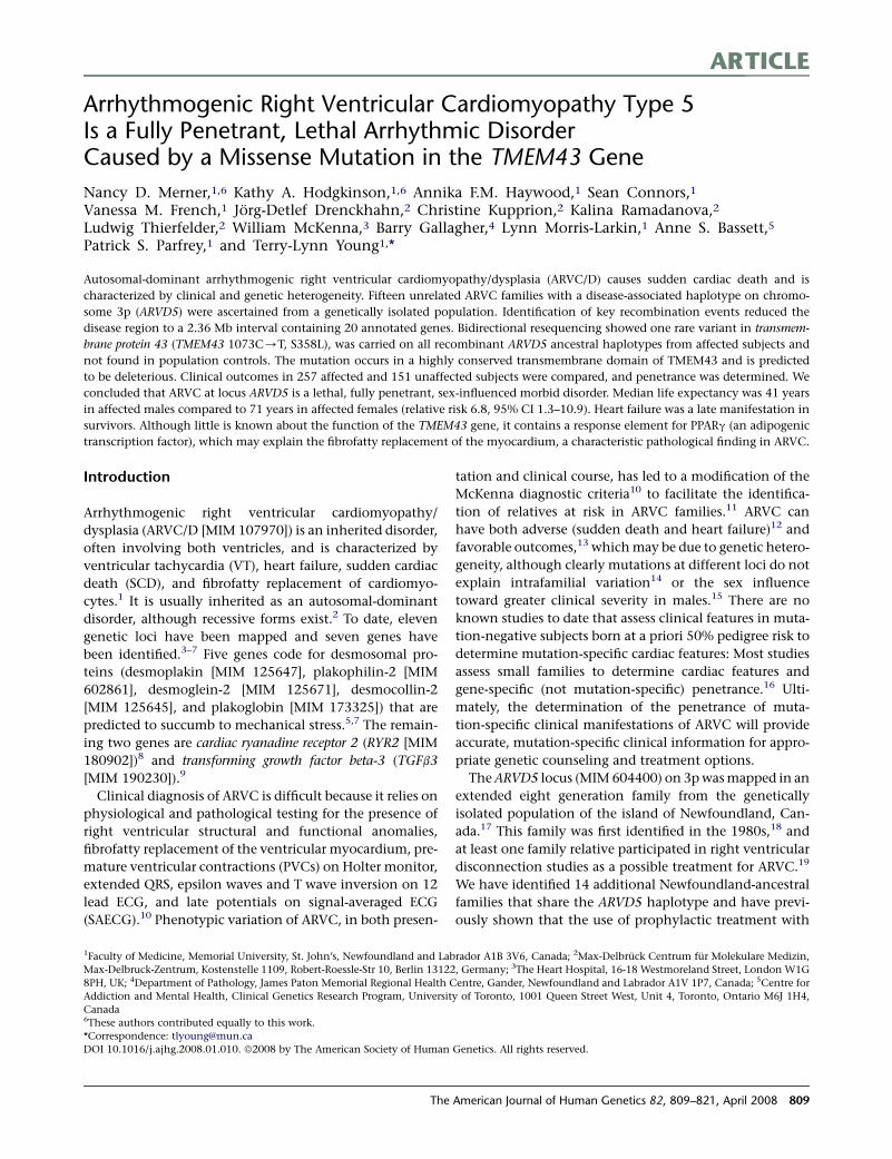
A total of 496 subjects born at a priori 50% risk (clinically
affected and their first-degree relatives) were available for study
across the 15 families. Subjects were determined to be clinically
affected if they fulfilled the ARVC modified criteria11 (Figure 2).
Blood samples from 295 subjects born at a priori 50% risk were
collected, and informed consent was obtained in compliance
Figure 1. ARVC Families Linked toChromosome 3p(A) Pedigrees of 15 autosomal-dominantARVC families linked to ARVD5. Affectedsubjects are shown as blackened squares(male) and circles (female). Subjects de-ceased because of SCD are noted by a circleabove the symbol.(B) Photomicrographs of paraffin-embed-ded postmortem right ventricular myocar-dium stained with masson trichromeshowing fibrofatty replacement of myo-cytes from a male teenager after suddencardiac death (left: 403 magnification)and a second-degree relative with suddencardiac death in his eighth decade (right:203 magnification). Pink represents nor-mal myocardium, blue represents fiber,and white represents fat.
with the Human Investigation Committee
requirements of the Eastern Health Cor-
poration of St. John’s, Newfoundland,
Canada (study number 00-176).
Fine Mapping of the ARVD5 LocusMapping the ARVD5 locus in family AR1
(Figure 1A) previously defined a 9.3 cM dis-
ease region between markers D3S3610 and
D3S3659.17 We genotyped 18 polymor-
phic microsatellite markers on clinically
affected subjects across all families to iden-
tify a disease-associated haplotype at 3p25
that was shared among families from this genetic isolate and pre-
sumed to be ancestral. We identified key recombination events on
the ARVD5 ancestral haplotype and used recombinations seen in
two or more families to narrow the region.
Screening Candidate ARVD5 GenesA mutation-screening panel was established that comprised seven
genomic DNA samples from four clinically affected subjects from
three families (AR1, AR8, and AR15; Figure 1A) and three spouses
(controls). All coding and noncoding exons and intron-exon
boundaries of positional candidate genes for ARVD5 were
sequenced. All sequences were amplified by polymerase-chain
reaction (PCR) assay from genomic DNA in 25 ml reaction
volume. Primer sequences are available from the authors on re-
quest. The PCR products were purified with 50% sephacryl
(Amersham Biosciences) and MultiScreen HTS filter plates (Milli-
pore Corporation). Purified PCR products were cycle sequenced in
both forward and reverse directions with the use of BigDye Termi-
nator V3.1 cycle sequencing kit on an automated ABI 3700 DNA
analyzer (Applied Biosystems). Sequencing electropherograms
were inspected manually and analyzed with Mutation Surveyor
software (Transition Technologies). Sequencing variants found
exclusively in clinically affected subjects on the mutation-screen-
ing panel were experimentally determined to reside on the
ARVD5 haplotype by segregation analysis in family AR14
(Figure 1A). The allele frequencies of the ARVD5 sequencing
810 The American Journal of Human Genetics 82, 809–821, April 2008
variants were determined with Newfoundland-population-based
controls obtained through random phone dialing, as part of
a large colorectal cancer study.21 Newfoundland is a known ge-
netic isolate where 98% of its residents are of English and Irish
descent.22,23 We analyzed key recombinant families (AR2 and
AR10) to determine which rare variants (<1% of the alleles
screened) were retained on recombinant ancestral haplotypes in
clinically affected subjects.
Expression of TMEM43 in Myocardium
and Lymphocytes of Clinically Affected SubjectsTotal RNA was extracted from Epstein Barr virus (EBV)-trans-
formed B lymphocytes from two affected subjects and one unaf-
fected control and also extracted from cardiac tissue from one
affected subject and an unrelated control with Trizol (Invitrogen);
this was followed by DNase1 treatment (Ambion). Complemen-
tary synthesis was performed and analyzed by both size fraction-
ation and direct sequencing with overlapping primers designed
to cover the complete coding sequence of TMEM43.
Bioinformatic AnalysisConservation of the TMEM43 protein across species was deter-
mined with ClustalW and Weblogo.24–26 Potential protein locali-
zation, function, structure, and posttranslational modification
Figure 2. Workflow and Mutation Sta-tus of Subjects Born at a Priori 50%Risk of ARVC
sites were predicted with the online tools
via the ExPASy web site.26 The effects of
amino acid substitutions on protein func-
tion were predicted.27–31 Information on
specific analyses is available upon request.
Clinical Assessment of Affected
versus Unaffected SubjectsClinical data were prospectively collected
over 11 years. This involved annual visits
by subjects to the genetics cardiomyopa-
thy clinic in which 12 lead ECGs, Holter
monitors, MRIs, signal-averaged ECGs,
and echocardiograms were done. All car-
diac anomalies were noted after clinical
testing. Clinical data were also obtained
retrospectively from medical records in-
cluding ‘‘at-risk’’ relatives not seen in clinic
and autopsy results. Subjects were catego-
rized as affected, unaffected, or unknown
(Figure 2). Only subjects from well-ascer-
tained sibships (in which disease status
was known in R50% of siblings) were
included in this study.
Anomalies on 12 lead ECGs were deter-
mined by two physicians blind to disease
status. Left ventricular enlargement (LVE)
was defined as R2 standard deviations
(SDs) above a predicted mean: left ventric-
ular end diastolic diameter (LVEDD)
>112% (>2 SD).32 Late potentials on SAECG were classified on
the basis of recognized criteria: QRS (filtered QRS duration)
>114 ms, LAS (low-amplitude signals) >38 ms, and RMS (root-
mean-square voltage of the terminal 40 ms of filtered QRS)
<20 ms.33 The presence of arrhythmias in the form of PVCs
was determined from Holter monitor analysis. Heart failure was
classified according to the New York Heart Association (NYHA)
functional classification.34 Subjects were identified with heart
failure (NYHA categories 1–4) if they presented prospectively in
the study with heart failure or it was documented in a medical re-
cord. Date of death was confirmed via autopsy records and archi-
val records.
Penetrance is defined as the proportion of subjects who have
a specified genotype known to cause a disease and who have any
signs or symptoms of the disease.35 To determine the disease pen-
etrance of ARVC at ARVD5, we assessed a subset of disease features
based on the ARVC modified diagnostic criteria11 from the first
available clinical test result to show an anomaly. We could then as-
sess which clinical features truly segregated with affection status in
this population. A Kaplan Meier analysis was then done with a sub-
set of subjects who were alive at the start of the prospective study
(1996), who had an available medical record, and who were either
mutation positive or an obligate carrier (60 males and 77 females).
On the basis of the analysis of prevalent disease features, a male
subject was considered ‘‘penetrant’’ at the age when any one of
The American Journal of Human Genetics 82, 809–821, April 2008 811

Figure 3. Physical Map of the ARVD5 Critical Region(A) Summary recombinant ARVD5 haplotypes identified across the 15 ARVC families with microsatellite markers (alleles are eithernumbered [1–9] or given in base pairs).(B) The physical map of the ARVD5 critical region. Physical distances were captured from the March 2006 freeze of the UCSC GenomeBrowser. Arrows show the direction of transcription of each annotated gene.
the following clinical events occurred: (1) SCD, (2) VT on clinical
testing, (3) heart failure, (4) >200 PVCs in a 24 hr period, (5) LVE
>2 SD, (6) QRS > 110 ms on 12 lead ECG, or (7) any late potential
on SAECG.11 Female subjects were considered penetrant for
all these features apart from QRS >110 ms on 12 lead ECG and
RMS or LAS on SAECG. We also determined the penetrance of
two major morbid outcomes of the disease, death and heart fail-
ure, using both the prospective and retrospective data set (affected
subjects n ¼ 257, 148 males, 109 females; unaffected subjects, n ¼151, 77 males, 74 females). For the heart-failure analysis, only
those from this group with an available medical record were used.
Cardiac tests were not available in all subjects at a priori 50%
risk, mainly because of SCD as a presenting feature for many
family members. For example, of 114 affected subjects who did
not have an available echocardiogram, 86 were deceased (75%),
whereas only two were dead in 50 unaffected subjects with no
echocardiogram. Testing is in progress in this latter group.
Statistical AnalysisComparisons between affected versus unaffected subjects were cal-
culated by the Kaplan Meier product limit method with censoring
occurring at the time of ICD therapy, heart transplantation, or last
follow-up (defined as the age at the last clinic visit). Relative risk
was calculated with Cox’s Regression model. A p value of <0.05
was considered significant (SPSS software, version 14, Chicago, IL).
812 The American Journal of Human Genetics 82, 809–821, April 20
Results
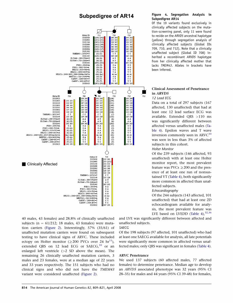
Fine Mapping and Candidate Gene Screening
Comparison of the ARVD5 ancestral haplotypes at 3p25
across clinically affected subjects identified key recombina-
tions and reduced the disease region to a 2.36 Mb interval
containing 20 annotated genes (Figures 3A and 3B; Table
1). Bidirectional resequencing of the 20 physical candidate
ARVD5 genes revealed 240 variants (Table S1 available
online). Nineteen variants were found exclusively in clini-
cally affected subjects on the mutation-screening panel,
and 11 were determined to reside on the ARVD5 ancestral
haplotype through segregation analysis in AR14 (Table 2
and Figure 4). Screening of population controls showed
that five of these variants were rare (<1% of the alleles
screened; Table 2). Only one of the five, TMEM43
1073C/T (S358L) (Figure 5D), was shared by all clinically
affected subjects across the 15 families and was retained
on key recombinant ARVD5 haplotypes identified in clini-
cally affected subjects from families AR10 (Figure 6) and
AR2 (data not shown). This suggested that TMEM43 is
ARVD5. Additional supportive evidence included clinically
unaffected adult subjects who shared distal sections of the
08

ARVD5 haplotype and lacked the TMEM43 mutation (e.g.,
Figure 4). All available spouses (n ¼ 47) and population
controls (n ¼ 161) were negative for the TMEM43 muta-
tion (416 mutation-negative chromosomes).
ARVC at Locus ARVD5 Is Caused by a Missense
Mutation in TMEM43
The longest isoform of transmembrane protein 43
(TMEM43, GenBank accession number NM_024334) has
Table 1. The 20 Physical Candidate Genes for ARVD5
Genes
Accession
Number
MIM
Number Strand
Genomic Position
ExonsStart End
IQSEC1 NM_014869 610166 � 13003536 12917079 13
NUP210 NM_024923 607703 � 13436809 13332737 40
HDAC11 NM_024827 607226 þ 13496824 13521834 10
FBLN2 NM_001004019 135821 þ 13565625 13654922 18
WNT7A NM_004625 601570 � 13896619 13835083 4
TPRXL AK092426 611167 þ 13953902 14082480 3
CHCHD4 NM_144636 611077 � 14141323 14128584 4
TMEM43 NM_024334 na þ 14141546 14160180 12
XPC NM_004628 278720 � 14195143 14161651 16
LSM3 NM_014463 607283 þ 14195341 14214840 4
SLC6A6 NM_003043 186854 þ 14419110 14503973 15
GRIP2 NM_001080423 na � 14558592 14510177 25
C3orf19 NM_016474 na þ 14668278 14689167 11
C3orf20 NM_032137 na þ 14691658 14789544 17
FGD5 NM_152536 na þ 14835810 14950899 20
NR2C2 NM_003298 601426 þ 14964240 15065782 15
MRPS25 NM_022497 na � 15081820 15065024 4
ZFYVE20 NM_022340 609511 � 15115659 15086584 14
CAPN7 NM_014296 606400 þ 15222737 15269426 21
SH3BP5 NM_004844 605612 � 15349108 15271250 9
Total 275
The
12 exons (Figure 5A) predicting a 400 amino acid protein
that is 98% similar to the mouse protein (Figure 7). This
conserved gene is found across all eukaryotic and pro-
karyotic species (Figure 7). We were able to extract full-
length TMEM43 cDNA from white blood cells and cardiac
tissue from both patients and controls, demonstrating
that TMEM43 is expressed in both blood and cardiac tis-
sue and that the TMEM43 1073C/T (S358L) mutation
does not appear to affect splicing (Figures 5B, 5C, and
5E).
Bioinformatic analysis of TMEM43 predicts it to be a
cytoplasmic membrane protein with several potential
posttranslation modification sites (Figure 8). Unlike the
transmembrane proteins of the desmosome (desmocollin
and desmoglein), TMEM43 does not have a cadherin
domain. Furthermore, protein sequence alignments with
desmocollin and desmoglein show less than 10% identity
and less than 12% similarity.26 The mutation, S358L,
occurs within the third predicted transmembrane domain
and is highly conserved in mammalian, avian, amphibian,
and insect orthologs (Figures 7A and 7B and Figure 8).
Interestingly, a leucine at this position is found in the
bacterium Rhizobium loti, but it is not found in any multi-
cellular organisms (Figures 7A and 7B). The S358L muta-
tion is predicted to be deleterious (Table 3), but little is
known about the function of TMEM43 protein.
TMEM43 Mutation Screening in ARVD5
Linked Families
We sequenced genomic DNA from all available subjects
born at a priori 50% risk (n ¼ 295) across the 15 ARVC
families for the presence of the 1073C/T TMEM43
mutation. All clinically affected subjects (n ¼ 83/83;
Table 2. Sequencing Variants Identified Exclusively in Clinically Affected Subjects
Gene Name Accession number Variant Nomenclature Classification Allele Frequency(# of chromosomes)
HDAC11 NM_024827 c.369þ18_369þ19insG Noncoding nd nd
TMEM43 NM_024334 c.1073C/T Missense (S > L) 0/322 0.00%*
TMEM43 NM_024334 c.1203þ115T/C Noncoding nd nd
XPC NM_004628 c.2823þ684G/C Noncoding nd nd
SLC6A6 NM_003043 c.1-27420G/A Noncoding nd nd
SLC6A6 NM_003043 c.599þ370A/G Noncoding dbSNP 46.00%
SLC6A6 NM_003043 c.733-1226A/G Noncoding 50/90 55.60%
FGD5 NM_152536 c.934G/A Missense (V > M) 2/318 0.60%*
FGD5 NM_152536 c.2186þ22G/A Noncoding nd nd
FGD5 NM_152536 c.2187-82G/A Noncoding nd nd
FGD5 NM_152536 c.2220G/T Synonymous (L > L) nd nd
FGD5 NM_152536 c.2613þ50C/T Noncoding nd nd
FGD5 NM_152536 c.3085-74G/A Noncoding 15/166 9.00%
NR2C2 NM_003298 c.855þ70G/A Noncoding 8/88 9.10%
NR2C2 NM_003298 c.1848þ365T/A Noncoding 16/90 17.80%
NR2C2 NM_003298 c.1848þ2965_1848þ2966insGATA Noncoding 23/126 18.30%
MRPS25 NM_022497 c.522þ1059G/A Noncoding 0/138 0.00%*
CAPN7 NM_014296 c.1289þ68C/T Noncoding 1/162 0.01%*
CAPN7 NM_014296 c.1430-28T/C Noncoding 0/160 0.00%*
Eleven of the 19 variants were determined to be in phase on the ARVD5 ancestral haplotype and were subsequently sequenced on population controls
(italics). The allele frequency of variants previously reported in the NCBI dbSNP was used. Rare variants (<1% of the alleles screened) are marked with
an asterisk.
American Journal of Human Genetics 82, 809–821, April 2008 813
40 males, 43 females) and 28.8% of clinically unaffected
subjects (n ¼ 61/212; 18 males, 43 females) were muta-
tion carriers (Figure 2). Interestingly, 57% (35/61) of
unaffected mutation carriers were found on subsequent
testing to have clinical signs of ARVC. These included
ectopy on Holter monitor (R200 PVCs over 24 hr11),
extended QRS on 12 lead ECG or SAECG,10 or an
enlarged left ventricle (>2 SD above the mean). The
remaining 26 clinically unaffected mutation carriers, 3
males and 23 females, were at a median age of 22 years
and 33 years respectively. The 151 subjects who had no
clinical signs and who did not have the TMEM43
variant were considered unaffected (Figure 2).
Figure 4. Segregation Analysis inSubpedigree AR14Of the 19 variants found exclusively inclinically affected subjects on the muta-tion-screening panel, only 11 were foundto reside on the ARVD5 ancestral haplotype(yellow) through segregation analysis ofclinically affected subjects (Global IDs709, 710, and 712). Note that a clinicallyunaffected subject (Global ID 708) in-herited a recombinant ARVD5 haplotypefrom her clinically affected mother thatlacks TMEM43. Alleles in brackets havebeen inferred.
Clinical Assessment of Penetrance
in ARVD5
12 Lead ECG
Data on a total of 297 subjects (167
affected, 130 unaffected) that had at
least one 12 lead surface ECG was
available. Extended QRS >110 ms
was significantly different between
affected versus unaffected males (Ta-
ble 4). Epsilon waves and T wave
inversion commonly seen in ARVC10
was seen in less than 3% of affected
subjects in this cohort.
Holter Monitor
Of the 239 subjects (146 affected, 93
unaffected) with at least one Holter
monitor report, the most prevalent
feature was PVCs R200 and the pres-
ence of at least one run of nonsus-
tained VT (Table 4), both significantly
more common in affected than unaf-
fected subjects.
Echocardiography
Of the 244 subjects (143 affected, 101
unaffected) that had at least one 2D
echocardiogram available for analy-
sis, the most prevalent feature was
LVE based on LVEDD (Table 4),32,36
and LVE was significantly different between affected and
unaffected subjects.
SAECG
Of the 198 subjects (97 affected, 101 unaffected) who had
at least one SAECG available for analysis, all late potentials
were significantly more common in affected versus unaf-
fected males; only QRS was significant in females (Table 4).
ARVC Penetrance
We used 137 subjects (60 affected males, 77 affected
females) to determine penetrance. Median age to develop
an ARVD5 associated phenotype was 32 years (95% CI
28–35) for males and 44 years (95% CI 39-48) for females,
814 The American Journal of Human Genetics 82, 809–821, April 2008

Figure 5. Gene Structure and Mutation Analysis of TMEM43(A) Gene structure of TMEM43. Exons are represented by boxes. Translated exons are solid green, and untranslated exons are clear. Intronsare represented by green lines.(B) Coverage of primers designed to amplify cDNA showing position of PCR primer pairs: exons 1–4 (purple), exons 4–9 (red), exons 9–12(green), exons 1–10 (blue), and 5–12 (yellow).(C) PCR products amplified from cDNA of EBV-transfected B cells of affected subjects from the mutation-screening panel (affected AR1 andaffected AR15) and unaffected (control) subjects. cDNA of heart tissue from both the left and right ventricle of an affected subject (AR13)and a heart biopsy from a control subject were size fractionated by electrophoresis.(D and E) Forward and reverse sequencing traces showing the TMEM43 1073C/T mutation of an affected subject’s (AR13) genomic andcDNA. The amino acid translations (top) shows the S358L amino acid substitution.
with 100% of males and females penetrant by 63 and 76
years, respectively (Figure 9 and Table 5). Males were twice
as likely to reveal the disease phenotype than females (RR
2, 99% CI 1.2–3.3) (p % 0.0001). The commonest clinical
features for which subjects were initially penetrant were
ectopy (44%) and LVE (27%), and those were followed by
VT (9%), QRS >110 ms (9%), late potentials on SAECG
(7%), SCD (2%), and heart failure (2%).
Heart Failure
Fourteen of 89 affected males developed heart failure at
a median age of 63 years (95% CI 41–84 years) compared
with no unaffected males (p % 0.0001, log rank)
(Figure 10Ai). One unaffected female at age 79 developed
heart failure compared to seven affected females (median
age 73 years, 95% CI 69–77 years) (p % 0.001, log rank)
The
(Figure 10Aii). Affected males were three times more likely
to develop heart failure than females (RR 3.4, 95% CI
1.36–8.57, p % 0.009) (Figure 10Aiii).
Life Expectancy
In affected subjects (n ¼ 257), there were 123 (48%) deaths
(99 males and 24 females). Sudden cardiac death occurred
in 86% (86/99) of the males and 42% (10/24) of the
females. Survival was significantly reduced in affected
versus unaffected subjects. Median survival was 41 com-
pared to 83 years (p % 0.0001, log rank) in affected versus
unaffected males (Figure 10Bi) and 71 compared to 83 years
in affected versus unaffected females (p % 0.002, log rank)
(Figure 10Bii). The relative risk of dying was 6.8 times
greater in affected males versus affected females (95% C.I:
4.3–10.9, p % 0.0001) (Figure 10Biii).
American Journal of Human Genetics 82, 809–821, April 2008 815

Discussion
Mutations in desmosomal genes underlie several genetic
subtypes of ARVC, suggesting that ARVC is primarily a dis-
ease of the desmosome.5,7 We used a positional mapping
approach and 15 ARVC families that shared a disease-asso-
ciated haplotype on 3p from a genetic isolate to identify
ARVD5. All clinically affected subjects that were screened
for the TMEM43 1073C/T mutation (n ¼ 83) were muta-
tion carriers. This mutation was not detected in spouses or
population controls. The TMEM43 gene is expressed in car-
diac tissue and is evolutionarily conserved, and the amino
acid substitution is predicted to be deleterious.
Recently, signaling pathways have been implicated in
ARVC pathogenesis.7,37 For example, plakoglobin, when
freed from desmosomal complexes, translocates to the
nucleus where it competes and opposes the action of
b-catenin and downregulates the canonical Wnt/b-catenin
signaling pathway.37 Suppression of the canonical Wnt/
b-catenin signaling upregulates two adipogenic transcrip-
tion factors, C/EBP-a (MIM 116897) and PPARg (MIM
Figure 6. Segregation Analysis inSubpedigree AR10Clinically affected subjects (Global IDs 794and 796) only have one of the five rare var-iants because of a historical recombinationevent on the ARVD5 haplotype (yellow).Alleles in brackets have been inferred.
601487).37 A genome-wide scan for
peroxisome proliferator response ele-
ments (PPREs) identified 1085 poten-
tial target genes of PPARg, including
TMEM43.38 If TMEM43 is a part of
an adipogenic pathway regulated by
PPARg, then perhaps dysregulation
of this pathway may explain the fi-
brofatty replacement of the myocar-
dium in ARVC patients.
Studies that assess the ARVC phe-
notype in patients with mutations
in plakophilin-2,39 desmoglein-2,16 and
plakoglobin2 typically evaluate clinical
features in mutation carriers only.
The assumption is that all cardiac
signs present in affected subjects are
due to the underlying genetic defect.
However, it may be that some signs
are due to the genetic background of
either the family or the source popu-
lation. The size of the ARVC cohort
in this study provides an opportunity
to compare and define ARVC-specific
clinical features in affected and unaf-
fected subjects.
By using the benefit of a control group, we evaluated
modified clinical criteria11 in affected versus unaffected
subjects. We included LVE, a clinical feature that is noted
in other ARVC studies40–44 and that is prevalent in this
cohort. For all clinical features examined, a significant dif-
ference was found between affected versus unaffected
males. Females were significantly different for all features
with the exception of RMS and LAS on SAECG and
a QRS >110 ms on 12 lead ECG.
For the penetrance study, we followed subjects who were
alive at the start of the study (1996), who had a medical
record, and who had a genetic diagnosis (mutation carriers
or obligate carriers). The results showed that ARVC was
fully penetrant in males by the age of 63 and in females
by the age of 76; ectopy and LVE were often the first
presenting features, and few presented with death or heart
failure. Therefore, in order to assess the penetrance of heart
failure and death, we used all affected subjects. Heart
failure and death were morbid outcomes at early ages in
both males and females, with far more serious early events
in males. In both sexes, heart failure occurs as a later
816 The American Journal of Human Genetics 82, 809–821, April 2008

Figure 7. Multiple Alignment of the TMEM43 Gene across Eight Eukaryotic and Prokaryotic Species(A) Clustal W align was used to align orthologs from Homo sapiens (NP_077310), Pan troglodytes (XP_516299), Canis familiaris(XP_541751), Mus musculus (NP_083042), Gallus gallus (XP_414378), Zenopus tropicalis (UP10004D5297), Tetraodon nigroviridis(Q4RXL8), Drosophila melanogaster (NP_64162), and Rhizobium loti (Q98HF3).25 The blue box outlines the DUF1625 domain, and the blackboxes outline predicted transmembrane domains. Completely conserved residues are red, strongly similar residues are green, and weaklysimilar residues are blue. The S358L mutation is marked by a yellow arrow.(B) The web logo format was used for aligning eukaryotic species. The third transmembrane domain is outlined (black box). The S358Lmutation is marked by a yellow arrow.
manifestation in subjects who did not succumb to SCD.
These major manifestations define ARVC, because of the
TMEM43 mutation, as a lethal, fully penetrant, sex-influ-
enced, autosomal-dominant disorder.
Biases exist in studies that use both retrospective and
prospective data; the use of well-ascertained sibships may
mitigate some of these. Although formal assessment of
right ventricular structure is not available for the majority
of subjects in this cohort, the cardiomyopathy fulfills
diagnostic criteria for ARVC,11 it has been historically diag-
nosed as ARVC,17,19 and fibrofatty replacement of the
myocardium is present. The involvement of the left ventri-
cle in this cohort supports the recent call to place ARVC
The
within a group called ‘‘arrhythmic cardiomyopathies,’’
which would take into account various presentations of
disease.45 These findings may not be applicable to other
forms of inherited ARVC. However, it is likely that muta-
tions in TMEM43 are present in other populations. Issues
of classification in the cardiomyopathies in general will
be solved by their underlying genetic etiology; thus,
disease-specific genotype/phenotype information will
become the most useful and relevant.
This study has the advantage of having access to past
medical records and extensive genealogical data, and a sin-
gle cardiac-care team followed all subjects. Clinical signs
due to the genetic background of either the family or the
American Journal of Human Genetics 82, 809–821, April 2008 817

source population were controlled for by the use of unaf-
fected subjects from well-ascertained sibships. This genetic
subtype does not show the typical ARVC ECG findings of T
wave inversion or epsilon waves.10 A direct benefit of this
process therefore has been the provision of accurate muta-
tion-specific clinical data for precise genetic counseling
and follow-up of those at-risk.20
We have identified the gene that causes ARVC at locus
ARVD5. A missense mutation in TMEM43 causes a fully
penetrant, sex-influenced lethal form of ARVC. Although
little is known about the function of this gene, it encodes
a transmembrane protein that could be a target of PPARg,
and this may explain the fibrofatty replacement of the
myocardium in ARVC patients. Future directions include
functional studies of the TMEM43 protein.
Supplemental Data
One table is available at http://www.ajhg.org/.
Acknowledgments
The authors are supported by a career award from the Canadian
Institutes of Health Research-Regional Partnerships Program (to
Figure 8. Predicted Topography of theTMEM43 ProteinIndicated are transmembrane domains(beige), phosphorylation sites (green), atransactivation domain (red), YingOYangsites (orange), a SUMO attachment site (pur-ple), and an O-glycosylation site (blueopen). The extracellular and cytoplasmicregions may be reversed: There is evidencesupporting either orientation.
Table 3. Prediction of the TMEM43 1073C/T, S358L,Mutation Effect
Program SIFT Panther PolyPhen SNPs3D PMut
S358L
mutation
deleterious deleterious Benign deleterious deleterious
Five different bioinformatic programs were used for predicting the effect of
the S358L mutation. Note that sequence homology used by the PolyPhen
analyses was calculated with alignments of orthologs from Eukaryota and
bacteria.
T.-L.Y.). K.H. and L.T. were supported by
a grant from the Ernst and Berta Grimmke
Foundation, Dusseldorf, Germany. This
project was funded by the Canadian Insti-
tutes of Health Research, Genome Canada
(Atlantic Medical Genetics and Genomics
Initiative), Newfoundland and Labrador
Centre for Applied Health Research, Jane-
way Children’s Hospital Foundation, Me-
morial University Opportunities Fund,
the General Hospital Foundation and St.
Judes Medical Research Grant, Canada.
We dedicate this paper to the ARVC families in Newfoundland,
who live daily with the reality of this disease. Without their con-
tinued support and participation, this research could not occur.
We thank Barbara Peddle, Pauline Ryan, Karen Williams, Carol
Tilley, Liz Kernaghan, Sara MacKay, Fiona Curtis, and Elizabeth
Dicks for clinical assistance, Dr. Roger Green for control samples,
and Dante Galutria, Sigrid Milan, Ingrid Pardoe, Nancy Whalen,
Yoella Teplisky, Susan Stuckless, and Fahad Chowdhury for techni-
cal assistance. Special thanks to Dr. Laura Gillespie and Dr. Gary
Paterno for help with protein prediction, Dr. Christina Templeton
and Dr. Suryakant Shah for pediatric cardiology consultation, and
Dr. Adrian Lear, Dr. William Marshall, and Dr. Elizabeth Ives for
their early interest in this disease.
Received: October 19, 2007
Revised: December 11, 2007
Accepted: January 8, 2008
Published online: February 28, 2008
Web Resources
The URLs for data presented herein are as follows:
NCBI dbSNP, http://www.ncbi.nlm.nih.gov/sites/entrez?db¼snp&cmd¼search&term¼
Online Mendelian Inheritance in Man (OMIM), http://www.ncbi.
nlm.nih.gov/Omim
UCSC Genome Browser, http://genome.ucsc.edu/
References
1. Fontaine, G., Fontaliran, F., and Frank, R. (1998). Arrhythmo-
genic right ventricular cardiomyopathies: Clinical forms and
main differential diagnoses. Circulation 97, 1532–1535.
2. Protonotarios, N., Tsatsopoulou, A., Anastasakis, A., Sevdalis,
E., McKoy, G., Stratos, K., Gatzoulis, K., Tentolouris, K., Spilio-
poulou, C., and Panagiotakos, D. (2001). Genotype-phenotype
818 The American Journal of Human Genetics 82, 809–821, April 2008

Table 4. Prevalent Cardiac Features on First Clinical Test
12 Lead ECG
Affected Unaffected Affected Males
versus Unaffected
Males, c2
Affected Females
versus Unaffected
Females, c2Male Female Total Male Female Total
Subjects Tested (N) 78 89 167 64 66 130
Mean Age (SD) 30.0 (12.1) 37.7 (15.8) 33.1 (16.0) 38.2 (13.2)
QRS >110 ms n (%) 25 (32) 8 (9) 33 (19.8) 4 (6) 2 (3) 6 (4.6) p % 0.001 p % 0.2 ns
Holter Monitor
Subjects Tested (N) 67 79 146 49 44 93
Mean Age (SD) 31.1 (13.3) 37.7 (15.5) 33.1 (15.9) 38.2 (12.8)
PVCs R200/24 hr n (%) 47 (70) 47 (59) 94 (64.4) 0 (0) 1 (2) 1 (1.1) p < 0.001 p < 0.001
R1 run ns VT n (%) 13 (19) 18 (23) 31 (21.2) 0 (0) 0 (0) 0 (0.0) p < 0.01 p < 0.001
Echocardiograph
Subjects Tested (N) 67 76 143 50 51 101
Mean Age (SD) 31.6 (13.2) 39.6 (15.1) 32.5 (14.9) 38.3 (13.3)
LVE >2 SD n (%) 35 (52) 27 (20) 62 (43.3) 10 (20) 4 (8) 14 (13.9) p < 0.001 p < 0.001
SAECG
Subjects Tested (N) 32 65 97 50 51 101
Mean Age (SD) 30.9 (14.5) 39.8 (14.5) 36.7 (16.3) 38.9 (11.0)
QRS >114 ms n (%) 19 (59) 19 (29) 38 (39.2) 10 (20) 2 (4) 12 (11.9) p % 0.001 p % 0.001 ns
RMS <20 ms n (%) 15 (47) 18 (28) 33 (34.0) 12 (24) 9 (18) 21 (20.8) p % 0.05 p % 0.3 ns
LAS >38 ms n (%) 17 (53) 21 (32) 38 (39.2) 14 (28) 13 (25) 27 (26.7) p % 0.025 p % 0.5 ns
This table shows 12 lead ECG, Holter, echocardiographic, and SAECG manifestations used in penetrance analysis in subjects from 15 families with ARVC due
to a mutation in TMEM43. Abbreviations are used as follows: PVC, premature ventricular complex; VT, ventricular tachycardia; LVE, left ventricular enlarge-
ment indexed to height and weight; RMS, root mean square voltage of the terminal 40 ms of filtered QRS; and LAS, low-amplitude signals.
assessment in autosomal recessive arrhythmogenic right ven-
tricular cardiomyopathy (Naxos disease) caused by a deletion
in plakoglobin. J. Am. Coll. Cardiol. 38, 1477–1484.
3. Heuser, A., Plovie, E.R., Ellinor, P.T., Grossmann, K.S., Shin,
J.T., Wichter, T., Basson, C.T., Lerman, B.B., Sasse-Klaassen,
S., Thierfelder, L., et al. (2006). Mutant desmocollin-2 causes
arrhythmogenic right ventricular cardiomyopathy. Am. J.
Hum. Genet. 79, 1081–1088.
4. Pilichou, K., Nava, A., Basso, C., Beffagna, G., Bauce, B., Loren-
zon, A., Frigo, G., Vettori, A., Valente, M., Towbin, J., et al.
Figure 9. ARVC Penetrance Caused by the TMEM43 1073C/TMutationCumulative proportion by age at which male (n ¼ 60) and female(n ¼ 77) mutation positive affected subjects manifested a firstclinical sign of ARVC. Subjects were censored at last follow-up.
The
(2006). Mutations in desmoglein-2 gene are associated with
arrhythmogenic right ventricularcardiomyopathy. Circula-
tion 113, 1171–1179.
5. Sen-Chowdhry, S., Syrris, P., and McKenna, W. (2005). Genet-
ics of right ventricular cardiomyopathy. J. Cardiovasc. Electro-
physiol. 16, 927–935.
6. Dokuparti, M.V., Pamuru, P.R., Thakkar, B., Tanjore, R.R., and
Nallari, P. (2005). Etiopathogenesis of arrhythmogenic right
ventricular cardiomyopathy. J. Hum. Genet. 50, 375–381.
7. MacRae, C.A., Birchmeier, W., and Thierfelder, L. (2006).
Arrhythmogenic right ventricular cardiomyopathy: moving
toward mechanism. J. Clin. Invest. 116, 1825–1828.
8. Tiso, N., Stephan, D.A., Nava, A., Bagattin, A., Devaney, J.M.,
Stanchi, F., Larderet, G., Brahmbhatt, B., Brown, K., Bauce,
B., et al. (2001). Identification of mutations in the cardiac
ryanodine receptor gene in families affected with arrhythmo-
genic right ventricular cardiomyopathy type 2 (ARVD2). Hum.
Mol. Genet. 10, 189–194.
9. Beffagna, G., Occhi, G., Nava, A., Vitiello, L., Ditadi, A., Basso,
C., Bauce, B., Carraro, G., Thiene, G., and Towbin, J.A. (2005).
Regulatory mutations in transforming growth factor-[beta]3
gene cause arrhythmogenic right ventricular cardiomyopathy
type 1. Cardiovasc. Res. 65, 366–373.
Table 5. Percentage of Subject’s Penetrant by Each Age
Age (years) 11–20 21–30 31–40 41–50 51–60 61–70 71–80
Males 13 39 68 89 96 100 100
Females 3 18 38 67 80 97 100
This table shows the cumulative percentage of subjects penetrant by
decade.
American Journal of Human Genetics 82, 809–821, April 2008 819

Figure 10. Time to Event Analysis ofHeart Failure and Death in AffectedSubjects(Ai–Aiii) Cumulative incidence of heart fail-ure; panels describe the following: (Ai)affected males (n ¼ 89, dark blue) com-pared to unaffected males (n ¼ 71, lightblue) (p % 0.0001: log rank); (Aii) affectedfemales (n ¼ 87, dark pink) compared tounaffected females (n ¼ 68, light pink)(p % 0.001, log rank); and (Aiii) affectedmales (dark blue) versus affected females(dark pink).(Bi–Biii) Life expectancy; panels describethe following: (Bi) time to death inaffected males (n ¼ 148, dark blue) andunaffected males (n ¼ 77, light blue);(Bii) time to death in affected females(n ¼ 109, dark pink) and unaffected(n ¼ 74, light pink); and (Biii) affectedmales (dark blue) versus affected females(dark pink).
10. McKenna, W.J., Thiene, G., Nava, A., Fontaliran, F., Blomstrom-
Lundquist, C., Fontaine, G., and Camererini, F. (1994). Diagno-
sis of arrhythmogenic right ventricular dysplasia/cardiomyopa-
thy. Task Forceof theWorking Group Myocardial andPericardial
disease of the European Society of Cardiology and the Scientific
Council on Cardiomyopathies of the International Society and
Federation of Cardiology. Br. Heart J. 71, 215–218.
11. Hamid, M.S., Norman, M., Quraishi, A., Firoozi, S., Thaman,
R., Gimeno, J.R., Sachdev, B., Rowland, E., Elliott, P.M., and
McKenna, W.J. (2002). Prospective evaluation of relatives for
familal arrhythmogenic right ventricular cardiomyopathy/
dysplasia reveals a need to broaden diagnostic criteria. J. Am.
Coll. Cardiol. 40, 1445–1450.
12. Corrado, D., Basso, C., and Thiene, G. (2000). CARDIOMYOP-
ATHY: Arrhythmogenic right ventricular cardiomyopathy:
Diagnosis, prognosis, and treatment. Heart 83, 588–595.
13. Nava, A., Bauce, B., Basso, C., Muriago, M., Rampazzo, A.,
Villanova, C., Daliento, L., Buja, G., Corrado, D., Danieli,
G.A., et al. (2000). Clinical profile and long-term follow-up
of 37 families with arrhythmogenic right ventricular cardio-
myopathy. J. Am. Coll. Cardiol. 36, 2226–2233.
14. Wlodarska, E.K., Konka, M., Zaleska, T., Ploski, R., Cedro, K.,
Pucilowska, B., Bekiesinska-Figatowska, M., Rydlewska-
Sadowska, W., Ruzyllo, W., and Hoffman, P. (2005). Arrhyth-
820 The American Journal of Human Genetics 82, 809–821, April 20
mogenic right ventricular cardiomyopathy in two pairs of
monozygotic twins. Int. J. Cardiol. 105, 126–133.
15. Blomstrom-Lundqvist, C., Sabel, K., and Olsson, S. (1987). A
long term follow-up of 15 patients with arrhythmogenic right
ventricular dysplasia. Br. Heart J. 58, 477–488.
16. Syrris, P., Ward, D., Asimaki, A., Evans, A., Sen-Chowdhry, S.,
Hughes, S.E., and McKenna, W.J. (2007). Desmoglein-2 muta-
tions in arrhythmogenic right ventricular cardiomyopathy: A
genotype-phenotype characterization of familial disease. Eur.
Heart J. 28, 581–588.
17. Ahmad, F., Li, D., Karibe, A., Gonzalez, O., Tapscott, T., Hill, R.,
Weilbaecher, D., Blackie, P., Furey, M., Gardner, M., et al.
(1998). Localisation of a gene responsible for arrhythmogenic
right ventricular dysplasia to chromosome 3p23. Circulation
98, 2791–2795.
18. Marshall, W., Furey, M., Larsen, B., Rose, J., Sharratt, G., Sus-
sex, B., Virmani, S., Ko, P., LeBlanc, P., and Nolan, R. (1988).
Right ventricular cardiomyopathy and sudden death in young
people. N. Engl. J. Med. 319, 174–175.
19. Guiraudon, G., Klein, G., Gulaumshein, S., Paiinvin, G.,
DelCompo, C., Gonzales, J., and Ko, P. (1983). Total disconnec-
tion of right ventricular free wall: Surgical treatment of right
ventricular tachycardia associated with right ventricular dyspla-
sia. Circulation 67, 463–470.
08
20. Hodgkinson, K., Parfrey, P., Bassett, A., Kupprion, C., Drenck-
hahn, J., Norman, M., Thierfelder, L., Stuckless, S., Dicks, E.,
McKenna, W., et al. (2005). The impact of implantable cardi-
overter defibrillator therapy on survival in autosomal domi-
nant arrhythmogenic right ventricular cardiomyopathy
(ARVD5). J. Am. Coll. Cardiol. 45, 400–408.
21. Green, R.C., Green, J.S., Buehler, S.K., Robb, J.D., Daftary, D.,
Gallinger, S., McLaughlin, J.R., Parfrey, P.S., and Younghus-
band, H.B. (2007). Very high incidence of familial colorectal
cancer in Newfoundland: A comparison with Ontario and
13 other population-based studies. Fam. Cancer 6, 53–62.
22. Mannion, J. (1977). The Peopling of Newfoundland: Essays in
Historical Geography. Institute of Social and Economic
Research (St. John’s: Memorial University).
23. Rahman, P., Jones, A., Curtis, J., Bartlett, S., Peddle, L., Fernan-
dez, B., and Freimer, N. (2003). The Newfoundland popula-
tion: A unique resource for genetic investigation of complex
diseases. Hum. Mol. Genet. 12, R167–R172.
24. Crooks, G.E., Hon, G., Chandonia, J.M., and Brenner, S.E.
(2004). WebLogo: A sequence logo generator. Genome Res.
14, 1188–1190.
25. Combet, C., Blanchet, C., Geourjon, C., and Deleage, G.
(2000). NPS@: Network protein sequence analysis. Trends Bio-
chem. Sci. 25, 147–150.
26. Gasteiger, E., Gattiker, A., Hoogland, C., Ivanyi, I., Appel, R.D.,
and Bairoch, A. (2003). ExPASy: The proteomics server for
in-depth protein knowledge and analysis. Nucleic Acids Res.
31, 3784–3788.
27. Ramensky, V., Bork, P., and Sunyaev, S. (2002). Human
non-synonymous SNPs: Server and survey. Nucleic Acids
Res. 30, 3894–3900.
28. Ng, P.C., and Henikoff, S. (2002). Accounting for human
polymorphisms predicted to affect protein function. Genome
Res. 12, 436–446.
29. Thomas, P.D., Kejariwal, A., Campbell, M.J., Mi, H., Diemer,
K., Guo, N., Ladunga, I., Ulitsky-Lazareva, B., Muruganujan,
A., Rabkin, S., et al. (2003). PANTHER: A browsable database
of gene products organized by biological function, using
curated protein family and subfamily classification. Nucleic
Acids Res. 31, 334–341.
30. Yue, P., Melamud, E., and Moult, J. (2006). SNPs3D: candidate
gene and SNP selection for association studies. BMC Bioinfor-
matics 7, 166.
31. Ferrer-Costa, C., Gelpi, J.L., Zamakola, L., Parraga, I., de la
Cruz, X., and Orozco, M. (2005). PMUT: A web-based tool
for the annotation of pathological mutations on proteins.
Bioinformatics 21, 3176–3178.
32. Henry, W.L., Gardin, J.M., and Ware, J.H. (1980). Echocardio-
graphic measurements in normal subjects from infancy to old
age. Circulation 62, 1054–1061.
33. Breithardt, G., Cain, M., and El-Sherif, N. (1991). Standards for
analysis of ventricular late potentials using high resolution or
The
signal-averaged electrocardiography. A statement by a task
force committee of the european society of cardiology, the
American Heart Association, and the American College of Car-
diology. J. Am. Coll. Cardiol. 17, 999–1006.
34. NYHA CC (1964). Nomenclature and Criteria for Diagnosis:
Diseases of the Heart and Blood Vessels (Boston: Little, Brown
& Co)., pp. 114.
35. Nussbaum, R., McInnes, R., and Willard, H. (2001). Thomp-
son and Thompson: Genetics in Medicine (Philadelphia:
W.B Saunders).
36. Henry, W.L., Ware, J., Gardin, J.M., Hepner, S.I., McKay, J., and
Weiner, M. (1978). Echocardiographic measurements in
normal subjects. Growth related changes that occur between
infancy and early adulthood. Circulation 57, 278–285.
37. Garcia-Gras, E., Lombardi, R., Giocondo, M.J., Willerson, J.T.,
Schneider, M.D., Khoury, D.S., and Marian, A.J. (2006). Sup-
pression of canonical Wnt/beta-catenin signaling by nuclear
plakoglobin recapitulates phenotype of arrhythmogenic right
ventricular cardiomyopathy. J. Clin. Invest. 116, 2012–2021.
38. Lemay, D.G., and Hwang, D.H. (2006). Genome-wide identifi-
cation of peroxisome proliferator response elements using in-
tegrated computational genomics. J. Lipid Res. 47, 1583–1587.
39. Syrris, P., Ward, D., Asimaki, A., Sen-Chowdhry, S., Ebrahim,
H.Y., Evans, A., Hitomi, N., Norman, M., Pantazis, A., Shaw,
A.L., et al. (2006). Clinical expression of plakophilin-2 muta-
tions in familial arrhythmogenic right ventricular cardiomy-
opathy. Circulation 113, 356–364.
40. Lindstrom, L., Nylander, E., Larsson, H., and Wranne, B.
(2005). Left ventricular involvement in arrhythmogenic right
ventricular cardiomyopathy - a scintigraphic and echocardio-
graphic study. Clin. Physiol. Funct. Imaging 25, 171–177.
41. Lobo, F.V., Silver, M.D., Butany, J., and Heggtveit, H.A. (1999).
Left ventricular involvement in right ventricular dysplasia/
cardiomyopathy. Can. J. Cardiol. 15, 1239–1247.
42. Matsuo, S., Sato, Y., Nakae, I., Masuda, D., Yomota, M., Ashi-
hara, T., and Horie, M. (2007). Left ventricular involvement
in arrhythmogenic right ventricular cardiomyopathy demon-
strated by multidetector-row computed tomography. Int.
J. Cardiol. 115, e129–e131.
43. Pinamonti, B., Pagnan, L., Bussani, R., Ricci, C., Silvestri, F.,
and Camerini, F. (1998). Right ventricular dysplasia with
biventricular involvement. Circulation 98, 1943–1945.
44. Norman, M., Simpson, M., Mogensen, J., Shaw, A., Hughes, S.,
Syrris, P., Sen-Chowdhry, S., Rowland, E., Crosby, A., and
McKenna, W.J. (2005). Novel mutation in desmoplakin causes
arrhythmogenic left ventricular cardiomyopathy. Circulation
112, 636–642.
45. Sen-Chowdhry, S., Syrris, P., Ward, D., Asimaki, A., Sevdalis, E.,
and McKenna, W. (2007). Clinical and genetic characteriza-
tion of families with arrhythmogenic right ventricular dyspla-
sia/cardiomyopathy provides novel insights into patterns of
disease expression. Circulation 115, 1710–1720.
American Journal of Human Genetics 82, 809–821, April 2008 821