
Diffusion stabilizes cavity solitons in bidirectional lasers
Upload
independentCategory
view
0download
0
Applications of a high repetition rate table top soft x-ray laser:laser ablation with a focused beam and reflectometry of materials
B.R.Benwarea, J.J.Roccaa A.OzOlsa, C.D. Macchiettoa, J.L.A. Chillaa, I.A.Artioukov', Yu.S. Kasjanov',v.v. Kondratenkob and A.V.Vinogradov'
a Electrical and Computer Engineering Department, Colorado State University, Fort Collins, CO 80523bPNLebedev Physical Institute, Leninsky Prospekt 53, Moscow 117924, Russia
ABSTRACTWe report the first applications of a soft x-ray laser in materials processing and diagnostics. We have focused a Ne-like Arcapillary discharge laser (46.9nm) operating at a repetition rate of 1Hz using a spherical SiISc multilayer coated mirror.The energy density obtained significantly exceeded the threshold for the ablation of metals. Ablation of stainless steel,aluminum and brass samples is reported. The laser ablation patterns on brass were used in combination with ray tracingcomputations to characterize the focused beam. The radiation intensity within the 2tm central region of the focal spot isestimated to be 1011 W/cm2, with a corresponding energy density of 100J/cm2. In a separate experiment we haveconducted laser reflectometry measurements for a number of different samples. These measurements resulted in thecharacterization of the reflectivity of Si/Sc multilayer mirrors as a function of angle and in the determination of opticalconstants for Si, GaP, InP, GaAs, GaAsP and Jr. The measurements of InP and GaAsP resulted in the first experimentalvalues to our knowledge for these materials at this wavelength.
Keywords: Soft x-ray lasers, XUV reflectometry, laser ablation.
1. INTRODUCTIONThe recent demonstration of a saturated high repetition rate table top soft x-ray laser [1,2] offers the possibility to generateshort wavelength coherent radiation ofhigh peak intensities at unprecedented high average powers. Such laser sources opennew applications for coherent short wavelength electromagnetic radiation. Herein we report the first applications of a soft x-ray laser to materials processing and characterization. We have used a capillary discharge pumped table top laser operatingat a repetition frequency of 1Hz to conduct laser ablation experiments and XUV reflectometry measurements in a variety ofmaterials.
The next section discusses the results of laser ablation experiments conducted by focusing the soft x-ray laser beam with amultilayer coated spherical mirror. The primary objective of this experiment has been to characterize the focused soft x-raylaser beam. Section 3 describes the experimental set up and procedure used in the reflectivity measurements and as exampleshows the measured variation ofthe reflectivity as a function ofangle for a Si sample and a Si/Sc multilayer minor.
2. LASER ABLATION WiTH A FOCUSED SOFT X-RAY BEAMFocused soft x-ray laser beams have the potential to reach very high intensities and energy densities that will open newapplications for short wavelength coherent electromagnetic radiation. Recently, nano-joule laser pulses from a 15.47nm Li-like Al recombination laser were focused into a sub-micrometer spot to obtain an intensity of 1x10⁷ W/cm2 [3]. Preliminaryresults of an attempt to focus laser pulses of a much higher energy from a collisional soft x-ray laser pumped by a largeoptical laser have been reported [4]. In this section we report the characterization of a focused 46.9nm laser beam generatedby a Ne-like Ar capillary discharge soft x-ray laser, and the results of the first laser ablation experiment with a soft x-raylaser.
The experimental set-up used to focus the laser beam and characterize its intensity distribution in the focal region is shown infigure 1. The laser pulses were generated at a repetition rate of 1Hz by amplification in an 18.2cm long Ar capillary plasmaexcited with a fast current pulse. The discharge parameters used to produce the laser pulses are those described in reference 1.With those operating parameters the capillary discharge laser produces laser pulses with an energy of about 0. l3mJ and l.2nsFWHM duration. The far field beam profile has been measured to have an annular shape with a peak to peak divergence ofabout 4.6mrad [1]. This annular intensity distribution is caused by refraction ofthe rays in the amplifier due to radial densitygradients [5,6]. The laser beam was focused by a spherical (R=lO cm) Si/Sc multilayer-coated minor located in a vacuumchamber at 256.5 cm from the exit of the capillary discharge amplifier. The multilayer coating was deposited using
Part of the SPIE Conference on Soft X-Ray Lasers
204and Applications III . Denver, Colorado . July 1999
SPIE Vol. 3776 • 0277-786X/99/$1 0.00
Downloaded From: http://proceedings.spiedigitallibrary.org/ on 06/22/2014 Terms of Use: http://spiedl.org/terms

magnetron sputtering ['7}on a 2.5 cm diameter super-polished borosilicate substrate having a surface roughness of O.lnm.Asshown in section 3, the mirror coatings used in this experiment have a nonnal incidence reflectivity of43% at 46.9nm. Themirror was positioned at normal incidence with the purpose of minimizing aberrations. Therefore, the reflected beam wasfocused on axis, where it impinged on the narrowest side of a thin (2mm thick) metal strip. It can be noticed that in this setup the target also intercepts part of the incoming beam. However, this is not very problematic because it only blocks a verysmall fraction ofthe incoming beam, which at that location has a peak to peak diameter ofabout 12mm.
The laser beam was observed to have sufficient intensity to ablate aluminum, stainless steel and brass samples within an axialregion that extends several hundred p.m from the focus. The characteristics of the imprints on the metal surface depend notonly on the intensity distribution, but also on the melting point and heat conductivity ofthe sample, and on the duration of thelaser pulse [8]. Nevertheless, they can give useful two-dimensional information of the focused laser beam intensitydistribution. To map the evolution of the laser intensity distribution along the propagation axis we mounted the target on atranslation stage driven by a computer controlled stepper motor. The axis ofmotion ofthe translation stage was positioned atan angle with respect to the optical axis. Series of imprints of the beam for positions along the optical axis were obtained bycontinuously moving the translation stage while repetitively firing the laser at a repetition rate of 1Hz.
Figure 2 (left) is a scanning electron microscope (SEM) photograph ofthe surface of a brass target showing the progressionof ablation patterns obtained as the target was moved away from the minor towards the focus. It covers an axial region ofabout 45Om near the focus that shows the convergence of the beam. The sampling distance along the optical axis betweenany two contiguous shots is 22.2pin (notice that this axial distance between patterns is smaller that it appears in the SEMphotographs because the sample is moved at an angle with respect to the optical axis). Each ablation pattern is the result of asingle laser shot. Figure 2 (right) shows a similar sequence of ablation patterns on a stainless steel sample. Figure 3 shows
205
Figure 1. Experimental set up used to focus and characterize the soft x-ray laser beam.
Figure 2. Sequence of ablation patterns obtained in brass (left) and stainless steel (right). Each pattern is the result of asingle laser pulse.
Downloaded From: http://proceedings.spiedigitallibrary.org/ on 06/22/2014 Terms of Use: http://spiedl.org/terms

206
Figure 3. Scanning electron microscope images of the ablation patterns obtained moving a brass sample toward thefocus. Each ablation pattern corresponds to a single laser shot.
Downloaded From: http://proceedings.spiedigitallibrary.org/ on 06/22/2014 Terms of Use: http://spiedl.org/terms
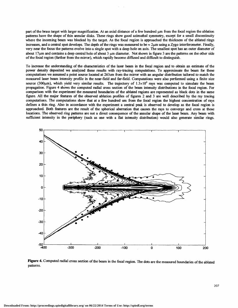
part ofthe brass target with larger magnification. At an axial distance ofa few hundred tm from the focal region the ablationpatterns have the shape of thin annular disks. These rings show good azimuthal symmetry, except for a small discontinuitywhere the incoming beam was blocked by the target. As the focal region is approached the thickness of the ablated ringsincreases, and a central spot develops. The depth ofthe rings was measured to be 2pm using a Zygo interferometer. Finally,very near the focus the patterns evolve into a single spot with a deep hole on axis. The smallest spot has an outer diameter ofabout 17gm and contains a deep central hole ofabout 3 ,un diameter. Not shown in figure 3 are the patterns on the other sideofthe focal region (farther from the mirror), which rapidly become diffused and difficult to distinguish.
To increase the understanding of the characteristics of the laser beam in the focal region and to obtain an estimate of thepower density deposited we analyzed these results with ray-tracing computations. To approximate the beam for thesecomputations we assumed a point source located at 263cm from the mirror with an angular distribution tailored to match themeasured laser beam intensity profile in the near-field and far-field. Computations were also performed using a finite sizesource (3OOtm), which yield very similar results. The trajectory of 1.3x10⁴ rays was computed to simulate the beampropagation. Figure 4 shows the computed radial cross section of the beam intensity distributions in the focal region. Forcomparison with the experiment the measured boundaries of the ablated regions are represented as black dots in the samefigure. All the major features of the observed ablation profiles of figures 2 and 3 are well described by the ray tracingcomputations. The computations show that at a few hundred urn from the focal region the highest concentration of raysdefines a thin ring. Also in accordance with the experiment a central peak is observed to develop as the focal region isapproached. Both features are the result of the spherical aberration that causes the rays to converge and cross at thoselocations. The observed ring patterns are not a direct consequence of the annular shape of the laser beam. Any beam withsufficient intensity in the periphery (such as one with a flat intensity distribution) would also generate similar rings.
200
Figure 4. Computed radial cross section of the beam in the focal region. The dots are the measured boundaries of the ablatedpatterns.
207
Downloaded From: http://proceedings.spiedigitallibrary.org/ on 06/22/2014 Terms of Use: http://spiedl.org/terms

208
Figure 6. Setup used for the series of ablation patterns in figure 5.
Figure 5. SEM images of ablation patterns on aluminum obtained moving the sample around the focus (left). Ablationhole resulting from 40 laser shots near the focus (right).
Target
Laser Beam
F tat
Mirror • 3O'
Translation /Stage
Downloaded From: http://proceedings.spiedigitallibrary.org/ on 06/22/2014 Terms of Use: http://spiedl.org/terms

Similarly, the spherical aberration causes the central peak, which begins to develop when the outermost rays converge on theaxis. Near the so-called "plane of minimum confusion" the intensity distribution is computed to be dominated by the sharpcentral peak, which is responsible for the deep central hole observed in the SEM. The average intensity within a 2timdiameter region is estimated to be lxi 0" W/cm2 from the computed fraction of rays that intersect this region. The analysisalso confirms that the minimum spot size obtained in this experiment is dominantly limited by the spherical aberration, aridnot by the partial spatial coherence of the laser beam [91.
Exposure of the targets to multiple laser shots in each position is observed to result in the ablation of deep holes. As anexample, figure 5 (left) shows a sequence of ablation patterns obtained in aluminum using 40 laser shots for each targetposition. Figure 5 (right) is a SEM image of an ablation hole near the focus, which is seen to have a diameter on the order ofl0l.tm. The experimental setup that was used for this particular series of ablation patterns is shown in figure 6. The beamwas first reflected using a flat Si/Sc multilayer mirror that has a 3 mm diameter hole at its center and was then focused backthrough the hole onto the target using a spherical Si/Sc multilayer mirror with radius of curvature R=2Ocm. While the flatmirror introduces an additional loss due to the 40% reflectivity, the intensity is still well above the threshold for ablationseveral hundred .tm from the smallest spot.
1. XUV REFLECTOMETRYI. Experimental setup
The experimental setup used to perform reflectometry at 46.%m is shown in Figure 7. The samples were illuminated with thebeam of the Ne-like Ar capillary discharge laser that operates in a single line at that wavelength. The characteristics of thistable-top soft x-ray laser were described in a previous publication [11. The measurements were conducted in a vacuumchamber placed at about 1.5 m from the exit of the laser. The samples were mounted on the axis of a rotational stage drivenby a stepper motor, which allowed for the selection of angles of incidence between 0 and 90 degrees. The intensity of thereflected beam was recorded with a vacuum photodiode (labeled "A" in figure 7), that was mounted on a lever arm thatfollowed the angular motion of the reflected beam. A 1mm diameter pinhole was placed at the entrance of the chamber toreduce the spot size of the laser beam incident on the sample, which allowed for measurements at grazing angles approachingzero degrees. To overcome scattering of the data due to shot to shot intensity variation of the laser, the intensity of thereflected beam was normalized by the intensity of the incident beam for each laser pulse. For this purpose a reference beamwas generated by placing a 50% transmissive gold-plated grid iii the path of the incident beam. The intensity of the referencebeam reflected by the grid was measured by a second fixed vacuum photodiode (labeled "B" in figure 7), and used for thenorrnali.zatioui. To obtain absolute reflectance measurements, the signal of the reference photodiode was calibrated withrespect to the intensity of the beam transmitted by the grid by removing the sample and positioning the rotating diode in thebeam path. This calibration was determined with an error of less than 0.5%. The angular dependence of the reflectivity wasmeasured by scanning the angle of incidence while repetitively firing the laser at a repetition frequency of 1 Hz. Thephotodiode signals corresponding to the intensity of the reflected beam and reference beams were recorded and stored for
z.4t /
Figure 7. Schematic diagram and photograph of the laser reflectometer used in the measurement of multi-layer mirror.
Downloaded From: http://proceedings.spiedigitallibrary.org/ on 06/22/2014 Terms of Use: http://spiedl.org/terms

every laser shot by a 500 MHz digitizing oscilloscope (Hewlett-Packard model 54825A).
Figure 8 is an example ofthe reflectance data obtained. It shows a single measurement run ofthe reflectivity as a function ofincident angle for a sample of polished crystalline Si. This data depicts a typical measurement that consisted of 300contiguous laser pulses for a 90 degree rotation ofthe sample. At small angles ofincidence, photodiode "A" blocks the beamfrom impinging on the sample limiting the minimum angle at which data could be obtained to 1.6 degrees. This angle, whichcorresponds to the first valid data point near normal incidence, was determined from the geometric dimensions of the system
>0G)
Figure 8. Example ofmeasured reflectivity vs incident angle dependence in the range 08 <0 < 908 for a Si sample. Thesample was treated with a 5% solution of HF in distilled water for 5 minutes and rinsed with acetone and methanolimmediately prior to its introduction into the vacuum chamber.
and was used to relate each data point to its corresponding angle. At the other extreme, as the incident angle approaches 90degrees, the projection of the incident beam on the sample becomes larger than the sample and therefore limited themaximum angle at which valid data could be obtained. In the specific case of the data for the Si sample shown in figure 8,the 1mm diameter of the beam limited the measurement to angles less than 85.5 degrees. This accounts for the apparentdecrease ofthe reflectivity at grazing angles that should otherwise approach 100%.
2. Angular dependent reflectivity of Si/Sc multilayer mirrorUtilizing the experimental setup described above, measurements were made to detennine the angular dependent reflectivityof Si/Sc multi-layer mirrors designed for use at a wavelength of 46.9nm. The multilayer coatings were deposited on super-polished borosilicate substrates by dc magnetron sputtering with a period of 18-27nm and a ratio of layer thickness'H(Sc)/H(Si)=0.786. The mirror parameters have been discussed in detail previously [7]. As an example, figure 9 shows themeasured reflectivity as a function of incidence angle for a mirror designed to operate at normal incidence. The graphcorresponds to the average of four runs. The runs varied in the nwnber of data points collected from 200 to 400 for a scanangle of 90 degrees. Each individual run yielded very similar data and the averaging was used only to reduce the randomnoise in the measurement. A very near normal incidence reflectivity of 43% was measured at 1.6 degrees. Again, theapparent roll off of the reflectivity at angles larger that 80 degrees is the result of the projected beam being larger than themirror.
3. Determination of optical constants of materialsMeasurements of the reflectivity as a function of angle were made for several materials with the purpose of determining theoptical constants at a wavelength of 46.9mn (26.5eV). The optical constants for Si, GaP lnP, GaAs, GaAsP and ft wereobtained by fitting the measured angular dependence of the reflectivity with the Fresnel formula. The high intensity of thelaser source is an advantage for the accurate measurement ofthe reflectivity at near normal incidence where the reflectivity ofmost materials is low. Our analysis of the data made use of models that take into account the presence of a surface layer ofcontamination, which develops on many materials in a natural atmospheric environment. Measurement of the same samplesthat had different surface layer characteristics gave similar optical constants for the bulk material. This suggests that theapproach used in this work is capable of yielding reliable values of the optical constants for the bulk material in the presenceof surface layer contaminants. The measurements of InP and GaAsP constitute, to the best of our knowledge, the first
210
Incidence Angle (deg.)
Downloaded From: http://proceedings.spiedigitallibrary.org/ on 06/22/2014 Terms of Use: http://spiedl.org/terms

I 0
0.8
>
0.4U,
0.2
0.0
Incidence Angle (deg.)
Figure 9. Reflectivity of a Si/Sc multilayer mirror at 46.9nm measured as a function of angle using a capillarydischarge table top soft x-ray laser.
experimental values at this wavelength, while for the rest of the materials, the values obtained are in most cases in goodagreement with tabulated values. These measurements are discussed in more detail in the paper by Artioukov et. aL in theseproceedings (3776-26).
4. SUMMARYIn summaiy, a spherical multilayer coated mirror was used to focus the beam of a table-top Ne-like Ar soft x-ray laserrealizing the first clear demonstration of material ablation with a coherent soft x-ray beam. The peak intensities obtained byfocusing O.l3mJ laser pulses are estimated to be '
WIcm2, detennined by spherical aberrations. In a separateexperiment we took advantage ofthe high repetition rate ofthe capillary discharge laser to conduct reflectivity measurementsas a function of angle. These measurements resulted in the determination of optical constants at A.=46.9nm for severalmaterials and in the characterization ofthe reflectivity of Si/Sc multilayer mirrors as a function of angle.
5. ACKNOWLEDGEMENTSThe authors are indebted to Yu.A.Uspenskii and Yu.P.Pershin for helpful discussions. This work was supported by NSFgrant DMR-9512282. We acknowledge the support of the crdf grant RP1-240 that allowed for the international collaborationthat resulted in the development of the multilayer optics. The CSU researchers acknowledge the support of the ColoradoAdvance Technology Institute.
6. REFERENCES1 . B.R. Benware, C.D. Macchietto, C.H. Moreno, and J.J. Rocca, "Demonstration of a High Average Power Table-Top Soft
X-Ray Laser," Phys. Rev. Left., vol. 81, pp. 5804-5807, 1998.2. Rocca et.al. these proceedings.3. T. Ohchi, N. Yamaguchi, C. Fujikawa and T. Hara, in Proc. 0f61h mt. Conf X-ray Lasers 1998, eds. Y. Kato, H. Takuma
and H. Daido, loP Conf. Proc. No. 159 (Institute ofPhysic publishing, Bristol and Philideiphia), pp. 683, 1998.4. Ph. Zeitoun, S. Sebban, K. Murai, H. Tang, et.al., in Proc. 0j61h jj ConfX-ray Lasers 1998, eds. Y. Kato, H. Takuma
and H. Daido, loP Conf. Proc. No. 159 (Institute ofPhysic publishing, Bristol and Philidelphia), pp. 677-680, 1998.5. C.H. Moreno, M.C. Marconi, V.N. Shlyaptsev, B.R. Benware, C.D. Macchietto, J.L.A. Chilla, J.J. Rocca, and A.L.
Osterheld, "Two-dimensional near-field and far-field imaging ofa Ne-like Ar capillary discharge table-top soft x-raylaser," Phys.Rev.A, vol. 58, pp. 1509-1514, 1998.
6. J.L.A. Chilla and J.J. Rocca, "Beam optics ofgain-guided soft x-ray lasers in cylindrical plasmas," J.Opt.Soc.Am.B, vol.13, pp. 2841-2851, 1996.
7. Yu.A. Uspenskii, V.E. Levashov, A.V. Vinogradov, A.I. Fedorenko, V.V. Kondratenko, Yu.P. Pershin, E.N. Zubarev,and V.Yu. Fedotov, "High reflectivity multilayer mirrors for a vacuum-ultraviolet interval of35-5Onm," Opt.Lett., vol.23, pp. 771-773, 1998.
8. John C. Miller and Richard F. Haglund, Laser ablation and desoption, San Diego: Academic Press, 1998.9. M.C. Marconi, J.L.A. Chilla, C.H. Moreno, B.R. Benware, and J.J. Rocca, "Measurement of the spatial coherence
buildup in a discharge pumped table-top soft x-ray laser," Phys.Rev.Lett., vol. 79, pp. 2799-2802, 1997.
211
15 30 45 60 75 90
Downloaded From: http://proceedings.spiedigitallibrary.org/ on 06/22/2014 Terms of Use: http://spiedl.org/terms
Copyright © 2022 FDOKUMEN