Effects of sodium butyrate in animal models of mania and depression
Upload
independentCategory
view
2download
0
Int. J. Cancer: 60,400-406 (1 995) 0 1995 Wiley-Liss, Inc.
Publication of the International Union Against Cancer Publication de I Union Internationale Contre le Cancer
APOPTOSIS IN COLORECTAL TUMOUR CELLS: INDUCTION BY THE SHORT CHAIN FATTY ACIDS BUTYRATE, PROPIONATE AND ACETATE AND BY THE BILE SALT DEOXYCHOLATE Angela HAGUE',? Douglas J.E. ELDER', Diane J. HICKS' and Christos PARASKEVA' I CRC Colorectal Tumour Biolog): Research Group, Bristol, UK.
The short chain fatty acids acetate, propionate and butyrate are produced when dietary fibre is fermented by the colonic bacteria. We have previously shown that sodium butyrate induces apoptosis in 3 colorectal turnour cell lines. We have extended our study to 3 adenoma and 4 carcinoma cell lines and investigated whether propionate and acetate also induce apop- tosis. All 3 short chain fatty acids induced apoptosis at physiologi- cal concentrations, but of the 3. butyrate was the most effective. Since these fatty acids are produced as a result of bacterial fermentation of dietary fibre, this may in part explain the correlation between a high-fibre diet and low colorectal cancer incidence. Sodium butyrate induced apoptosis in all 7 of the cell lines studied; however, 2 of the 4 carcinoma cell lines (PC/JW/FI and S/ KS/FI) were more resistant to butyrate-induced apopto- sis than the 3 adenoma cell lines, suggesting that at least some carcinomas may evolve mechanisms to protect the cells from the induction of apoptosis. The bile acid deoxycholic acid has previously been reported as a possible tumour promoter in the large intestine and i ts levels are reduced by dietary fibre. Concentrations of between 10 nM and 0. I rnM had no effect on either the proliferation or apoptosis of colonic tumour cells in vitro. However, a significant induction of apoptosis was obtained at a concentration of 0.5 mM. These results may have signifi- cance for the aetiology of colorectal cancer. o 1995 Wile)?-Liss, Inc.
Thc colorectal epithelium is in direct contact with the contents of the large bowel, and as a rcsult dietary factors, as well as its own internal growth controls, may have profound cffects on the patterns of growth, differentiation and cell death. Indeed. epidemiological studies indicate that diet plays an important role in colorectal carcinogenesis and may be a major contributory factor to the high incidence of colorectal cancer in Westernised countries. These studies suggest a link with a high-fat, low-fibre diet (Howe et a/., 1992). Dietary fibre consists of non-starch polysaccharides such as celluloses, hemicelluloses. pectin and gums. Fibre may protect against colorectal cancer as a result of a number of effects. It incrcases intestinal transit rate and bulk (thereby decreasing exposure to carcinogens in the diet), adsorbs carcinogens, modifies intesti- nal microflora (and therefore may alter bile salt and carcino- gen metabolism), modifies faecal bile salt excretion. lowers the colonic pH and increases the concentrations of short chain fatty acids in the colon and rectum (reviewed in McIntyre etul., 1993). Non-starch polysaccharides and some starch escape digestion in thc small intestine and arc fermented by the anacrobic bacteria of the large bowel gencrating short chain fatty acids (acetate, propionate and butyrate) at a rate of at least 300 mmol per day (Cummings, 1984; Cummings et al.. 1987). The more soluble fibres are most readily fermented, and the sugar composition of the fibre determines which fatty acid by-products are most commonly produced. Fermentation of xylosc tends to yield butyrate. glucose yields propionate and uronic acids produce acetic acid (Salvador et al., 1993). Dietary wheat bran results in higher conccntrations of butyrate than either oat bran or guar gum (McIntyre er al., 1993). Butyrate is preferentially taken up by the colonic epithelium, where it is actively mctabolised to producc energy (Roediger, 1982). Butyrate is, therefore, an important energy source for thc colonocyte and is metaboliscd in prefcrence to glucose o r glutamine, accounting for 70?6 of the total energy consump-
tion (Scheppach, 1994). In vitro studies have shown that butyrate inhibits cell proliferation and induces a more differen- tiated phenotype in colorectal tumour cell lines (Augeron and Laboisse, 1984; Whitehead et ul., 1986; Gamet et al., 1992; Deng et al., 1992).
In vivo studics using rats demonstrated that dietary fibre supplementation led to increased butyrate levels, which were associated with reduced colonic cell proliferation (Boffa et al., 1992) and lowered tumour mass in rats treated with dimethyl- hydrazine (McIntyre et al., 1993). The reduction of colonic cell proliferation and induction of differentiation in colonic epithe- lial cells have led to clinical trials of butyrate enemas in the treatment of ulcerative colitis, in which proliferation in the basal regions of the colonic crypts is retained but there is a reduction in the upper crypt labelling index (Scheppach et al., 1992). Butyrate has been considered for therapeutic purposes in the treatment of colorectal cancer, and i t has been suggested that butyrate could bc targeted to carcinoma cells using a tumour-specific monoclonal antibody and delivering a mea- sured dose in a liposomc capsule (Otaka et al., 1989).
In a self-renewing tissue such as the colonic epithelium the regulation of cell number must be strictly controlled such that the rate of new cell production balances the rate at which cells undergo growth arrest, apoptosis or exfoliation (Wyllie et al.. 1984). We have previously demonstrated that the naturally occurring 4 carbon fatty acid sodium butyrate can induce apoptosis in 3 colorectal tumour cell lines (2 adenoma and 1 carcinoma) (Hague et a/. , 1993). These findings have been confirmed by Heerdt et ul., (1994) using the 2 colorectal carcinoma cell lines HT29 and SW620. Similarly, butyrate has also been shown to induce apoptosis in the HL60 myeloid leukaemia cell line (Calabresse et al., 1993) and in the Burkitt's lymphoma cell line BL-30 (Filippovich et al., 1994). We have now extended our study of the effects of butyrate (C4) to 3 cell lines derived from colorectal adenomas and 4 cell lines from colorectal carcinomas and have also investigated the effects of the shorter chain fatty acids acetate (C2) and propionate (C3) on apoptosis. Other components of the lumenal contents have also been implicated in colorectal carcinogenesis. Reports that thc bile salt deoxycholate may act as a tumour promoter in the large intestine have come from studies of populations at high risk for the development of colon cancer (Hill et al., 1971) and from animal model systems (Narisawaet al., 1974). In addition, deoxycholic acid concentrations in the gut vary according to diet, dietary wheat bran reducing the concentration (Reddy et al., 1992). We have investigated the effects of deoxycholate on the growth of colonic tumour cells and have shown that this bile salt can induce apoptosis in these cells. The possible significance of this result in terms of the tumour-promoting activity of deoxycholic acid is discussed.
'To whom correspondence and reprint requests should be sent, at University of Bristol, CRC Colorectal Tumour Biology Research Group, Department of Pathology and Microbiology, School of Medical Sciences, University Walk, Bristol. BS8 ITD, UK. Fax: 0044 272 287806.
Received: August 2, 1903 and in revised form October 11.1994.
APOPTOSIS lNDUCED BY DIETARY FACTORS 401
METHODS C’cll 1inL.s
cell lines used in this study were clonogenic (could be pas<aged with trypsin) and 3T3 feeder independent. The :idcnoma-(lerivctl cell lines were M i C l (Williams et a/., 1990)% RG/CZ (Hague et al., 1992) and RR/CL (Williamseta(., IYYZ). The carcinoma cell lines were LS174T (obtained from the Imperial Cariccr Rcsearch Fund, London, UK), 2 sporadic carcinomas at early passage SICMbIFI and S/KS/FI (cstab- lished in this laboratory) and the carcinoma cell line from a familial polyposis patient, PC.IJW/FI (Paraskeva et a/., 1984; Bcrry and Paraskeva, 1988). All 3 adenoma cell lines are non-tumorigcnic in athymic nude mice and anchorage- dependent, whereas all the carcinoma cell lines used were anchorage-iridependeiit and tumorigenic.
Cultirrc, conditioris For assessment of the cells‘ response to the sodium salts of
the short chain fatty acids and deoxycholic acid all cell lines were grown on tissue culturc plastic in DMEM (Flow, Thame, UK) supplemented with 205% FBS (batch selected), 2 mM glutamine, 100 unitsiml penicillin, 100 pgiml streptomycin, 0.2 unitslml insulin and 1 kg/ml hydrocortisone sodium succinate.
Measurcrneiit of apoptosis We have previously reported that during routine culture of
colorcctlil adenoma and carcinoma cell lines, wc noticed that some cells detached spontaneously from the flasks and floated in the medium. These floating cells were demonstrated to be apoptotic (,Hague et al., 1993). Apoptosis was characterised niorphologically by acridine orange staining, which demon- strates the condensed chromatin of apoptotic cells (Gregory et 01.. 199 1 ) . and biochemically by inter-nucleosomal fragmenta- tion o f the DNA into multiples of 180-200 b.p., which produces a “DNA ladder” on an electrophoretic gel (Wyllie, 1980). After treatment with the fatty acid sodium butyrate, we showed an incrcase i n the number of floating cells relative to the number of adherent cells, and the floating cells obtained after trsatment were again demonstrated to be apoptotic. The proportion of floating cells that were apoptotic was the same in both treatcd and control cultures, showing that the increase in floating cells was not due to necrosis (Hague et a/.. 1993). The proportion of cells floating was therefore used as a measure of thc exlent of apoptosis. in conjunction with examination of both adherent and floating cell populations by acridinc orange staining and DNA laddering.
Acridirie oraitge staining Cells were stained with 5 p.g/ml acridine orange in PBS for
1 0 min and werc viewed immediately by fluorescence micros- copy, as described by Gregory et al., (1991). Cells with condcnscd, bright chromatin were scored as apoptotic. At least 300 cells were counted per treatment.
DNA hddent7g ThL: method used for assessing oligonucleosomal fragmenta-
t i o n was that of Smith et al. (1989). DNA samples from 10” cells were run on a 2 % agarose (wiv) gel containing 0.1 &ml ethidium bromide at 40 V until the dye front had migrated 4-5 cm. DNA from floating and attached cells was run separately.
Preparation of thymocyrc cell cultures Mouse thymocytes. which apoptose in response to corticoste-
roids (Wyllie, 1980). were used as positive controls for elcctro- phorctic gels demonstrating DNA laddering. MF-I mice (origi- nally obtained from Olac, Bicester, UK, bred and maintained in our anirnal facility) were sacrificed at 2-3 months of age. The thymus was dissected out and thymocytes were released by pressing through a sterilc gauze. Thymocytes were suspended in KPMI mcdium supplemented with 10% FBS, 2 mM
glutamine, 100 units/ml penicillin and 100 kgiml streptomycin at a density of 106 cells per ml and were treated with dexamethasone ( lW7M) to induce apoptosis. Samples were prepared 18 hr after treatment with dexamethasone.
Treutmenf with sodium buyrate For comparative purposes, trypsinised cells were seeded at a
density such that there were approximately 2.5 X loh cells per T25 flask when treated. At time of treatment the cells had entered the exponential growth phase. For most cell lines cells were treated 3 days after seeding; however, the cell line R R i C l had a longer lag phase and was therefore treated 5 days after seeding. Cells were treated with 2 and 4 mM sodium butyrate (Sigma, Poole, UK) for 4 days. After this time, the attached and floating cells were counted separately, then stained with acridine orange (5 &ml) for examination by fluorescence microscopy. Representative samples were taken for DNA laddering.
Treatment with sodium acetate /propionate Using the same 3 cell lines that have previously been shown
to be responsive to sodium butyrate-induced apoptosis (Hague eta/ . , 1993), trypsinised cells were seeded at 1 x lo6 cells per T25 flask and 3 days later werc treated with either sodium acetate or sodium propionate (Sigma) at a range of concentra- tions. Attached and floating cells were counted 4 days later and examined by acridine orange staining (Hague et al., 1993). Representative samples were also examined for DNA ladder- ing.
Treutment with soditrtn deo-xyrholute The 3 cell lines RGIC2, AA/Cl and PCIJWIFI were used
to determine whether deoxycholic acid had any effect on the frequency of apoptosis. Trypsinised cells were seeded at 1 x 10” cells per T25 flask and 3 days later were treated with sodium deoxycholate (Sigma) at concentrations between 10 nM and 10 mM. Attached and floating cells were counted 4 days later and examined by acridine orange staining (Hague et al.. 1993). Representative samples were also examined for DNA laddering.
RESULTS Effects of sodium b q r a t e on adcnoma and carcinoniu cell hies
We have previously reported that butyrate induces apopto- sis, which is detectable as cells that detach and float in the medium (Hague et al., 1993). Examination of the floating and attached cells by acridine orange staining showed that the floating cell population was comprised largely of apoptotic cells with the classical condensed chromatin (Wyllie, 1980; Gregory et ul., 1991). whereas the proportion of apoptotic cells in the attached compartment was always low, typically around 1%-3% (Hague et ul., 1993). This observation was confirmed by DNA gel electrophoresis. The DNA from the floating cells produced ladders characteristic of inter-nucleosomal fragmen- tation, which is a biochemical marker of apoptosis (Wyllie, 1980), whereas the adherent cells retained high m.w. DNA with no evidence of laddering. The proportion of cells that were apoptotic in the adherent fraction was too low to be detected by electrophoresis. For these reasons we used the proportion of cells floating as a mcasure of the frequency of apoptosis in the cultures but always examined thc floating and attached cells for the frequency of apoptotic cells by acridine orange staining to ensure that the induction of floating cells was due to apoptosis and not necrosis. In this study 3 adenoma and 4 carcinoma cell lines were treated in exponential growth for 4 days with the fatty acid sodium butyrate to determine whether adenonia cells were more sensitive to growth inhibi- tion and induction of apoptosis by butyrate than carcinoma cells. For all 7 of the cell lincs described in this report the percentage of floating cells which were apoptotic was high and
402 HAGUE ETAL
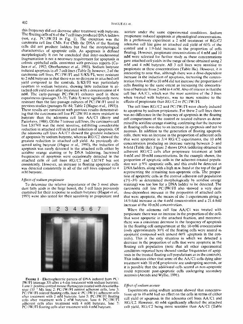
this frequency did not decrease after treatment with butyrate. The floating cells of 6 of the 7 cell lines produced DNA ladders (see, e.g., PCIJWIFI in Fig. 1). The exception was the carcinoma cell line CMb. For CMb the DNA of the floating cells did not produce ladders but had the morphological characteristics of apoptotic cells. As apoptosis is defined morphologically it can be concluded that inter-nucleosomal fragmentation is not a necessary requirement for apoptosis In colonic epithelial cells, consistent with previous reports (Co- hen et al., 1992; Oberhammer et al., 1993). Sodium butyrate induced apoptosis in all 7 cell lines (Table I); however, 2 of the carcinoma cell lines, PCIJWIFI and SIKSIFI, were resistant to 2 mM butyrate in that there was no decrease in attached cell yield compared to the controls. SIKSIFI was particularly resistant to sodium butyrate, showing little reduction in at- tached cell yield even after treatment with a concentration of 4 mM. The carly-passage PCIJWIFI cultures uscd in these experiments (passages 33-35; Table I) were significantly more resistant than the late-passage cultures of PCIJWIFI used in previous studies (passages 81-84; Table I (Hague et al., 1993)). These results are consistent with previous results demonstrat- ing that the carcinoma cell line PC/JW/FI is more resistant to butyrate than the adenoma cell line AAIC1 (Berry and Paraskeva, 1988). Of the 7 tumour cell lines, the carcinoma cell line LS174T was the most sensitive, exhibiting considerable reduction in attached cell yield and induction of apoptosis. Of the adenoma cell lines AAiCl showed the greatest induction of apoptosis by sodium butyrate, whereas RGIC2 showed the greatest reduction in attached cell yield. As previously ob- served using butyrate (Hague et al., 1993), the induction of apoptosis was rarely detected in the attached cells either by acridine orange staining or by DNA laddering. Increased frequencies of apoptosis were occasionally detected in the attached cells of cell lines RG/C2 and LS174T but not consistently. However, an increase in floating apoptotic cells was detected consistently in all of the cell lines exposed to 4 mM butyrate.
Efects of sodium propionate To determine the relative importance of the 3 most abun-
dant fatty acids in the large bowel, the 3 cell lines previously examined for their response to sodium butyrate (Hague e f al., 1993) were also tested for their sensitivity to propionate and
FIGURE 1 - Electrophoretic pattern of DNA isolated from PC/ JW/FI (passage 33) after a 4-day treatment with sodium butyrate. Lane 1: positive control mouse thymocytes treated with dexametha- sone (lo-’ M), lane 2: PCiJWiFI control adherent cells, lane 3: PC/JW/FI control floating cells, lane 4: PCIJWiFI adherent cells after treatment with 2 mM butyrate, lane 5: PCiJWiFI floating cells after treatment with 2 mM butyrate, lane 6: PCiJWiFI adherent cells after treatment with 4 mM butyrate, lane 7: PC/JW/FI floating cells after treatment with 4 mM butyrate.
acetate under the same experimental conditions. Sodium Propionate induced apoptosis at physiological concentrations. In a preliminary experiment a 2 mM treatment of RGiC2 adenoma cell line gave an attached cell yield of 60% of the control and a 1.9-fold increase in the proportion of cells floating. However, propionate concentrations of 4 mM and 10 mM were selected for further study as these concentrations gave attached cell yields in the range of those obtained using 2 mM and 4 mM butyrate. All 3 cell lines were sensitive to propionate at these concentrations (Table Ira). However. it is interesting to note that, although there was a dose-dependent increase in the induction of apoptosis, increasing the concen- tration from 4 mM to 10 mM did not increase the proportion of cells floating to the same extent as increasing the concentra- tion of butyrate from 2 mM to 4 mM. Also of interest is that the cell line AAIC1, which was the most sensitive of the 3 lines when treated with butyrate, was no more sensitive to the effects of propionate than RGIC2 or PCIJWIFI.
The cell lines RG/C2 and PCiJWiFI were clearly induced to apoptose by sodium propionate. For these 2 cell lines there was no difference in the frequency of apoptosis in the floating cell compartment of the control or treated cultures as deter- mined by acridine orange staining, confirming that the increase in floating cells was due to an increase in apoptosis and not to necrosis. In addition to the generation of floating apoptotic cells, there was an increase in the proportion of adherent cells that were apoptotic in 3/4 RGIC2 experiments, the 10 mM concentration producing an increase varying between 2- and 8-fold (Table Ha). Figure 2 shows DNA laddering obtained in adherent RGIC2 cells after propionate treatment at both 4-mM and 10-mM concentrations. In the example shown, the proportion of apoptotic cells in the adherent-treated popula- tion was 2 9% apoptotic cells, and this could be detected as DNA ladders, along with a high m.w. band at the top of the gel represcnting the remaining non-apoptotic cells. The propor- tion of apoptotic cells in the control adherent cell population (1%-3% as determined morphologically by acridine orange staining) was too low for a DNA ladder to be detected. The carcinoma cell line PCIJWIFI also showed a very clear dose-dependent increase in the proportion of attached cells that were apoptotic, the means of the 3 experiments giving a 14.9-fold increase at the 4-mM concentration and a 21.4-fold increase at the 10-mM concentration.
When the adenoma cell line AA/Cl was treated with propionate there was no increase in the proportion of the cells that were apoptotic in the attached fraction, and moreover, there was a consistent decrease in the frequency of apoptosis in the floating cell compartment at the 10-mM concentration (only approximately 30% of the floating cells were scored as apoptotic compared with around 60% apoptosis in the con- trols). This is the only situation in which we detected a decrease in the proportion of cells that were apoptotic in the floating cell population (note that all other experimental conditions reported here showed similar frequencies of apop- tosis in the treated floating cell populations as in the controls). This indicates either that some of the AA/Cl cells dying after treatment with 10 mM propionate are undergoing necrosis, or it is possible that the additional cells scored as non-apoptotic could represent post-apoptotic cells undergoing secondary necrosis (Arends and Wyllie. 1991).
Effect of sodium acetate Experiments using sodium acetate showed that concentra-
tions up to 10 mM had no effect on the cells in terms of either cell yield or apoptosis in the adenoma cell lines AA/Cl and RG/C2. However, 40 mM significantly affected the attached cell yield, RGIC2 being more sensitive than AA/Cl (Table

403 APOPTOSIS I N D U C E D BY D I E T A R Y F A C T O R S
T4BLE 1. - RESPONSE OF 7 COLORECTAL TUMOUR CELL LINES T O A &DAY TREATMENT WITH SODIUM BUTYRATE AT CONCENTRATIONS O F 2 AND 4 mM --
Percentage of cells floating Attached cell yield Passage (C; control) numbers Cell 1111(.
2 mM hutyrate 4 mM hutyrate Control 2 mM hutyrate 4 mM hutyrate
Adenomas AAICI RGIC2 RRiCl
79-81 75.3 2 9.7 41.1 f 7.3 1.61 t 0.13 3.74 t 0.13 14.63 2 2.38 3 3 4 3 72.7 f 10.8 31.8 t 6.0 11.00 f 1.28 15.33 2 1.90 40.46 2 6.13 52-54 70.8 t 3.9 52.9 f 5.7 8.68 2 0.88 9.87 f 1.09 22.08 2 3.43
Carcinomas 4 1-47 54.6 f 15.4 35.2 t 19.1 6.18 f 0.71 14.62 2 3.16 36.49 f 6.55 CMb
LS I74T 151-154 41.4 t 7.9 18.1 2 2.9 0.85 f 0.24 2.85 2 0.93 19.86 f 3.27 PCIJWIFI early 33-35 106.9 & 11.2 66.7 f 12.0 19.91 t 1.18 22.60 f 3.05 28.74 2 2.60 PCIJWiFI late 8 1-84 65.3 2 16.1 45.1 f 11.4 15.88 f 2.94 22.75 f 2.81 36.40 f 2.87 S i KSI Fl 28-32 109.0 f 9.8 83.5 t 11.6 7.90 f 0.60 5.72 t 0.05 9.22 5 1.40
Cells (2.5 x 10h) were treated in exponential growth. Results are means of at least 3 experiments f S.E.M.
TABLE 11 - EFFECTS OF A 4-DAY TREATMENT WITH SODIUM PROPIONATE ( a ) AND SODIUM ACETATE ( h ) ON COLORECTAL TUMOUR CELL LINES
Percentage of cell5 floating Attached cell yield hssage (Q control) -
(11 I (‘cll line rnimher
-- 4 mM propionate 1U mM propionate Control 4 mM propionate 10 mM propionate ._
AAiCl 86-87 (15.2 f 3.2 45.2 t 5.1 3.90 t 0.51 6.61 f 0.33 11.28 2 1.67 RGiC2 33-36 54.0 f 3.8 37.9 2 2.4 10.18 t 0.95 20.68 t 3.55 26.29 t 3.19 PC i JW IF1 86-89 51.5 t 3.9 32.5 f 15.7 13.63 ? 3.91 21.71 t 1.84 31.31 ? 2.96
Percentage of cellc Hoating Attached cell yield Paxsage (C control) nu mhe r - ( h )
C c l l Ilne 40 mM acetate 80 mM acetate Control 40 mM acetate XO mM acetate
AAICl 92--93 87.8 t 16.6 44.8 t 2.3 2.17 -+ 0.42 2.81 t 0.80 6.53 t 0.91 RGIC? 45 71.0 t 7.2 34.7 2 0.2 17.49 ? 1.14 22.77 t 2.38 29.51 2 1.39
For each agent loh cells were seeded and treatment was commenced 3 days later when cells were in exponential growth. Results are means of at least 3 experiments f S.E.M. ( a ) Response of the adenoma cell lines AAiC1 and RGIC? and the carcinoma cell line PCIJWiFI to 4 and 10 mM propionare (b) Response of the adenoma cell lines AAiCl and RGIC? to 40 and 80 mM acetate.
FIGURE 2 - Electrophoretic pattern of DNA isolated from RGIC2 (passage 42) after a 4-day treatment with sodium propio- nate. Lane I : positive control mouse thymocytes treated with dexarnethasone (I&’ M), lane 2: RGIC2 control adherent cells, lane 3 : RGiC2 control lloating cells, lane 4: RGIC2 adherent cells after treatment with 4 mM propionate, lane 5: RGIC2 floating cells after treatment with 4 mM propionate, lane 6: RGiC2 adherent cells after treatment with LO mM propionate, lane 7: RGIC2 floating cells ailer treatment with 10 mM propionate.
116). There was also a 1.3-fold increase in the proportion of cells floating for both cell lines. A concentration of 80 mM produced a further decrease in attached cell yield and an increase in the proportion of cells floating (3-fold for AAIC1, 1.7-fold for RGiC2). Neither of these concentrations in- creased the proportion of attached cells that were apoptotic. However, there was no decrease in the proportion of cells in the floating cell population that were apoptotic after treat- ment, indicating that the increase in the proportion of cells floating was due to apoptosis rather than necrosis.
Effects of deoxycholic acid Concentrations of 10 nM to 10 mM sodium deoxycholate
were initially tested for their effects on the adenoma cell line RGIC2. Concentrations up to 0.1 mM had no significant effect on the cell yield or the frequency of apoptosis. At 0.5 mM, however, the attached cell yield was reduced to 45.6 5 7.1% and there was a 2-fold increase in the percentage of cells floating (Fig. 3). Concentrations of 0.1, 0.2 and 0.5 mM deoxycholic acid were used in further studies. In RGIC2, 0.2 mM deoxycholic acid increased the extent of apoptosis in 3 of 5 experiments; however, the same concentration had no effect on the extent of apoptosis in the adenoma cell line AAiCl or the carcinoma cell line PCIJWIFI. A concentration of 0.5 mM induced significant apoptosis in both RGIC2 and PCIJWIFI. PCIJWIFI was the most sensitive at this concentration. AAiCl was the least sensitive of the 3 cell lines. Although the extent of apoptosis was consistently higher in the treated cultures compared to the controls, this was not statistically significant at this dose (Fig. 3). Like butyrate, deoxycholic acid did not increase the proportion of cells apoptotic in the attached cell population. This was also evidenced by the lack of DNA laddering from the adherent cells.

404 HAGUE ETAL
A.
B.
AAlC 1 RGlC2 Cell line
1.4-
C
PCIJWIFI
40 453
AAlC 1 RGIC2 PCIJWIFI
Control
0.5mM
FIGURE 3 -Effect of a +day treatment with 0.5 mM sodium deoxycholate on the adenoma cell lines A A i C l and RGiC2 and the carcinoma cell line PC/JW/FI. Results shown for RG/C2 are means f S.E.M. of 5 experiments, PC/JW/FI means of 4 experiments and AAiCl means of 3 experiments. ( a ) Attached cell yield after deoxycholate treatment expressed as a percentage of the control cultures. (6,c) Apoptosis estimated by the proportion of cells floating in the culture medium. The results obtained for AAiCl (b) are plotted on a different scale from the results for RGiC2 and PC/JW/FI (c) since there was a very low level of apoptosis in AAICI .
DISCUSSION
The bacterial fermentation of dietary fibre in the large intestine generates short chain fatty acids as a by-product. We have previously demonstrated that the 4 carbon fatty acid butyrate induces apoptosis in colorectal tumour cells at physi- ological concentrations, as evidenced by the shedding into the culture medium of cells which have the morphological charac- teristics of apoptosis and produce DNA ladders on an electro- phoretic gel, whereas at the same levels of growth inhibition
transforming growth factor did not induce apoptosis (Hague et d., 1993). This has been confirmed by Heerdt et af. (1994), who reported that butyrate induced apoptosis in the carcinoma cell lines HT29 and SW620 but that the butyrate derivatives isobutyric acid and heptoff uorobutyric acid did not induce apoptosis. Our initial studies were of 3 cell lines, the 2 adenoma cell lines AAiCl and RGiC2 and the carcinoma cell line PC/JW/FI (Hague et d., 1993). In the present study we have compared the effects of butyrate on our initial 3 cell lines

APOPTOSIS INDUCED BY DIETARY FACTORS 405
and 4 others. Sodium butyrate induced apoptosis in all 7 of the cell lines tcsted. However, 2 of the carcinoma cell lines, S/KS/FI and PCIJWIFI. were relatively resistant to butyrate. A subset of carcinomas may therefore be resistant to sodium butyratc-induced apoptosis. The mechanism by which sodium butyrate induces apoptosis is unclear. Butyrate treatment results in hyperacetylation of the histones H3 and H4 due to its inhibition of histonc deacetylase, and resultant changes in chromatin structure lead to altered gene expression and modification of the accessibility of the chromatin to DNases (revicwed in Kruh, 1982). One of the other major targets of butyratc is the cytoskeleton, modifying microfilament and microtubule assembly. and it also has effects on the external cytoskeletal matrix (reviewed in Kruh, 1982). We have found that butyrate induces tissue transglutaminase in association with apoptosis and that this induction is seen in the adherent and floating cells (data not shown). This is consistent with tissue transglutaminase as an early marker of apoptosis (Piacen- tini etal., 1093). The results of Heerdt etal. (1994) that alkaline phosphatase is increased in floating apoptotic cells suggest that the apoptosis induc’ed by sodium butyrate may represent the culmination of a differentiation pathway.
The 3 and 2 carbon fatty acids propionate and acetate are also produced as by-products of the bacterial fermentation of dietary fibre. I t was therefore of interest whether these fatty acids also induced apoptosis in colorectal tumour cells. Using the cell lines AAICI. RGIC2 and PCIJWIFI, sodium propio- nate was shown to induce apoptosis, though it was less effective than sodium butyratc. This finding reflects previous studies of the cffectf of short chain fatty acids on cell proliferation (Whitchcad et ul., 1986; Garnet et id., 1992). Interestingly, the adenoma cell line k4IC1, which rcsponded well to butyrate, was the least responsivc of the 3 cell lines when the apoptosis- inducing agent was propionate. The other 2 cell lines, RG/C2 and PCIJWIFI. after treatment with propionate, showed an increase in the frequency of apoptosis in the attached cells, an effect only rarely seen in cultures treated with sodium butyrate. Acetate was the least effective of the 3 fatty acids but did induce apoptosis at concentrations of 40 mM and above. In the colon the molar ratios of acetate:propionate:butyrate are 57:22:21 and the ccinccntration of acetate in the colonic contents may reach around 60 mmol per kg (Cummings et ul., 1987). Considering the relative abundance of acetate in the human colorectum. it may be that the concentrations required to induce apoptosis would be physiological.
Thc bile salt deoxycholate is thought to act as a tumour promoter in the large intestine (Reddyetal., 1992), though the mechanism of its activity is unclear. Of interest is our finding that deoxycholate induces apoptosis at high physiological concentrations. Significant apoptosis was induced in the RG/C2 and PCiJWiFI cell lines after treatment with 0.5 mM deoxycho- late, whcreas AAICl ‘was relatively resistant at this dose. The carcinoma cell line PCiJWIFI has no p53 protein (mutant or wild type) (Baker et id, 1990); therefore, as was found for butyrate (Hague et al.. 1993). wild-type p53 is not required for the induction of apoptosis by deoxycholate.
Although butyrate can induce apoptosis, there may be conditions under which cells lose their responsiveness to butyrate (Hague et al., 1993). This is further suggested by the relative resistance of the 2 carcinoma cell lines PCiJWIFI and SiKSiFI to sodium butyrate-induced apoptosis. Whether butyrate induces apoptosis in viva may depend on the alterna- tive energy sources available to the cells, the genetic back- ground of the cells or the presence of mutagens in the diet. Indeed, sodium butyrate has previously been used in the transformation of the pre-malignant adenoma cell line AAICl to a malignant phenotype (Williams et al.. 1990). Only AAIC l cells which had been pretreated with sodium butyrate pro- gressed to a tumorigenic phenotype after treatment with the point mutagen and carcinogen N-methyl-N’-nitro-N-nitrosogua- nidine (MNNG). This may have occurred due to selection of a sub-population of cells resistant to apoptosis by butyrate, which allowed survival of damaged cells after the subsequent carcinogenic insult. Similarly, the presence of apoptosis- resistant cells in carcinomas of the colon may explain the poor response of these tumours to chemotherapy.
There has been considerable interest in the role of dietary factors in colorectal cancer, and both the fatty acids and the bile acids have attracted attention. We have shown that the 3 most common short chain fatty acids produced by bacterial fermentation of dietary fibre (butyrate, propionate and ac- etate) induce apoptosis in colorectal epithelial cells. Of these butyrate is the most effective inducer of apoptosis, followed by propionate. Our results suggest that the short chain fatty acids may be important in modulating proliferation and apoptosis in colonic epithelial cells. However, 2 of the carcinoma cell lines were resistant to apoptosis after treatment with 2 mM butyr- ate. There is some evidence, therefore, that certain carcinoma cells may become relatively resistant to the induction of apoptosis by butyrate. In addition, we have also demonstrated that the bile salt sodium deoxycholate induces apoptosis at high physiological concentrations. If deoxycholate acts as a tumour promoter in the large bowel, this may be a result of selection for sub-populations of cells resistant to deoxycholate- induced apoptosis. Clearly, further investigations are required into the similarities and differences in the mechanisms by which these two dietary agents induce apoptosis. In particular, it will be important to determine the mechanisms by which resistance to the induction of apoptosis by both agents is achieved and whether this confers resistance to apoptosis induced by chemotherapeutic agents.
ACKNOWLEDGEMENTS
This research was supported by the Cancer Research Campaign of Great Britain. The authors thank Mr. P. Sum- mers and Mrs. J. Baker for photography and Miss T. Collard for technical assistance.
REFERENCES
AKENIx. M.J. and Wvuii:. A.H., Apoptosis: mechanisms and roles in pathology. In(. Rot’. C . Y ~ J . P t i r l i~~L, 32, 223-253 (1991 ). AUGEKON. C. and LABOISSE. C.L., Emergence of permanently differen- tiated cell clones i n a hurnan colonic cancer cell line after treatment with sodium butyrate. Cancw Res., 44,3961-3969 (1984). BAKER. S.J., PREISINGER, A.C., JESSUP, J.M., PARASKEVA, C.. MARKO- V I l L . s., WII.LSC)N. J.K.V.. IJAMILTON, s. and VOGELSTEIN, B.,pS3 gene mutaticons occur in combination with 17p allelic deletions as late events in colorectal tumorigenesi:s. Cancer Res., 50. 7717-7722 (1990).
BOI-FA, L.C.. LUPTON, J.R.. MARIANI, M.R., CEPPI, M., NEWMARK, H.L., SCALMATI. A. and LIPKIN, M., Modulation of colonic epithelial cell proliferation, histone acetylation, and luminal short chain fatty acids by variation of dietary fiber (wheat bran) in rats. Cancer Rex. 52, 5906-5912 (1992). CALABRESSE. C., VENTURINI, L.. RONCO, G., VILLA. P., C H O M l t N N E , C. and BELPOMME, D.. Butyric acid and its monosaccharide ester induce apoptosis in the HL-60 cell line. Blockern. hloplzys. Res. Cornm.. 195, 31-38 (1993).
BERRY, R.D. and PARASIEVA. C., Expression of carcinoernbryonic antigen by adenoma and carcinoma derived epithelial cell lines: possible marker of tumour progression and modulation of expression
COHEN, G.M., SUN, X.M.. SNOWDEN, R.T., DINSDALE, D. and SKILL- ErER. D.N.. Key morphological features o f apoptosis may occur in the absence of internucleosomal DNA fragmentation. Biochem. J . . 286,
by sodium butyrate. Corcinogeriesis. 9,447-450 (1988). 331-334 (1092).

406 H A G U E t T A L
CUMMINGS, J.H., Colonic absorption: the importance of short chain fatty acids in man. Scad. J. Gastroentero/., 19, S93, 89-99 (1984). CUMMINGS, J.H., POMARE, E.W., BRANCH, W.J., NAYLOR. C.P.E. and MACFARLANE. G.T.. Short chain fatty acids in human large intestine, portal. hepatic and venous blood. Cur, 28,1221-1227 (1987). DENG. G., LIU. G., Hu, L., GUM, J.R. and KIM, Y.S., Transcriptional regulation of the human placental-like alkaline phosphatase gene and mechanisms involved in its induction by sodium butyrate. Cancer Res., 52,3378-3383 (1992). FILIPPOVICH, I. , SOROKINA, N., KHANNA, K.K. and LAVIN. M.F., Butyrate induced apoptosis in lymphoid cells preceded by transient over-expression of Hsp70 mRNA. Biochn. hiophys. Rex Comm., 198,
GAMET, L., DAVIAUD, D., DENIS-POUXVIEL, C., REMESY, C. and MURAT, J.C., Effects of short-chain fatty acids on growth and differen- tiation of the human colon-cancer cell line HT29. Int . J . Cuncer. 52,
GREGORY, C.D., DIVE, C.. HENDERSON, S.. SMITH, C.A., WILLIAMS, G.T., GORDON, J. and RICKINSON, A.B.. Activation of Epstein-Barr virus latent genes protects human B cells from death by apoptosis. Nature (Lond.). 349,612-614 (1991). HAGUE, A,, HANLON, K.A. and PARASKEVA, C., Clonal evolution and tumor progression in two human colorectal adenoma-derived cell lines in vitro: the involvement of chromosome 1 abnormalities. h i . J. Uncol.,
HAGLIE, A,. MANNING. A.M., HANLON, K.A., HUSCHTSCHA, L.I., HART, D. and PARASKEVA, C., Sodium butyrate induces apoptosis in human colonic tumour cell lines in a p53-independent pathway-implications for the possible role of dietary fibre in the prevention of large-bowel cancer. bit . J. Cancer, 55,498-505 (1993). HtERDT, B.G., HousroN, M.A. and AUGENLICHT, L.H., Potentiation by short chain fatty acids of differentiation and apoptosis in human colonic carcinoma cell lines. Cancer Rex. 54,3288-3294 (1994). HILL, M.J., DRASAR, B.S., ARIES, V., CROWTHER, J.S., HAWKESWORTH. G. and WILLIAMS, R.E.O. Bacteria and the aetiologyof cancer of large bowel. Lancer, 1,95-100 (1971). HOWE, G.R., BENITO, E., CASTELLETO. R. and 22 OTHERS, Dietary intake of fiber and decreased risk of cancers of the colon and rectum-evidence from the combined analysis of 13 case-control studies. J. nar. Cancer In.st., 84, 1887-1896 (1992). KRUH, J., Effects ofsodium butyrate, a new pharmacological agent, on cells in culture. Mo1. cell. Biochem., 42,65-82 (1982). MCIUTYRE, A., GIBSON. P.R. and YOUNG. G.P., Butyrate production from dietary fibre and protection against large bowel cancer in a rat model. Girt, 34,386-391 (1993). NARISAWA, T., MAGADIA, N.E.. WEISBURGER. J.H. and WYNDER, E.L. Promoting effect of bile acids on colon carcinogenesis after intrarectal installation of N-methyl-N'-nitro-N-nitrosoguanidine in rats. J. nut. Cancer Inst., 53,1093-1097 (1974). OBERHAMMER, F., WILSON. J.W., DIVE, C.. MORRIS, I.D., HICKMAN. J.A., WAKELING, A.E., WALKER, P.R. and SIKORSKI, M., Apoptotic
257-265 (1994).
286-289 (1992).
1,201-208 (1992).
death in epithelial cells-cleavage of DNA to 300 and/or 50 kb fragments prior to or in the absence of internucleosomal fragmenta- tion. EMBO J. , 12,3679-3684 (1993). OTAKA, M., SINGHAI., A. and HAKOMORI, S., Antibody-mediated targeting of differentiation inducers to tumor cells: inhibition of colonic cancer cell growth in vitro and in rivo. Biochem. biophys. Res. Comm., 158,202-208 (1989). PARASKEVA, C., BUCKLE, B.G., SHEER, D. and WIGLEY, C.B., The isolation and characterization of colorectal epithelial cell lines at different stages in malignant transformation from familial polyposis coli patients. fnr. J. Cancer, 34,49-56 (1984). PIACENTINI, M., FEW, L. and MELINO, G., Multiple cell cycle access to the apoptotic death programme in human neuroblastoma cells. FEES Lett., 320. 150-154 (1993). REDDY, B.S., ENGLE, A., SIMI. B. and GOLDMAN. M., Effect of dietary fiber on colonic bacterial enzymes and bile acids in relation to colon cancer. Gastroenterology, 102, 1475-1482 (1992). ROEDIGER, W.E.W.. Utilization of nutrients by isolated epithelial cells of the rat colon. Gastroenturology, 83.424-429 (1982). SALVADOR, V.. CHERBUT. C., BARRY. J.L., BERTRAND, D., BONNET. C. and DELOHT-LAVAL. J., Sugar composition of dietary fibre and short- chain fatty acid production during in vitro fermentation by human bacteria. Brit. J. Nurr., 70, 189-197 (1993). SCHEPPACH, W., Effects of short chain fatty acids on gut morphology and function. Gut, S1, S35-S38 (1994). SCHEPPACH, W., SOMMER, H., KIRCHNER, T.. PAGANELLI, G.M.. BARrRAM, P., CHRISTL, S., RICHTER, F.. DUSEL. G. and U S P E R , H., Effect of butyrate enemas on the colonic mucosa in distal ulcerative colitis. Gastroenterology, 103,Sl-56 (1992). SM~TH, C.A.. WILI-IAMS, G.T., KINGSTON, R., JENKINSON. E.J. and OWEN, J.J.T.. Antibodies to CD3iT-cell receptor complex induce death by apoptosis in immature T cells in thymic cultures. Nature
WHITEHEAD, R.H., YOUNG, G.P. and BHATHAL. P.S.. Effects of short chain fatty acids on a new human colon carcinoma cell line (LIM
WILLIAMS, A.C., HARPER, S.J., MARSHALL, C.J., GILL, R.W., MOUNT- FORD, R.A. and PARASKEVA. C., Specific cytogenetic abnormalities and k-rus mutation in two new human colorectal adenoma-derived cell lines. fnt. J. Cancer, 52,785-790 (1992). WILLIAMS, A.C.. HARPER, S.J. and PARASKEVA, C.. Neoplastic transfor- mation of a human colonic epithelial cell line: in titro evidence for the adenoma to carcinoma sequence. Cancer Res., 50,47244730 (1990). WYLLIE, A.H., Glucocorticoid-induced thymocyte apoptosis is associ- ated with endogenous endonuclease activation. Nature (Lond.), 284,
WYLLIE. A.H.. MORRIS, R.G., SMITH, A.L. and DUNLOP, D., Chroma- tin cleavage in apoptosis: association with condensed chromatin morphology and dependence on macromolecular synthesis. J. Patliol., 142,67-77 (1984).
(Lord.), 337, 181-184 (1989).
1215). Gut. 27,1457-1463 (1986).
555-556 (1980).
Copyright © 2022 FDOKUMEN