A Chemical, Genetic, and Structural Analysis of the Nuclear Bile Acid Receptor FXR
14
Molecular Cell, Vol. 11, 1079–1092, April, 2003, Copyright 2003 by Cell Press A Chemical, Genetic, and Structural Analysis of the Nuclear Bile Acid Receptor FXR Introduction In vertebrates, the liver and the intestine maintain lipid Michael Downes, 1 Mark A. Verdecia, 2 A.J. Roecker, 3 Robert Hughes, 3 John B. Hogenesch, 4 Heidi R. Kast-Woelbern, 5 Marianne E. Bowman, 2 homeostasis by regulating acquisition, synthesis, and Jean-Luc Ferrer, 6 Andrew M. Anisfeld, 5 metabolism of cholesterol (Chawla et al., 2000). Excess Peter A. Edwards, 5 John M. Rosenfeld, 1 dietary cholesterol is either converted into bile acids in Jacqueline G.A. Alvarez, 1 Joseph P. Noel, 2 the liver or undergoes biliary excretion into the intestine K.C Nicolaou, 3,7 and Ronald M. Evans 1, * (Chiang, 2002). The nuclear hormone receptor (NHR) 1 Howard Hughes Medical Institute farnesoid X receptor (FXR, NRIH4) has been implicated Gene Expression Laboratory in the regulation of both of these metabolic processes. 2 Structural Biology Laboratory FXR is expressed in the liver and intestine as well as The Salk Institute for Biological Studies other cholesterol-rich tissues such as the adrenal gland. 10010 North Torrey Pines Road Knockout mice deficient in FXR expression display de- 3 Department of Chemistry and fects in bile acid (BA) homeostasis when exposed to The Skaggs Institute for Chemical Biology dietary stresses, including elevated serum BA, reduced The Scripps Research Institute bile acid pools, and reduced fecal BA secretion (Sinal 10550 North Torrey Pines Road et al., 2000). In the liver, the rate-limiting step for the La Jolla, California 92037 conversion of excess cholesterol into BAs is catalyzed 4 The Genomics Institute of the Novartis by the cytochrome p450 enzyme cholesterol 7-- Research Foundation hydroxylase (CYP7A1). A second enzyme, sterol 12-- San Diego, California 92121 hydroxylase (CYP8B), is key for regulating the ratio of 5 Departments of Biological Chemistry and Medicine cholic acid (CA) to chenodeoxycholic acid (CDCA) dur- University of California, Los Angeles ing BA biosynthesis (Kerr et al., 2002; Wang et al., 2002; Los Angeles, California 90095 Edwards et al., 2002). In mammals, expression of these 6 The Institut de Biologie Structurale genes is indirectly regulated by FXR via the NHR homo- 38027 Grenoble Cedex 1 log SHP (Lu et al., 2000; Goodwin et al., 2000). Physiolog- France ical concentrations of specific BAs bind and activate 7 Department of Chemistry and Biochemistry FXR, the most potent being CDCA, a major primary BA University of California, San Diego found in human bile (Makishima et al., 1999; Parks et 9500 Gilman Drive al., 1999; Wang et al., 1999). Activation enables FXR La Jolla, California 92093 to act as a transcriptional sensor for BAs, in directly repressing the transcription of both CYP7A and CYP8B genes by increasing the levels of the inhibitory nuclear receptor SHP. The ability of SHP to bind and inhibit Summary the liver receptor homolog (LRH-1), a NHR required for CYP7A gene expression, allows FXR activation to exert The farnesoid X receptor (FXR) functions as a bile acid a negative influence on cholesterol metabolism (Lu et (BA) sensor coordinating cholesterol metabolism, lipid al., 2000; Goodwin et al., 2000). homeostasis, and absorption of dietary fats and vita- FXR belongs to a family of ligand-inducible transcrip- mins. However, BAs are poor reagents for characteriz- tion factors whose members share two structurally con- ing FXR functions due to multiple receptor indepen- served domains: a central DNA binding domain that dent properties. Accordingly, using combinatorial targets the receptor to specific DNA sequences, and a chemistry we evolved a small molecule agonist termed ligand binding domain (LBD) that binds small lipophilic fexaramine with 100-fold increased affinity relative to hormones (Evans, 1988). The LBD functions as the mo- natural compounds. Gene-profiling experiments con- lecular switch. Binding of the appropriate hormone to ducted in hepatocytes with FXR-specific fexaramine the LBD brings about conformational changes that re- versus the primary BA chenodeoxycholic acid (CDCA) sult in the release of bound corepressor proteins and produced remarkably distinct genomic targets. Highly the recruitment of coactivator proteins to the site of DNA diffracting cocrystals (1.78 A ˚ ) of fexaramine bound to binding culminating in the transcription of target genes. the ligand binding domain of FXR revealed the agonist The regulation of NHR transcription factors by small sequestered in a 726 A ˚ 3 hydrophobic cavity and sug- lipophilic hormones makes this family of transcription gest a mechanistic basis for the initial step in the BA factors ideal targets for the design and synthesis of signaling pathway. The discovery of fexaramine will small molecule probes (Blumberg and Evans, 1998). allow us to unravel the FXR genetic network from the The current hypothesis that FXR senses BA levels BA network and selectively manipulate components of and mediates the transcriptional repression of genes the cholesterol pathway that may be useful in treating responsible for the conversion of excess cholesterol into cholesterol-related human diseases. BAs as well as the induction of genes necessary for BA transport makes FXR an attractive pharmacological target. The availability of high-affinity synthetic agonists for FXR is a critical step required for the validation of *Correspondence: [email protected]
Transcript of A Chemical, Genetic, and Structural Analysis of the Nuclear Bile Acid Receptor FXR
Molecular Cell, Vol. 11, 1079–1092, April, 2003, Copyright 2003 by Cell Press
A Chemical, Genetic, and Structural Analysisof the Nuclear Bile Acid Receptor FXR
Introduction
In vertebrates, the liver and the intestine maintain lipid
Michael Downes,1 Mark A. Verdecia,2 A.J. Roecker,3
Robert Hughes,3 John B. Hogenesch,4
Heidi R. Kast-Woelbern,5 Marianne E. Bowman,2
homeostasis by regulating acquisition, synthesis, andJean-Luc Ferrer,6 Andrew M. Anisfeld,5
metabolism of cholesterol (Chawla et al., 2000). ExcessPeter A. Edwards,5 John M. Rosenfeld,1
dietary cholesterol is either converted into bile acids inJacqueline G.A. Alvarez,1 Joseph P. Noel,2
the liver or undergoes biliary excretion into the intestineK.C Nicolaou,3,7 and Ronald M. Evans1,*(Chiang, 2002). The nuclear hormone receptor (NHR)1Howard Hughes Medical Institutefarnesoid X receptor (FXR, NRIH4) has been implicatedGene Expression Laboratoryin the regulation of both of these metabolic processes.2 Structural Biology LaboratoryFXR is expressed in the liver and intestine as well asThe Salk Institute for Biological Studiesother cholesterol-rich tissues such as the adrenal gland.10010 North Torrey Pines RoadKnockout mice deficient in FXR expression display de-3 Department of Chemistry andfects in bile acid (BA) homeostasis when exposed toThe Skaggs Institute for Chemical Biologydietary stresses, including elevated serum BA, reducedThe Scripps Research Institutebile acid pools, and reduced fecal BA secretion (Sinal10550 North Torrey Pines Roadet al., 2000). In the liver, the rate-limiting step for theLa Jolla, California 92037conversion of excess cholesterol into BAs is catalyzed4 The Genomics Institute of the Novartisby the cytochrome p450 enzyme cholesterol 7-�-Research Foundationhydroxylase (CYP7A1). A second enzyme, sterol 12-�-San Diego, California 92121hydroxylase (CYP8B), is key for regulating the ratio of5 Departments of Biological Chemistry and Medicinecholic acid (CA) to chenodeoxycholic acid (CDCA) dur-University of California, Los Angelesing BA biosynthesis (Kerr et al., 2002; Wang et al., 2002;Los Angeles, California 90095Edwards et al., 2002). In mammals, expression of these6 The Institut de Biologie Structuralegenes is indirectly regulated by FXR via the NHR homo-38027 Grenoble Cedex 1log SHP (Lu et al., 2000; Goodwin et al., 2000). Physiolog-Franceical concentrations of specific BAs bind and activate7 Department of Chemistry and BiochemistryFXR, the most potent being CDCA, a major primary BAUniversity of California, San Diegofound in human bile (Makishima et al., 1999; Parks et9500 Gilman Driveal., 1999; Wang et al., 1999). Activation enables FXRLa Jolla, California 92093to act as a transcriptional sensor for BAs, in directlyrepressing the transcription of both CYP7A and CYP8Bgenes by increasing the levels of the inhibitory nuclearreceptor SHP. The ability of SHP to bind and inhibitSummarythe liver receptor homolog (LRH-1), a NHR required forCYP7A gene expression, allows FXR activation to exertThe farnesoid X receptor (FXR) functions as a bile acida negative influence on cholesterol metabolism (Lu et(BA) sensor coordinating cholesterol metabolism, lipidal., 2000; Goodwin et al., 2000).homeostasis, and absorption of dietary fats and vita-
FXR belongs to a family of ligand-inducible transcrip-mins. However, BAs are poor reagents for characteriz-tion factors whose members share two structurally con-ing FXR functions due to multiple receptor indepen-served domains: a central DNA binding domain that
dent properties. Accordingly, using combinatorialtargets the receptor to specific DNA sequences, and a
chemistry we evolved a small molecule agonist termed ligand binding domain (LBD) that binds small lipophilicfexaramine with 100-fold increased affinity relative to hormones (Evans, 1988). The LBD functions as the mo-natural compounds. Gene-profiling experiments con- lecular switch. Binding of the appropriate hormone toducted in hepatocytes with FXR-specific fexaramine the LBD brings about conformational changes that re-versus the primary BA chenodeoxycholic acid (CDCA) sult in the release of bound corepressor proteins andproduced remarkably distinct genomic targets. Highly the recruitment of coactivator proteins to the site of DNAdiffracting cocrystals (1.78 A) of fexaramine bound to binding culminating in the transcription of target genes.the ligand binding domain of FXR revealed the agonist The regulation of NHR transcription factors by smallsequestered in a 726 A3 hydrophobic cavity and sug- lipophilic hormones makes this family of transcriptiongest a mechanistic basis for the initial step in the BA factors ideal targets for the design and synthesis ofsignaling pathway. The discovery of fexaramine will small molecule probes (Blumberg and Evans, 1998).allow us to unravel the FXR genetic network from the The current hypothesis that FXR senses BA levelsBA network and selectively manipulate components of and mediates the transcriptional repression of genesthe cholesterol pathway that may be useful in treating responsible for the conversion of excess cholesterol intocholesterol-related human diseases. BAs as well as the induction of genes necessary for
BA transport makes FXR an attractive pharmacologicaltarget. The availability of high-affinity synthetic agonistsfor FXR is a critical step required for the validation of*Correspondence: [email protected]
Molecular Cell1080
Development of a High-Affinity FXR Agonist1081
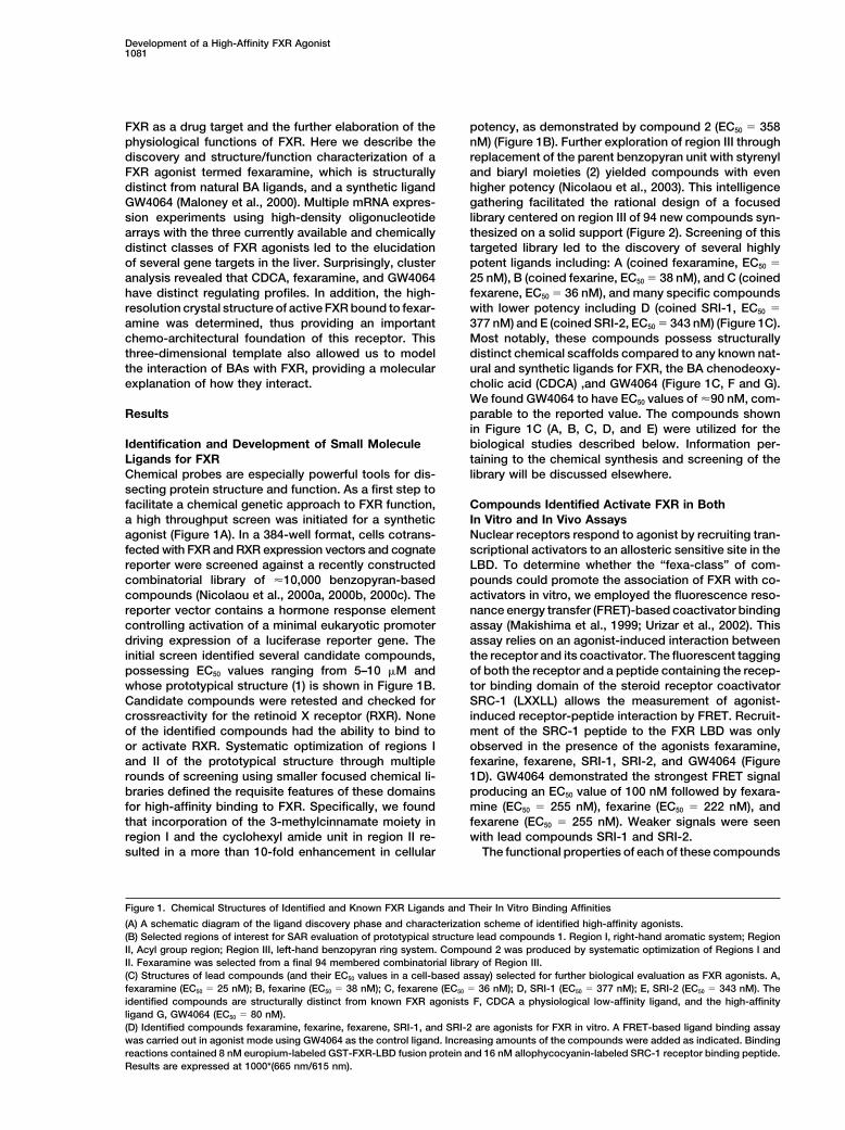
FXR as a drug target and the further elaboration of the potency, as demonstrated by compound 2 (EC50 � 358nM) (Figure 1B). Further exploration of region III throughphysiological functions of FXR. Here we describe the
discovery and structure/function characterization of a replacement of the parent benzopyran unit with styrenyland biaryl moieties (2) yielded compounds with evenFXR agonist termed fexaramine, which is structurally
distinct from natural BA ligands, and a synthetic ligand higher potency (Nicolaou et al., 2003). This intelligencegathering facilitated the rational design of a focusedGW4064 (Maloney et al., 2000). Multiple mRNA expres-
sion experiments using high-density oligonucleotide library centered on region III of 94 new compounds syn-thesized on a solid support (Figure 2). Screening of thisarrays with the three currently available and chemically
distinct classes of FXR agonists led to the elucidation targeted library led to the discovery of several highlypotent ligands including: A (coined fexaramine, EC50 �of several gene targets in the liver. Surprisingly, cluster
analysis revealed that CDCA, fexaramine, and GW4064 25 nM), B (coined fexarine, EC50 � 38 nM), and C (coinedfexarene, EC50 � 36 nM), and many specific compoundshave distinct regulating profiles. In addition, the high-
resolution crystal structure of active FXR bound to fexar- with lower potency including D (coined SRI-1, EC50 �377 nM) and E (coined SRI-2, EC50 � 343 nM) (Figure 1C).amine was determined, thus providing an important
chemo-architectural foundation of this receptor. This Most notably, these compounds possess structurallydistinct chemical scaffolds compared to any known nat-three-dimensional template also allowed us to model
the interaction of BAs with FXR, providing a molecular ural and synthetic ligands for FXR, the BA chenodeoxy-cholic acid (CDCA) ,and GW4064 (Figure 1C, F and G).explanation of how they interact.We found GW4064 to have EC50 values of �90 nM, com-parable to the reported value. The compounds shownResultsin Figure 1C (A, B, C, D, and E) were utilized for thebiological studies described below. Information per-Identification and Development of Small Molecule
Ligands for FXR taining to the chemical synthesis and screening of thelibrary will be discussed elsewhere.Chemical probes are especially powerful tools for dis-
secting protein structure and function. As a first step tofacilitate a chemical genetic approach to FXR function, Compounds Identified Activate FXR in Both
In Vitro and In Vivo Assaysa high throughput screen was initiated for a syntheticagonist (Figure 1A). In a 384-well format, cells cotrans- Nuclear receptors respond to agonist by recruiting tran-
scriptional activators to an allosteric sensitive site in thefected with FXR and RXR expression vectors and cognatereporter were screened against a recently constructed LBD. To determine whether the “fexa-class” of com-
pounds could promote the association of FXR with co-combinatorial library of �10,000 benzopyran-basedcompounds (Nicolaou et al., 2000a, 2000b, 2000c). The activators in vitro, we employed the fluorescence reso-
nance energy transfer (FRET)-based coactivator bindingreporter vector contains a hormone response elementcontrolling activation of a minimal eukaryotic promoter assay (Makishima et al., 1999; Urizar et al., 2002). This
assay relies on an agonist-induced interaction betweendriving expression of a luciferase reporter gene. Theinitial screen identified several candidate compounds, the receptor and its coactivator. The fluorescent tagging
of both the receptor and a peptide containing the recep-possessing EC50 values ranging from 5–10 �M andwhose prototypical structure (1) is shown in Figure 1B. tor binding domain of the steroid receptor coactivator
SRC-1 (LXXLL) allows the measurement of agonist-Candidate compounds were retested and checked forcrossreactivity for the retinoid X receptor (RXR). None induced receptor-peptide interaction by FRET. Recruit-
ment of the SRC-1 peptide to the FXR LBD was onlyof the identified compounds had the ability to bind toor activate RXR. Systematic optimization of regions I observed in the presence of the agonists fexaramine,
fexarine, fexarene, SRI-1, SRI-2, and GW4064 (Figureand II of the prototypical structure through multiplerounds of screening using smaller focused chemical li- 1D). GW4064 demonstrated the strongest FRET signal
producing an EC50 value of 100 nM followed by fexara-braries defined the requisite features of these domainsfor high-affinity binding to FXR. Specifically, we found mine (EC50 � 255 nM), fexarine (EC50 � 222 nM), and
fexarene (EC50 � 255 nM). Weaker signals were seenthat incorporation of the 3-methylcinnamate moiety inregion I and the cyclohexyl amide unit in region II re- with lead compounds SRI-1 and SRI-2.
The functional properties of each of these compoundssulted in a more than 10-fold enhancement in cellular
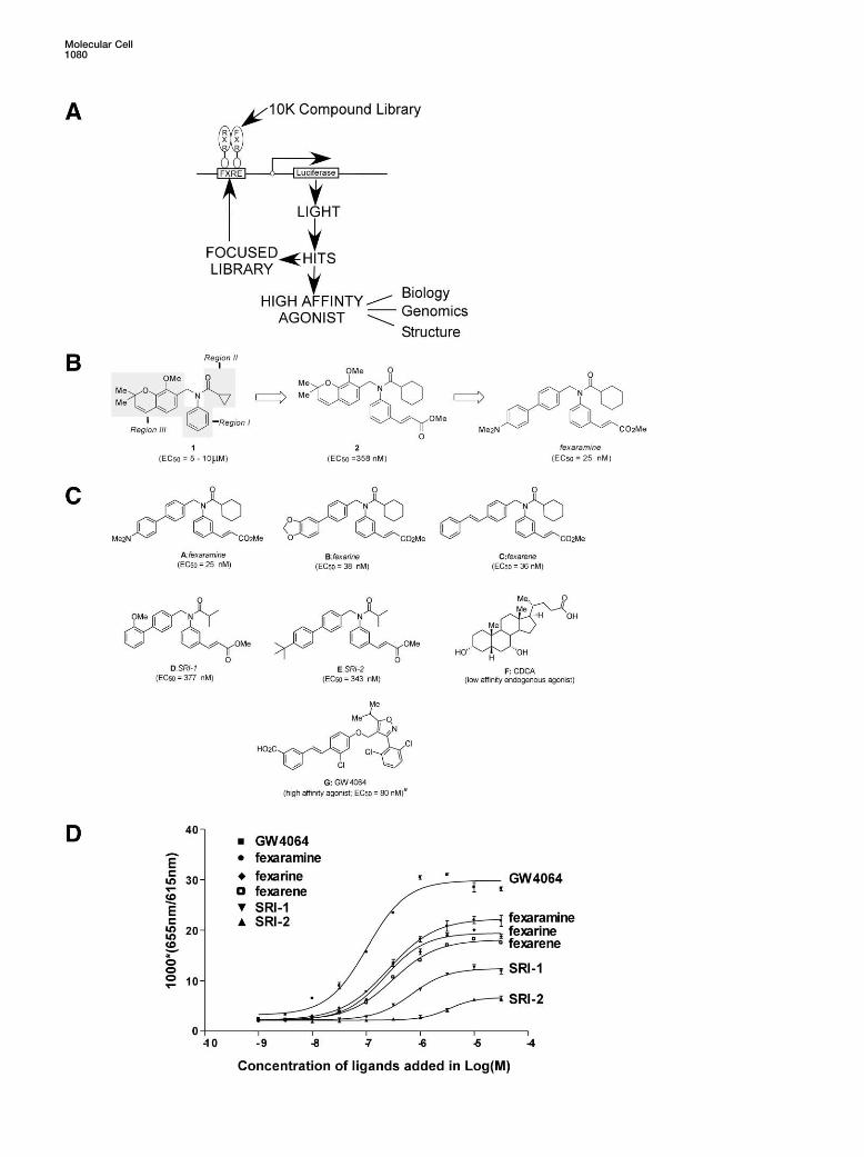
Figure 1. Chemical Structures of Identified and Known FXR Ligands and Their In Vitro Binding Affinities
(A) A schematic diagram of the ligand discovery phase and characterization scheme of identified high-affinity agonists.(B) Selected regions of interest for SAR evaluation of prototypical structure lead compounds 1. Region I, right-hand aromatic system; RegionII, Acyl group region; Region III, left-hand benzopyran ring system. Compound 2 was produced by systematic optimization of Regions I andII. Fexaramine was selected from a final 94 membered combinatorial library of Region III.(C) Structures of lead compounds (and their EC50 values in a cell-based assay) selected for further biological evaluation as FXR agonists. A,fexaramine (EC50 � 25 nM); B, fexarine (EC50 � 38 nM); C, fexarene (EC50 � 36 nM); D, SRI-1 (EC50 � 377 nM); E, SRI-2 (EC50 � 343 nM). Theidentified compounds are structurally distinct from known FXR agonists F, CDCA a physiological low-affinity ligand, and the high-affinityligand G, GW4064 (EC50 � 80 nM).(D) Identified compounds fexaramine, fexarine, fexarene, SRI-1, and SRI-2 are agonists for FXR in vitro. A FRET-based ligand binding assaywas carried out in agonist mode using GW4064 as the control ligand. Increasing amounts of the compounds were added as indicated. Bindingreactions contained 8 nM europium-labeled GST-FXR-LBD fusion protein and 16 nM allophycocyanin-labeled SRC-1 receptor binding peptide.Results are expressed at 1000*(665 nm/615 nm).
Molecular Cell1082
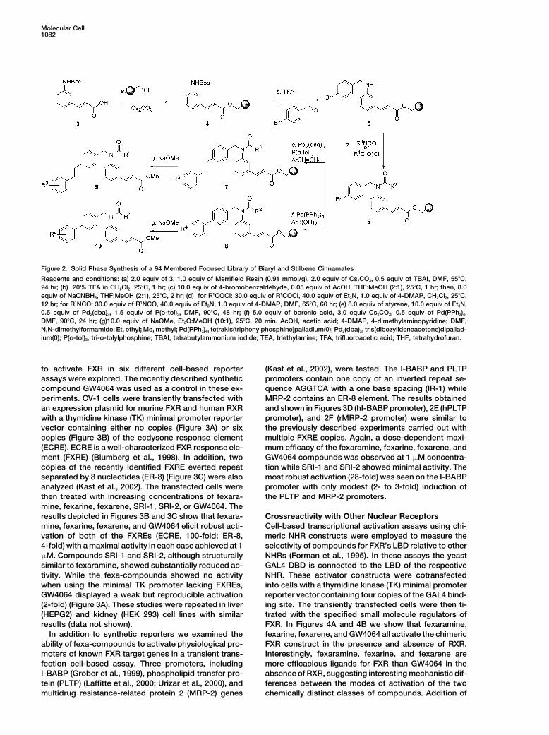
Figure 2. Solid Phase Synthesis of a 94 Membered Focused Library of Biaryl and Stilbene Cinnamates
Reagents and conditions: (a) 2.0 equiv of 3, 1.0 equiv of Merrifield Resin (0.91 mmol/g), 2.0 equiv of Cs2CO3, 0.5 equiv of TBAI, DMF, 55�C,24 hr; (b) 20% TFA in CH2Cl2, 25�C, 1 hr; (c) 10.0 equiv of 4-bromobenzaldehyde, 0.05 equiv of AcOH, THF:MeOH (2:1), 25�C, 1 hr; then, 8.0equiv of NaCNBH3, THF:MeOH (2:1), 25�C, 2 hr; (d) for R1COCl: 30.0 equiv of R1COCl, 40.0 equiv of Et3N, 1.0 equiv of 4-DMAP, CH2Cl2, 25�C,12 hr; for R1NCO: 30.0 equiv of R1NCO, 40.0 equiv of Et3N, 1.0 equiv of 4-DMAP, DMF, 65�C, 60 hr; (e) 8.0 equiv of styrene, 10.0 equiv of Et3N,0.5 equiv of Pd2(dba)3, 1.5 equiv of P(o-tol)3, DMF, 90�C, 48 hr; (f) 5.0 equiv of boronic acid, 3.0 equiv Cs2CO3, 0.5 equiv of Pd(PPh3)4,DMF, 90�C, 24 hr; (g)10.0 equiv of NaOMe, Et2O:MeOH (10:1), 25�C, 20 min. AcOH, acetic acid; 4-DMAP, 4-dimethylaminopyridine; DMF,N,N-dimethylformamide; Et, ethyl; Me, methyl; Pd(PPh3)4, tetrakis(triphenylphosphine)palladium(0); Pd2(dba)3, tris(dibezylideneacetone)dipallad-ium(0); P(o-tol)3, tri-o-tolylphosphine; TBAI, tetrabutylammonium iodide; TEA, triethylamine; TFA, trifluoroacetic acid; THF, tetrahydrofuran.
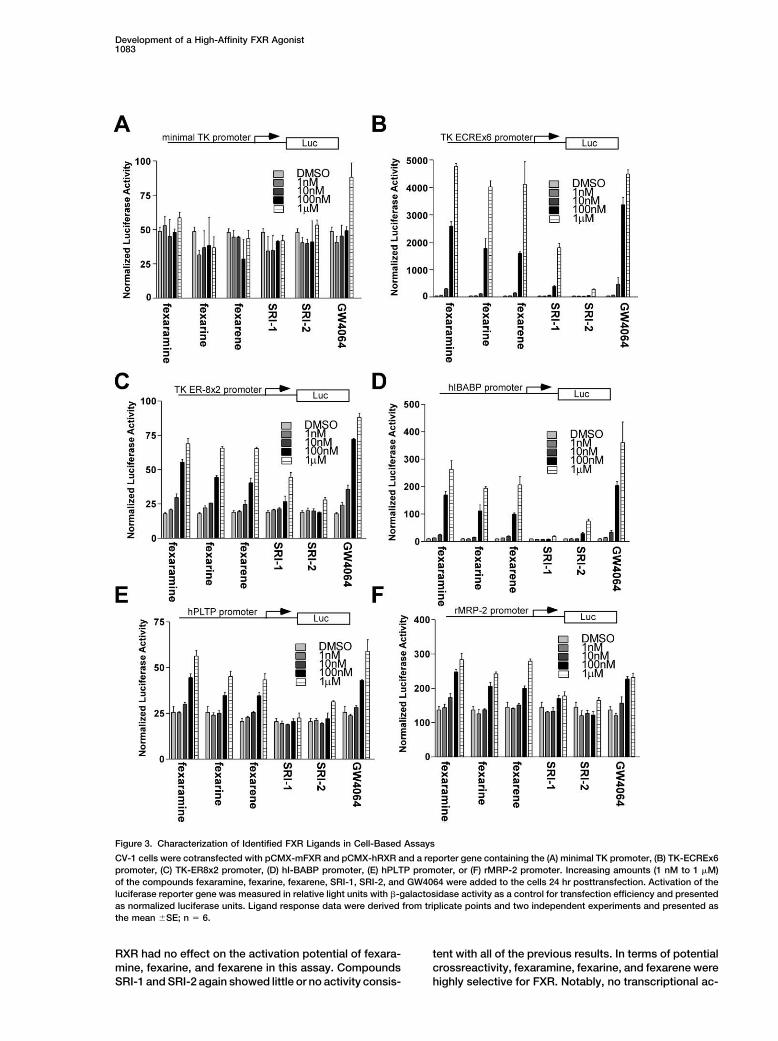
to activate FXR in six different cell-based reporter (Kast et al., 2002), were tested. The I-BABP and PLTPpromoters contain one copy of an inverted repeat se-assays were explored. The recently described synthetic
compound GW4064 was used as a control in these ex- quence AGGTCA with a one base spacing (IR-1) whileMRP-2 contains an ER-8 element. The results obtainedperiments. CV-1 cells were transiently transfected with
an expression plasmid for murine FXR and human RXR and shown in Figures 3D (hI-BABP promoter), 2E (hPLTPpromoter), and 2F (rMRP-2 promoter) were similar towith a thymidine kinase (TK) minimal promoter reporter
vector containing either no copies (Figure 3A) or six the previously described experiments carried out withmultiple FXRE copies. Again, a dose-dependent maxi-copies (Figure 3B) of the ecdysone response element
(ECRE). ECRE is a well-characterized FXR response ele- mum efficacy of the fexaramine, fexarine, fexarene, andGW4064 compounds was observed at 1 �M concentra-ment (FXRE) (Blumberg et al., 1998). In addition, two
copies of the recently identified FXRE everted repeat tion while SRI-1 and SRI-2 showed minimal activity. Themost robust activation (28-fold) was seen on the I-BABPseparated by 8 nucleotides (ER-8) (Figure 3C) were also
analyzed (Kast et al., 2002). The transfected cells were promoter with only modest (2- to 3-fold) induction ofthe PLTP and MRP-2 promoters.then treated with increasing concentrations of fexara-
mine, fexarine, fexarene, SRI-1, SRI-2, or GW4064. Theresults depicted in Figures 3B and 3C show that fexara- Crossreactivity with Other Nuclear Receptors
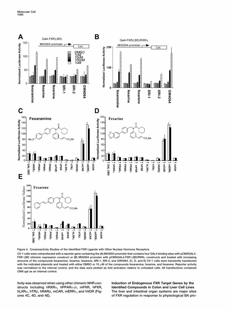
Cell-based transcriptional activation assays using chi-mine, fexarine, fexarene, and GW4064 elicit robust acti-vation of both of the FXREs (ECRE, 100-fold; ER-8, meric NHR constructs were employed to measure the
selectivity of compounds for FXR’s LBD relative to other4-fold) with a maximal activity in each case achieved at 1�M. Compounds SRI-1 and SRI-2, although structurally NHRs (Forman et al., 1995). In these assays the yeast
GAL4 DBD is connected to the LBD of the respectivesimilar to fexaramine, showed substantially reduced ac-tivity. While the fexa-compounds showed no activity NHR. These activator constructs were cotransfected
into cells with a thymidine kinase (TK) minimal promoterwhen using the minimal TK promoter lacking FXREs,GW4064 displayed a weak but reproducible activation reporter vector containing four copies of the GAL4 bind-
ing site. The transiently transfected cells were then ti-(2-fold) (Figure 3A). These studies were repeated in liver(HEPG2) and kidney (HEK 293) cell lines with similar trated with the specified small molecule regulators of
FXR. In Figures 4A and 4B we show that fexaramine,results (data not shown).In addition to synthetic reporters we examined the fexarine, fexarene, and GW4064 all activate the chimeric
FXR construct in the presence and absence of RXR.ability of fexa-compounds to activate physiological pro-moters of known FXR target genes in a transient trans- Interestingly, fexaramine, fexarine, and fexarene are
more efficacious ligands for FXR than GW4064 in thefection cell-based assay. Three promoters, includingI-BABP (Grober et al., 1999), phospholipid transfer pro- absence of RXR, suggesting interesting mechanistic dif-
ferences between the modes of activation of the twotein (PLTP) (Laffitte et al., 2000; Urizar et al., 2000), andmultidrug resistance-related protein 2 (MRP-2) genes chemically distinct classes of compounds. Addition of
Development of a High-Affinity FXR Agonist1083
Figure 3. Characterization of Identified FXR Ligands in Cell-Based Assays
CV-1 cells were cotransfected with pCMX-mFXR and pCMX-hRXR and a reporter gene containing the (A) minimal TK promoter, (B) TK-ECREx6promoter, (C) TK-ER8x2 promoter, (D) hI-BABP promoter, (E) hPLTP promoter, or (F) rMRP-2 promoter. Increasing amounts (1 nM to 1 �M)of the compounds fexaramine, fexarine, fexarene, SRI-1, SRI-2, and GW4064 were added to the cells 24 hr posttransfection. Activation of theluciferase reporter gene was measured in relative light units with �-galactosidase activity as a control for transfection efficiency and presentedas normalized luciferase units. Ligand response data were derived from triplicate points and two independent experiments and presented asthe mean �SE; n � 6.
RXR had no effect on the activation potential of fexara- tent with all of the previous results. In terms of potentialcrossreactivity, fexaramine, fexarine, and fexarene weremine, fexarine, and fexarene in this assay. Compounds
SRI-1 and SRI-2 again showed little or no activity consis- highly selective for FXR. Notably, no transcriptional ac-
Molecular Cell1084
Figure 4. Crossreactivity Studies of the Identified FXR Ligands with Other Nuclear Hormone Receptors
CV-1 cells were cotransfected with a reporter gene containing the (A) MH2004 promoter that contains four GAL4 binding sites with pCMXGAL4-FXR LBD chimeric expression construct or (B) MH2004 promoter with pCMXGAL4-FXR LBD/RXR� constructs and treated with increasingamounts of the compounds fexaramine, fexarine, fexarene, SRI-1, SRI-2, and GW4064. (C, D, and E) CV-1 cells were transiently transfectedwith the indicated plasmids and treated with either DMSO or 10 �M of the compounds fexaramine, fexarine, and fexarene. Reporter activitywas normalized to the internal control, and the data were plotted as fold activation relative to untreated cells. All transfections containedCMX-gal as an internal control.
tivity was observed when using other chimeric NHR con- Induction of Endogenous FXR Target Genes by theIdentified Compounds in Colon and Liver Cell Linesstructs including hRXR�, hPPAR���, mPXR, hPXR,
hLXR�, hTR�, hRAR�, mCAR, mERR�, and hVDR (Fig- The liver and intestinal organ systems are major sitesof FXR regulation in response to physiological BA pro-ures 4C, 4D, and 4E).
Development of a High-Affinity FXR Agonist1085
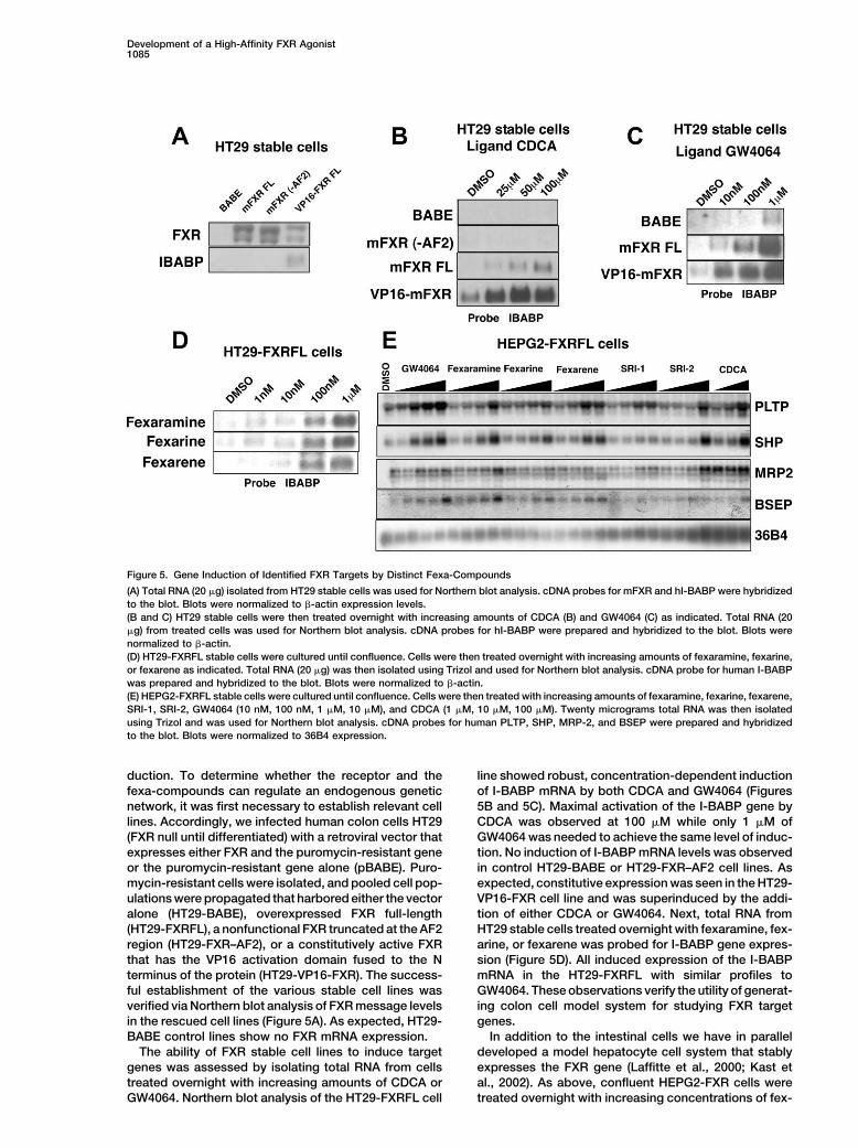
Figure 5. Gene Induction of Identified FXR Targets by Distinct Fexa-Compounds
(A) Total RNA (20 �g) isolated from HT29 stable cells was used for Northern blot analysis. cDNA probes for mFXR and hI-BABP were hybridizedto the blot. Blots were normalized to �-actin expression levels.(B and C) HT29 stable cells were then treated overnight with increasing amounts of CDCA (B) and GW4064 (C) as indicated. Total RNA (20�g) from treated cells was used for Northern blot analysis. cDNA probes for hI-BABP were prepared and hybridized to the blot. Blots werenormalized to �-actin.(D) HT29-FXRFL stable cells were cultured until confluence. Cells were then treated overnight with increasing amounts of fexaramine, fexarine,or fexarene as indicated. Total RNA (20 �g) was then isolated using Trizol and used for Northern blot analysis. cDNA probe for human I-BABPwas prepared and hybridized to the blot. Blots were normalized to �-actin.(E) HEPG2-FXRFL stable cells were cultured until confluence. Cells were then treated with increasing amounts of fexaramine, fexarine, fexarene,SRI-1, SRI-2, GW4064 (10 nM, 100 nM, 1 �M, 10 �M), and CDCA (1 �M, 10 �M, 100 �M). Twenty micrograms total RNA was then isolatedusing Trizol and was used for Northern blot analysis. cDNA probes for human PLTP, SHP, MRP-2, and BSEP were prepared and hybridizedto the blot. Blots were normalized to 36B4 expression.
duction. To determine whether the receptor and the line showed robust, concentration-dependent inductionof I-BABP mRNA by both CDCA and GW4064 (Figuresfexa-compounds can regulate an endogenous genetic
network, it was first necessary to establish relevant cell 5B and 5C). Maximal activation of the I-BABP gene byCDCA was observed at 100 �M while only 1 �M oflines. Accordingly, we infected human colon cells HT29
(FXR null until differentiated) with a retroviral vector that GW4064 was needed to achieve the same level of induc-tion. No induction of I-BABP mRNA levels was observedexpresses either FXR and the puromycin-resistant gene
or the puromycin-resistant gene alone (pBABE). Puro- in control HT29-BABE or HT29-FXR–AF2 cell lines. Asexpected, constitutive expression was seen in the HT29-mycin-resistant cells were isolated, and pooled cell pop-
ulations were propagated that harbored either the vector VP16-FXR cell line and was superinduced by the addi-tion of either CDCA or GW4064. Next, total RNA fromalone (HT29-BABE), overexpressed FXR full-length
(HT29-FXRFL), a nonfunctional FXR truncated at the AF2 HT29 stable cells treated overnight with fexaramine, fex-arine, or fexarene was probed for I-BABP gene expres-region (HT29-FXR–AF2), or a constitutively active FXR
that has the VP16 activation domain fused to the N sion (Figure 5D). All induced expression of the I-BABPmRNA in the HT29-FXRFL with similar profiles toterminus of the protein (HT29-VP16-FXR). The success-
ful establishment of the various stable cell lines was GW4064. These observations verify the utility of generat-ing colon cell model system for studying FXR targetverified via Northern blot analysis of FXR message levels
in the rescued cell lines (Figure 5A). As expected, HT29- genes.In addition to the intestinal cells we have in parallelBABE control lines show no FXR mRNA expression.
The ability of FXR stable cell lines to induce target developed a model hepatocyte cell system that stablyexpresses the FXR gene (Laffitte et al., 2000; Kast etgenes was assessed by isolating total RNA from cells
treated overnight with increasing amounts of CDCA or al., 2002). As above, confluent HEPG2-FXR cells weretreated overnight with increasing concentrations of fex-GW4064. Northern blot analysis of the HT29-FXRFL cell
Molecular Cell1086
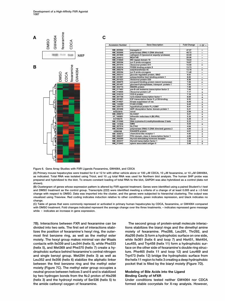
aramine, fexarine, fexarene, SRI-1, SRI-2, or the control scription factor 1, 3-fold), and amino acid transport(SCL7A2, 4-fold). Confirmations of gene induction byligands GW4064 and CDCA. Total RNA was isolated,
and the expression of the FXR target genes SHP, MRP-2, FXR agonists of many of the target genes reported inthis list were done by Northern blot analysis and will beBSEP, and PLTP was measured by Northern blot analy-
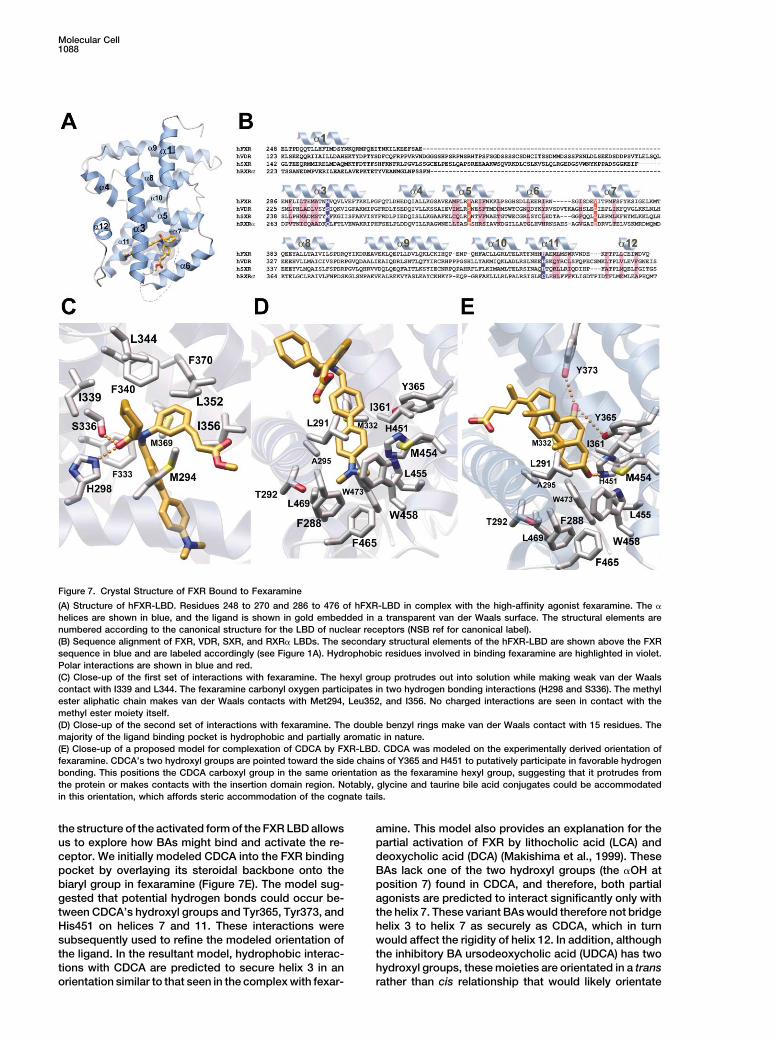
sis (Figure 5E). The control ligands CDCA and GW4064 the subject of a more detailed paper.showed similar patterns of induction to what has beenpreviously reported. Of our fexa-compounds, fexara- Structural Basis of FXR-Mediatedmine was the most effective inducer of target genes, Fexaramine Recognitionalthough strong and comparative inductions were also To understand the molecular determinants of ligandobserved with fexarine and fexarene. Interestingly, al- binding as well as to gain insight into the physical prop-though GW4064 showed slightly better induction of the erties of the active FXR receptor, we solved and refinedFXR target genes PLTP and SHP, fexaramine-matched the crystal structure of the ligand binding domain (LBD)GW4064 induced activation of the BSEP and MRP-2 of human FXR (amino acids 248–472) in a complex withgenes. Interestingly, even the weak compound SRI-1 fexaramine to 1.78 A resolution. The hFXR-LBD adoptsdisplayed remarkably effective induction of PLTP al- a 12 �-helix bundle as seen in all NHR LBD structuresthough it weakened on other genes. These results dem- described to date (RXR� [Egea et al., 2000], PXR/SXRonstrate that the fexa-compounds can be used to iden- [Watkins et al., 2001], PPAR� [Xu et al., 2001], and ROR�tify FXR-dependent target genes in liver and intestinal [Stehlin et al., 2001]) (Figures 7A and 7B). The mostcell lines. The somewhat (�10) reduced sensitivity of significant difference between other NHRs (RXR, VDR,the hepatic cells may reflect the ability of the liver hepa- and PPARs) and FXR is in the replacement of the �-turntocytes to mount a xenobiotic response or may be af- found following helix 5 with a more pronounced helix 6fected by cell-specific permeability characteristics of in FXR (Figure 7A).the compounds. For these reasons a multiplicity of in- In addition, the 15 residue insertion region betweenducers can be valuable in determining the FXR regula- helices 1 and 3 is completely disordered in the FXRtory network. crystal structure (Figures 7A and 7B). RXR�, which most
closely resembles FXR in both primary sequence andlength of the insertion region, has an additional helixGene Profiling of FXR Agonist in Primary
Hepatocyte Cells (helix 2) in this position in the absence of ligand thatunfolds and becomes disordered upon binding of 9-cisHaving established fexaramine as a potent FXR-specific
agonist in both cell culture systems, we next compared retinoic acid (Egea et al., 2000). This region of RXR�has been proposed to act as a molecular spring whichfexaramine’s gene activation profile with CDCA and
GW4064 in human primary hepatocytes. Hepatocytes accommodates the large conformational movements ofhelix 3 upon ligand binding. The insertion region maywere treated with either DMSO (control group), fexara-
mine (10 �M), CDCA (100 �M), or GW4064 (10 �M), and serve a similar role in hFXR, facilitating helix 3 re-arrangements upon ligand binding. In the PPARs, thistotal RNA was isolated at 6 and 12 hr time points. Prior
to gene profiling experiments, the samples were verified region contains a helix 2, and this region is the proposedligand access site for the small molecule binding pocket.by Northern blot analysis for induction of SHP, a known
FXR target gene (Figure 6A). Subsequently, biotinylated In SXR (Watkins et al., 2001) and VDR (Rochel et al.,2000) the insertion domain region is significantly longercRNAs prepared from mRNA samples were indepen-
dently labeled and hybridized to duplicate sets of high- (Figure 7B). Analysis of root-mean-square deviations(rmsd) between the apo and ligand bound structuresdensity microarrays (U-133A set, Affymetrix, Palo Alto,
CA). A total of 222 transcripts were identified whose of SXR and VDR revealed no significant differences,suggesting that a shorter insertion domain region mayexpression changed relative to the DMSO control using
a paired Student’s t test and each of the three agonists. be responsible for regulating large rearrangements ofhelix 3. Significantly, the activation function-2 domainThese genes were then subjected to hierarchal cluster-
ing and visualized (Cluster and Treeview, Mike Eisen). (AF2 or helix 12), essential for transcriptional activationof the receptor, is packed against the body of FXR,The most surprising observation was the distinct ex-
pression profiles of the �30,000 genes seen using the positioned between helices 3 and 4 (Figure 7A). Thiscompact conformation is a signature feature that en-chemically distinct FXR agonists (Figure 6B). Indeed,
relatively few genes were observed whose expression ables stable interactions between NHRs and their co-activator partners (Xu et al., 2001). By analogy, coactiva-profiles changed in a similar fashion using all three ago-
nists. As mentioned above, this may be due in part to tors would bind in the hydrophobic pocket formed byhelices 3, 4, 5, and 12. This extended and complemen-CDCA as a physiological BA, exerting a multiplicity of
effects via non-FXR pathways. For example, the recent tary pocket would interact with the hydrophobic face ofthe LXXLL helix located within coactivator proteins.knockout of SHP reveals that BAs act through at least
two pathways to mediate repression of the CYP7A en- The ligand binding cavity of the hFXR-LBD is predomi-nantly hydrophobic in nature and is formed by 25 aminozyme. In addition, a small subset of genes (Figure 6C)
exhibited 3-fold changes in expression when using any acid side chains (Figures 7C and 7D). The binding pockethas a volume of 726 A3 that is smaller than that seen inof the three FXR ligands (genes below the 3-fold cut-
off like SHP [2-fold] are not documented but have being SXR (1150 A3 ) (Watkins et al., 2001) but larger than thatof RXR� (439 A3 ) (Egea et al., 2000) (Figure 7E). Fexara-actively investigated). This list suggests additional roles
for FXR in the bilirubin biosynthetic pathway (BLVRA, mine is sequestered between helices 3 and 7 and makessignificant contacts with helices 5, 6, 11, and 12 (Figure5-fold), thyroid metabolism (TSHR, 3-fold; thyroid tran-
Development of a High-Affinity FXR Agonist1087
Figure 6. Gene Array Studies with FXR Ligands Fexaramine, GW4064, and CDCA
(A) Primary mouse hepatocytes were treated for 6 or 12 hr with either vehicle alone or 100 �M CDCA, 10 �M fexaramine, or 10 �M GW4064,as indicated. Total RNA was isolated using Trizol, and 10 �g total RNA was used for Northern blot analysis. The human SHP probe wasprepared and hybridized to the blot. To ensure constant loading of total RNA to the blot, GAPDH was also hybridized as a control (data notshown).(B) Clustergram of genes whose expression pattern is altered by FXR agonist treatment. Genes were identified using a paired Student’s t testand DMSO treatment as the control group. Transcripts (222) were identified meeting a criteria of a change of at least 0.005 and a 2-foldchange with respect to DMSO. Data was imported into the cluster, and the genes were subjected to hierarchal clustering. The output wasvisualized using Treeview. Red coding indicates induction relative to other conditions, green indicates repression, and black indicates nochange.(C) Table of genes that were commonly repressed or activated in primary human hepatocytes by CDCA, fexaramine, or GW4064 comparedwith DMSO treatment. Fold changes indicated represent the average change over the three treatments. – indicates repressed gene messagewhile � indicates an increase in gene expression.
7B). Interactions between FXR and fexaramine can be The second group of protein-small molecule interac-tions stabilizes the biaryl rings and the dimethyl aminedivided into two sets. The first set of interactions stabi-
lizes the position of fexaramine’s hexyl ring, the outer- moiety of fexaramine. Phe288, Leu291, Thr292, andAla295 (helix 3) form a hydrophobic surface on one side,most first benzene ring, as well as the methyl ester
moiety. The hexyl group makes minimal van der Waals while Ile361 (helix 6 and loop 7) and His451, Met454,Leu455, and Trp458 (helix 11) form a hydrophobic sur-contacts with Ile339 and Leu344 (helix 5), while Phe333
(helix 5), and Met369 and Phe370 (helix 7) create a hy- face on the other side of fexaramine’s double ring struc-ture. Phe465 (helix 11 and loop 12) and Leu469 anddrophobic surface behind fexaramine’s central nitrogen
and single benzyl group. Met294 (helix 3) as well as Trp473 (helix 12) bridge the hydrophobic surface fromthe helix 11 region to helix 3 creating a deep hydrophobicLeu352 and Ile356 (helix 6) stabilize the aliphatic linker
between the first benzene ring and the methyl ester pocket that is filled by the biaryl moiety (Figure 7D).moiety (Figure 7C). The methyl ester group occupies aneutral groove between helices 3 and 6 and is stabilized Modeling of Bile Acids into the Ligand
Binding Cavity of hFXRby two hydrogen bonds from the N�2 proton of His298(helix 3) and the hydroxyl moiety of Ser336 (helix 5) to Under conditions tested neither GW4064 nor CDCA
formed stable cocrystals for X-ray analysis. However,the amide carbonyl oxygen of fexaramine.
Molecular Cell1088
Figure 7. Crystal Structure of FXR Bound to Fexaramine
(A) Structure of hFXR-LBD. Residues 248 to 270 and 286 to 476 of hFXR-LBD in complex with the high-affinity agonist fexaramine. The �
helices are shown in blue, and the ligand is shown in gold embedded in a transparent van der Waals surface. The structural elements arenumbered according to the canonical structure for the LBD of nuclear receptors (NSB ref for canonical label).(B) Sequence alignment of FXR, VDR, SXR, and RXR� LBDs. The secondary structural elements of the hFXR-LBD are shown above the FXRsequence in blue and are labeled accordingly (see Figure 1A). Hydrophobic residues involved in binding fexaramine are highlighted in violet.Polar interactions are shown in blue and red.(C) Close-up of the first set of interactions with fexaramine. The hexyl group protrudes out into solution while making weak van der Waalscontact with I339 and L344. The fexaramine carbonyl oxygen participates in two hydrogen bonding interactions (H298 and S336). The methylester aliphatic chain makes van der Waals contacts with Met294, Leu352, and I356. No charged interactions are seen in contact with themethyl ester moiety itself.(D) Close-up of the second set of interactions with fexaramine. The double benzyl rings make van der Waals contact with 15 residues. Themajority of the ligand binding pocket is hydrophobic and partially aromatic in nature.(E) Close-up of a proposed model for complexation of CDCA by FXR-LBD. CDCA was modeled on the experimentally derived orientation offexaramine. CDCA’s two hydroxyl groups are pointed toward the side chains of Y365 and H451 to putatively participate in favorable hydrogenbonding. This positions the CDCA carboxyl group in the same orientation as the fexaramine hexyl group, suggesting that it protrudes fromthe protein or makes contacts with the insertion domain region. Notably, glycine and taurine bile acid conjugates could be accommodatedin this orientation, which affords steric accommodation of the cognate tails.
the structure of the activated form of the FXR LBD allows amine. This model also provides an explanation for thepartial activation of FXR by lithocholic acid (LCA) andus to explore how BAs might bind and activate the re-
ceptor. We initially modeled CDCA into the FXR binding deoxycholic acid (DCA) (Makishima et al., 1999). TheseBAs lack one of the two hydroxyl groups (the �OH atpocket by overlaying its steroidal backbone onto the
biaryl group in fexaramine (Figure 7E). The model sug- position 7) found in CDCA, and therefore, both partialagonists are predicted to interact significantly only withgested that potential hydrogen bonds could occur be-
tween CDCA’s hydroxyl groups and Tyr365, Tyr373, and the helix 7. These variant BAs would therefore not bridgehelix 3 to helix 7 as securely as CDCA, which in turnHis451 on helices 7 and 11. These interactions were
subsequently used to refine the modeled orientation of would affect the rigidity of helix 12. In addition, althoughthe inhibitory BA ursodeoxycholic acid (UDCA) has twothe ligand. In the resultant model, hydrophobic interac-
tions with CDCA are predicted to secure helix 3 in an hydroxyl groups, these moieties are orientated in a transrather than cis relationship that would likely orientateorientation similar to that seen in the complex with fexar-
Development of a High-Affinity FXR Agonist1089
UDCA to create a more open ligand binding pocket. This Biological and functional studies were undertaken tofurther characterize fexaramine activity. In vitro assaysarrangement, in turn, may force a suboptimal orientationestablished that fexaramine and related ligands robustlyof helix 12 and result in partial inhibition of the coactiva-recruited the coactivator SRC-1 peptide to FXR in ator interaction.manner comparable to that of GW4064. Cell-based inModeling of the recently identified synthetic BA ago-vivo assays with FXR response elements and naturalnist 6-�-ethyl-chenodeoxycholic acid (6-ECDCA) ontopromoters of known target genes demonstrated thatthe positional coordinates for the CDCA model furtherthese ligands potently activate FXR in a concentration-supports the validity of the model and suggests a mech-dependent manner. Crossreactivity experiments re-anism for its efficacy as well (Pellicciari et al., 2002).vealed the specificity of this fexa-class of ligands. Unlike6-ECDCA differs from CDCA by an additional aliphaticthe fexa-compounds, GW4064 required cotransfectionmoiety at the 6� position. The ethyl substituent at thisof RXR to achieve maximal efficacy in the chimericposition would be predicted to fit snugly into a hy-GAL4DBD-FXR-LBD protein. This suggests that the indrophobic pocket formed by Met332 and Phe333 fromvivo binding of GW4064 to FXR may preferentially recog-helix 5. Furthermore, it was demonstrated that either anize the FXR/RXR heterodimer. Induction of known tar-methyl substituent or a bulkier group at this positionget genes in both intestinal and liver cell systems dem-reduced efficacy (Pellicciari et al., 2002). This modelonstrated the usefulness of the identified compounds inwould predict a less than optimal interaction of a methylstudying FXR target genes. In intestinal cells, treatmentsubstituent with FXR since the smaller methyl groupwith fexaramine robustly induced the I-BABP gene in adoes not fill the hydrophobic pocket as well as the largerconcentration-dependent manner with efficacy similarethyl group. The resultant loss of binding energy throughto GW4064. Likewise, in the HEPG2 liver cell system,a decrease in contact surface area would result in a lossstrong induction of target genes SHP, PLTP BSEP, andof efficacy. Bulkier substituents would also be unfavor-MRP-2 was achieved at comparable concentrations ofable, as they would surpass the 0.3 A limit allowed for infexaramine and GW4064.van der Waals overlap resulting in a significant repulsive
Having confirmed the biological specificity and effi-force in the FXR ligand binding site.cacy of fexaramine, we then investigated FXR targetFexaramine is a much stronger activator of FXR-medi-genes. The structural dissimilarity of the FXR agonistsated transcriptional activity than even FXR’s most po-discussed here was reflected in the effects these com-tent natural ligand. Our model suggests that fexara-pounds had on global gene regulation. Each moleculemine’s potency appears to be mediated using twohad a distinct signature of gene expression, in additionmechanistic paths. First, the fexaramine methyl esterto a smaller subset of genes that were affected similarlygroup provides a significant number of contacts withby all three ligands (Figures 6C and 5E). The differenceshelix 3 that are absent in our model of CDCA binding.between the synthetic compounds could be due to ef-The methyl ester aliphatic chain effectively bridges helixfects mediated by heterogeneous FXR-containing com-3 with helix 6 through van der Waals contacts. FXRplexes due to structural differences in the ligand, differ-further stabilizes helix 3 against the remainder of theential clearance rates in primary liver hepatocytes, orstructure via interactions between Asn297 from helix 3through non-FXR-mediated signaling mechanisms. Inand Arg335 from helix 5, in addition to interactions fromcontrast, the distinct profile seen by CDCA is likely aAsn286 (helix 3) and Arg354 (helix 6). The second mecha-consequence of non-FXR dependent activation of thenism seems to be a function of fexaramine’s lengthxenobiotic receptor PXR and the c-Jun N-terminal ki-which by comparison to fexarene and fexarine and thenase JNK (Wang et al., 2002). In addition, it has beenBAs suggests that the sequential hydrophobic ringshown that partial agonists such as tamoxifen or raloxi-structures of these compounds penetrate deeper intofene for human estrogen receptor can display strikinglythe ligand binding pocket to increase the number ofdifferent gene expression profiles. We suggest that the
stable contacts with the LBD’s binding pocket. Thedifferences between the fexa-compounds and GW4064
larger volume of fexaramine (461 A3 ) compared to CDCAmight be because of their individual association with the
(339 A3 ) more effectively fills the ligand binding cavity. LBD achieving specificity by aggregating with distinctAnalysis of buried surface area in the absence and pres- cofactor complexes. This has yet to be shown for anyence of fexaramine reveals an additional 9 A2 of buried synthetic compound but is consistent with the knownhydrophobic surface when fexaramine is bound. This mechanism of action. This exemplifies the difficulties ofcorresponds to an increase of approximately 1 kJ/M in investigating NHR function using a natural ligand andstabilizing energy. Fexaramine also appears to make highlights the need for specific synthetic ligands to iso-direct contact with helix 12 to enhance rigidity and pre- late NHR’s dependent pathways. However, even high-sumably stabilize coactivator binding. affinity synthetic compounds may have nonspecific
effects. This potential limitation indicates the utility ofDiscussion employing multiple synthetic ligands to accurately dis-
cern the common core signaling pathways.We describe the development of a synthetic natural To complete this study, we next analyzed the FXRproduct-like molecule, fexaramine, which binds FXR and protein using a series of detailed structural studies.facilitates analysis of complex physiologic events such CDCA and other BAs, which bind to FXR with relativelyas cholesterol metabolism. To that end, fexaramine has low affinity, failed to promote receptor crystallizationrevealed an FXR-specific genomic profile distinct from while, in contrast, fexaramine cocrystallizes with FXR.CDCA and played a critical role in obtaining a high- This provided not only a high-resolution structure but
also enabled the subsequent modeling of CDCA with aresolution structure of the activated receptor.
Molecular Cell1090
pling (Pd[PPh3]4, Cs2CO3) with eighteen boronic acids to yield stil-high degree of confidence into the ligand binding pocketbene resins (5) and biaryl resins (6), respectively. Cleavage of theof FXR. This model provides a molecular explanationresulting compounds from resins (5) and (6) with NaOMe yieldedfor the selectivity of BAs on FXR and highlights themethyl cinnamates (7) and (8). Analysis of the library by LCMS after
importance of position and orientation of the hydroxyl purification showed the average purity of these compounds togroups at positions 3 and 7 in binding affinity. This model be 95%. Further details of the chemistry involved in this project
can be found elsewhere (Nicolaou et al., 2003).provides a possible rationale for the beneficial effectsof UDCA in the treatment of primary biliary cirrhosis.
RNA Isolation and Northern Blot HybridizationAlthough UDCA has two hydroxyl groups that couldUnless otherwise indicated, HepG2- or HT29-derived cell lines werepotentially form hydrogen bonds with FXR in the ligandcultured in medium containing superstripped FBS for 24 hr beforebinding cavity, their trans configuration creates a morethe addition of a ligand solution or DMSO (vehicle) for an additional
open ligand binding pocket that would destabilize helix 24–48 hr. Total RNA was isolated using Trizol reagent and was12 and thereby inhibit activation of the receptor. resolved (20 �g/lane) on a 1% agarose, 2.2 M formaldehyde gel,
The body of work presented in this paper was the transferred to a nylon membrane, and UV crosslinked. cDNA probeswere radiolabeled with [�-32P]dCTP using the high-prime labelingproduct of combining chemical, genetic, and structuralkit. Membranes were hybridized using the QuikHyb hybridizationapproaches to the analysis of FXR. In doing so we havesolution according to the manufacturer’s protocol (Stratagene).not only gained a valuable chemical probe, fexaramine,Blots were normalized for loading with control ribosomal 18 S cDNA
to study the mechanism of receptor signaling, but we or 36B4 protein probes. The RNA levels were quantified using ahave also begun to unravel the FXR genetic network PhosphorImager in addition to being exposed to X-ray film.from the BA network and gained the ability to manipulateselective components of the pathway. Protein Expression and Purification
The plasmid pHIS8-3-hFXR LBD (residues 248 to 476) was trans-formed into E. coli strain BL21 (DE3), and cells were grown at 37�CExperimental Proceduresuntil an OD600nm of 1.0. Expression was induced by adding isopropyl-1-thio-�-D-galactopyrauoside to 0.1 mM, and cells were grown forConstructs
The pCMX expression plasmids and luciferase reporter plasmids an additional 6 hr at 20�C. Bacteria were harvested by centrifugationat 8000 g, and pellets were stored at �70�C. Cell pellets werehave been described elsewhere (Blumberg et al., 1998; Kast et al.,
2002). The hPLTP-luc promoter was kindly provided by Dr. Dennis thawed and resuspended in 50 mM Tris-Cl (pH 8.0), 500 mM NaCl,10 mM imidazole (pH 8.0), 10% (v/v) glycerol, 1% (v/v) Tween 20,Dowhan, and the hIBABP-luc promoter was created from a plasmid
provided by Dr. Philippe Besnard. and 10 mM �-mercaptoethanol (�-ME) at 4�C. Resuspended cellswere sonicated, and lysates were centrifuged at 100,000 g atResidues 248 to 476 of human FXR LBD were PCR amplified and
subcloned into the BamHI or NcoI/BamHI sites of pGEX and pHIS, 4�C. Supernatants were purified by Ni2�-chelation chromatography(QIAGEN, Valencia, CA). After washing bound protein sample wasrespectively, to generate protein expression vectors (Jez et al.,
2000). eluted using 50 mM Tris-Cl (pH 8.0), 500 mM NaCl, 250 mM imidazole(pH 8.0), 10% (v/v) glycerol, and 10 mM �-ME. The N-terminal octa-The retroviral plasmids were constructed by cloning FXRFL, FXR-
AF2, and VP16-FXR cDNAs into the BamHI site of the established histidine tag was removed by thrombin (Sigma, St. Louis, MO) diges-tion during dialysis against 50 mM Tris (pH 8.0), 500 mM NaCl, andpBABE retroviral backbone vector. Viral extracts were established
using published procedures and used to infect HT29 colon cells. 10 mM dithiothretiol (DTT) at 4�C for 24 hr. The dialyzed and cleavedsample was purified using a Superdex 200 26/60 gel filtration columnAfter exposure for 24 hr, cells were selected by the addition of 4
�g/ml of puromycin. Cells that survived this selection procedure (Amersham Biosciences, Piscataway, NJ) equilibrated in dialysis/thrombin cleavage buffer. Peak fractions were collected and dia-were then pooled and analyzed for the expression of the FXR gene.
All constructs were verified by sequencing to confirm identity lyzed against 5 mM Tris-HCL (pH 8.0) 62.5 mM NaCl, and 1 mMDTT, concentrated to 15 mg/ml using a Centricon 10 (Amicon, Bed-and reading frame. Detailed information regarding each construct
is available upon request. ford, MA), and stored at �70�C. Selenomethionine-substituted pro-tein (SeMet) was obtained from E. coli grown in minimal media usingthe methionine pathway inhibition methods (Doublie, 1997) and wasTransfectionspurified similarly to the native FXR-LBD.CV-1 and HEPG2 cells were grown in DMEM supplemented with
10% FBS, 50 U/ml penicillin G, and 50 �g/ml streptomycin sulfateat 37�C in 7% CO2. CV-1 cells (60%–70% confluence, 48-well plate) Crystallization and Structure Determinationwere cotransfected with 16.6 ng of the appropriate expression vec- Fexaramine was solubilized in dimethylsulfoxide (DMSO) to a finaltor, 100 ng of reporter plasmid, and 100 ng of pCMX-LacZ in 200 �l concentration of 10 mM; hFXR-LBD (15 mg/ml) was incubated withof DMEM containing 10% FBS by the Lipofectamine 2000 procedure fexaramine at a 1:2 molar ratio. Crystals of the hFXR-LBD/fexara-(Invitrogen, Carlsbad, CA). After 24 hr, the medium was replaced, mine mixture were grown by the hanging drop vapor diffusionand cells were harvested and assayed for luciferase activity 36–48 method at 4�C by mixing 1.0 �l of hFXR-LBD/fexaramine complexhr after transfection. The luciferase activity was normalized by with 1.0 �l of a reservoir solution containing 15%–20% (w/v) PEG�-galactosidase activity. Each transfection was performed in tripli- 8000, 100 mM HEPES-Na� (pH 7.5), 0.2 M MgCl2, 1 mM DTT. Crystalscate and repeated at least three times. of selenomethionine-substituted hFXR-LBD were grown similarly
using 10 mM DTT. Crystals were stabilized in 10%–15% (v/v) glyc-erol, 20% (w/v) PEG 8000, 0.2 M MgCl2, 100 mM HEPES-Na� (pHSolid Phase Synthesis of Small Molecule Combinatorial
Libraries and Ligands 7.5), and 10 mM DTT and rapidly frozen in a 100 K stream of nitrogengas. MAD data to 2.1 A were collected around the Se edge at theThe synthesis of the chemical compounds was carried out on solid
phase supports in parallel as summarized in Figure 2. In brief, Boc- European Synchrotron Radiation Facility (ESRF, Grenoble, France)on beamline FIP (BM30A). Native data to 1.78 A were collected atprotected cinnamic acid (1) was immobilized on Merrifield resin
using Cs2CO3 to afford conjugate (2). The Boc group was removed the Stanford Synchrotron Radiation Laboratory, beamline 9-1. Alldata were processed with DENZO and SCALEPACK (Otwinowskiby treatment with 20% (v/v) TFA (for abbreviations see legend to
Figure 2) in CH2Cl2, and the resultant resin-bound amine was reduc- and Minor, 1997). The crystals contain one molecule per asymmetricunit (52.9% solvent) and belong to the space group P212121 (a �tively alklylated with 4-bromobenzaldehyde in the presence of
NaCNBH3 to yield amino resin (3). Resin (3) was acylated with one 36.656, b � 56.776, c � 117.646, � � 9, � � 9, � � 90.0�). Threewavelength MAD data were scaled to the �3. Seven of nine Se sitesof three acyl groups to give amide or urea resins (4). The acylated
resins (4) were then subjected to either a Heck coupling (Pd2[dba]3, were located and MAD phasing was accomplished using SOLVE(Terwilliger and Berendzen, 1992), and density modification wasP[o-tol]3, Et3N]) with thirteen substituted styrenes or a Suzuki cou-
Development of a High-Affinity FXR Agonist1091
carried out with RESOLVE (Terwilliger, 2000). The initial model was Moore, L.B., Galardi, C., Wilson, J.G., Lewis, M.C., Roth, M.E., et al.(2000). A regulatory cascade of the nuclear receptors FXR, SHP-1,built into the experimental electron density maps displayed in O
(Jones et al., 1991). The resulting model was positionally refined and LRH-1 represses bile acid biosynthesis. Mol. Cell 6, 517–526.against all of the high-resolution native data set using the default Grober, J., Zaghini, I., Fujii, H., Jones, S.A., Kliewer, S.A., Willson,bulk solvent model in CNS with maximum likelihood targets (Brunger T.M., Ono, T., and Besnard, P. (1999). Identification of a bile acid-et al., 1998). The structure of the FXR-LBD/fexaramine complex was responsive element in the human ileal bile acid-binding protein gene.refined to a Rcryst and a Rfree value of 23.0% and 27.5%, respectively, Involvement of the farnesoid X receptor/9-cis-retinoic acid receptorusing all data extending to 1.78 A resolution. The R factor � �|Fobs � heterodimer. J. Biol. Chem. 274, 29749–29754.Fcalc|/�Fobs, where summation is over the data used for refinement
Jez, J.M., Ferrer, J.-L., Bowman, M.E., Dixon, R.A., and Noel, J.P.and the Rfree was calculated using 5% of the reflection data chosen(2000). Dissection of malonyl-coenzyme A decarboxylation fromand excluded from refinement. The model consists of residues 248polyketide formation in the reaction mechanism of a plant polyketideto 270 and 286 to 475 of human FXR, 1 fexaramine molecule, andsynthase. Biochemistry 39, 890–902.340 water molecules. PROCHECK (Laskowski et al., 1993) revealedJones, A.T., Zou, J.-Y., Cowan, S., and Kjeldgaard, M. (1991). Im-a total of 92% of the residues in the most favored region of theproved methods for building protein models in electron densityRamachandran plot and 8% in the additionally allowed region. Mainmaps and the location of errors in these models. Acta Crystallogr.chain and side chain structural parameters were consistently betterA 47, 110–119.than average (overall G value of 0.16).
Kast, H.R., Goodwin, B., Tarr, P.T., Jones, S.A., Anisfeld, A.M., Stoltz,Acknowledgments C.M., Tontonoz, P., Kliewer, S., Willson, T.M., and Edwards, P.A.
(2002). Regulation of multidrug resistance-associated protein 2We thank Drs. Peter Ordentlich and David Egan for their helpful (ABCC2) by the nuclear receptors pregnane X receptor, farnesoidadvice and Drs. Ann Atkins, Ruth Yu, and Joyce Havstad for proof- X-activated receptor, and constitutive androstane receptor. J. Biol.reading. R.M.E. is an Investigator of the Howard Hughes Medical Chem. 277, 2908–2915.Institute at the Salk Institute and March of Dimes Chair in Molecular Kerr, T.A., Saeki, S., Schneider, M., Schaefer, K., Berdy, S., Redder,and Developmental Biology. This work was supported by the How- T., Shan, B., Russell, D.W., and Schwarz, M. (2002). Loss of nuclearard Hughes Medical Institute. M.D. and R.M.E. acknowledge funding receptor SHP impairs but does not eliminate negative feedbacksupport through the NIH NURSA orphan receptor program, grant regulation of bile acid synthesis. Dev. Cell 2, 713–720.number U19DK62434-01. K.C.N. thanks the National Institutes of
Laffitte, B.A., Kast, H.R., Nguyen, C.M., Zavacki, A.M., Moore, D.D.,Health (NIH) and the Skaggs Institute for Chemical Biology for fund-and Edwards, P.A. (2000). Identification of the DNA binding specific-ing support. J.P.N. received funding from the NIH, grant numberity and potential target genes for the farnesoid X-activated receptor.CA54418. P.A.E. received funding from the NIH, grant numbersJ. Biol. Chem. 275, 10638–10647.HL30568 and HL68445.Laskowski, R.A., MacArthur, M.W., Moss, D.S., and Thornton, J.M.(1993). PROCHECK: a program to check the stereochemical qualityReceived: December 5, 2002of protein structures. J. Appl. Crystallogr. 26, 283–291.Revised: February 5, 2003
Accepted: March 4, 2003 Lu, T.T., Makishima, M., Repa, J.J., Schoonjans, K., Kerr, T.A., Au-Published: April 24, 2003 werx, J., and Mangelsdorf, D.J. (2000). Molecular basis for feedback
regulation of bile acid synthesis by nuclear receptors. Mol. Cell 6,References 507–515.
Makishima, M., Okamoto, A.Y., Repa, J.J., Tu, H., Learned, R.M.,Blumberg, B., and Evans, R.M. (1998). Orphan nuclear receptors— Luk, A., Hull, M.V., Lustig, K.D., Mangelsdorf, D.J., and Shan, B.new ligands and new possibilities. Genes Dev. 12, 3149–3155. (1999). Identification of a nuclear receptor for bile acids. Science.Blumberg, B., Sabbagh, W., Jr., Juguilon, H., Bolado, J., Jr., van 284, 1362–1365.Meter, C.M., Ong, E.S., and Evans, R.M. (1998). SXR, a novel steroid Maloney, P.R., Parks, D.J., Haffner, C.D., Fivush, A.M., Chandra, G.,and xenobiotic-sensing nuclear receptor. Genes Dev. 12, 3195– Plunket, K.D., Creech, K.L., Moore, L.B., Wilson, J.G., Lewis, M.C.,3205. et al. (2000). Identification of a chemical tool for the orphan nuclearBrunger, A.T., Adams, P.D., Clore, G.M., DeLano, W.L., Gros, P., receptor FXR. J. Med. Chem. 43, 2971–2974.Grosse-Kunstleve, R.W., Jiang, J.S., Kuszewski, J., Nilges, M.,
Nicolaou, K.C., Pfefferkorn, J.A., Roecker, A.J., Cao, G.-Q., Bar-Pannu, N.S., et al. (1998). Crystallography & NMR system: A new
luenga, S., and Mitchell, H.J. (2000a). Natural product-like combina-software suite for macromolecular structure determination. Acta
torial libraries based on privileged structutres. 1. General principlesCrystallogr. D Biol. Crystallogr. 54, 905–921.
and solid-phase synthesis of benzopyrans. J. Am. Chem. Soc. 122,Chawla A., Saez E., and Evans RM. (2000). “Don’t know much bile- 9939–9953.ology”. Cell. 103, 1–4.
Nicolaou, K.C., Pfefferkorn, J.A., Mitchell, H.J., Roecker, A.J., Bar-Chiang, J.Y. (2002). Bile acid regulation of gene expression: roles luenga, S., Cao, G.-Q., Affleck, R.L., and Lillig, J.E. (2000b). Naturalof nuclear hormone receptors. Endocr. Rev. 23, 443–463. product-like combinatorial libraries based on privileged structures.Doublie, S. (1997). Preparation of selenomethionyl proteins for 2. Construction of a 10000-membered benzopyran library by di-phase determination. Methods Enzymol. 276, 523–530. rected split-and-pool chemistry using nanokans and optical encod-
ing. J. Am. Chem. Soc. 122, 9954–9967.Edwards, P.A., Kast, H.R., and Anisfeld, A.M. (2002). BAREing it all:the adoption of LXR and FXR and their roles in lipid homeostasis. Nicolaou, K.C., Pfefferkorn, J.A., Barluenga, S., Mitchell, H.J.,J. Lipid Res. 43, 2–12. Roecker, A.J., and Cao, G.-Q. (2000c). Natural product-like combina-
torial libraries based on privileged structutres. 3. The “libraries fromEgea, P.F., Mitschler, A., Rochel, N., Ruff, M., Chambon, P., andlibraries” principle for diversity enhancement of benzopyran librar-Moras, D. (2000). Crystal structure of the human RXR� ligand-bind-ies. J. Am. Chem. Soc. 122, 9968–9976.ing domain to its natural ligand: 9-cis retinoic acid. EMBO J. 19,
2592–2601. Nicolaou, K.C. Evans, R.M., Roecker, A.J., Hughes, R., Downes, M.,and Pfefferkorn, J.A. (2003). Discovery of four classes of potenet,Evans RM. (1988). The steroid and thyroid hormone receptor super-non-steroidal FXR agonists originating from natural product-like li-family. Science 240, 889–895.braries. Org. Biomol. Chem. 1, 908–920.Forman, B.M., Goode, E., Chen, J., Oro, A.E., Bradley, D.J., Per-Otwinowski, Z., and Minor, W. (1997). Processing of X-ray diffractionlmann, T., Noonan, D.J., Burka, L.T., McMorris, T., Lamph, W.W., etdata collected in oscillation mode. Methods Enzymol. 276, 307–326.al. (1995). Identification of a nuclear receptor that is activated by
farnesol metabolites. Cell 81, 687–693. Parks, D.J., Blanchard, S.G., Bledsoe, R.K., Chandra, G., Consler,T.G., Kliewer, S.A., Stimmel, J.B., Willson, T.M., Zavacki, A.M.,Goodwin, B., Jones, S.A., Price, R.R., Watson, M.A., McKee, D.D.,
Molecular Cell1092
Moore, D.D., and Lehmann, J.M. (1999). Bile acids: natural ligandsfor an orphan nuclear receptor. Science 284, 1365–1368.
Pellicciari, R., Fiorucci, S., Camaioni, E., Clerici, C., Costantino, G.,Maloney, P.R., Morelli, A., Parks, D.J., and Willson, T.M. (2002).6alpha-ethyl-chenodeoxycholic acid (6-ECDCA), a potent and se-lective FXR agonist endowed with anticholestatic activity. J. Med.Chem. 45, 3569–3572.
Sinal, C.J., Tohkin, M., Miyata, M., Ward, J.M., Lambert, G., andGonzalez, F.J. (2000). Targeted disruption of the nuclear receptorFXR/BAR impairs bile acid and lipid homeostasis. Cell 102, 731–744.
Stehlin, C., Wurtz, J.M., Steinmetz, A., Greiner, E., Schule, R., Moras,D., and Renaud, J.P. (2001). X-ray structure of the orphan nuclearreceptor RORbeta ligand-binding domain in the active conforma-tion. EMBO J. 20, 5822–5831.
Rochel, N., Wurtz, J.M., Mitschler, A., Klaholz, B., and Moras, D.(2000). The crystal structure of the nuclear receptor for vitamin Dbound to its natural ligand. Mol. Cell 5, 173–179.
Terwilliger, T.C. (2000). Maximum likelihood density modification.Acta Crystallogr. D Biol. Crystallogr. 56, 965–972.
Terwilliger, T.C., and Berendzen, J. (1992). Automated MAD and MIRstructure solution. Acta Crystallogr. D Biol. Cystallogr. 55, 849–861.
Urizar, N.L., Dowhan, D.H., and Moore, D.D. (2000). The farnesoidX-activated receptor mediates bile acid activation of phospholipidtransfer protein gene expression. J. Biol. Chem. 275, 39313–39317.
Urizar, N.L., Liverman, A.B., Dodds, D.T., Silva, F.V., Ordentlich, P.,Yan, Y., Gonzalez, F.J., Heyman, R.A., Mangelsdorf, D.J., and Moore,D.D. (2002). A natural product that lowers cholesterol as an antago-nist ligand for FXR. Science 296, 1703–1706.
Wang, H., Chen, J., Hollister, K., Sowers, L.C., and Forman, B.M.(1999). Endogenous bile acids are ligands for the nuclear receptorFXR/BAR. Mol. Cell 3, 543–553.
Wang, L., Lee, Y.K., Bundman, D., Han, Y., Thevananther, S., Kim,C.S., Chua, S.S., Wei, P., Heyman, R.A., Karin, M., and Moore, D.D.(2002). Redundant pathways for negative feedback regulation ofbile acid production. Dev. Cell 2, 721–731.
Watkins, R.E., Wisely, G.B., Moore, L.B., Collins, J.L., Lambert, M.H.,Williams, S.P., Willson, T.M., Kliewer, S.A., and Redinbo, M.R. (2001).The human nuclear xenobiotic receptor PXR: structural determi-nants of directed promiscuity. Science 292, 2329–2333.
Xu, H.E., Lambert, M.H., Montana, V.G., Plunket, K.D., Moore, L.B.,Collins, J.L., Oplinger, J.A., Kliewer, S.A., Gampe, R.T., Jr., McKee,D.D., et al. (2001). Structural determinants of ligand binding selectiv-ity between the peroxisome proliferator-activated receptors. Proc.Natl. Acad. Sci. USA 98, 13919–13924.
Accession Numbers
The Protein Data Bank code for the FXR-feraramine X-ray structureis 1OSH.pdb, and the code for the FXR-CDCA model is 1OSK.pdb.