Cohorts of arbuscular mycorrhizal fungi (AMF) in Vitis vinifera, a typical Mediterranean fruit crop
Upload
khangminh22Category
view
0download
0
HAL Id: tel-03117766https://tel.archives-ouvertes.fr/tel-03117766
Submitted on 21 Jan 2021
HAL is a multi-disciplinary open accessarchive for the deposit and dissemination of sci-entific research documents, whether they are pub-lished or not. The documents may come fromteaching and research institutions in France orabroad, or from public or private research centers.
L’archive ouverte pluridisciplinaire HAL, estdestinée au dépôt et à la diffusion de documentsscientifiques de niveau recherche, publiés ou non,émanant des établissements d’enseignement et derecherche français ou étrangers, des laboratoirespublics ou privés.
Antitumoral actions of natural stilbenes derived fromVitis vinifera
Iris Aja Perez
To cite this version:Iris Aja Perez. Antitumoral actions of natural stilbenes derived from Vitis vinifera. Cellular Biology.Université de Bordeaux; Universidad del País Vasco. Facultad de ciencias, 2020. English. �NNT :2020BORD0115�. �tel-03117766�

THÈSE EN COTUTELLE PRÉSENTÉE POUR OBTENIR LE GRADE DE
DOCTEUR DE L’UNIVERSITÉ DE BORDEAUX
ET DE L’UNIVERSITÉ DU PAYS BASQUE
ÉCOLE DOCTORALE SCIENCES DE LA VIE ET DE LA SANTÉ SPÉCIALITÉ:BIOCHIMIE
ÉCOLE DE MASTER ET DE DOCTORAT (MDe) SPÉCIALITÉ: RECHERCHE BIOMÉDICALE
Par Iris AJA PÉREZ
Antitumoral actions of natural stilbenes derived from Vitis vinifera
Sous la direction de Tristan RICHARD et de José Ignacio RUIZ SANZ
Soutenue le 25 Septembre 2020
Membres du jury :
CSIC Président Université du Pays Basque Examinateur CSIC Examinateur Université de Séville Examinateur Université de Bordeaux Examinateur Université de Lisbonne Rapporteur
M. PALMA MARTÍNEZ, José Manuel Mme. MARTINEZ SAN PELAYO, María José Mme. GARCÍA CONESA, María Teresa Mme. GARCÍA PARRILLA, María Carmen Mme. GAUDIN, Karen M. A TUNE , FernandoM. FRAGA, Cesar Université de Buenos Aires Rapporteur


Titre: Propriétés anti-tumorales de stilbènes naturels dérivés de Vitis vinifera
L'objectif principal de cette thèse de doctorat est d'étudier l'activité anti-
inflammatoire et anti-tumorale potentielle du resvératrol et d’autres stilbènes
obtenus à partir d’extraits de V. vinifera. Pour atteindre cet objectif, l'effet anti-
inflammatoire des stilbènes est d'abord abordé sur un modèle de macrophage
murin activé par les lipopolysaccharides (LPS), puis l'activité antitumorale est
évaluée sur un modèle de carcinome hépatocellulaire humain.
Les stilbènes utilisés pour l'analyse des activités biologiques comprennent 8
monomères (resvératrol, picéide, piceatannol, astringine A, pterostilbene,
oxyresveratrol, isorhapontine et isorhapontigenine), 7 dimères ( -viniférine, -
viniférine, -viniférine, ampélopsine A, scirpusine A, pallidol et vitisinol C), un
trimère (miyabenol C) et 4 tétramères (hopeaphenol, isohopeaphenol, R2-
viniférine et R-viniférine).
Concernant les effets anti-inflammatoires, tous les stilbènes ont été testés sur une
lignée de macrophages murins activés par du LPS, en analysant à la fois leur
cytotoxicité et leur capacité à inhiber la production de NO. Leurs CI30 ou CI50
ont été calculées. A partir de ces travaux, 6 composés ont été sélectionnés :
piceatannol, -viniférine, δ-viniférine, hopéaphenol et l'isohopéaphenol. Sur ces
composés, la libération de cytokines inflammatoires et la production de ROS a été
étudié. L’hopeaphenol et le piceatannol inhibent la production de ROS. La δ-
viniférine ne diminue pas la libération des cytokines IL-1β et TNF-α. Il a été
montré que le piceatannol, l’ -viniférine, l’hopeaphenol et l’isohopeaphenol ont
un effet sur la réponse inflammatoire produite par l'activation que le LPS exerce
sur les macrophages murins. Cette observation justifie de poursuivre l'étude dans

des modèles plus complexes étant donné son utilité possible dans le traitement
des maladies à base inflammatoire.
Pour l'étude de l'activité antitumorale, la CI50 obtenue après traitement par 6
stilbènes (monomère : resvératrol ; dimères : ampélopsine A et -viniférine ;
tétramères : hopeaphenol, isohopeaphenol, R2-viniférine et R-viniférine) de 2
lignées d'hépatomes a été comparée à une lignée d'hépatocytes non transformés.
La R2-viniférine présente l'activité cytotoxique la plus élevée sur la lignée HepG2
(CI50 <10 μM), sans être toxique à cette concentration pour la lignée HH4. Les
cellules Hep3B sont nettement plus résistantes à la R2-viniférine (CI50 48 μM). Le
stilbène a produit une augmentation des cellules HepG2 en phase SubG0, sans
altération des cellules Hep3B. La réponse différenciée des lignées cellulaires à
l’action de ce stilbène peut être due au statut différent de p53. En effet,
l’extinction de p53 dans les cellules HepG2 a augmenté leur résistance au
traitement, tandis que la transfection de p53 dans les cellules Hep3B les a rendues
plus sensibles. La R2-viniférine provoque la phosphorylation de p53 sur les
lignées HepG2. L'activation des caspases 3 et 9, l'augmentation du rapport des
protéines Bax/Bcl2 et la libération de LDH intracellulaire ont mis en évidence la
mort cellulaire par apoptose et nécrose provoquée par l’action de la R2-viniférine.
Dans la lignée HepG2, Cette action est modulable par PI3K/Akt et ERK1/2. De
plus, l'expression de l'histone -H2AX a montré les dommages que le traitement
par la R2-viniférine produit sur l'ADN. Le rôle clé que jouent les radicaux libres
dans tout ce processus a été observé en raison de l'augmentation de la production
de ROS et de H2O2, et de l'augmentation de l'activité Mn-SOD. Il a également été
démontré que la R2-viniférine diminue la capacité de migration et de formation
de colonies de cellules HepG2.
Il est conclu que la R2-viniférine, un tétramère du resvératrol, exerce une activité
antitumorale par le biais de mécanismes d'apoptose liés à p53. Ce composé est un

agent prometteur avec des effets potentiels pour la chimioprévention et le
traitement du cancer du foie.
Mots clés : polyphénols, stilbènes, inflammation, activité anticancéreuse, HepG2 cellules, RAW 264.7 cellules.
Title: Antitumoral actions of natural stilbenes derived from Vitis vinifera
The main objective of this Doctoral Thesis is to study the potential anti-
inflammatory and anti-tumor activity of resveratrol and other stilbenes present in
Vitis vinifera. To achieve this goal, the anti-inflammatory effect of stilbenes is
firstly addressed in a lipopolysaccharide (LPS) -activated murine macrophage
model, and secondly, antitumor activity in a model of human hepatocarcinoma is
analyzed.
The stilbenes used for the analysis of antitumor activity comprise 8 monomers
(resveratrol, piceide, piceatannol, astringin A, pterostilbene, oxyresveratrol,
isorhapontine and isorhapontigenine), 7 dimers ( -viniferin, -viniferin, -
viniferin, ampelopsin A escirpusin A, pallidol and vitisinol C), a trimer
(miyabenol C) and 4 tetramers (hopeaphenol, isohopeaphenol, R2-viniferin and
R-viniferin). All were tested in the LPS-activated murine macrophage line,
analyzing both their cytotoxicity and their ability to inhibit NO production. From
these experiments, their IC30 or IC50, respectively, were calculated. The stilbenes
selected based on the results obtained were piceatanol, -viniferin, δ-viniferin,
hopeaphenol and isohopeaphenol, with which the effect that they exert on the
release of inflammatory cytokines and the production of ROS was studied.

Hopeaphenol and piceatannol inhibit ROS production, and only δ-viniferin does
not decrease the release of cytokines IL-1β and TNF-α. It is concluded that the
derivatives of resveratrol piceatanol, -viniferin, hopeaphenol and
isohopeaphenol have interesting effects on the inflammatory response produced
by the activation that LPS exerts on murine macrophages. This finding justifies
continuing the study in more complex models given its possible usefulness in the
treatment of diseases with an inflammatory basis.
For the study of antitumor activity, the IC50 obtained after treatment with 6
stilbenes from the 2 hepatoma lines were compared with the line of non-
transformed hepatocytes. Resveratrol (monomer), ampelopsin A and -viniferin
(dimers) and the tetramers hopeaphenol, isohopeaphenol, R2-viniferin and R-
viniferin were analyzed. The R2-viniferin tetramer shows the highest cytotoxic
activity in HepG2 (IC50 <10 μM), without being toxic at this concentration for
HH4. Hep3B cells are notably more resistant to R2-viniferin (IC50 48 μM).
Stilbene produced increased HepG2 cells in SubG0, without alteration in Hep3B.
The different response to this stilbene turned out to be due to the different status
of p53, since silencing p53 in HepG2 cells increased their resistance to treatment,
while transfection of p53 into Hep3B made them more sensitive. R2-viniferin
causes phosphorylation of p53 in HepG2.
The activation of caspase 3 and 9, the increase in the ratio of Bax / Bcl2 proteins
and the release of intracellular LDH evidenced cell death by apoptosis and
necrosis caused by that stilbene, which can be modulated by the PI3K/Akt and
ERK1/2 on HepG2. Furthermore, the expression of histone -H2AX showed the
damage that treatment with R2-viniferin produces in DNA. The key role that free
radicals play in this entire process was elucidated due to the increased
production of ROS and H2O2 and the increase in Mn-SOD activity. It was also
shown that R2-viniferin decreases the migration and colony formation capacity of
HepG2.
It is concluded that the R2-viniferin tetramer exerts its anti-tumor activity
through apoptosis mechanisms linked to p53 and is a promising compound with
potential effects for chemoprevention and treatment of liver cancer.
Keywords : polyphenols, stilbenes, inflammation, anticancer activity, HepG2 cells, RAW 264.7 cells.
Unité de recherche OEnologie EA 4577, USC 1366 INRA, Axe Molécules d’Intérêt Biologique Institut des Sciences de la Vigne et du Vin, 210 Chemin de Leysotte, 33882 Villenave d’Ornon, Département de physiologie Free Radicals and Oxydative Stress (FROS) Université du Pays Basque, Sarriena s/n 48940, Leioa


A Jesús, Carmen, Coral y Hugo
<< La vie est courte. Transgressez les règles, pardonnez rapidement, embrassez lentement, aimez véritablement, riez sans contrôle et, ne regrettez jamais quelque chose qui vous a fait sourire >>


Agradecimientos
Ya han pasado 3 años desde que empezó esta intensa aventura
gracias a la cuál he aprendido tanto y que me ha dado la
oportunidad de conocer a grandes personas. Han sido unos años
llenos de metas y aprendizaje, pero también de momentos de
frustración y desánimo. Pero gracias a todas las personas que han
formado parte de mi vida durante esta etapa, puedo decir que han
sido 3 años maravillosos e inolvidables (pandemia por Covid-19
incluida).
Gracias a mis directores José Ignacio y Tristan por confiar en mí, por
apoyarme, por permitirme formar parte de vuestros grupos de
investigación y guiarme durante la tesis. Me habéis permitido
investigar sin tener que renunciar a otras facetas de mi vida y me
habéis dado la libertad de gestionarme el tiempo. No he podido
tener más suerte. Y por supuesto gracias MªBego, porque has sido un
pilar fundamental.
He tenido la suerte de tener grandes compañeros de Radicales
Libres. Jeni, tú me enseñaste a dar los primeros pasos y la persona a
la que sigo recurriendo cuando tengo alguna duda. ¡Qué memoria
tienes! ¡¡Después de tantos años aún recuerdas que la catalasa está en
el primer cajón de la nevera 4!!
Irantzu, cuánto te he echado de menos este último año. Gracias por
enseñarme a usar el lector, animarme en mis momentos de bajón y
por los audios de whatsapp. Me encantó estar a tu lado durante tu
tesis y en ti encontré a la compañera de laboratorio perfecta.
A mis compañeros de colesterol: Bea, Diego, Fran, Mikel, Ane,
Maider…¡Gracias! ¡Qué buenos ratos hemos pasado en el comedor,
tomando el café al solecito en el banco o yendo a desayunar a la
cafetería! Al final, esos recuerdos son los mejores y los que nunca se
olvidan.
Tengo que agradecer también al resto de personas del departamento
de fisiología, porque de una manera o de otra han formado parte de
esta etapa de mi vida. Y por supuesto a Josean y Carlos (técnicos de
laboratorio) y Ricardo y Alex (técnicos SGIKER).
Realizar la tesis doctoral en cotutela era un desafío para mí, por tener
que irme a Burdeos a trabajar con un equipo nuevo y vivir alejada de
mi familia en un país del que no conocía el idioma. Al final ha
resultado ser una gran experiencia, de la que me llevo un gran
recuerdo y gracias a la cuál he podido conocer personas fantásticas.
Je voudrais remercier également toute l’équipe del Axe Molecules
d´Interest Biologique d´Institut des Sciences de la vie et du Vin
(ISVV) (Julien, Louis, David, Toni, Aleksandra, Pauline, Caroline,
Eric, Pierre, Sthéphanie Cluzet…) et je tiens à remercier
spécialement à M. Arno Courtois and Mme. Stéphanie Krisa pour sa
patience, sa disponibilité et surtout ses judicieux conseils.

Ma chérie, Marie Laure, ici ta poule numéro 2. Tu as un cœur
tellement grand… tes bras et bisous toutes les matins étaient super !
Ruth y Josep, mi sevillana y mi catalán favoritos. Desde el primer
momento me integrasteis con el resto, y el primer jueves ya hubo
apèro en casa de Josep. Además de reírme con vosotros también me
di cuenta de que como científicos sois muy buenos. Bon courage et á
bientôt mes amis. ¡Gracias por todo!
Gracias a Bea y a Iñaki, mis compañeros de cotutela, somos los
grandes incomprendidos. Todo este proceso ha sido más fácil gracias
a vosotros.
No puedo olvidarme de mis compañeros de Cellulis S.L. Rober,
Andoni, Natalia, Patri y Elena. Con vosotros aprendí lo que era el
mundo de la empresa y lo difícil que lo tienen las empresas
biotecnológicas para salir adelante. Ojalá nuestros caminos vuelvan a
cruzarse.
Gracias a mis Biólogas porque después de tantos años seguimos
juntas y lo que nos reímos cuando por fin nos reunimos no está
pagado.
Gracias a mis Majas, mis amigas del alma. Me habéis salido
aventureras y por desgracia no os tengo todo lo cerca que me
gustaría, pero cuando nos juntamos es como si el tiempo no hubiese
pasado. Sois la familia que he escogido.
Finalmente, gracias a mi familia.

A mis abuelos, que no terminan de entender de qué va esto del
doctorado pero siempre me han enseñado que lo primero es
estudiar.
A mis padres, que han luchado toda la vida para que mi hermana y
yo tengamos un futuro y me han apoyado siempre en todas mis
decisiones. Gracias por tanto.
A mi Marru, mi hermana. Mi reflejo más fiel. Gracias hija porque
siempre has estado ahí para escucharme y apoyarme. Contigo la
vida es más fácil.
Y por último, gracias Hugo. No puedo terminar esta etapa sin
agradecerte que me animaras a dar un giro radical a mi vida para
que siguiera avanzando. Porque tú eres el autentico culpable de que
hoy esté escribiendo esta tesis. Gracias por ser el mejor compañero
de vida y aventuras que pudiera tener. El futuro es nuestro.
Ahora puedo decir, que bailó la pena.


The doctoral thesis work presented has been organized as follows:
1. Introduction. Polyphenols and specifically stilbenes are described,
as well as their main biological effects. The human hepatocarcinoma
is described as a study model used; briefly exposing its etiology and
the treatments that currently exist. Finally, the most relevant
molecular mechanisms involved in its formation and progress are
detailed, such as inflammation, free radical formation and cell death.
The revision made in this section allows establishing the bases from
which the hypothesis and the objectives are defined.
2. Hypothesis and Objectives. The starting hypothesis is proposed
and the objectives to be developed are described.
3. Results and Discussion. The results and discussion are presented
in 4 chapters, which have been developed by grouping several
aspects of the thesis. Each chapter corresponds to an original
research article. (Published, pending submission).
3.1 Chapter I. In the first chapter the cytotoxicity of novel
stilbenes isolated from the woody parts of Vitis vinifera is
tested in HepG2.
3.2. Chapter II. A screenning with 20 natural stilbenes is
performed to analyze its ability to reduce the inflammatory
effect induced in murine macrophages by stimulation with
LPS. Its cytotoxic activity is analyzed, as well as its ability to
reduce the production of reactive oxygen species (ROS), and

nitric oxide (NO). Finally, its influence on the production of
inflammatory cytokines such as TNF-α and IL-1β is analyzed.
3.3. Chapter III. In this chapter, a screening with 6 stilbenes is
performed to analyze its effect on the HepG2 and Hep3B
human hepatoma lines and on the HH4 non-transformed
hepatocyte line. Derived from this first screening, stilbene
with the greatest cytotoxic potential in tumor cells is selected
without being in non-tumor cells and studies are initiated to
determine its mechanism of action. To do this, we proceed to
analyze its effect on the cell cycle, the production of ROS and
O2-, the activation of caspases and, finally, the ratio of pro-
and anti-apoptotic proteins Bax and Bcl2.
3.4. Chapter IV. In the last chapter, studies continue to try to
detail the mechanism of action of R2-viniferin on the human
hepatocarcinoma HepG2 line. To this end, p53 transfection
assays, lactate dehydrogenase (LDH) release, H2O2, and
analysis of possible proteins involved by western blotting are
performed. Finally, the possible effects of the pretreatment of
these cells with stilbene on cell viability, the ability to form
colonies and their ability to migrate are analyzed.
4. Conclusions. This section defines the final conclusions drawn
from the results of the 4 chapters of the doctoral thesis.

INDEX


INDEX
ABBREVIATIONS .................................................................................... 9
SUMMARY ............................................................................................... 17
RESUMEN ................................................................................................ 23
RÉSUMÉ .................................................................................................... 30
INTRODUCTION ................................................................................... 37
1. Polyphenols ........................................................................................ 39
1.1 Biosynthesis ................................................................................... 39
1.2 Classification ................................................................................. 40
1.2.1 Stilbenes ................................................................................... 41
1.2.2 Biological effects ..................................................................... 48
2. Hepatocellular carcinoma ................................................................ 50
2.1 Aethiology ..................................................................................... 51
2.2 HCC treatments ............................................................................ 54
3. Molecular mechanisms of HCC ....................................................... 56
3.1 Inflammation ................................................................................. 57
3.1.1 NF‐κB pathway ....................................................................... 58
3.2 Free radicals: ROS and RNS ........................................................ 59
3.2.1 ROS ........................................................................................... 60
3.3 Cell death ....................................................................................... 63
3.3.1 Apoptosis ................................................................................. 65
3.3.1.1 Extrinsic pathway ............................................................. 67
3.3.1.2 Intrinsic pathway .............................................................. 67
4. References ........................................................................................... 69

HYPOTHESIS AND OBJECTIVES ..................................................... 89
RESULTS AND DISCUSSION ............................................................ 93
CHAPTER I ........................................................................................... 95
Abstract................................................................................................ 97
1. Introduction ................................................................................ 99
2. Materials and methods ........................................................... 101
2.1. Plant material and reagents .................................................. 101
2.2. Extraction and isolation procedures .................................... 101
2.3. NMR and mass spectrometry ............................................... 103
2.4. Cell culture and cell viability assay ..................................... 103
2.5. Statistical analysis .................................................................. 105
3. Results and discussion ............................................................ 106
4. Conclusion ................................................................................. 112
5. References .................................................................................. 114
CHAPTER II ........................................................................................ 117
Abstract .............................................................................................. 119
1. Introduction ................................................................................... 121
2. Materials and methods ................................................................ 125
2.1 Stilbene compounds ................................................................ 125
2.2 Reagents and material ............................................................. 125
2.3 Cell culture ............................................................................... 126
2.4 Cytoxicity assays ...................................................................... 126
2.5 Measurement of NO production ........................................... 127
2.6 Measurement of ROS production.......................................... 127
2.7 Measurement of TNF‐α and IL‐1β production ................... 128

2.8 Statistical analysis .................................................................... 129
3. Results ............................................................................................ 130
4. Discussion ...................................................................................... 138
5. References ...................................................................................... 146
CHAPTER III ....................................................................................... 167
Abstract .............................................................................................. 169
1. Introduction ................................................................................... 171
2. Materials and methods ................................................................ 174
2.1. Stilbenes from Vitis Vinifera ................................................... 174
2.2. Cell Culture ............................................................................. 174
2.3. Cell Viability Assay ................................................................ 175
2.4. Cell Cycle Analysis ................................................................. 176
2.5. Intracellular ROS and Mitochondrial O2− Measurement ... 177
2.6. Caspase‐3 Activity .................................................................. 178
2.7. Western Blot Analysis ............................................................ 178
2.8. Statistical Analysis .................................................................. 180
3. Results ............................................................................................ 181
4. Discussion ...................................................................................... 191
5. Conclusions ................................................................................... 196
6. References ...................................................................................... 199

CHAPTER IV ...................................................................................... 219
Abstract.............................................................................................. 221
1. Introduction .................................................................................. 223
2. Materials and methods ................................................................ 225
2.1. Reagents and antibodies ....................................................... 225
2.2. Stilbene extraction from Vitis vinifera .................................. 226
2.3. Cell cultures ............................................................................ 226
2.4. Colony formation assay ........................................................ 226
2.5. Transwell migration assay .................................................... 227
2.6. Transient p53 silencing in HepG2 cells ............................... 228
2.7. Transient p53 transfection into Hep3B cells ....................... 228
2.8. Western blot analysis ............................................................. 229
2.9. Lactate dehydrogenase release assay .................................. 230
2.10. Hydrogen peroxide measurement ..................................... 230
2.11. Statistical analysis ................................................................ 230
3. Results ............................................................................................ 231
4. Discussion ..................................................................................... 244
5. References ..................................................................................... 249
CONCLUSIONS ................................................................................... 255


ABBREVIATIONS


Abbreviations
11
ABBREVIATIONS
AP‐1 Activatorprotein1
ATM Ataxia‐telangiectasiamutated
BCLC Barcelonacliniclivercancer
BSA Bovineserumalbumin
CDKs Cyclin‐dependentkinases
CHD Coronaryheartdisease
COX Cyclo‐oxygenase
CPC Centrifugalpartitionchromatography
DCF 2´,7´‐Dichlorofluorescein
DISC Death‐inducingsignalingcomplex
DMEM Dulbecco'sModifiedEagleMedium
DMSO Dimethylsulfoxide
DR Deathreceptors
DTT Dithiothreitol
EASL EuropeanAssociationfortheStudyoftheLiver
ECL Enhancedchemiluminescence
EDTA Ethylenediaminetetraaceticacid
EGTA Egtazicacid
ERK Extracellular‐signal‐regulatedkinase
Fas‐L Fasligand
FBS Fetalbovineserum
FDA Foodanddrugadministration
GAPDH 3Glyceraldehyde
GSH Glutathione
H2DCF‐DA 2´7´‐dichlorodihydrofluoresceindiacetate
H2O2 Hydrogenperoxide

Abbreviations
12
HBV HepatitisB‐virus
HCC Hepatocellularcarcinoma
HCV HepatitisC‐virus
HEPES 4‐(2‐hydroxyethyl)‐1‐piperazineethanesulfonicacid
HPCs Hepaticprogenitorcells
HPLC Reversephasehigh‐performanceliquidchromatography
HSCs HepaticStellateCells
IAPs Inhibitorsoftheapoptosisproteins
IC30 30%maximalinhibitoryconcentration
IC50 Halfmaximalinhibitoryconcentration
IKK IKBkinase
IL‐1 Interleukin‐1
IL‐1α Interleukina‐1α
IL‐1 Interleukin‐1
IL‐6 Interleukina‐6
IL‐8 Interleukina‐8
iNOS Nitricoxidesynthase
IκB InhibitorofNF‐κB
KCs KupperCells
LDH Lactatedehydrogenase
LPS Lipopolysaccharide
LPS Lipopolysaccharide
MLKL Mixedlineagekinasedomain‐likepseudokinaseprotein
MTT Thiazolylbluetetrazoliumbromide
NAFLD Non‐alcoholicfattyliverdisease
NASH Non‐alcoholsteatosis
NF‐ Nuclearfactorkappa
NO Nitricoxide
NO2 Nitrogendioxide

Abbreviations
13
NO2‐ Nitrite
NO3‐ Peroxynitrite
Nrf2 Nuclearfactorerythroid2‐realtedfactor2
O2− Superoxideanion
OH‐ Hydroxyl
PBS Phosphatebuffersaline
PInsP2 Phosphatidylinositol4,5‐bisphosphate
PInsP3 Phosphatidylinositol(3,4,5)‐trisphosphate
ROS Reactiveoxygenspecies
scRNA ScrambledsiRNA
SE Standarderror
Sirt1 Sirtuin1
SOD Superoxidedismutase
TMS Trans‐3,5,4´‐trimethoxystilbene
TNF‐α Tumornecrosisfactor‐α
TRAIL TNF‐ relatedapoptosis‐inducingligand
UPLC Ultra‐performanceliquidchromatography


SUMMARYRESUMEN/RÉSUMÉ


Summary
17
SUMMARY
The use of natural compounds for cancer treatment has increased
considerably in recent years. In fact, about half of the molecules
tested as potential chemotherapeutic agents are compounds derived
from plants. The secondary metabolism of plants, also called
specialized metabolism, produces a large number of molecules, one
of their main objectives being the plant protection from both biotic
and abiotic stress (climatic factors, pathogens, etc.). In their
secondary metabolism, plants produce innumerable compounds
whose purpose is to protect them from stressful agents (pathogens,
ultraviolet light). The structural diversity of these chemical
compounds is enormous, polyphenols being a very large group,
They are classified according to their chemical structure into
flavonoids, phenolic acids, lignans and stilbenes, the latter being the
object of study in this thesis. Stilbenes are polyphenols widely
distributed in the plant kingdom, present in our diet, those that come
from the domestic vine (Vitis vinifera L.), such as those present in
wine and grapes, being an important source. The basic structure of
stilbenes is composed of two phenolic rings linked by a double bond,
from which monomers, dimers, trimers and tetramers are generated.
The most studied stilbene is resveratrol, of which antioxidant and
potentially antitumor properties, among others, have been described
in the scientific literature. However, the analysis of the possible
benefits of the rest of the oligomers present in Vitis vinifera is very
scarce. The structural diversity of stilbenes can explain the different

Summary
18
activity exerted by each of them in the different biological models
used. Several of these compounds show higher effects than
resveratrol.
Hepatocarcinoma represents approximately 90% of all primary types
of liver cancer, and is therefore considered a worldwide problem. To
date, Sorafenib is the drug with which the best results have been
achieved in the treatment of advanced hepatocarcioma. Sorafenib is
a multi‐kinase inhibitor that increases the patientʹs life expectancy
from 3 to 5 months, but on many occasions it produces side effects
that require discontinuation of therapy.
Hepatocarcinoma is a complex process in which many mechanisms
are involved. Liver cirrhosis stands out as the main risk factor
associated with the development of this tumor; liver cirrhosis results
from chronic inflammation following continued damage. In this
chronic inflammation inflammatory mediators such as TNF‐ or IL‐
1 are released, and oxidative stress is established as a consequence
of the imbalance between the production and elimination of free
radicals. All these events initiate the necessary mechanisms to
provoke phenotypic changes in the liver and damage the healthy
cell, causing its death and creating a microenvironment that favors
the development and growth of tumor cells.
The HYPOTHESIS of this work was that Vitis vinifera contains
bioactive stilbenes, some of which show cytotoxic activities against

Summary
19
human hepatoma cells higher than resveratrol, the most studied
stilbene.
The main OBJECTIVE of the present Doctoral Thesis was first to
carry out a preliminary screening of a series of novel stilbenes from
Vitis vinifera vine for their cytotoxic and anti‐inflammatory potential,
and second to select the most potent one to establish its mechanism of
action.
The following OPERATIVE OBJECTIVES were raised:
1. To test the cytotoxic activity of novel glucosylated derivatives of
resveratrol isolated from the woody parts of Vitis vinifera in HepG2.
2. To screen 20 stilbenes for their anti‐inflammatory activity in a
cellular model of lipopolysacharide‐stimulated murine
macrophages.
3. To analyze in the murine model the anti‐inflammatory effects of
the most potent stilbenes in terms of the production of nitric oxide,
reactive oxygen species (ROS), and pro‐inflammatory TNFα and IL‐
cytokines.
4. To screen the cytotoxicity for seven oligoestilbenes from Vitis
vinifera vine in two human hepatoma cell lines with different p53
status (wild‐type p53 HepG2 and p53‐null Hep3B), and in non‐
transformed human hepatocytes.

Summary
20
5. To stablish the involvement of p53 in the toxic effects of the
selected stilbene (R2‐viniferin) in the hepatoma cells.
6. To unravel the cytotoxic mechanisms of R2‐viniferin in HepG2,
studying specifically the production of superoxide anion, hydrogen
peroxide, cell death pathways, and the influence of the stilbene on
cell cycle, migration, and capacity of colony formation.
To achieve the operative objectives, 4 cell lines were used. The study
of the anti‐inflammatory effect of stilbenes was carried out on the
RAW 264.7 murine macrophage cell line. The HepG2 and Hep3B
human hepatocarcinoma lines and the HH4 untransformed human
hepatocyte line were the models for studying the antitumor potential
of stilbenes. All the stilbenes used for the development of this
doctoral thesis had a purity of at least 95% and were extracted from a
natural extract rich in stilbenes, called Vineatrol®30, made from vine
shoots, except resveratrol (Sigma) , oxyresveratrol and piceatannol
(TCI chemicals).
The novel glucosylated stilbenes, which had been previously isolated
from the woody parts of Vitis vinifera, were tested in HepG2 cells to
evaluate their cytotoxic activity. All these glucosylated stilbenes
showed a moderate but significative cytotoxicity in HepG2 cells,
resulting the resveratrol rutinoside as the compound with higher
cytotoxicty.

Summary
21
The stilbenes used for the analysis of the anti‐inflamatory activity
comprised 8 monomers, 7 dimers, a trimer and 4 tetramers. All were
tested in the LPS‐activated murine macrophage line, analyzing both
their cytotoxicity and their ability to inhibit NO production. From
these experiments, their IC30 or IC50, respectively, were calculated.
The stilbenes selected based on the results obtained were piceatanol,
‐viniferin, δ‐viniferin, hopeaphenol and isohopeaphenol, with
which the effect that they exerted on the release of inflammatory
cytokines and the production of ROS was studied. Hopeaphenol and
piceatannol inhibited ROS production, and only δ‐viniferin did not
decrease the release of cytokines IL‐1β and TNF‐α. It was concluded
that the derivatives of resveratrol piceatanol, ‐viniferin,
hopeaphenol and isohopeaphenol had interesting effects on the
inflammatory response produced by the activation that LPS exerts
on murine macrophages. This finding justified continuing the study
in more complex models given its possible usefulness in the
treatment of diseases with an inflammatory basis.
For the study of antitumor activity, the IC50 obtained after treatment
with 6 stilbenes from the 2 hepatoma lines were compared with the
line of non‐transformed hepatocytes. Resveratrol (monomer),
ampelopsin A and ‐viniferin (dimers) and the tetramers
hopeaphenol, isohopeaphenol, R2‐viniferin and R‐viniferin were
analyzed. The R2‐viniferin tetramer showed the highest cytotoxic
activity in HepG2 (IC50 <10 μM), without being toxic at this
concentration for HH4. Hep3B cells were notably more resistant to

Summary
22
R2‐viniferin. Stilbene produced an increase of HepG2 cells in SubG0,
without alteration in Hep3B. The different response to this stilbene
turned out to be due to the different status of p53, since silencing p53
in HepG2 cells (wild‐type p53) increased their resistance to
treatment, while transfection of p53 into Hep3B (p53‐null) made
them more sensitive. R2‐viniferin caused phosphorylation of p53 in
HepG2.
The activation of caspase 3 and 9, the increase in the ratio of Bax /
Bcl2 proteins and the release of intracellular LDH evidenced cell
death by apoptosis and necrosis caused by that stilbene, which can
be modulated by the PI3K/Akt and ERK1/2 on HepG2. Furthermore,
the expression of histone ‐H2AX showed the damage that treatment
with R2‐viniferin produces in DNA. The key role that free radicals
play in this entire process was elucidated due to the increased
production of ROS and H2O2 and the increase in Mn‐SOD activity. It
was also shown that R2‐viniferin decreased the migration and colony
formation capacity of HepG2.
It was concluded that the R2‐viniferin tetramer exerts its anti‐tumor
activity through apoptosis mechanisms linked to p53 and is a
promising compound with potential effects for chemoprevention
and treatment of liver cancer.

Resumen
23
RESUMEN
El uso de compuestos naturales para el tratamiento del cáncer ha
sufrido un aumento considerable en los últimos años. De hecho,
aproximadamente la mitad de las moléculas ensayadas como
potenciales agentes quimioterapéuticos son compuestos derivados
de plantas. El metabolismo secundario de las plantas, también
denominado metabolismo especializado, produce gran cantidad de
moléculas, siendo uno de sus objetivos principales protegerlas de los
agentes estresantes tanto bióticos como abióticos (factores climáticos,
patógenos, etc.). La diversidad estructural de estos compuestos
químicos es enorme, suponiendo un grupo muy numeroso los
polifenoles, que se clasifican atendiendo a su estructura química en
flavonoides, ácidos fenólicos, lignanos y estilbenos, siendo estos
últimos objeto de estudio en la presente tesis. Los estilbenos son
polifenoles distribuidos ampliamente en el reino vegetal, presentes
en nuestra dieta, siendo una importante fuente los que provienen de
la viña doméstica (Vitis vinifera L.), como los presentes en el vino y
las uvas). La estructura básica de los estilbenos está compuesta por
dos anillos fenólicos unidos por un doble enlace, a partir de los
cuales se generan monómeros, dímeros, trímeros y tetrámeros. El
estilbeno más estudiado es el resveratrol, del que se han descrito en
la literatura científica, entre otras, propiedades antioxidantes y
potencialmente antitumorales. Sin embargo, el análisis de los
posibles beneficios del resto de oligómeros presentes en Vitis vinifera
es muy escaso. La diversidad estructural de los estilbenos puede

Resumen
24
explicar la diferente actividad ejercida por cada uno de ellos en los
distintos modelos biológicos empleados. Algunos de estos
compuestos presentan efectos superiores a los ejercidos por el
resveratrol.
El hepatocarcinoma representa aproximadamente el 90% de todos
los tipos de cáncer de hígado primarios, por lo que es considerado
como un problema mundial. A día de hoy el Sorafenib es el fármaco
con el cual se han conseguido los mejores resultados en el
tratamiento del hepatocarcioma avanzado. El sorafenib es un
inhibidor multiquinasa que aumenta la esperanza de vida del
paciente de 3 a 5 meses, pero en muchas ocasiones produce efectos
secundarios que obligan a suspender la terapia.
El hepatocarcinoma es un proceso complejo en el cual están
implicados muchos mecanismos. La cirrosis hepática destaca como
principal factor de riesgo asociado con el desarrollo de este tumor; la
cirrosis hepática es consecuencia de una inflamación crónica tras un
daño continuado. En esta inflamación crónica se liberan mediadores
inflamatorios como TNF‐α o IL‐1β, y se instaura un estrés oxidativo
como consecuencia del desequilibrio entre la producción y
eliminación de radicales libres. Todos estos eventos inician los
mecanismos necesarios para provocar cambios fenotípicos en el
hígado y dañar la célula sana, provocando su muerte y creando un
microambiente que favorece el desarrollo y crecimiento de las células
tumorales.

Resumen
25
La HIPÓTESIS de este trabajo fue que Vitis vinifera contiene
estilbenos bioactivos, algunos de los cuales poseen mayor actividad
citotóxica contra las células de hepatoma humano que el resveratrol,
el estilbeno más estudiado.
El OBJETIVO principal de la presente Tesis Doctoral fue en primer
lugar, realizar una evaluación preliminar de una serie de nuevos
estilbenos de la vid Vitis vinifera por su potencial citotóxico y
antiinflamatorio, y segundo seleccionar el más potente para
establecer su mecanismo de acción.
Los OBJETIVOS OPERATIVOS fueron los siguientes:
1. Determinar la citotoxicidad en HepG2 de unos novedosos
derivados glucosiladosos del resveratrol aislados de las partes
leñosas de Vitis vinifera.
2. Examinar la actividad antiinflamatoria de 20 estilbenos en un
modelo celular de macrófagos murinos estimulados con
lipopolisacárido.
3. Analizar en el modelo murino los efectos antiinflamatorios de los
estilbenos más potentes en términos de producción de óxido nítrico,
especies reactivas de oxígeno (ROS) y citocinas proinflamatorias
TNFα e IL‐1β.

Resumen
26
4. Detectar la citotoxicidad de siete oligoestilbenos de la vid Vitis
vinifera en dos líneas celulares de hepatoma humano con diferente
estado de p53 (HepG2 p53 salvaje y Hep3B p53 deficiente) y en
hepatocitos humanos no transformados.
5. Establecer la implicación de p53 en los efectos tóxicos del estilbeno
seleccionado (R2‐viniferina) en las células de hepatoma.
6. Descifrar los mecanismos citotóxicos de la R2‐viniferina en
HepG2, estudiando específicamente la producción de anión
superóxido, peróxido de hidrógeno, vías de muerte celular y
la influencia del estilbeno en el ciclo celular, la migración y
la capacidad de formación de colonias.
Para la consecución de los objetivos operativos se emplearon 4 líneas
celulares. El estudio del efecto antiinflamatorio de los estilbenos se
llevó a cabo en la línea celular de macrófagos murinos RAW 264.7.
Las líneas de hepatocarcinoma humano HepG2 y Hep3B y la línea de
hepatocitos humanos no transformados HH4 fueron los modelos
para el estudio del potencial antitumoral de los estilbenos. Todos los
estilbenos usados para el desarrollo de esta tesis doctoral poseían
una pureza de al menos el 95% y fueron extraídos de un extracto
natural rico en estilbenos, llamado Vineatrol®30, elaborado a partir
de sarmientos de vid, excepto el resveratrol (Sigma), oxirresveratrol
y piceatanol (TCI chemicals).

Resumen
27
Los novedosos estilbenos glucosilados, que previamente habían sido
aislados de las partes leñosas de Vitis vinifera, fueron testados sobre
la línea HepG2 para evaluar su actividad citotóxica. Todos estos
estilbenos glucosilados mostraron una moderada pero significativa
citotoxicidad sobre las HepG2, siendo el resveratrol rutinósido el
compuesto con mayor citotoxicidad.
Los estilbenos utilizados para el análisis de la actividad
antiinflamatoria comprendieron 8 monómeros, 7 dímeros, un
trímero y 4 tetrámeros. Todos se ensayaron en la línea de macrófagos
murinos activados por LPS, analizando tanto su citotoxicidad como
su capacidad para inhibir la producción de NO. A partir de dichos
experimentos se calcularon sus IC30 o IC50, respectivamente. Los
estilbenos seleccionados en base a los resultados obtenidos fueron
piceatanol, ‐viniferina, δ‐viniferina, hopeafenol e isohopeafenol,
con los que se estudió el efecto que ejercen sobre la liberación de
citoquinas inflamatorias y la producción de ROS. Hopeafenol y el
piceatanol inhibieron la producción de ROS, y únicamente δ‐
viniferina no disminuyó la liberación de las citoquinas IL‐1β y TNF‐
α. Se concluyó que los derivados del resveratrol piceatanol, ‐
viniferina, hopeafenol e isohopeafenol poseían interesantes efectos
sobre la respuesta inflamatoria producida por la activación que el
LPS ejerce en los macrófagos murinos. Este hallazgo justificó
continuar el estudio en modelos más complejos ante su posible
utilidad en el tratamiento de enfermedades con base inflamatoria.

Resumen
28
Para el estudio de la actividad antitumoral, se compararon los IC50
obtenidos tras el tratamiento con 6 estilbenos de las 2 líneas de
hepatoma con la línea de hepatocitos no transformados. Se
analizaron el resveratrol (monómero), ampelopsina A y ‐viniferina
(dímeros) y los tetrámeros hopeafenol, isohopeafenol, R2‐viniferina y
R‐viniferina. El tetrámero R2‐viniferina presentó la mayor actividad
citotóxica en HepG2 (IC50 < 10 μM), sin resultar tóxico a dicha
concentración para HH4. Las células Hep3B fueron sensiblemente
más resistentes a R2‐viniferina. El estilbeno produjo aumento de
células HepG2 en SubG0, sin alteración en Hep3B. La diferente
respuesta a este estilbeno resultó ser debida al diferente estatus de
p53, puesto que al silenciar p53 en las células HepG2 (p53 salvaje)
aumentaba su resistencia al tratamiento, en tanto que la transfección
de p53 en Hep3B (p53 deficiente) las hizo más sensibles. R2‐
viniferina provocó la fosforilación de p53 en HepG2.
La activación de caspasa 3 y 9, el incremento del ratio de las
proteínas Bax/Bcl2 y la liberación de LDH intracelular evidenciaron
la muerte celular por apoptosis y necrosis producida por dicho
estilbeno, la cual puede ser modulada por las rutas de PI3K/Akt y
ERK1/2 en HepG2. Además, la expresión de la histona ‐H2AX
mostró el daño que el tratamiento con R2‐viniferina produce en el
DNA. El papel clave que juegan los radicales libres en todo este
proceso fue dilucidado debido al incremento de la producción de
ROS y H2O2 y al aumento de la actividad Mn‐SOD. También se

Resumen
29
evidenció que la R2‐viniferina disminuye la capacidad migratoria y
de formación de colonias de HepG2.
Se concluyó que el tetrámero R2‐viniferina ejerce su actividad
antitumoral a través de mecanismos de apoptosis ligados a p53 y es
un prometedor compuesto con potenciales efectos para la
quimioprevención y el tratamiento de cáncer de hígado.

Résumé
30
RÉSUMÉ
Lʹutilisation de composés naturels pour prévenir ou lutter contre le
cancer a connu un engouement considérable ces dernières années. En
effet, près de la moitié des molécules testées comme agents
chimiothérapeutiques potentiels sont des composés issus de plantes.
Le métabolisme secondaire des plantes, appelé également
métabolisme spécialisé, produit de nombreuses molécules dont l’un
des buts est de les protéger de facteurs de stress biotiques ou
abiotiques (facteurs climatiques, agents pathogènes, etc.). La
diversité structurale de ces composés chimiques est très grande.
Parmi ces molécules les composés phénoliques occupent une place
singulière. Ces composés, en fonction de leur structure chimique,
sont classés en différentes familles : flavonoïdes au sens large, acides
phénoliques, lignanes, stilbènes. Cette dernière famille fait lʹobjet du
présent travail. Les stilbènes sont des polyphénols présents dans
différentes sources végétales. Bien que présents dans certains fruits
rouges, ceux de notre alimentation proviennent principalement de la
vigne domestique (Vitis vinifera L.) par la consommation de raisin et
de vin. La structure de base des stilbènes est composée de deux
cycles phénoliques reliés par une double liaison. A partir de laquelle
sont formés différents monomères, dimères, trimères et tétramères.
Le stilbène le plus étudié est le resvératrol, dont les propriétés
antioxydantes et potentiellement antitumorales sont bien décrites
dans la littérature scientifique. Cependant, lʹanalyse des propriétés
biologiques des autres stilbènes de la vigne reste un domaine de

Résumé
31
recherche relativement vierge. La diversité structurale des stilbènes
peut expliquer les différentes activités exercées par chacun dʹeux sur
divers modèles biologiques. Certains de ces composés présentant des
effets supérieurs à ceux du resvératrol.
Le carcinome hépatocellulaire représente près de 90% des cancers du
foie, ce qui en fait un problème de santé publique majeur à l’échelle
mondiale. Aujourdʹhui, le sorafénib est le médicament avec lequel les
meilleurs résultats ont été obtenus dans le traitement du carcinome
hépatocellulaire avancé. Le sorafénib est un inhibiteur multi‐kinase
qui augmente lʹespérance de vie du patient de 3 à 5 mois, mais à de
nombreuses reprises, il produit des effets secondaires qui nécessitent
lʹarrêt du traitement.
Le carcinome hépatocellulaire est un processus complexe dans lequel
de nombreux mécanismes sont impliqués. La cirrhose du foie est le
principal facteur de risque associé au développement de cette
tumeur; la cirrhose du foie résulte dʹune inflammation chronique
suite à des dommages continus. Dans cette inflammation chronique,
des médiateurs inflammatoires tels que le TNF‐α ou lʹIL‐1β sont
libérés, et un stress oxydant survient en réponse au déséquilibre
entre la production et lʹélimination des radicaux libres. Tous ces
événements provoquent des changements phénotypiques dans le
foie et endommagent les cellules saines, induisant sa mort et créant
un micro‐environnement qui favorise le développement et la
croissance des cellules tumorales.

Résumé
32
Compte tenu de ce contexte, l’HYPOTHÈSE de base de ce travail
était que d’autres stilbènes présents dans Vitis vinifera puissent avoir
une plus grande activité cytotoxique sur des cellules du carcinome
hépatocellulaire que le resvératrol, le stilbène de référence pour la
communauté scientifique.
LʹOBJECTIF PRINCIPAL de cette thèse de doctorat était dʹabord de
réaliser une sélection préliminaire dʹune série de nouveaux stilbènes
de Vitis vinifera vine pour leur potentiel cytotoxique et anti‐
inflammatoire, et ensuite de sélectionner le plus puissant pour établir
son mécanisme dʹaction.
Pour cela, les OBJECTIFS OPÉRATIONNELS suivants étaient
proposés:
1. Tester lʹactivité cytotoxique de nouveaux dérivés glucosylés du
resvératrol isolés des parties ligneuses de Vitis vinifera dans HepG2.
2. Analyse de la capacité cytotoxique et inhibitrice de la production
dʹoxyde nitrique de 20 stilbènes issus d’extraits de Vitis vinifera sur
des macrophages murins activés par le LPS, et sélection de ceux
ayant le potentiel anti‐inflammatoire le plus élevé.
3. Étude de la capacité anti‐inflammatoire des stilbènes sélectionnés
sur des macrophages murins, en termes de libération de cytokines
inflammatoires, telles que IL‐1 et TNF‐, et production dʹespèces
réactives oxygénées (ERO).

Résumé
33
4. Analyse de lʹactivité cytotoxique de 7 stilbènes sur deux lignées
cellulaires dʹhépatome humain (HepG2 et Hep3B) ayant un statut
p53 différent, en utilisant des hépatocytes humains non transformés
(HH4) comme modèle de référence.
5. Étude de lʹinfluence du statut de p53 sur la réponse aux stilbènes
ayant la plus grande activité antitumorale (R2‐viniférine).
6. Analyse du mécanisme d’induction de la cytotoxicité sur des
cellules HepG2 par la R2‐viniférine en termes de production de
EROs, la production dʹanion superoxyde et le peroxyde dʹhydrogène,
mécanismes de mort cellulaire, lʹinfluence du stilbène sur le cycle
cellulaire,migration et capacité de formation de colonies.
Pour atteindre les objectifs, 4 lignées cellulaires étaient utilisées.
Lʹétude de lʹeffet anti‐inflammatoire des stilbènes était réalisée sur la
lignée cellulaire macrophage murine RAW 264.7. Les lignées de
carcinome hépatocellulaire humain HepG2 et Hep3B et la lignée
dʹhépatocytes humains non transformés HH4 étaient les modèles
utilisés pour étudier le potentiel antitumoral des stilbènes. Tous les
stilbènes utilisés pour lʹélaboration de cette thèse de doctorat avaient
une pureté dʹau moins 95% et étaient extraits dʹun extrait naturel
riche en stilbènes, appelé Vineatrol®30, issu de sarments de vigne, à
lʹexception du resvératrol (Sigma), oxyresveratrol et piceatannol (TCI
chemicals).

Résumé
34
Les nouveaux stilbènes glycosylés, qui avaient été isolés des parties
ligneuses de Vitis vinifera, ont été testés sur la lignée HepG2 pour
évaluer leur activité cytotoxique. Tous ces stilbènes glycosylés ont
montré une cytotoxicité modérée mais significative sur HepG2, le
resvératrol rutinoside étant le composé ayant la cytotoxicité la plus
élevée.
Les stilbènes utilisés pour lʹanalyse des activités biologiques
comprenaient 8 monomères, 7 dimères, un trimère et 4 tétramères.
Concernant les effets anti‐inflammatoires, tous les stilbènes ont été
testés sur une lignée de macrophages murins activés par du LPS, en
analysant à la fois leur cytotoxicité et leur capacité à inhiber la
production de NO. Leurs CI30 ou CI50 ont été calculées. A partir de
ces travaux, 6 composés ont été sélectionnés : piceatannol, ‐
viniférine, δ‐viniférine, hopéaphenol et lʹisohopéaphenol. Sur ces
composés, la libération de cytokines inflammatoires et la production
de ROS a été étudié. L’hopeaphenol et le piceatannol inhibent la
production de ROS. La δ‐viniférine ne diminue pas la libération des
cytokines IL‐1β et TNF‐α. Il a été montré que le piceatannol, l’‐
viniférine, l’hopeaphenol et l’isohopeaphenol avaient un effet sur la
réponse inflammatoire produite par lʹactivation que le LPS exerce
sur les macrophages murins. Cette observation justifiait de
poursuivre lʹétude dans des modèles plus complexes étant donné
son utilité possible dans le traitement des maladies à base
inflammatoire.

Résumé
35
Pour lʹétude de lʹactivité anti‐inflammatoire, la CI50 obtenue après
traitement par 6 stilbènes (monomère : resvératrol ; dimères :
ampélopsine A et ‐viniférine ; tétramères : hopeaphenol,
isohopeaphenol, R2‐viniférine et R‐viniférine) de 2 lignées
dʹhépatomes a été comparée à une lignée dʹhépatocytes non
transformés. La R2‐viniférine présentait lʹactivité cytotoxique la plus
élevée sur la lignée HepG2 (CI50 <10 μM), sans être toxique à cette
concentration pour la lignée HH4. Les cellules Hep3B étaient
nettement plus résistantes à la R2‐viniférine. Le stilbène a produit
une augmentation des cellules HepG2 en phase SubG0, sans
altération des cellules Hep3B. La réponse différenciée des lignées
cellulaires à l’action de ce stilbène peut être due au statut différent de
p53. En effet, l’extinction de p53 dans les cellules HepG2 (p53‐
sauvage) a augmenté leur résistance au traitement, tandis que la
transfection de p53 dans les cellules Hep3B (p53‐déficient) les a
rendues plus sensibles. La R2‐viniférine provoquait la
phosphorylation de p53 sur les lignées HepG2.
Lʹactivation des caspases 3 et 9, lʹaugmentation du rapport des
protéines Bax/Bcl2 et la libération de LDH intracellulaire avaient mis
en évidence la mort cellulaire par apoptose et nécrose provoquée par
l’action de la R2‐viniférine. Dans le lignée HepG2, cette action est
modulable par PI3K/Akt et ERK1/2. De plus, lʹexpression de lʹhistone
‐H2AX a montré les dommages que le traitement par la R2‐
viniférine produit sur lʹADN. Le rôle clé que jouent les radicaux
libres dans tout ce processus a été observé en raison de

Résumé
36
lʹaugmentation de la production de ROS et de H2O2, et de
lʹaugmentation de lʹactivité Mn‐SOD. Il a également été démontré
que la R2‐viniférine diminue la capacité de migration et de formation
de colonies de cellules HepG2.
Ces résultats montraient que la R2‐viniférine, un tétramère du
resvératrol, exerce une activité antitumorale par le biais de
mécanismes dʹapoptose liés à p53. Ce composé est un agent
prometteur avec des effets potentiels pour la chimioprévention et le
traitement du cancer du foie.

INTRODUCTION


Introduction
39
1. Polyphenols
Plant‐derived compounds have gained more interest in cancer
chemotherapy because they usually exert less toxicity to non‐
tumoral cells, perhaps as a consequence of they have evolved with
the target sites in biological systems (Mishra and Tiwari 2011). In
fact, between 1940 and 2014, nearby half of the small molecules
tested as chemotherapy agents were natural products, compared to
the 17% amounting the synthetic agents (Newman and Cragg 2016).
Among natural products with antitumor properties, secondary
metabolites stand out. Secondary metabolites are small organic
molecules produced by plants which are no essential for the growth,
development and reproduction of the organism, but that are
considered as phytoalexins because of their involvement in defense
against ultraviolet radiation or aggression by pathogens (Seca and
Pinto 2018).
1.1 Biosynthesis
According to the pathway by which they are synthesized, secondary
metabolites can be classified in phenolic compounds,
terpenoids/isoprenoids and alkaloids/glucosinolates (Aharoni and
Galili 2011).

Introduction
40
Natural polyphenols can be originated through two pathways that
can occur independently or together. One pathway involves the
binding of two‐carbon units to form polyketides, which undergo
subsequent cyclisation into polyphenols (Cutrim and Cortez 2018).
But the major mechanism by which most polyphenols are
biosynthesized is shikimate pathway. Via this pathway, the derived
carbohydrate precursors from the glycolysis and pentose phosphate
pathways are converted to the aromatic amino acids phenylalanine,
tyrosine and tryptophan. The deletion of an ammonia molecule to
form cinnamic acid and the subsequent conversion to p‐OH‐
cinnamic acid, followed by the sequential addition of hydroxyl and
methoxy groups, give rise to the formation of caffeic and ferulic
acids, which are precursors of the largest group of polyphenols, that
is flavonoids (O’Connell and Fox 2001; Hopkins, W.G Hüner N.P
2004; Taiz, L. & Zeigner, E. 2002).
1.2 Classification
The structure of polyphenols consists of a benzene ring with at least
one hydroxyl group attached to it. Attending to the number of
phenol rings presented and to the structural components that bind
these rings together, polyphenols can be broadly divided in four
categories: flavonoids, phenolic acids, stilbenes and lignans (Manach
et al. 2004).

Introduction
41
Figure 1. Chemical structures and classification of polyphenols (Goszcz et
al. 2017).
1.2.1 Stilbenes
Stilbenes, or stilbenoids, are polyphenols whose basic structure is
formed by two phenol rings. Ring A carries two hydroxyl groups in
the metha‐position, while ring B can carry hydroxy and methoxy
groups in the ortho‐ (R2), metha‐ (R3) and/or para‐ (R4) position. Their
precursors are cinnamic acid derivatives, and the substitution
pattern of the acid determines the substitution pattern in the ring B
of the stilbene (Cassidy, Hanley, and Lamuela‐Raventos 2000).

Introduction
42
Figure 2. Trans‐stilbene structure.
Stilbenes can be found in a wide range of plant families, as
Celastraceae, Cyperaceae, Dipterocarpaceae, Fabaceae, Gnetaceae,
Iridaceae, Moraceae, Paeoniaceae and Vitaceae (Rivière, Pawlus, and
Mérillon 2012). They paly a protective role against biotic and abiotic
stress factors. Despite of its wide distribution in plants, in human
diet is limited to a few foods, being the most important western
dietary source of stilbenes the derivatives of Vitis vinifera L., as wine
(98.4% of the intake) and berries and juice grapes (1.6%), followed in
a lower quantity by peanuts and other berries (<0.01%) (Zamora‐Ros
et al. 2008).
In the classification proposed by Pawlus et al. among 100 stilbenes
identified in the genus Vitis, only 17 monomers, 24 dimers, 6 trimers
and 14 tetramers resulted to be present in Vitis vinifera, being diverse
its distribution in the different parts of the vine, and only some of
them present in wine (Figure 3).
AB
HO
HO
R4
R3R2

Introduction
43
Be
rrie
sM
onom
er: E
-Pic
eata
nnol
, E
-/Z
-Pic
eid,
E-P
tero
stilb
ene,
E
-Res
vera
trol
Ro
ots
Dim
er: (
+)-
Vin
ifere
ther
A a
nd B
, E-
-Vin
iferin
Trim
er: G
netin
HT
etra
mer
: Hop
eaph
enol
, (+
) –V
inife
rolE
, E-V
itisi
nB
Ste
ms
Mon
omer
: E-P
icea
tann
ol, E
-/Z
-P
icei
d, E
-/Z
-Res
vera
trol
, E-
Res
vera
trol
-2-C
-gl
ucos
ide
Dim
er: (
+) –
Am
pelo
psin
A a
nd F
, (-)
–M
alib
atol
A, P
allid
ol,
Sci
rpus
inA
, V
inife
rifur
an, (
+)-
E-
-Vin
iferin
, E-
-Vin
iferin
Trim
er: E
-tran
s-M
iyab
enol
C, (
+)-
Vin
ifero
lDT
etra
mer
: Hop
eaph
enol
, Is
ohop
eaph
enol
, (+
)-V
inife
rolA
, B a
nd C
, (+
)-V
itisi
fura
nA
and
B,V
itisi
nA
, E-V
itisi
nB
and
C
Le
av
es
Mon
omer
: E-P
icei
d, E
-Pte
rost
ilben
e, E
-/Z
-Res
vera
trol
Dim
er: A
mpe
lops
inD
, Pal
lidol
, Q
uadr
angu
linA
, E-
-V
inife
rin,
Z-
-Vin
iferin
, (+
)-E
--V
inife
rin, E
--V
inife
rin, E
-/Z
--V
inife
rinT
rimer
: E-/
Z-tr
ans-
Miy
aben
olC
, E-c
is-M
iyab
enol
C,
α-V
inife
rinT
etra
mer
: Am
pelo
psin
H, H
opea
phen
ol,
Isoh
opea
phen
ol,
Vat
ican
olC
isom
er
Ce
lls
us
pe
ns
ion
cu
ltu
reM
onom
er: E
-/Z
-Ast
ringi
n, E
-/Z
-P
icei
d, E
-/Z
-Res
vera
trol
, E-Z
-R
esve
ratr
ol-3
,4́-
O-β
-dig
luco
side
,E
-/Z
-R
esve
ratr
ol-3
,5-O
-β-d
iglu
cosi
de, Z
-Res
vera
trol
-3,5
,4´-
O-β
-trig
luco
side
, E-/
Z-R
esve
ratr
olos
ide
Dim
er: P
allid
ol, E
-δ-V
inife
rin, E
-δ-V
inife
rin-1
1-O
-β
-D-g
luco
pyra
nosi
de, E
-δ-V
inife
rin-1
1´-O
-β
-D-g
luco
pyra
nosi
de
Win
eM
onom
er: E
-Z-A
strin
gin,
E-P
icea
tann
ol,
E-Z
-Pic
eid,
E-Z
-Res
vera
trol
, E-R
esve
ratr
ol-2
-C-
gluc
osid
e,2,
4,6-
Trih
ydro
xyph
enan
thre
ne-2
-O-
gluc
osid
eD
imer
: Pal
lidol
, P
allid
ol-3
,3”-
digl
ucos
ide,
Pal
lidol
-3-
O-g
luco
side
, Par
then
ocis
sin
A,E
-δ-V
inife
rin, Z
--
Vin
iferin
, E-
-Vin
iferin
, E-/
Z-
-Vin
iferin
-dig
luco
side
Tet
ram
er: H
opea
phen
ol
Figure 3. Stilbenes in cell suspension culture, berries, stems, leaves, roots and wine of Vitis vinifera according
to Paw
lus et al. 2012.

Introduction
44
Resveratrol is the most studied stilbene. Its synthesis is catalyzed by
stilbene synthase in three different condensation reactions among
three molecules of malonyl‐CoA and one molecule of coumaroyl‐
CoA (Soleas, Diamandis, and Goldberg 1997). There are a high
number of resveratrol analogues and oligomers, differing in the
number and position of substituents (hydroxyl, methoxyl,
halogenated, glycosylated, esterified), the presence of stilbenic
double bonds and different stereoisomers (Perrone et al. 2017).
Oligomers occur as dimers, trimers, and tetramers in plants. Dimers
are almost universally generated by an oxidative radical coupling,
and the others oligomers seem to be the product of oxidative
dimerizations (Langcake and Pryce 1977). Trimers are formed by
three resveratrol monomers, and tetramers are originated from four
monomers, or two different dimmers, or a monomer and a trimer
(Xue et al. 2014).

Introduction
45
Resveratrol
MONOMERS
Piceatannol Piceid Astringin A
Pterostilbene Oxyresveratrol Isorhapontigenin Isorhapontin
DIMERS
-Viniferin -Viniferin -Viniferin Pallidol
Vitisinol C Scirpusin A Ampelopsin A
TRIMER
Miyabenol C

Introduction
46
Figure 4. Chemical structure of stilbenes. Obtained from https://mib‐
polyphenol.eu/
The characterization of the total stilbene content from raw grapevine
shoot extract rendered the identification of 13 compounds (Biais et
al. 2017). Stilbene amounted 48.8% of the raw material, and ‐
viniferin was the most abundant stilbene (16.5%), followed by
resveratrol (9.3%), isohopeaphenol (4.4%), ampelopsin A (3.9%) and
R2‐viniferin (also named as Vitisin A) (3.2%) (Biais et al. 2017).
Hopeaphenol Isohopeaphenol
R2-viniferin ( or Vitisin A) R-viniferin (or Vitisin B)
TETRAMERS

Introduction
47
Figure 5. Stilbene content in raw grapevine shoot extract. Data adapted
from (Biais et al. 2017).
The degree of oligomerization is an important factor in terms of
bioavailability, absorption and distribution, so each stilbene has a
different pattern, presenting monomers the highest rates. While
vitisin B (also named as R‐viniferin) is not able to reach the
bloodstream after an oral administration, viniferins do. Better rates
are reach by monomers oxyresveratrol, gnetol and pinostilbene
(around 20%), presenting the highest bioavailavility the pterostilbene
(around 60%). Moreover, pterostilbene and its metabolites have been
found in different organs, so they are well distributed (El Khawand
et al. 2018). The presence of two methoxy groups in the pterostilbene
0,4
0,5
0,6
1,3
1,8
1,9
2,1
2,9
3,2
3,9
4,4
9,3
16,5
0 2 4 6 8 10 12 14 16 18
Vitisinol C
δ‐Viniferin
Piceatannol
ω‐Viniferin
Hopeaphenol
R‐viniferin
Pallidol
Miyabenol C
R2‐viniferin
Ampelopsin A
Isohopeaphenol
Resveratrol
ε‐Viniferin
% (w/w)
Introduction
48
structure makes it more lipophilic and thus more bioavailable
(Kapetanovic et al. 2011). The same authors (El Khawand et al. 2018)
hypothesized the possibility that oligomers exert a local action in the
intestine while monomers present a systemic action.
1.2.2 Biological effects
It could be said that interest in the human health benefits of stilbenes
arose from the year 1958, when Bronte‐Stewart 1958 publish a study
in which shown that in spite the European countries had
comparables amounts of dietary fat intakes, France had the lower
mortality rates due to coronary heart disease (CHD). In 1979, Leger,
Cochrane, and Moore (1979) described the same phenomena
comparing France to other development countries such as the United
Kingdom and United States.

Introduction
49
Figure 6. Relation between age‐standardised death rate from CHD (mean
for the men and women) and compsumption of dairy fat in countries
reporting wine consumption. (Renaud and de Lorgeril 1992)
But it was in 1992 when Renaud and de Lorgeril tried to explain the
French Paradox postulating that it might be due to the ability of wine
consumption to counteract the negative effects of fat intake. From
there, biological activities of stilbenes, particularly for their ability to
prevent various diseases associated with oxidative stress, like
cancers, cardiovascular and neurodegenerative diseases, have been
extensively studied (Akinwumi, Bordun, and Anderson 2018).

Introduction
50
Figure 7. Biological activities of stilbenes. Adapted from Akinwumi,
Bordun, and Anderson (2018).
2. Hepatocellular carcinoma
According to the last European Association for the Study of the Liver
(EASL) Clinical Practice Guidelines (Galle et al. 2018), hepatocellular
carcinoma (HCC) is considered a global health problem that
represents about 90% of primary liver cancers worldwide.
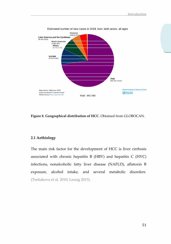
Attending to the geographical distribution providded by
GLOBOCAN 2018, a striking imbalance is observed, with the highest
incidence rates in Asia (72.5%), followed by Europe (9.8%), Africa
(7.7 %) and North America (5%), where the incidence is lower.
In last years the incidence of HCC has suffered a significant growth,
standing out that new cases of HCC increased by 75% between 1990
and 2015, mainly due to changes in the populationʹs age pyramid
and the demographic increase (Akinyemiju et al. 2017).
Cardioprotective
•Reduce systolic bloodpressure, TNF-α and IL-1β
•IncreaseSOD
•Inhibits COX-2 and LDL oxidation in endothelial cells
•Activate AMPK
•Upregulate eNOS
Cancer
• Activate apoptosis
• Inhibit angiogenesis and proliferation
Neuroprotection
• Decrease amyloidplaques in brain,cerebral infarctvolume and neuronal ROS generation
• Inhibitcholinesterases
Obesity
• Inhibit lipogenesis
• Increase lipolysis
• Activte AMPK, SIRTand PGC-1α
Diabetes
•Enhace insulinsensivity
•Increaseglucoseuptake and AMPK-dependentmitochondrialbiogenesis
•Decrease ROSgeneration
Introduction
51
Figure 8. Geographical distribution of HCC. Obtained from GLOBOCAN.
2.1 Aethiology
The main risk factor for the development of HCC is liver cirrhosis
associated with chronic hepatitis B (HBV) and hepatitis C (HVC)
infections, nonalcoholic fatty liver disease (NAFLD), aflatoxin B
exposure, alcohol intake, and several metabolic disorders
(Tretiakova et al. 2010; Leung 2015).
Introduction
52
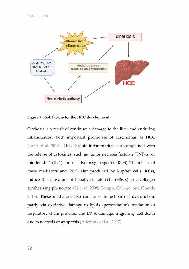
Figure 9. Risk factors for the HCC development.
Cirrhosis is a result of continuous damage to the liver and enduring
inflammation, both important promoters of carcinomas as HCC
(Tang et al. 2018). This chronic inflammation is accompained with
the release of cytokines, such as tumor necrosis factor‐α (TNF‐α) or
interleukin‐1 (IL‐1) and reactive oxygen species (ROS). The release of
these mediators and ROS, also produced by kupffer cells (KCs),
induce the activation of hepatic stellate cells (HSCs) to a collagen
synthesizing phenotype (Li et al. 2008; Campo, Gallego, and Grande
2018). These mediators also can cause mitochondrial dysfunction,
partly via oxidative damage to lipids (peroxidation), oxidation of
respiratory chain proteins, and DNA damage, triggering cell death
due to necrosis or apoptosis (Ademowo et al. 2017).

Introduction
53
Figure 10. Most frequent causes of HCC
Approximately 54% of cases can be attributed to HBV infection
worldwide, while 31% can be attributed to HCV infection, and the
remaining 15% associated with other causes (Tang et al. 2018). Both
hepatitis viruses promote the development of HCC by oncogenic
viral protein expression that carry on the mutations and malignant
transformation of infected cells. In addition, HVC induces hepatic
inflammation and fibrosis, both of which contribute to
carcinogenesis and apoptosis, as mentioned before (J. D. Yang et al.
2011; Lemon and McGivern 2012). In 2018, 54.5% of HCC worldwide
cases were attributable to HBV infection, while 31.2% were to HCV
infection (de Martel et al. 2020). The risk of development HCC
among those with chronic HVB or HVC infection increases with co‐
infection, male sex, older age, chronic alcohol or tobacco use,
aflatoxin exposition, diabetes and obesity (Rapti 2015).
Despite alcohol abuse, non‐alcoholic fatty liver disease (NAFLD) is a
major risk also for HCC development. In patients with NAFLD an

Introduction
54
excess of triglycerides accumulates in liver cells and, along other
manifestations such as obesity and type‐2 diabetes mellitus, results
in non‐alcohol steatosis (NASH) and, finally, in cirrhosis
development (Massarweh and El‐Serag 2017).
Aflatoxins are secondary metabolites produced by Aspergillus flavus
and Aspergillus parasiticus that contaminate a wide variety of maize,
groundnuts, wheat, soy and rice, especially is countries with a warm
and humid environment (Valdes 2017). Aflatoxin is mainly
metabolized in the liver, and generates hypermutation in p53 tumor
suppression gene, that could lead the HCC development (Adam et
al. 2017).
2.2 HCC treatments
The actual EASL Clinical Practice Guidelines (Galle et al. 2018),
propose a modified Barcelona Clinic Liver Cancer (BCLC) staging
system and treatment strategy, in which patients with HCC are
classified into five stages according to pre‐established prognosis
variables, and therapies are allocated according to treatment‐related
status.
During the early stages of HCC, surgical approaches like local
ablation, liver resection or liver transplantation, and nonsurgical
approaches such as chemoembolization, are the most common
treatments. However, in patients with advanced cancer‐related

Introduction
55
symptoms like macroscopic vascular invasion, lymph node
involvement or metastases, other approaches are necessary. In 2007,
the Food and Drug Administration (FDA) approved the use of
Sorafenib (a multi‐tyrosine kinase inhibitor) in patients with
advanced HCC. The great results of Sorafenib supported the
searching and evaluation of other first‐line therapies as Lenvatinib,
second‐line therapies as Regorafenib, Cabozantinib, Ramucirumab
and immune checkpoint inhibitors as Nivolumab or Pembrolizumab
(Montironi, Montal, and Llovet 2019).
Figure 11. Modified BCLC staging system and treatment strategy. EASL
Clinical Practice Guidelines.
In spite of being the standard treatment for advanced HCC,
Sorafenib efficacy is moderate, with a tumor response of around
30%, and an average survival rate increased by 3‐5 months (Llovet et

Introduction
56
al. 2008). Generally, it is well‐tolerated, but some patients show
severe toxicities, such as skin reactions, diarrhea, fatigue or liver
dysfunction and in rare cases, but fateful, cardiovascular events,
arterial thromboembolic events and bleeding (Llovet et al. 2008;
Kane et al. 2009; Philip 2009). Due to the side effects, in many cases
the Sorafenib treatment is dose‐reduced or discontinued (Philip
2009) and no alternative effective therapeutics are available after the
failure of treatment with Sorafenib (Bahman et al. 2018).
3. Molecular mechanisms of HCC
HCC is a multistep process and the molecular basis of HCC
progression may differ depending on diverse factors and a number
of mechanisms might be involved (Alotaibi et al. 2016). As
mentioned, in majority of cases, HCC occurs as result of cirrhosis.
The cirrhotic nodules support a favorable microenvironment for the
malignant transformation of hepatocytes which leads to HCC
development through the successive accumulation of genetic and
epigenetic changes (Alqahtani et al. 2019).

Introduction
57
Figure 12. The multistep process of the development of liver cancer. The
risk factors, inflammatory cytokines and chemokines, and
hepatocarcinogenic pathways are related to the inflammation‐cancer
transformation during the development of primary liver cancer. From
(Chen et al. 2018).
3.1 Inflammation
Exposure to HBV or HVC viruses, aflatoxins or lipopolysaccharide
(LPS), triggers hepatic inflammation. As result of this chronic
damage, the homeostasis of liver parenchymal cells is upset and
macrophages and mast cells immediately triggers the inflammatory
process by releasing soluble mediators as cytokines, chemokines,
ROS and bioactive mediators as histamine, which induce leukocytes
mobilization and infiltration at the site of injury (B. Vendramini‐
Costa and E. Carvalho 2012). Chronic inflammation leads to
excessive tissue remodeling, loss of tissue architecture and
modifications on DNA and proteins as consequence of oxidative
stress, enhancing the risks for cancer development (Chen et al. 2018).

Introduction
58
Many of the immune cells, including macrophages, neutrofils and
eoisinofils, are involved directly or indirectly in pathology of chronic
inflammation by cytokine production. Tumor necrosis factor‐α
(TNF‐α) and interleukin family (IL‐6, IL‐1β) are well known
cytokines. IL‐6 is produced by activated kupffer cells in chronic
heptatitis. It promotes local inflammatory response and activates
hepatocyte proliferation leading to cancerous hepatocytes (Naugler
and Karin 2008). IL‐1β is found to promote hepatic stellate cell
proliferation, activation and transdifferentiation into the
myofibroblastic phenotype (Han et al. 2004). This cytokine can
promote hepatic inflammation by inducing the production of C‐
reactive protein, a sensitive marker of infection and inflammation,
independently of IL‐6 (Weinhold and RüTher 1997, 6). It has been
identified that TNF‐α causes DNA damage through the induction of
ROS (Montfort et al. 2019). Recently, (Jing et al. 2018), have described
that the overexpression of TNF‐α promotes HCC through the
activation of hepatic progenitor cells (HPCs); the knocking down of
TNF‐α inhibited the HPC activation and proliferation, which reduces
tumor incidence. All these data confirmed that TNF‐α plays an
important role in liver injury and prognosis.
3.1.1 NF‐κB pathway
NF‐κB is a transcription factor that regulates innate immunity and
inflammation. It has been demonstrated to play an essential role in

Introduction
59
the regulation of inflammatory signaling pathways in the liver (Xiao
and Ghosh 2005) (Muriel 2009), being a critical link between
inflammation and cancer (Taniguchi and Karin 2018).
NF‐κB complex is constituted by five proteins: RelA (p65), c‐Rel,
RelB, NF‐κB1 (p50/p105), and NF‐κB2 (p52/p100) (Ghosh and
Karin 2002). In physiological conditions, the dimmer formed by the
p50 and p65 subunits rests inactive in the cytoplasm through binding
to the inhibitory protein of NF‐κB (IκB) (Hoffmann and
Baltimore 2006). In response to pro‐inflammatory stimuli, including
inflammatory cytokines (such as TNF‐α and IL‐1β), the IκB kinase
(IKK) complex phosphorylates IκB and promotes its degradation by
the proteasome (Karin and Ben‐Neriah 2000; West et al. 2006).
Consequently, the activated NF‐κB dimmer is released and
translocates to the nucleus. There, binds specific DNA sequences,
and stimulates transcription of several target genes involved in
inflammation, immune responses, cell proliferation, and cell survival
(Pahl 1999; Ghosh and Karin 2002).
3.2 Free radicals: ROS and RNS
Free radicals are molecules containing unpaired electrons in their
orbitals, and are capable of have independent existence. They act as
redox signaling molecules and they are extremely unstable and
highly reactive. Cells produce two different types of redox signaling
molecules: reactive oxygen species (ROS), such as the superoxide

Introduction
60
anion radical (O2−), hydrogen peroxide (H2O2), and the hydroxyl
radical (•OH) and reactive nitrogen species (RNS), such as the nitric
oxide radical (•NO), the nitrogen dioxide radical (•NO2), and
peroxynitrite (ONOO−) (Nathan and Ding 2010).
3.2.1 ROS
Reactive oxygen species (ROS) are oxygen‐containing radicals that
are produced by organisms as a result of normal cellular metabolism.
They have an important role in physiological cell processes. Under
normal physiological conditions, antioxidants are able to modulate
the internal levels of ROS. However, several types of stress, as
cytokines produced by chronic inflammation, leads that the ROS
production overpasses the capacity to scavenge them. In such cases,
they trigger detrimental modifications to lipids, proteins and DNA,
leading a pathological cell process. This unbalance between oxidant
and antioxidant capacity is known as “oxidative stress.”

Introduction
61
Figure 13. Imbalance between ROS production and ROS scavengers
produce oxidative stress, which is dangerous for the fate of the cell.
The superoxide anion radical (O•−2) hydrogen peroxide (H2O2), and
the hydroxyl radical (•OH) are the more common ROS. These
molecules are produced as by‐products during the mitochondrial
electron transport, or by oxydoreductase enzymes and metal
catalyzed oxidation. Cells have a variety of defense mechanisms to
ameliorate their harmful effects. Among antioxidants are included
enzymatic scavengers as superoxide dismutase (SOD), catalase and
glutathione peroxidase, as well as non‐enzymatic scavengers such as
vitamins C and E, glutathione (GSH), lipoic acid, carotenoids and
iron chelators (Redza‐Dutordoir and Averill‐Bates 2016).
ROS production
ROS scavengers
Oxidativestress

Introduction
62
SOD is the responsible of catalyze the transformation of two
superoxide anions into a molecule of hydrogen peroxide (H2O2) and
oxygen (O2). In eukaryotic peroxisomes, the enzyme catalase
converts H2O2 to H2O and O2, and thus concludes the detoxification
initiated by SOD. Glutathione peroxidase is a family of enzymes
containing selenium, which also catalyze the degradation of
hydrogen peroxide, as well as break down organic peroxides to
alcohol.
In addition to ROS, RNS also play an important role. In
physiological conditions these reactive species are detoxified,
however, when they are overproduced, as occurs in many diseases
related to chronic inflammation, these reactive species can cause
nitrative and oxidative DNA damage (Cerutti and Trump 1991).
Nitric oxide (NO) is an important mediator between chronic
inflammation and hepatocarcinoma, which is produced by hepatic
parenchymal and non‐parenchymal cells through the inducible nitric
oxide synthase (iNOS) (Bishayee 2014). The expression of iNOS is
induced in a wide variety of tissues in response to endotoxin,
endogenous mediators of inflammation, and other stimuli such as
hypoxia (Weigert and Brüne 2008). In the presence of important level
of O2.−, the NO generated by iNOS may react with the radical in a n‐
enzymatic reaction to generate peroxynitrite (ONOO−). All the
reactive species, at a non‐counterbalanced concentration, damage

Introduction
63
macromolecules, and ultimately drive cells to death by processes
such as apoptosis and/or necrosis (Halliwell 2011).
Figure 14. Major pathways of cellular ROS/RNS generation and
detoxification. Antioxidant elements are represented in italics characters.
Blunted grey arrows represent an inhibitory effect against oxidative stress
by antioxidant enzymes or by antioxidant compounds. (L. Poirrier et al.
2010).
3.3 Cell death
Cell death includes two broadly defined mechanisms: programmed
cell death and necrosis (Fuchs and Steller 2011). While necrosis is the
result of an unordered cellular explosion in response to severe and

Introduction
64
irresistible trauma (Jan and Chaudhry 2019), programmed cell death
is a genetically programmed process of cell suicide in response to
particular signals (Gorski and Marra 2002; Fuchs and Steller 2011). It
is an extended error to equate programmed cell death with
apoptosis, despite of existing non‐apoptotic forms of programmed
cell death. Therefore, programmed cell death and apoptosis could
not be used as synonymous (Jan and Chaudhry 2019).
Figure 15. General mode of cancer cell death. Cancer cell death involves
two broadly defined mechanisms: programmed cell death and necrosis.
Programmed cell death mainly apoptosis and nonapoptosis base cell death,
such as autophagy, necroptosis and apoptosis like programmed cell death.
(Jan and Chaudhry 2019).

Introduction
65
3.3.1 Apoptosis
Apoptosis is a complex process that proceeds through at least two
main pathways, extrinsic and intrinsic pathways. Both of them
converge on the execution pathway which is initiated by the
activation by cleaving of caspase‐3, resulting in DNA fragmentation,
degradation of cytoskeletal and nuclear proteins, crosslinking of
proteins, formation of apoptotic bodies, expression of ligand for
phagocytic cell receptors and, finally, uptake by phagocytic cells
(Elmore 2007).
Caspases are the main effectors of apoptosis and belongs to a family
of proteins with proteolytic activity because they are able to cleave
proteins in aspartic acid residues (McIlwain, Berger, and Mak 2013).
Caspases are widely expressed as zymogens in most cells, and once
proteolytically processed can activate other procaspases, leading to a
cascade by which the apoptotic signal is amplified and results in cell
death promotion (Elmore 2007). These enzymes are classified in
three types according to their functions. Caspases‐1, 5, 12 and 14
have an important role as inflammation mediators. In relation with
apoptosis, capases‐2, 8, 9 and 10 play a role as apoptosis initiators,
while caspases‐3, 6 and 7 are executioner caspases (Behzadi and
Ranjbar 2015).

Introduction
66
Figure 16. Pathways of apoptosis. Apoptosis consists of two main
pathways. Extrinsic pathway triggered by external stimuli or ligand
molecules and particularly involved death receptors (DRs). The intrinsic
pathway is mediated by Bax/Bak insertion into mitochondrial membrane.
Subsequently, cytochrome c is released, which combines with Apaf–1 and
procaspase‐9 to produce apoptosome followed by the activation of caspase‐
3 cascade of apoptosis. TNF related apoptosis inducing ligand (TRAIL),
cellular FLICE inhibitory proteins (cFLIP), truncated bid (tBid), B‐cell
lymphoma protein 2 (Bcl‐2), Bcl‐2 homologue splice variants (Bcl‐xL),
cytochrome (Cyt C), second mitochondrial activator of caspases (SMAC),
inhibitor of apoptosis proteins (IAPs). (Jan and Chaudhry 2019).

Introduction
67
3.3.1.1 Extrinsic pathway
This apoptotic pathway is activated when extracellular ligands such
as TNF‐α, Fas ligand (Fas‐L) and TNF‐related apoptosis‐inducing
ligand (TRAIL) recognize the extracellular domain of the death
receptors (DR) and active them. This activation results in the
formation of a death‐inducing signaling complex (DISC) (J. W. Kim,
Choi, and Joe 2000). Stimulation of death receptors results in
activation of the initiator caspase‐8, which can propagate the
apoptosis signal by direct cleavage of downstream effector caspases
such as caspase‐3 (Walczak and Krammer 2000). Death receptors
mediated apoptosis can be inhibited by a protein named c‐FLIP
(Guicciardi and Gores 2009). There is a link between the extrinsic
and intrinsic pathway that starts when active caspase‐8 cleaves Bid
and truncated Bid interacts with mitochondria. From this point is not
possible distinguish between the two apoptotic pathways (Kantari
and Walczak 2011).
3.3.1.2 Intrinsic pathway
This apoptosis pathway is mediated mainly by mitochondria. The
intrinsic pathway is triggered by extra and intra‐cellular stresses like
oxidative stress, irradiation and treatment with cytotoxic drugs (Jan
and Chaudhry 2019). These stimuli causes changes in the inner
mitochondrial membrane that leads the Bax/Bak insertion into
mitochondrial membrane and subsequently, cytochrome c release
Introduction
68
from the mitochondrial membrane space into the cytosol (R. Kim
2005). In non‐apoptotic conditions, the anti‐apoptotic proteins
(members of Bcl‐2 family) prevents the release of cytochrome c (Cory
and Adams 2002). The tumor suppressor p53 has a critical role in
regulation the expression of Bcl‐2 family proteins. p53 increases the
ratio pro‐apoptotic (Bax, Bad, Bid)/anti‐apoptotic (Bcl‐2, Bcl‐x, Bcl‐
XL) proteins triggering the release of cytochrome c, caspase
activation and finally apoptosis (Fridman and Lowe 2003). The
release of cytochrome c into the cytosol triggers caspase‐3 activation
through formation of the cytochrome c/Apaf‐1/caspase‐9‐containing
apoptosome complex, whereas Smac/DIABLO promote caspase
activation through neutralizing the inhibitory effects to the inhibitors
of the apoptosis proteins (IAPs) (Fulda and Debatin 2006).

Introduction
69
4. References
Adam, Mowaffaq Adam Ahmed, Yasser M. Tabana, Khirun Binti Musa, andDoblin Anak Sandai. 2017. “Effects of Different Mycotoxins onHumans, Cell Genome and Their Involvement in Cancer.” OncologyReports37(3):1321–36.https://doi.org/10.3892/or.2017.5424.
Ademowo,O.S.,H.K. I.Dias,D.G.A.Burton,andH.R.Griffiths.2017. “Lipid(per) Oxidation in Mitochondria: An Emerging Target in the AgeingProcess?” Biogerontology 18 (6): 859–79.https://doi.org/10.1007/s10522‐017‐9710‐z.
Aharoni, Asaph, and Gad Galili. 2011. “Metabolic Engineering of the PlantPrimary–Secondary Metabolism Interface.” Current Opinion inBiotechnology 22 (2): 239–44.https://doi.org/10.1016/j.copbio.2010.11.004.
Aja, Iris, M. Begoña Ruiz‐Larrea, Arnaud Courtois, Stéphanie Krisa, TristanRichard, and José‐Ignacio Ruiz‐Sanz. 2020. “Screening of NaturalStilbene Oligomers from Vitis Vinifera for Anticancer Activity onHuman Hepatocellular Carcinoma Cells.” Antioxidants 9 (6): 469.https://doi.org/10.3390/antiox9060469.
Akdis,Mübeccel,AlarAab,CanAltunbulakli,KursatAzkur,RitaA.Costa,RetoCrameri, Su Duan, et al. 2016. “Interleukins (from IL‐1 to IL‐38),Interferons, Transforming Growth Factor β, and TNF‐α: Receptors,Functions, and Roles in Diseases.” Journal of Allergy and ClinicalImmunology 138 (4): 984–1010.https://doi.org/10.1016/j.jaci.2016.06.033.
Akinwumi, Bolanle, Kimberly‐Ann Bordun, and Hope Anderson. 2018.“BiologicalActivitiesofStilbenoids.”InternationalJournalofMolecularSciences19(3):792.https://doi.org/10.3390/ijms19030792.
Akinyemiju, Tomi, Semaw Abera, Muktar Ahmed, Noore Alam, MulubirhanAssefaAlemayohu,ChristineAllen,RajaaAl‐Raddadi,etal.2017.“TheBurdenofPrimaryLiverCancerandUnderlyingEtiologiesFrom1990to2015attheGlobal,Regional,andNationalLevel:ResultsFromtheGlobal Burden ofDisease Study 2015.” JAMAOncology 3 (12): 1683.https://doi.org/10.1001/jamaoncol.2017.3055.
Alotaibi,Hani,NeseAtabey,KasımDiril,EsraErdal,andMehmetOzturk.2016.“MolecularMechanismsofHepatocellularCarcinoma.”HepatocellularCarcinoma,43–63.https://doi.org/10.1007/978‐3‐319‐34214‐6_3.

Introduction
70
Alqahtani, Khan, Alloghbi, Said Ahmed, Ashraf, and Hammouda. 2019.“Hepatocellular Carcinoma: Molecular Mechanisms and TargetedTherapies.” Medicina 55 (9): 526.https://doi.org/10.3390/medicina55090526.
Asenstorfer, Robert E., Andrew J. Markides, Patrick G. Iland, and Graham P.Jones. 2003. “Formation of Vitisin A during RedWine FermentationandMaturation.”AustralianJournalofGrapeandWineResearch9(1):40–46.https://doi.org/10.1111/j.1755‐0238.2003.tb00230.x.
B.Vendramini‐Costa,D.,andJ.E.Carvalho.2012.“MolecularLinkMechanismsbetweenInflammationandCancer.”CurrentPharmaceuticalDesign18(26):3831–52.https://doi.org/10.2174/138161212802083707.
Baechler, Simone A., Anika Schroeter, Martina Dicker, Pablo Steinberg, andDoris Marko. 2014. “Topoisomerase II‐Targeting Properties of aGrapevine‐Shoot Extract and Resveratrol Oligomers.” Journal ofAgricultural and Food Chemistry 62 (3): 780–88.https://doi.org/10.1021/jf4046182.
Bahman, Abdulmajeed, Mohamed Abaza, Sarah Khoushiash, and RajaaAl‑Attiyah. 2018. “Sequence‑dependent Effect of Sorafenib inCombination with Natural Phenolic Compounds on Hepatic CancerCellsand thePossibleMechanismofAction.” International JournalofMolecularMedicine,June.https://doi.org/10.3892/ijmm.2018.3725.
Baur, Joseph A., and David A. Sinclair. 2006. “Therapeutic Potential ofResveratrol:The inVivoEvidence.”NatureReviewsDrugDiscovery5(6):493–506.https://doi.org/10.1038/nrd2060.
Behzadi,Payam,andRezaRanjbar.2015.“CaspasesandApoptosis.”MolecularEnzymology and Drug Targets 01 (02).https://doi.org/10.21767/2572‐5475.10006.
Bi,XiuLi, JingYuYang,YingXuDong, JiMingWang,YongHongCui,TakashiIkeshima,YuQingZhao,andChunFuWu.2005.“ResveratrolInhibitsNitric Oxide and TNF‐α Production by Lipopolysaccharide‐ActivatedMicroglia.” International Immunopharmacology 5 (1): 185–93.https://doi.org/10.1016/j.intimp.2004.08.008.
Biais, Benoit, Stéphanie Krisa, Stéphanie Cluzet, Grégory Da Costa, PierreWaffo‐Teguo, Jean‐Michel Mérillon, and Tristan Richard. 2017.“Antioxidant and Cytoprotective Activities of Grapevine Stilbenes.”Journal of Agricultural and Food Chemistry 65 (24): 4952–60.https://doi.org/10.1021/acs.jafc.7b01254.

Introduction
71
Bishayee, Anupam. 2014. “The Inflammation and Liver Cancer.” InInflammation and Cancer, edited by Bharat B. Aggarwal, BokyungSung,andSubashChandraGupta,816:401–35.Basel:SpringerBasel.https://doi.org/10.1007/978‐3‐0348‐0837‐8_16.
Bradford,MarionM.1976.“ARapidandSensitiveMethodfortheQuantitationofMicrogramQuantitiesofProteinUtilizing thePrincipleofProtein‐Dye Binding.” Analytical Biochemistry 72 (1–2): 248–54.https://doi.org/10.1016/0003‐2697(76)90527‐3.
Bronte‐Stewart,B.1958.“TheEffectOfDietaryFatsOnTheBloodLipidsAndTheirRelationToIschaemicHeartDisease.”BritishMedicalBulletin14(3):243–52.https://doi.org/10.1093/oxfordjournals.bmb.a069691.
Bush,Andrew.2019.“PathophysiologicalMechanismsofAsthma.”FrontiersinPediatrics7(March).https://doi.org/10.3389/fped.2019.00068.
Caggiu, Elisa, Giannina Arru, Sepideh Hosseini, Magdalena Niegowska,GianPietroSechi,IgnazioRobertoZarbo,andLeonardoA.Sechi.2019.“Inflammation,InfectiousTriggers,andParkinson’sDisease.”Frontiersin Neurology 10 (February).https://doi.org/10.3389/fneur.2019.00122.
Campo, José A Del, Paloma Gallego, and Lourdes Grande. 2018. “Role ofInflammatory Response in Liver Diseases: Therapeutic Strategies.”World Journal of Hepatology 10 (1): 1–7.https://doi.org/10.4254/wjh.v10.i1.1.
Cassidy, Aedin, Bryan Hanley, and Rosa M Lamuela‐Raventos. 2000.“Isoflavones, Lignans and Stilbenes ‐ Origins, Metabolism andPotentialImportancetoHumanHealth.”JournaloftheScienceofFoodand Agriculture 80 (7): 1044–62.https://doi.org/10.1002/(SICI)1097‐0010(20000515)80:7<1044::AID‐JSFA586>3.0.CO;2‐N.
Cerutti, P. A., and B. F. Trump. 1991. “Inflammation and Oxidative Stress inCarcinogenesis.”CancerCells(ColdSpringHarbor,N.Y.:1989)3(1):1–7.
Chalons,Pauline,SouheilaAmor,FlavieCourtaut,EmmaCantos‐Villar,TristanRichard, Cyril Auger, Philippe Chabert, Valérie Schni‐Kerth, VirginieAires, and Dominique Delmas. 2018. “Study of Potential Anti‐InflammatoryEffectsofRedWineExtractandResveratrol throughaModulationofInterleukin‐1‐BetainMacrophages.”Nutrients10(12):1856.https://doi.org/10.3390/nu10121856.

Introduction
72
Chang, Chi‐I,Wei‐Chu Chien, Kai‐XinHuang, and Jue‐LiangHsu. 2017. “Anti‐InflammatoryEffectsofVitisinolAandFourOtherOligostilbenesfromAmpelopsis Brevipedunculata Var. Hancei.”Molecules 22 (7): 1195.https://doi.org/10.3390/molecules22071195.
Chen,Hong‐Jin,Ming‐HuaHu,Fang‐GuiXu,Hao‐JunXu,Jun‐JunShe,andHong‐Ping Xia. 2018. “Understanding the Inflammation‐CancerTransformation in the Development of Primary Liver Cancer.”Hepatoma Research 4 (7): 29. https://doi.org/10.20517/2394‐5079.2018.18.
Cheng,ChakKwong,Jiang‐YunLuo,ChiWaiLau,Zhen‐YuChen,XiaoYuTian,and Yu Huang. 2020. “Pharmacological Basis and New Insights ofResveratrol Action in the Cardiovascular System.” British Journal ofPharmacology177(6):1258–77.https://doi.org/10.1111/bph.14801.
Chiou,Yi‐Siou,Mei‐LingTsai,KalyanamNagabhushanam,Yin‐JenWang,Chih‐HsiungWu,Chi‐TangHo,andMin‐HsiungPan.2011.“PterostilbeneIsMore Potent than Resveratrol in Preventing Azoxymethane (AOM)‐Induced Colon Tumorigenesis via Activation of the NF‐E2‐RelatedFactor 2 (Nrf2)‐Mediated Antioxidant Signaling Pathway.” Journal ofAgricultural and Food Chemistry 59 (6): 2725–33.https://doi.org/10.1021/jf2000103.
Cichocki, Michal, Jaroslaw Paluszczak, Hanna Szaefer, Adriana Piechowiak,AgnesM.Rimando,andWandaBaer‐Dubowska.2008.“PterostilbeneIs Equally Potent as Resveratrol in Inhibiting 12‐O‐Tetradecanoylphorbol‐13‐Acetate Activated NFκB, AP‐1, COX‐2, andINOSinMouseEpidermis.”MolecularNutrition&FoodResearch,June.https://doi.org/10.1002/mnfr.200700466.
Colin, Didier, Allan Lancon, Dominique Delmas, Gerard Lizard, JessicaAbrossinow, Edmond Kahn, Brigitte Jannin, and Norbert Latruffe.2008. “Antiproliferative Activities of Resveratrol and RelatedCompounds in Human Hepatocyte Derived HepG2 Cells AreAssociated with Biochemical Cell Disturbance Revealed byFluorescence Analyses.” Biochimie 90 (11–12): 1674–84.https://doi.org/10.1016/j.biochi.2008.06.006.
Colombo, Francesca, Chiara Di Lorenzo, Luca Regazzoni, Marco Fumagalli,EnricoSangiovanni,LuísPeresdeSousa,LuigiBavaresco,etal.2019.“Phenolic Profiles and Anti‐Inflammatory Activities of Sixteen TableGrape ( Vitis Vinifera L.) Varieties.” Food & Function 10 (4): 1797–1807.https://doi.org/10.1039/C8FO02175A.
Introduction
73
Coppa, Tania,Maria Claudia Lazzè, Ornella Cazzalini, Paola Perucca, RobertoPizzala, Livia Bianchi, Lucia Anna Stivala, et al. 2011. “Structure–ActivityRelationshipofResveratrolandItsAnalogue,4,4′‐Dihydroxy‐Trans ‐Stilbene, Toward the Endothelin Axis in Human EndothelialCells.” Journal of Medicinal Food 14 (10): 1173–80.https://doi.org/10.1089/jmf.2010.0272.
Cory,Suzanne,andJerryM.Adams.2002.“TheBcl2Family:RegulatorsoftheCellularLife‐or‐Death Switch.”NatureReviewsCancer 2 (9): 647–56.https://doi.org/10.1038/nrc883.
Courtois,Arnaud,ClaudeAtgié,AxelMarchal,RuthHornedo‐Ortega,CarolineLapèze, Chrystel Faure, Tristan Richard, and Stéphanie Krisa. 2018.“Tissular Distribution and Metabolism of Trans‐ε‐Viniferin afterIntraperitoneal Injection in Rat.” Nutrients 10 (11): 1660.https://doi.org/10.3390/nu10111660.
Courtois, Arnaud, Manon Garcia, Stéphanie Krisa, Claude Atgié, PatrickSauvant,TristanRichard,andChrystelFaure.2019.“Encapsulationofε‐Viniferin in Onion‐Type Multi‐Lamellar Liposomes Increases ItsSolubility and Its Photo‐Stability and Decreases Its Cytotoxicity onCaco‐2 Intestinal Cells.” Food & Function 10 (5): 2573–82.https://doi.org/10.1039/C9FO00420C.
Crozier, Alan, Indu B. Jaganath, and Michael N. Clifford. 2009. “DietaryPhenolics: Chemistry, Bioavailability and Effects on Health.”NaturalProductReports26(8):1001.https://doi.org/10.1039/b802662a.
Csiszar,Anna,NazarLabinskyy,AndrejPodlutsky,PawelM.Kaminski,MichaelS. Wolin, Cuihua Zhang, Partha Mukhopadhyay, et al. 2008.“Vasoprotective Effects of Resveratrol and SIRT1: Attenuation ofCigarette Smoke‐Induced Oxidative Stress and ProinflammatoryPhenotypic Alterations.” American Journal of Physiology‐Heart andCirculatory Physiology 294 (6): H2721–35.https://doi.org/10.1152/ajpheart.00235.2008.
Cutrim,CamilaSampaio,andMarcoAntonioSlobodaCortez.2018.“AReviewonPolyphenols:Classification,BeneficialEffectsandTheirApplicationinDairy Products.” International JournalofDairyTechnology 71 (3):564–78.https://doi.org/10.1111/1471‐0307.12515.
Dandona, P. 2004. “Inflammation: The Link between Insulin Resistance,Obesity and Diabetes.” Trends in Immunology 25 (1): 4–7.https://doi.org/10.1016/j.it.2003.10.013.

Introduction
74
Delmas, D, B Jannin, M Cherkaoui Malki, and N Latruffe. 2000. “InhibitoryEffect of Resveratrol on the Proliferation ofHuman andRatHepaticDerived Cell Lines.” Oncology Reports, July.https://doi.org/10.3892/or.7.4.847.
Djoko, Bambang, Robin Y.‐Y. Chiou, Jia‐Jen Shee, and Yi‐Wen Liu. 2007.“Characterization of Immunological Activities of Peanut Stilbenoids,Arachidin‐1, Piceatannol, and Resveratrol on Lipopolysaccharide‐Induced Inflammation of RAW 264.7 Macrophages.” Journal ofAgricultural and Food Chemistry 55 (6): 2376–83.https://doi.org/10.1021/jf062741a.
ElKhawand,Toni,ArnaudCourtois,JosepValls,TristanRichard,andStéphanieKrisa. 2018. “A Review of Dietary Stilbenes: Sources andBioavailability.” Phytochemistry Reviews 17 (5): 1007–29.https://doi.org/10.1007/s11101‐018‐9578‐9.
Elmore, Susan. 2007. “Apoptosis: A Review of Programmed Cell Death.”Toxicologic Pathology 35 (4): 495–516.https://doi.org/10.1080/01926230701320337.
Esatbeyoglu,Tuba,PhilippEwald,YoshiakiYasui,HarukaYokokawa,AnikaE.Wagner, Seiichi Matsugo, Peter Winterhalter, and Gerald Rimbach.2016. “Chemical Characterization, Free Radical Scavenging, andCellularAntioxidantandAnti‐InflammatoryPropertiesofaStilbenoid‐Rich Root Extract of Vitis Vinifera.” OxidativeMedicine and CellularLongevity2016:1–11.https://doi.org/10.1155/2016/8591286.
Espinoza,J.Luis,andPleiadesT.Inaoka.2017.“Gnetin‐CandOtherResveratrolOligomers with Cancer Chemopreventive Potential: ResveratrolOligomerswithAnticancerPotential.”AnnalsoftheNewYorkAcademyofSciences1403(1):5–14.https://doi.org/10.1111/nyas.13450.
Fridman, Jordan S, and ScottW Lowe. 2003. “Control of Apoptosis by P53.”Oncogene22(56):9030–40.https://doi.org/10.1038/sj.onc.1207116.
Fuchs,Yaron,andHermannSteller.2011.“ProgrammedCellDeath inAnimalDevelopment and Disease.” Cell 147 (4): 742–58.https://doi.org/10.1016/j.cell.2011.10.033.
Fulda, S, and K‐M Debatin. 2006. “Extrinsic versus Intrinsic ApoptosisPathways in Anticancer Chemotherapy.” Oncogene 25 (34): 4798–4811.https://doi.org/10.1038/sj.onc.1209608.

Introduction
75
Gabaston,Julien,ToniElKhawand,PierreWaffo‐Teguo,AlainDecendit,TristanRichard, Jean‐Michel Mérillon, and Roman Pavela. 2018. “Stilbenesfrom Grapevine Root: A Promising Natural Insecticide againstLeptinotarsaDecemlineata.” JournalofPestScience 91 (2): 897–906.https://doi.org/10.1007/s10340‐018‐0956‐2.
Galle,PeterR.,AlejandroForner, JosepM.Llovet,VincenzoMazzaferro,FabioPiscaglia, Jean‐Luc Raoul, Peter Schirmacher, and Valérie Vilgrain.2018. “EASL Clinical Practice Guidelines: Management ofHepatocellular Carcinoma.” Journal of Hepatology 69 (1): 182–236.https://doi.org/10.1016/j.jhep.2018.03.019.
Gao, S., and M. Hu. 2010. “Bioavailability Challenges Associated withDevelopment of Anti‐Cancer Phenolics.” Mini‐Reviews in MedicinalChemistry 10 (6): 550–67.https://doi.org/10.2174/138955710791384081.
Gordon, Siamon, and Annette Plüddemann. 2017. “Tissue Macrophages:Heterogeneity and Functions.” BMC Biology 15 (1).https://doi.org/10.1186/s12915‐017‐0392‐4.
Gorski,S.,andM.Marra.2002.“ProgrammedCellDeathTakesFlight:Geneticand Genomic Approaches to Gene Discovery in Drosophila.”Physiological Genomics 9 (2): 59–69.https://doi.org/10.1152/physiolgenomics.00114.2001.
Goszcz,Katarzyna,GarryGDuthie,DerekStewart,StephenJLeslie,andIanLMegson. 2017. “Bioactive Polyphenols and Cardiovascular Disease:Chemical Antagonists, Pharmacological Agents or Xenobiotics ThatDrive an Adaptive Response?: Bioactive Polyphenols andCardiovascular Disease.” British Journal of Pharmacology 174 (11):1209–25.https://doi.org/10.1111/bph.13708.
Guerrero, Raul F., Josep Valls‐Fonayet, Tristan Richard, and Emma Cantos‐Villar. 2020. “A Rapid Quantification of Stilbene Content inWine byUltra‐High Pressure Liquid Chromatography – Mass Spectrometry.”Food Control 108 (February): 106821.https://doi.org/10.1016/j.foodcont.2019.106821.
Guicciardi,MariaEugenia,andGregoryJ.Gores.2009.“LifeandDeathbyDeathReceptors.” The FASEB Journal 23 (6): 1625–37.https://doi.org/10.1096/fj.08‐111005.
Halliwell,Barry.2011.“FreeRadicalsandAntioxidants–QuoVadis?”TrendsinPharmacological Sciences 32 (3): 125–30.https://doi.org/10.1016/j.tips.2010.12.002.

Introduction
76
Han,Yuan‐Ping,LingZhou, JiaohongWang,ShigangXiong,WarrenL.Garner,SamuelW.French,andHidekazuTsukamoto.2004.“EssentialRoleofMatrix Metalloproteinases in Interleukin‐1‐Induced MyofibroblasticActivation of Hepatic Stellate Cell in Collagen.” Journal of BiologicalChemistry 279 (6): 4820–28.https://doi.org/10.1074/jbc.M310999200.
Harbeoui, H., A.Hichami,W. AidiWannes, J. Lemput,M. Saidani Tounsi, andN.A.Khan.2019.“Anti‐InflammatoryEffectofGrape(VitisViniferaL.)Seed Extract through the Downregulation of NF‐ΚB and MAPKPathways in LPS‐Induced RAW264.7 Macrophages.” South AfricanJournal of Botany 125 (September): 1–8.https://doi.org/10.1016/j.sajb.2019.06.026.
Hopkins,W.GHünerN.P. 2004. Introduction toPlantPhysiology. 3rdEdition.Inc.JohnWiley&Sons,Inc,Hoboken.
Huang,Fangfang,ZuisuYang,DiYu, JiabinWang,RongLi,andGuofangDing.2012. “Sepia Ink Oligopeptide Induces Apoptosis in Prostate CancerCellLinesviaCaspase‐3ActivationandElevationofBax/Bcl‐2Ratio.”Marine Drugs 10 (12): 2153–65.https://doi.org/10.3390/md10102153.
Hussain, S P, J Schwank, F Staib, X W Wang, and C C Harris. 2007. “TP53Mutations and Hepatocellular Carcinoma: Insights into the Etiologyand Pathogenesis of Liver Cancer.” Oncogene 26 (15): 2166–76.https://doi.org/10.1038/sj.onc.1210279.
Hussain, S. Perwez, and Curtis C. Harris. 1998. “Molecular Epidemiology ofHuman Cancer: Contribution of Mutation Spectra Studies of TumorSuppressorGenes.”CancerResearch58(18):4023.
Islam, Shamima, Ferdaus Hassan, Mya Mya Mu, Hiroyasu Ito, Naoki Koide,Isamu Mori, Tomoaki Yoshida, and Takashi Yokochi. 2004.“PiceatannolPreventsLipopolysaccharide(LPS)‐InducedNitricOxide(NO)ProductionandNuclearFactor(NF)‐ΚBActivationbyInhibitingIκB Kinase (IKK).” Microbiology and Immunology 48 (10): 729–36.https://doi.org/10.1111/j.1348‐0421.2004.tb03598.x.
Ivanova, Donika, Zhivko Zhelev, Severina Semkova, Ichio Aoki, and RumianaBakalova. 2019. “Resveratrol Modulates the Redox‐Status andCytotoxicity of Anticancer Drugs by Sensitizing LeukemicLymphocytes and Protecting Normal Lymphocytes.” AnticancerResearch 39 (7): 3745–55.https://doi.org/10.21873/anticanres.13523.

Introduction
77
Jan, Rehmat, and Gul‐e‐Saba Chaudhry. 2019. “Understanding Apoptosis andApoptotic Pathways Targeted Cancer Therapeutics.” AdvancedPharmaceutical Bulletin 9 (2): 205–18.https://doi.org/10.15171/apb.2019.024.
Jiang, Yu, Qiu‐Ju Han, and Jian Zhang. 2019. “Hepatocellular Carcinoma:Mechanisms of Progression and Immunotherapy.”World Journal ofGastroenterology 25 (25): 3151–67.https://doi.org/10.3748/wjg.v25.i25.3151.
Jing,Yingying,KaiSun,WentingLiu,DandanSheng,ShanminZhao,LuGao,andLixinWei. 2018. “TumorNecrosis Factor‐α PromotesHepatocellularCarcinogenesis through the Activation of Hepatic Progenitor Cells.”Cancer Letters 434 (October): 22–32.https://doi.org/10.1016/j.canlet.2018.07.001.
Johansson,Martin,and JennyPersson.2008. “CancerTherapy:TargetingCellCycle Regulators.” Anti‐Cancer Agents inMedicinal Chemistry 8 (7):723–31.https://doi.org/10.2174/187152008785914833.
Kane,R.C.,A.T.Farrell,R.Madabushi,B.Booth,S.Chattopadhyay,R.Sridhara,R. Justice, and R. Pazdur. 2009. “Sorafenib for the Treatment ofUnresectable Hepatocellular Carcinoma.” TheOncologist 14 (1): 95–100.https://doi.org/10.1634/theoncologist.2008‐0185.
Kantari,Chahrazade,andHenningWalczak.2011.“Caspase‐8andBid:CaughtintheActbetweenDeathReceptorsandMitochondria.”BiochimicaetBiophysica Acta (BBA) ‐Molecular Cell Research 1813 (4): 558–63.https://doi.org/10.1016/j.bbamcr.2011.01.026.
Kapetanovic,IzetM.,MiguelMuzzio,ZhihuaHuang,ThomasN.Thompson,andDavid L. McCormick. 2011. “Pharmacokinetics, Oral Bioavailability,and Metabolic Profile of Resveratrol and Its Dimethylether Analog,Pterostilbene, in Rats.” Cancer Chemotherapy and Pharmacology 68(3):593–601.https://doi.org/10.1007/s00280‐010‐1525‐4.
Keylor,M.H.,B.S.Matsuura,M.Griesser, J.‐P.R.Chauvin,R.A.Harding,M.S.Kirillova,X.Zhu,O.J.Fischer,D.A.Pratt,andC.R.J.Stephenson.2016.“Synthesis of Resveratrol Tetramers via a Stereoconvergent RadicalEquilibrium.” Science 354 (6317): 1260–65.https://doi.org/10.1126/science.aaj1597.
Khan,Md. Asaduzzaman, Han‐chun Chen, Xin‐xingWan,Mousumi Tania, Ai‐hua Xu, Fang‐zhi Chen, and Dian‐zheng Zhang. 2013. “Erratum to‘Regulatory Effects of Resveratrol on Antioxidant Enzymes: AMechanism of Growth Inhibition and Apoptosis Induction in CancerCells.’” Molecules and Cells 35 (4): 355–355.https://doi.org/10.1007/s10059‐013‐1259‐3.
Introduction
78
Khoury, Laure, Daniel Zalko, and Marc Audebert. 2015. “Evaluation of FourHuman Cell Lines with Distinct Biotransformation Properties forGenotoxic Screening.” Mutagenesis, August, gev058.https://doi.org/10.1093/mutage/gev058.
Kim,JinWoo,Eui‐JuChoi,andCheolOJoe.2000.“ActivationofDeath‐InducingSignaling Complex (DISC) by pro‐Apoptotic C‐Terminal Fragment ofRIP.” Oncogene 19 (39): 4491–99.https://doi.org/10.1038/sj.onc.1203796.
Kim,Jiseon,JeeSunMin,DoyunKim,YuFenZheng,KarabasappaMailar,WonJun Choi, Choongho Lee, and Soo Kyung Bae. 2017. “A Simple andSensitive Liquid Chromatography–Tandem Mass SpectrometryMethodforTrans‐ε‐ViniferinQuantificationinMousePlasmaandItsApplication to a Pharmacokinetic Study in Mice.” Journal ofPharmaceutical and Biomedical Analysis 134 (February): 116–21.https://doi.org/10.1016/j.jpba.2016.11.044.
Kim, Ryungsa. 2005. “Recent Advances in Understanding the Cell DeathPathwaysActivatedbyAnticancerTherapy.”Cancer103(8):1551–60.https://doi.org/10.1002/cncr.20947.
Kunst, Claudia, Marika Haderer, Sebastian Heckel, Sophie Schlosser, andMartinaMüller.2016.“TheP53FamilyinHepatocellularCarcinoma.”Translational Cancer Research 5 (6): 632–38.https://doi.org/10.21037/tcr.2016.11.79.
Kurin,Elena,AtanasAtanasov,OliverDonath,ElkeHeiss,VerenaDirsch, andMilanNagy. 2012. “Synergy Study of the Inhibitory Potential of RedWine Polyphenols on Vascular Smooth Muscle Cell Proliferation.”Planta Medica 78 (08): 772–78. https://doi.org/10.1055/s‐0031‐1298440.
L. Poirrier, A., J. Pincemail, P. Van Den Ackerveken, P. P. Lefebvre, and B.Malgrange.2010.“OxidativeStressintheCochlea:AnUpdate.”CurrentMedicinal Chemistry 17 (30): 3591–3604.https://doi.org/10.2174/092986710792927895.
Langcake, P., and R. J. Pryce. 1977. “A New Class of Phytoalexins fromGrapevines.” Experientia 33 (2): 151–52.https://doi.org/10.1007/BF02124034.
Leger, A.S.St, A.L Cochrane, and F Moore. 1979. “Factors Associated WithCardiacMortality In Developed CountriesWith Particular ReferenceTo The Consumption Of Wine.” The Lancet 313 (8124): 1017–20.https://doi.org/10.1016/S0140‐6736(79)92765‐X.

Introduction
79
Lemon, Stanley M., and David R. McGivern. 2012. “Is Hepatitis C VirusCarcinogenic?” Gastroenterology 142 (6): 1274–78.https://doi.org/10.1053/j.gastro.2012.01.045.
Leung, Christopher. 2015. “Characteristics of Hepatocellular Carcinoma inCirrhoticandNon‐CirrhoticNon‐AlcoholicFattyLiverDisease.”WorldJournal of Gastroenterology 21 (4): 1189.https://doi.org/10.3748/wjg.v21.i4.1189.
Li,Jing‐Ting,Zhang‐XiuLiao,JiePing,DanXu,andHuiWang.2008.“MolecularMechanism of Hepatic Stellate Cell Activation and AntifibroticTherapeutic Strategies.” Journal of Gastroenterology 43 (6): 419–28.https://doi.org/10.1007/s00535‐008‐2180‐y.
Liao,Weiguo,JieLiu,BinLiu,XiaojieHuang,YongxinYin,DeCai,MingyiLi,andRunzhiZhu.2018.“JIB‑04InducesCellApoptosisviaActivationoftheP53/Bcl‑2/CaspasePathwayinMHCC97HandHepG2Cells.”OncologyReports,September.https://doi.org/10.3892/or.2018.6737.
Libby, Peter, and Sebastian Kobold. 2019. “Inflammation: A CommonContributor to Cancer, Aging, and Cardiovascular Diseases—Expanding theConceptofCardio‐Oncology.”CardiovascularResearch115(5):824–29.https://doi.org/10.1093/cvr/cvz058.
Link, Tim, and Tomoo Iwakuma. 2017. “Roles of P53 in Extrinsic Factor‐Induced Liver Carcinogenesis.” Hepatoma Research 3 (6): 95.https://doi.org/10.20517/2394‐5079.2017.07.
Llovet, Josep M., Sergio Ricci, Vincenzo Mazzaferro, Philip Hilgard, EdwardGane, Jean‐Frédéric Blanc, Andre Cosme de Oliveira, et al. 2008.“Sorafenib in Advanced Hepatocellular Carcinoma.” New EnglandJournal of Medicine 359 (4): 378–90.https://doi.org/10.1056/NEJMoa0708857.
Loisruangsin,Arthorn,KiyomiHikita,NorikazuSeto,MasatakeNiwa,YoshiakiTakaya,andNorioKaneda.2019.“StructuralAnalysisoftheInhibitoryEffects of Polyphenols, (+)‐hopeaphenol and (−)‐isohopeaphenol, onHuman SIRT1.” BioFactors 45 (2): 253–58.https://doi.org/10.1002/biof.1479.
Loupit,Grégoire,SylvainPrigent,CélineFranc,GillesDeRevel,TristanRichard,Sarah Jane Cookson, and Josep Valls Fonayet. 2020. “PolyphenolProfiles of Just Pruned Grapevine Canes fromWild Vitis Accessionsand Vitis Vinifera Cultivars.” Journal of Agricultural and FoodChemistry,April.https://doi.org/10.1021/acs.jafc.9b08099.

Introduction
80
Luo,Hongmei,AiminYang,BradleyA.Schulte,MichaelJ.Wargovich,andGavinY.Wang. 2013. “Resveratrol Induces Premature Senescence in LungCancerCellsviaROS‐MediatedDNADamage.”EditedbyAamirAhmad.PLoS ONE 8 (3): e60065.https://doi.org/10.1371/journal.pone.0060065.
Manach,Claudine,AugustinScalbert,ChristineMorand,ChristianRémésy,andLiliana Jiménez. 2004. “Polyphenols: Food Sources andBioavailability.”TheAmericanJournalofClinicalNutrition79(5):727–47. https://doi.org/10.1093/ajcn/79.5.727.
Martel,Catherinede,DamienGeorges,FreddieBray,JacquesFerlay,andGaryMClifford.2020. “GlobalBurdenofCancerAttributable to Infectionsin2018:AWorldwideIncidenceAnalysis.”TheLancetGlobalHealth8(2):e180–90.https://doi.org/10.1016/S2214‐109X(19)30488‐7.
Martín,AntonioRamón,IsabelVillegas,MarinaSánchez‐Hidalgo,andCatalinaAlarcónde laLastra.2006.“TheEffectsofResveratrol,aPhytoalexinDerived from Red Wines, on Chronic Inflammation Induced in anExperimentally Induced Colitis Model.” British Journal ofPharmacology 147 (8): 873–85.https://doi.org/10.1038/sj.bjp.0706469.
Martinez, Javier, and Juan J Moreno. 2000. “Effect of Resveratrol, a NaturalPolyphenolic Compound, on Reactive Oxygen Species andProstaglandinProduction.”BiochemicalPharmacology59(7):865–70.https://doi.org/10.1016/S0006‐2952(99)00380‐9.
Massarweh, Nader N., and Hashem B. El‐Serag. 2017. “Epidemiology ofHepatocellular Carcinoma and Intrahepatic Cholangiocarcinoma.”Cancer Control 24 (3): 107327481772924.https://doi.org/10.1177/1073274817729245.
Matsuura,BryanS.,MitchellH.Keylor,BoLi,YuXuanLin,ShelbyAllison,DerekA. Pratt, and Corey R. J. Stephenson. 2015. “A Scalable BiomimeticSynthesis of Resveratrol Dimers and Systematic Evaluation of TheirAntioxidant Activities.” Angewandte Chemie International Edition 54(12):3754–57.https://doi.org/10.1002/anie.201409773.
McIlwain, D. R., T. Berger, and T. W. Mak. 2013. “Caspase Functions in CellDeathandDisease.”ColdSpringHarborPerspectives inBiology5 (4):a008656–a008656.https://doi.org/10.1101/cshperspect.a008656.

Introduction
81
MiJeongSung,MunkhtugsDavaatseren,WonKim,SungKwangPark,Soon‐HeeKim, Haeng Jeon Hur, Myung Sunny Kim, Young‐Sup Kim, and DaeYoungKwon.2009.“VitisinASuppressesLPS‐InducedNOProductionby Inhibiting ERK, P38, and NF‐ΚB Activation in RAW 264.7 Cells.”International Immunopharmacology 9 (3): 319–23.https://doi.org/10.1016/j.intimp.2008.12.005.
Mishra,BhuwanB.,andVinodK.Tiwari.2011.“NaturalProducts:AnEvolvingRole in Future Drug Discovery.” European Journal of MedicinalChemistry 46 (10): 4769–4807.https://doi.org/10.1016/j.ejmech.2011.07.057.
Mittal, Sahil, andHashemB. El‐Serag. 2013. “Epidemiology ofHepatocellularCarcinoma: Consider the Population.” Journal of ClinicalGastroenterology 47 (July): S2–6.https://doi.org/10.1097/MCG.0b013e3182872f29.
Montfort, Anne, Céline Colacios, Thierry Levade, Nathalie Andrieu‐Abadie,NicolasMeyer, and Bruno Ségui. 2019. “The TNF Paradox in CancerProgressionandImmunotherapy.”Frontiers inImmunology10(July).https://doi.org/10.3389/fimmu.2019.01818.
Montironi, Carla, Robert Montal, and Josep M. Llovet. 2019. “New DrugsEffective in the Systemic Treatment of Hepatocellular Carcinoma.”ClinicalLiverDisease14(2):56–61.https://doi.org/10.1002/cld.796.
Muenzner, Julienne K., Philipp Kunze, Pablo Lindner, Sandra Polaschek, KiraMenke,MarkusEckstein,CarolI.Geppert,etal.2018.“GenerationandCharacterization of Hepatocellular Carcinoma Cell Lines withEnhanced Cancer Stem Cell Potential.” Journal of Cellular andMolecular Medicine 22 (12): 6238–48.https://doi.org/10.1111/jcmm.13911.
Muriel, Pablo. 2009. “NF‐κ B in LiverDiseases: ATarget forDrugTherapy.”Journal of Applied Toxicology 29 (2): 91–100.https://doi.org/10.1002/jat.1393.
Nassra, Merian, Stéphanie Krisa, Yorgos Papastamoulis, Gilbert Kapche,Jonathan Bisson, Caroline André, Jan‐Pieter Konsman, Jean‐MarieSchmitter, Jean‐Michel Mérillon, and Pierre Waffo‐Téguo. 2013.“Inhibitory Activity of Plant Stilbenoids against Nitric OxideProductionbyLipopolysaccharide‐ActivatedMicroglia.”PlantaMedica79(11):966–70.https://doi.org/10.1055/s‐0032‐1328651.
Nathan,Carl,andAihaoDing.2010.“SnapShot:ReactiveOxygenIntermediates(ROI).” Cell 140 (6): 951‐951.e2.https://doi.org/10.1016/j.cell.2010.03.008.

Introduction
82
Naugler,WillscottE.,andMichaelKarin.2008.“TheWolf inSheep’sClothing:The Role of Interleukin‐6 in Immunity, Inflammation and Cancer.”Trends in Molecular Medicine 14 (3): 109–19.https://doi.org/10.1016/j.molmed.2007.12.007.
Neveu,V.,J.Perez‐Jimenez,F.Vos,V.Crespy,L.duChaffaut,L.Mennen,C.Knox,etal.2010.“Phenol‐Explorer:AnOnlineComprehensiveDatabaseonPolyphenol Contents in Foods.”Database 2010 (0): bap024–bap024.https://doi.org/10.1093/database/bap024.
Newman,DavidJ.,andGordonM.Cragg.2016.“NaturalProductsasSourcesofNewDrugs from 1981 to 2014.” Journal ofNaturalProducts 79 (3):629–61.https://doi.org/10.1021/acs.jnatprod.5b01055.
O’Connell, J.E., and P.F. Fox. 2001. “Significance andApplications of PhenolicCompoundsintheProductionandQualityofMilkandDairyProducts:A Review.” International Dairy Journal 11 (3): 103–20.https://doi.org/10.1016/S0958‐6946(01)00033‐4.
Pan,Min‐Hsiung,Yen‐HuiChang,Mei‐LingTsai,Ching‐ShuLai,Sheng‐YowHo,VladimirBadmaev,andChi‐TangHo.2008.“PterostilbeneSuppressedLipopolysaccharide‐Induced Up‐Expression of INOS and COX‐2 inMurineMacrophages.” JournalofAgriculturalandFoodChemistry 56(16):7502–9.https://doi.org/10.1021/jf800820y.
Pandey,KantiBhooshan,andSyedIbrahimRizvi.2009.“PlantPolyphenolsasDietary Antioxidants in Human Health and Disease.” OxidativeMedicine and Cellular Longevity 2 (5): 270–78.https://doi.org/10.4161/oxim.2.5.9498.
Pandey, Kanti, and Syed Rizvi. 2009. “Current Understanding of DietaryPolyphenolsandTheirRoleinHealthandDisease.”CurrentNutrition& Food Science 5 (4): 249–63.https://doi.org/10.2174/157340109790218058.
Pawlus,AlisonD.,PierreWaffo‐Téguo,JonahShaver,andJean‐MichelMérillon.2012.“StilbenoidChemistryfromWineandtheGenusVitis,aReview.”OENO One 46 (2): 57. https://doi.org/10.20870/oeno‐one.2012.46.2.1512.
Perrone,Donatella,MariaPiaFuggetta,FatimaArdito,AndreaCottarelli,AnnaDeFilippis,GiampietroRavagnan,SalvatoreDeMaria,andLorenzoLoMuzio. 2017. “Resveratrol (3,5,4′‐Trihydroxystilbene) and ItsProperties in Oral Diseases.”Experimental andTherapeuticMedicine14(1):3–9.https://doi.org/10.3892/etm.2017.4472.

Introduction
83
Philip, Philip A. 2009. “Safety and Efficacy of Sorafenib in the Treatment ofHepatocellularCarcinoma.”OncoTargetsandTherapy,November,261.https://doi.org/10.2147/OTT.S5548.
Pugajeva, Iveta, Ingus Perkons, and Paweł Górnaś. 2018. “Identification andDeterminationofStilbenesbyQ‐TOFinGrapeSkins,Seeds, JuiceandStems.”JournalofFoodCompositionandAnalysis74(December):44–52. https://doi.org/10.1016/j.jfca.2018.09.007.
Qureshi,AsafA, XiuGuan, Julia CReis, Christopher J Papasian, Sandra Jabre,David C Morrison, and Nilofer Qureshi. 2012. “Inhibition of NitricOxide and Inflammatory Cytokines in LPS‐Stimulated MurineMacrophagesbyResveratrol,aPotentProteasomeInhibitor.”LipidsinHealth and Disease 11 (1): 76. https://doi.org/10.1186/1476‐511X‐11‐76.
Ramírez‐Garza, Sonia, Emily Laveriano‐Santos, María Marhuenda‐Muñoz,Carolina Storniolo, Anna Tresserra‐Rimbau, Anna Vallverdú‐Queralt,and Rosa Lamuela‐Raventós. 2018. “Health Effects of Resveratrol:Results from Human Intervention Trials.” Nutrients 10 (12): 1892.https://doi.org/10.3390/nu10121892.
Rapti,Irene.2015.“RiskforHepatocellularCarcinomaintheCourseofChronicHepatitisBVirus Infectionand theProtectiveEffectofTherapywithNucleos(t)Ide Analogues.”World Journal of Hepatology 7 (8): 1064.https://doi.org/10.4254/wjh.v7.i8.1064.
Redondo‐Blanco, Saúl, Javier Fernández, Ignacio Gutiérrez‐del‐Río, Claudio J.Villar, and Felipe Lombó. 2017. “New Insights toward ColorectalCancerChemotherapyUsingNaturalBioactiveCompounds.”Frontiersin Pharmacology 8 (March).https://doi.org/10.3389/fphar.2017.00109.
Redza‐Dutordoir, Maureen, and Diana A. Averill‐Bates. 2016. “Activation ofApoptosis Signalling Pathways by Reactive Oxygen Species.”Biochimica et Biophysica Acta (BBA) ‐Molecular Cell Research 1863(12):2977–92.https://doi.org/10.1016/j.bbamcr.2016.09.012.
Renaud,S.,andM.deLorgeril.1992.“Wine,Alcohol,Platelets,andtheFrenchParadox forCoronaryHeartDisease.”TheLancet 339 (8808):1523–26. https://doi.org/10.1016/0140‐6736(92)91277‐F.
Richard,Nathalie,DeboraPorath,AlexanderRadspieler,andJosephSchwager.2005. “Effects of Resveratrol, Piceatannol, Tri‐Acetoxystilbene, andGenisteinon the InflammatoryResponseofHumanPeripheralBloodLeukocytes.” Molecular Nutrition & Food Research 49 (5): 431–42.https://doi.org/10.1002/mnfr.200400099.

Introduction
84
Rivière, Céline, Alison D. Pawlus, and Jean‐Michel Mérillon. 2012. “NaturalStilbenoids:Distribution in the PlantKingdomandChemotaxonomicInterest in Vitaceae.” Natural Product Reports 29 (11): 1317.https://doi.org/10.1039/c2np20049j.
Romier, Béatrice, Yves‐Jacques Schneider, Yvan Larondelle, and AlexandrineDuring. 2009. “Dietary Polyphenols Can Modulate the IntestinalInflammatory Response.” Nutrition Reviews 67 (7): 363–78.https://doi.org/10.1111/j.1753‐4887.2009.00210.x.
Sáez, Vania, Edgar Pastene, Carola Vergara, Claudia Mardones, IsidroHermosín‐Gutiérrez,SergioGómez‐Alonso,M.VictoriaGómez,CristinaTheoduloz, Sebastián Riquelme, and Dietrich von Baer. 2018.“Oligostilbenoids in Vitis Vinifera L. Pinot Noir Grape Cane Extract:Isolation, Characterization, in Vitro Antioxidant Capacity and Anti‐ProliferativeEffectonCancerCells.”FoodChemistry265(November):101–10.https://doi.org/10.1016/j.foodchem.2018.05.050.
Saez‐Rodriguez, Julio, Aidan MacNamara, and Simon Cook. 2015. “ModelingSignaling Networks to Advance New Cancer Therapies.” AnnualReview of Biomedical Engineering 17 (1): 143–63.https://doi.org/10.1146/annurev‐bioeng‐071813‐104927.
Schulze, Kornelius, Sandrine Imbeaud, Eric Letouzé, Ludmil B Alexandrov,Julien Calderaro, Sandra Rebouissou, Gabrielle Couchy, et al. 2015.“Exome Sequencing of Hepatocellular Carcinomas Identifies NewMutational Signatures and Potential Therapeutic Targets.” NatureGenetics47(5):505–11.https://doi.org/10.1038/ng.3252.
Seca,Ana,andDianaPinto.2018.“PlantSecondaryMetabolitesasAnticancerAgents: Successes in Clinical Trials and Therapeutic Application.”International Journal of Molecular Sciences 19 (1): 263.https://doi.org/10.3390/ijms19010263.
Serino,Alexa,andGloriaSalazar.2018.“ProtectiveRoleofPolyphenolsagainstVascular Inflammation, Aging andCardiovascularDisease.”Nutrients11(1):53.https://doi.org/10.3390/nu11010053.
Shahidi, Fereidoon, and JuDong Yeo. 2018. “Bioactivities of Phenolics byFocusingonSuppressionofChronicDiseases:AReview.”InternationalJournal of Molecular Sciences 19 (6): 1573.https://doi.org/10.3390/ijms19061573.

Introduction
85
Shin,Deokil,Hee‐YoungKwon,EunJungSohn,MoonSikNam,JungHyoKim,Jae Chul Lee, Shi‐Yong Ryu, Byungchun Park, and Sung‐Hoon Kim.2015. “UpregulationofDeathReceptor5andProductionofReactiveOxygenSpeciesMediateSensitizationofPC‐3ProstateCancerCellstoTRAIL Induced Apoptosis by Vitisin A.” Cellular Physiology andBiochemistry36(3):1151–62.https://doi.org/10.1159/000430286.
Singh, Udai P., Narendra P. Singh, Balwan Singh, Lorne J. Hofseth, Robert L.Price,MitziNagarkatti,andPrakashS.Nagarkatti.2010.“Resveratrol(Trans‐3,5,4′‐Trihydroxystilbene) Induces Silent Mating TypeInformationRegulation‐1andDown‐RegulatesNuclearTranscriptionFactor‐ΚB Activation to Abrogate Dextran Sulfate Sodium‐InducedColitis.” JournalofPharmacologyandExperimentalTherapeutics 332(3):829–39.https://doi.org/10.1124/jpet.109.160838.
Snyder, Scott A., Andreas Gollner, andMaria I. Chiriac. 2011. “RegioselectiveReactionsforProgrammableResveratrolOligomerSynthesis.”Nature474(7352):461–66.https://doi.org/10.1038/nature10197.
Soleas, George J., Eleftherios P. Diamandis, and David M. Goldberg. 1997.“Resveratrol:AMoleculeWhoseTimeHasCome?AndGone?”ClinicalBiochemistry 30 (2): 91–113. https://doi.org/10.1016/S0009‐9120(96)00155‐5.
Son,Yong,Hun‐TaegChung, andHyun‐OckPae.2014. “DifferentialEffectsofResveratrol and Its Natural Analogs, Piceatannol and 3,5,4′‐ Trans ‐Trimethoxystilbene, on Anti‐Inflammatory Heme Oxigenase‐1ExpressioninRAW264.7Macrophages:DifferentialEffectsofRes,Pic,and TMS.” BioFactors 40 (1): 138–45.https://doi.org/10.1002/biof.1108.
Son, Yong, Ju Hwan Lee, Hun‐Taeg Chung, and Hyun‐Ock Pae. 2013.“Therapeutic Roles of Heme Oxygenase‐1 in Metabolic Diseases:Curcumin and Resveratrol Analogues as Possible Inducers of HemeOxygenase‐1.”OxidativeMedicineandCellularLongevity 2013: 1–12.https://doi.org/10.1155/2013/639541.
Sun,AlbertY.,QunWang,AgnesSimonyi,andGraceY.Sun.2010.“Resveratrolas a Therapeutic Agent for Neurodegenerative Diseases.”MolecularNeurobiology 41 (2–3): 375–83. https://doi.org/10.1007/s12035‐010‐8111‐y.
Szaefer, Hanna, Michał Cichocki, Violetta Krajka‐Kuźniak, Tomasz Stefański,Stanisław Sobiak, Barbara Licznerska, and Wanda Baer‐Dubowska.2014.“TheEffectofResveratrolandItsMethylthio‐DerivativesonNF‐ΚB and AP‐1 Signaling Pathways in HaCaT Keratinocytes.”Pharmacological Reports 66 (5): 732–40.https://doi.org/10.1016/j.pharep.2014.03.012.

Introduction
86
Taiz,L.&Zeigner,E.2002.PlantPhysiology.SinauerAssociates,Inc.
Tang, An, Oussama Hallouch, Victoria Chernyak, Aya Kamaya, and Claude B.Sirlin. 2018. “Epidemiology of Hepatocellular Carcinoma: TargetPopulation for Surveillance and Diagnosis.” AbdominalRadiology 43(1):13–25.https://doi.org/10.1007/s00261‐017‐1209‐1.
Taniguchi,Koji,andMichaelKarin.2018.“NF‐ΚB,Inflammation,ImmunityandCancer:ComingofAge.”NatureReviews Immunology18(5):309–24.https://doi.org/10.1038/nri.2017.142.
Trepiana, Jenifer,SusanaMeijide,RosauraNavarro,M.LuisaHernández, JoséIgnacio Ruiz‐Sanz, and M. Begoña Ruiz‐Larrea. 2017. “Influence ofOxygen Partial Pressure on the Characteristics of HumanHepatocarcinoma Cells.” Redox Biology 12 (August): 103–13.https://doi.org/10.1016/j.redox.2017.02.004.
Tretiakova,MariaS,MeerTShabani‐Rad,KellyGuggisberg,JohnHart,RobertAAnders, and Zu‐hua Gao. 2010. “Genomic and ImmunophenotypicalDifferences between Hepatocellular Carcinoma with and withoutCirrhosis: Carcinogenesis of Hepatocellular Carcinoma.”Histopathology 56 (6): 683–93. https://doi.org/10.1111/j.1365‐2559.2010.03554.x.
Truong,Van‐Long,MiraJun,andWoo‐SikJeong.2018.“RoleofResveratrolinRegulation of Cellular Defense Systems against Oxidative Stress:CellularDefenseSystemsagainstOxidativeStress.”BioFactors44(1):36–49.https://doi.org/10.1002/biof.1399.
Valdes, Salvador Lopez. 2017. “The Relationship of Aflatoxin B1 andHepatocellularCarcinoma:AMiniReview.” JournalofLiverResearch,Disorders & Therapy 3 (6).https://doi.org/10.15406/jlrdt.2017.03.00073.
Vervandier‐Fasseur, Dominique, and Norbert Latruffe. 2019. “The PotentialUse of Resveratrol for Cancer Prevention.”Molecules 24 (24): 4506.https://doi.org/10.3390/molecules24244506.
Walczak,Henning,andPeterH.Krammer.2000.“TheCD95(APO‐1/Fas)andthe TRAIL (APO‐2L) Apoptosis Systems.”Experimental CellResearch256(1):58–66.https://doi.org/10.1006/excr.2000.4840.
Walle, Thomas. 2011. “Bioavailability of Resveratrol: ResveratrolBioavailability.”AnnalsoftheNewYorkAcademyofSciences1215(1):9–15.https://doi.org/10.1111/j.1749‐6632.2010.05842.x.

Introduction
87
Weigert, Andreas, and Bernhard Brüne. 2008. “Nitric Oxide, Apoptosis andMacrophagePolarizationduringTumorProgression.”NitricOxide19(2):95–102.https://doi.org/10.1016/j.niox.2008.04.021.
Weinhold, Birgit, and Ulrich RüTher. 1997. “Interleukin‐6‐Dependent and ‐Independent Regulation of the Human C‐Reactive Protein Gene.”Biochemical Journal 327 (2): 425–29.https://doi.org/10.1042/bj3270425.
Willenberg,Ina,WiebkeBrauer,MichaelT.Empl,andNilsHelgeSchebb.2012.“Development of a Rapid LC‐UV Method for the Investigation ofChemicalandMetabolicStabilityofResveratrolOligomers.”JournalofAgricultural and Food Chemistry 60 (32): 7844–50.https://doi.org/10.1021/jf302136t.
Xiao, Changchun, and Sankar Ghosh. 2005. “NF‐ΚB, an EvolutionarilyConserved Mediator of Immune and Inflammatory Responses.” InMechanismsofLymphocyteActivationandImmuneRegulationX,editedby Sudhir Gupta, William E. Paul, and Ralph Steinman, 560:41–45.Boston,MA:SpringerUS.https://doi.org/10.1007/0‐387‐24180‐9_5.
Xue,You‐Qiu,Jin‐MingDi,YunLuo,Ke‐JunCheng,XingWei,andZhiShi.2014.“ResveratrolOligomersforthePreventionandTreatmentofCancers.”Oxidative Medicine and Cellular Longevity 2014: 1–9.https://doi.org/10.1155/2014/765832.
Yang, Chuen‐Mao, Yu‐Wen Chen, Pei‐Ling Chi, Chih‐Chung Lin, and Li‐DerHsiao.2017.“ResveratrolInhibitsBK‐InducedCOX‐2TranscriptionbySuppressing Acetylation of AP‐1 and NF‐ΚB in Human RheumatoidArthritisSynovialFibroblasts.”BiochemicalPharmacology132(May):77–91.https://doi.org/10.1016/j.bcp.2017.03.003.
Yang, Ju Dong, Pierre Hainaut, Gregory J. Gores, Amina Amadou, AmeliePlymoth,andLewisR.Roberts.2019.“AGlobalViewofHepatocellularCarcinoma: Trends, Risk, Prevention and Management.” NatureReviews Gastroenterology & Hepatology 16 (10): 589–604.https://doi.org/10.1038/s41575‐019‐0186‐y.
Yang,JuDong,W.RayKim,RitikaCoelho,TeresaA.Mettler,JoanneT.Benson,SchuylerO.Sanderson,TerryM.Therneau,BohyunKim,andLewisR.Roberts.2011.“CirrhosisIsPresentinMostPatientsWithHepatitisBand Hepatocellular Carcinoma.” Clinical Gastroenterology andHepatology9(1):64–70.https://doi.org/10.1016/j.cgh.2010.08.019.

Introduction
88
Yen,Chun‐Ming,Chia‐WenTsai,Wen‐ShinChang,Yi‐ChinYang,Yi‐WenHung,Hsu‐Tung Lee, Chiung‐Chyi Shen, et al. 2018. “Novel Combination ofArsenicTrioxide(As2O3)PlusResveratrolinInducingProgrammedCellDeathofHumanNeuroblastomaSK‐N‐SHCells.”CancerGenomics‐Proteomics15(6):453–60.https://doi.org/10.21873/cgp.20104.
Zamora‐Ros,Raul, CristinaAndres‐Lacueva,RosaM. Lamuela‐Raventós, ToniBerenguer, Paula Jakszyn, Carmen Martínez, María J. Sánchez, et al.2008. “Concentrations of Resveratrol and Derivatives in Foods andEstimation of Dietary Intake in a Spanish Population: EuropeanProspective Investigation into Cancer and Nutrition (EPIC)‐SpainCohort.” British Journal of Nutrition 100 (1): 188–96.https://doi.org/10.1017/S0007114507882997.
Zhu, Xiangyun, Chunhua Wu, Shanhu Qiu, Xuelu Yuan, and Ling Li. 2017.“Effects of Resveratrol on Glucose Control and Insulin Sensitivity inSubjectswithType2Diabetes:SystematicReviewandMeta‐Analysis.”Nutrition&Metabolism14(1).https://doi.org/10.1186/s12986‐017‐0217‐z.

HYPOTHESISANDOBJECTIVES


HypothesisandObjectives
91
HYPOTHESIS AND OBJECTIVES
The interest in identifying new chemotherapeutic agents has greatly
increased in recent years, specifically natural products derived from
plants, because of their anti‐inflammatory and antitumor activities.
Among these natural products, those with a phenolic structure have
focused a great attention. Stilbenes are polyphenols produced by
plants as secondary metabolites against infections. The most studied
stilbene is resveratrol, which has shown properties against diseases
such as cancer. Other members of the stilbene family include those
based on the resveratrol structure, with different number and
position of hydroxyl groups, sugars, methyl or methoxy groups, and
oligomers (dimers, trimers and tetramers). Grapes and red wine are
the main sources of resveratrol, but other parts of the grapevine
(wood, canes and roots), which are waste in the wine industry, are
important and cheap sources for bioactive stilbenes. Despite being
present at high quantities, there are few reports of the antitumoral
activities of these resveratrol‐derived stilbenes.
The HYPOTHESIS of this work is that Vitis vinifera contains bioactive
stilbenes, some of which show cytotoxic activities against human
hepatoma cells higher than resveratrol, the most studied stilbene.
The main OBJECTIVE of the present Doctoral Thesis was first to
carry out a preliminary screening of a series of novel stilbenes from

HypothesisandObjectives
92
Vitis vinifera vine for their cytotoxic and anti‐inflammatory potential,
and second to select the most potent one to establish its mechanism
of action.
The following OPERATIVE OBJECTIVES were raised:
1. To test the cytotoxic activity of novel glucosylated derivatives of
resveratrol isolated from the woody parts of Vitis vinifera in HepG2.
2. To screen 20 stilbenes for their anti‐inflammatory activity in a
cellular model of lipopolysacharide‐stimulated murine
macrophages.
3. To analyze in the murine model the anti‐inflammatory effects of
the most potent stilbenes in terms of the production of nitric oxide,
reactive oxygen species (ROS), and pro‐inflammatory TNFα and IL‐
cytokines.
4. To screen the cytotoxicity for seven oligoestilbenes from Vitis
vinifera vine in two human hepatoma cell lines with different p53
status (wild‐type p53 HepG2 and p53‐null Hep3B), and in non‐
transformed human hepatocytes.
5. To stablish the involvement of p53 in the toxic effects of the
selected stilbene (R2‐viniferin) in the hepatoma cells.
6. To unravel the cytotoxic mechanisms of R2‐viniferin in HepG2,
studying specifically the production of superoxide anion, hydrogen
peroxide, cell death pathways, and the influence of the stilbene on
cell cycle, migration, and capacity of colony formation.

HypothesisandObjectives
93
RESULTSANDDISCUSSION


CHAPTERI
Unusual stilbene glucosides from Vitis vinifera roots
This chapter has been published in:
Iris Aja, Grégory Da Costa, Eric Pedrot, Marie‐Laure Iglesias,
Antonio Palos‐Pinto, Josep Valls, Nassima Chaher, M. Begoña Ruiz‐
Larrea, Jean‐Michel Mérillon, Djebbar Atmani, José Ignacio Ruiz‐
Sanz and Tristan Richard. “Unusual stilbene glucosides from Vitis
vinifera roots”. OENO One 2019, 3, 573‐579 Doi:10.20870/oeno‐
one.2019.53.3.2462


ChapterI
97
Abstract
Aim: Stilbenes are well‐known phytoalexins present in vine and
wine. Stilbene glucosides have been identified in wine; however,
other than piceid, these compounds have never been reported to
be present in the woody parts of grapevine. The aims of this study
were to investigate the presence of stilbene glucosides in the
woody parts of the vine and to evaluate their cytotoxic activity, in
comparison with that of resveratrol, in human hepatoma HepG2
cells.
Methods and results: Stilbene glucosides were isolated from a
Vitis vinifera root extract. The extract was partitioned with ethyl
acetate and fractionated by polyamide gel column
chromatography. Pure compounds were obtained by
semipreparative high‐performance liquid chromatography.
These were then identified by mass spectrometry and nuclear
magnetic resonance (NMR) analyses, including analysis of two‐
dimensional NMR spectra. In addition to resveratrol, five stilbene
glucosides were found: resveratroloside, resveratrol rutinoside,
trans‐ε‐ viniferin diglucoside, cis‐ε‐viniferin diglucoside and
piceid. Of these, the first four showed cytotoxic effects against
HepG2 cells when the crystal violet assay was used to determine cell
viability.
Conclusion: In addition to resveratrol and piceid, this is the first
report of the presence of four glucosylated derivatives of
resveratrol (resveratroloside, resveratrol rutinoside, and trans‐ and
cis‐ε‐viniferin diglucosides) in the woody parts of vine. These
compounds showed significant cytotoxicity against HepG2 cells.
Significance and impact of the study: Stilbenes are well‐known
biological compounds. Grapevine is one of the main sources of this
family of polyphenols. Other than piceid, stilbene glucosides have

ChapterI
98
been identified in wine but never in the woody parts of vine. This is
the first study in which five glucosylated derivatives of resveratrol
were isolated from woody parts of vine. They were also shown to
exert antiproliferative effects in human hepatoma HepG2 cells.
Keywords: antiproliferative effects, cytotoxicity, HepG2 cells,
phytoalexins, resveratrol, stilbene glucoside

ChapterI
99
1. Introduction
Stilbenes are natural compounds present in several
plants (Rivière et al., 2012). One of their main vegetal sources is
grapevine. They are present throughout the plant; however,
the woody parts (e.g. wood and roots) are significantly
richer in stilbenes than the vegetative and generative parts
(Gabaston et al., 2017). Stilbenes are known particularly for their
antimicrobial properties (Schnee et al., 2013); they also act as
grapevine phytoalexins (Langcake and Pryce, 1977; Jeandet
et al., 2002).
Members of the stilbene family are based on the resveratrol
structure (3,5,4‐trihydroxystilbene), differing in the number and
position of hydroxyl groups, sugars, and methyl or methoxy
groups. Furthermore, oxidation of resveratrol leads to the
formation of various oligomers, including ε‐viniferin, which
is a dimer of resveratrol and one of the main stilbenes in
grapevine stem extract (Biais et al., 2017). In grapevine berries,
stilbene glucosides are usually found in significantly higher
concentrations than those of stilbene aglycones (Pawlus et al.,
2012). However, other than piceid, no stilbene glucoside has been
reported in the woody parts of grapevine.
Resveratrol is a well‐known bioactive polyphenol with
many biological activities associated with life extension and
some of the health benefits of wine (Vang et al., 2011). Several

ChapterI
100
studies have shown that resveratrol exerts, via diverse
mechanisms, proapoptotic and antiproliferative effects in a number
of different cancer cell lines (Ko et al., 2017). Piceid is a
resveratrol glucoside and the major resveratrol derivative in grape
juices and wine (Neveu et al., 2010). It may have similar beneficial
actions against various pathologies to those of resveratrol
(Fabris et al., 2008). Su and colleagues have compared the
antioxidant and antiproliferative effects of resveratrol and piceid
in different cell lines (Su et al., 2013). Their results have shown
that piceid has stronger radical‐scavenging activity than
resveratrol and inhibits proliferation in the human hepatoma
HepG2 and human breast cancer MDA‐MB‐231 and MCF‐7 cell
lines.
Our aims in carrying out the present study were to investigate the
presence of stilbene glucosides in grapevine root extracts and to
evaluate the antiproliferative activity of these compounds in vitro,
using the hepatoma HepG2 cell line.

ChapterI
101
2. Materials and methods
2.1. Plant material and reagents
Grapevine roots were kindly provided by Actichem
S.A. (Montauban, France). Healthy grapevine roots, namely SO4
rootstocks (Vitis riparia ´ Vitis berlandieri), were harvested in the
Saint Christoly de Blaye vineyard in the Bordeaux area of France.
The roots were dried at room temperature for 2 months under
conditions of no light, and then crushed in powder. Extraction
was carried out using an ethanol–water mixture (85:15, v/v)
under agitation at 60 °C. The ethanol was removed by evaporation
in vacuo. The aqueous phase was then lyophilized, producing a
brown powder.
Analytical‐grade methanol, formic acid and ethyl acetate were
supplied by Fisher Scientific (Waltham, MA, USA) or Sigma–
Aldrich (St Louis, MO, USA). Acetonitrile for ultra–high‐
performance liquid chromatography (UPLC)–mass
spectroscopy (MS) was of high‐ purity grade and purchased from
Sigma–Aldrich. Deuterated solvents were purchased from
Eurisotop (Saint‐Aubin, France).
2.2. Extraction and isolation procedures
The crude root extract (10 g) was partitioned with ethyl
acetate (100 mL). After removal of the ethyl acetate, the
precipitate (3 g) was reconstituted in water (30 mL). Column

ChapterI
102
chromatography was carried out using a glass column loaded
with polyamide gel. The sample solution was loaded at the top of
the polyamide and eluted with 500 mL of methanol, at a flow rate
of 10 mL/min; this was done eight times. The eight fractions
thereby obtained were subjected to evaporation in vacuo. They
were screened for the presence of stilbenes by reverse‐ phase high‐
performance liquid chromatography (HPLC) and ultra‐
performance liquid chromatography (UPLC)–MS in the negative‐
ion mode (Gabaston et al., 2017). Fraction 3 (440 mg) was
found to contain five stilbene glucosides.
This fraction was rechromatographed on a semipreparative
HPLC Varian Pro Star equipped with an Agilent Zorbax C18
column (7 μm, 250 ´ 25 mm) (Agilent, Santa Clara, CA, USA). The
solvent programme was a gradient systemA, water with 0.1 %
formic acid; B, acetonitrile with 0.1 % formic acid. The elution
programme at 15 mL/min was as follows: 10 % B (0–5 min); 10–20
% B (5–10 min); 20–30 % B (10–20 min); 30–35 % B (20–25 min); 35–60 %
B (25–35 min); 60–100 % B (35–45 min); and 100 % (45–50
min). Chromatograms were monitored at 280 and 306 nm. Final
purification yielded resveratroloside (3 mg), resveratrol
rutinoside (1 mg), piceid (4 mg), trans‐viniferin diglucoside (2 mg)
and cis‐viniferin diglucoside (1 mg).

ChapterI
103
2.3. NMR and mass spectrometry
Nuclear magnetic resonance spectra were recorded
on a Bruker Avance III 600 NMR spectrometer (Bruker,
Billerica, MA, USA). Mass spectra were recorded by an Agilent
1290 series UHPLC apparatus connected to an Esquire LC‐ESI‐
MS/MS from Bruker Daltonics. Mass spectra were recorded in
negative mode, with the capillary set at 1500 V, the end plate at –
500 V, the capillary exit at –120.4 V, dry gas at 330 °C, gas flow at
11 L/min, the nebulizer at 60 p.s.i., target mass at m/z 500, scan
range from m/z 100 to 3000, helium as the collision gas, and MS/MS
fragmentation amplitude at 1.0 V. An analytical C18 column
(Zorbax C18, 100 ´ 2.1 mm, 1.8 μm, Agilent) was used, with a
flow rate of 0.4 mL/min (solvent system: 0.1 % [v/v] formic acid
[A], acetonitrile [B]). Gradient started over 0 min at 5 % B, 1.7 min
at 10 % B, 3.4 min at 20 % B, 5.1 min at 30 % B, 6.8 min at 30 % B,
8.5 min at 35 % B, 11.9 min at 60 % B, 15.3 min at 100 % B and
17.0 min at 100 % B, and resulted in 17.3 min at 10% B.
2.4. Cell culture and cell viability assay
The human hepatoma cell line HepG2 was purchased
from the American Type Culture Collection (Manassas, VA,
USA). The HepG2 cells were cultured in Eagle’s Minimum
Essential Medium (Sigma‐Aldrich) supplemented with 10 %
heat‐inactivated fetal bovine serum, 2 mM L‐glutamine, 0.1

ChapterI
104
mg/mL streptomycin and 100 U/mL penicillin (Sigma‐ Aldrich).
The cells were grown at 37 °C in a humidified 5 % CO2
atmosphere, with the medium replaced every 2–3 days. When
the culture reached approximately 80 % confluence, the cells were
detached by a solution of 0.1 % trypsin–0.04 % EDTA and
harvested for use in the cell viability assays.
The HepG2 cells were seeded onto 96‐well plates (5 103 cells/well),
24 h before treatment. Solutions of stilbenes (dissolved in
culture medium) were added to the wells at increasing
concentrations. After at least 72 h, the viability of the cells was
determined using the crystal violet assay. The assay
procedure was as follows. First, the medium was removed.
The cells were then washed once with phosphate‐ buffered
saline (PBS), before being fixed by exposure to a 3.7 %
formaldehyde solution for 15 min at room temperature. Next, the
cells were washed twice with PBS, before being stained by exposure
to a 0.25 % crystal violet solution (Merck, Darmstadt, Germany)
for 20 min in the dark. The microplates were then washed with
running water and dried at 37 °C. After the crystal violet had
been dissolved by adding 150 μL of a 33 % acetic acid solution, a
Synergy HT microplate reader (BioTek, Winooski, VT, USA) was
used to measure absorbance at 590 nm.

ChapterI
105
2.5. Statistical analysis
Means (± standard error, SE) were calculated from data
yielded from at least three independent experiments. Means
of related groups were compared by paired‐samples
Student’s t‐test, using the SPSS 17.0 statistical package (SPSS Inc.,
Chicago, IL, USA).
ChapterI
106
3. Results and discussion
The main stilbenes in different grapevine extracts
have been identified and quantified by previous studies (Biais et
al., 2017; Gabaston et al., 2017). Thirteen stilbenes have been
identified: ampelopsin A, hopeaphenol, isohopeaphenol,
myabenol C, pallidol, piceatannol, resveratrol, δ‐viniferin, ε‐
viniferin, ω‐viniferin, vitisin A, vitisin B and vitisinol C.
Additionally, their antioxidant and cytotoxic activities in PC12
cells have been evaluated using oxygen radical absorbance
capacity (Biais et al., 2017).
Surprisingly, liquid chromatography–MS analyses of extracts
from different parts of grapevine, including canes, wood and
roots, showed the presence of stilbene glucosides mainly in the
root extracts. In an effort to isolate and identify these compounds,
the grapevine root extract was fractionated further. First, the
extract was partitioned with ethyl acetate. The precipitate was
then subjected to column chromatography on a polyamide gel.
Subsequent elution with methanol yielded eight fractions.
Each fraction was subjected to evaporation, reconstituted in
water–methanol (1:1, v/v), and analysed by UPLC‐MS or MS
spectroscopy in negative mode. In addition to resveratrol, five
putative glycosylated stilbenes were found to be present in
fractions 3 and 4. These compounds were isolated and purified to
enable elucidation of their structures. Purification was carried out
using reverse‐phase semipreparative HPLC. The structures were

ChapterI
107
deduced from two‐dimensional NMR experiments. Assignments
of proton and carbon resonances were based on homonuclear and
heteronuclear experiments, including COSY, HSQC, HMBC and
ROESY experiments. The five stilbenes were identified as
resveratroloside, resveratrol rutinoside, piceid, trans‐viniferin
diglucoside and cis‐viniferin diglucoside. Their structures are
shown in Figure 1.
Figure 1. Structures of resveratrol glucosides isolated from grapevine root
extract.
1. Determination of the structure of the compounds
The structures of the stilbenes isolated are as follows:
Resveratroloside (3,5,4′‐trihydroxystilbene‐4′‐ O‐b‐glucoside):
ESI‐MS m/z 389 [M – H]–; 1H‐ NMR (DMSO‐d6) δ 7.49 (2H,
d, J = 8.7 Hz, H‐ 2′,6′), 7.00 (2H, d, J = 8.6 Hz, H‐3′,5′), 6.98
(1H, d, J = 16.3 Hz, H‐8), 6.91 (1H, d, J = 16.3 Hz, H‐7), 6.39

ChapterI
108
(2H, d, J = 2.0 Hz, H‐2,6), 6.11 (1H, brs, H‐4), 4.86 (1H, d, J
= 7.6 Hz, H‐1″), 4.01 (1H, dd, J = 11.1, 4.0 Hz, H‐6″a), 3.87
(1H, dd,J = 11.1, 6.5 Hz, H‐6″b), 3.60–3.40 (4H, m, H‐
2″,3″,4″,5″) (Jayatilake et al., 1993).
Resveratrol rutinoside (3,5,4′‐ trihydroxystilbene‐3‐O‐b‐
rutinoside): ESI‐MS m/z 578 [M – H]– ; 1H‐NMR (DMSO‐
d6): δ 7.38 (2H, d, J = 8.6 Hz, H‐2′,6′), 7.01 (1H, d, J =
16.3 Hz, H‐8), 6.82 (1H, d, J = 16.3 Hz, H‐ 7), 6.75 (2H, d, J =
8.6 Hz, H‐3′,5′), 6.70 (1H, d, brs, H‐2′), 6.53 (1H, d, brs, H‐
6′), 6.29 (1H, t, J = 2.1 Hz, H‐4′), 5.12 (1H, d, J = 1.1 Hz, H‐
1’), 4.91 (1H, d, J = 7.4 Hz, H‐1″), 3.90–3.20 (10 sugar
protons), 1.19 (d, J = 6.2 Hz, OCH3) (Joseph et al., 2007).
Piceid (3,5,4′‐trihydroxystilbene‐3‐O‐b‐ glucoside): ESI‐
MS m/z 389 [M – H]–; 1H‐NMR (DMSO‐d6): δ 7.36 (2H, d, J
= 8.6 Hz, H‐2′,6′), 7.02 (1H, d, J = 16.3 Hz, H‐8), 6.85
(1H, d, J = 16.3 Hz, H‐7), 6.79 (1H, br s, H‐2), 6.77 (2H, d, J =
8.6 Hz, H‐3′,5′), 6.62 (1H, brs, H‐6), 6.46 (1H, t, J = 2.1 Hz,
H‐4), 4.89 (1H, d, J = 7.3 Hz, H‐1″), 3.93 (1H, dd, J = 12.1,
2.5 Hz, H‐6″a), 3.72 (1H, dd, J = 12.1, 5.6 Hz, H‐6″b), 3.50–
3.35 (4 sugar protons).
Trans‐ε‐viniferin‐diglucoside: ESI‐MS m/z 777 [M – H]–; 1H‐
NMR (DMSO‐d6): δ 7.16 (2H, d, J = 8.6 Hz, H‐2a,6a), 7.14

ChapterI
109
(2H, d, J = 8.6 Hz, H‐ 2b,6b), 6.85 (1H, d, J = 16.3 Hz, H‐
8b), 6.74 (2H, d, J = 8.6 Hz, H‐3a,5a), 6.68 (2H, d, J =
8.6 Hz, H‐3b,5b), 6.61 (1H, t, J = 1.9 Hz, H‐ 14b), 6.59 (1H, d, J
= 1.9 Hz, H‐12a), 6.61 (1H, t, J = 1.9 Hz, H‐14b), 6.56 (1H, d,
J = 16.3 Hz, H‐7b), 6.41 (2H, d, J = 1.9 Hz, H‐10a,14a), 6.26
(1H, t, J = 1.9 Hz, H‐12b), 5.34 (1H, t, J = 4.2 Hz, H‐
8a), 4.77 (2H, d, J = 7.5 Hz, H‐1′,1″), 4.58 (1H, d, J = 4.2 Hz,
H‐7a), 3.64 (2H, dd, J = 11.4, 5.8 Hz, H‐6a′,6a″), 3.43 (2H, m,
H‐6b′,6b″), 3.30–310 (8 sugar protons) (Baderschneider
and Winterhalter, 2000).
Cis‐ε‐viniferin‐diglucoside: ESI‐MS m/z 777 [M – H]–; 1H‐
NMR (DMSO‐d6): δ 6.97 (2H, d, J = 8.6 Hz, H‐2a,6a), 6.93
(2H, d, J = 8.6 Hz, H‐ 2b,6b) , 6.70 (2H, d, J = 8.6 Hz, H‐
3a,5a), 6.60 (2H, d, J = 8.6 Hz, H‐3b,5b), 6.57 (1H, d, J
= 1.9 Hz, H‐12a), 6.21 (2H, d, J = 1.9 Hz, H‐ 10a,14a), 6.20
(1H, d, J = 12.2 Hz, H‐8b), 6.19 (2H, brs, H‐12b,14b), 5.94
(1H, d, J = 12.2 Hz, H‐7b), 5.17 (1H, t, J = 5.3 Hz, H‐8a),
4.77 (2H, d, J = 7.5 Hz, H‐1′, 1″), 4.00 (1H, d, J = 5.3 Hz, H‐
7a), 3.66 (2H, dd, J = 11.4, 5.8 Hz, H‐6a′,6a″), 3.44 (2H, m,
H‐6b′,6b″), 3.30–310 (8 sugar protons) (Baderschneider
and Winterhalter, 2000).

ChapterI
110
Resveratroloside has previously been isolated from roots of
Polygonum cuspidatum (Jayatilake et al., 1993) and in cell cultures of
Vitis vinifera (Waffo Teguo et al., 1998). However, to the best of our
knowledge, the present study is the first to find resveratroloside in
the woody parts of grapevine. Resveratrol‐3‐O‐b‐rutinoside
has previously been described in root bark of Terminalia
sericea (Joseph et al., 2007); this is the first report of the presence of
resveratrol‐ rutinoside in the Vitis genus. Piceid has been
identified in different parts of grapevine, including stems
and berries (Pawlus et al., 2012). Both the trans‐ and cis‐ε‐
viniferin diglucosides have previously been identified in wine
(Baderschneider and Winterhalter, 2000), but this is the first
report of their presence in grapevine extract.
2. Cytotoxicity in human hepatoma HepG2 cells
The cytotoxic activity of resveratroloside, resveratrol
rutinoside, and trans‐ and cis‐ε‐ viniferin diglucosides in
HepG2 cells was evaluated, using resveratrol as a positive control
(Figure 2). The results for resveratrol showed this stilbene to be
cytotoxic in a dose‐dependent manner, with a half‐maximal
inhibitory concentration (mean ± SE) of 37.2 ± 9.9 μM at 72 h. It
showed significant cytotoxicity at concentrations as low as
20 μM, well below previously reported cytotoxic concentrations
(Su et al., 2013). All the glucosylated stilbenes showed

ChapterI
111
moderate but significant cytotoxicity against HepG2 cells. The
most active compound was resveratrol rutinoside, which at the
lowest concentration tested, 20 μM, decreased cell viability by
30%.
Figure 2. Effect of resveratrol (A), resveratroloside (B), resveratrol rutinoside
(C), trans‐ε‐viniferin diglucoside (D) and cis‐ε‐viniferin diglucoside (E) on
the viability of HepG2 cells. CTRL, negative control (no added stilbenes).
Compared with control group: *p < 0.05, **p < 0.001
Although trans‐ε‐viniferin diglucoside was not active at 20 μM, it
reduced viability significantly, by 20%, when assayed at higher

ChapterI
112
concentrations (50–200 μM). The cis‐ε‐viniferin diglucoside
isomer was less cytoxic, producing an 11% decrease in
viability. These results highlight the importance of the trans
stereoisomer in cytotoxicity. In the case of trans and cis isomers of
resveratrol and piceid, the former are more thermodynamically
stable and quantitatively more abundant in nature than the latter
(Cicero et al., 2019).
The finding that glucoside derivatives exert lower toxic effects
than resveratrol could be due to the lower availability in the cell
of the free active forms of these glucoside compounds, as has
previously been suggested for piceid in comparison with
resveratrol (Su et al., 2013).
4. Conclusion
This is the first study to show the presence of glucosylated
derivatives of resveratrol, other than piceid, in the woody parts
of grapevine. In addition to piceid, four compounds were
identified: resveratroloside, resveratrol rutinoside, and the
trans‐ and cis‐ε‐viniferin diglucosides. All the compounds
showed moderate cytotoxicity against the human hepatoma
HepG2 cell line. However, they showed lower biological
activity than resveratrol.
ChapterI
113
Acknowledgements: This research was funded in part by a
Ph.D. research fellowship in the context of Campus Euro‐regional
Bordeaux/Euskampus. The work was supported by the ANR
LabCom programme (Stilbene Innovation project, ANR‐14‐LAB5‐
0005‐ 01), the Bordeaux Metabolome Facility and MetaboHUB
(ANR‐11‐INBS‐0010 project), and the University of the Basque
Country – UPV/EHU (to research groups, reference GIU16/62).

ChapterI
114
5. References
BaderschneiderB.andWinterhalterP.,2000.Isolationandcharacterizationofnovel stilbenederivatives fromRieslingwine. JournalofAgriculturalandFoodChemistry,48(7),2681–2686.doi:10.1021/jf991348k
Biais B., Krisa S., Cluzet S., Da Costa G., Waffo‐ Teguo P., Mérillon J.‐M. andRichard T., 2017. Antioxidant and cytoprotective activities ofgrapevine stilbenes. Journal of Agricultural and Food Chemistry,65(24),4952–4960.doi:10.1021/acs.jafc.7b01254
Cicero A.F.G., Ruscica M. and Banach M., 2019. Resveratrol and cognitivedecline: a clinician perspective. Archives of medical science : AMS,15(4),936‐943.doi:10.5114/aoms.2019.85463
FabrisS.,MomoF.,RavagnanG.andStevanatoR.,2008.Antioxidantpropertiesof resveratrol and piceid on lipid peroxidation in micelles andmonolamellar liposomes. Biophysical Chemistry, 135(1), 76–83. doi:10.1016/j.bpc.2008.03.005
Gabaston J., Cantos‐VillarE.,BiaisB.,Waffo‐TeguoP.,RenoufE., Corio‐CostetM.‐F.,RichardT.andMérillonJ.‐M.,2017.StilbenesfromVitisviniferaL.waste:asustainabletoolforcontrollingPlasmoparaviticola.Journalof Agricultural and Food Chemistry, 65(13), 2711–2718. doi:10.1021/acs.jafc.7b00241
Jayatilake G.S., Jayasuriya H., Lee E.‐S., Koonchanok N.M., Geahlen R.L.,AshendelC.L.,McLaughlin J.L.andChangC.J.,1993.Kinase inhibitorsfrom Polygonum cuspidatum. Journal of Natural Products, 56(10),1805–1810.
JeandetP.,Douillet‐BreuilA.‐C.,BessisR.,DebordS.,SbaghiM.andAdrianM.,2002.Phytoalexins fromtheVitaceae:biosynthesis,phytoalexingeneexpression in transgenic plants, antifungal activity, andmetabolism.JournalofAgriculturalandFoodChemistry,50(10),2731–2741.doi:10.1021/jf011429s
Joseph C.C., Moshi M.J., Innocent E. and NkunyaM.H.H., 2007. Isolation of astilbene glycoside and other constituents of Terminalia sericeae.African Journal of Traditional, Complementary and AlternativeMedicines,4(4),383–386.
KoJ.‐H.,SethiG.,UmJ.‐Y.,ShanmugamM.K.,ArfusoF.,KumarA.P.,BishayeeA.and Ahn K.S., 2017. The role of resveratrol in cancer therapy.International Journal of Molecular Sciences, 18(12), 2589. doi:10.3390/ijms18122589

ChapterI
115
LangcakeP.andPryceR.J.,1977.Anewclassofphytoalexinsfromgrapevines.Experientia,33(2),151–152.
NeveuV.,Perez‐JiménezJ.,VosF.,CrespyV.,duChaffautL.,MennenL.,KnoxC.,EisnerR.,CruzJ,WishartD.andScalbertA.,2010.Phenol‐Explorer:anonline comprehensive database on polyphenol contents in foods.Database,2010,bap024.doi:10.1093/database/bap024
Pawlus A.D., Waffo‐Téguo P., Shaver J. and Mérillon J.M., 2012. Stilbenoidchemistry from wine and the genus Vitis, a review. JournalInternationaldesSciencesdelaVigneetduVin,46(2),57–111.
Rivière C., Pawlus A.D. and Mérillon J.‐M., 2012. Natural stilbenoids:distribution in the plant kingdom and chemotaxonomic interest inVitaceae.NaturalProductReports,29(11),1317–1333.doi:10.1039/C2NP20049J
SchneeS.,QueirozE.F.,VoinescoF.,MarcourtL.,DubuisP.‐H.,Wolfender J.‐L.andGindroK., 2013.Vitis vinifera canes, a new source of antifungalcompounds against Plasmopara viticola, Erysiphe necator, and Botrytis cinerea. Journal of Agricultural and Food Chemistry, 61(23),5459–5467.doi:10.1021/jf4010252
SuD., Cheng Y., LiuM., LiuD., CuiH., ZhangB., Zhou S., Yang T. andMeiQ.,2013. Comparision of piceid and resveratrol in antioxidation andantiproliferation activities in vitro. PLoS One, 8(1), e54505. doi:10.1371/journal.pone.0054505
VangO.,AhmadN.,BaileC.A.,BaurJ.A.,BrownK.,CsiszarA.,DasD.K.,DelmasD.,GottfriedC.,LinH.‐Y.,MaQ.‐Y.,MukhopadhyayP.,NaliniN.,PezzutoJ.M.,RichardT.,ShuklaY.,SurhY.‐J.,SzekeresT.,SzkudelskiT.,WalleT.and Wu J.M., 2011. What is new for an old molecule? Systematicreview and recommendations on the use of resveratrol. PLoS One,6(6),e19881.doi:10.1371/journal.pone.0019881
Waffo Teguo P., Fauconneau B., Deffieux G., Huguet F., Vercauteren J. andMérillon J.‐M., 1998. Isolation, identification, andantioxidant activityof three stilbene glucosides newly extracted from Vitis vinifera cellcultures. Journal of Natural Products, 61(5), 655–657. doi:10.1021/np9704819


ChapterII
117
CHAPTERII
Stilbenes at low micromolar concentrations mitigate the NO, TNF-α, IL-1β and ROS production in LPS-stimulated murine macrophages
This chapter has been sent for publication in:
Iris Aja Pérez, Stéphanie Krisa, Ruth Hornedo‐Ortega, M. Begoña
Ruiz Larrea, Jose I. Ruiz‐Sanz, Tristan Richard, Arnaud Courtois.
“Stilbenes at low micromolar concentrations mitigate the NO, TNF‐
α, IL‐1β and ROS production in LPS‐stimulated murine
macrophages”. Natural Product Research (submitted).


ChapterII
119
Abstract
Stilbenes, of which resveratrol is the most studied, were
described as antioxidant and anti‐ inflammatory molecules.
Presumably, other stilbenes have similar biological properties, but
evidences are poorly documented. Therefore, 20 stilbenes from
resveratrol monomers to tetramers were screened as anti‐
inflammatory and antioxidant agents in a cellular model of LPS‐
stimulated murine macrophages. Piceatannol (monomer), ε‐
viniferin and δ‐viniferin (dimers), and hopeaphenol and
isohopeaphenol (tetramers) were the most powerful compounds to
inhibit NO and ROS generation upon LPS exposure. Among these
compounds, hopeaphenol (5‐10 μM) showed the highest efficiency.
Nevertheless, ε‐Viniferin was the most powerful to inhibit TNF‐α
release whereas isohopeaphenol was the greatest reducing IL‐1β
upon LPS stimulation. Thus, this work provided evidence that the
chemical structure of stilbenes is highly relevant on the behavior of
their activities. These results will allow the selection of potential
bioactive stilbenes to develop further studies in order to elucidate
their molecular mechanism of action.
Keywords: stilbenes, macrophages, anti‐inflammatory, antioxidant.


ChapterII
121
1. Introduction
Stilbenes are naturally occurring molecules that belong to
the wide family of polyphenolic compounds. These compounds
are present in various plants, but their presence in the human diet
is limited to a few foods such as grapes, red wine and some types of
nuts and berries (Neveu et al. 2010). These secondary metabolites
are considered as phytoalexins, associated with the mechanisms of
defense of plants. Moreover, they were described as antioxidant
and anti‐ inflammatory agents, in particular the major stilbene
studied, i.e. resveratrol. Indeed, in various cellular or animal
models, antitumoral, anti‐obesity, cardioprotective, and
neuroprotective properties were also reported that highlight their
interest for human health (K. Pandey and Rizvi 2009; Crozier,
Jaganath, and Clifford 2009).
Resveratrol presents a chemical structure of two phenolic rings
linked by a methylene bridge. Oligomers can be formed by the
oxidative polymerization of two to eight resveratrol units. The
biological activities of stilbenes are dependent on structural
variations such as polymerization, glycosylation, prenylation,
methoxylation, and various hydroxylation patterns (Coppa et al.
2011).
Inflammation is a first line of physiological defense response that
appears to combat an external aggression upon exposure to
infectious, chemical or physical agents. Macrophages are the

ChapterII
122
predominant cell types in the early stage of inflammation response
and have been identified as a key factor in the progression of tissue
inflammation (Gordon and Plüddemann 2017). After bacterial
infection, macrophages are stimulated and can secrete noxious
agents, such as nitric oxide (NO), in order to destroy the infectious
agent, and produce pro‐inflammatory cytokines such as tumor
necrosis factor‐α (TNF‐α), or interleukin 1β (IL‐1β) that participate
in the regulation of the host‐response against the pathogen.
Inflammation thus appears as a beneficial response. Nevertheless,
when occurring chronically, it has some detrimental consequences.
In fact, during inflammation, an enhanced reactive oxygen species
(ROS) generation can damage lipids, proteins and nucleic acids.
Moreover, chronic inflammation and oxidative stress are both
involved in the physiopathological process of several human
diseases such as asthma, arthritis, inflammatory bowel diseases,
cardiovascular diseases, neurodegenerative disorders and cancer
(Romier et al. 2009; Bush 2019; Libby and Kobold 2019; Caggiu et
al. 2019).
The anti‐inflammatory properties of stilbenes are widely described
in in vitro and in vivo studies (Serino and Salazar 2018; Shahidi and
Yeo 2018). It was demonstrated that resveratrol could inhibit the
NO production through the inhibition of inducible nitric oxide
synthase (iNOS) at protein and mRNA levels, and could inhibit
the release of pro‐inflammatory cytokines (Qureshi et al. 2012;

ChapterII
123
Djoko et al. 2007; Martín et al. 2006; Bi et al. 2005). The anti‐
inflammatory activity of resveratrol is associated with the
inhibition of the nuclear translocation of nuclear factor‐ kappa B
(NF‐κB) that regulates the expression of cyclo‐oxygenase (COX)
and iNOS (Szaefer et al. 2014; Cichocki et al. 2008; Martinez and
Moreno 2000). Resveratrol could also modulate inflammation
through the activation of sirtuin1 (Sirt1) which is a member of the
group of histone deacetylases, and its activation is involved in cell
survival and longevity in diverse species (Singh et al. 2010; Csiszar
et al. 2008).
Other stilbenes were also described to exert some anti‐
inflammatory properties. As an example some monomers such as
piceatannol (a hydroxylated derivative of resveratrol) or
pterostilbene (a dimethoxy derivative of resveratrol) could
suppress the LPS‐induced inflammatory response in murine
macrophages (Djoko et al. 2007; Islam et al. 2004; Pan et al. 2008).
Moreover, piceatannol suppressed the gene expression of LPS‐
induced TNF‐α more significantly than resveratrol (Richard et al.
2005; Son, Chung, and Pae 2014). Several oligomers such as a
resveratrol dimer (ε‐viniferin) and resveratrol tetramers (vitisin A,
vitisin B and hopeaphenol) could suppress NO production by
inhibiting iNOS and COX mRNA, as it has been described in LPS‐
stimulated macrophages (Chang et al. 2017). Finally, roots extracts
from Vitis vinifera, that contained a mix of resveratrol monomer,

ChapterII
124
trimers and tetramers, were shown to prevent DNA damage
induced by hydrogen peroxide in macrophages. This effect
occurred through the induction of heme oxygenase‐1 and γ‐
glutamylcysteine synthetase after the stimulation of the nuclear
factor erythroid 2‐related factor 2 (Nrf2) pathway (Esatbeyoglu et
al. 2016).
According to literature data, resveratrol is the stilbene that exhibits
the most important quantity of results on anti‐inflammatory
properties. By contrast, it is suggested that other stilbenoids also
have anti‐inflammatory activities, but the evidence is poorly
documented. Therefore, in the present study, we screened the anti‐
inflammatory properties of 20 stilbenes mainly isolated from Vitis
vinifera, from monomers to tetramers, in a cellular model of LPS‐
induced inflammatory response in murine macrophages. Their
effects on inflammatory markers such as NO, TNF‐α and IL‐1β
production, and their potential to limit oxidative stress were
described.

ChapterII
125
2. Materials and methods
2.1 Stilbene compounds
Resveratrol and 19 of its analogues were evaluated for their
potential to inhibit NO production by LPS‐activated RAW 264.7
macrophages. Oxyresveratrol and piceatannol were purchased
from TCI chemicals (Paris, France); piceid and resveratrol were
purchased from Sigma‐Aldrich (Lyon, France). The others stilbenes
were isolated and purified from Vitis vinifera shoots and Epicea abies
in our laboratory. Their purity (at least ≥ 96%) was evaluated by
Nuclear Magnetic Resonance on a Bruker Avance III 600 MHz
spectrometer equipped with a 5‐mm triple‐resonance probe
(Bruker‐Daltonics, Billerica, MA, USA).
2.2 Reagents and material
Culture media, dimethyl sulfoxide (DMSO), thiazolyl blue
tetrazolium bromide (MTT), phosphate‐buffered saline (PBS),
lipopolysaccharide (LPS), 2’,7’‐dichlorodihydrofluorescein
diacetate (H2DCF‐DA), Griess reagent, and Neutral Red were
purchased from Sigma‐Aldrich (Lyon, France). IL‐1β and TNF‐α
ELISA kits were obtained by Biolegend (London, UK) and
InmunoTools (Friesoythe, Germany), respectively.

ChapterII
126
2.3 Cell culture
RAW 264.7 murine macrophage cells, obtained from the
European Collection of Authenticated Cell Cultures, were cultured
in Dulbecco’s modified Eagle’s medium high glucose
supplemented with 10% heat‐inactivated fetal bovine serum at 37ºC
in a 5% CO2 humidified incubator. Sub‐confluent cell cultures (80‐
90%) are split by gentle scraping twice a week.
For the cytotoxicity studies and the screening of the activity of the
20 stilbenes on NO and ROS production, RAW 264.7 were plated
at a density of 20.000 cells per well in 96‐well culture plates. For
IL‐1β and TNF‐ production assay, cells were plated at a density
of 200.000 cells in 48‐well plates.
2.4 Cytoxicity assays
Three days after seeding, cells were treated with or without
LPS (0.1 μg/mL) in the presence or in the absence of increasing
concentrations of stilbenes (from 1 to 50 μM). After 24h of
exposure, MTT and Neutral Red assays were performed. Briefly,
the medium was discarded and cells were incubated with MTT
solution (0.5 mg/mL) or Neutral Red solution (40 μg/mL) at 37ºC
and 5% CO2. After 3h of incubation, the medium was discarded
and 100 μL of DMSO or 150 μL of Neutral Red dissolution
solution (acetic acid/ethanol/water, 1/49/50) were added in order
to dissolve MTT formazan product or reveal Neutral Red

ChapterII
127
accumulation, respectively. Finally, the absorbance was measured
using a microplate reader (Fluostar Omega; BMG Labtech,
Offenburg, Germany) at 540 nm for MTT and at 490 nm for Neutral
Red.
2.5 Measurement of NO production
Two days after seeding, RAW 264.7 were activated with
LPS (0.1 μg/mL) in the absence or in the presence of increasing
concentrations of stilbenes (from 1 to 50 μM). After 24h of
exposure, culture media were analyzed for nitrite (NO2) content by
the Griess reaction.
Briefly, 50 μL of Griess reagent was mixed with an equal volume of
cell culture supernatants and incubated at room temperature for
15min. Color development was measured at 550 nm in a microplate
reader (Fluostar Omega; BMG Labtech, Offenburg, Germany). The
amount of NO2 in media was calculated using a NaNO2 standard
curve.
2.6 Measurement of ROS production
Two days after seeding, RAW 264.7 were stimulated with
LPS (0.1 μg/mL) in the presence or in the absence of increasing
concentrations of stilbenes (from 1 to 50 μM). After 24h of exposure,
medium was discarded and H2DCFDA (10 μM, in phosphate

ChapterII
128
buffered saline) was added to the cells for 30 min at 37 °C.
Fluorescence intensity (λEx 485 nm; λEm 550 nm) was measured
using a microplate reader (Fluostar Omega; BMG Labtech,
Offenburg, Germany).
2.7 Measurement of TNF‐α and IL‐1β production
Two days after seeding, RAW 264.7 were exposed to LPS
(0.1 μg/mL) in absence or in presence of increasing concentrations
of stilbenes (from 10 to 50 μM). After 24h of exposure,
measurement of IL‐1β and TNF‐α concentration in culture media
supernatants was performed by ELISA sandwich assay. Briefly,
supernatants were collected and diluted to 1/10 for TNF‐α but not
for IL‐1β and processed according to manufacturer’s instructions.
Capture and detection antibodies were diluted at 1:100 (TNF‐α) or
1:200 (IL‐1β) and then cytokines were detected with a biotinylated
secondary antibody and an avidin peroxidase conjugate with TMB
as detection reagent. After 30min, the reaction was stopped by
addition of the stop solution. The absorbance was measured at 450
nm using a microplate reader (Fluostar Omega; BMG Labtech,
Offenburg, Germany). The amount of TNF‐α and IL‐1β secreted by
the macrophages was obtained by using TNF‐α (0 ‐ 3000 pg/mL) or
IL‐1β (0 ‐ 2000 pg/mL) standard curves.
ChapterII
129
2.8 Statistical analysis
Data are shown as mean ± SEM obtained from at least three
independent experiments. Statistical analysis of the data was
carried out by one‐way analysis of variance (ANOVA) followed by
Dunnett´s post hoc test, using GradPad Prism Software 5.0 (San
Diego, CA, USA). Statistical significance was set at *p<0.05,
**p<0.01 and ***p<0.001.

ChapterII
130
3. Results
A total of 20 stilbenes (resveratrol and its 19 analogues) were
screened for the measurement of their cytotoxicity and their ability
to inhibit NO production in RAW 264.7 cells after 24h of stimulation
with LPS (0.1 μg/mL). The 30% inhibitory concentration (IC30) for cell
cytotoxicity was calculated according to the International Standard
Organization (ISO) 10993‐5:2009 for in vitro cytotoxicity tests. Indeed,
if the cell viability for the highest tested concentration of the
compound is superior or equal to 70% (corresponding to its IC30) of
the control group, then the compound can be considered as non‐
cytotoxic. These values were determined directly from the obtained
dose‐response curves. In the same manner, the IC50 (concentration
of stilbene required to inhibit the 50% of NO production) was
calculated (Table 1).
Piceatannol was the only monomer that showed cytotoxicity
measured with MTT assay according to the calculated IC30. The other
monomers showed no cytotoxicity at concentrations as high as 50
μM. Treatment of RAW 264.7 cells with LPS did not affect the
viability of cells but produced a significant increase in NO
production (6.1 ± 3.8 and 25.4 ± 6.1 μM for control and LPS‐treated
cells, respectively, data not shown). Among monomers, resveratrol
and piceatannol showed the lowest IC50 value for the NO
production (28 and 7.3 μM, respectively). Piceid, astringin A and
isorhapontin, three glycoside derivatives and also oxyresveratrol, the

ChapterII
131
IC50 could not be determined since it was higher than 50 μM,
whereas pterostilbene and isorhapontigenin, two methylated
derivatives, showed an IC50 of approximately 30 μM (Table 1).
Among the dimers, ε‐ and δ‐viniferin showed the highest
cytotoxicity according to their lowest IC30 measured with MTT assay.
Vitisinol C and ω‐viniferin were cytotoxic at 29.1 and 41.3 μM,
respectively, whereas all others dimers (ampelopsin A, scirpusin A
and pallidol) showed no cytotoxicity at concentration up to 50 μM.
Among the seven studied dimers, ε‐viniferin and δ‐viniferin showed
the lowest IC50 values for inhibiting NO production (15.6 and 12.9
μM, respectively). Ampelopsin A, scirpusin A and pallidol showed
no effects on NO production at concentrations up to 50 μM.
Moreover, vitisinol C and ω‐viniferin showed intermediate
inhibitory effects against NO production with IC50 values of 33.6
and 27.3 μM, respectively (Table 1).
The miyabenol C trimer showed any cytotoxic neither inhibitory
effect on NO production at the concentrations tested up to 50 μM
(Table 1).
Among the tetramers, vitisin A and hopeaphenol showed the highest
cytotoxicity. Isohopeaphenol and vitisin B were cytotoxic at 16.3 μM
and 20.1 μM, respectively. With regard to NO production,
hopeaphenol and isohopeaphenol had the IC50 values of 2.6 and 8
μM, respectively. However, for vitisin A and vitisin B, the IC50
could not be calculated.

ChapterII
132
The effects for the most active molecules were explored on the
production of inflammatory cytokines elicited by LPS, an inducer of
an inflammatory reaction that is also known to induce an oxidative
stress in stimulated macrophages. The bioactive stilbenes were
selected according to two different criteria. Firstly, the stilbene has to
reduce the NO production by 50% of the control at concentrations
lower than 20 μM; and secondly, the stilbene should not be cytotoxic
at that concentration. According to these criteria and the cytotoxicity
results, five stilbenes were selected: one resveratrol monomer
(piceatannol), two dimers (ε‐viniferin and δ‐viniferin) and two
tetramers (hopeaphenol and isohopeaphenol).

ChapterII
133
Table 1. Inhibitory concentrations on cell viability and NO production
for the 20 stilbenes in LPS stimulated murine macrophages.
IC30 (μM) values for cell viability were analyzed by MTT or neutral red assays and
IC50 (μM) values for NO production were analyzed by Griess reaction. The
concentrations of stilbenes used were 10, 20, 30, 40 and 50 μM. aIC30: Stilbene
concentration that induced 30% of cytotoxicity relative to the LPS control. bIC50:
Stilbene concentration that reduced 50% the production of NO relative to the LPS
control. Untreated cells produced 6.1 ± 3.8 μM NO whereas activated cells with 0.1
μg/mL of LPS produced 25.4 ± 6.1 μM NO. nd, not determined when the compound
was toxic at the concentration which reduced the production of NO by 50%.
Cellviability NO production
CompoundMTTassayIC30(µM)a
NeutralRedassayIC30(µM)a
GriessassayIC50(µM)b
Monomers
Resveratrol >50 24.1 28Piceid >50 >50 >50Piceatannol 25.3 31.8 7.3AstringinA >50 >50 >50Pterostilbene >50 >50 34.7Oxyresveratrol >50 >50 >50Isorhapontin >50 >50 >50Isorhapontigenin >50 >50 31.0
Dimers
‐Viniferin 19.1 18.3 15.6
‐Viniferin 41.3 >50 27.3
‐Viniferin 18.9 22.6 12.9AmpelopsinA >50 >50 >50ScirpusinA >50 >50 >50Pallidol >50 >50 >50VitisinolC 29.1 25.2 33.6
Trimers
MiyabenolC >50 >50 >50
Tetramers
Hopeaphenol 12.3 16.8 2.6Isohopeaphenol 16.3 18.5 10VitisinA <10 10.7 ndVitisinB 20.1 36.9 nd

ChapterII
134
As shown in Figure 1A, all compounds significantly suppressed
LPS‐induced NO production. The most active stilbene was
hopeaphenol, followed by piceatannol, isohopeaphenol, ε‐ viniferin
and δ‐viniferin. At 10 μM concentration, these compounds reduced
the NO production of 91.8% for hopeaphenol, 73% for isopeaphenol,
63.6% for piceatannol, 37.4% for ε‐viniferin, and 31.1% for δ‐
viniferin. Interestingly, hopeaphenol at 5 μM exhibited more than
90% inhibition of NO production.
Figure 1. Effect of stilbenes on the NO production (A) or ROS formation (B)
in LPS‐activated RAW 264.7 cells. Data are represented as means (% relative
to the LPS control) ± SEM of at least three independent experiments.
*p<0.05, **p<0.01, ***p<0.001, significantly different from LPS‐activated
RAW 264.7 cells. Pt=Piceatannol, Ɛ‐V= Ɛ‐Viniferin, δ‐V=δ‐Viniferin,
Hp=Hopeaphenol, Ip=Isohopephenol.
As well as for the NO production, treatment of cells with LPS (0.1
μg/mL) also induced a 1.8 folds increase in ROS levels as compared
to the untreated cells. As displayed in Figure 1B, piceatannol and
hopeaphenol significantly decreased LPS‐induced ROS. The most
active compound was hopeaphenol which could reduce the ROS
NO
(%
of L
PS
con
trol
)
Pt -V -V Hp Ip0
20
40
60
80
100
120
140 5M10M
**
***
***
*****
*** ***
*
***
RO
S(%
of L
PS
con
trol
)
Pt -V -V Hp Ip0
20
40
60
80
100
120
140 5 M10 M
***
**
*
A) B)

ChapterII
135
generation (50 and 60% at 5 and 10 μM, respectively, as compared to
the LPS treated cells). ε‐viniferin, δ‐viniferin and isohopeaphenol did
not significantly decreased LPS‐induced ROS formation in RAW
267.4 macrophages at the tested concentrations.
The levels of pro‐inflammatory cytokines TNF‐α and IL‐1β in the
culture supernatants of RAW 264.7 cells were measured by ELISA
kits as described in Materials and Methods section. LPS treatment
produced a significant increase of TNF‐α production, around 10
folds (4016.8 ± 167.9 vs. 39513.7 ± 5007.5 pg/mL for control and LPS‐
treated cells, respectively) and IL‐1β, around 2.3 folds (52.4 ± 2.2 vs.
118.3 ± 3.8 pg/mL for control and LPS‐treated cells, respectively), as
compared with the untreated control group. As illustrated in Figure
2A, piceatannol, ε‐viniferin, hopeaphenol and isohopeaphenol
significantly decreased LPS‐ induced TNF‐α production in a dose‐
dependent manner. ε‐Viniferin actively decreased TNF‐ α
production (40 and 56% at 5 and 10 μM, respectively). By contrast, δ‐
viniferin showed no effect on LPS‐induced TNF‐α production.
Piceatannol, ε‐viniferin, hopeaphenol and isohopeaphenol decreased
the LPS‐induced IL‐1β production in a dose dependent manner
(Figure 2B). Isohopeaphenol showed the highest inhibitory potency
(30 and 52% at 5 and 10 μM, respectively). Similarly, to TNF‐α
production, δ‐viniferin had no effect on LPS‐induced secretion of IL‐
1β (Figure 2B).

ChapterII
136
Figure 2. Effect of stilbenes on TNF‐α (A) and IL‐1β (B) secretion by LPS‐
activated RAW 264.7 cells. Data are represented as means (% relative to the
LPS control) ± SEM of at least two independent experiments. *p<0.05,
**p<0.01, significantly different from LPS‐activated RAW 264.7 cells.
Pt=Piceatannol, Ɛ‐V=Ɛ‐Viniferin, δ‐V=δ‐Viniferin, Hp=Hopeaphenol,
Ip=Isohopephenol.
In the last set of experiments, we tried to understand if stilbenes
could have additive effects on the inhibition of NO and ROS
production in LPS treated RAW 264.7 macrophages. Each individual
stilbene at the concentration of 1 μM did not reduce the LPS‐induced
production of markers of inflammatory response and oxidative
stress. In addition, the treatment by all these stilbenes together in
order to reach a cumulative concentration of 5 μM did not reduce the
LPS‐induced production of NO and ROS in RAW 264.7 macrophages
(Data not shown).
The general effects of the five stilbenes are resumed in the Table 2.
From that table it could be observed that the concentration of 10 μM
showed the highest effect between the two concentrations tested. All
stilbenes except δ‐viniferin have important effects in the inhibition of
TN
F
(%
of L
PS
con
trol
)
Pt -V -V Hp Ip0
20
40
60
80
100
120 5 M
10 M
***
***
****
*
IL-1
(% o
f LP
S c
ontr
ol)
Pt -V -V Hp Ip0
20
40
60
80
100
120 5 M
10 M
* ***
* * *
A) B)

ChapterII
137
NO, ROS and cytokines production triggered by LPS treatment. The
most active stilbenes were the tetramers that showed the highest
inhibitory activity.
Table 2. Resume of the inhibitory effects of the five more active stilbenes
on the NO, reactive oxygen species, TNF‐α and IL1‐β production in LPS
stimulated murine macrophages.
Macrophages were activated by incubation with 0.1 μg/mL LPS and co‐
incubated with the addition of 5 or 10 μM of corresponding stilbene. After
24 h incubation period, NO, reactive oxygen species (ROS) and cytokines
TNF‐α and IL‐1β production in cells were determined according to
Materials and Methods. One arrow represents a reduction greater than 25%
respect to control cells; two arrows represent a reduction greater than 50%.
InhibitionNOproduction
InhibitionROS
production
InhibitionTNF‐α
production
InhibitionIL‐1β
production
5µM 10µM 5µM 10µM 5µM 10µM 5µM10
µM
Piceatannol ↓ ↓↓ ↓ ↓ ↓ ↓
‐viniferin ↓ ↓ ↓ ↓↓ ↓ ↓
‐Viniferin ↓
Hopeaphenol ↓↓ ↓↓ ↓ ↓↓ ↓ ↓ ↓ ↓
Isohopeaphenol ↓ ↓↓ ↓ ↓ ↓ ↓↓

ChapterII
138
4. Discussion
Chronic and low grade inflammation is involved in the
pathophysiology of many diseases such as atherosclerosis, diabetes,
rheumatoid arthritis and cancer (Dandona 2004). Therefore, in order
to counteract this reaction, there is a growing interest to identify
bioactive anti‐inflammatory compounds. Stilbenes, a class of
polyphenols, are known to possess some anti‐inflammatory
properties and due to their occurrence in our daily food, they could
represent an interesting alternative to avoid the use of chronic
medicine treatment.
In the present study we have demonstrated the anti‐inflammatory
and anti‐oxidant activities of natural stilbenes present in Vitis vinifera
and Epicea abies. We have tested resveratrol and 19 of its derivatives;
seven monomers, seven dimers, one trimer and four tetramers for
their anti‐inflammatory and antioxidant properties in a murine cell
model of LPS‐activated macrophages. Among stilbenes, we found a
strong variation concerning their bioactivity that in some cases can
be explained taking into account the chemical structure and
substituents of each molecule. However, when stilbenes are more
complex (oligomers) there is no available information that may help
to do a well‐stablished hypothesis that relate the structure‐activity
relationship.
Among derivatives, the monomer piceatannol and the tetramers
hopeaphenol and isohopeaphenol were the most active stilbenes that

ChapterII
139
inhibited NO production highlighting that there is no correlation
between their bioactivity and the degree of oligomerisation. It
seemed that some chemical residues could modulate the activity of
the compounds. Indeed, the presence of a glycoside moiety reduced
the anti‐inflammatory activity, as it has been observed for piceid
(Figure 3), astringin A, and isorhapontin (Nassra et al. 2013). In
addition, the position of the hydroxyl groups on B ring seems to be
important for monomers activity. For example, piceatannol with
hydroxyl groups at the ortho position, displayed anti‐inflammatory
properties whereas oxyresveratrol with hydroxyl groups at meta
position did not (Figure 3). These features were already observed in
another model of microglial cells (Nassra et al. 2013). Literature data
and our results indicate that glycosylation and the position of
hydroxyl groups in the B ring constitute a key point for the anti‐
inflammatory activities of those monomers (Nassra et al. 2013). In
fact, some of those features correspond to piceatannol a non‐
glycosylated compound with a hydroxyl group at the ortho position.
The monomer piceatannol, the two dimers ε‐viniferin and δ‐
viniferin, and the two tetramers hopeaphenol and isohopeaphenol
were selected among the 20 stilbenes because they were not cytotoxic
at the effective concentrations able to decrease the NO production
upon LPS exposure. Their structures are indicated in Figure 3.
In macrophages, NO is produced by iNOS and it plays a key role in
the immune response to infectious agents. Therefore, NO production

ChapterII
140
may reflect inflammation and allow the measurement of the
potential activity of biological agents on inflammatory process. To
further confirm the anti‐inflammatory activity of the five tested
stilbenes, we measured their potential to inhibit the release of the
pro‐inflammatory mediators TNF‐α and IL‐1β. These two cytokines
exert pleiotropic effects on a variety of cells and play a crucial roles
in acute and chronic inflammatory and immune disorders (Akdis et
al. 2016). After exposure to LPS, our results clearly showed that
piceatannol, ε‐viniferin, hopeaphenol and isohopeaphenol were able
to diminish the secretion of pro‐inflammatory cytokines in
macrophages, although to different extent. These results again
highlighted the different responses between molecules toward the
inhibition of pro‐inflammatory cytokines secretion, as it was already
noticed for NO production.

ChapterII
141
Figure 3. Chemical structures of some of the stilbenes studied
Resveratrol Piceatannol
Oxyresveratrol Piceid
δ-viniferin Ɛ-viniferin
Hopeaphenol Isohopeaphenol

ChapterII
142
The most active molecule was the dimer ε‐viniferin since it strongly
suppressed the TNF‐α and IL‐1β secretion after LPS exposure.
Surprisingly, the other dimer δ‐viniferin did not exert any activity,
whereas both dimers showed similar inhibitory activity toward NO
production. This differential activity was already observed with
other stilbenes. Indeed resveratrol, piceatannol and trans‐3,5,4’‐
trimethoxystilbene (TMS) were all able to inhibit cytokine release
upon LPS exposure, but only resveratrol and piceatannol could
induce heme oxygenase‐1 activity, whereas TMS did not (Son,
Chung, and Pae 2014). Authors suggested that the hydroxyl groups
on the phenyl rings could be one of the important structural features
necessary to suppress LPS‐induced cytokines production. However,
further studies masking some hydroxyl groups by hemi‐ synthesis in
order to obtain viniferin derivatives would be interesting in order to
answer this issue.
As oxidative stress is closely associated with inflammation,
antioxidant properties of stilbenes were further studied. A good
correlation between their ability to inhibit NO production and
decrease oxidative stress did occur except in the case of
isohopeaphenol at 10 μM, which significantly inhibited NO
production without decreasing oxidative stress. Polyphenols are well
known to exhibit antioxidant properties by scavenging ROS, and
according to previous experiments ε‐viniferin was able to scavenge
ROS in the ORAC acellular assay (Biais et al. 2017; Sáez et al. 2018).
ChapterII
143
Nevertheless, in the present study ε‐viniferin did not produce any
significant effect to limit ROS production in more biological context.
Also, hopeaphenol, which possess a lesser in vitro scavenging
activity than ε‐viniferin was able to exert an antioxidant activity by
limiting ROS production by macrophages upon LPS exposure. Thus,
it appeared in the present study that the capacity of stilbenes to exert
antioxidative properties may rely on their ability to modulate the
antioxidant defence system rather than to directly scavenge ROS.
Other ways to counteract oxidative stress did occur through the
induction of cellular defence mechanisms. Resveratrol or
pterostilbene were described to induce the expression of anti‐
oxidant enzymes such as heme oxygenase‐1 (Chiou et al. 2011). This
induction occurred through the induction of the transcription factor
Nrf2 that is able to bind to the anti‐oxidant responsive element
present in the promoter region of genes involved in the anti‐oxidant
defence mechanisms. In addition, it was shown that resveratrol
inhibited the activity of transcription factors NF‐κB and AP‐1, two
regulators of the expression and activity of cyclooxygenases and
iNOS that produce pro‐inflammatory and pro‐oxidant mediators (C.‐
M. Yang et al. 2017). In our study the antioxidant and anti‐
inflammatory activities of the stilbenes did certainly occur through a
similar mechanism. It was reported that the antioxidant and anti‐
inflammatory effect of stilbenes was associated with the induction of
heme oxygenase‐1 (Son et al. 2013; Truong, Jun, and Jeong 2018). It
would be then interesting to study the effect of these stilbenes on the
ChapterII
144
expression on heme oxygenase‐because it would help us to identify
stilbenes that exhibit antioxidant or anti‐inflammatory properties, or
both.
Several studies have shown an anti‐inflammatory effect of whole
extracts derived from plants or relative products such as wine
(Chalons et al. 2018; Colombo et al. 2019; Harbeoui et al. 2019). These
extracts are very complex and contain several molecules including
numerous polyphenols. Sometimes the concentration of each
molecule in the extract is below from the effective concentration
when this molecule is studied alone. Considering that the
combination of several molecules of the extract contributes to the
activity of the whole extract, it is interesting to measure the potential
synergic effects of a stilbene mixture; each molecule bringing its own
effect or some molecule acting as synergistic way with others. For
this reason, in our study we tried to test the combination of several
stilbenes at an ineffective concentration in order to identify an
additive or synergistic effect. Unfortunately, the combination of five
of the most active stilbenes did not produce any inhibitory effect on
NO or ROS production. This suggests that stilbenes exert their
activity on their own and did not act in an additive nor in a
synergistic manner; moreover, they could trigger their anti‐
inflammatory action through the stimulation of different signalling
pathways. In line with this, some studies have previously shown that
resveratrol in combination with quercetin exhibited a synergistic

ChapterII
145
effect on the inhibition of vascular smooth muscle cell proliferation
(Kurin et al. 2012).
In conclusion this study provided evidence that monomers and
oligomers of resveratrol as piceatannol, ε‐viniferin, hopeaphenol and
isohopeaphenol have potential interesting effects in the inhibition of
an inflammatory response to LPS activation via inhibiting the NO,
ROS, TNF‐ α and IL‐1β production. Thus, our results indicate that
these stilbenes may be of potential interest in treatment of
inflammatory related diseases. However, more studies are required
in order to elucidate their mechanism of action.

ChapterII
146
5. References
Adam, Mowaffaq Adam Ahmed, Yasser M. Tabana, Khirun Binti Musa, andDoblin Anak Sandai. 2017. “Effects of Different Mycotoxins onHumans, Cell Genome and Their Involvement in Cancer.” OncologyReports37(3):1321–36.https://doi.org/10.3892/or.2017.5424.
Ademowo,O.S.,H.K. I.Dias,D.G.A.Burton,andH.R.Griffiths.2017. “Lipid(per) Oxidation in Mitochondria: An Emerging Target in the AgeingProcess?” Biogerontology 18 (6): 859–79.https://doi.org/10.1007/s10522‐017‐9710‐z.
Aharoni, Asaph, and Gad Galili. 2011. “Metabolic Engineering of the PlantPrimary–Secondary Metabolism Interface.” Current Opinion inBiotechnology 22 (2): 239–44.https://doi.org/10.1016/j.copbio.2010.11.004.
Aja, Iris, M. Begoña Ruiz‐Larrea, Arnaud Courtois, Stéphanie Krisa, TristanRichard, and José‐Ignacio Ruiz‐Sanz. 2020. “Screening of NaturalStilbene Oligomers from Vitis Vinifera for Anticancer Activity onHuman Hepatocellular Carcinoma Cells.” Antioxidants 9 (6): 469.https://doi.org/10.3390/antiox9060469.
Akdis,Mübeccel,AlarAab,CanAltunbulakli,KursatAzkur,RitaA.Costa,RetoCrameri, Su Duan, et al. 2016. “Interleukins (from IL‐1 to IL‐38),Interferons, Transforming Growth Factor β, and TNF‐α: Receptors,Functions, and Roles in Diseases.” Journal of Allergy and ClinicalImmunology 138 (4): 984–1010.https://doi.org/10.1016/j.jaci.2016.06.033.
Akinwumi, Bolanle, Kimberly‐Ann Bordun, and Hope Anderson. 2018.“BiologicalActivitiesofStilbenoids.”InternationalJournalofMolecularSciences19(3):792.https://doi.org/10.3390/ijms19030792.
Akinyemiju, Tomi, Semaw Abera, Muktar Ahmed, Noore Alam, MulubirhanAssefaAlemayohu,ChristineAllen,RajaaAl‐Raddadi,etal.2017.“TheBurdenofPrimaryLiverCancerandUnderlyingEtiologiesFrom1990to2015attheGlobal,Regional,andNationalLevel:ResultsFromtheGlobal Burden ofDisease Study 2015.” JAMAOncology 3 (12): 1683.https://doi.org/10.1001/jamaoncol.2017.3055.
Alotaibi,Hani,NeseAtabey,KasımDiril,EsraErdal,andMehmetOzturk.2016.“Molecular Mechanisms of Hepatocellular Carcinoma.” InHepatocellular Carcinoma, edited by Brian I. Carr, 43–63. Cham:Springer International Publishing. https://doi.org/10.1007/978‐3‐319‐34214‐6_3.

ChapterII
147
Alqahtani, Khan, Alloghbi, Said Ahmed, Ashraf, and Hammouda. 2019.“Hepatocellular Carcinoma: Molecular Mechanisms and TargetedTherapies.” Medicina 55 (9): 526.https://doi.org/10.3390/medicina55090526.
Asenstorfer, Robert E., Andrew J. Markides, Patrick G. Iland, and Graham P.Jones. 2003. “Formation of Vitisin A during RedWine FermentationandMaturation.”AustralianJournalofGrapeandWineResearch9(1):40–46.https://doi.org/10.1111/j.1755‐0238.2003.tb00230.x.
B.Vendramini‐Costa,D.,andJ.E.Carvalho.2012.“MolecularLinkMechanismsbetweenInflammationandCancer.”CurrentPharmaceuticalDesign18(26):3831–52.https://doi.org/10.2174/138161212802083707.
Baechler, Simone A., Anika Schroeter, Martina Dicker, Pablo Steinberg, andDoris Marko. 2014. “Topoisomerase II‐Targeting Properties of aGrapevine‐Shoot Extract and Resveratrol Oligomers.” Journal ofAgricultural and Food Chemistry 62 (3): 780–88.https://doi.org/10.1021/jf4046182.
Bahman, Abdulmajeed, Mohamed Abaza, Sarah Khoushiash, and RajaaAl‑Attiyah. 2018. “Sequence‑dependent Effect of Sorafenib inCombination with Natural Phenolic Compounds on Hepatic CancerCellsand thePossibleMechanismofAction.” International JournalofMolecularMedicine,June.https://doi.org/10.3892/ijmm.2018.3725.
Baur, Joseph A., and David A. Sinclair. 2006. “Therapeutic Potential ofResveratrol:The inVivoEvidence.”NatureReviewsDrugDiscovery5(6):493–506.https://doi.org/10.1038/nrd2060.
Behzadi,Payam,andRezaRanjbar.2015.“CaspasesandApoptosis.”MolecularEnzymology and Drug Targets 01 (02).https://doi.org/10.21767/2572‐5475.10006.
Bi,XiuLi, JingYuYang,YingXuDong, JiMingWang,YongHongCui,TakashiIkeshima,YuQingZhao,andChunFuWu.2005.“ResveratrolInhibitsNitric Oxide and TNF‐α Production by Lipopolysaccharide‐ActivatedMicroglia.” International Immunopharmacology 5 (1): 185–93.https://doi.org/10.1016/j.intimp.2004.08.008.
Biais, Benoit, Stéphanie Krisa, Stéphanie Cluzet, Grégory Da Costa, PierreWaffo‐Teguo, Jean‐Michel Mérillon, and Tristan Richard. 2017.“Antioxidant and Cytoprotective Activities of Grapevine Stilbenes.”Journal of Agricultural and Food Chemistry 65 (24): 4952–60.https://doi.org/10.1021/acs.jafc.7b01254.

ChapterII
148
Bishayee, Anupam. 2014. “The Inflammation and Liver Cancer.” InInflammation and Cancer, edited by Bharat B. Aggarwal, BokyungSung,andSubashChandraGupta,816:401–35.Basel:SpringerBasel.https://doi.org/10.1007/978‐3‐0348‐0837‐8_16.
Bradford,MarionM.1976.“ARapidandSensitiveMethodfortheQuantitationofMicrogramQuantitiesofProteinUtilizing thePrincipleofProtein‐Dye Binding.” Analytical Biochemistry 72 (1–2): 248–54.https://doi.org/10.1016/0003‐2697(76)90527‐3.
Bronte‐Stewart,B.1958.“TheEffectOfDietaryFatsOnTheBloodLipidsAndTheirRelationToIschaemicHeartDisease.”BritishMedicalBulletin14(3):243–52.https://doi.org/10.1093/oxfordjournals.bmb.a069691.
Bush,Andrew.2019.“PathophysiologicalMechanismsofAsthma.”FrontiersinPediatrics7(March).https://doi.org/10.3389/fped.2019.00068.
Caggiu, Elisa, Giannina Arru, Sepideh Hosseini, Magdalena Niegowska,GianPietroSechi,IgnazioRobertoZarbo,andLeonardoA.Sechi.2019.“Inflammation,InfectiousTriggers,andParkinson’sDisease.”Frontiersin Neurology 10 (February).https://doi.org/10.3389/fneur.2019.00122.
Campo, José A Del, Paloma Gallego, and Lourdes Grande. 2018. “Role ofInflammatory Response in Liver Diseases: Therapeutic Strategies.”World Journal of Hepatology 10 (1): 1–7.https://doi.org/10.4254/wjh.v10.i1.1.
Cassidy, Aedin, Bryan Hanley, and Rosa M Lamuela‐Raventos. 2000.“Isoflavones, Lignans and Stilbenes ‐ Origins, Metabolism andPotentialImportancetoHumanHealth.”JournaloftheScienceofFoodand Agriculture 80 (7): 1044–62.https://doi.org/10.1002/(SICI)1097‐0010(20000515)80:7<1044::AID‐JSFA586>3.0.CO;2‐N.
Cerutti, P. A., and B. F. Trump. 1991. “Inflammation and Oxidative Stress inCarcinogenesis.”CancerCells(ColdSpringHarbor,N.Y.:1989)3(1):1–7.
Chalons,Pauline,SouheilaAmor,FlavieCourtaut,EmmaCantos‐Villar,TristanRichard, Cyril Auger, Philippe Chabert, Valérie Schni‐Kerth, VirginieAires, and Dominique Delmas. 2018. “Study of Potential Anti‐InflammatoryEffectsofRedWineExtractandResveratrol throughaModulationofInterleukin‐1‐BetainMacrophages.”Nutrients10(12):1856.https://doi.org/10.3390/nu10121856.

ChapterII
149
Chang, Chi‐I,Wei‐Chu Chien, Kai‐XinHuang, and Jue‐LiangHsu. 2017. “Anti‐InflammatoryEffectsofVitisinolAandFourOtherOligostilbenesfromAmpelopsis Brevipedunculata Var. Hancei.”Molecules 22 (7): 1195.https://doi.org/10.3390/molecules22071195.
Chen,Hong‐Jin,Ming‐HuaHu,Fang‐GuiXu,Hao‐JunXu,Jun‐JunShe,andHong‐Ping Xia. 2018. “Understanding the Inflammation‐CancerTransformation in the Development of Primary Liver Cancer.”Hepatoma Research 4 (7): 29. https://doi.org/10.20517/2394‐5079.2018.18.
Cheng,ChakKwong,Jiang‐YunLuo,ChiWaiLau,Zhen‐YuChen,XiaoYuTian,and Yu Huang. 2020. “Pharmacological Basis and New Insights ofResveratrol Action in the Cardiovascular System.” British Journal ofPharmacology177(6):1258–77.https://doi.org/10.1111/bph.14801.
Chiou,Yi‐Siou,Mei‐LingTsai,KalyanamNagabhushanam,Yin‐JenWang,Chih‐HsiungWu,Chi‐TangHo,andMin‐HsiungPan.2011.“PterostilbeneIsMore Potent than Resveratrol in Preventing Azoxymethane (AOM)‐Induced Colon Tumorigenesis via Activation of the NF‐E2‐RelatedFactor 2 (Nrf2)‐Mediated Antioxidant Signaling Pathway.” Journal ofAgricultural and Food Chemistry 59 (6): 2725–33.https://doi.org/10.1021/jf2000103.
Cichocki, Michal, Jaroslaw Paluszczak, Hanna Szaefer, Adriana Piechowiak,AgnesM.Rimando,andWandaBaer‐Dubowska.2008.“PterostilbeneIs Equally Potent as Resveratrol in Inhibiting 12‐O‐Tetradecanoylphorbol‐13‐Acetate Activated NFκB, AP‐1, COX‐2, andINOSinMouseEpidermis.”MolecularNutrition&FoodResearch,June.https://doi.org/10.1002/mnfr.200700466.
Colin, Didier, Allan Lancon, Dominique Delmas, Gerard Lizard, JessicaAbrossinow, Edmond Kahn, Brigitte Jannin, and Norbert Latruffe.2008. “Antiproliferative Activities of Resveratrol and RelatedCompounds in Human Hepatocyte Derived HepG2 Cells AreAssociated with Biochemical Cell Disturbance Revealed byFluorescence Analyses.” Biochimie 90 (11–12): 1674–84.https://doi.org/10.1016/j.biochi.2008.06.006.
Colombo, Francesca, Chiara Di Lorenzo, Luca Regazzoni, Marco Fumagalli,EnricoSangiovanni,LuísPeresdeSousa,LuigiBavaresco,etal.2019.“Phenolic Profiles and Anti‐Inflammatory Activities of Sixteen TableGrape ( Vitis Vinifera L.) Varieties.” Food & Function 10 (4): 1797–1807.https://doi.org/10.1039/C8FO02175A.

ChapterII
150
Coppa, Tania,Maria Claudia Lazzè, Ornella Cazzalini, Paola Perucca, RobertoPizzala, Livia Bianchi, Lucia Anna Stivala, et al. 2011. “Structure–ActivityRelationshipofResveratrolandItsAnalogue,4,4′‐Dihydroxy‐Trans ‐Stilbene, Toward the Endothelin Axis in Human EndothelialCells.” Journal of Medicinal Food 14 (10): 1173–80.https://doi.org/10.1089/jmf.2010.0272.
Cory,Suzanne,andJerryM.Adams.2002.“TheBcl2Family:RegulatorsoftheCellularLife‐or‐Death Switch.”NatureReviewsCancer 2 (9): 647–56.https://doi.org/10.1038/nrc883.
Courtois,Arnaud,ClaudeAtgié,AxelMarchal,RuthHornedo‐Ortega,CarolineLapèze, Chrystel Faure, Tristan Richard, and Stéphanie Krisa. 2018.“Tissular Distribution and Metabolism of Trans‐ε‐Viniferin afterIntraperitoneal Injection in Rat.” Nutrients 10 (11): 1660.https://doi.org/10.3390/nu10111660.
Courtois, Arnaud, Manon Garcia, Stéphanie Krisa, Claude Atgié, PatrickSauvant,TristanRichard,andChrystelFaure.2019.“Encapsulationofε‐Viniferin in Onion‐Type Multi‐Lamellar Liposomes Increases ItsSolubility and Its Photo‐Stability and Decreases Its Cytotoxicity onCaco‐2 Intestinal Cells.” Food & Function 10 (5): 2573–82.https://doi.org/10.1039/C9FO00420C.
Crozier, Alan, Indu B. Jaganath, and Michael N. Clifford. 2009. “DietaryPhenolics: Chemistry, Bioavailability and Effects on Health.”NaturalProductReports26(8):1001.https://doi.org/10.1039/b802662a.
Csiszar,Anna,NazarLabinskyy,AndrejPodlutsky,PawelM.Kaminski,MichaelS. Wolin, Cuihua Zhang, Partha Mukhopadhyay, et al. 2008.“Vasoprotective Effects of Resveratrol and SIRT1: Attenuation ofCigarette Smoke‐Induced Oxidative Stress and ProinflammatoryPhenotypic Alterations.” American Journal of Physiology‐Heart andCirculatory Physiology 294 (6): H2721–35.https://doi.org/10.1152/ajpheart.00235.2008.
Cutrim,CamilaSampaio,andMarcoAntonioSlobodaCortez.2018.“AReviewonPolyphenols:Classification,BeneficialEffectsandTheirApplicationinDairy Products.” International JournalofDairyTechnology 71 (3):564–78.https://doi.org/10.1111/1471‐0307.12515.
Dandona, P. 2004. “Inflammation: The Link between Insulin Resistance,Obesity and Diabetes.” Trends in Immunology 25 (1): 4–7.https://doi.org/10.1016/j.it.2003.10.013.
Delmas, D, B Jannin, M Cherkaoui Malki, and N Latruffe. 2000. “InhibitoryEffect of Resveratrol on the Proliferation ofHuman andRatHepaticDerived Cell Lines.” Oncology Reports, July.https://doi.org/10.3892/or.7.4.847.

ChapterII
151
Djoko, Bambang, Robin Y.‐Y. Chiou, Jia‐Jen Shee, and Yi‐Wen Liu. 2007.“Characterization of Immunological Activities of Peanut Stilbenoids,Arachidin‐1, Piceatannol, and Resveratrol on Lipopolysaccharide‐Induced Inflammation of RAW 264.7 Macrophages.” Journal ofAgricultural and Food Chemistry 55 (6): 2376–83.https://doi.org/10.1021/jf062741a.
ElKhawand,Toni,ArnaudCourtois,JosepValls,TristanRichard,andStéphanieKrisa. 2018. “A Review of Dietary Stilbenes: Sources andBioavailability.” Phytochemistry Reviews 17 (5): 1007–29.https://doi.org/10.1007/s11101‐018‐9578‐9.
Elmore, Susan. 2007. “Apoptosis: A Review of Programmed Cell Death.”Toxicologic Pathology 35 (4): 495–516.https://doi.org/10.1080/01926230701320337.
Esatbeyoglu,Tuba,PhilippEwald,YoshiakiYasui,HarukaYokokawa,AnikaE.Wagner, Seiichi Matsugo, Peter Winterhalter, and Gerald Rimbach.2016. “Chemical Characterization, Free Radical Scavenging, andCellularAntioxidantandAnti‐InflammatoryPropertiesofaStilbenoid‐Rich Root Extract of Vitis Vinifera.” OxidativeMedicine and CellularLongevity2016:1–11.https://doi.org/10.1155/2016/8591286.
Espinoza,J.Luis,andPleiadesT.Inaoka.2017.“Gnetin‐CandOtherResveratrolOligomers with Cancer Chemopreventive Potential: ResveratrolOligomerswithAnticancerPotential.”AnnalsoftheNewYorkAcademyofSciences1403(1):5–14.https://doi.org/10.1111/nyas.13450.
Fridman, Jordan S, and ScottW Lowe. 2003. “Control of Apoptosis by P53.”Oncogene22(56):9030–40.https://doi.org/10.1038/sj.onc.1207116.
Fuchs,Yaron,andHermannSteller.2011.“ProgrammedCellDeath inAnimalDevelopment and Disease.” Cell 147 (4): 742–58.https://doi.org/10.1016/j.cell.2011.10.033.
Fulda, S, and K‐M Debatin. 2006. “Extrinsic versus Intrinsic ApoptosisPathways in Anticancer Chemotherapy.” Oncogene 25 (34): 4798–4811.https://doi.org/10.1038/sj.onc.1209608.
Gabaston,Julien,ToniElKhawand,PierreWaffo‐Teguo,AlainDecendit,TristanRichard, Jean‐Michel Mérillon, and Roman Pavela. 2018. “Stilbenesfrom Grapevine Root: A Promising Natural Insecticide againstLeptinotarsaDecemlineata.” JournalofPestScience 91 (2): 897–906.https://doi.org/10.1007/s10340‐018‐0956‐2.

ChapterII
152
Galle,PeterR.,AlejandroForner, JosepM.Llovet,VincenzoMazzaferro,FabioPiscaglia, Jean‐Luc Raoul, Peter Schirmacher, and Valérie Vilgrain.2018. “EASL Clinical Practice Guidelines: Management ofHepatocellular Carcinoma.” Journal of Hepatology 69 (1): 182–236.https://doi.org/10.1016/j.jhep.2018.03.019.
Gao, S., and M. Hu. 2010. “Bioavailability Challenges Associated withDevelopment of Anti‐Cancer Phenolics.” Mini‐Reviews in MedicinalChemistry 10 (6): 550–67.https://doi.org/10.2174/138955710791384081.
Gordon, Siamon, and Annette Plüddemann. 2017. “Tissue Macrophages:Heterogeneity and Functions.” BMC Biology 15 (1).https://doi.org/10.1186/s12915‐017‐0392‐4.
Gorski,S.,andM.Marra.2002.“ProgrammedCellDeathTakesFlight:Geneticand Genomic Approaches to Gene Discovery in Drosophila.”Physiological Genomics 9 (2): 59–69.https://doi.org/10.1152/physiolgenomics.00114.2001.
Goszcz,Katarzyna,GarryGDuthie,DerekStewart,StephenJLeslie,andIanLMegson. 2017. “Bioactive Polyphenols and Cardiovascular Disease:Chemical Antagonists, Pharmacological Agents or Xenobiotics ThatDrive an Adaptive Response?: Bioactive Polyphenols andCardiovascular Disease.” British Journal of Pharmacology 174 (11):1209–25.https://doi.org/10.1111/bph.13708.
Guerrero, Raul F., Josep Valls‐Fonayet, Tristan Richard, and Emma Cantos‐Villar. 2020. “A Rapid Quantification of Stilbene Content inWine byUltra‐High Pressure Liquid Chromatography – Mass Spectrometry.”Food Control 108 (February): 106821.https://doi.org/10.1016/j.foodcont.2019.106821.
Guicciardi,MariaEugenia,andGregoryJ.Gores.2009.“LifeandDeathbyDeathReceptors.” The FASEB Journal 23 (6): 1625–37.https://doi.org/10.1096/fj.08‐111005.
Halliwell,Barry.2011.“FreeRadicalsandAntioxidants–QuoVadis?”TrendsinPharmacological Sciences 32 (3): 125–30.https://doi.org/10.1016/j.tips.2010.12.002.
Han,Yuan‐Ping,LingZhou, JiaohongWang,ShigangXiong,WarrenL.Garner,SamuelW.French,andHidekazuTsukamoto.2004.“EssentialRoleofMatrix Metalloproteinases in Interleukin‐1‐Induced MyofibroblasticActivation of Hepatic Stellate Cell in Collagen.” Journal of BiologicalChemistry 279 (6): 4820–28.https://doi.org/10.1074/jbc.M310999200.

ChapterII
153
Harbeoui, H., A.Hichami,W. AidiWannes, J. Lemput,M. Saidani Tounsi, andN.A.Khan.2019.“Anti‐InflammatoryEffectofGrape(VitisViniferaL.)Seed Extract through the Downregulation of NF‐ΚB and MAPKPathways in LPS‐Induced RAW264.7 Macrophages.” South AfricanJournal of Botany 125 (September): 1–8.https://doi.org/10.1016/j.sajb.2019.06.026.
Hopkins,W.GHünerN.P. 2004. Introduction toPlantPhysiology. 3rdEdition.Inc.JohnWiley&Sons,Inc,Hoboken.
Huang,Fangfang,ZuisuYang,DiYu, JiabinWang,RongLi,andGuofangDing.2012. “Sepia Ink Oligopeptide Induces Apoptosis in Prostate CancerCellLinesviaCaspase‐3ActivationandElevationofBax/Bcl‐2Ratio.”Marine Drugs 10 (12): 2153–65.https://doi.org/10.3390/md10102153.
Hussain, S P, J Schwank, F Staib, X W Wang, and C C Harris. 2007. “TP53Mutations and Hepatocellular Carcinoma: Insights into the Etiologyand Pathogenesis of Liver Cancer.” Oncogene 26 (15): 2166–76.https://doi.org/10.1038/sj.onc.1210279.
Hussain, S. Perwez, and Curtis C. Harris. 1998. “Molecular Epidemiology ofHuman Cancer: Contribution of Mutation Spectra Studies of TumorSuppressorGenes.”CancerResearch58(18):4023.
Islam, Shamima, Ferdaus Hassan, Mya Mya Mu, Hiroyasu Ito, Naoki Koide,Isamu Mori, Tomoaki Yoshida, and Takashi Yokochi. 2004.“PiceatannolPreventsLipopolysaccharide(LPS)‐InducedNitricOxide(NO)ProductionandNuclearFactor(NF)‐ΚBActivationbyInhibitingIκB Kinase (IKK).” Microbiology and Immunology 48 (10): 729–36.https://doi.org/10.1111/j.1348‐0421.2004.tb03598.x.
Ivanova, Donika, Zhivko Zhelev, Severina Semkova, Ichio Aoki, and RumianaBakalova. 2019. “Resveratrol Modulates the Redox‐Status andCytotoxicity of Anticancer Drugs by Sensitizing LeukemicLymphocytes and Protecting Normal Lymphocytes.” AnticancerResearch 39 (7): 3745–55.https://doi.org/10.21873/anticanres.13523.
Jan, Rehmat, and Gul‐e‐Saba Chaudhry. 2019. “Understanding Apoptosis andApoptotic Pathways Targeted Cancer Therapeutics.” AdvancedPharmaceutical Bulletin 9 (2): 205–18.https://doi.org/10.15171/apb.2019.024.

ChapterII
154
Jiang, Yu, Qiu‐Ju Han, and Jian Zhang. 2019. “Hepatocellular Carcinoma:Mechanisms of Progression and Immunotherapy.”World Journal ofGastroenterology 25 (25): 3151–67.https://doi.org/10.3748/wjg.v25.i25.3151.
Jing,Yingying,KaiSun,WentingLiu,DandanSheng,ShanminZhao,LuGao,andLixinWei. 2018. “TumorNecrosis Factor‐α PromotesHepatocellularCarcinogenesis through the Activation of Hepatic Progenitor Cells.”Cancer Letters 434 (October): 22–32.https://doi.org/10.1016/j.canlet.2018.07.001.
Johansson,Martin,and JennyPersson.2008. “CancerTherapy:TargetingCellCycle Regulators.” Anti‐Cancer Agents inMedicinal Chemistry 8 (7):723–31.https://doi.org/10.2174/187152008785914833.
Kane,R.C.,A.T.Farrell,R.Madabushi,B.Booth,S.Chattopadhyay,R.Sridhara,R. Justice, and R. Pazdur. 2009. “Sorafenib for the Treatment ofUnresectable Hepatocellular Carcinoma.” TheOncologist 14 (1): 95–100.https://doi.org/10.1634/theoncologist.2008‐0185.
Kantari,Chahrazade,andHenningWalczak.2011.“Caspase‐8andBid:CaughtintheActbetweenDeathReceptorsandMitochondria.”BiochimicaetBiophysica Acta (BBA) ‐Molecular Cell Research 1813 (4): 558–63.https://doi.org/10.1016/j.bbamcr.2011.01.026.
Kapetanovic,IzetM.,MiguelMuzzio,ZhihuaHuang,ThomasN.Thompson,andDavid L. McCormick. 2011. “Pharmacokinetics, Oral Bioavailability,and Metabolic Profile of Resveratrol and Its Dimethylether Analog,Pterostilbene, in Rats.” Cancer Chemotherapy and Pharmacology 68(3):593–601.https://doi.org/10.1007/s00280‐010‐1525‐4.
Keylor,M.H.,B.S.Matsuura,M.Griesser, J.‐P.R.Chauvin,R.A.Harding,M.S.Kirillova,X.Zhu,O.J.Fischer,D.A.Pratt,andC.R.J.Stephenson.2016.“Synthesis of Resveratrol Tetramers via a Stereoconvergent RadicalEquilibrium.” Science 354 (6317): 1260–65.https://doi.org/10.1126/science.aaj1597.
Khan,Md. Asaduzzaman, Han‐chun Chen, Xin‐xingWan,Mousumi Tania, Ai‐hua Xu, Fang‐zhi Chen, and Dian‐zheng Zhang. 2013. “Erratum to‘Regulatory Effects of Resveratrol on Antioxidant Enzymes: AMechanism of Growth Inhibition and Apoptosis Induction in CancerCells.’” Molecules and Cells 35 (4): 355–355.https://doi.org/10.1007/s10059‐013‐1259‐3.

ChapterII
155
Khoury, Laure, Daniel Zalko, and Marc Audebert. 2015. “Evaluation of FourHuman Cell Lines with Distinct Biotransformation Properties forGenotoxic Screening.” Mutagenesis, August, gev058.https://doi.org/10.1093/mutage/gev058.
Kim,JinWoo,Eui‐JuChoi,andCheolOJoe.2000.“ActivationofDeath‐InducingSignaling Complex (DISC) by pro‐Apoptotic C‐Terminal Fragment ofRIP.” Oncogene 19 (39): 4491–99.https://doi.org/10.1038/sj.onc.1203796.
Kim,Jiseon,JeeSunMin,DoyunKim,YuFenZheng,KarabasappaMailar,WonJun Choi, Choongho Lee, and Soo Kyung Bae. 2017. “A Simple andSensitive Liquid Chromatography–Tandem Mass SpectrometryMethodforTrans‐ε‐ViniferinQuantificationinMousePlasmaandItsApplication to a Pharmacokinetic Study in Mice.” Journal ofPharmaceutical and Biomedical Analysis 134 (February): 116–21.https://doi.org/10.1016/j.jpba.2016.11.044.
Kim, Ryungsa. 2005. “Recent Advances in Understanding the Cell DeathPathwaysActivatedbyAnticancerTherapy.”Cancer103(8):1551–60.https://doi.org/10.1002/cncr.20947.
Kunst, Claudia, Marika Haderer, Sebastian Heckel, Sophie Schlosser, andMartinaMüller.2016.“TheP53FamilyinHepatocellularCarcinoma.”Translational Cancer Research 5 (6): 632–38.https://doi.org/10.21037/tcr.2016.11.79.
Kurin,Elena,AtanasAtanasov,OliverDonath,ElkeHeiss,VerenaDirsch, andMilanNagy. 2012. “Synergy Study of the Inhibitory Potential of RedWine Polyphenols on Vascular Smooth Muscle Cell Proliferation.”Planta Medica 78 (08): 772–78. https://doi.org/10.1055/s‐0031‐1298440.
L. Poirrier, A., J. Pincemail, P. Van Den Ackerveken, P. P. Lefebvre, and B.Malgrange.2010.“OxidativeStressintheCochlea:AnUpdate.”CurrentMedicinal Chemistry 17 (30): 3591–3604.https://doi.org/10.2174/092986710792927895.
Langcake, P., and R. J. Pryce. 1977. “A New Class of Phytoalexins fromGrapevines.” Experientia 33 (2): 151–52.https://doi.org/10.1007/BF02124034.
Leger, A.S.St, A.L Cochrane, and F Moore. 1979. “Factors Associated WithCardiacMortality In Developed CountriesWith Particular ReferenceTo The Consumption Of Wine.” The Lancet 313 (8124): 1017–20.https://doi.org/10.1016/S0140‐6736(79)92765‐X.

ChapterII
156
Lemon, Stanley M., and David R. McGivern. 2012. “Is Hepatitis C VirusCarcinogenic?” Gastroenterology 142 (6): 1274–78.https://doi.org/10.1053/j.gastro.2012.01.045.
Leung, Christopher. 2015. “Characteristics of Hepatocellular Carcinoma inCirrhoticandNon‐CirrhoticNon‐AlcoholicFattyLiverDisease.”WorldJournal of Gastroenterology 21 (4): 1189.https://doi.org/10.3748/wjg.v21.i4.1189.
Li,Jing‐Ting,Zhang‐XiuLiao,JiePing,DanXu,andHuiWang.2008.“MolecularMechanism of Hepatic Stellate Cell Activation and AntifibroticTherapeutic Strategies.” Journal of Gastroenterology 43 (6): 419–28.https://doi.org/10.1007/s00535‐008‐2180‐y.
Liao,Weiguo,JieLiu,BinLiu,XiaojieHuang,YongxinYin,DeCai,MingyiLi,andRunzhiZhu.2018.“JIB‑04InducesCellApoptosisviaActivationoftheP53/Bcl‑2/CaspasePathwayinMHCC97HandHepG2Cells.”OncologyReports,September.https://doi.org/10.3892/or.2018.6737.
Libby, Peter, and Sebastian Kobold. 2019. “Inflammation: A CommonContributor to Cancer, Aging, and Cardiovascular Diseases—Expanding theConceptofCardio‐Oncology.”CardiovascularResearch115(5):824–29.https://doi.org/10.1093/cvr/cvz058.
Link, Tim, and Tomoo Iwakuma. 2017. “Roles of P53 in Extrinsic Factor‐Induced Liver Carcinogenesis.” Hepatoma Research 3 (6): 95.https://doi.org/10.20517/2394‐5079.2017.07.
Llovet, Josep M., Sergio Ricci, Vincenzo Mazzaferro, Philip Hilgard, EdwardGane, Jean‐Frédéric Blanc, Andre Cosme de Oliveira, et al. 2008.“Sorafenib in Advanced Hepatocellular Carcinoma.” New EnglandJournal of Medicine 359 (4): 378–90.https://doi.org/10.1056/NEJMoa0708857.
Loisruangsin,Arthorn,KiyomiHikita,NorikazuSeto,MasatakeNiwa,YoshiakiTakaya,andNorioKaneda.2019.“StructuralAnalysisoftheInhibitoryEffects of Polyphenols, (+)‐hopeaphenol and (−)‐isohopeaphenol, onHuman SIRT1.” BioFactors 45 (2): 253–58.https://doi.org/10.1002/biof.1479.
Loupit,Grégoire,SylvainPrigent,CélineFranc,GillesDeRevel,TristanRichard,Sarah Jane Cookson, and Josep Valls Fonayet. 2020. “PolyphenolProfiles of Just Pruned Grapevine Canes fromWild Vitis Accessionsand Vitis Vinifera Cultivars.” Journal of Agricultural and FoodChemistry,April.https://doi.org/10.1021/acs.jafc.9b08099.

ChapterII
157
Luo,Hongmei,AiminYang,BradleyA.Schulte,MichaelJ.Wargovich,andGavinY.Wang. 2013. “Resveratrol Induces Premature Senescence in LungCancerCellsviaROS‐MediatedDNADamage.”EditedbyAamirAhmad.PLoS ONE 8 (3): e60065.https://doi.org/10.1371/journal.pone.0060065.
Manach,Claudine,AugustinScalbert,ChristineMorand,ChristianRémésy,andLiliana Jiménez. 2004. “Polyphenols: Food Sources andBioavailability.”TheAmericanJournalofClinicalNutrition79(5):727–47.https://doi.org/10.1093/ajcn/79.5.727.
Martel,Catherinede,DamienGeorges,FreddieBray,JacquesFerlay,andGaryMClifford.2020. “GlobalBurdenofCancerAttributable to Infectionsin2018:AWorldwideIncidenceAnalysis.”TheLancetGlobalHealth8(2):e180–90.https://doi.org/10.1016/S2214‐109X(19)30488‐7.
Martín,AntonioRamón,IsabelVillegas,MarinaSánchez‐Hidalgo,andCatalinaAlarcónde laLastra.2006.“TheEffectsofResveratrol,aPhytoalexinDerived from Red Wines, on Chronic Inflammation Induced in anExperimentally Induced Colitis Model.” British Journal ofPharmacology 147 (8): 873–85.https://doi.org/10.1038/sj.bjp.0706469.
Martinez, Javier, and Juan J Moreno. 2000. “Effect of Resveratrol, a NaturalPolyphenolic Compound, on Reactive Oxygen Species andProstaglandinProduction.”BiochemicalPharmacology59(7):865–70.https://doi.org/10.1016/S0006‐2952(99)00380‐9.
Massarweh, Nader N., and Hashem B. El‐Serag. 2017. “Epidemiology ofHepatocellular Carcinoma and Intrahepatic Cholangiocarcinoma.”Cancer Control 24 (3): 107327481772924.https://doi.org/10.1177/1073274817729245.
Matsuura,BryanS.,MitchellH.Keylor,BoLi,YuXuanLin,ShelbyAllison,DerekA. Pratt, and Corey R. J. Stephenson. 2015. “A Scalable BiomimeticSynthesis of Resveratrol Dimers and Systematic Evaluation of TheirAntioxidant Activities.” Angewandte Chemie International Edition 54(12):3754–57.https://doi.org/10.1002/anie.201409773.
McIlwain, D. R., T. Berger, and T. W. Mak. 2013. “Caspase Functions in CellDeathandDisease.”ColdSpringHarborPerspectives inBiology5 (4):a008656–a008656.https://doi.org/10.1101/cshperspect.a008656.

ChapterII
158
MiJeongSung,MunkhtugsDavaatseren,WonKim,SungKwangPark,Soon‐HeeKim, Haeng Jeon Hur, Myung Sunny Kim, Young‐Sup Kim, and DaeYoungKwon.2009.“VitisinASuppressesLPS‐InducedNOProductionby Inhibiting ERK, P38, and NF‐ΚB Activation in RAW 264.7 Cells.”International Immunopharmacology 9 (3): 319–23.https://doi.org/10.1016/j.intimp.2008.12.005.
Mishra,BhuwanB.,andVinodK.Tiwari.2011.“NaturalProducts:AnEvolvingRole in Future Drug Discovery.” European Journal of MedicinalChemistry 46 (10): 4769–4807.https://doi.org/10.1016/j.ejmech.2011.07.057.
Mittal, Sahil, andHashemB. El‐Serag. 2013. “Epidemiology ofHepatocellularCarcinoma: Consider the Population.” Journal of ClinicalGastroenterology 47 (July): S2–6.https://doi.org/10.1097/MCG.0b013e3182872f29.
Montfort, Anne, Céline Colacios, Thierry Levade, Nathalie Andrieu‐Abadie,NicolasMeyer, and Bruno Ségui. 2019. “The TNF Paradox in CancerProgressionandImmunotherapy.”Frontiers inImmunology10(July).https://doi.org/10.3389/fimmu.2019.01818.
Montironi, Carla, Robert Montal, and Josep M. Llovet. 2019. “New DrugsEffective in the Systemic Treatment of Hepatocellular Carcinoma.”ClinicalLiverDisease14(2):56–61.https://doi.org/10.1002/cld.796.
Muenzner, Julienne K., Philipp Kunze, Pablo Lindner, Sandra Polaschek, KiraMenke,MarkusEckstein,CarolI.Geppert,etal.2018.“GenerationandCharacterization of Hepatocellular Carcinoma Cell Lines withEnhanced Cancer Stem Cell Potential.” Journal of Cellular andMolecular Medicine 22 (12): 6238–48.https://doi.org/10.1111/jcmm.13911.
Muriel, Pablo. 2009. “NF‐κ B in LiverDiseases: ATarget forDrugTherapy.”Journal of Applied Toxicology 29 (2): 91–100.https://doi.org/10.1002/jat.1393.
Nassra, Merian, Stéphanie Krisa, Yorgos Papastamoulis, Gilbert Kapche,Jonathan Bisson, Caroline André, Jan‐Pieter Konsman, Jean‐MarieSchmitter, Jean‐Michel Mérillon, and Pierre Waffo‐Téguo. 2013.“Inhibitory Activity of Plant Stilbenoids against Nitric OxideProductionbyLipopolysaccharide‐ActivatedMicroglia.”PlantaMedica79(11):966–70.https://doi.org/10.1055/s‐0032‐1328651.
Nathan,Carl,andAihaoDing.2010.“SnapShot:ReactiveOxygenIntermediates(ROI).” Cell 140 (6): 951‐951.e2.https://doi.org/10.1016/j.cell.2010.03.008.

ChapterII
159
Naugler,WillscottE.,andMichaelKarin.2008.“TheWolf inSheep’sClothing:The Role of Interleukin‐6 in Immunity, Inflammation and Cancer.”Trends in Molecular Medicine 14 (3): 109–19.https://doi.org/10.1016/j.molmed.2007.12.007.
Neveu,V.,J.Perez‐Jimenez,F.Vos,V.Crespy,L.duChaffaut,L.Mennen,C.Knox,etal.2010.“Phenol‐Explorer:AnOnlineComprehensiveDatabaseonPolyphenol Contents in Foods.”Database 2010 (0): bap024–bap024.https://doi.org/10.1093/database/bap024.
Newman,DavidJ.,andGordonM.Cragg.2016.“NaturalProductsasSourcesofNewDrugs from 1981 to 2014.” Journal ofNaturalProducts 79 (3):629–61.https://doi.org/10.1021/acs.jnatprod.5b01055.
O’Connell, J.E., and P.F. Fox. 2001. “Significance andApplications of PhenolicCompoundsintheProductionandQualityofMilkandDairyProducts:A Review.” International Dairy Journal 11 (3): 103–20.https://doi.org/10.1016/S0958‐6946(01)00033‐4.
Pan,Min‐Hsiung,Yen‐HuiChang,Mei‐LingTsai,Ching‐ShuLai,Sheng‐YowHo,VladimirBadmaev,andChi‐TangHo.2008.“PterostilbeneSuppressedLipopolysaccharide‐Induced Up‐Expression of INOS and COX‐2 inMurineMacrophages.” JournalofAgriculturalandFoodChemistry 56(16):7502–9.https://doi.org/10.1021/jf800820y.
Pandey,KantiBhooshan,andSyedIbrahimRizvi.2009.“PlantPolyphenolsasDietary Antioxidants in Human Health and Disease.” OxidativeMedicine and Cellular Longevity 2 (5): 270–78.https://doi.org/10.4161/oxim.2.5.9498.
Pandey, Kanti, and Syed Rizvi. 2009. “Current Understanding of DietaryPolyphenolsandTheirRoleinHealthandDisease.”CurrentNutrition& Food Science 5 (4): 249–63.https://doi.org/10.2174/157340109790218058.
Pawlus,AlisonD.,PierreWaffo‐Téguo,JonahShaver,andJean‐MichelMérillon.2012.“StilbenoidChemistryfromWineandtheGenusVitis,aReview.”OENO One 46 (2): 57. https://doi.org/10.20870/oeno‐one.2012.46.2.1512.
Perrone,Donatella,MariaPiaFuggetta,FatimaArdito,AndreaCottarelli,AnnaDeFilippis,GiampietroRavagnan,SalvatoreDeMaria,andLorenzoLoMuzio. 2017. “Resveratrol (3,5,4′‐Trihydroxystilbene) and ItsProperties in Oral Diseases.”Experimental andTherapeuticMedicine14(1):3–9.https://doi.org/10.3892/etm.2017.4472.

ChapterII
160
Philip, Philip A. 2009. “Safety and Efficacy of Sorafenib in the Treatment ofHepatocellularCarcinoma.”OncoTargetsandTherapy,November,261.https://doi.org/10.2147/OTT.S5548.
Pugajeva, Iveta, Ingus Perkons, and Paweł Górnaś. 2018. “Identification andDeterminationofStilbenesbyQ‐TOFinGrapeSkins,Seeds, JuiceandStems.”JournalofFoodCompositionandAnalysis74(December):44–52.https://doi.org/10.1016/j.jfca.2018.09.007.
Qureshi,AsafA, XiuGuan, Julia CReis, Christopher J Papasian, Sandra Jabre,David C Morrison, and Nilofer Qureshi. 2012. “Inhibition of NitricOxide and Inflammatory Cytokines in LPS‐Stimulated MurineMacrophagesbyResveratrol,aPotentProteasomeInhibitor.”LipidsinHealth and Disease 11 (1): 76. https://doi.org/10.1186/1476‐511X‐11‐76.
Ramírez‐Garza, Sonia, Emily Laveriano‐Santos, María Marhuenda‐Muñoz,Carolina Storniolo, Anna Tresserra‐Rimbau, Anna Vallverdú‐Queralt,and Rosa Lamuela‐Raventós. 2018. “Health Effects of Resveratrol:Results from Human Intervention Trials.” Nutrients 10 (12): 1892.https://doi.org/10.3390/nu10121892.
Rapti,Irene.2015.“RiskforHepatocellularCarcinomaintheCourseofChronicHepatitisBVirus Infectionand theProtectiveEffectofTherapywithNucleos(t)Ide Analogues.”World Journal of Hepatology 7 (8): 1064.https://doi.org/10.4254/wjh.v7.i8.1064.
Redondo‐Blanco, Saúl, Javier Fernández, Ignacio Gutiérrez‐del‐Río, Claudio J.Villar, and Felipe Lombó. 2017. “New Insights toward ColorectalCancerChemotherapyUsingNaturalBioactiveCompounds.”Frontiersin Pharmacology 8 (March).https://doi.org/10.3389/fphar.2017.00109.
Redza‐Dutordoir, Maureen, and Diana A. Averill‐Bates. 2016. “Activation ofApoptosis Signalling Pathways by Reactive Oxygen Species.”Biochimica et Biophysica Acta (BBA) ‐Molecular Cell Research 1863(12):2977–92.https://doi.org/10.1016/j.bbamcr.2016.09.012.
Renaud,S.,andM.deLorgeril.1992.“Wine,Alcohol,Platelets,andtheFrenchParadox forCoronaryHeartDisease.”TheLancet 339 (8808):1523–26.https://doi.org/10.1016/0140‐6736(92)91277‐F.
Richard,Nathalie,DeboraPorath,AlexanderRadspieler,andJosephSchwager.2005. “Effects of Resveratrol, Piceatannol, Tri‐Acetoxystilbene, andGenisteinon the InflammatoryResponseofHumanPeripheralBloodLeukocytes.” Molecular Nutrition & Food Research 49 (5): 431–42.https://doi.org/10.1002/mnfr.200400099.

ChapterII
161
Rivière, Céline, Alison D. Pawlus, and Jean‐Michel Mérillon. 2012. “NaturalStilbenoids:Distribution in the PlantKingdomandChemotaxonomicInterest in Vitaceae.” Natural Product Reports 29 (11): 1317.https://doi.org/10.1039/c2np20049j.
Romier, Béatrice, Yves‐Jacques Schneider, Yvan Larondelle, and AlexandrineDuring. 2009. “Dietary Polyphenols Can Modulate the IntestinalInflammatory Response.” Nutrition Reviews 67 (7): 363–78.https://doi.org/10.1111/j.1753‐4887.2009.00210.x.
Sáez, Vania, Edgar Pastene, Carola Vergara, Claudia Mardones, IsidroHermosín‐Gutiérrez,SergioGómez‐Alonso,M.VictoriaGómez,CristinaTheoduloz, Sebastián Riquelme, and Dietrich von Baer. 2018.“Oligostilbenoids in Vitis Vinifera L. Pinot Noir Grape Cane Extract:Isolation, Characterization, in Vitro Antioxidant Capacity and Anti‐ProliferativeEffectonCancerCells.”FoodChemistry265(November):101–10.https://doi.org/10.1016/j.foodchem.2018.05.050.
Saez‐Rodriguez, Julio, Aidan MacNamara, and Simon Cook. 2015. “ModelingSignaling Networks to Advance New Cancer Therapies.” AnnualReview of Biomedical Engineering 17 (1): 143–63.https://doi.org/10.1146/annurev‐bioeng‐071813‐104927.
Schulze, Kornelius, Sandrine Imbeaud, Eric Letouzé, Ludmil B Alexandrov,Julien Calderaro, Sandra Rebouissou, Gabrielle Couchy, et al. 2015.“Exome Sequencing of Hepatocellular Carcinomas Identifies NewMutational Signatures and Potential Therapeutic Targets.” NatureGenetics47(5):505–11.https://doi.org/10.1038/ng.3252.
Seca,Ana,andDianaPinto.2018.“PlantSecondaryMetabolitesasAnticancerAgents: Successes in Clinical Trials and Therapeutic Application.”International Journal of Molecular Sciences 19 (1): 263.https://doi.org/10.3390/ijms19010263.
Serino,Alexa,andGloriaSalazar.2018.“ProtectiveRoleofPolyphenolsagainstVascular Inflammation, Aging andCardiovascularDisease.”Nutrients11(1):53.https://doi.org/10.3390/nu11010053.
Shahidi, Fereidoon, and JuDong Yeo. 2018. “Bioactivities of Phenolics byFocusingonSuppressionofChronicDiseases:AReview.”InternationalJournal of Molecular Sciences 19 (6): 1573.https://doi.org/10.3390/ijms19061573.

ChapterII
162
Shin,Deokil,Hee‐YoungKwon,EunJungSohn,MoonSikNam,JungHyoKim,Jae Chul Lee, Shi‐Yong Ryu, Byungchun Park, and Sung‐Hoon Kim.2015. “UpregulationofDeathReceptor5andProductionofReactiveOxygenSpeciesMediateSensitizationofPC‐3ProstateCancerCellstoTRAIL Induced Apoptosis by Vitisin A.” Cellular Physiology andBiochemistry36(3):1151–62.https://doi.org/10.1159/000430286.
Singh, Udai P., Narendra P. Singh, Balwan Singh, Lorne J. Hofseth, Robert L.Price,MitziNagarkatti,andPrakashS.Nagarkatti.2010.“Resveratrol(Trans‐3,5,4′‐Trihydroxystilbene) Induces Silent Mating TypeInformationRegulation‐1andDown‐RegulatesNuclearTranscriptionFactor‐ΚB Activation to Abrogate Dextran Sulfate Sodium‐InducedColitis.” Journal ofPharmacologyandExperimentalTherapeutics 332(3):829–39.https://doi.org/10.1124/jpet.109.160838.
Snyder, Scott A., Andreas Gollner, andMaria I. Chiriac. 2011. “RegioselectiveReactionsforProgrammableResveratrolOligomerSynthesis.”Nature474(7352):461–66.https://doi.org/10.1038/nature10197.
Soleas, George J., Eleftherios P. Diamandis, and David M. Goldberg. 1997.“Resveratrol:AMoleculeWhoseTimeHasCome?AndGone?”ClinicalBiochemistry 30 (2): 91–113. https://doi.org/10.1016/S0009‐9120(96)00155‐5.
Son,Yong,Hun‐TaegChung, andHyun‐OckPae.2014. “DifferentialEffectsofResveratrol and Its Natural Analogs, Piceatannol and 3,5,4′‐ Trans ‐Trimethoxystilbene, on Anti‐Inflammatory Heme Oxigenase‐1ExpressioninRAW264.7Macrophages:DifferentialEffectsofRes,Pic,and TMS.” BioFactors 40 (1): 138–45.https://doi.org/10.1002/biof.1108.
Son, Yong, Ju Hwan Lee, Hun‐Taeg Chung, and Hyun‐Ock Pae. 2013.“Therapeutic Roles of Heme Oxygenase‐1 in Metabolic Diseases:Curcumin and Resveratrol Analogues as Possible Inducers of HemeOxygenase‐1.”OxidativeMedicineandCellularLongevity 2013: 1–12.https://doi.org/10.1155/2013/639541.
Sun,AlbertY.,QunWang,AgnesSimonyi,andGraceY.Sun.2010.“Resveratrolas a Therapeutic Agent for Neurodegenerative Diseases.”MolecularNeurobiology 41 (2–3): 375–83. https://doi.org/10.1007/s12035‐010‐8111‐y.
Szaefer, Hanna, Michał Cichocki, Violetta Krajka‐Kuźniak, Tomasz Stefański,Stanisław Sobiak, Barbara Licznerska, and Wanda Baer‐Dubowska.2014.“TheEffectofResveratrolandItsMethylthio‐DerivativesonNF‐ΚB and AP‐1 Signaling Pathways in HaCaT Keratinocytes.”Pharmacological Reports 66 (5): 732–40.https://doi.org/10.1016/j.pharep.2014.03.012.

ChapterII
163
Taiz,L.&Zeigner,E.2002.PlantPhysiology.SinauerAssociates,Inc.
Tang, An, Oussama Hallouch, Victoria Chernyak, Aya Kamaya, and Claude B.Sirlin. 2018. “Epidemiology of Hepatocellular Carcinoma: TargetPopulation for Surveillance and Diagnosis.” AbdominalRadiology 43(1):13–25.https://doi.org/10.1007/s00261‐017‐1209‐1.
Taniguchi,Koji,andMichaelKarin.2018.“NF‐ΚB,Inflammation,ImmunityandCancer:ComingofAge.”NatureReviews Immunology18(5):309–24.https://doi.org/10.1038/nri.2017.142.
Trepiana, Jenifer,SusanaMeijide,RosauraNavarro,M.LuisaHernández, JoséIgnacio Ruiz‐Sanz, and M. Begoña Ruiz‐Larrea. 2017. “Influence ofOxygen Partial Pressure on the Characteristics of HumanHepatocarcinoma Cells.” Redox Biology 12 (August): 103–13.https://doi.org/10.1016/j.redox.2017.02.004.
Tretiakova,MariaS,MeerTShabani‐Rad,KellyGuggisberg,JohnHart,RobertAAnders, and Zu‐hua Gao. 2010. “Genomic and ImmunophenotypicalDifferences between Hepatocellular Carcinoma with and withoutCirrhosis: Carcinogenesis of Hepatocellular Carcinoma.”Histopathology 56 (6): 683–93. https://doi.org/10.1111/j.1365‐2559.2010.03554.x.
Truong,Van‐Long,MiraJun,andWoo‐SikJeong.2018.“RoleofResveratrolinRegulation of Cellular Defense Systems against Oxidative Stress:CellularDefenseSystemsagainstOxidativeStress.”BioFactors44(1):36–49.https://doi.org/10.1002/biof.1399.
Valdes, Salvador Lopez. 2017. “The Relationship of Aflatoxin B1 andHepatocellularCarcinoma:AMiniReview.” JournalofLiverResearch,Disorders & Therapy 3 (6).https://doi.org/10.15406/jlrdt.2017.03.00073.
Vervandier‐Fasseur, Dominique, and Norbert Latruffe. 2019. “The PotentialUse of Resveratrol for Cancer Prevention.”Molecules 24 (24): 4506.https://doi.org/10.3390/molecules24244506.
Walczak,Henning,andPeterH.Krammer.2000.“TheCD95(APO‐1/Fas)andthe TRAIL (APO‐2L) Apoptosis Systems.”Experimental CellResearch256(1):58–66.https://doi.org/10.1006/excr.2000.4840.
Walle, Thomas. 2011. “Bioavailability of Resveratrol: ResveratrolBioavailability.”AnnalsoftheNewYorkAcademyofSciences1215(1):9–15.https://doi.org/10.1111/j.1749‐6632.2010.05842.x.

ChapterII
164
Weigert, Andreas, and Bernhard Brüne. 2008. “Nitric Oxide, Apoptosis andMacrophagePolarizationduringTumorProgression.”NitricOxide19(2):95–102.https://doi.org/10.1016/j.niox.2008.04.021.
Weinhold, Birgit, and Ulrich RüTher. 1997. “Interleukin‐6‐Dependent and ‐Independent Regulation of the Human C‐Reactive Protein Gene.”Biochemical Journal 327 (2): 425–29.https://doi.org/10.1042/bj3270425.
Willenberg,Ina,WiebkeBrauer,MichaelT.Empl,andNilsHelgeSchebb.2012.“Development of a Rapid LC‐UV Method for the Investigation ofChemicalandMetabolicStabilityofResveratrolOligomers.”JournalofAgricultural and Food Chemistry 60 (32): 7844–50.https://doi.org/10.1021/jf302136t.
Xiao, Changchun, and Sankar Ghosh. 2005. “NF‐ΚB, an EvolutionarilyConserved Mediator of Immune and Inflammatory Responses.” InMechanismsofLymphocyteActivationandImmuneRegulationX,editedby Sudhir Gupta, William E. Paul, and Ralph Steinman, 560:41–45.Boston,MA:SpringerUS.https://doi.org/10.1007/0‐387‐24180‐9_5.
Xue,You‐Qiu,Jin‐MingDi,YunLuo,Ke‐JunCheng,XingWei,andZhiShi.2014.“ResveratrolOligomersforthePreventionandTreatmentofCancers.”Oxidative Medicine and Cellular Longevity 2014: 1–9.https://doi.org/10.1155/2014/765832.
Yang, Chuen‐Mao, Yu‐Wen Chen, Pei‐Ling Chi, Chih‐Chung Lin, and Li‐DerHsiao.2017.“ResveratrolInhibitsBK‐InducedCOX‐2TranscriptionbySuppressing Acetylation of AP‐1 and NF‐ΚB in Human RheumatoidArthritisSynovialFibroblasts.”BiochemicalPharmacology132(May):77–91.https://doi.org/10.1016/j.bcp.2017.03.003.
Yang, Ju Dong, Pierre Hainaut, Gregory J. Gores, Amina Amadou, AmeliePlymoth,andLewisR.Roberts.2019.“AGlobalViewofHepatocellularCarcinoma: Trends, Risk, Prevention and Management.” NatureReviews Gastroenterology & Hepatology 16 (10): 589–604.https://doi.org/10.1038/s41575‐019‐0186‐y.
Yang,JuDong,W.RayKim,RitikaCoelho,TeresaA.Mettler,JoanneT.Benson,SchuylerO.Sanderson,TerryM.Therneau,BohyunKim,andLewisR.Roberts.2011.“CirrhosisIsPresentinMostPatientsWithHepatitisBand Hepatocellular Carcinoma.” Clinical Gastroenterology andHepatology9(1):64–70.https://doi.org/10.1016/j.cgh.2010.08.019.

ChapterII
165
Yen,Chun‐Ming,Chia‐WenTsai,Wen‐ShinChang,Yi‐ChinYang,Yi‐WenHung,Hsu‐Tung Lee, Chiung‐Chyi Shen, et al. 2018. “Novel Combination ofArsenicTrioxide(As2O3)PlusResveratrolinInducingProgrammedCellDeathofHumanNeuroblastomaSK‐N‐SHCells.”CancerGenomics‐Proteomics15(6):453–60.https://doi.org/10.21873/cgp.20104.
Zamora‐Ros,Raul, CristinaAndres‐Lacueva,RosaM. Lamuela‐Raventós, ToniBerenguer, Paula Jakszyn, Carmen Martínez, María J. Sánchez, et al.2008. “Concentrations of Resveratrol and Derivatives in Foods andEstimation of Dietary Intake in a Spanish Population: EuropeanProspective Investigation into Cancer and Nutrition (EPIC)‐SpainCohort.” British Journal of Nutrition 100 (1): 188–96.https://doi.org/10.1017/S0007114507882997.
Zhu, Xiangyun, Chunhua Wu, Shanhu Qiu, Xuelu Yuan, and Ling Li. 2017.“Effects of Resveratrol on Glucose Control and Insulin Sensitivity inSubjectswithType2Diabetes:SystematicReviewandMeta‐Analysis.”Nutrition&Metabolism14(1).https://doi.org/10.1186/s12986‐017‐0217‐z.


CHAPTERIII
Screening of natural stilbene oligomers from Vitis vinifera for anticancer activity on human hepatocellular carcinoma cells
This chapter has been published in:
Iris Aja, M. Begoña Ruiz‐Larrea, Arnaud Courtois, Stéphanie Krisa,
Tristan Richard, and José Ignacio Ruiz‐Sanz. “Screening of natural
stilbene oligomers from Vitis vinifera for anticancer activity on
human hepatocellular carcinoma cells”. Antioxidants 2020, 9(6), 469;
https://doi.org/10.3390/antiox9060469.

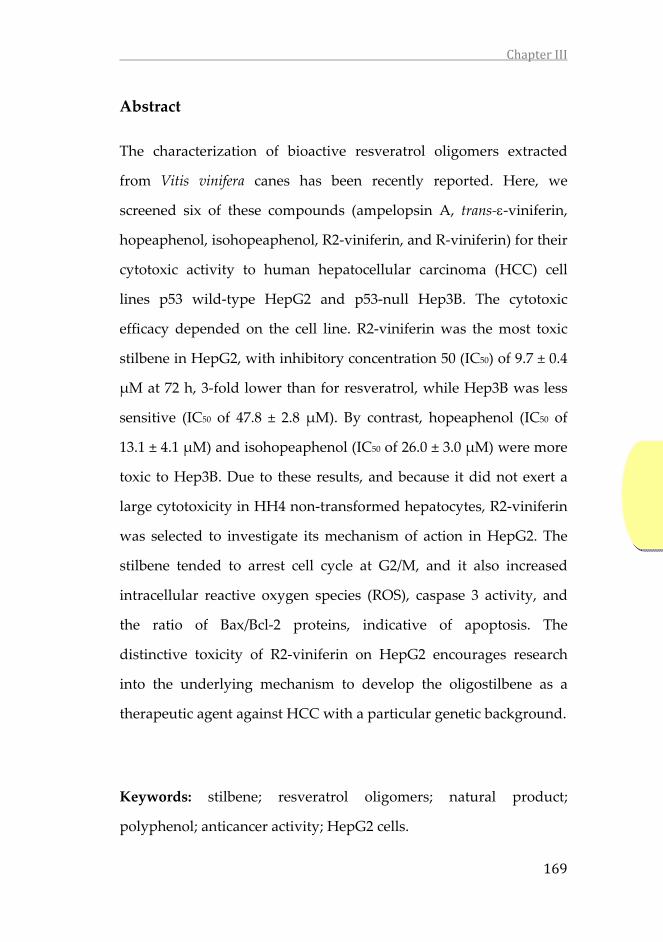
ChapterIII
169
Abstract
The characterization of bioactive resveratrol oligomers extracted
from Vitis vinifera canes has been recently reported. Here, we
screened six of these compounds (ampelopsin A, trans‐‐viniferin,
hopeaphenol, isohopeaphenol, R2‐viniferin, and R‐viniferin) for their
cytotoxic activity to human hepatocellular carcinoma (HCC) cell
lines p53 wild‐type HepG2 and p53‐null Hep3B. The cytotoxic
efficacy depended on the cell line. R2‐viniferin was the most toxic
stilbene in HepG2, with inhibitory concentration 50 (IC50) of 9.7 ± 0.4
μM at 72 h, 3‐fold lower than for resveratrol, while Hep3B was less
sensitive (IC50 of 47.8 ± 2.8 μM). By contrast, hopeaphenol (IC50 of
13.1 ± 4.1 μM) and isohopeaphenol (IC50 of 26.0 ± 3.0 μM) were more
toxic to Hep3B. Due to these results, and because it did not exert a
large cytotoxicity in HH4 non‐transformed hepatocytes, R2‐viniferin
was selected to investigate its mechanism of action in HepG2. The
stilbene tended to arrest cell cycle at G2/M, and it also increased
intracellular reactive oxygen species (ROS), caspase 3 activity, and
the ratio of Bax/Bcl‐2 proteins, indicative of apoptosis. The
distinctive toxicity of R2‐viniferin on HepG2 encourages research
into the underlying mechanism to develop the oligostilbene as a
therapeutic agent against HCC with a particular genetic background.
Keywords: stilbene; resveratrol oligomers; natural product;
polyphenol; anticancer activity; HepG2 cells.


ChapterIII
171
1. Introduction
Hepatocellular carcinoma (HCC) is the most frequently
diagnosed primary cancer of the liver (Mittal and El‐Serag 2013) and
the fourth most common cause of cancer‐related death worldwide (J.
D. Yang et al. 2019). There is currently no effective treatment for
HCC due to the high heterogeneity of the cancer. Thus, extrinsic
factors and different mutations contribute to the induction of liver
cancer (S. Perwez Hussain and Harris 1998; Link and Iwakuma
2017). Among the genetic factors, mutations in the p53 tumor
suppressor gene (TP53) are frequent, especially in HCC from
populations exposed to environmental carcinogens, such as dietary
aflatoxin B1 (S P Hussain et al. 2007). In these cases, anti‐tumor
strategies include p53 activation. However, in a high proportion of
hepatocarcinomas, p53 is retained, and recently, it has been proven
that depending on the specific isoform it could also have an
oncogenic role. Thus, the specific p53 family isoform is the target of
novel anticancer therapies against HCC (Kunst et al. 2016).
Several tumor cell lines with different phenotypes that
resemble the various types of liver cancers have been used. These
approaches are valid and allow the results to be extrapolated to an in
vivo situation (Khoury, Zalko, and Audebert 2015).
Since the discovery of the natural antitumor drug paclitaxel
in 1960s, the interest in identifying new natural chemotherapeutic
agents, particularly those with phenolic structure, has greatly
increased. Stilbenes are a class of polyphenols characterized by the

ChapterIII
172
presence of a 1,2‐diphenylethylene moiety. They are produced as
plant secondary metabolites and exert protective actions against
environmental challenge, acting mainly as antifungal phytoalexins
(K. B. Pandey and Rizvi 2009). Stilbenes are present in foods and
beverages such as blueberries, peanuts, grapes, and red wine (El
Khawand et al. 2018). Grapevine is one of the richest sources of
stilbenes currently known. The cane and grape stems and seeds
show high concentrations of stilbenes (Pugajeva, Perkons, and
Górnaś 2018; Loupit et al. 2020) which are also present in grape skin
and juice and in red wines (Guerrero et al. 2020). Resveratrol is the
most widely studied stilbene for its actions on human health.
Different works have reported its properties against diseases such as
diabetes (Zhu et al. 2017), cancer (Vervandier‐Fasseur and Latruffe
2019), cardiovascular diseases (Cheng et al. 2020), and
neurodegenerative diseases (Sun et al. 2010). However, in the
biosynthetic pathway, as the result of various oxidative
condensations of resveratrol monomer, several dimers, trimers and
tetramers are formed. Despite the numerous works describing the
beneficial effects of resveratrol on health, other natural stilbenes,
particularly oligomers, have received far less attention. The wine
industry generates a high quantity of waste (wood, cane, and root)
and oligostilbens are the main stilbenes extracted from these wastes,
which constitute a cheap source of bioactive products (Loupit et al.
2020). In this work, we have studied the cytotoxic potential of a
range of resveratrol oligomers (dimers and tetramers), extracted and

ChapterIII
173
purified from the Vitis vinifera grapevine cane, on hepatoma cell
lines. The effects were compared with those of resveratrol. After
selecting the most active compound, we investigated the mechanism
of its action.

ChapterIII
174
2. Materials and methods
2.1. Stilbenes from Vitis Vinifera
Ampelopsin A, trans‐‐viniferin, hopeaphenol,
isohopeaphenol, R2‐viniferin, and R‐viniferin were obtained from
extracts of grapevine cane (Vitis vinifera), named Vineatrol®30. The
extract was kindly provided by Actichem S.A. (Montauban, France).
The isolation, characterization and purification of the stilbenes were
carried out as described previously (Gabaston et al. 2018). The purity
of the compounds was estimated to be ≥95%. trans‐Resveratrol was
purchased from Sigma‐Aldrich (Lyon, France).
2.2. Cell Culture
The human hepatoma cell line HepG2 was obtained from the
American Tissue Culture Collection (ATCC, Manassas, VA, USA).
The human hepatoma cell line Hep3B was obtained from the
European Collection of Authenticated Cell Cultures (ECACC, Porton
Down, Salisbury, Wiltshire, UK). HH4 non‐transformed human
hepatocyte cell line (kindly provided by Dr. I. Fabregat, Molecular
Oncology, Bellvitge Biomedical Research Institute, Spain) was
established as described (Trepiana et al. 2017).
HepG2 and Hep3B cell lines were maintained in Eagle’s
Minimum Essential Medium and HH4 cell line was grown in Ham´s
F12 Nutrient Mixture (Sigma‐Aldrich, St Louis, MO, USA). Both

ChapterIII
175
culture media were supplemented with 10% heat inactivated fetal
bovine serum, 2 mM L‐glutamine and antibiotics (0.1 mg/mL
streptomycin and 100 U/mL penicillin) (Sigma‐Aldrich, St Louis,
MO, USA). Cell cultures were grown in an incubator at 37 °C with
5% CO2 atmosphere. After reaching approximately 80% of
confluence, cells were detached in a solution of 0.1% trypsin and
0.04% EDTA and plated as required for further experiments.
2.3. Cell Viability Assay
Cell lines were seeded into 96‐well plates at 5 × 10³ (HepG2)
and 3 × 10³ (Hep3B and HH4) cells/well 24 h before treatment.
Increasing concentrations of the monomer trans‐resveratrol, the
dimers ampelopsin A and trans‐‐viniferin and the tetramers
isohopeaphenol, hopeaphenol, R2‐viniferin and R‐viniferin were
then added and cells were incubated for 24, 48, and 72 h. Stilbenes
were dissolved in dimethyl sulfoxide (DMSO), Sigma‐Aldrich, St
Louis, MO, USA) at a final concentration of 0.01%. The same amount
of DMSO was added to control cells. After treatment, the cell
viability was determined using the crystal violet assay (Trepiana et
al. 2017). The absorbance was recorded at 590 nm in a Synergy HT
microplate reader (BioTek, Winooski, VT, USA). Cell viability was
calculated as the percentage of viable cells treated with stilbene
versus untreated control cells using the following equation: Cell
viability (%) = [OD (Treatment) − OD (Blank)]/[OD (Control) – OD
(Blank)] × 100. The cytotoxic effect of stilbenes was determined by

ChapterIII
176
calculating IC50 values using non‐linear regression analysis
(GraphPad Prism 6, San Diego, CA, USA).
2.4. Cell Cycle Analysis
Exponentially growing cells were seeded in 6‐wells plates
(200,000 cells HepG2 and 180,000 cells Hep3B) 24 h before R2‐
viniferin treatment. On the day of treatment, the culture medium
was replaced with culture medium containing R2‐viniferin or vehicle
solution (control).
After a treatment, cells were detached by trypsinization and
fixed overnight at 4 °C in 70% ice‐cold ethanol diluted in phosphate
buffer saline (PBS). The ethanol was then discarded and the cells
were washed with ice‐cold PBS and finally stained with 25 μg/mL of
propidium iodide (Sigma‐Aldrich, St Louis, MO, USA) in the
presence of 200 μg/mL ribonuclease A (Roche Biochemicals,
Indianapolis, IN, USA) for 30 min at room temperature in the dark.
Flow cytometry analysis (Beckman Coulter Gallios) was carried out
from a total number of 10,000 events acquired in the General
Research Services SGIker of the UPV/EHU. Analysis of the data was
performed using Summit 4.3 software (Dako, Glostrup,
Hovedstaden, Denmark).

ChapterIII
177
2.5. Intracellular ROS and Mitochondrial O2− Measurement
Intracellular levels of reactive oxygen species (ROS) were
measured using the cell‐permeant 2’,7’ dichlorodihydrofluorescein
diacetate (H2DCF‐DA) probe (Molecular Probes, Eugene, OR, USA),
which permeates living cells and is deacetylated and oxidized upon
ROS exposure inside the cell, forming the fluorescent 2’,7’‐
dichlorofluorescein (DCF).
The mitochondrial superoxide anion (O2−) levels were
measured using MitoSOX™Red reagent (Molecular Probes, Eugene,
OR, USA), which permeates living cells where it selectively targets
mitochondria and is oxidized by superoxide.
HepG2 cells were cultured in a 6‐well cell culture plate at a
density of 2 × 10⁵ cells per well 24 h before starting the treatment
with R2‐viniferin. The medium was renewed and cells were
incubated with R2‐viniferin as described above. After treatment, cells
were washed and incubated with 20 μM H2DCF‐DA or 4 μM
MitoSOX™ for 30 min at 37 °C in the dark. The probe solution was
then withdrawn and, after washing with PBS, the cells were
trypsinized. The fluorescence intensity from living cells was
measured by flow cytometry (DCF λex = 485/20 and λem = 528/20,
MitoSOX λex = 485/20 and λem = 620/20) in a Beckman Coulter
Gallios Flow cytometer in the General Research Services SGIker of
the UPV/EHU. At least 10,000 events were detected. Data obtained
from flow cytometry were analyzed using Summit 4.3 software

ChapterIII
178
(Dako, Hovedstaden, Denmark). Intracellular ROS and
mitochondrial O2− were expressed as the percentage of the
fluorescence intensity in control cells at the same time of incubation.
2.6. Caspase‐3 Activity
The activity of caspase‐3 was measured using the specific
synthetic tetrapeptide fluorogenic substrate Ac‐DEVD‐AMC (BD
Pharmigen Biosciences, San Diego, CA, USA). The assay was carried
out in 96‐well plates in a total volume of 100 μL, with 37 μM of the
substrate and 50 μg of protein in the assay medium in 100 mM
HEPES, pH 7.4, containing 200 mM NaCl, 0.2% CHAPS, 2 mM
EDTA, 20% glycerol, and 5 mM dithiothreitol (DTT).
In the presence of caspase‐3, the Ac‐DEVD‐AMC substrate is
hydrolyzed and the fluorogenic compound AMC is released. The
activity was determined by continuous recording of the fluorescence
at λex = 360 nm and λem = 460 nm at 37 °C for 2 h every 5 min in a
Synergy HT microplate reader (BioTek, Winooski, VT, USA). Results
were expressed as the percentage of the fluorescence in control cells.
2.7. Western Blot Analysis
HepG2 cells were seeded in Petri dishes, incubated for 24 h
and then treated with 5 and 10 μM R2‐viniferin. Following 24 h of
treatment, the cells were lysed in ice for 30 min with lysis buffer (20
mM HEPES pH 7.5, 1 mM NaF, 10 mM EGTA, 40 mM β‐
glycerophosphate, 1% NP‐40, 2.5 mM MgCl2, 2 mM orthovanadate,

ChapterIII
179
and 1 mM DTT) to which 10 μL/mL of a protease inhibitor cocktail
(Sigma‐Aldrich, St Louis, USA) was added just before use. Cellular
fragments were removed by centrifugation at 12,000× g for 10 min at
4 °C, and total protein concentration was determined by Bradford
assay [19]. The cellular protein extracts were boiled at 95 °C for 5 min
in Laemmli buffer and separated by sodium dodecyl sulfate–
polyacrylamide gel electrophoresis in 15% polyacrylamide gels.
Proteins were transferred onto polyvinylidene difluoride (PVDF)
membranes by electro‐blotting with constant amperage (1 mA/cm2)
for 2 h in a wet chamber. After blocking for 1 h in 5% bovine serum
albumin (BSA) Tris‐buffered saline containing Tween 20 at room
temperature, membranes were incubated overnight at 4 °C with
antibodies to Bax (Santa Cruz Biotechnology, 1:500), Bcl‐2 (Santa
Cruz Biotechnology, 1:500) and glyceraldehyde‐3‐phosphate
dehydrogenase (GAPDH, Abcam, 1:2000). After washing,
membranes were probed with their corresponding secondary
antibody conjugated to horseradish peroxidase for 1 h at room
temperature. Specific proteins were detected using the enhanced
chemiluminescence (ECL) substrate kit (Clarity Western ECL
substrate, Bio‐Rad, Hercules, California, USA) and the blots were
imaged with the C‐DiGit LI‐COR blot scanner (Bonsai Advanced
technologies S.L. Madrid, Spain). Intensities of target protein bands
were determined by densitometry and normalized to the intensity of
the loading control GAPDH protein.
ChapterIII
180
2.8. Statistical Analysis
The statistical package SPSS 19.0 (SPSS Inc. Chicago, IL, USA)
was used for data analysis. The results were expressed as mean ±
standard error (SE) from at least three experiments. Statistical
significance for the differences of the means was estimated by
parametric Student’s t‐test. Differences between means were
considered statistically significant for p < 0.05. IC50 values were
derived from fitting the data to a sigmoidal dose‐response curve
with a three‐parameter logistic model using GraphPad Prism 6.

ChapterIII
181
3. Results
In this work, we have investigated the cytotoxicity of one
stilbene monomer: trans‐resveratrol; two stilbene dimers:
ampelopsin A and trans‐‐viniferin; and four resveratrol tetramers:
hopeaphenol, isohopeaphenol, R2‐viniferin, and R‐viniferin. Their
chemical structures and that of resveratrol are shown in Figure 1.
Figure 1. Chemical structures of resveratrol and resveratrol oligomers.
HOOH
HO
trans‐resveratrolampelopsin A
trans‐ε‐viniferin
hopeaphenol isohopeaphenol
R2‐viniferin
R‐viniferin

ChapterIII
182
Figure 2. Effect of different stilbenoids on cell viability of HepG2, Hep3B
and HH4. Cell viability was measured by crystal violet assay. Results are
the mean + SE of n = 3–5 experiments. *p < 0.05, **p < 0.01, ***p < 0.001,
compared with control at the same time.
Re
sve
ratr
ol
Am
pe
lop
sin
AIs
oh
op
ea
ph
en
ol
trans
--V
inif
eri
nH
op
ea
ph
en
ol
R2-
Vin
ife
rin
R-V
inif
eri
nHepG2 Hep3B HH4
10 25 50 100 200
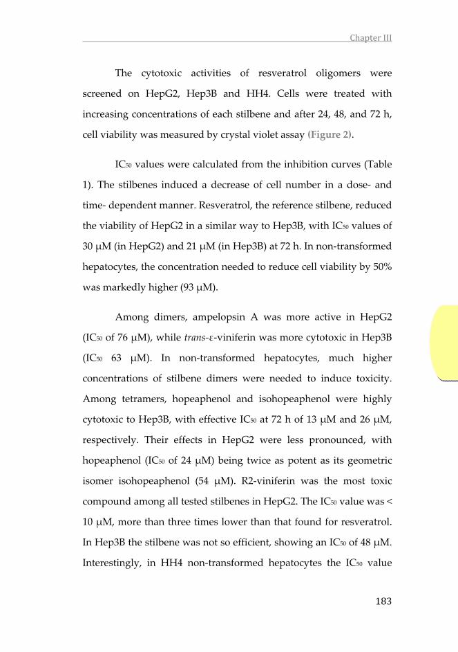
ChapterIII
183
The cytotoxic activities of resveratrol oligomers were
screened on HepG2, Hep3B and HH4. Cells were treated with
increasing concentrations of each stilbene and after 24, 48, and 72 h,
cell viability was measured by crystal violet assay (Figure 2).
IC50 values were calculated from the inhibition curves (Table
1). The stilbenes induced a decrease of cell number in a dose‐ and
time‐ dependent manner. Resveratrol, the reference stilbene, reduced
the viability of HepG2 in a similar way to Hep3B, with IC50 values of
30 μM (in HepG2) and 21 μM (in Hep3B) at 72 h. In non‐transformed
hepatocytes, the concentration needed to reduce cell viability by 50%
was markedly higher (93 μM).
Among dimers, ampelopsin A was more active in HepG2
(IC50 of 76 μM), while trans‐ε‐viniferin was more cytotoxic in Hep3B
(IC50 63 μM). In non‐transformed hepatocytes, much higher
concentrations of stilbene dimers were needed to induce toxicity.
Among tetramers, hopeaphenol and isohopeaphenol were highly
cytotoxic to Hep3B, with effective IC50 at 72 h of 13 μM and 26 μM,
respectively. Their effects in HepG2 were less pronounced, with
hopeaphenol (IC50 of 24 μM) being twice as potent as its geometric
isomer isohopeaphenol (54 μM). R2‐viniferin was the most toxic
compound among all tested stilbenes in HepG2. The IC50 value was <
10 μM, more than three times lower than that found for resveratrol.
In Hep3B the stilbene was not so efficient, showing an IC50 of 48 μM.
Interestingly, in HH4 non‐transformed hepatocytes the IC50 value

ChapterIII
184
was higher than 200 μM (Table 1). In the case of R‐viniferin,
containing one less free hydroxyl group than R2‐viniferin,
concentrations as high as 200 μM were necessary to induce cell death
in HepG2 and Hep3B (Figure 2).
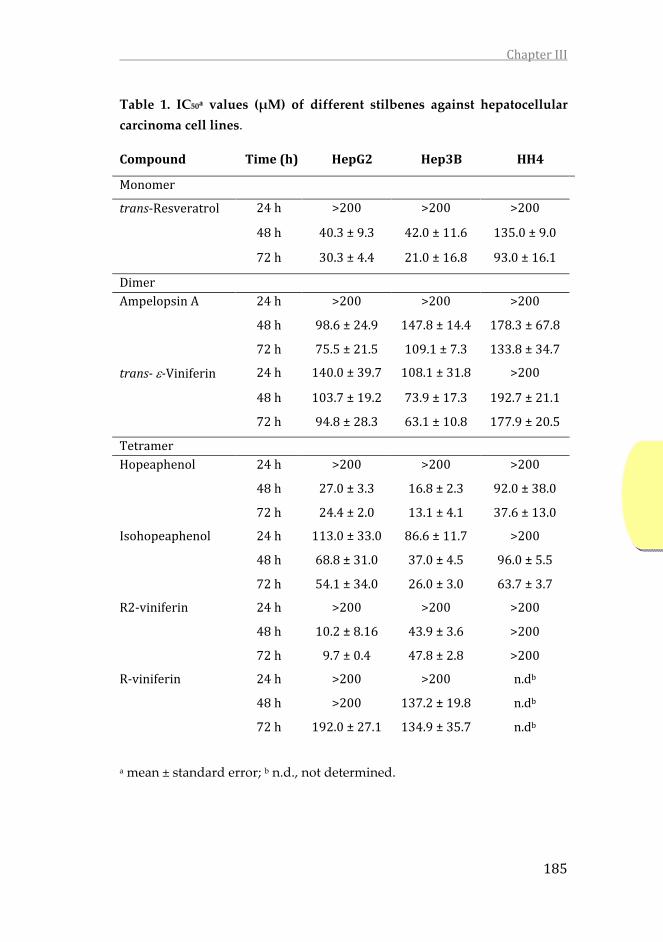
ChapterIII
185
Table 1. IC50a values (μM) of different stilbenes against hepatocellular
carcinoma cell lines.
Compound Time(h) HepG2 Hep3B HH4
Monomer
trans‐Resveratrol 24h >200 >200 >200
48h 40.3±9.3 42.0±11.6 135.0±9.0
72h 30.3±4.4 21.0±16.8 93.0±16.1
DimerAmpelopsinA 24h >200 >200 >200
48h 98.6±24.9 147.8±14.4 178.3±67.8
72h 75.5±21.5 109.1±7.3 133.8±34.7
trans‐‐Viniferin 24h 140.0±39.7 108.1±31.8 >200
48h 103.7±19.2 73.9±17.3 192.7±21.1
72h 94.8±28.3 63.1±10.8 177.9±20.5
TetramerHopeaphenol 24h >200 >200 >200
48h 27.0±3.3 16.8±2.3 92.0±38.0
72h 24.4±2.0 13.1±4.1 37.6±13.0
Isohopeaphenol 24h 113.0±33.0 86.6±11.7 >200
48h 68.8±31.0 37.0±4.5 96.0±5.5
72h 54.1±34.0 26.0±3.0 63.7±3.7
R2‐viniferin 24h >200 >200 >200
48h 10.2±8.16 43.9±3.6 >200
72h 9.7±0.4 47.8±2.8 >200
R‐viniferin 24h >200 >200 n.db
48h >200 137.2±19.8 n.db
72h 192.0±27.1 134.9±35.7 n.db
a mean ± standard error; b n.d., not determined.

ChapterIII
186
In view of these results, R2‐viniferin was selected to study the
mechanisms of its cytotoxicity in hepatocellular carcinoma cells. The
effect of R2‐viniferin on cell cycle distribution in both HepG2 and
Hep3B was studied by flow cytometry. As can be seen in Figure 3,
HepG2 treated with 10 μM R2‐viniferin showed a progressive
increase in the number of cells in subG0 phase over time. This effect
was accompanied by a concomitant, though not significant, increase
in the percentage of cells in the G2/M phase, suggesting an arrest in
this phase. In Hep3B, however, R2‐viniferin did not alter cell cycle at
this concentration (Figure 4).
Figure 3. Cell cycle distribution of HepG2 treated with R2‐viniferin. (A)
Representative graphs of cell cycle analysis by flow cytometry at 72 h. (B)
Statistical analysis of cell cycle distribution. Data are expressed as the
percentage (%) of cells at different stages of cell cycle. In the case of cells in
subG0, data are expressed as the percentage of total cells. Results are the
mean ± SE of three experiments. *p<0.001, significantly different from
control.
A) Control R2‐Viniferin
B)
Control R2‐Viniferin
24 h 48 h 72 h 24 h 48 h 72 h
SubG0 1.0 ± 0.2 1.1 ± 0.5 1.9 ± 0.6 8.5 ± 2.7 22.4 ± 11.0 71.0 ± 2.1*
Cell cycle
G0/G1 72.5 ± 3.2 78.5 ± 3.0 87.3 ± 1.7 65.6 ± 11.7 71.7 ± 10.9 75.5 ± 7.8
S 12.4 ± 2.5 8.8 ± 1.4 3.0 ± 0.6 11.9 ± 3.2 9.0 ± 4.5 9.5 ± 5.6
G2/M 15.1 ± 0.6 12.7 ± 1.6 9.7 ± 1.2 22.5 ± 8.5 19.3 ± 6.5 15.0 ± 2.2

ChapterIII
187
Figure 4. Cell cycle distribution of Hep3B treated with R2‐viniferin. (A)
Representative graphs of cell cycle analysis by flow cytometry at 72 h. (B)
Statistical analysis of cell cycle distribution. Data are expressed as the
percentage (%) of cells at different stages of cell cycle. In the case of cells in
subG0, data are expressed as the percentage of total cells. Results are the
mean ± SE of three experiments.
R2‐viniferin increased the intracellular ROS concentration dose‐
dependently from the first time assayed (Figure 5A). At the highest
concentration used (10 μM), ROS remained significantly elevated up
to 72 h. In the case of mitochondrial O2−, no significant effect could be
observed at any of the stilbene doses or times assayed (Figure 5B).
A) Control R2‐Viniferin
B)
Control R2‐Viniferin
24 h 48 h 72 h 24 h 48 h 72 h
SubG0 1.3 ± 0.4 1.0 ± 0.3 3.6 ± 1.3 2.0 ± 0.6 1.8 ± 0.4 3.3 ± 0.7
Cell cycle
G0/G1 62.8 ± 3.5 74.8 ± 3.6 82.2 ± 2.4 63.6 ± 8.0 73.7 ± 3.4 75.5 ± 3.9
S 13.6 ± 1.1 9.6 ± 1.4 6.5 ± 1.6 15.7 ± 3.7 10.4 ± 1.6 9.8 ± 1.2
G2/M 23.6 ± 3.1 15.6 ± 2.4 11.3 ± 1.2 20.7 ± 6.1 16.5 ± 2.0 14.7 ± 2.9

ChapterIII
188
Figure 5. Effect of R2‐viniferin on (A) intracellular ROS and (B)
mitochondrial O2‐ levels in HepG2. Cells were treated with 5 and 10 μM of
R2‐viniferin for the indicated times. Reactive species were detected by flow
cytometry. (A) ROS were determined by H2DCF‐DA assay. (B) O2− was
determined by MitoSOX probe. Results are expressed as the percentage (%)
of the control values at the same time, and are the mean ± SE of 3
experiments. *p < 0.05; **p < 0.01; ***p < 0.001.
R2‐viniferin (10 μM) increased the caspase‐3 activity significantly
from 24 h to 72 h. At 5 μM concentration the stilbene increased
caspase‐3 activity at 72 h (Figure 6). These data indicate that R2‐
viniferin induces HepG2 death through a caspase‐dependent
mechanism, in which the executioner caspase‐3 is involved.
A) B)

ChapterIII
189
Figure 6. Effect of R2‐viniferin on caspase‐3 activity in HepG2. Cells were
incubated with R2‐viniferin (5 μM and 10 μM) at the indicated times.
Results are expressed as the percentage (%) of the control values, and are
the mean ± SE of 4 experiments. *p < 0.05, **p < 0.01, compared with
controls.
We then analyzed by the effect of R2‐viniferin on the apoptosis‐
related Bcl‐2 protein family. The resveratrol oligomer upregulated
the expression of the proapoptotic Bax protein and downregulated
anti‐apoptotic Bcl‐2 proteins (Figure 7). The Bax/Bcl‐2 ratio increased
dose‐dependently (39% and 123%) over control at 5 μM and 10 μM,
respectively.

ChapterIII
190
Figure 7. Western blot analysis of Bax and Bcl‐2 expression in HepG2 cells
incubated with R2‐viniferin (5 and 10 μM). The bar graph shows the
Bax/Bcl2 ratio, calculated by densitometric analysis of the anti‐apoptotic
proteins expressed as the percentage of the ratio (molecule of
interest/GAPDH protein) detected in the control. Results are the mean + SE
of n = 6 experiments. **p < 0.01, ***p < 0.001, compared with control.
Bax
Bcl-2
GAPDH
GAPDH
0 5 10 µM
Bax
/Bcl
-2 r
atio
(%
)
0
200
300 5 M
10 M
***
**
**
100

ChapterIII
191
4. Discussion
The results presented herein show for the first time that several
natural oligomeric products of resveratrol, in particular tetramers,
are cytotoxic to human hepatocellular carcinoma cells, inhibiting cell
proliferation and triggering death of the tumor cells. Resveratrol has
been widely described to have antioxidant and protective actions
against a wide range of diseases, particularly cardiovascular diseases
(Cheng et al. 2020) and cancer (Vervandier‐Fasseur and Latruffe
2019). However, other natural products obtained from the same
source, the Vitis vinifera cane, in particular tetramers, have not been
tested before for their antiproliferative activity against hepatoma
cells. As resveratrol, some of these compounds, such as
hopeaphenol, isohopeaphenol and R2‐viniferin, were also identified
in red wines and only recently R‐viniferin and R2‐viniferin could be
detected at very low concentrations in certain red wines (Willenberg
et al. 2012).
The dimer of resveratrol ‐viniferin was cytotoxic to various
leukemia, HeLa cervix cancer, breast cancer, melanoma, and HepG2
cell lines (Espinoza and Inaoka 2017). The stilbene induced apoptosis
in these cells and has been shown to inhibit topoisomerase IIa
(Baechler et al. 2014). In HepG2, ‐viniferin showed slightly lower
antiproliferative potential than resveratrol (Colin et al. 2008), and
this was confirmed in the present work. The authors did not test
toxicity in non‐malignant liver cells. In our work, the resveratrol

ChapterIII
192
dimer also induced toxicity to HH4 cells, although the IC50 was high
(178 μM). This result rules out the possibility to develop this product
as a unique anticancer agent against hepatocellular carcinomas.
However, the development of antitumor therapies could be
established based on combinations of stilbenes at low concentrations
with anticancer drugs, which may exert synergistic effects in the
prevention or treatment of liver cancers, as it has been described for
several cancer cells (Ivanova et al. 2019; Yen et al. 2018; Redondo‐
Blanco et al. 2017).
The results of this screening study showed that the cytotoxic effects
of the natural stilbenes varied from one compound to another, and
seemed to be dependent on the cellular model used. For example, in
HepG2 hopeaphenol was two times more potent than its tetrameric
geometric isomer isohopeaphenol. We do not know the cause of
these differences in the cytotoxic activity of the two isomers. In a
study reported by Loisruangsin et al. it was found that both
tetramers acted as competitive inhibitors of sirtuin 1 (SIRT1), a key
histone deacetylase in the regulation of cellular processes, suggesting
that inhibitors could suppress the growth of tumor cells
(Loisruangsin et al. 2019). Hopeaphenol proved to be a more efficient
inhibitor than isohopeaphenol. The authors showed, by computer‐
assisted modeling, that there were differences in the bonds that were
established in the enzyme complex resulting from the inhibition,
with more hydrogen bonds being formed in the case of hopeaphenol
than with isohopeaphenol. As regards the different efficacy

ChapterIII
193
depending of the cell line, hopeaphenol and isohopeaphenol, similar
to the resveratrol monomer, were more toxic to Hep3B than to
HepG2. By contrast, R2‐viniferin, a tetramer formed from two
dimers of ‐viniferin and ampelopsin B, was the most potent stilbene
to induce cell death in HepG2 (with an IC50 three times lower than
that of resveratrol), while Hep3B cells were less sensitive to this
tetramer. The structural diversity of tested compounds could explain
the variability of activity on different cellular models. To better
understand these structural specificities, additional studies will be
necessary to look at the structure–activity relationship.
Scientific reports regarding the R2‐viniferin stilbene are quite scarce.
There is great confusion about this oligostilbene, since R2‐viniferin is
also called vitisin A, a name that also refers to another compound
with a pyranoanthocyanin structure found in red wines (Asenstorfer
et al. 2003). Two studies reported that this oligostilbene induced cell
death in prostate cancer (Shin et al. 2015) and leukemia cell lines (Mi
Jeong Sung et al. 2009). In these latter cells, R2‐viniferin induced cell
apoptosis, as well as the inhibition of ERK, p38, and NF‐kB
pathways. In our work, the differences in susceptibility to R2‐
viniferin and the other resveratrol oligomers may be related to
differences in the genetic background of the hepatocarcinoma cell
lines. Thus, HepG2 cells carry wild‐type p53, while Hep3B are p53‐
null. Tumor protein p53 induces cell cycle arrest and apoptosis
through the transcriptional regulation of BAX gene. The protein also
upregulates Bak expression, and the activation of Bax and Bak

ChapterIII
194
induces activation of caspase‐3 (Liao et al. 2018). In HepG2 cells, we
have found that R2‐viniferin tended to arrest cell cycle at G2/M
phase, increased intracellular ROS levels, and the Bax/Bcl2 ratio in a
dose‐dependent manner. The induced effect on apoptosis is more
dependent on the balance between Bcl‐2 and Bax than on Bcl‐2
quantity alone (Huang et al. 2012). When tested at the same
concentration of 10 μM, R2‐viniferin was unable to inhibit cell
proliferation and induce cell cycle blockage in p53‐null Hep3B cells.
In a previous work, resveratrol was reported to inhibit HepG2 cell
proliferation by blocking cell cycle at the G2/M transition (Delmas et
al. 2000). Since cyclins and their cyclin‐dependent kinases (CDKs) are
key regulators of cell cycle (Johansson and Persson 2008), the cell
cycle inhibition by resveratrol in cancer cells has been attributed to
disturbances in cyclins‐CDKs complexes. We propose a similar
mechanism of cell proliferation inhibition for R2‐viniferin.
We have seen that R2‐viniferin increased intracellular ROS levels
without affecting the mitochondrial O2‐ determined by the
MitoSOX™Red fluorescent probe. We do not know the nature and
source of these ROS. It has been described that resveratrol induces
the expression of the transmembrane enzyme NADPH oxidase‐5
(Nox5) in lung cancer cells. This enzyme generates O2‐ which is
converted by action of superoxide dismutase (SOD) into hydrogen
peroxide (Luo et al. 2013). In HepG2 resveratrol also causes the
upregulation of SOD, without affecting glutathione peroxidase,
which contributes to the formation and accumulation of hydrogen

ChapterIII
195
peroxide inside the cells (Khan et al. 2013). Although speculative, R2‐
viniferin could exert similar effects in HepG2 in generating hydrogen
peroxide, which would lead to cell apoptosis.
Our results show cytotoxic actions of stilbenes from Vitis vinifera
cane in vitro. A major limitation of the study is the bioavailability of
stilbenes in vivo. Several works have reported a weak bioavailability
of resveratrol, mainly as a result of its low cell accessibility and its
fast metabolism in the intestine and liver (Baur and Sinclair 2006;
Walle 2011). Therefore, a series of synthetic resveratrol derivatives
that are more hydrophobic and with higher cell permeability are
being developed to test their biological activity (Snyder, Gollner, and
Chiriac 2011; Matsuura et al. 2015; Keylor et al. 2016). To our
knowledge, there are no reports on the bioavailability of the stilbene
oligomers screened in this study, with the exception of the dimer ‐
viniferin (El Khawand et al. 2018; J. Kim et al. 2017; Courtois et al.
2018), which showed by flow cytometry a cellular uptake kinetics
similar to that of resveratrol (Colin et al. 2008). The bioavailability of
stilbenes depends on many factors, among them their stability, the
molecular size, the chemical structure, and the
hydrophilicity/hydrophobicity properties of the compound (Gao and
Hu 2010). Encapsulation into nanoparticles or liposomes of bioactive
compounds with low water solubility may be a promising approach
to facilitate their stability, absorption, transport to target cells and,
therefore, their action. This challenge has recently been described for
‐viniferin in Caco‐2 intestinal cells (Courtois et al. 2019).

ChapterIII
196
5. Conclusions
We have described the cytotoxic activities of several natural
resveratrol oligomers isolated from Vitis vinifera cane extracts against
human hepatocellular carcinoma cells, and in comparison to HH4
non‐transformed human hepatocytes. The cellular efficacies varied
depending on the cell type. From the compounds tested, the tetramer
R2‐viniferin at concentrations below 10 μM was the most potent
cytotoxic stilbene in p53‐wilde type HepG2 cells, increasing
intracellular ROS, and inducing cell apoptosis. The stilbene was
innocuous in normal hepatocytes. These results suggest that R2‐
viniferin is a promising compound to develop in the
chemoprevention and treatment of liver cancer. Further studies will
be required in order to improve its bioavailability and to unravel its
mechanism of action for potential clinical application in the
treatment of liver cancer.

ChapterIII
197
Abbreviations:
BSA Bovine serum albumin
CDKs Cyclin‐dependent kinases
DCF 2´,7´‐Dichlorofluorescein
DMSO Dimethyl sulfoxide
DTT Dithiothreitol
ECL Enhanced chemiluminescence
EDTA Ethylenediaminetetraacetic acid
EGTA Ethylene glycol‐bis(β‐aminoethyl ether)‐N,N,N′,N′‐ tetraacetic acid
ERK Extracellular‐signal‐regulated kinase
GAPDH Glyceraldehyde‐3‐phosphate dehydrogenase
H2DCF‐DA 2′,7′ Dichlorodihydrofluorescein diacetate
HCC Hepatocellular carcinoma
HEPES 4‐(2‐Hydroxyethyl)‐1‐piperazineethanesulfonic acid
NF‐kB Nuclear factor kappa B
O2− Superoxide anion
PBS Phosphate buffer saline
ROS Reactive oxygen species

ChapterIII
198
Author Contributions: Conceptualization, M.B.R., T.R., and J.I.R.;
Methodology, I.A, M.B.R., and J.I.R.; validation, I.A. and J.I.R.;
formal analysis, I.A. and J.I.R.; investigation, I.A., S.K., and J.I.R.;
resources, M.B.R., A.C., T.R., and J.I.R.; data curation, I.A., M.B.R.,
and J.I.R.; writing—original draft preparation, I.A., M.B.R., and J.I.R.;
writing—review and editing, I.A., M.B.R., S.K., A.C., T.R., and J.I.R.;
visualization, I.A., M.B.R., T.R., and J.I.R; supervision, M.B.R., T.R.,
and J.I.R.; project administration, M.B.R., T.R., and J.I.R.; funding
acquisition, M.B.R., T.R., and J.I.R.
Funding: This research was funded by a PhD Research Fellowship in
the context of Campus Euro‐regional Bordeaux/Euskampus (ref.
PIFBUR16‐01). The work was supported by the University of the
Basque Country UPV/EHU (to Research Groups, ref. GIU16/62).
Moreover, it was also supported by the Bordeaux Metabolome
Facility and MetaboHUB (ANR‐11‐INBS‐0010 project). The authors
thank J.A. López, M‐L. Iglesias and A. Palos‐Pinto for their technical
assistance.
Conflicts of Interest: The authors declare no conflict of interest. The
funders had no role in the design of the study; in the collection,
analyses, or interpretation of data; in the writing of the manuscript,
or in the decision to publish the results.

ChapterIII
199
6. References
Adam, Mowaffaq Adam Ahmed, Yasser M. Tabana, Khirun Binti Musa, andDoblin Anak Sandai. 2017. “Effects of Different Mycotoxins onHumans, Cell Genome and Their Involvement in Cancer.” OncologyReports37(3):1321–36.https://doi.org/10.3892/or.2017.5424.
Ademowo,O.S.,H.K. I.Dias,D.G.A.Burton,andH.R.Griffiths.2017. “Lipid(per) Oxidation in Mitochondria: An Emerging Target in the AgeingProcess?” Biogerontology 18 (6): 859–79.https://doi.org/10.1007/s10522‐017‐9710‐z.
Aharoni, Asaph, and Gad Galili. 2011. “Metabolic Engineering of the PlantPrimary–Secondary Metabolism Interface.” Current Opinion inBiotechnology 22 (2): 239–44.https://doi.org/10.1016/j.copbio.2010.11.004.
Aja, Iris, M. Begoña Ruiz‐Larrea, Arnaud Courtois, Stéphanie Krisa, TristanRichard, and José‐Ignacio Ruiz‐Sanz. 2020. “Screening of NaturalStilbene Oligomers from Vitis Vinifera for Anticancer Activity onHuman Hepatocellular Carcinoma Cells.” Antioxidants 9 (6): 469.https://doi.org/10.3390/antiox9060469.
Akdis,Mübeccel,AlarAab,CanAltunbulakli,KursatAzkur,RitaA.Costa,RetoCrameri, Su Duan, et al. 2016. “Interleukins (from IL‐1 to IL‐38),Interferons, Transforming Growth Factor β, and TNF‐α: Receptors,Functions, and Roles in Diseases.” Journal of Allergy and ClinicalImmunology 138 (4): 984–1010.https://doi.org/10.1016/j.jaci.2016.06.033.
Akinwumi, Bolanle, Kimberly‐Ann Bordun, and Hope Anderson. 2018.“BiologicalActivitiesofStilbenoids.”InternationalJournalofMolecularSciences19(3):792.https://doi.org/10.3390/ijms19030792.
Akinyemiju, Tomi, Semaw Abera, Muktar Ahmed, Noore Alam, MulubirhanAssefaAlemayohu,ChristineAllen,RajaaAl‐Raddadi,etal.2017.“TheBurdenofPrimaryLiverCancerandUnderlyingEtiologiesFrom1990to2015attheGlobal,Regional,andNationalLevel:ResultsFromtheGlobal Burden ofDisease Study 2015.” JAMAOncology 3 (12): 1683.https://doi.org/10.1001/jamaoncol.2017.3055.
Alotaibi,Hani,NeseAtabey,KasımDiril,EsraErdal,andMehmetOzturk.2016.“Molecular Mechanisms of Hepatocellular Carcinoma.” InHepatocellular Carcinoma, edited by Brian I. Carr, 43–63. Cham:Springer International Publishing. https://doi.org/10.1007/978‐3‐319‐34214‐6_3.

ChapterIII
200
Alqahtani, Khan, Alloghbi, Said Ahmed, Ashraf, and Hammouda. 2019.“Hepatocellular Carcinoma: Molecular Mechanisms and TargetedTherapies.” Medicina 55 (9): 526.https://doi.org/10.3390/medicina55090526.
Asenstorfer, Robert E., Andrew J. Markides, Patrick G. Iland, and Graham P.Jones. 2003. “Formation of Vitisin A during RedWine FermentationandMaturation.”AustralianJournalofGrapeandWineResearch9(1):40–46.https://doi.org/10.1111/j.1755‐0238.2003.tb00230.x.
B.Vendramini‐Costa,D.,andJ.E.Carvalho.2012.“MolecularLinkMechanismsbetweenInflammationandCancer.”CurrentPharmaceuticalDesign18(26):3831–52.https://doi.org/10.2174/138161212802083707.
Baechler, Simone A., Anika Schroeter, Martina Dicker, Pablo Steinberg, andDoris Marko. 2014. “Topoisomerase II‐Targeting Properties of aGrapevine‐Shoot Extract and Resveratrol Oligomers.” Journal ofAgricultural and Food Chemistry 62 (3): 780–88.https://doi.org/10.1021/jf4046182.
Bahman, Abdulmajeed, Mohamed Abaza, Sarah Khoushiash, and RajaaAl‑Attiyah. 2018. “Sequence‑dependent Effect of Sorafenib inCombination with Natural Phenolic Compounds on Hepatic CancerCellsand thePossibleMechanismofAction.” International JournalofMolecularMedicine,June.https://doi.org/10.3892/ijmm.2018.3725.
Baur, Joseph A., and David A. Sinclair. 2006. “Therapeutic Potential ofResveratrol:The inVivoEvidence.”NatureReviewsDrugDiscovery5(6):493–506.https://doi.org/10.1038/nrd2060.
Behzadi,Payam,andRezaRanjbar.2015.“CaspasesandApoptosis.”MolecularEnzymology and Drug Targets 01 (02).https://doi.org/10.21767/2572‐5475.10006.
Bi,XiuLi, JingYuYang,YingXuDong, JiMingWang,YongHongCui,TakashiIkeshima,YuQingZhao,andChunFuWu.2005.“ResveratrolInhibitsNitric Oxide and TNF‐α Production by Lipopolysaccharide‐ActivatedMicroglia.” International Immunopharmacology 5 (1): 185–93.https://doi.org/10.1016/j.intimp.2004.08.008.
Biais, Benoit, Stéphanie Krisa, Stéphanie Cluzet, Grégory Da Costa, PierreWaffo‐Teguo, Jean‐Michel Mérillon, and Tristan Richard. 2017.“Antioxidant and Cytoprotective Activities of Grapevine Stilbenes.”Journal of Agricultural and Food Chemistry 65 (24): 4952–60.https://doi.org/10.1021/acs.jafc.7b01254.

ChapterIII
201
Bishayee, Anupam. 2014. “The Inflammation and Liver Cancer.” InInflammation and Cancer, edited by Bharat B. Aggarwal, BokyungSung,andSubashChandraGupta,816:401–35.Basel:SpringerBasel.https://doi.org/10.1007/978‐3‐0348‐0837‐8_16.
Bradford,MarionM.1976.“ARapidandSensitiveMethodfortheQuantitationofMicrogramQuantitiesofProteinUtilizing thePrincipleofProtein‐Dye Binding.” Analytical Biochemistry 72 (1–2): 248–54.https://doi.org/10.1016/0003‐2697(76)90527‐3.
Bronte‐Stewart,B.1958.“TheEffectOfDietaryFatsOnTheBloodLipidsAndTheirRelationToIschaemicHeartDisease.”BritishMedicalBulletin14(3):243–52.https://doi.org/10.1093/oxfordjournals.bmb.a069691.
Bush,Andrew.2019.“PathophysiologicalMechanismsofAsthma.”FrontiersinPediatrics7(March).https://doi.org/10.3389/fped.2019.00068.
Caggiu, Elisa, Giannina Arru, Sepideh Hosseini, Magdalena Niegowska,GianPietroSechi,IgnazioRobertoZarbo,andLeonardoA.Sechi.2019.“Inflammation,InfectiousTriggers,andParkinson’sDisease.”Frontiersin Neurology 10 (February).https://doi.org/10.3389/fneur.2019.00122.
Campo, José A Del, Paloma Gallego, and Lourdes Grande. 2018. “Role ofInflammatory Response in Liver Diseases: Therapeutic Strategies.”World Journal of Hepatology 10 (1): 1–7.https://doi.org/10.4254/wjh.v10.i1.1.
Cassidy, Aedin, Bryan Hanley, and Rosa M Lamuela‐Raventos. 2000.“Isoflavones, Lignans and Stilbenes ‐ Origins, Metabolism andPotentialImportancetoHumanHealth.”JournaloftheScienceofFoodand Agriculture 80 (7): 1044–62.https://doi.org/10.1002/(SICI)1097‐0010(20000515)80:7<1044::AID‐JSFA586>3.0.CO;2‐N.
Cerutti, P. A., and B. F. Trump. 1991. “Inflammation and Oxidative Stress inCarcinogenesis.”CancerCells(ColdSpringHarbor,N.Y.:1989)3(1):1–7.
Chalons,Pauline,SouheilaAmor,FlavieCourtaut,EmmaCantos‐Villar,TristanRichard, Cyril Auger, Philippe Chabert, Valérie Schni‐Kerth, VirginieAires, and Dominique Delmas. 2018. “Study of Potential Anti‐InflammatoryEffectsofRedWineExtractandResveratrol throughaModulationofInterleukin‐1‐BetainMacrophages.”Nutrients10(12):1856.https://doi.org/10.3390/nu10121856.

ChapterIII
202
Chang, Chi‐I,Wei‐Chu Chien, Kai‐XinHuang, and Jue‐LiangHsu. 2017. “Anti‐InflammatoryEffectsofVitisinolAandFourOtherOligostilbenesfromAmpelopsis Brevipedunculata Var. Hancei.”Molecules 22 (7): 1195.https://doi.org/10.3390/molecules22071195.
Chen,Hong‐Jin,Ming‐HuaHu,Fang‐GuiXu,Hao‐JunXu,Jun‐JunShe,andHong‐Ping Xia. 2018. “Understanding the Inflammation‐CancerTransformation in the Development of Primary Liver Cancer.”Hepatoma Research 4 (7): 29. https://doi.org/10.20517/2394‐5079.2018.18.
Cheng,ChakKwong,Jiang‐YunLuo,ChiWaiLau,Zhen‐YuChen,XiaoYuTian,and Yu Huang. 2020. “Pharmacological Basis and New Insights ofResveratrol Action in the Cardiovascular System.” British Journal ofPharmacology177(6):1258–77.https://doi.org/10.1111/bph.14801.
Chiou,Yi‐Siou,Mei‐LingTsai,KalyanamNagabhushanam,Yin‐JenWang,Chih‐HsiungWu,Chi‐TangHo,andMin‐HsiungPan.2011.“PterostilbeneIsMore Potent than Resveratrol in Preventing Azoxymethane (AOM)‐Induced Colon Tumorigenesis via Activation of the NF‐E2‐RelatedFactor 2 (Nrf2)‐Mediated Antioxidant Signaling Pathway.” Journal ofAgricultural and Food Chemistry 59 (6): 2725–33.https://doi.org/10.1021/jf2000103.
Cichocki, Michal, Jaroslaw Paluszczak, Hanna Szaefer, Adriana Piechowiak,AgnesM.Rimando,andWandaBaer‐Dubowska.2008.“PterostilbeneIs Equally Potent as Resveratrol in Inhibiting 12‐O‐Tetradecanoylphorbol‐13‐Acetate Activated NFκB, AP‐1, COX‐2, andINOSinMouseEpidermis.”MolecularNutrition&FoodResearch,June.https://doi.org/10.1002/mnfr.200700466.
Colin, Didier, Allan Lancon, Dominique Delmas, Gerard Lizard, JessicaAbrossinow, Edmond Kahn, Brigitte Jannin, and Norbert Latruffe.2008. “Antiproliferative Activities of Resveratrol and RelatedCompounds in Human Hepatocyte Derived HepG2 Cells AreAssociated with Biochemical Cell Disturbance Revealed byFluorescence Analyses.” Biochimie 90 (11–12): 1674–84.https://doi.org/10.1016/j.biochi.2008.06.006.
Colombo, Francesca, Chiara Di Lorenzo, Luca Regazzoni, Marco Fumagalli,EnricoSangiovanni,LuísPeresdeSousa,LuigiBavaresco,etal.2019.“Phenolic Profiles and Anti‐Inflammatory Activities of Sixteen TableGrape ( Vitis Vinifera L.) Varieties.” Food & Function 10 (4): 1797–1807.https://doi.org/10.1039/C8FO02175A.
Coppa, Tania,Maria Claudia Lazzè, Ornella Cazzalini, Paola Perucca, RobertoPizzala, Livia Bianchi, Lucia Anna Stivala, et al. 2011. “Structure–ActivityRelationshipofResveratrolandItsAnalogue,4,4′‐Dihydroxy‐

ChapterIII
203
Trans ‐Stilbene, Toward the Endothelin Axis in Human EndothelialCells.” Journal of Medicinal Food 14 (10): 1173–80.https://doi.org/10.1089/jmf.2010.0272.
Cory,Suzanne,andJerryM.Adams.2002.“TheBcl2Family:RegulatorsoftheCellularLife‐or‐Death Switch.”NatureReviewsCancer 2 (9): 647–56.https://doi.org/10.1038/nrc883.
Courtois,Arnaud,ClaudeAtgié,AxelMarchal,RuthHornedo‐Ortega,CarolineLapèze, Chrystel Faure, Tristan Richard, and Stéphanie Krisa. 2018.“Tissular Distribution and Metabolism of Trans‐ε‐Viniferin afterIntraperitoneal Injection in Rat.” Nutrients 10 (11): 1660.https://doi.org/10.3390/nu10111660.
Courtois, Arnaud, Manon Garcia, Stéphanie Krisa, Claude Atgié, PatrickSauvant,TristanRichard,andChrystelFaure.2019.“Encapsulationofε‐Viniferin in Onion‐Type Multi‐Lamellar Liposomes Increases ItsSolubility and Its Photo‐Stability and Decreases Its Cytotoxicity onCaco‐2 Intestinal Cells.” Food & Function 10 (5): 2573–82.https://doi.org/10.1039/C9FO00420C.
Crozier, Alan, Indu B. Jaganath, and Michael N. Clifford. 2009. “DietaryPhenolics: Chemistry, Bioavailability and Effects on Health.”NaturalProductReports26(8):1001.https://doi.org/10.1039/b802662a.
Csiszar,Anna,NazarLabinskyy,AndrejPodlutsky,PawelM.Kaminski,MichaelS. Wolin, Cuihua Zhang, Partha Mukhopadhyay, et al. 2008.“Vasoprotective Effects of Resveratrol and SIRT1: Attenuation ofCigarette Smoke‐Induced Oxidative Stress and ProinflammatoryPhenotypic Alterations.” American Journal of Physiology‐Heart andCirculatory Physiology 294 (6): H2721–35.https://doi.org/10.1152/ajpheart.00235.2008.
Cutrim,CamilaSampaio,andMarcoAntonioSlobodaCortez.2018.“AReviewonPolyphenols:Classification,BeneficialEffectsandTheirApplicationinDairy Products.” International JournalofDairyTechnology 71 (3):564–78.https://doi.org/10.1111/1471‐0307.12515.
Dandona, P. 2004. “Inflammation: The Link between Insulin Resistance,Obesity and Diabetes.” Trends in Immunology 25 (1): 4–7.https://doi.org/10.1016/j.it.2003.10.013.
Delmas, D, B Jannin, M Cherkaoui Malki, and N Latruffe. 2000. “InhibitoryEffect of Resveratrol on the Proliferation ofHuman andRatHepaticDerived Cell Lines.” Oncology Reports, July.https://doi.org/10.3892/or.7.4.847.

ChapterIII
204
Djoko, Bambang, Robin Y.‐Y. Chiou, Jia‐Jen Shee, and Yi‐Wen Liu. 2007.“Characterization of Immunological Activities of Peanut Stilbenoids,Arachidin‐1, Piceatannol, and Resveratrol on Lipopolysaccharide‐Induced Inflammation of RAW 264.7 Macrophages.” Journal ofAgricultural and Food Chemistry 55 (6): 2376–83.https://doi.org/10.1021/jf062741a.
ElKhawand,Toni,ArnaudCourtois,JosepValls,TristanRichard,andStéphanieKrisa. 2018. “A Review of Dietary Stilbenes: Sources andBioavailability.” Phytochemistry Reviews 17 (5): 1007–29.https://doi.org/10.1007/s11101‐018‐9578‐9.
Elmore, Susan. 2007. “Apoptosis: A Review of Programmed Cell Death.”Toxicologic Pathology 35 (4): 495–516.https://doi.org/10.1080/01926230701320337.
Esatbeyoglu,Tuba,PhilippEwald,YoshiakiYasui,HarukaYokokawa,AnikaE.Wagner, Seiichi Matsugo, Peter Winterhalter, and Gerald Rimbach.2016. “Chemical Characterization, Free Radical Scavenging, andCellularAntioxidantandAnti‐InflammatoryPropertiesofaStilbenoid‐Rich Root Extract of Vitis Vinifera.” OxidativeMedicine and CellularLongevity2016:1–11.https://doi.org/10.1155/2016/8591286.
Espinoza,J.Luis,andPleiadesT.Inaoka.2017.“Gnetin‐CandOtherResveratrolOligomers with Cancer Chemopreventive Potential: ResveratrolOligomerswithAnticancerPotential.”AnnalsoftheNewYorkAcademyofSciences1403(1):5–14.https://doi.org/10.1111/nyas.13450.
Fridman, Jordan S, and ScottW Lowe. 2003. “Control of Apoptosis by P53.”Oncogene22(56):9030–40.https://doi.org/10.1038/sj.onc.1207116.
Fuchs,Yaron,andHermannSteller.2011.“ProgrammedCellDeath inAnimalDevelopment and Disease.” Cell 147 (4): 742–58.https://doi.org/10.1016/j.cell.2011.10.033.
Fulda, S, and K‐M Debatin. 2006. “Extrinsic versus Intrinsic ApoptosisPathways in Anticancer Chemotherapy.” Oncogene 25 (34): 4798–4811.https://doi.org/10.1038/sj.onc.1209608.
Gabaston,Julien,ToniElKhawand,PierreWaffo‐Teguo,AlainDecendit,TristanRichard, Jean‐Michel Mérillon, and Roman Pavela. 2018. “Stilbenesfrom Grapevine Root: A Promising Natural Insecticide againstLeptinotarsaDecemlineata.” JournalofPestScience 91 (2): 897–906.https://doi.org/10.1007/s10340‐018‐0956‐2.

ChapterIII
205
Galle,PeterR.,AlejandroForner, JosepM.Llovet,VincenzoMazzaferro,FabioPiscaglia, Jean‐Luc Raoul, Peter Schirmacher, and Valérie Vilgrain.2018. “EASL Clinical Practice Guidelines: Management ofHepatocellular Carcinoma.” Journal of Hepatology 69 (1): 182–236.https://doi.org/10.1016/j.jhep.2018.03.019.
Gao, S., and M. Hu. 2010. “Bioavailability Challenges Associated withDevelopment of Anti‐Cancer Phenolics.” Mini‐Reviews in MedicinalChemistry 10 (6): 550–67.https://doi.org/10.2174/138955710791384081.
Gordon, Siamon, and Annette Plüddemann. 2017. “Tissue Macrophages:Heterogeneity and Functions.” BMC Biology 15 (1).https://doi.org/10.1186/s12915‐017‐0392‐4.
Gorski,S.,andM.Marra.2002.“ProgrammedCellDeathTakesFlight:Geneticand Genomic Approaches to Gene Discovery in Drosophila.”Physiological Genomics 9 (2): 59–69.https://doi.org/10.1152/physiolgenomics.00114.2001.
Goszcz,Katarzyna,GarryGDuthie,DerekStewart,StephenJLeslie,andIanLMegson. 2017. “Bioactive Polyphenols and Cardiovascular Disease:Chemical Antagonists, Pharmacological Agents or Xenobiotics ThatDrive an Adaptive Response?: Bioactive Polyphenols andCardiovascular Disease.” British Journal of Pharmacology 174 (11):1209–25.https://doi.org/10.1111/bph.13708.
Guerrero, Raul F., Josep Valls‐Fonayet, Tristan Richard, and Emma Cantos‐Villar. 2020. “A Rapid Quantification of Stilbene Content inWine byUltra‐High Pressure Liquid Chromatography – Mass Spectrometry.”Food Control 108 (February): 106821.https://doi.org/10.1016/j.foodcont.2019.106821.
Guicciardi,MariaEugenia,andGregoryJ.Gores.2009.“LifeandDeathbyDeathReceptors.” The FASEB Journal 23 (6): 1625–37.https://doi.org/10.1096/fj.08‐111005.
Halliwell,Barry.2011.“FreeRadicalsandAntioxidants–QuoVadis?”TrendsinPharmacological Sciences 32 (3): 125–30.https://doi.org/10.1016/j.tips.2010.12.002.
Han,Yuan‐Ping,LingZhou, JiaohongWang,ShigangXiong,WarrenL.Garner,SamuelW.French,andHidekazuTsukamoto.2004.“EssentialRoleofMatrix Metalloproteinases in Interleukin‐1‐Induced MyofibroblasticActivation of Hepatic Stellate Cell in Collagen.” Journal of BiologicalChemistry 279 (6): 4820–28.https://doi.org/10.1074/jbc.M310999200.

ChapterIII
206
Harbeoui, H., A.Hichami,W. AidiWannes, J. Lemput,M. Saidani Tounsi, andN.A.Khan.2019.“Anti‐InflammatoryEffectofGrape(VitisViniferaL.)Seed Extract through the Downregulation of NF‐ΚB and MAPKPathways in LPS‐Induced RAW264.7 Macrophages.” South AfricanJournal of Botany 125 (September): 1–8.https://doi.org/10.1016/j.sajb.2019.06.026.
Hopkins,W.GHünerN.P. 2004. Introduction toPlantPhysiology. 3rdEdition.Inc.JohnWiley&Sons,Inc,Hoboken.
Huang,Fangfang,ZuisuYang,DiYu, JiabinWang,RongLi,andGuofangDing.2012. “Sepia Ink Oligopeptide Induces Apoptosis in Prostate CancerCellLinesviaCaspase‐3ActivationandElevationofBax/Bcl‐2Ratio.”Marine Drugs 10 (12): 2153–65.https://doi.org/10.3390/md10102153.
Hussain, S P, J Schwank, F Staib, X W Wang, and C C Harris. 2007. “TP53Mutations and Hepatocellular Carcinoma: Insights into the Etiologyand Pathogenesis of Liver Cancer.” Oncogene 26 (15): 2166–76.https://doi.org/10.1038/sj.onc.1210279.
Hussain, S. Perwez, and Curtis C. Harris. 1998. “Molecular Epidemiology ofHuman Cancer: Contribution of Mutation Spectra Studies of TumorSuppressorGenes.”CancerResearch58(18):4023.
Islam, Shamima, Ferdaus Hassan, Mya Mya Mu, Hiroyasu Ito, Naoki Koide,Isamu Mori, Tomoaki Yoshida, and Takashi Yokochi. 2004.“PiceatannolPreventsLipopolysaccharide(LPS)‐InducedNitricOxide(NO)ProductionandNuclearFactor(NF)‐ΚBActivationbyInhibitingIκB Kinase (IKK).” Microbiology and Immunology 48 (10): 729–36.https://doi.org/10.1111/j.1348‐0421.2004.tb03598.x.
Ivanova, Donika, Zhivko Zhelev, Severina Semkova, Ichio Aoki, and RumianaBakalova. 2019. “Resveratrol Modulates the Redox‐Status andCytotoxicity of Anticancer Drugs by Sensitizing LeukemicLymphocytes and Protecting Normal Lymphocytes.” AnticancerResearch 39 (7): 3745–55.https://doi.org/10.21873/anticanres.13523.
Jan, Rehmat, and Gul‐e‐Saba Chaudhry. 2019. “Understanding Apoptosis andApoptotic Pathways Targeted Cancer Therapeutics.” AdvancedPharmaceutical Bulletin 9 (2): 205–18.https://doi.org/10.15171/apb.2019.024.

ChapterIII
207
Jiang, Yu, Qiu‐Ju Han, and Jian Zhang. 2019. “Hepatocellular Carcinoma:Mechanisms of Progression and Immunotherapy.”World Journal ofGastroenterology 25 (25): 3151–67.https://doi.org/10.3748/wjg.v25.i25.3151.
Jing,Yingying,KaiSun,WentingLiu,DandanSheng,ShanminZhao,LuGao,andLixinWei. 2018. “TumorNecrosis Factor‐α PromotesHepatocellularCarcinogenesis through the Activation of Hepatic Progenitor Cells.”Cancer Letters 434 (October): 22–32.https://doi.org/10.1016/j.canlet.2018.07.001.
Johansson,Martin,and JennyPersson.2008. “CancerTherapy:TargetingCellCycle Regulators.” Anti‐Cancer Agents inMedicinal Chemistry 8 (7):723–31.https://doi.org/10.2174/187152008785914833.
Kane,R.C.,A.T.Farrell,R.Madabushi,B.Booth,S.Chattopadhyay,R.Sridhara,R. Justice, and R. Pazdur. 2009. “Sorafenib for the Treatment ofUnresectable Hepatocellular Carcinoma.” TheOncologist 14 (1): 95–100.https://doi.org/10.1634/theoncologist.2008‐0185.
Kantari,Chahrazade,andHenningWalczak.2011.“Caspase‐8andBid:CaughtintheActbetweenDeathReceptorsandMitochondria.”BiochimicaetBiophysica Acta (BBA) ‐Molecular Cell Research 1813 (4): 558–63.https://doi.org/10.1016/j.bbamcr.2011.01.026.
Kapetanovic,IzetM.,MiguelMuzzio,ZhihuaHuang,ThomasN.Thompson,andDavid L. McCormick. 2011. “Pharmacokinetics, Oral Bioavailability,and Metabolic Profile of Resveratrol and Its Dimethylether Analog,Pterostilbene, in Rats.” Cancer Chemotherapy and Pharmacology 68(3):593–601.https://doi.org/10.1007/s00280‐010‐1525‐4.
Keylor,M.H.,B.S.Matsuura,M.Griesser, J.‐P.R.Chauvin,R.A.Harding,M.S.Kirillova,X.Zhu,O.J.Fischer,D.A.Pratt,andC.R.J.Stephenson.2016.“Synthesis of Resveratrol Tetramers via a Stereoconvergent RadicalEquilibrium.” Science 354 (6317): 1260–65.https://doi.org/10.1126/science.aaj1597.
Khan,Md. Asaduzzaman, Han‐chun Chen, Xin‐xingWan,Mousumi Tania, Ai‐hua Xu, Fang‐zhi Chen, and Dian‐zheng Zhang. 2013. “Erratum to‘Regulatory Effects of Resveratrol on Antioxidant Enzymes: AMechanism of Growth Inhibition and Apoptosis Induction in CancerCells.’” Molecules and Cells 35 (4): 355–355.https://doi.org/10.1007/s10059‐013‐1259‐3.

ChapterIII
208
Khoury, Laure, Daniel Zalko, and Marc Audebert. 2015. “Evaluation of FourHuman Cell Lines with Distinct Biotransformation Properties forGenotoxic Screening.” Mutagenesis, August, gev058.https://doi.org/10.1093/mutage/gev058.
Kim,JinWoo,Eui‐JuChoi,andCheolOJoe.2000.“ActivationofDeath‐InducingSignaling Complex (DISC) by pro‐Apoptotic C‐Terminal Fragment ofRIP.” Oncogene 19 (39): 4491–99.https://doi.org/10.1038/sj.onc.1203796.
Kim,Jiseon,JeeSunMin,DoyunKim,YuFenZheng,KarabasappaMailar,WonJun Choi, Choongho Lee, and Soo Kyung Bae. 2017. “A Simple andSensitive Liquid Chromatography–Tandem Mass SpectrometryMethodforTrans‐ε‐ViniferinQuantificationinMousePlasmaandItsApplication to a Pharmacokinetic Study in Mice.” Journal ofPharmaceutical and Biomedical Analysis 134 (February): 116–21.https://doi.org/10.1016/j.jpba.2016.11.044.
Kim, Ryungsa. 2005. “Recent Advances in Understanding the Cell DeathPathwaysActivatedbyAnticancerTherapy.”Cancer103(8):1551–60.https://doi.org/10.1002/cncr.20947.
Kunst, Claudia, Marika Haderer, Sebastian Heckel, Sophie Schlosser, andMartinaMüller.2016.“TheP53FamilyinHepatocellularCarcinoma.”Translational Cancer Research 5 (6): 632–38.https://doi.org/10.21037/tcr.2016.11.79.
Kurin,Elena,AtanasAtanasov,OliverDonath,ElkeHeiss,VerenaDirsch, andMilanNagy. 2012. “Synergy Study of the Inhibitory Potential of RedWine Polyphenols on Vascular Smooth Muscle Cell Proliferation.”Planta Medica 78 (08): 772–78. https://doi.org/10.1055/s‐0031‐1298440.
L. Poirrier, A., J. Pincemail, P. Van Den Ackerveken, P. P. Lefebvre, and B.Malgrange.2010.“OxidativeStressintheCochlea:AnUpdate.”CurrentMedicinal Chemistry 17 (30): 3591–3604.https://doi.org/10.2174/092986710792927895.
Langcake, P., and R. J. Pryce. 1977. “A New Class of Phytoalexins fromGrapevines.” Experientia 33 (2): 151–52.https://doi.org/10.1007/BF02124034.
Leger, A.S.St, A.L Cochrane, and F Moore. 1979. “Factors Associated WithCardiacMortalityInDevelopedCountriesWithParticularReferencetoThe Consumprion Of Wine.” The Lancet 313 (8124): 1017–20.https://doi.org/10.1016/S0140‐6736(79)92765‐X.

ChapterIII
209
Lemon, Stanley M., and David R. McGivern. 2012. “Is Hepatitis C VirusCarcinogenic?” Gastroenterology 142 (6): 1274–78.https://doi.org/10.1053/j.gastro.2012.01.045.
Leung, Christopher. 2015. “Characteristics of Hepatocellular Carcinoma inCirrhoticandNon‐CirrhoticNon‐AlcoholicFattyLiverDisease.”WorldJournal of Gastroenterology 21 (4): 1189.https://doi.org/10.3748/wjg.v21.i4.1189.
Li,Jing‐Ting,Zhang‐XiuLiao,JiePing,DanXu,andHuiWang.2008.“MolecularMechanism of Hepatic Stellate Cell Activation and AntifibroticTherapeutic Strategies.” Journal of Gastroenterology 43 (6): 419–28.https://doi.org/10.1007/s00535‐008‐2180‐y.
Liao,Weiguo,JieLiu,BinLiu,XiaojieHuang,YongxinYin,DeCai,MingyiLi,andRunzhiZhu.2018.“JIB‑04InducesCellApoptosisviaActivationoftheP53/Bcl‑2/CaspasePathwayinMHCC97HandHepG2Cells.”OncologyReports,September.https://doi.org/10.3892/or.2018.6737.
Libby, Peter, and Sebastian Kobold. 2019. “Inflammation: A CommonContributor to Cancer, Aging, and Cardiovascular Diseases—Expanding theConceptofCardio‐Oncology.”CardiovascularResearch115(5):824–29.https://doi.org/10.1093/cvr/cvz058.
Link, Tim, and Tomoo Iwakuma. 2017. “Roles of P53 in Extrinsic Factor‐Induced Liver Carcinogenesis.” Hepatoma Research 3 (6): 95.https://doi.org/10.20517/2394‐5079.2017.07.
Llovet, Josep M., Sergio Ricci, Vincenzo Mazzaferro, Philip Hilgard, EdwardGane, Jean‐Frédéric Blanc, Andre Cosme de Oliveira, et al. 2008.“Sorafenib in Advanced Hepatocellular Carcinoma.” New EnglandJournal of Medicine 359 (4): 378–90.https://doi.org/10.1056/NEJMoa0708857.
Loisruangsin,Arthorn,KiyomiHikita,NorikazuSeto,MasatakeNiwa,YoshiakiTakaya,andNorioKaneda.2019.“StructuralAnalysisoftheInhibitoryEffects of Polyphenols, (+)‐hopeaphenol and (−)‐isohopeaphenol, onHuman SIRT1.” BioFactors 45 (2): 253–58.https://doi.org/10.1002/biof.1479.
Loupit,Grégoire,SylvainPrigent,CélineFranc,GillesDeRevel,TristanRichard,Sarah Jane Cookson, and Josep Valls Fonayet. 2020. “PolyphenolProfiles of Just Pruned Grapevine Canes fromWild Vitis Accessionsand Vitis Vinifera Cultivars.” Journal of Agricultural and FoodChemistry,April.https://doi.org/10.1021/acs.jafc.9b08099.

ChapterIII
210
Luo,Hongmei,AiminYang,BradleyA.Schulte,MichaelJ.Wargovich,andGavinY.Wang. 2013. “Resveratrol Induces Premature Senescence in LungCancerCellsviaROS‐MediatedDNADamage.”EditedbyAamirAhmad.PLoS ONE 8 (3): e60065.https://doi.org/10.1371/journal.pone.0060065.
Manach,Claudine,AugustinScalbert,ChristineMorand,ChristianRémésy,andLiliana Jiménez. 2004. “Polyphenols: Food Sources andBioavailability.”TheAmericanJournalofClinicalNutrition79(5):727–47.https://doi.org/10.1093/ajcn/79.5.727.
Martel,Catherinede,DamienGeorges,FreddieBray,JacquesFerlay,andGaryMClifford.2020. “GlobalBurdenofCancerAttributable to Infectionsin2018:AWorldwideIncidenceAnalysis.”TheLancetGlobalHealth8(2):e180–90.https://doi.org/10.1016/S2214‐109X(19)30488‐7.
Martín,AntonioRamón,IsabelVillegas,MarinaSánchez‐Hidalgo,andCatalinaAlarcónde laLastra.2006.“TheEffectsofResveratrol,aPhytoalexinDerived from Red Wines, on Chronic Inflammation Induced in anExperimentally Induced Colitis Model.” British Journal ofPharmacology 147 (8): 873–85.https://doi.org/10.1038/sj.bjp.0706469.
Martinez, Javier, and Juan J Moreno. 2000. “Effect of Resveratrol, a NaturalPolyphenolic Compound, on Reactive Oxygen Species andProstaglandinProduction.”BiochemicalPharmacology59(7):865–70.https://doi.org/10.1016/S0006‐2952(99)00380‐9.
Massarweh, Nader N., and Hashem B. El‐Serag. 2017. “Epidemiology ofHepatocellular Carcinoma and Intrahepatic Cholangiocarcinoma.”Cancer Control 24 (3): 107327481772924.https://doi.org/10.1177/1073274817729245.
Matsuura,BryanS.,MitchellH.Keylor,BoLi,YuXuanLin,ShelbyAllison,DerekA. Pratt, and Corey R. J. Stephenson. 2015. “A Scalable BiomimeticSynthesis of Resveratrol Dimers and Systematic Evaluation of TheirAntioxidant Activities.” Angewandte Chemie International Edition 54(12):3754–57.https://doi.org/10.1002/anie.201409773.
McIlwain, D. R., T. Berger, and T. W. Mak. 2013. “Caspase Functions in CellDeathandDisease.”ColdSpringHarborPerspectives inBiology5 (4):a008656–a008656.https://doi.org/10.1101/cshperspect.a008656.

ChapterIII
211
MiJeongSung,MunkhtugsDavaatseren,WonKim,SungKwangPark,Soon‐HeeKim, Haeng Jeon Hur, Myung Sunny Kim, Young‐Sup Kim, and DaeYoungKwon.2009.“VitisinASuppressesLPS‐InducedNOProductionby Inhibiting ERK, P38, and NF‐ΚB Activation in RAW 264.7 Cells.”International Immunopharmacology 9 (3): 319–23.https://doi.org/10.1016/j.intimp.2008.12.005.
Mishra,BhuwanB.,andVinodK.Tiwari.2011.“NaturalProducts:AnEvolvingRole in Future Drug Discovery.” European Journal of MedicinalChemistry 46 (10): 4769–4807.https://doi.org/10.1016/j.ejmech.2011.07.057.
Mittal, Sahil, andHashemB. El‐Serag. 2013. “Epidemiology ofHepatocellularCarcinoma: Consider the Population.” Journal of ClinicalGastroenterology 47 (July): S2–6.https://doi.org/10.1097/MCG.0b013e3182872f29.
Montfort, Anne, Céline Colacios, Thierry Levade, Nathalie Andrieu‐Abadie,NicolasMeyer, and Bruno Ségui. 2019. “The TNF Paradox in CancerProgressionandImmunotherapy.”Frontiers inImmunology10(July).https://doi.org/10.3389/fimmu.2019.01818.
Montironi, Carla, Robert Montal, and Josep M. Llovet. 2019. “New DrugsEffective in the Systemic Treatment of Hepatocellular Carcinoma.”ClinicalLiverDisease14(2):56–61.https://doi.org/10.1002/cld.796.
Muenzner, Julienne K., Philipp Kunze, Pablo Lindner, Sandra Polaschek, KiraMenke,MarkusEckstein,CarolI.Geppert,etal.2018.“GenerationandCharacterization of Hepatocellular Carcinoma Cell Lines withEnhanced Cancer Stem Cell Potential.” Journal of Cellular andMolecular Medicine 22 (12): 6238–48.https://doi.org/10.1111/jcmm.13911.
Muriel, Pablo. 2009. “NF‐κ B in LiverDiseases: ATarget forDrugTherapy.”Journal of Applied Toxicology 29 (2): 91–100.https://doi.org/10.1002/jat.1393.
Nassra, Merian, Stéphanie Krisa, Yorgos Papastamoulis, Gilbert Kapche,Jonathan Bisson, Caroline André, Jan‐Pieter Konsman, Jean‐MarieSchmitter, Jean‐Michel Mérillon, and Pierre Waffo‐Téguo. 2013.“Inhibitory Activity of Plant Stilbenoids against Nitric OxideProductionbyLipopolysaccharide‐ActivatedMicroglia.”PlantaMedica79(11):966–70.https://doi.org/10.1055/s‐0032‐1328651.
Nathan,Carl,andAihaoDing.2010.“SnapShot:ReactiveOxygenIntermediates(ROI).” Cell 140 (6): 951‐951.e2.https://doi.org/10.1016/j.cell.2010.03.008.

ChapterIII
212
Naugler,WillscottE.,andMichaelKarin.2008.“TheWolf inSheep’sClothing:The Role of Interleukin‐6 in Immunity, Inflammation and Cancer.”Trends in Molecular Medicine 14 (3): 109–19.https://doi.org/10.1016/j.molmed.2007.12.007.
Neveu,V.,J.Perez‐Jimenez,F.Vos,V.Crespy,L.duChaffaut,L.Mennen,C.Knox,etal.2010.“Phenol‐Explorer:AnOnlineComprehensiveDatabaseonPolyphenol Contents in Foods.”Database 2010 (0): bap024–bap024.https://doi.org/10.1093/database/bap024.
Newman,DavidJ.,andGordonM.Cragg.2016.“NaturalProductsasSourcesofNewDrugs from 1981 to 2014.” Journal ofNaturalProducts 79 (3):629–61.https://doi.org/10.1021/acs.jnatprod.5b01055.
O’Connell, J.E., and P.F. Fox. 2001. “Significance andApplications of PhenolicCompoundsintheProductionandQualityofMilkandDairyProducts:A Review.” International Dairy Journal 11 (3): 103–20.https://doi.org/10.1016/S0958‐6946(01)00033‐4.
Pan,Min‐Hsiung,Yen‐HuiChang,Mei‐LingTsai,Ching‐ShuLai,Sheng‐YowHo,VladimirBadmaev,andChi‐TangHo.2008.“PterostilbeneSuppressedLipopolysaccharide‐Induced Up‐Expression of INOS and COX‐2 inMurineMacrophages.” JournalofAgriculturalandFoodChemistry 56(16):7502–9.https://doi.org/10.1021/jf800820y.
Pandey,KantiBhooshan,andSyedIbrahimRizvi.2009.“PlantPolyphenolsasDietary Antioxidants in Human Health and Disease.” OxidativeMedicine and Cellular Longevity 2 (5): 270–78.https://doi.org/10.4161/oxim.2.5.9498.
Pandey, Kanti, and Syed Rizvi. 2009. “Current Understanding of DietaryPolyphenolsandTheirRoleinHealthandDisease.”CurrentNutrition& Food Science 5 (4): 249–63.https://doi.org/10.2174/157340109790218058.
Pawlus,AlisonD.,PierreWaffo‐Téguo,JonahShaver,andJean‐MichelMérillon.2012.“StilbenoidChemistryfromWineandtheGenusVitis,aReview.”OENO One 46 (2): 57. https://doi.org/10.20870/oeno‐one.2012.46.2.1512.
Perrone,Donatella,MariaPiaFuggetta,FatimaArdito,AndreaCottarelli,AnnaDeFilippis,GiampietroRavagnan,SalvatoreDeMaria,andLorenzoLoMuzio. 2017. “Resveratrol (3,5,4′‐Trihydroxystilbene) and ItsProperties in Oral Diseases.”Experimental andTherapeuticMedicine14(1):3–9.https://doi.org/10.3892/etm.2017.4472.

ChapterIII
213
Philip, Philip A. 2009. “Safety and Efficacy of Sorafenib in the Treatment ofHepatocellularCarcinoma.”OncoTargetsandTherapy,November,261.https://doi.org/10.2147/OTT.S5548.
Pugajeva, Iveta, Ingus Perkons, and Paweł Górnaś. 2018. “Identification andDeterminationofStilbenesbyQ‐TOFinGrapeSkins,Seeds, JuiceandStems.”JournalofFoodCompositionandAnalysis74(December):44–52. https://doi.org/10.1016/j.jfca.2018.09.007.
Qureshi,AsafA, XiuGuan, Julia CReis, Christopher J Papasian, Sandra Jabre,David C Morrison, and Nilofer Qureshi. 2012. “Inhibition of NitricOxide and Inflammatory Cytokines in LPS‐Stimulated MurineMacrophagesbyResveratrol,aPotentProteasomeInhibitor.”LipidsinHealth and Disease 11 (1): 76. https://doi.org/10.1186/1476‐511X‐11‐76.
Ramírez‐Garza, Sonia, Emily Laveriano‐Santos, María Marhuenda‐Muñoz,Carolina Storniolo, Anna Tresserra‐Rimbau, Anna Vallverdú‐Queralt,and Rosa Lamuela‐Raventós. 2018. “Health Effects of Resveratrol:Results from Human Intervention Trials.” Nutrients 10 (12): 1892.https://doi.org/10.3390/nu10121892.
Rapti,Irene.2015.“RiskforHepatocellularCarcinomaintheCourseofChronicHepatitisBVirus Infectionand theProtectiveEffectofTherapywithNucleos(t)Ide Analogues.”World Journal of Hepatology 7 (8): 1064.https://doi.org/10.4254/wjh.v7.i8.1064.
Redondo‐Blanco, Saúl, Javier Fernández, Ignacio Gutiérrez‐del‐Río, Claudio J.Villar, and Felipe Lombó. 2017. “New Insights toward ColorectalCancerChemotherapyUsingNaturalBioactiveCompounds.”Frontiersin Pharmacology 8 (March).https://doi.org/10.3389/fphar.2017.00109.
Redza‐Dutordoir, Maureen, and Diana A. Averill‐Bates. 2016. “Activation ofApoptosis Signalling Pathways by Reactive Oxygen Species.”Biochimica et Biophysica Acta (BBA) ‐Molecular Cell Research 1863(12):2977–92.https://doi.org/10.1016/j.bbamcr.2016.09.012.
Renaud,S.,andM.deLorgeril.1992.“Wine,Alcohol,Platelets,andtheFrenchParadox forCoronaryHeartDisease.”TheLancet 339 (8808):1523–26. https://doi.org/10.1016/0140‐6736(92)91277‐F.
Richard,Nathalie,DeboraPorath,AlexanderRadspieler,andJosephSchwager.2005. “Effects of Resveratrol, Piceatannol, Tri‐Acetoxystilbene, andGenisteinon the InflammatoryResponseofHumanPeripheralBloodLeukocytes.” Molecular Nutrition & Food Research 49 (5): 431–42.https://doi.org/10.1002/mnfr.200400099.

ChapterIII
214
Rivière, Céline, Alison D. Pawlus, and Jean‐Michel Mérillon. 2012. “NaturalStilbenoids:Distribution in the PlantKingdomandChemotaxonomicInterest in Vitaceae.” Natural Product Reports 29 (11): 1317.https://doi.org/10.1039/c2np20049j.
Romier, Béatrice, Yves‐Jacques Schneider, Yvan Larondelle, and AlexandrineDuring. 2009. “Dietary Polyphenols Can Modulate the IntestinalInflammatory Response.” Nutrition Reviews 67 (7): 363–78.https://doi.org/10.1111/j.1753‐4887.2009.00210.x.
Sáez, Vania, Edgar Pastene, Carola Vergara, Claudia Mardones, IsidroHermosín‐Gutiérrez,SergioGómez‐Alonso,M.VictoriaGómez,CristinaTheoduloz, Sebastián Riquelme, and Dietrich von Baer. 2018.“Oligostilbenoids in Vitis Vinifera L. Pinot Noir Grape Cane Extract:Isolation, Characterization, in Vitro Antioxidant Capacity and Anti‐ProliferativeEffectonCancerCells.”FoodChemistry265(November):101–10.https://doi.org/10.1016/j.foodchem.2018.05.050.
Saez‐Rodriguez, Julio, Aidan MacNamara, and Simon Cook. 2015. “ModelingSignaling Networks to Advance New Cancer Therapies.” AnnualReview of Biomedical Engineering 17 (1): 143–63.https://doi.org/10.1146/annurev‐bioeng‐071813‐104927.
Schulze, Kornelius, Sandrine Imbeaud, Eric Letouzé, Ludmil B Alexandrov,Julien Calderaro, Sandra Rebouissou, Gabrielle Couchy, et al. 2015.“Exome Sequencing of Hepatocellular Carcinomas Identifies NewMutational Signatures and Potential Therapeutic Targets.” NatureGenetics47(5):505–11.https://doi.org/10.1038/ng.3252.
Seca,Ana,andDianaPinto.2018.“PlantSecondaryMetabolitesasAnticancerAgents: Successes in Clinical Trials and Therapeutic Application.”International Journal of Molecular Sciences 19 (1): 263.https://doi.org/10.3390/ijms19010263.
Serino,Alexa,andGloriaSalazar.2018.“ProtectiveRoleofPolyphenolsagainstVascular Inflammation, Aging andCardiovascularDisease.”Nutrients11(1):53.https://doi.org/10.3390/nu11010053.
Shahidi, Fereidoon, and JuDong Yeo. 2018. “Bioactivities of Phenolics byFocusingonSuppressionofChronicDiseases:AReview.”InternationalJournal of Molecular Sciences 19 (6): 1573.https://doi.org/10.3390/ijms19061573.
Shin,Deokil,Hee‐YoungKwon,EunJungSohn,MoonSikNam,JungHyoKim,Jae Chul Lee, Shi‐Yong Ryu, Byungchun Park, and Sung‐Hoon Kim.2015. “UpregulationofDeathReceptor5andProductionofReactiveOxygenSpeciesMediateSensitizationofPC‐3ProstateCancerCellsto

ChapterIII
215
TRAIL Induced Apoptosis by Vitisin A.” Cellular Physiology andBiochemistry36(3):1151–62.https://doi.org/10.1159/000430286.
Singh, Udai P., Narendra P. Singh, Balwan Singh, Lorne J. Hofseth, Robert L.Price,MitziNagarkatti,andPrakashS.Nagarkatti.2010.“Resveratrol(Trans‐3,5,4′‐Trihydroxystilbene) Induces Silent Mating TypeInformationRegulation‐1andDown‐RegulatesNuclearTranscriptionFactor‐ΚB Activation to Abrogate Dextran Sulfate Sodium‐InducedColitis.” JournalofPharmacologyandExperimentalTherapeutics 332(3):829–39.https://doi.org/10.1124/jpet.109.160838.
Snyder, Scott A., Andreas Gollner, andMaria I. Chiriac. 2011. “RegioselectiveReactionsforProgrammableResveratrolOligomerSynthesis.”Nature474(7352):461–66.https://doi.org/10.1038/nature10197.
Soleas, George J., Eleftherios P. Diamandis, and David M. Goldberg. 1997.“Resveratrol:AMoleculeWhoseTimeHasCome?AndGone?”ClinicalBiochemistry 30 (2): 91–113. https://doi.org/10.1016/S0009‐9120(96)00155‐5.
Son,Yong,Hun‐TaegChung, andHyun‐OckPae.2014. “DifferentialEffectsofResveratrol and Its Natural Analogs, Piceatannol and 3,5,4′‐ Trans ‐Trimethoxystilbene, on Anti‐Inflammatory Heme Oxigenase‐1ExpressioninRAW264.7Macrophages:DifferentialEffectsofRes,Pic,and TMS.” BioFactors 40 (1): 138–45.https://doi.org/10.1002/biof.1108.
Son, Yong, Ju Hwan Lee, Hun‐Taeg Chung, and Hyun‐Ock Pae. 2013.“Therapeutic Roles of Heme Oxygenase‐1 in Metabolic Diseases:Curcumin and Resveratrol Analogues as Possible Inducers of HemeOxygenase‐1.”OxidativeMedicineandCellularLongevity 2013: 1–12.https://doi.org/10.1155/2013/639541.
Sun,AlbertY.,QunWang,AgnesSimonyi,andGraceY.Sun.2010.“Resveratrolas a Therapeutic Agent for Neurodegenerative Diseases.”MolecularNeurobiology 41 (2–3): 375–83. https://doi.org/10.1007/s12035‐010‐8111‐y.
Szaefer, Hanna, Michał Cichocki, Violetta Krajka‐Kuźniak, Tomasz Stefański,Stanisław Sobiak, Barbara Licznerska, and Wanda Baer‐Dubowska.2014.“TheEffectofResveratrolandItsMethylthio‐DerivativesonNF‐ΚB and AP‐1 Signaling Pathways in HaCaT Keratinocytes.”Pharmacological Reports 66 (5): 732–40.https://doi.org/10.1016/j.pharep.2014.03.012.

ChapterIII
216
Taiz,L.&Zeigner,E.2002.PlantPhysiology.SinauerAssociates,Inc.
Tang, An, Oussama Hallouch, Victoria Chernyak, Aya Kamaya, and Claude B.Sirlin. 2018. “Epidemiology of Hepatocellular Carcinoma: TargetPopulation for Surveillance and Diagnosis.” AbdominalRadiology 43(1):13–25.https://doi.org/10.1007/s00261‐017‐1209‐1.
Taniguchi,Koji,andMichaelKarin.2018.“NF‐ΚB,Inflammation,ImmunityandCancer:ComingofAge.”NatureReviews Immunology18(5):309–24.https://doi.org/10.1038/nri.2017.142.
Trepiana, Jenifer,SusanaMeijide,RosauraNavarro,M.LuisaHernández, JoséIgnacio Ruiz‐Sanz, and M. Begoña Ruiz‐Larrea. 2017. “Influence ofOxygen Partial Pressure on the Characteristics of HumanHepatocarcinoma Cells.” Redox Biology 12 (August): 103–13.https://doi.org/10.1016/j.redox.2017.02.004.
Tretiakova,MariaS,MeerTShabani‐Rad,KellyGuggisberg,JohnHart,RobertAAnders, and Zu‐hua Gao. 2010. “Genomic and ImmunophenotypicalDifferences between Hepatocellular Carcinoma with and withoutCirrhosis: Carcinogenesis of Hepatocellular Carcinoma.”Histopathology 56 (6): 683–93. https://doi.org/10.1111/j.1365‐2559.2010.03554.x.
Truong,Van‐Long,MiraJun,andWoo‐SikJeong.2018.“RoleofResveratrolinRegulation of Cellular Defense Systems against Oxidative Stress:CellularDefenseSystemsagainstOxidativeStress.”BioFactors44(1):36–49.https://doi.org/10.1002/biof.1399.
Valdes, Salvador Lopez. 2017. “The Relationship of Aflatoxin B1 andHepatocellularCarcinoma:AMiniReview.” JournalofLiverResearch,Disorders & Therapy 3 (6).https://doi.org/10.15406/jlrdt.2017.03.00073.
Vervandier‐Fasseur, Dominique, and Norbert Latruffe. 2019. “The PotentialUse of Resveratrol for Cancer Prevention.”Molecules 24 (24): 4506.https://doi.org/10.3390/molecules24244506.
Walczak,Henning,andPeterH.Krammer.2000.“TheCD95(APO‐1/Fas)andthe TRAIL (APO‐2L) Apoptosis Systems.”Experimental CellResearch256(1):58–66.https://doi.org/10.1006/excr.2000.4840.
Walle, Thomas. 2011. “Bioavailability of Resveratrol: ResveratrolBioavailability.”AnnalsoftheNewYorkAcademyofSciences1215(1):9–15.https://doi.org/10.1111/j.1749‐6632.2010.05842.x.

ChapterIII
217
Weigert, Andreas, and Bernhard Brüne. 2008. “Nitric Oxide, Apoptosis andMacrophagePolarizationduringTumorProgression.”NitricOxide19(2):95–102.https://doi.org/10.1016/j.niox.2008.04.021.
Weinhold, Birgit, and Ulrich RüTher. 1997. “Interleukin‐6‐Dependent and ‐Independent Regulation of the Human C‐Reactive Protein Gene.”Biochemical Journal 327 (2): 425–29.https://doi.org/10.1042/bj3270425.
Willenberg,Ina,WiebkeBrauer,MichaelT.Empl,andNilsHelgeSchebb.2012.“Development of a Rapid LC‐UV Method for the Investigation ofChemicalandMetabolicStabilityofResveratrolOligomers.”JournalofAgricultural and Food Chemistry 60 (32): 7844–50.https://doi.org/10.1021/jf302136t.
Xiao, Changchun, and Sankar Ghosh. 2005. “NF‐ΚB, an EvolutionarilyConserved Mediator of Immune and Inflammatory Responses.” InMechanismsofLymphocyteActivationandImmuneRegulationX,editedby Sudhir Gupta, William E. Paul, and Ralph Steinman, 560:41–45.Boston,MA:SpringerUS.https://doi.org/10.1007/0‐387‐24180‐9_5.
Xue,You‐Qiu,Jin‐MingDi,YunLuo,Ke‐JunCheng,XingWei,andZhiShi.2014.“ResveratrolOligomersforthePreventionandTreatmentofCancers.”Oxidative Medicine and Cellular Longevity 2014: 1–9.https://doi.org/10.1155/2014/765832.
Yang, Chuen‐Mao, Yu‐Wen Chen, Pei‐Ling Chi, Chih‐Chung Lin, and Li‐DerHsiao.2017.“ResveratrolInhibitsBK‐InducedCOX‐2TranscriptionbySuppressing Acetylation of AP‐1 and NF‐ΚB in Human RheumatoidArthritisSynovialFibroblasts.”BiochemicalPharmacology132(May):77–91.https://doi.org/10.1016/j.bcp.2017.03.003.
Yang, Ju Dong, Pierre Hainaut, Gregory J. Gores, Amina Amadou, AmeliePlymoth,andLewisR.Roberts.2019.“AGlobalViewofHepatocellularCarcinoma: Trends, Risk, Prevention and Management.” NatureReviews Gastroenterology & Hepatology 16 (10): 589–604.https://doi.org/10.1038/s41575‐019‐0186‐y.
Yang,JuDong,W.RayKim,RitikaCoelho,TeresaA.Mettler,JoanneT.Benson,SchuylerO.Sanderson,TerryM.Therneau,BohyunKim,andLewisR.Roberts.2011.“CirrhosisIsPresentinMostPatientsWithHepatitisBand Hepatocellular Carcinoma.” Clinical Gastroenterology andHepatology9(1):64–70.https://doi.org/10.1016/j.cgh.2010.08.019.

ChapterIII
218
Yen,Chun‐Ming,Chia‐WenTsai,Wen‐ShinChang,Yi‐ChinYang,Yi‐WenHung,Hsu‐Tung Lee, Chiung‐Chyi Shen, et al. 2018. “Novel Combination ofArsenicTrioxide(As2O3)PlusResveratrolinInducingProgrammedCellDeathofHumanNeuroblastomaSK‐N‐SHCells.”CancerGenomics‐Proteomics15(6):453–60.https://doi.org/10.21873/cgp.20104.
Zamora‐Ros,Raul, CristinaAndres‐Lacueva,RosaM. Lamuela‐Raventós, ToniBerenguer, Paula Jakszyn, Carmen Martínez, María J. Sánchez, et al.2008. “Concentrations of Resveratrol and Derivatives in Foods andEstimation of Dietary Intake in a Spanish Population: EuropeanProspective Investigation into Cancer and Nutrition (EPIC)‐SpainCohort.” British Journal of Nutrition 100 (1): 188–96.https://doi.org/10.1017/S0007114507882997.
Zhu, Xiangyun, Chunhua Wu, Shanhu Qiu, Xuelu Yuan, and Ling Li. 2017.“Effects of Resveratrol on Glucose Control and Insulin Sensitivity inSubjectswithType2Diabetes:SystematicReviewandMeta‐Analysis.”Nutrition&Metabolism14(1).https://doi.org/10.1186/s12986‐017‐0217‐z.
© 2020 by the authors. Submitted for possible open access publication
under the terms and conditions of the Creative Commons Attribution
(CC BY) license (http://creativecommons.org/licenses/by/4.0/).

CHAPTERIV
R2-viniferin induces toxicity in human hepatocellular carcinoma HepG2 through a p53-dependent mechanism
This chapter has been sent for publication in:
Iris Aja, Rosaura Navarro, María Luisa Hernández, José Luis
Zugaza, Tristan Richard, M. Begoña Ruiz‐Larrea and José Ignacio
Ruiz‐Sanz.
“R2‐viniferin induces toxicity in human hepatocellular carcinoma
HepG2 through a p53‐dependent mechanism”. Free Radical Biology
and Medicine 2020 (submitted).


ChapterIV
221
Abstract
The natural R2‐viniferin stilbene is a novel resveratrol tetramer, very
abundant in vine canes. We have recently reported that R2‐viniferin
caused toxicity in wild‐type p53 HepG2 cells, while it was less toxic
in p53‐null Hep3B and innocuous in normal hepatocytes. In this
work, we have investigated the mechanism by which R2‐viniferin
exerts toxic actions in HepG2, and specifically whether p53 is
involved in this effect. To investigate the implication of this tumor
suppressor, p53 was transiently silenced by siRNA in HepG2 and
transfected with plasmid coding for the protein in Hep3B. Results
showed an almost lack of effect in p53‐silenced HepG2 (IC50 > 200
μM), and a higher sensitivity to R2‐viniferin of p53‐transfected
Hep3B (40% reduction of IC50). The oligostilbene induced the
expression of phospho‐p53, phospho‐ɣH2AX, and p21, increased the
intracellular H2O2 levels, and upregulated mitochondrial MnSOD in
HepG2. The tetramer induced apoptosis through activation of
caspase‐9. Results by western blot also indicated alterations in
ERK1/2 and PI3K/Akt signaling pathways, suggesting their
implication in the apoptotic cascade. R2‐viniferin eventually led to a
permanent inhibition of clonogenicity and migration of HepG2.
These results confirm the involvement of p53 in the toxic action of
R2‐viniferin, and represent an important advance in research for the
development of chemotherapies against p53‐positive human
hepatocellular carcinomas based on this natural compound.


ChapterIV
223
1. Introduction
Hepatocellular carcinoma (HCC) is one of the most common types of
cancer and has become a critical global health problem. The
incidence of this cancer has progressively increased over time (Galle
et al. 2018). The main cause of HCC is the cirrhosis produced by
chronic infection with hepatitis B or C virus. Additional main risk
derives from chronic inflammation such as alcoholic and non‐
alcoholic fatty liver disease (NAFLD), or from genetic factors
(Muenzner et al. 2018). Mutations in the tumor suppressor gene TP53
are associated with HCC patients (Schulze et al. 2015). The TP53
family gene regulates a variety of cellular processes, involving cell
cycle, cell death, apoptosis, senescence, and metabolism. The p53
protein product of the gene is a transcription factor that regulates a
number of genes that are under its control. It stabilizes the genome
by coordinating the response to repair the DNA when it is damaged.
p53 induces cell cycle arrest, mainly mediated by the transcriptional
activation of p21, until repair is completed and cells come back to the
normal cell cycle. If the damage is too serious to be repaired, p53
activates the mechanism to induce cell death by programmed or
non‐programmed mechanisms. Therefore, mutations in TP53 results
in deregulation of responses to external challenge that normally
maintain cell integrity. In contrast, the wild‐type p53 is also present
in numerous human HCC that retain functional p53, and specific
isoforms of the family protein have been proposed to be oncogenic
(Kunst et al. 2016; Kim et al. 2019). This oncogenic role of p53 has
ChapterIV
224
been recently reviewed (Jiang et al. 2019). Therefore, the p53 family
is the target for therapeutic strategies in HCC.
The positive results of the treatment of advanced HCC patients with
sorafenib, an oral multikinase inhibitor, triggered the search for, and
evaluation of, other specific agents, especially the naturally occurring
anti‐cancer compounds. Resveratrol is the major natural stilbene that
has been widely studied for its beneficial actions and anticancer
activities in a large number of works, both in in vitro models and in
clinical trials (Ramírez‐Garza et al. 2018). R2‐Viniferin is a novel
natural polyphenol, with a tetrameric structure derived from
resveratrol monomer, which is obtained in large quantities from vine
canes. Recently, we have reported that R2‐viniferin was highly toxic
(at concentrations three‐times lower than those observed for
resveratrol) to the HCC wild‐type p53 HepG2 cell line. The p53‐null
Hep3B was less sensitive to the stilbene (Aja et al. 2020). The results
suggested a p53‐dependent mechanism of toxicity. In this work, we
have investigated this mechanism in detail, and in particular,
whether p53 is involved in the antitumor activity of R2‐viniferin.

ChapterIV
225
2. Materials and methods
2.1. Reagents and antibodies
Vineatrol®30 was from Actichem S.A. (Montauban, France).
The human hepatoma cell line HepG2 was purchased from the
American Tissue Culture Collection (Manassas, VA, USA) and
Hep3B from the European Collection of Cell Cultures (Porton Down,
Salisbury, Wiltshire, UK). Amplex Red reagent, dimethyl sulfoxide,
penicillin, and propidium iodide were from Sigma‐Aldrich (St Louis,
MO, USA), RNase A from Roche Biochemicals (Indianapolis, IN,
USA), Lipofectamine RNAiMAX from Thermo Fisher Scientific
(Waltham, MA, USA), scrambled siRNA from Santa Cruz
Biotechnology Inc. (Dallas, TX, USA), and X‐tremeGene 9 DNA
Transfection Reagent from Roche Diagnostics (Mannheim,
Germany). Human recombinant Cu,Zn‐SOD and Mn‐SOD proteins
were purchased from ProSpec‐Tany TechnoGene Ltd., Israel. The
primary antibodies used were anti‐ERK (sc‐9102), anti‐p53 (sc‐126),
and anti‐p21 (sc‐271610) from Santa Cruz Biotechnology (Dallas, TX),
anti‐caspase 9 (ab185719), anti‐glyceraldehyde 3‐phosphate
dehydrogenase (GAPDH, ab8245), anti‐‐H2AX (ab22551), anti‐
MLKL (ab184718), anti‐phospho‐ERK (ab214362), and anti‐phospho‐
MLKL (ab187091) from Abcam (Cambridge, MA), anti‐Akt (cs‐9272),
anti‐phospho‐Akt (cs‐9271) and anti‐phospho‐p53 (cs‐9284) from Cell
Signaling Technology (Danvers, MA), anti‐MnSOD (06‐984) from

ChapterIV
226
Millipore (Darmstadt, Germany), and anti‐Cu/ZnSOD from
Calbiochem (La Jolla, CA, USA).
2.2. Stilbene extraction from Vitis vinifera
R2‐viniferin was obtained from Vineatrol®30, a vine (Vitis
vinifera) shoot extract. The isolation, characterization and purification
of the stilbene has been described previously (Gabaston et al., 2018).
The estimated purity of R2‐viniferin was ≥95%.
2.3. Cell cultures
Cells were seeded in Eagle’s Minimum Essential Medium
supplemented with 10% heat inactivated fetal bovine serum (FBS), 2
mM L‐glutamine, 0.1 mg/ml streptomycin, and 100 U/ml penicillin,
and cultured in a humidified incubator with 5% CO2 at 37 ºC. When
the cells reached approximately 80% of confluence, they were
detached using 0.1% trypsin‐0.04% EDTA, and then harvested to
perform subsequent assays. R2‐viniferin was dissolved in dimethyl
sulfoxide at a final concentration of 0.01%. Cell viability was
determined by crystal violet assay as described previously (Trepiana
et al. 2017).
2.4. Colony formation assay
HepG2 cells were plated in Petri dishes and treated with R2‐
viniferin (5 and 10 μM) for 24 h. After treatment, cells were
ChapterIV
227
trypsinized and 1,000‐2,000 cells/well were seeded in 6 well plates
with fresh culture medium for 10 days until visible colonies were
formed. The colonies were fixed with 4% formaldehyde, stained with
crystal violet, and then the number of colonies was manually
counted.
2.5. Transwell migration assay
HepG2 cells were cultured in 24‐well plates containing
transwell inserts of 8 μm pore size, according to the manufacturer’s
instructions (Greiner bio‐one, Switzerland). Briefly, 37,500 cells
previously treated with R2‐viniferin for 24 h were diluted in 0.4 ml
serum free medium, placed into the insert immersed in 0.6 ml
complete medium, and incubated for 24 and 48 h. After incubation,
non‐migrating cells were removed from the upper surface of the
membrane by scrubbing, and migrating cells on the lower surface of
the membrane were fixed in ice‐cold 70% ethanol overnight.
Subsequently, cells were stained overnight with a solution
containing 25 μg/ml propidium and 200 μg/ml RNase A. The inserts
were photographed under an Olympus Fluoview FV500 confocal
microscope in the General Research Services SGIker of the
UPV/EHU. The number of migrated cells was calculated using the
ImageJ software (NIH, Bethesda, Maryland, USA).

ChapterIV
228
2.6. Transient p53 silencing in HepG2 cells
HepG2 cells were seeded into 96 well plates at a density of
3,000 cells/well. Cells were transfected with 300 nM of p53 or
scrambled siRNA (scRNA) using Lipofectamine RNAiMAX,
according to the protocol provided by the manufacturer. After 48 h
of silencing, the medium was discarded, and cells were treated with
increasing concentrations of R2‐viniferin (10‐200 μM) for up to 72 h.
Results were compared with those in cells transfected with scRNA.
2.7. Transient p53 transfection into Hep3B cells
The plasmid pCB6‐p53 containing human wild‐type p53
cDNA, was kindly provided by Dr. Font de Mora (Precision
Medicine Unit, La Fe Hospital Research Institute, Valencia). The
transient transfection experiments were performed using the non‐
liposomal X‐tremeGene 9 DNA Transfection Reagent, according to
the manufacturer’s instructions. Hep3B cells were seeded (1,500
cells/well) in 96 well plates in complete culture medium without
antibiotics, and maintained at 37 °C in a humidified 5% CO2
atmosphere. Non‐stable transfection was conducted by the addition
of 1 μg of plasmid DNA (p53‐plasmid or empty vector). After 48 h,
the medium was removed, and cells were incubated for 72 h in fresh
medium with increasing concentrations of R2‐viniferin (10‐200 μM).
Results were compared with those in cells transfected with an empty
vector.

ChapterIV
229
2.8. Western blot analysis
Cells were seeded in Petri dishes, incubated for 24 h and then
treated with 5 and 10 μM of R2‐viniferin for 24, 48 and 72 h. Cell
extracts were processed and the proteins were separated by western
blot as described previously (Aja et al. 2020). The dilutions used for
the primary antibodies to p53 and p21 were 1:500 and 1:1000 for the
rest of proteins (Akt, phospho‐Akt phospho‐ERK, ‐H2AX, phospho‐
MLKL, MLKL, caspase‐9, GAPDH, MnSOD, and Cu/ZnSOD). The
protein concentration was determined as described by Bradford
(Bradford 1976). GAPDH levels in the cell lysates were used as
loading control. The membrane images were digitized by scanning
with the C‐DiGit LI‐COR blot scanner (Bonsai Advanced
technologies S.L., Madrid, Spain). For normalization, band intensity
of each sample was determined by densitometry, and the intensity of
the target protein was divided by the intensity of the loading control
protein.
To quantify the absolute levels of cytosolic and mitochondrial SOD
isoforms, we prepared standard curves with commercially available
human recombinant SOD proteins. The ranges were 6.6‐33 ng
(Cu,Zn‐SOD) and 1.6‐7.9 ng (Mn‐SOD). The intensity of the bands in
the samples was translated into absolute amounts by interpolating it
in the standard curve in each blot (Trepiana et al. 2018).

ChapterIV
230
2.9. Lactate dehydrogenase release assay
HepG2 cells were grown in 6‐well culture plates (180,000/cell)
in Dulbeccoʹs Modified Eagle Medium (DMEM) supplemented with
10% FBS for 24 h. The cells were then exposed to R2‐viniferin in
culture medium without phenol red for 24‐72 h. After treatment,
media were centrifuged for 5 min at 13,000xg at 4 °C. Lactate
dehydrogenase (LDH) was measured in the supernatants. The cells
adhered to the plates were detached by scrapping with 1.5 ml of
milliQ H2O, lysed by two cycles of freezing‐thawing in liquid
nitrogen, and centrifuged for 5 min at 13,000 xg at 4°C. Supernatants
were used for measuring intracellular LDH.
2.10. Hydrogen peroxide measurement
H2O2 was determined in the culture medium using Amplex
Red, as described before (Trepiana et al. 2018).
2.11. Statistical analysis
Data are shown as the mean ± standard error (SE) from at least three
experiments. Statistical significance for the differences of the means
was analyzed by parametric Studentʹs t‐test, using the Statistical
Package for the Social Sciences (SPSS Inc., Chicago, IL, USA), version
19.0. Differences were considered statistically significant for p<0.05.

ChapterIV
231
3. Results
Previous studies from our research group have shown that R2‐
viniferin was cytotoxic to HepG2 at low micromolar doses (Aja et al.
2020). Here, we have used 5 and 10 μM for up to 48 h and have
compared the induced toxicity to that obtained from a transient
exposition to the stilbene. In this case, cells were challenged with R2‐
viniferin for 24 h and after thoroughly washing, cells were cultured
in fresh medium for additional 24 and 48 h. As previously reported,
continuous treatment with 10 μM R2‐viniferin showed significant
cytotoxicity at 48 h (approximately 50% cell viability reduction)
(Figure 1A). The transient incubation of cells with the stilbene
reduced viability by 30%, although this modification was not
statistically significant (Figure 1B).
Figure 1. Effects of R2‐viniferin on cell viability of HepG2 A)
continuously and B) transiently exposed to the stilbene. In A) cells were
treated with R2‐viniferin for 24 and 48 h. B) Cells were treated with R2‐
viniferin for 24 h and after washing, viability was determined at 24 h and 48
h post‐treatment. Cell viability was measured by crystal violet assay.
Results are the mean + SE of 3 experiments. **p<0.01 compared with control
at the same time.
A)
0
20
40
60
80
100
**
Cel
l via
bili
ty (
%)
24 h 48 h
B)
0
20
40
60
80
100
**
Cel
l via
bili
ty (
%)
24 h 48 h
5 M10 M

ChapterIV
232
We further analyzed the possible permanent effects of the transient
exposure of HepG2 to R2‐viniferin on the potential survival through
the cellular colony‐forming ability. The oligostilbene (10 μM)
significantly (p < 0.001) reduced by 58% the number of colonies
(Figure 2).
Figure 2. Colony‐forming ability of HepG2 after pre‐treatment with R2‐
viniferin. Cells were exposed to R2‐viniferin (5 and 10 μM) for 24 h and
after extensive washing, cells were cultured for 10 days before the colony
count. A) Representative images of colony‐forming assay. B) Rate (%) of
colonies formed after R2‐viniferin pre‐treatment. ***p<0.001 compared with
control (no additions).
Regarding modulation of cell migration, quantitative analysis
showed that pre‐treatment with 10 μM R2‐viniferin partially
abolished the migration ability of HepG2 at 24 h (44%) and 48 h
(25%). Intriguing, a lower concentration of the stilbene temporarily
conferred the cells a higher migrating ability, although not
statistically significant (Figure 3).
A) B)
0
50
100
150
***
5 M 10 M
1000
cel
ls20
00 c
ells
Control 5 M 10 M
5 M 10 M
Co
lon
ies
(%)

ChapterIV
233
Figure 3. Effect of R2‐viniferin pre‐treatment on HepG2 cell migration.
After 24 h of treatment with R2‐viniferin (5 and 10 μM), cells were detached
and seeded in the upper‐chamber of transwell inserts of 8 μm pore size in
the presence of serum free medium. Inserts were immersed in complete
medium for 24 and 48 h, and then stained with propidium iodide and
RNAse A. The number of migrating cells were photographed and counted.
Results are the mean + SE of n=3 experiments **p<0.01 compared with
control.
We have reported different sensitivities of hepatoma cell lines to R2‐
viniferin, as evidenced by the IC50 values (the concentration required
to inhibit cell viability by 50%). In HepG2, R2‐viniferin showed high
toxicity, with IC50 < 10 μM, while in Hep3B the oligostilbene was less
toxic, with IC50 = 47.5 μM (Aja et al. 2020). We addressed the
involvement of p53 in the mechanism of toxicity by transient
silencing of p53 with siRNAs in HepG2 and p53 transfection in
Hep3B. The sensitivity to R2‐viniferin of each cell line was compared
with that of the corresponding unmodified cell line. As shown in
Figure 4A, the siRNA silencing markedly reduced the expression of
Mig
rate
d c
ells
(%
)
24 h 48 h0
50
100
150
200
****
5 M10 M

ChapterIV
234
p53 protein, with recovering at the end of the assay. This silencing
was successful in the time window that allowed a cytotoxicity assay.
p53 silenced HepG2 revealed lower sensitivity (IC50 > 200 μM) to R2‐
viniferin than wild‐type p53 cells (IC50 < 10 μM) (Figure 4B).
Figure 4. Effects of R2‐viniferin on toxicity of wild‐type and p53 silenced
HepG2. A) Levels of p53 protein in wild‐type and 24, 48 and 72 h after
siRNA silencing. B) Cell viability at 72 h of wild‐type and p53‐silenced
HepG2. Results are expressed as the percentage of the control values.
In the case of Hep3B, the p53 protein levels increased 6‐fold at 24 h
after transfection, gradually reverting to non‐transfected cell levels
over time (Figure 5A). At 72 h of treatment with R2‐viniferin, IC50 in
p53‐/‐ Hep3B was 59.4 μM, while p53 transfection made the cells
more sensitive to the stilbene, decreasing IC50 to 37.5 μM.
A) B)
GAPDH
p53
WTp53 0 h 24 h 48 h 72 h
siRNA p53
P53
/GA
PD
H
10 M25 M50 M100 M200 M
WTp53 siRNA p530
20
40
60
80
100C
ell v
iab
ility
(%
)
**
** ** *
**
**

ChapterIV
235
Figure 5. Effects of R2‐viniferin on toxicity of wild‐type and p53‐
transfected Hep3B. A) Levels of p53 protein in wild‐type p53 Hep3B and
24, 48 and 72 h after transfection. B) Cell viability at 72 h of treatment of
p53‐null and p53‐transfected Hep3B. Results are expressed as the
percentage of the control values.
Under a cellular stress challenge, the p53 protein is subjected to
numerous post‐translational modifications, with phosphorylation of
serines and/or threonines being the most commonly reported. R2‐
viniferin significantly increased the phosphorylation of p53 on Ser15
at 24 h (Figure 6A). Moreover, this effect was accompanied by an
increase in the levels of p21, a protein with a key role in cellular
apoptosis (Figure 6B).
p53
GAPDH
p53-/- 0 h 24 h 48 h 72 h
p53 transfected
A)
0.0
0.2
0.4
0.6
P53
/GA
PD
H
B)
p53 -/- p53 transfected0
20
40
60
80
100
Cel
l via
bili
ty (
%)
10 M25 M50 M100 M200 M

ChapterIV
236
Figure 6. Effects of R2‐viniferin on the protein expression of A)
phosphorylated‐p53 and B) p21 in HepG2. After exposure to R2‐viniferin
(5 and 10 μM) for 24, 48 and 72 h, protein expression was detected by
western blot. A) The bar graph shows phospo‐p53/p53 ratio compared to
the control (no additions). B) The expression levels of p21 were normalized
to glucose 6‐phosphate dehydrogenase (GAPDH). Results are expressed as
percentage values respecting control cells. Results are the mean + SE of n=3‐
7 experiments. *p<0.05; **p<0.01, compared with control.
We had previously found that R2‐viniferin induced the activation of
the executioner caspase‐3 in HepG2 (Aja et al. 2020). In order to
determine the upstream caspases responsible of this activation, we
analyzed the involvement of initiator caspases 2, 8 and 9. The
activities of caspase‐2 and 8 were tested by fluorometric assays, but
we could not detect caspase activities (data not shown). Caspase‐9
activation was analyzed by western blot. The enzyme was activated
A)
p-p53
p53
GAPDH
24 h 48 h 72 h
M 0 5 10 0 5 10 0 5 10
0
100
200
300
400
500
600
*
*
*
% c
on
tro
l5 M10 M
24 h 48 h 72 h
p21
GAPDH
24 h 48 h 72 h
M 0 5 10 0 5 10 0 5 10
B)
0
50
100
150
200
**
**
****%
co
ntr
ol
5 M10 M
24 h 48 h 72 h

ChapterIV
237
at 48 h of treatment and remained activated with time at the higher
concentration of the stilbene (Figure 7).
Figure 7. Effect of R2‐viniferin on caspase‐9 levels in HepG2. Cells were
incubated without (control) and with R2‐viniferin (5 and 10 μM) at the
indicated times. Results are expressed as the percentage of the ratio cleaved
caspase/procaspase, compared with control at the same time, and are the
mean + SE of 3 experiments. *p<0.05, compared with control.
The death mechanism of necrosis was addressed by determining
LDH activity. R2‐viniferin dose‐ and time‐dependently decreased
intracellular LDH activity, with a 70% reduction at the higher
concentration at 72 h (Figure 8). These results indicate the rupture of
the plasma membrane, reflecting a necrotic process.
0
50
100
150
200
** *
procaspase-9
caspase-9 caspase-9
24 h 48 h 72 h
M 0 5 10 0 5 10 0 5 10
5 M10 M
24 h 48 h 72 h
% c
on
tro
l

ChapterIV
238
Figure 8. Effect of R2‐viniferin on intracellular LDH activity. After
treatment with R2‐viniferin at the indicated times, the LDH activity in
extra‐ and intracellular compartments was measured by monitoring the
NADH decrease at 340 nm. Results are the mean + SE from 3 experiments.
*p<0.05, **p<0.01.
As there is a cross‐talk among the different cell death mechanisms,
the regulated necrotic cell death named necroptosis was studied.
Western blot was used to analyze the phosphorylated statuts of the
mixed lineage kinase domain‐like pseudokinase (MLKL) protein, the
terminal mediator in the necroptotic pathway. No significant
modifications of the levels of phosphorylated MLKL were detected
at any time or dose (Figure 9).
In order to determine the causes of the ROS increase induced by R2‐
viniferin in HepG2 (Aja et al. 2020), we analyzed the expression of
the antioxidant enzymes Cu,Zn‐SOD and Mn‐SOD. The cytosolic
isoform (basal levels, 2525 ± 649 ng/mg of protein) showed no
% c
on
tro
l
0
50
100
150
**
*
*
5 M10 M
24 h 48 h 72 h

ChapterIV
239
significant changes in treated cells (Figure 10A). However,
mitochondrial SOD (basal levels, 327 ± 111 ng/mg of protein)
increased significantly by nearly 50% at 24 h and progressively
returned to control values at 72 h (Figure 10B).
Figure 9. Effect of R2‐viniferin on the protein expression of
phosphorylated MLKL in HepG2. After treatment without (control) and
with R2‐viniferin at indicated times, phosphorylated and total MLKL levels
were measured by western blot. The bar graph shows phospo‐
MLKL/MLKL ratio compared to the control. Results are the mean + SE of 3
experiments.
pMLKL
MLKL
24 h 48 h 72 h
M 0 5 10 0 5 10 0 5 10
0
50
100
150 5 M10 M
% c
on
tro
l
24 h 48 h 72 h

ChapterIV
240
Figure 10. Effect of R2‐viniferin on the protein levels of A) Cu,Zn‐SOD
and B) Mn‐SOD in HepG2. Cells were incubated without (control) and
with R2‐viniferin (5 and 10 μM) for 24‐72 h. The proteins were quantified by
immunoblotting, Human recombinant proteins were used to construct
standard curves (6.6‐33 ng Cu,Zn‐SOD and 1.6‐7.9 ng Mn‐SOD) to quantify
absolute amounts of the proteins. Results are the mean + SE of 3
experiments. *p<0.05, different from control.
0
50
100
150
200
**
*
0
50
100
150
A)
24 h 48 h 72 h
0 5 10 0 5 10 0 5 10Standard M
B)
24 h 48 h 72 h
0 5 10 0 5 10 0 5 10Standard M
5 M10 M
5 M10 M
24 h 48 h 72 h
% c
on
tro
l%
co
ntr
ol
24 h 48 h 72 h
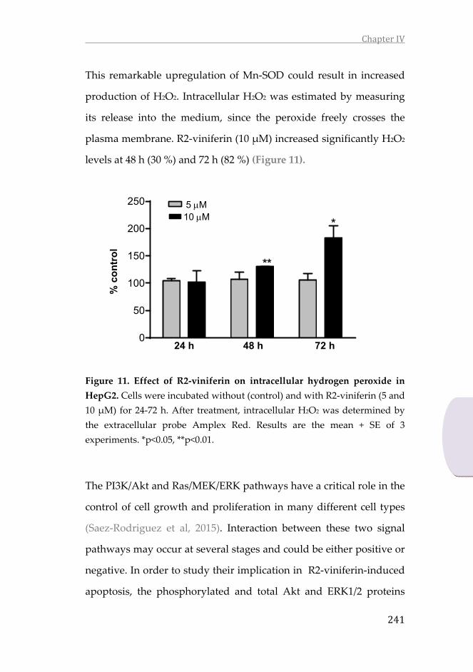
ChapterIV
241
This remarkable upregulation of Mn‐SOD could result in increased
production of H2O2. Intracellular H2O2 was estimated by measuring
its release into the medium, since the peroxide freely crosses the
plasma membrane. R2‐viniferin (10 μM) increased significantly H2O2
levels at 48 h (30 %) and 72 h (82 %) (Figure 11).
Figure 11. Effect of R2‐viniferin on intracellular hydrogen peroxide in
HepG2. Cells were incubated without (control) and with R2‐viniferin (5 and
10 μM) for 24‐72 h. After treatment, intracellular H2O2 was determined by
the extracellular probe Amplex Red. Results are the mean + SE of 3
experiments. *p<0.05, **p<0.01.
The PI3K/Akt and Ras/MEK/ERK pathways have a critical role in the
control of cell growth and proliferation in many different cell types
(Saez‐Rodriguez et al, 2015). Interaction between these two signal
pathways may occur at several stages and could be either positive or
negative. In order to study their implication in R2‐viniferin‐induced
apoptosis, the phosphorylated and total Akt and ERK1/2 proteins
0
50
100
150
200
250
**
*
5 M10 M
24 h 48 h 72 h
% c
on
tro
l

ChapterIV
242
were determined by western blot. The oligostilbene triggered
dephosphorylation of Akt at 72 h, but increased ERK1/2
phosphorylation at the same time (Figure 12). These data suggest
that R2‐viniferin‐induced apoptosis could be modulated by the
PI3K/Akt and ERK1/2 pathways.
Figure 12. Effect of R2‐viniferin on protein phosphorylation of A) Akt
and B) ERK1/2 in HepG2. Cells were incubated without (control) and with
R2‐viniferin (5 and 10 μM) at the indicated times. The bar graph shows
phospho‐Akt/Akt or phospho‐ERK/ERK ratio compared to the control. The
bands shown are representative of at least 3 independent experiments.
Results are the mean + SE of 3 experiments. *p<0.05, **p<0.01, ***p<0.001,
compared with control.
24 h 48 h 72 h0
50
100
150
200
***
*
24 h 48 h 72 h0
50
100
150
***
*
p-Akt
Akt
24 h 48 h 72 h
M 0 5 10 0 5 10 0 5 10
A)
p-ERK1/2
ERK1/2
24 h 48 h 72 h
M 0 5 10 0 5 10 0 5 10
B)
% c
on
tro
l
% c
on
tro
l
24 h 48 h 72 h 24 h 48 h 72 h
5 M10 M
5 M10 M

ChapterIV
243
‐H2AX histone represents an early event in the cellular response
against DNA double‐strand breaks and plays a central role in
sensing and repairing DNA damage. R2‐viniferin prompted the
increase ‐H2AX protein levels in a dose‐ and time‐dependent
manner (Figure 13), suggesting that the stilbene induces DNA
damage that is detected earlier than the apoptotic response.
Figure 13. Effect of R2‐viniferin on ‐H2AX levels in HepG2. Cells were
incubated without (control) and with R2‐viniferin (5 and 10 μM) at the
indicated times. Proteins were analyzed by western blot.
0
250
500800
900
1000
-H2AX
GAPDH
24 h 48 h 72 h
M 0 5 10 0 5 10 0 5 10
5 M10 M
% c
on
tro
l
24 h 48 h 72 h

ChapterIV
244
4. Discussion
The aim of this work was to investigate the mechanism by which R2‐
viniferin exerts toxic actions in HepG2 and specifically whether p53
is mediating this effect. To investigate the implication of this tumor
suppressor, p53 was transiently silenced by siRNA in HepG2 and
transfected with plasmid coding for the protein in Hep3B. Results
showed an almost lack of effect in p53‐silenced HepG2 and a higher
sensitivity to R2‐viniferin of p53‐transfected Hep3B. These results,
together with a higher phosphorylation of the protein and the
upregulation of p21, confirm that the induced toxicity is due, at least
in part, to p53 status. p53 is subjected to numerous post‐translational
modifications (phosphorylation, modification with ubiquitin and
other ubiquitin‐like proteins, acetylation, methylation, glycosylation,
farnesylation, hydroxylation and ADP ribosylation). These post‐
translational modifications, in concert with the interaction of p53
with a variety of protein‐binding partners, regulate the subcellular
localization as well as its transcriptional and transcriptionally
independent functions (Gu et al. 2012). p53 transcriptionally
activates the expression of several pro‐apoptotic Bcl‐2 family
proteins, and directly interacts with various pro‐apoptotic and anti‐
apoptotic proteins in the cytoplasm and at the mitochondrial
membrane (Green et al. 2009). Under pro‐apoptotic conditions, p53
interacts with Bcl2, Bcl‐XL and Bak, de‐repressing Bax and Bak
(Chipuk et al. 2004). p53 also participates in cell repair. Thus,
following DNA damage, p53‐activated cell cycle arrest can suspend

ChapterIV
245
cell cycle progression, allowing DNA lesions to be repaired. The
main transcriptional target of p53 involved in establishing this
process is CDKN1A (encoding p21), which can induce a temporary
cell cycle arrest in addition to contributing to senescence. In response
to DNA double‐strand breaks the ataxia‐telangiectasia mutated
(ATM) kinase phosphorylates ‐H2AX and p53 at Ser15 (Menolfi et
al. 2020). We have shown that R2‐viniferin triggered ‐H2AX
phosphorylation and activation of p53, as well as induced
transcription of p21. In the previous study, we detected a cycle arrest
at G2/M in cells treated with R2‐viniferin (Aja et al., 2020). It should
be taken into account that ATM‐mediated phosphorylation of p53
and Chk2 leads to a series of cell cycle G1 and G2 checkpoints,
preventing cell cycle progression and thus avoiding genomic
instability after DNA damage (Matsuoka et al. 2000). A similar
mechanism of toxicity could be the case of R2‐viniferin in HepG2.
The Bcl‐2 promotor contains a p53‐negative response element
(Seto et al. 1992), raising the possibility that Bcl‐2 may be a direct
target of p53‐mediated transrepression (Wu et al. 2001). In this work,
we have shown that p53 transfection in Hep3B rendered the cells
more responsive to R2‐viniferin and, by contrast, p53 silencing in
HepG2 triggered resistance to the oligostilbene. The polyphenol
treatment activated p53, as determined by Ser15 phosphorylation and
p21 upregulation. These changes were accompanied by a highly
significant decrease in Bcl‐2 expression, rendering a Bax/Bcl‐2 ratio
more prone to apoptosis. We detected the activation of caspase‐9, as

ChapterIV
246
shown by the procaspase‐9 cleavage. p53 may trigger apoptosis
through both mitochondrial and death‐receptor pathways (Henry et
al. 2012). We were unable to detect caspase‐8 activity, so our results
points to a p53‐related activation of the intrinsic apoptotic pathway.
Activation of MLKL, a biochemical marker specific for
necroptosis, is associated with LDH release presumably caused by
cell membrane permeabilization. At the plasma membrane,
phospho‐MLKL sequesters the phosphoinositides,
phosphatidylinositol 4,5‐bisphosphate (PInsP2) and
phosphatidylinositol (3,4,5)‐trisphosphate (PInsP3), during assembly
of a pore‐forming cytotoxic complex. This precludes access to and
activation of phosphoinositide‐3 kinase, which may account for the
reduced phosphorylation of Akt (Dondelinger et al. 2014). We have
actually detected a decline in the phosphorylation status of Akt as a
consequence of R2‐viniferin treatment. However, this loss of pro‐
survival signaling was not accompanied by an increased in MLKL
phosphorylation. R2‐viniferin prompted LDH release into the
medium, pointing to a class of necrotic death that would not be
associated with necroptosis.
The early ROS accumulation induced by R2‐viniferin
previously reported (Aja et al., 2020) could be due to an
overproduction of the O2‐ that was converted into the H2O2 signaling
molecule. In fact, in this work we specifically demonstrated the
increase in the intracellular levels of H2O2 and a remarkable
upregulation of the mitochondrial Mn‐SOD enzyme, which would

ChapterIV
247
be responsible of the imbalance of H2O2 levels. As already suggested
for the antitumor drug paclitaxel in human lung cancer cells, the
early accumulation of mitochondrial H2O2 is a crucial event in the
induced toxicity (Alexander et al., 2006), and this could be the case of
R2‐viniferin. Despite the recognized antioxidant properties in both in
vitro and in vivo models, polyphenols induce apoptosis increasing
ROS production in different cancer cell lines, but not in normal cells
(Hadi et al. 2010; Khan et al. 2014; Liu et al. 2019; Aja et al., 2020).
This dual behavior is attributed in part to the pro‐oxidant properties
of polyphenols in the presence of transition metals, specifically iron
and copper. Various reports indicate that elevated intracellular iron
and copper levels is a hallmark in cancerous cells (Gupte et al. 2008;
Ebara et al. 2000; Yoshida et al. 1993). The presence of high levels of
these metals together with a higher production of H2O2 leads to
Fenton reaction and lipid peroxidation. This will eventually cause
cell death through ferroptosis, a type of programmed necrotic death
(Dixon et al. 2019). Although not addressed in this work, it is
conceivable that R2‐viniferin, through the increased production of
hydrogen peroxide, could trigger the activation of this programmed
cell death.
In summary, we have shown that R2‐viniferin exerts
cytotoxic actions in human hepatoma HepG2 cells by a p53‐
dependent mechanism. It involves DNA damage, the
phosphorylation and activation of p53 and the induction of p21
expression. The oligostilbene also causes induced apoptosis through

ChapterIV
248
caspase‐9 activation, an increase in the intracellular H2O2 levels, and
the upregulation of mitochondrial MnSOD. PI3K/Akt and ERK1/2
pathways are suggested to modulate R2‐viniferin‐induced apoptosis.
The effects exerted by the oligostilbene leads eventually to a
permanent inhibition of clonogenicity and migration. The proposed
mechanism of action exhibited by R2‐viniferin is outlined in Figure
14. The exact mechanism exerted by this novel stilbene represents an
important advance in the research for the development of this
natural compound in chemotherapies against p53‐positive human
hepatocellular carcinomas.
Figure 14. Proposed mechanism of toxicity exerted by R2‐viniferin in
HepG2.

ChapterIV
249
5. References
Aja, Iris, M. Begoña Ruiz‐Larrea, Arnaud Courtois, Stéphanie Krisa, TristanRichard, and José‐Ignacio Ruiz‐Sanz. 2020. “Screening of NaturalStilbene Oligomers from Vitis Vinifera for Anticancer Activity onHuman Hepatocellular Carcinoma Cells.” Antioxidants 9 (6): 469.https://doi.org/10.3390/antiox9060469.
Alexandre, Jérôme,FrédéricBatteux,CaroleNicco,ChristianeChéreau,AlexisLaurent,LoïcGuillevin,BernardWeill,andFrançoisGoldwasser.2006.“AccumulationofHydrogenPeroxide IsanEarlyandCrucialStep forPaclitaxel‐Induced Cancer Cell Death Bothin Vitro Andin Vivo.”International Journal of Cancer 119 (1): 41–48.https://doi.org/10.1002/ijc.21685.
Bradford,MarionM.1976.“ARapidandSensitiveMethodfortheQuantitationofMicrogramQuantitiesofProteinUtilizing thePrincipleofProtein‐Dye Binding.” Analytical Biochemistry 72 (1–2): 248–54.https://doi.org/10.1016/0003‐2697(76)90527‐3.
Chipuk, J. E. 2004. “Direct Activation of Bax by P53MediatesMitochondrialMembrane Permeabilization and Apoptosis.” Science 303 (5660):1010–14.https://doi.org/10.1126/science.1092734.
Dixon, ScottJ., KathrynM. Lemberg, MichaelR. Lamprecht, Rachid Skouta,EleinaM. Zaitsev, CarolineE. Gleason, DarpanN. Patel, et al. 2012.“Ferroptosis: An Iron‐Dependent Form of Nonapoptotic Cell Death.”Cell149(5):1060–72.https://doi.org/10.1016/j.cell.2012.03.042.
Dondelinger, Yves, Wim Declercq, Sylvie Montessuit, Ria Roelandt, AmandaGoncalves, Inge Bruggeman, Paco Hulpiau, et al. 2014. “MLKLCompromises Plasma Membrane Integrity by Binding toPhosphatidylinositol Phosphates.” Cell Reports 7 (4): 971–81.https://doi.org/10.1016/j.celrep.2014.04.026.
Ebara,Masaaki,HiroyukiFukuda,RyoujiHatano,HiromitsuSaisho,YoshinoriNagato, Keiji Suzuki, Katuyuki Nakajima, et al. 2000. “Relationshipbetween Copper, Zinc and Metallothionein in HepatocellularCarcinoma and Its Surrounding Liver Parenchyma.” Journal ofHepatology 33 (3): 415–22. https://doi.org/10.1016/S0168‐8278(00)80277‐9.

ChapterIV
250
Gabaston,Julien,ToniElKhawand,PierreWaffo‐Teguo,AlainDecendit,TristanRichard, Jean‐Michel Mérillon, and Roman Pavela. 2018. “Stilbenesfrom Grapevine Root: A Promising Natural Insecticide againstLeptinotarsaDecemlineata.” JournalofPestScience91(2):897–906.https://doi.org/10.1007/s10340‐018‐0956‐2.
Galle,PeterR.,AlejandroForner, JosepM.Llovet,VincenzoMazzaferro,FabioPiscaglia, Jean‐Luc Raoul, Peter Schirmacher, and Valérie Vilgrain.2018. “EASL Clinical Practice Guidelines: Management ofHepatocellular Carcinoma.” Journal of Hepatology 69 (1): 182–236.https://doi.org/10.1016/j.jhep.2018.03.019.
Green, Douglas R., and Guido Kroemer. 2009. “Cytoplasmic Functions of theTumour Suppressor P53.” Nature 458 (7242): 1127–30.https://doi.org/10.1038/nature07986.
Gu, Bo, and Wei‐Guo Zhu. 2012. “Surf the Post‐Translational ModificationNetwork of P53 Regulation.” International Journal of BiologicalSciences8(5):672–84.https://doi.org/10.7150/ijbs.4283.
Gupte,Anshul, andRussell J.Mumper.2009. “ElevatedCopperandOxidativeStress in Cancer Cells as a Target for Cancer Treatment.” CancerTreatment Reviews 35 (1): 32–46.https://doi.org/10.1016/j.ctrv.2008.07.004.
Hadi,S.M.,M.F.Ullah,A.S.Azmi,A.Ahmad,U.Shamim,H.Zubair,andH.Y.Khan. 2010. “Resveratrol Mobilizes Endogenous Copper in HumanPeripheral Lymphocytes Leading to Oxidative DNA Breakage: APutativeMechanismforChemopreventionofCancer.”PharmaceuticalResearch27(6):979–88.https://doi.org/10.1007/s11095‐010‐0055‐4.
Henry, Ryan E, Zdenek Andrysik, Ramiro París, Matthew D Galbraith, andJoaquínMEspinosa.2012.“ADR4:TBIDAxisDrivestheP53ApoptoticResponse byPromotingOligomerization of PoisedBAX:MechanismsofCellFateChoicetoP53Activation.”TheEMBOJournal31(5):1266–78.https://doi.org/10.1038/emboj.2011.498.
Jiang, Yu, Qiu‐Ju Han, and Jian Zhang. 2019. “Hepatocellular Carcinoma:Mechanisms of Progression and Immunotherapy.” World Journal ofGastroenterology 25 (25): 3151–67.https://doi.org/10.3748/wjg.v25.i25.3151.

ChapterIV
251
Khan, Husain Yar, Haseeb Zubair, Mohd Faisal, Mohd Fahad Ullah, MohdFarhan, Fazlul H. Sarkar, Aamir Ahmad, and Sheikh Mumtaz Hadi.2014. “Plant Polyphenol Induced Cell Death in Human Cancer CellsInvolves Mobilization of Intracellular Copper Ions and ReactiveOxygen Species Generation: A Mechanism for CancerChemopreventive Action.” Molecular Nutrition & Food Research 58(3):437–46.https://doi.org/10.1002/mnfr.201300417.
Kim,Jinchul,LiliYu,WanchengChen,YanxiaXu,MengWu,DilyanaTodorova,Qingshuang Tang, et al. 2019. “Wild‐Type P53 Promotes CancerMetabolic Switch by Inducing PUMA‐Dependent Suppression ofOxidative Phosphorylation.” Cancer Cell 35 (2): 191‐203.e8.https://doi.org/10.1016/j.ccell.2018.12.012.
Kunst, Claudia, Marika Haderer, Sebastian Heckel, Sophie Schlosser, andMartinaMüller.2016.“TheP53FamilyinHepatocellularCarcinoma.”Translational Cancer Research 5 (6): 632–38.https://doi.org/10.21037/tcr.2016.11.79.
Liu,Zheng‐Hong,Cheng‐XiYang,LeiZhang,Cong‐YingYang,andXiu‐QuanXu.2019.“Baicalein,asaProoxidant,TriggersMitochondrialApoptosisinMCF‐7 Human Breast Cancer Cells Through Mobilization ofIntracellular Copper and Reactive Oxygen Species Generation.”OncoTargets and Therapy Volume 12 (December): 10749–61.https://doi.org/10.2147/OTT.S222819.
Matsuoka,S.,G.Rotman,A.Ogawa,Y.Shiloh,K.Tamai,andS.J.Elledge.2000.“Ataxia Telangiectasia‐Mutated Phosphorylates Chk2 in Vivo and inVitro.” Proceedings of the National Academy of Sciences 97 (19):10389–94.https://doi.org/10.1073/pnas.190030497.
Menolfi,Demis, andShanZha.2020. “ATM,ATRandDNA‐PKcsKinases—theLessons from the Mouse Models: Inhibition ≠ Deletion.” Cell &Bioscience10(1).https://doi.org/10.1186/s13578‐020‐0376‐x.
Muenzner, Julienne K., Philipp Kunze, Pablo Lindner, Sandra Polaschek, KiraMenke,MarkusEckstein,CarolI.Geppert,etal.2018.“GenerationandCharacterization of Hepatocellular Carcinoma Cell Lines withEnhanced Cancer Stem Cell Potential.” Journal of Cellular andMolecular Medicine 22 (12): 6238–48.https://doi.org/10.1111/jcmm.13911.

ChapterIV
252
Ramírez‐Garza, Sonia, Emily Laveriano‐Santos, María Marhuenda‐Muñoz,Carolina Storniolo, Anna Tresserra‐Rimbau, Anna Vallverdú‐Queralt,and Rosa Lamuela‐Raventós. 2018. “Health Effects of Resveratrol:Results from Human Intervention Trials.” Nutrients 10 (12): 1892.https://doi.org/10.3390/nu10121892.
Saez‐Rodriguez, Julio, Aidan MacNamara, and Simon Cook. 2015. “ModelingSignaling Networks to Advance New Cancer Therapies.” AnnualReview of Biomedical Engineering 17 (1): 143–63.https://doi.org/10.1146/annurev‐bioeng‐071813‐104927.
Schulze, Kornelius, Sandrine Imbeaud, Eric Letouzé, Ludmil B Alexandrov,Julien Calderaro, Sandra Rebouissou, Gabrielle Couchy, et al. 2015.“Exome Sequencing of Hepatocellular Carcinomas Identifies NewMutational Signatures and Potential Therapeutic Targets.” NatureGenetics47(5):505–11.https://doi.org/10.1038/ng.3252.
Seto,E.,A.Usheva,G.P.Zambetti,J.Momand,N.Horikoshi,R.Weinmann,A.J.Levine, and T. Shenk. 1992. “Wild‐Type P53 Binds to the TATA‐Binding Protein and Represses Transcription.” Proceedings of theNational Academy of Sciences 89 (24): 12028–32.https://doi.org/10.1073/pnas.89.24.12028.
Trepiana, Jenifer,SusanaMeijide,RosauraNavarro,M.LuisaHernández, JoséIgnacio Ruiz‐Sanz, and M. Begoña Ruiz‐Larrea. 2017. “Influence ofOxygen Partial Pressure on the Characteristics of HumanHepatocarcinoma Cells.” Redox Biology 12 (August): 103–13.https://doi.org/10.1016/j.redox.2017.02.004.
Trepiana, Jenifer, M. Begoña Ruiz‐Larrea, and José Ignacio Ruiz‐Sanz. 2018.“UnravelingtheinVitroAntitumorActivityofVismiaBacciferaagainstHepG2: Role of Hydrogen Peroxide.” Heliyon 4 (6): e00675.https://doi.org/10.1016/j.heliyon.2018.e00675.
Wu, Yu‐ling, John W Mehew, Caroline A Heckman, Magdalena Arcinas, andLindaMBoxer.2001.“NegativeRegulationofBcl‐2ExpressionbyP53in Hematopoietic Cells.” Oncogene 20 (2): 240–51.https://doi.org/10.1038/sj.onc.1204067.
Yoshida, Daizo, Yukio Ikeda, and Shozo Nakazawa. 1993. “QuantitativeAnalysis of Copper, Zinc and Copper/Zinc Ratio in Selected HumanBrain Tumors.” Journal of Neuro‐Oncology 16 (2): 109–15.https://doi.org/10.1007/BF01324697.

ChapterIV
253
Declaration of interest
The authors declare that there is no conflict of interest that could be
perceived as prejudicing the impartiality of the research reported.
Funding
This research was funded by a PhD Research Fellowship in the
context of Campus Euro‐regional Bordeaux/Euskampus (ref.
PIFBUR16‐01 to I.A.). The work was supported by the University of
the Basque Country UPV/EHU (to Research Groups, ref. GIU16/62)
and the Bordeaux Metabolome Facility and MetaboHUB (ANR‐11‐
INBS‐0010 project). The authors thank J.A. López for his technical
assistance.


CONCLUSIONS


Conclusions
257
CONCLUSIONS
1. The woody parts (roots) of the Vitis vinifera grapevine contain
the glucosylated derivatives of resveratrol: resveratroloside,
resveratrol rutinoside, and trans‐ and cis‐‐viniferin
diglucosides, which show moderate/low biological activity.
2. The piceatannol monomer and the resveratrol
oligomers,viniferin, hopeaphenol and isohopeaphenol,
inhibit the inflammatory response to lipopolysacharide‐
stimulated murine macrophages, decreasing the levels of
nitric oxide, reactive oxygen species, and the pro‐
inflammatory TNF‐ and IL‐1cytokines.
3. From several natural resveratrol oligomers isolated from Vitis
vinifera cane (ampelopsin A, trans‐‐viniferin, hopeaphenol,
isohopeaphenol, R2‐viniferin, and R‐viniferin), the R2‐
viniferin tetramer is the most potent cytotoxic stilbene in
inducing cell death of human HepG2 hepatoma cell line,
without damaging non‐transformed human hepatocyes.
4. R2‐viniferin exerts cytotoxic actions in human hepatoma
HepG2 cells by a p53‐dependent mechanism. It involves
DNA damage, the phosphorylation and activation of p53 and
the induction of p21 expression. The oligostilbene also causes
apoptosis through caspase‐9 activation, an increase in the
intracellular H2O2 levels, and the upregulation of
mitochondrial MnSOD.
From the results, we conclude that Vitis vinifera cane is a source of
R2‐viniferin oligostilbene, a promising natural compound in the
development of chemotherapies against p53‐positive human
hepatocellular carcinomas. Further studies will be required in order

Conclusions
258
to improve its bioavailability and to unravel its mechanism of action
both in vitro and vivo models for its potential clinical application in
the treatment of liver cancer.


Copyright © 2022 FDOKUMEN


![Fluorescence properties of a potential antitumoral benzothieno[3,2-b]pyrrole in solution and lipid membranes](https://static.fdokumen.com/doc/165x107/63440ec5df19c083b1076b23/fluorescence-properties-of-a-potential-antitumoral-benzothieno32-bpyrrole-in.jpg)