Antifouling Mortars for Underwater Restoration - MDPI
14
Citation: Ricca, M.; Ruffolo, S.A.; La Russa, M.F.; Rispoli, C.; Grifa, C.; Sierra-Fernández, A.; Fort, R.; Randazzo, L. Antifouling Mortars for Underwater Restoration. Nanomaterials 2022, 12, 1498. https:// doi.org/10.3390/nano12091498 Academic Editor: Andreu Cabot Received: 11 April 2022 Accepted: 27 April 2022 Published: 28 April 2022 Publisher’s Note: MDPI stays neutral with regard to jurisdictional claims in published maps and institutional affil- iations. Copyright: © 2022 by the authors. Licensee MDPI, Basel, Switzerland. This article is an open access article distributed under the terms and conditions of the Creative Commons Attribution (CC BY) license (https:// creativecommons.org/licenses/by/ 4.0/). nanomaterials Article Antifouling Mortars for Underwater Restoration Michela Ricca 1 , Silvestro Antonio Ruffolo 1, * , Mauro Francesco La Russa 1 , Concetta Rispoli 2 , Celestino Grifa 3 , Aranzazu Sierra-Fernández 4 , Rafael Fort 4 and Luciana Randazzo 5 1 Dipartimento di Biologia, Ecologia e Scienze della Terra, Università della Calabria, Via Pietro Bucci, 87036 Arcavacata di Rende, Italy; [email protected] (M.R.); [email protected] (M.F.L.R.) 2 Dipartimento di Scienze della Terra, dell’Ambiente e delle Risorse, University of Naples Federico II, Complesso Universitario Monte Sant’Angelo, ED. 10, Via Cintia 26, 80126 Naples, Italy; [email protected] 3 Dipartimento di Scienze e Tecnologie, Università degli Studi del Sannio di Benevento, Via De Sanctis snc, 82100 Benevento, Italy; [email protected] 4 Instituto de Geociencias (CSIC, UCM) C/Severo Ochoa 7, 28040 Madrid, Spain; [email protected] (A.S.-F.); [email protected] (R.F.) 5 Dipartimento di Scienze delle Terra e del Mare, Università di Palermo, Via Archirafi, 26, 90123 Palermo, Italy; [email protected] * Correspondence: [email protected] Abstract: This research has focused on the assessment of the compositional features and mechanical and antifouling performances of two different mortars formulated for an underwater setting, and which contain Mg(OH) 2 as an antifouling agent. Regarding the mechanical characterization, the uniaxial compressive strength and flexural strength were measured. The composition of the materials was explored by differential thermal/thermogravimetric analysis (DTA-TG), X-ray diffraction analysis (XRPD), and scanning electronic microscopy (SEM) coupled with EDS microanalysis. The assessment of the biological colonization was evaluated with colorimetric analysis and image analysis. The results suggest that both mortars have good mechanical resistance once set underwater. Moreover, the adding of Mg(OH) 2 improves the resistance toward biofouling; this was observed both in laboratory and sea-exposed specimens. Keywords: magnesium hydroxide; mortars; submerged sites; biofouling; geomaterials; restoration; nanoparticles 1. Introduction In recent decades, there has been an increasing interest in the study of the degradation phenomena affecting the archaeological sites located in submarine environments [1,2] The most recent guidelines about underwater heritage are focused on in situ conserva- tion (i.e., UNESCO Convention on the Protection of the Underwater Cultural Heritage, 2 November 2001). Since their issue, several techniques and materials have been proposed for their conservation [3–7]. The main cause of the decay of natural (e.g., stone) and artificial geomaterials (e.g., mortars, pottery) in an underwater environment is biodeterioration [8,9] in the form of biofouling and bioerosion phenomena [10–13]. Biological weathering, com- monly called biofouling, is a consequence of physical, chemical, and biological factors that contribute to the decay of submerged materials. It is a natural process occurring on underwater items through the colonization and overgrowth of epilithic and endolithic organisms and represents a major economic problem in both archaeological sites and maritime industries [14]. The first events in biofouling formation are the deposition of mul- tilayered organic matter (biofilm), followed by colonization with specific bacterial species (microfouling) [11]. This primary microbial film can prepare the surface for subsequent colonization by extracellular polymeric substances production, which are responsible for the adhesion and growth of new microorganisms, plants, algae, and sessile organisms. The Nanomaterials 2022, 12, 1498. https://doi.org/10.3390/nano12091498 https://www.mdpi.com/journal/nanomaterials
-
Upload
khangminh22 -
Category
Documents
-
view
1 -
download
0
Transcript of Antifouling Mortars for Underwater Restoration - MDPI

Citation: Ricca, M.; Ruffolo, S.A.; La
Russa, M.F.; Rispoli, C.; Grifa, C.;
Sierra-Fernández, A.; Fort, R.;
Randazzo, L. Antifouling Mortars for
Underwater Restoration.
Nanomaterials 2022, 12, 1498. https://
doi.org/10.3390/nano12091498
Academic Editor: Andreu Cabot
Received: 11 April 2022
Accepted: 27 April 2022
Published: 28 April 2022
Publisher’s Note: MDPI stays neutral
with regard to jurisdictional claims in
published maps and institutional affil-
iations.
Copyright: © 2022 by the authors.
Licensee MDPI, Basel, Switzerland.
This article is an open access article
distributed under the terms and
conditions of the Creative Commons
Attribution (CC BY) license (https://
creativecommons.org/licenses/by/
4.0/).
nanomaterials
Article
Antifouling Mortars for Underwater RestorationMichela Ricca 1 , Silvestro Antonio Ruffolo 1,* , Mauro Francesco La Russa 1, Concetta Rispoli 2 ,Celestino Grifa 3, Aranzazu Sierra-Fernández 4, Rafael Fort 4 and Luciana Randazzo 5
1 Dipartimento di Biologia, Ecologia e Scienze della Terra, Università della Calabria, Via Pietro Bucci,87036 Arcavacata di Rende, Italy; [email protected] (M.R.); [email protected] (M.F.L.R.)
2 Dipartimento di Scienze della Terra, dell’Ambiente e delle Risorse, University of Naples Federico II,Complesso Universitario Monte Sant’Angelo, ED. 10, Via Cintia 26, 80126 Naples, Italy;[email protected]
3 Dipartimento di Scienze e Tecnologie, Università degli Studi del Sannio di Benevento, Via De Sanctis snc,82100 Benevento, Italy; [email protected]
4 Instituto de Geociencias (CSIC, UCM) C/Severo Ochoa 7, 28040 Madrid, Spain; [email protected] (A.S.-F.);[email protected] (R.F.)
5 Dipartimento di Scienze delle Terra e del Mare, Università di Palermo, Via Archirafi, 26, 90123 Palermo, Italy;[email protected]
* Correspondence: [email protected]
Abstract: This research has focused on the assessment of the compositional features and mechanicaland antifouling performances of two different mortars formulated for an underwater setting, andwhich contain Mg(OH)2 as an antifouling agent. Regarding the mechanical characterization, theuniaxial compressive strength and flexural strength were measured. The composition of the materialswas explored by differential thermal/thermogravimetric analysis (DTA-TG), X-ray diffraction analysis(XRPD), and scanning electronic microscopy (SEM) coupled with EDS microanalysis. The assessmentof the biological colonization was evaluated with colorimetric analysis and image analysis. Theresults suggest that both mortars have good mechanical resistance once set underwater. Moreover, theadding of Mg(OH)2 improves the resistance toward biofouling; this was observed both in laboratoryand sea-exposed specimens.
Keywords: magnesium hydroxide; mortars; submerged sites; biofouling; geomaterials; restoration;nanoparticles
1. Introduction
In recent decades, there has been an increasing interest in the study of the degradationphenomena affecting the archaeological sites located in submarine environments [1,2]The most recent guidelines about underwater heritage are focused on in situ conserva-tion (i.e., UNESCO Convention on the Protection of the Underwater Cultural Heritage,2 November 2001). Since their issue, several techniques and materials have been proposedfor their conservation [3–7]. The main cause of the decay of natural (e.g., stone) and artificialgeomaterials (e.g., mortars, pottery) in an underwater environment is biodeterioration [8,9]in the form of biofouling and bioerosion phenomena [10–13]. Biological weathering, com-monly called biofouling, is a consequence of physical, chemical, and biological factorsthat contribute to the decay of submerged materials. It is a natural process occurring onunderwater items through the colonization and overgrowth of epilithic and endolithicorganisms and represents a major economic problem in both archaeological sites andmaritime industries [14]. The first events in biofouling formation are the deposition of mul-tilayered organic matter (biofilm), followed by colonization with specific bacterial species(microfouling) [11]. This primary microbial film can prepare the surface for subsequentcolonization by extracellular polymeric substances production, which are responsible forthe adhesion and growth of new microorganisms, plants, algae, and sessile organisms. The
Nanomaterials 2022, 12, 1498. https://doi.org/10.3390/nano12091498 https://www.mdpi.com/journal/nanomaterials

Nanomaterials 2022, 12, 1498 2 of 14
materials used for underwater restoration may be prone to rapid biodeterioration as well,and among them, mortar-based materials are common in the conservation of underwa-ter archaeological patrimony. In order to be suitable for underwater application, a goodhydraulic behaviour and mineralogical stability are required for restoration mortars [15];moreover, a low susceptibility to bio-colonization is expected during their formulation.
In this study, attention was paid to magnesium hydroxide Mg(OH)2 as an antifoulingagent [16] in different mortar pastes. This material had shown a promising biocidal activitywhen used in coatings for stone materials, especially in the form of nanoparticles withdimensions ranging from ~30 and ~450 nm and well-defined hexagonal shapes [17–20].Magnesium-based materials have generated considerable interest as antimicrobial agentsin a wide variety of applications, such as in the treatment of stone materials used in culturalheritage [19,21,22]. In particular, the use of these nanostructured inorganic materials hasproved to be a highly effective means to prevent bio-colonization as well as to carry outa consolidating action. Commonly, nanostructured materials possess specific physicochem-ical properties that, in combination with their large surface area and dimensions, allowthem to interact and internalize within cells, displaying a broad spectrum of antibacterialactivity [23,24]. Moreover, their modular nature means that a library of relatively low-costmaterials with different sizes, shapes, surface properties, and chemical compositions canbe developed, leading to a great potential for developing effective antimicrobial agentswith high stability under harsh environmental conditions. It is important to note that theantibacterial effect often depends on the size, shape, chemical composition, and surfaceproperties (e.g., hydrophobicity) of the nanoparticles [22,25,26].
The behavior of these formulations in terms of mechanical strength, compositionalcharacteristics and resistance to biodeterioration when exposed to the underwater environ-ment was evaluated in two different exposure conditions (i.e., in lab and in situ trials).
2. Materials and Methods
For the experiment, two types of natural hydraulic mortars were selected: (i) one isbased on Volteco Microlime Gel (Volteco, Ponzano Veneto, Italy) (hereinafter named VM),a mixture composed of aerial and hydraulic binders loaded with organic thickeners whichprovide pseudoplastic behavior; (ii) the second is based on NHL 3.5 Saint Astier (CESA,Saint-Astier, France) (hereinafter named SA), 3.5 type natural hydraulic lime (NHL).
A pozzolanic aggregate was mixed in both starting materials; for this purpose, thePozzolana fine (Rime 1, Srl, Rome, Italy) was added, with a particle size from 75 microns to1 mm. The mortars were prepared with a binder/aggregate ratio equal to 1/2 and a wateramount of 30% w/w with respect to the binder amount. To explore the use of magnesiumhydroxide (Mg(OH)2) nanoparticles as antifouling agents for mortars, 44 specimens weremixed with 1% w/w magnesium hydroxide Mg(OH)2 nanoparticles, synthesized accordingto Sierra-Fernandez et al., 2019. A total of 22.02 g of magnesium methoxide (Mg(OCH3)2was dissolved in ethanol (85.2 mL) at 60 ◦C. After that, 25.2 mL of deionized water wasadded to the above solution drop-wise and allowed to reflux at 60 ◦C for 24 h. Thereafter,the solution was centrifuged and washed several times with mixtures of ethanol: distilledwater. Finally, the sample was dried under an inert gas atmosphere at 100 ◦C for 4 h. Twoseries of specimens were prepared to evaluate the performance of artificial stone materialsbefore and after exposure to seawater, under different exposure conditions. Respectively,the samples were of dimensions 16 × 4 × 4 cm for mechanical tests and 10 × 5 × 0.8 cmfor the evaluation of biological growth. Before the exposure of the mortars to the differentexperimental conditions, a curing time of 28 days was achieved. Specifically, blends werepoured into molds and then were transferred to a cabin storage after 24 h, where they werekept under controlled conditions of 20 ± 2 ◦C, and relative humidity >95% for 28 days. Asummary of the specimens used in the two experimental sets, with a brief description andanalytical techniques used, are shown in Figures 1 and 2 and summarized in Table 1.
Nanomaterials 2022, 12, 1498 3 of 14Nanomaterials 2022, 12, x FOR PEER REVIEW 3 of 15
Figure 1. (a) Steel mold scheme consisting of three horizontal compartments for prismatic speci-mens’ preparation (16 × 4 × 4 cm); (b) cabin storage for curing specimens; (c) specimens after 28 days of curing time; (d) compressive strength testing machine; (e) wood mold for specimen preparation (10 × 8 × 0.8 cm); (f) rectangular glass tank for lab experimentation.
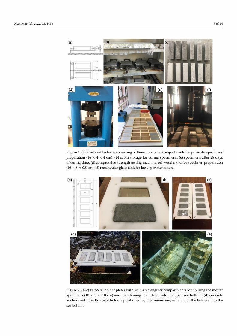
Figure 2. (a–c) Ertacetal holder plates with six (6) rectangular compartments for housing the mortar specimens (10 × 5 × 0.8 cm) and maintaining them fixed into the open sea bottom; (d) concrete an-chors with the Ertacetal holders positioned before immersion; (e) view of the holders into the sea bottom.
Figure 1. (a) Steel mold scheme consisting of three horizontal compartments for prismatic specimens’preparation (16 × 4 × 4 cm); (b) cabin storage for curing specimens; (c) specimens after 28 daysof curing time; (d) compressive strength testing machine; (e) wood mold for specimen preparation(10 × 8 × 0.8 cm); (f) rectangular glass tank for lab experimentation.
Nanomaterials 2022, 12, x FOR PEER REVIEW 3 of 15
Figure 1. (a) Steel mold scheme consisting of three horizontal compartments for prismatic speci-mens’ preparation (16 × 4 × 4 cm); (b) cabin storage for curing specimens; (c) specimens after 28 days of curing time; (d) compressive strength testing machine; (e) wood mold for specimen preparation (10 × 8 × 0.8 cm); (f) rectangular glass tank for lab experimentation.
Figure 2. (a–c) Ertacetal holder plates with six (6) rectangular compartments for housing the mortar specimens (10 × 5 × 0.8 cm) and maintaining them fixed into the open sea bottom; (d) concrete an-chors with the Ertacetal holders positioned before immersion; (e) view of the holders into the sea bottom.
Figure 2. (a–c) Ertacetal holder plates with six (6) rectangular compartments for housing the mortarspecimens (10 × 5 × 0.8 cm) and maintaining them fixed into the open sea bottom; (d) concreteanchors with the Ertacetal holders positioned before immersion; (e) view of the holders into thesea bottom.
Nanomaterials 2022, 12, 1498 4 of 14
Table 1. Summary of the specimens used in the two experimental sets, with a brief description andanalytical techniques used.
Sample Code Lab Experiment In Situ Experiment
MechanicalTest
ColorimetricAnalysis XRD SEM/EDS TG/DTA Image Analysis
Size of Specimens (cm) 16 × 16 × 4 10 × 5 × 0.8 10 × 5 × 0.8 (n = 16)
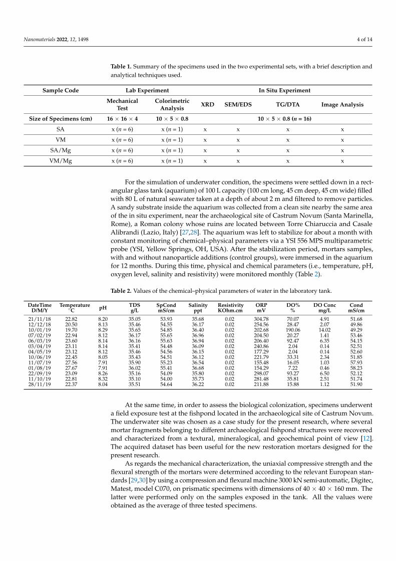
SA x (n = 6) x (n = 1) x x x x
VM x (n = 6) x (n = 1) x x x x
SA/Mg x (n = 6) x (n = 1) x x x x
VM/Mg x (n = 6) x (n = 1) x x x x
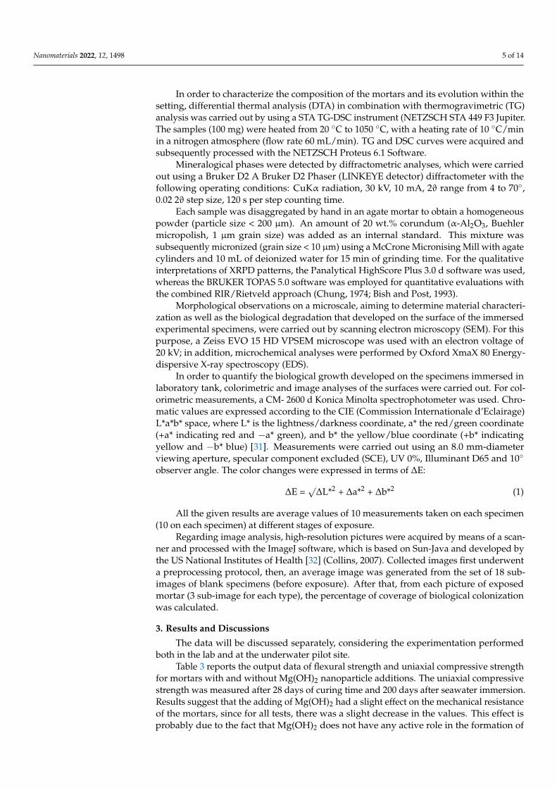
For the simulation of underwater condition, the specimens were settled down in a rect-angular glass tank (aquarium) of 100 L capacity (100 cm long, 45 cm deep, 45 cm wide) filledwith 80 L of natural seawater taken at a depth of about 2 m and filtered to remove particles.A sandy substrate inside the aquarium was collected from a clean site nearby the same areaof the in situ experiment, near the archaeological site of Castrum Novum (Santa Marinella,Rome), a Roman colony whose ruins are located between Torre Chiaruccia and CasaleAlibrandi (Lazio, Italy) [27,28]. The aquarium was left to stabilize for about a month withconstant monitoring of chemical–physical parameters via a YSI 556 MPS multiparametricprobe (YSI, Yellow Springs, OH, USA). After the stabilization period, mortars samples,with and without nanoparticle additions (control groups), were immersed in the aquariumfor 12 months. During this time, physical and chemical parameters (i.e., temperature, pH,oxygen level, salinity and resistivity) were monitored monthly (Table 2).
Table 2. Values of the chemical–physical parameters of water in the laboratory tank.
DateTimeD/M/Y
Temperature◦C pH TDS
g/LSpCondmS/cm
Salinityppt
ResistivityKOhm.cm
ORPmV
DO%%
DO Concmg/L
CondmS/cm
21/11/18 22.82 8.20 35.05 53.93 35.68 0.02 304.78 70.07 4.91 51.6812/12/18 20.50 8.13 35.46 54.55 36.17 0.02 254.56 28.47 2.07 49.8610/01/19 19.70 8.29 35.65 54.85 36.40 0.02 202.68 190.06 14.02 49.2907/02/19 22.94 8.12 36.17 55.65 36.96 0.02 204.50 20.27 1.41 53.4606/03/19 23.60 8.14 36.16 55.63 36.94 0.02 206.40 92.47 6.35 54.1503/04/19 23.11 8.14 35.41 54.48 36.09 0.02 240.86 2.04 0.14 52.5104/05/19 23.12 8.12 35.46 54.56 36.15 0.02 177.29 2.04 0.14 52.6010/06/19 22.45 8.05 35.43 54.51 36.12 0.02 221.79 33.31 2.34 51.8511/07/19 27.56 7.91 35.90 55.23 36.54 0.02 155.48 16.05 1.03 57.9301/08/19 27.67 7.91 36.02 55.41 36.68 0.02 154.29 7.22 0.46 58.2322/09/19 23.09 8.26 35.16 54.09 35.80 0.02 298.07 93.27 6.50 52.1211/10/19 22.81 8.32 35.10 54.00 35.73 0.02 281.48 35.81 2.51 51.7428/11/19 22.37 8.04 35.51 54.64 36.22 0.02 211.88 15.88 1.12 51.90
At the same time, in order to assess the biological colonization, specimens underwenta field exposure test at the fishpond located in the archaeological site of Castrum Novum.The underwater site was chosen as a case study for the present research, where severalmortar fragments belonging to different archaeological fishpond structures were recoveredand characterized from a textural, mineralogical, and geochemical point of view [12].The acquired dataset has been useful for the new restoration mortars designed for thepresent research.
As regards the mechanical characterization, the uniaxial compressive strength and theflexural strength of the mortars were determined according to the relevant European stan-dards [29,30] by using a compression and flexural machine 3000 kN semi-automatic, Digitec,Matest, model C070, on prismatic specimens with dimensions of 40 × 40 × 160 mm. Thelatter were performed only on the samples exposed in the tank. All the values wereobtained as the average of three tested specimens.
Nanomaterials 2022, 12, 1498 5 of 14
In order to characterize the composition of the mortars and its evolution within thesetting, differential thermal analysis (DTA) in combination with thermogravimetric (TG)analysis was carried out by using a STA TG-DSC instrument (NETZSCH STA 449 F3 Jupiter.The samples (100 mg) were heated from 20 ◦C to 1050 ◦C, with a heating rate of 10 ◦C/minin a nitrogen atmosphere (flow rate 60 mL/min). TG and DSC curves were acquired andsubsequently processed with the NETZSCH Proteus 6.1 Software.
Mineralogical phases were detected by diffractometric analyses, which were carriedout using a Bruker D2 A Bruker D2 Phaser (LINKEYE detector) diffractometer with thefollowing operating conditions: CuKα radiation, 30 kV, 10 mA, 2ϑ range from 4 to 70◦,0.02 2ϑ step size, 120 s per step counting time.
Each sample was disaggregated by hand in an agate mortar to obtain a homogeneouspowder (particle size < 200 µm). An amount of 20 wt.% corundum (α-Al2O3, Buehlermicropolish, 1 µm grain size) was added as an internal standard. This mixture wassubsequently micronized (grain size < 10 µm) using a McCrone Micronising Mill with agatecylinders and 10 mL of deionized water for 15 min of grinding time. For the qualitativeinterpretations of XRPD patterns, the Panalytical HighScore Plus 3.0 d software was used,whereas the BRUKER TOPAS 5.0 software was employed for quantitative evaluations withthe combined RIR/Rietveld approach (Chung, 1974; Bish and Post, 1993).
Morphological observations on a microscale, aiming to determine material characteri-zation as well as the biological degradation that developed on the surface of the immersedexperimental specimens, were carried out by scanning electron microscopy (SEM). For thispurpose, a Zeiss EVO 15 HD VPSEM microscope was used with an electron voltage of20 kV; in addition, microchemical analyses were performed by Oxford XmaX 80 Energy-dispersive X-ray spectroscopy (EDS).
In order to quantify the biological growth developed on the specimens immersed inlaboratory tank, colorimetric and image analyses of the surfaces were carried out. For col-orimetric measurements, a CM- 2600 d Konica Minolta spectrophotometer was used. Chro-matic values are expressed according to the CIE (Commission Internationale d’Eclairage)L*a*b* space, where L* is the lightness/darkness coordinate, a* the red/green coordinate(+a* indicating red and −a* green), and b* the yellow/blue coordinate (+b* indicatingyellow and −b* blue) [31]. Measurements were carried out using an 8.0 mm-diameterviewing aperture, specular component excluded (SCE), UV 0%, Illuminant D65 and 10◦
observer angle. The color changes were expressed in terms of ∆E:
∆E =√
∆L*2 + ∆a*2 + ∆b*2 (1)
All the given results are average values of 10 measurements taken on each specimen(10 on each specimen) at different stages of exposure.
Regarding image analysis, high-resolution pictures were acquired by means of a scan-ner and processed with the ImageJ software, which is based on Sun-Java and developed bythe US National Institutes of Health [32] (Collins, 2007). Collected images first underwenta preprocessing protocol, then, an average image was generated from the set of 18 sub-images of blank specimens (before exposure). After that, from each picture of exposedmortar (3 sub-image for each type), the percentage of coverage of biological colonizationwas calculated.
3. Results and Discussions
The data will be discussed separately, considering the experimentation performedboth in the lab and at the underwater pilot site.
Table 3 reports the output data of flexural strength and uniaxial compressive strengthfor mortars with and without Mg(OH)2 nanoparticle additions. The uniaxial compressivestrength was measured after 28 days of curing time and 200 days after seawater immersion.Results suggest that the adding of Mg(OH)2 had a slight effect on the mechanical resistanceof the mortars, since for all tests, there was a slight decrease in the values. This effect isprobably due to the fact that Mg(OH)2 does not have any active role in the formation of

Nanomaterials 2022, 12, 1498 6 of 14
chemical bonds, it has no reactivity, and it is just an inert component. Moreover, as willbe shown later by TGA, the mortar samples with Mg(OH)2 nanoparticle additions andexposed to in-situ conditions presented a higher degree of hydration, which inhibitedthe carbonation rate. Therefore, the lower rate of carbonation detected in these samplestogether with the presence of hydrated phases with deficient stability could be an importantfactor explaining this slight reduction in their strength development. Additionally, there isno clear variation of the compressive strength over time; there is an increase in all valuesafter 200 days, although they have the same magnitude of the standard deviations. Withregards to the comparison between the prepared mortars and the values given by theproducers, there is a slight deviation, in particular, the data is better for the SA mortar,whereas the VM values in terms of compressive strength are comparable. This could belinked to an increased pozzolanic activity due to the further addition of pozzolans to themortars, particularly in the SA typology.
Table 3. Flexural strength and uniaxial compressive strength measured for mortar specimens. * Valuesgiven by the producers, n.a. = data not available.
Flexural Strength28 Days (MPa)
Uniaxial CompressiveStrength 28 Days (MPa)
Uniaxial CompressiveStrength 200 Days (MPa)
SA 2.4 ± 0.5 4.6 ± 0.9 4.8 ± 0.9
SA/Mg 2.0 ± 0.1 3.8 ± 0.1 3.9 ± 0.1
VM 3.8 ± 0.3 7.1 ± 0.8 7.5 ± 0.9
VM/Mg 3.2 ± 0.3 5.5 ± 0.2 5.8 ± 0.2
SA * n.a. 1.88 n.a.
VM * >1.5 >8 n.a.
Colorimetric analysis was carried out on samples after 28 days of immersion inlaboratory tank, and after 200 days as well (Table 4). For all samples, just low chromaticvariations were detected, however, the samples containing Mg(OH)2 showed a lowerchromatic variation. The color change over time is mainly due to biological colonization;this suggests that Mg(OH)2 exerts an antifouling effect.
Table 4. Results of colorimetric analysis.
28 Days of Immersion 200 Days of Immersion
L a b L a b ∆L* ∆a* ∆b* ∆E
SA 75.5 ± 2.2 0.4 ± 0.1 4.8 ± 0.8 76.6 ± 3.8 0.2 ± 0.1 5.0 ± 1.1 1.1 −0.2 0.3 1.1 ± 0.4
VM 80.8 ± 1.7 0.2 ± 0.1 5.1 ± 0.9 79.3 ± 2.9 0.1 ± 0.1 5.3 ± 1.5 −1.5 −0.1 0.2 1.5 ± 0.3
SA/Mg 77.7 ± 2.5 −0.1 ± 0.1 5.8 ± 0.7 78.2 ± 3.1 0.0 ± 0.1 5.3 ± 1.7 0.5 0.1 −0.5 0.7 ± 0.3
VM/Mg 76.9 ± 1.8 −0.1 ± 0.1 5.5 ± 0.6 77.1 ± 2.7 −0.1 ± 0.1 4.7 ± 1.9 0.2 0.0 −0.8 0.8 ± 0.3
In-Situ Experimentation
Table 5 it reports the mineralogical composition according to XRD analysis, showingall the specimens investigated both prior and after the exposure to sea water. There isa compositional difference between SA and VM mortars, maybe due to a different initialcomposition of the binder and the subsequent reactions between the binder and aggregatethat occurred. Specifically, VM is characterized by a greater amount of mineralogicalphases belonging to sandy aggregates such as quartz, plagioclase, pyroxene and leucitewith respect to SA mortar. In addition, VM showed a relatively lower amount in calcite.Such values could affect the entity of the pozzolanic reaction that occurred in the mortars,which is related to the amount of amorphous phases (hydrated compounds such as C-S-A-H or hydrated calcium silicates and aluminates). From a performance point of view,

Nanomaterials 2022, 12, 1498 7 of 14
the major development of neoformation mineralogical phases in SA, linked to pozzolanicreactions, would make the mortar more suitable for consolidation purposes, due to anincrease in resistance and cohesion properties.
Table 5. Results of quantitative X-ray analysis.
Sample Code Cal Qz Anl Lct Cpx Pl Mca Hl LOAP
SA (Control test) 33.0 5.0 3.1 7.2 16.7 3.3 0.1 − 31.6SA 3 Months 33.6 4.1 3.6 6.5 17.0 3.5 0.1 tr 31.5SA 6 Months 30.6 4.8 3.8 7.2 17.6 4.1 0.1 1.1 30.7
SA 12 Months 33.4 5.6 4.3 7.4 18.1 6.2 0.1 tr 24.8SA/Mg 3 Months 33.5 4.7 3.7 7.3 18.3 3.4 0.2 0.22 28.6SA/Mg 6 Months 31.5 5.7 4.3 6.3 18.1 4.5 0.2 0.18 29.3SA/Mg 12 Months 28.4 4.9 3.9 6.9 18.5 3.9 0.2 tr 33.3VM (Control test) 31.5 13.0 4.4 8.4 19.4 6.7 0.1 − 16.0
VM 3 Months 31.6 10.5 4.4 7.5 19.0 5.6 0.2 0.54 21.0VM 6 Months 31.7 5.0 4.4 7.4 18.6 4.6 0.2 − 28.2
VM/Mg 6 Months 30.2 5.2 4.0 7.3 18.0 4.9 0.1 tr 30.2
Legend: Cal, Calcite; Qz, Quartz; Pl, Plagioclase; Cpx, Clinopyroxene; Lct, leucite; Anl, Analcime; Mca, Mica;Hl, halite; Am, Amorphous phases; tr, traces; − not detected.
However, this seems to be disproved by mechanical measurements, since VM samplesare slightly more resistant than SA ones. Such results could depend on the exposuresite, i.e., from the tank and in situ field tests. Moreover, differences in the degree ofcarbonation reaction associated with the incorporation of nanoparticles in the samplescould be established. In this way, a higher content of calcite was detected in sampleswithout nanoparticles (Table 5). In contrast, mortars with Mg(OH)2 nanoparticle additionsshow the major content in amorphous phases after 12 months of exposition, since morehydrated phases could be formed in them. These results are in line with the following TGAfindings, in which a higher amount of water content and hydrated phases (weight loss inregion 40–200 ◦C) and a reduction in the temperature range attributed to the decompositionof carbonates (600–850 ◦C) were determined in these specimens. On the other hand, nonoteworthy difference is evident with regards to the time of exposure to seawater.
Thermal analysis was performed on samples (with and without magnesium hydroxideat different stages of exposure, i.e., 3, 6 and 12 months) in a static air atmosphere, in thetemperature range of 20–1000 ◦C, with a gradient of 10 ◦C/min. Usually, in the mortarcharacterization, the thermal transformations that occur during the heating of the sampleare generally associated: (a) with a loss of hygroscopic water (T < 120 ◦C); (b) with a lossof crystallization water of hydrated salts, such as gypsum (120 < T < 200 ◦C) [33]; (c) witha loss of structurally bound water belonging to hydraulic components, such as hydratedcalcium silicate (CSH) or hydrated calcium aluminates (CAH) (120 < T < 600 ◦C) [34,35];and (d) with the decomposition of carbonates (600 < T < 900 ◦C) [33]. However, due tothe different thermal stability of nanocrystals [36], and the fact that metastable calciumcarbonate polymorphs and hydrated amorphous calcium carbonate are decomposed inthe same temperature range (400–600 ◦C) [37], the interpretation of thermal analysis in themortars can be more complex.
It was not possible to carry out the analytical sequence on all the types of formu-lated mortars. In particular, the data regarding VM (12 months), VM/Mg-3 months and12 months are missing. This was due to strong storms that affected the archaeological siteduring the experiment period. As a result, some of the experimental mortars housed in thesample holder were swept away and it was not possible to recover them in any way.
The recognition and quantitative evaluation of the thermally reactive phases of theaggregate and of the binder were achieved by thermogravimetric analyses (Table 6) thatare considered a widely accepted approach for the hydraulic classification of ancientmortars [38,39]. In the first considered interval of temperature (40–200 ◦C), a weightloss (on average 3 wt%) due to dehydratation of non-chemically bonded water occurred,

Nanomaterials 2022, 12, 1498 8 of 14
the following temperature range (200–600 ◦C) shows the thermal effects of the weightloss due to Structural Bound Water (SBW), generally related to the presence of hydratedcompounds such as calcium silicate hydrate (CSH) and/or calcium aluminate hydrate(CAH); the weight loss ranges from 4.12 to 7.74 wt%. In the 600–850 ◦C the decompositionof calcite and other carbonates is the most prominent thermal effect (on average 10 wt%).The calculated stoichiometric calcite ranges from 18.73 to 29.37 wt%. Finally, at highesttemperatures (>850 ◦C), the weight loss was ca. 1 wt%, likely attributable to halidescomponent. DSC highlights that all thermal reactions are endothermic and consideringthe CO2/SBW vs. CO2 ratio (Figure 3), all the samples can be considered pozzolanicmortars [40]. Remarkably, the addition of Mg(OH)2 nanoparticles provoked, both inthe mortars composed by a mixture of aerial and hydraulic binders loaded with organicthickeners (VM) and the specimens based on natural hydraulic lime (SA), a decrease incarbonation rate (Table 6). Different levels of carbonation between combinations could bedue to the different levels of pozzolanic reactions. Thus, the lowest value corresponding tothe carbonation rate (600–850 ◦C) belonged to mortar samples with nanoparticle additionsand which were exposed to longer times (VM/Mg 6 months and SA/Mg 12 months).Moreover, these samples presented more weight losses in the range attributed to the watercontent (40–200 ◦C). In this way, after six (6) months of exposition, the sample VM withMg(OH)2 nanoparticle addition (VM/Mg 6 months) experienced an increase of 17.1% incomparison with its analogue without nanoparticles (VM 6 months). Likewise, the mortarsbased on natural hydraulic binder (SA) with nanoparticle additions (SA/Mg 12 months)showed an increase of 10.9% in comparison with the specimens without nanoparticles(SA/12 months) (Table 6). This water content may come from hydrated silica originatingfrom the CSH carbonation, making pozzolanic reactions dominate over the carbonationreaction in these specimens [41].
Figure 3. Hydraulic classification of mortars through the CO2/SBW ratio. CO2 refers to the loss inweight (%) in the range of 600–850 ◦C, and SBW (Structural Bound Water) refers to that in the rangeof 200–600 ◦C.
Scanning electron microscope observations were carried out on the surface portion ofthe samples analyzed to evaluate the effectiveness of Mg(OH)2 nanoparticles as additiveswith antifouling properties.

Nanomaterials 2022, 12, 1498 9 of 14
Table 6. Simultaneous Thermal Analysis TG-DTG-DSC.
SamplesCode
Dehydration Dehydration of Phyllosilicates andDecomposition of Organic Substance Decomposition of Carbonates Polymorphic Transformation
and Sintering R.M.(%)
Cal(%)
40–200 ◦C 200–600 ◦C 600–850 ◦C >850 ◦C
∆W(%) DTG (◦C) DSC (a) (◦C) ∆W
(%) DTG (◦C) DSC (a,b) (◦C) ∆W(%) DTG (◦C) DSC (a) (◦C) ∆W
(%) DTG (◦C) DSC (a,b)(◦C)
VM (Control test) 2.36 99.4 97.3 a 3.58 427.9 423.3 a 11.32 780.4 772.4 a 0.09 - 868 a 82.65 25.70
VM (3 months) 2.25 100.1 92.2 a 4.12 432.1 421.1 a–567.0 a 11.37 753.6 754.9 a 0.94 897.2 - 81.32 25.81
VM (6 months) 3.98 98.7 98.2 a 7.03 436.5–561.5 423.0 a–553.1 a 8.97 740.5 743.9 a 1.14 926.9 - 78.88 20.36
VM/Mg (6 months) 4.66 101.5 99.6 a 7.73 440.5–535.5 430.1 a–536.6 a 8.67 734.7 741.9 a 1.28 928.8 - 77.66 19.68SA (Control test) 2.34 112.3 107.5 a 4.23 441.7 447.8 a 12.94 790.7 790.7 a 0 - - 80.49 29.37
SA (3 months) 3.89 106.9 102.4 a 8.08 450 413.0 a 8.41 661.8–721.8 651.0 a–730.0 a 1.83 973.4 - 77.79 19.09
SA (6 months) 3.87 96.3 96.1 a 7.68 442 444.9 a 9.28 736.6 737.3 a 1.8 902.6–1024.6 - 77.37 21.07
SA (12 months) 4.4 108.3 98.7 a 6.79 444.8 447.9 a 8.89 725.7 735.4 a 1.22 910.7 - 78.70 20.18
SA/Mg (3 months) 4.18 106.7 110.4 a 7.21 442.1–591.3 433.0 a–543.8 a 9.83 758.6 612.0 a–759.8 a 1.26 1057.3 - 77.52 22.31
SA/Mg (6 months) 4.51 94.4 95.3 a 7.2 432.8 422.8 a–547.5 a 9.39 741 741.9 a 1.05 913.4 - 77.85 21.32
SA/Mg (12 months) 4.88 103.1 107.9 a 7.19 441.7–591.9 432.8 a–526.1 a–584.8 a 8.25 755 754.9 a 1.19 961.5 - 78.49 18.73
Legend: (a) = endothermic; (b) = esothermic; ∆W = weight loss; R.M. = residual mass; Cal = calcite; deh. = dehydroxylation; dec. = decomposition.

Nanomaterials 2022, 12, 1498 10 of 14
As can be seen from the images acquired and reported below (Figure 4), colonizationshows similar characteristics regardless of the base mortar used for the formulation of thespecimens. The only noteworthy differences are a higher presence of biological speciesfound on the surface of the specimens without additives, compared to those with additiveswith Mg(OH)2 nanoparticles. The macrofouling growth on untreated mortars samplesis primarily characterized by coralline algae showing calcareous honeycomb-like thallialong with diatoms contamination. On the contrary, the mortars with additives showthe almost absence of biomass, with surfaces almost entirely covered by newly formedmineralogical phases.
Nanomaterials 2022, 12, x FOR PEER REVIEW 11 of 15
Scanning electron microscope observations were carried out on the surface portion of the samples analyzed to evaluate the effectiveness of Mg(OH)2 nanoparticles as additives with antifouling properties.
As can be seen from the images acquired and reported below (Figure 4), colonization shows similar characteristics regardless of the base mortar used for the formulation of the specimens. The only noteworthy differences are a higher presence of biological species found on the surface of the specimens without additives, compared to those with addi-tives with Mg(OH)2 nanoparticles. The macrofouling growth on untreated mortars sam-ples is primarily characterized by coralline algae showing calcareous honeycomb-like thalli along with diatoms contamination. On the contrary, the mortars with additives show the almost absence of biomass, with surfaces almost entirely covered by newly formed mineralogical phases.
Figure 4. Representative SEM-BSE images of mortar specimens without (a–c) and with magnesium-based additives (d–f). Specifically: (a,b) deposits of crustose calcareous algae with honeycomb-like thalli; (c) diatoms; (d–f) new mineral phases, different in shape and size, covering the entire inves-tigated area.
An assessment of biological colonization of mortars exposed to real conditions was carried out by image analysis and summarized in Figure 5. Once collected and dried from seawater, samples were brushed to remove the incoherent material. The images of ex-posed surfaces were then scanned, and then an area was selected and transformed into an 8-bit image. After a thresholding and a subtracting process, the percentage of coverage was established for each image. The results of such calculations are summarized in Figure 6. There is a different behavior between SA and VM samples, since SA suffer less coloni-zation than VM mortars; this could be due to the fact that VM contains a certain amount of organic matter which can play an active role in the colonization process. Regarding the adding of Mg(OH)2, results suggest that this material is effective in reducing biological colonization; this is in accordance with measurements carried out on laboratory-exposed samples and investigated by colorimetric and microscopic observations by SEM. SA/Mg samples exhibited better behavior against colonization.
Figure 4. Representative SEM-BSE images of mortar specimens without (a–c) and with magnesium-based additives (d–f). Specifically: (a,b) deposits of crustose calcareous algae with honeycomb-like thalli; (c) diatoms; (d–f) new mineral phases, different in shape and size, covering the entireinvestigated area.
An assessment of biological colonization of mortars exposed to real conditions wascarried out by image analysis and summarized in Figure 5. Once collected and dried fromseawater, samples were brushed to remove the incoherent material. The images of exposedsurfaces were then scanned, and then an area was selected and transformed into an 8-bitimage. After a thresholding and a subtracting process, the percentage of coverage wasestablished for each image. The results of such calculations are summarized in Figure 6.There is a different behavior between SA and VM samples, since SA suffer less colonizationthan VM mortars; this could be due to the fact that VM contains a certain amount oforganic matter which can play an active role in the colonization process. Regarding theadding of Mg(OH)2, results suggest that this material is effective in reducing biologicalcolonization; this is in accordance with measurements carried out on laboratory-exposedsamples and investigated by colorimetric and microscopic observations by SEM. SA/Mgsamples exhibited better behavior against colonization.

Nanomaterials 2022, 12, 1498 11 of 14Nanomaterials 2022, 12, x FOR PEER REVIEW 12 of 15
Figure 5. Images involved in the image analysis process for assessing the percentage of coverage. (a) Picture of the samples to be analyzed; (b) selected area; (c) reduction to an 8-bit image; (d) thresh-olding and subtracting process; (e) binary image ready to be analyzed.
Figure 6. Summary of Coverage evaluation performed on sea-exposed samples.
Figure 5. Images involved in the image analysis process for assessing the percentage of cover-age. (a) Picture of the samples to be analyzed; (b) selected area; (c) reduction to an 8-bit image;(d) thresholding and subtracting process; (e) binary image ready to be analyzed.
Nanomaterials 2022, 12, x FOR PEER REVIEW 12 of 15
Figure 5. Images involved in the image analysis process for assessing the percentage of coverage. (a) Picture of the samples to be analyzed; (b) selected area; (c) reduction to an 8-bit image; (d) thresh-olding and subtracting process; (e) binary image ready to be analyzed.
Figure 6. Summary of Coverage evaluation performed on sea-exposed samples. Figure 6. Summary of Coverage evaluation performed on sea-exposed samples.

Nanomaterials 2022, 12, 1498 12 of 14
4. Conclusions
This study assessed the compositional features and the mechanical performances oftwo different mortars formulated for underwater setting. The first one (named VM) wasbased on mixture composed of aerial and hydraulic binders, loaded with organic thickenerswhich provide pseudoplastic behavior, whereas the second (named SA) was based onnatural hydraulic lime and pozzolanic aggregate. In addition, it tested the antifouling fea-tures for the mortars added with Mg(OH)2. Measurements were carried out on specimensexposed in a laboratory tank and in the sea, in real conditions. The results suggested thatboth mortars have good mechanical resistance once set underwater, although VM mortarsseem to be more resistant than SA. The adding of Mg(OH)2 improves the resistance towardbiofouling; this was observed both in laboratory and sea exposed specimens, in particular,SA formulation containing Mg(OH)2 showed the best performance.
Author Contributions: Conceptualization, M.F.L.R., L.R., M.R. and S.A.R.; methodology, L.R., M.R.and M.F.L.R.; formal analysis, L.R., M.R., C.R., A.S.-F. and C.G.; investigation, L.R. and M.R.; resources,M.F.L.R. and M.R.; data curation, M.R. and L.R.; writing—original draft preparation, L.R., M.R. andS.A.R.; writing—review and editing, L.R., M.R., S.A.R., C.R., C.G. and R.F.; visualization, M.F.L.R.,L.R., M.R. and S.A.R.; supervision, M.F.L.R. and L.R. All authors have read and agreed to thepublished version of the manuscript.
Funding: This research was conducted within the frame of the MaTaCoS project (Advanced materialsand technologies applied to the conservation of underwater cultural heritage) funded by MISE (ItalianMinistry of Economic Development), PON “Ricerca e Innovazione” 2014–2020—Fondo sociale eu-ropeo, Azione 1.2 “Mobilità dei Ricercatori” (AIM “Attraction and International Mobility”—LINEA 1.Both projects concern the design of advanced tools and methods for the protection of UnderwaterCultural Heritage, through specific cleaning and consolidating procedures to be applied directly insitu) and TOP Heritage project (P2018/NMT-4372) of the Community of Madrid.
Conflicts of Interest: The authors declare no conflict of interest.
References1. Davidde, B. Underwater archaeological parks: A new perspective and a challenge for conservation—The Italian panorama. Int. J.
Naut. Archaeol. 2002, 31, 83–88.2. Aloise, P.; Ricca, M.; La Russa, M.F.; Ruffolo, S.A.; Belfiore, C.M.; Padeletti, G.; Crisci, G.M. Diagnostic analysis of stone materials
from underwater excavations: The case study of the Roman archaeological site of Baia (Naples, Italy). Appl. Phys. A Mater. Sci.Processing 2014, 114, 655–662. [CrossRef]
3. Hamilton, D.L. Basic Methods of Conserving Underwater Archaeological Material Culture; Legacy Resource Management Program,U.S. Department of Defense: Washington, DC, USA, 1996.
4. Crisci, G.M.; La Russa, M.F.; Macchione, M.; Malagodi, M.; Palermo, A.M.; Ruffolo, S.A. Study of archaeological underwaterfinds: Deterioration and conservation. Appl. Phys. A Mater. Sci. Processing 2010, 100, 855–863. [CrossRef]
5. Gregory, D.; Jensen, P.; Strætkvern, K. Conservation and in situ preservation of wooden shipwrecks from marine environments.J. Cult. Herit. 2012, 13 (Suppl. S3), S139–S148. [CrossRef]
6. Bruno, F.; Muzzupappa, M.; Barbieri, L.; Gallo, A.; Ritacco, G.; Lagudi, A.; La Russa, M.F.; Ruffolo, S.A.; Crisci, G.M.; Ricca, M.;et al. The CoMAS project: New materials and tools for improving the in situ documentation, restoration, and conservation ofunderwater archaeological remains. Mar. Technol. Soc. J. 2016, 50, 108–118. [CrossRef]
7. Ricca, M.; Cámara, B.; Fort, R.; Álvarez de Buergo, M.; Randazzo, L.; Davidde Petriaggi, B.; La Russa, M.F. Definition of analyticalcleaning procedures for archaeological pottery from underwater environments: The case study of samples from Baia (Naples,South Italy). Mater. Des. 2021, 197, 109278. [CrossRef]
8. La Russa, M.F.; Ruffolo, S.A.; Ricca, M.; Ricci, S.; Davidde, B.; Barca, D.; Capristo, V. A multidisciplinary approach to the study ofunderwater artefacts: The case of a Tritone Barbato marble statue (Grotta Azzurra, Island of Capri, Naples). Period. Di Mineral.2013, 82, 101–111.
9. Ricci, S.; Sacco Perasso, C.; Antonelli, F.; Davidde Petriaggi, B. Marine bivalves colonizing Roman artefacts recovered in the Gulfof Pozzuoli and in the Blue Grotto in Capri (Naples, Italy): Boring and nestling species. Int. Biodeterior. Biodegrad. 2015, 98, 89–100.[CrossRef]
10. Ricca, M.; La Russa, M.F.; Ruffolo, S.A.; Davidde, B.; Barca, D.; Crisci, G.M. Mosaic marble tesserae from the underwaterarchaeological site of Baia (Naples, Italy): Determination of the provenance. Eur. J. Mineral. 2014, 26, 323–331. [CrossRef]
11. Cámara, B.; de Buergo, M.Á.; Bethencourt, M.; Fernández-Montblanc, T.; La Russa, M.F.; Ricca, M.; Fort, R. Biodeterioration ofmarble in an underwater environment. Sci. Total Environ. 2017, 609, 109–122. [CrossRef]

Nanomaterials 2022, 12, 1498 13 of 14
12. Randazzo, L.; Ricca, M.; Ruffolo, S.; Aquino, M.; Petriaggi, B.D.; Enei, F.; La Russa, M.F. An integrated analytical approachto define the compositional and textural features of mortars used in the underwater archaeological site of castrum novum(Santa Marinella, Rome, Italy). Minerals 2019, 9, 268. [CrossRef]
13. Ricca, M.; La Russa, M.F. Challenges for the protection of underwater cultural heritage (UCH), from waterlogged and weatheredstone materials to conservation strategies: An overview. Heritage 2020, 3, 24. [CrossRef]
14. Wahl, M. Marine epibioses: 1 Fouling and antifouling: Some basic aspects. Mar. Ecol. Prog. Ser. 1989, 58, 175–189. [CrossRef]15. Dabral, K.; Selvaraj, R.; Simon, J. Principles and Experimental Methods for Underwater Concrete Formulations. IOSR J. Mech. Civ.
Eng. (IOSR-JMCE) 2016, 13, 31–34. [CrossRef]16. Sierra-Fernandez, A.; De la Rosa-García, S.C.; Yañez-Macías, R.; Guerrero-Sanchez, C.; Gomez-Villalba, L.S.; Gómez-Cornelio,
S.; Quintana, P. Sol-gel synthesis of Mg(OH)2 and Ca(OH)2 nanoparticles: A comparative study of their antifungal activity inpartially quaternized p(DMAEMA) nanocomposite films. J. Sol-Gel Sci. Technol. 2019, 89, 310–321. [CrossRef]
17. La Russa, M.F.; Ricca, M.; Belfiore, C.M.; Ruffolo, S.A.; Ballester, M.B.; Crisci, G.M. The contribution of earth sciences to thepreservation of underwater archaeological stone materials: An analytical approach. Int. J. Conserv. Sci. 2015, 6, 335–348.
18. Ruffolo, S.A.; Ricca, M.; Macchia, A.; La Russa, M.F. Antifouling coatings for underwater archaeological stone materials. Prog.Org. Coat. 2017, 104, 64–71. [CrossRef]
19. Sierra-Fernandez, A.; De La Rosa-García, S.C.; Gomez-Villalba, L.S.; Gómez-Cornelio, S.; Rabanal, M.E.; Fort, R.; Quintana,P. Synthesis, Photocatalytic, and Antifungal Properties of MgO, ZnO and Zn/Mg Oxide Nanoparticles for the Protection ofCalcareous Stone Heritage. ACS Appl. Mater. Interfaces 2017, 9, 24873–24886. [CrossRef]
20. Randazzo, L.; Ricca, M.; Pellegrino, D.; La Russa, D.; Marrone, A.; Macchia, A.; Rivaroli, L.; Enei, F.; La Russa, M.F. Anti-foulingadditives for the consolidation of archaeological mortars in underwater environment: Efficacy tests performed on the apsidalfishpond of Castrum Novum (Rome, Italy). Int. J. Conserv. Sci. 2020, 11, 243–250.
21. Dei, L.; Salvadori, B. Nanotechnology in cultural heritage conservation: Nanometric slaked lime saves architectonic and artisticsurfaces from decay. J. Cult. Herit. 2006, 7, 110–115. [CrossRef]
22. Castillo, I.F.; De Matteis, L.; Marquina, C.; Guillen, E.G.; de La Fuente, J.M.; Mitchell, S.G. Protection of 18th century paper usingantimicrobial nano-magnesium oxide. Int. Biodeterior. Biodegrad. 2019, 141, 79–86. [CrossRef]
23. He, Y.; Ingudam, S.; Reed, S.; Gehring, A.; Strobaugh, T.P.; Irwin, P. Study on the mechanism of antibacterial action of magnesiumoxide nanoparticles against foodborne pathogens. J. Nanobiotechnol. 2016, 14, 1–9. [CrossRef]
24. Tang, Z.-X.; Fang, X.-J.; Zhang, Z.-L.; Zhou, T.; Zhang, X.-Y.; Shi, L.-E. Nanosize MgO as antibacterial agent: Preparation andcharacteristics. Braz. J. Chem. Eng. 2012, 29, 775–781. [CrossRef]
25. Raghupathi, K.R.; Koodali, R.T.; Manna, A.C. Size-dependent bacterial growth inhibition and mechanism of antibacterial activityof zinc oxide nanoparticles. Langmuir 2011, 27, 4020–4028. [CrossRef] [PubMed]
26. Hajipour, M.J.; Fromm, K.M.; Ashkarran, A.A.; Jiménez de Aberasturi, D.; de Larramendi, I.R.; Rojo, T. Antibacterial properties ofnanoparticles. Trends Biotechnol. 2012, 30, 499–511. [CrossRef]
27. Enei, F.; Haack, M.L.; Nardi-Combescure, S.; Poccardi, G. Castrum Novum. Storia e Archeologia di una Colonia Romana nel Territoriodi Santa Marinella; Quaderno 1: Santa Marinella, Italy, 2011.
28. Desibio, L.; Enei, F.; Nardi-Combescure, S.; Poccardi, G.; Sia, V.; Levanto, M.T.; Squaglia, A. The CastrumNovum Project: Historyand Archaeology of a Roman Colony (Santa Marinella, Rome, Italy). Int. J. Archaeol. Archaeol. Sci. 2015, 3, 62–75. [CrossRef]
29. EN 1926; Natural Stone Test Methods Determination of Uniaxial Compressive Strength. Standards Policy and Strategy Committee:London, UK, 2006.
30. EN 12372; Natural Stone Test Methods Determination of Flexural Strength under Concentrated Load. Standards Policy andStrategy Committee: London, UK, 2006.
31. ISO/CIE 11664-4:2019; Colours and Measurement of Light. CIE International Commission on Illumination: Vienna, Austria, 2019.32. Collins, T.J. ImageJ for microscopy. BioTechniques 2007, 43, 25–30. [CrossRef]33. Genestar, C.; Pons, C.; Más, A. Analytical characterization of ancient mortars from the archaeological Roman city of Pollentia
(Balearic Islands, Spain). Anal. Chim. Acta 2006, 557, 373–379. [CrossRef]34. Schueremans, L.; Cizer, O.; Janssens, E.; Serre, G.; Van Balen, K. Characterization of repair mortars for the assessment of their
compatibility in restoration projects: Research and practice. Constr. Build. Mater. 2011, 25, 4338–4350. [CrossRef]35. Frankeová, D.; Koudelková, V. Influence of ageing conditions on the mineralogical micro-character of natural hydraulic lime
mortars. Constr. Build. Mater. 2020, 264, 120205. [CrossRef]36. Marty, N.; Grangeon, S.; Warmont, F.; Lerouge, C. Alteration of nanocrystalline calcium silicate hydrate (C-S-H) at pH 9.2 and
room temperature: A combined mineralogical and chemical study. Mineral. Mag. 2015, 79, 437–458. [CrossRef]37. Morandeau, A.; Thiéry, M.; Dangla, P. Investigation of the carbonation mechanism of CH and C-S-H- in terms of kinetic,
microstructure changes and moisture properties. Cem. Concr. Res. 2014, 56, 153–170. [CrossRef]38. Adams, J.E.; Kneller, A.W. Thermal Analysis of Medieval Mortars from Gothic Cathedrals in France (1988) in Engineering Geology of
Ancient Works; Balkema: Rotterdam, The Netherlands, 1988; pp. 1019–1026.

Nanomaterials 2022, 12, 1498 14 of 14
39. Biscontin, G.; Pellinzon Birelli, M.; Zendri, E. Characterization of binders employed in the manifacture of Venetian historicalmortars. J. Cult. Herit. 2002, 3, 31–37. [CrossRef]
40. Izzo, F.; Grifa, C.; Germinario, C.; Mercurio, M.; De Bonis, A.; Tomay, L.; Langella, A. Production technology of mortar-basedbuilding materials from the Arch of Trajan and the Roman Theatre in Benevento, Italy. Eur. Phys. J. Plus 2018, 133, 363. [CrossRef]
41. Cizer, O.; Van Balen, K.; Van Gemert, D. Competition Between Hydration and Carbonation in Hydraulic Lime and Lime-PozzolanaMortars. Adv. Mat. Res. 2010, 133–134, 241–246. [CrossRef]