Antenatal maternal stress alters functional brain responses in adult offspring during conditioned...
19
Antenatal Maternal Stress Alters Functional Brain Responses In Adult Offspring During Conditioned Fear Theodore R. Sadler 1 , Peter T. Nguyen 2 , Jun Yang 2 , Tina K. Givrad 5 , Emeran A. Mayer 6,7 , Jean-Michel I. Maarek 5 , David R. Hinton 1 , and Daniel P. Holschneider 2,3,4,5 1 Department of Pathology, USC Keck School of Medicine, Los Angeles, CA 90033, USA 2 Department of Psychiatry and the Behavioral Sciences, USC Keck School of Medicine, Los Angeles, CA 90033, USA 3 Department of Cell and Neurobiology, USC Keck School of Medicine, Los Angeles, CA 90033, USA 4 Department of Neurology, USC Keck School of Medicine, Los Angeles, CA 90033, USA 5 Department of Biomedical Engineering, USC Viterbi School of Engineering, Los Angeles, CA, 90089, USA 6 Center for Neurobiology of Stress, UCLA, Los Angeles, CA, 90095, USA 7 Department of Medicine, UCLA, Los Angeles, CA, 90095, USA Abstract Antenatal maternal stress has been shown in rodent models and in humans to result in altered behavioral and neuroendocrine responses, yet little is known about its effects on functional brain activation. Pregnant female rats received a daily foot-shock stress or sham-stress two days after testing plug-positive and continuing for the duration of their pregnancy. Adult male offspring (age 14 weeks) with and without prior maternal stress (MS) were exposed to an auditory fear conditioning (CF) paradigm. Cerebral blood flow (CBF) was assessed during recall of the tone cue in the nonsedated, nontethered animal using the 14 C-iodoantipyrine method, in which the tracer was administered intravenously by remote activation of an implantable minipump. Regional CBF distribution was examined by autoradiography and analyzed by statistical parametric mapping in the three-dimensionally reconstructed brains. Presence of fear memory was confirmed by behavioral immobility (‘freezing’). Corticosterone plasma levels during the CF paradigm were measured by ELISA in a separate group of rats. Antenatal MS exposure altered functional brain responses to the fear conditioned cue in adult offspring. Rats with prior MS exposure compared to those without demonstrated heightened fear responsivity, exaggerated and prolonged corticosterone release, increased functional cerebral activation of limbic/paralimbic regions (amygdala, ventral hippocampus, insula, ventral striatum, nucleus acumbens), the locus coeruleus, and white matter, and deactivation of medial prefrontal cortical regions. Dysregulation of corticolimbic circuits may represent risk factors in the future development of anxiety disorders and associated alterations in emotional regulation. © 2010 Elsevier B.V. All rights reserved. Corresponding author: Daniel P. Holschneider, MD, University of Southern California, 1333 San Pablo St., BMT 403, MC9112, Los Angeles, CA 90033, Tel: (323)442-1536, Fax: (323)442-1587, [email protected]. Publisher's Disclaimer: This is a PDF file of an unedited manuscript that has been accepted for publication. As a service to our customers we are providing this early version of the manuscript. The manuscript will undergo copyediting, typesetting, and review of the resulting proof before it is published in its final citable form. Please note that during the production process errors may be discovered which could affect the content, and all legal disclaimers that apply to the journal pertain. NIH Public Access Author Manuscript Brain Res. Author manuscript; available in PMC 2012 April 18. Published in final edited form as: Brain Res. 2011 April 18; 1385: 163–174. doi:10.1016/j.brainres.2011.01.104. NIH-PA Author Manuscript NIH-PA Author Manuscript NIH-PA Author Manuscript
-
Upload
independent -
Category
Documents
-
view
1 -
download
0
Transcript of Antenatal maternal stress alters functional brain responses in adult offspring during conditioned...
Antenatal Maternal Stress Alters Functional Brain Responses InAdult Offspring During Conditioned Fear
Theodore R. Sadler1, Peter T. Nguyen2, Jun Yang2, Tina K. Givrad5, Emeran A. Mayer6,7,Jean-Michel I. Maarek5, David R. Hinton1, and Daniel P. Holschneider2,3,4,51Department of Pathology, USC Keck School of Medicine, Los Angeles, CA 90033, USA2Department of Psychiatry and the Behavioral Sciences, USC Keck School of Medicine, LosAngeles, CA 90033, USA3Department of Cell and Neurobiology, USC Keck School of Medicine, Los Angeles, CA 90033,USA4Department of Neurology, USC Keck School of Medicine, Los Angeles, CA 90033, USA5Department of Biomedical Engineering, USC Viterbi School of Engineering, Los Angeles, CA,90089, USA6Center for Neurobiology of Stress, UCLA, Los Angeles, CA, 90095, USA7Department of Medicine, UCLA, Los Angeles, CA, 90095, USA
AbstractAntenatal maternal stress has been shown in rodent models and in humans to result in alteredbehavioral and neuroendocrine responses, yet little is known about its effects on functional brainactivation. Pregnant female rats received a daily foot-shock stress or sham-stress two days aftertesting plug-positive and continuing for the duration of their pregnancy. Adult male offspring (age14 weeks) with and without prior maternal stress (MS) were exposed to an auditory fearconditioning (CF) paradigm. Cerebral blood flow (CBF) was assessed during recall of the tone cuein the nonsedated, nontethered animal using the 14C-iodoantipyrine method, in which the tracerwas administered intravenously by remote activation of an implantable minipump. Regional CBFdistribution was examined by autoradiography and analyzed by statistical parametric mapping inthe three-dimensionally reconstructed brains. Presence of fear memory was confirmed bybehavioral immobility (‘freezing’). Corticosterone plasma levels during the CF paradigm weremeasured by ELISA in a separate group of rats. Antenatal MS exposure altered functional brainresponses to the fear conditioned cue in adult offspring. Rats with prior MS exposure compared tothose without demonstrated heightened fear responsivity, exaggerated and prolongedcorticosterone release, increased functional cerebral activation of limbic/paralimbic regions(amygdala, ventral hippocampus, insula, ventral striatum, nucleus acumbens), the locus coeruleus,and white matter, and deactivation of medial prefrontal cortical regions. Dysregulation ofcorticolimbic circuits may represent risk factors in the future development of anxiety disorders andassociated alterations in emotional regulation.
© 2010 Elsevier B.V. All rights reserved.Corresponding author: Daniel P. Holschneider, MD, University of Southern California, 1333 San Pablo St., BMT 403, MC9112,Los Angeles, CA 90033, Tel: (323)442-1536, Fax: (323)442-1587, [email protected]'s Disclaimer: This is a PDF file of an unedited manuscript that has been accepted for publication. As a service to ourcustomers we are providing this early version of the manuscript. The manuscript will undergo copyediting, typesetting, and review ofthe resulting proof before it is published in its final citable form. Please note that during the production process errors may bediscovered which could affect the content, and all legal disclaimers that apply to the journal pertain.
NIH Public AccessAuthor ManuscriptBrain Res. Author manuscript; available in PMC 2012 April 18.
Published in final edited form as:Brain Res. 2011 April 18; 1385: 163–174. doi:10.1016/j.brainres.2011.01.104.
NIH
-PA Author Manuscript
NIH
-PA Author Manuscript
NIH
-PA Author Manuscript

Keywordsprenatal; stress; brain mapping; cerebral blood flow; amygdala; fear
1. INTRODUCTIONMaternal psychological stress during pregnancy has been implicated as a risk factor for thedevelopment of affective disorders and schizophrenia in exposed offspring, as well as fordecrements in their cognitive and language abilities (Koenig et al., 2002, Kofman, 2002,Weinstock, 2008, Laplante et al., 2008). Antenatal maternal stress (MS) has been shown toelicit changes in brain structure (Kawamura et al., 2006, McClure et al., 2004, Murmu et al.,2006, Salm et al., 2004, Wiggins and Gottesfeld, 1986, Zhu et al., 2004), brainneurochemistry (Adrover et al., 2007, Son et al., 2007, Van den Hove et al., 2006, Barros etal., 2006), stress hormone release (Kapoor et al., 2006) and behavior (Bowman et al., 2004,Lemaire et al., 2000, Louvart et al., 2005, Sternberg and Ridgway, 2003, Takahashi et al.,1992). The effects early life stress has on adult brain functional activation are just beginningto be examined. Prior work examining the effects of MS on functional brain activationduring an acute stress challenge have been limited to a few studies documenting alterationsin c-fos expression in isolated brain areas (hypothalamus, locus coeruleus) (Del Cerro et al.,2010, Fujita et al., 2010, Humm et al., 1995, Viltart et al., 2006). Using a rodent model, ourstudy is the first to examine functional brain activation using whole brain perfusion mappingduring a stress challenge in adult rats with or without a prior history of MS.
2. RESULTS2.1. Antenatal maternal stress heightens adult psychophysiological responses
Effects of MS were examined in a classic auditory fear conditioning paradigm. During thebaseline, prior to receiving tone/footshock pairings, MS and no maternal stress (NMS) ratswere actively engaged in exploratory behavior in the training chamber with no significantgroup difference in anxiety-like responses (percent ‘freezing’, range 0.0–26.0%, Fig. 1A).During the CF training phase, animals with prior exposure to MS showed greater anxiety-like behavior compared to NMS animals (freezing response during minutes 4–15; ‘MS’,F1,36 =13.2, P<0.001). Surprisingly, the effects of MS were most apparent in animals thathad not received footshocks (CON: MS vs. NMS, freezing 63.4±13.0% vs. 32.2±12.2%,mean±SD, Fig. 1B). This increased response may be attributed to the tone, which itself mayhave been interpreted as an unfamiliar, fearful stimulus. A ceiling effect was noted for ratsexposed to the footshock (CF: MS vs. NMS, freezing 66.5±14.9% vs. 56.1±16.0%, Fig. 1A),with no significant group differences between MS/CF and NMS/CF, except during the initialfreezing responses to the footshocks (‘Time × CF × MS’, F14,23 =2.6, P<0.05). Similarly,histograms plotting the number of 30-second intervals displaying freezing behavior duringtraining showed a significant separation in the MS and NMS animals that had not receivedthe footshocks (MS/CON vs. NMS/CON, P<0.05), with a nonsignificant trend in the CFanimals (MS/CF vs. NMS/CF)(Figs. 1A–B, right panels).
Twenty-four hours later during reexposure to the tone cue (recall), CF-trained animals hadsignificantly elevated freezing behavior compared to controls (‘CF’, F1,36 =58.9, P<0.0005).Comparison of MS/CF to NMS/CF animals showed statistically significant differences infreezing between these groups (‘MS’, F1,36 =10.8, P<0.002, CF: MS vs. NMS, 99.3±1.4%vs. 88.0±12.4%, CON: MS vs. NMS, 65.9±12.2% vs. 44.0±13.0%, Figs. 1D–E, left panels).NMS/CF animals gradually increased their freezing response to near maximal levels (~97%)over two minutes, whereas MS/CF animals were 100% motionless starting with the first 30seconds of tone playback, remaining so throughout the entire recall. Histograms also showed
Sadler et al. Page 2
Brain Res. Author manuscript; available in PMC 2012 April 18.
NIH
-PA Author Manuscript
NIH
-PA Author Manuscript
NIH
-PA Author Manuscript
a significant separation in the MS and NMS populations during fear conditioned recall (MS/CF vs. NMS/CF, P<0.05; Figs 1D–E, right panels).
Blood corticosterone (BC) results paralleled those noted in the behavioral scoring. Prior toCF training, BC levels of NMS and MS rats were both within ranges of previously reportedbasal values (24–56ng/mL and 32–65ng/mL, respectively) (Cordero et al., 1998). After thefirst tone/foot-shock pairing, BC levels increased to 68±15ng/mL in NMS/CF rats, whilelevels in MS/CF animals dramatically increased to 235±64 ng/mL (‘CF’, F1,28 =7.1, P<0.01)(Fig. 1C). Significant differences in BC levels between MS/CF and NMS/CF rats persistedduring the duration of tone exposure (MS/CF 230–280ng/ml, NMS/CF 68–107ng/ml), withcontinued elevation even 120 minutes after the last tone-shock pairing for MS/CF rats(142±58ng/ml) (‘MS’, F1,28 =9.9, P<0.004). As noted in the behavioral measures, exposureto ‘tone alone’ elicited greater BC levels in MS/CON than in NMS/CON animals (MS/CON172±54ng/ml, NMS/CON 57±20ng/ml), suggesting that the auditory stimulus was perceivedas being more stressful by animals with prior MS exposure. Twenty-four hours later duringrecall, significant effects were noted for MS and CF (‘MS’, F1,28 =10.8, P<0.003, ‘CF’,F1,28 =19.6, P<0.0005). MS/CF rats again demonstrated elevations in BC levels compared toNMS/CF rats (MS/CF 136±19ng/mL and NMS/CF 79±13ng/mL) (Fig. 1F).
2.2. Functional brain activation is reorganized by antenatal maternal stressGroup subcortical differences in the distribution of regional cerebral blood flow (rCBF) areshown as color-coded statistical parametric maps superimposed on the brain coronal andtransverse slices (Fig. 2). Lists of cortical and subcortical regions of interest for which groupdifferences were significant for the SPM analysis are shown in Table 1, with full resultsavailable in a supplementary table online.
2.2.1. Amygdala—Exposure to the conditioned tone (MS: CF vs. CON and NMS: CF vs.CON) resulted in a significant increase in rCBF in the lateral nucleus of the amygdala, aregion considered the primary sensory interface for unimodal, sensory processes associatedwith auditory stimuli in the acquisition and expression of CF. Significant decreases in rCBFwere noted in anterior portions of the basolateral and basomedial amygdala, a sensoryinterface for multimodal, complex, configural, conditioned stimuli (Pare et al., 2004, Yanivet al., 2004). Regional CBF was decreased in the amygdala’s central nucleus (CE) for theNMS/CF versus NMS/CON comparison. Deactivation of the CE may have been the result ofthe fact that rCBF measurements were obtained during the later stages of recall (after 2 min.continuous tone exposure). In human subjects during CF exposure, it has been suggestedthat learning-related activation occurs only during early acquisition, whereas deactivation isseen at later stages of retention and extinction (Buchel et al., 1998, LaBar et al., 1998),possibly due to inhibitory modulation arising from the medial prefrontal cortex (Quirk et al.,2003). Maternal stress accentuated functional activation of the amygdala during exposure tothe conditioned tone (CF: MS vs. NMS), as well as during exposure to the tone in theabsence of prior conditioning (CON: MS vs. NMS). Significant increases in rCBF noted inMS rats compared to NMS rats were broadly expressed in the lateral amygdala and in themedial, basal and central nuclei. Because the time of injection of the perfusion tracer (2min.) occurred immediately following the point of maximal behavioral differences betweenMS and NMS rats (0–60 sec), it is likely that changes in amygdalar perfusion and freezingbehavior offer different sensitivities and/or temporal profiles in detecting anxiety-likebehavior.
2.2.2. Hippocampal Formation—Rats conditioned to the tone cue compared to controls(NMS: CF vs. CON and MS: CF vs. CON) showed significant increases of rCBF in theposterior ventral HPC (CA1), the fimbria, and ventral subiculum, while a decrease was
Sadler et al. Page 3
Brain Res. Author manuscript; available in PMC 2012 April 18.
NIH
-PA Author Manuscript
NIH
-PA Author Manuscript
NIH
-PA Author Manuscript
noted in the dorsal HPC (CA1), dentate gyrus and the dorsal subiculum. MS compared toNMS animals showed significantly greater increases of rCBF in the fimbria andsignificantly greater decreases of rCBF in the ventral posterior HPC (CF: MS vs. NMS), aswell as the dorsal posterior HPC (CA1, dentate gyrus) and dorsal subiculum (CF: MS vs.NMS and CON: MS vs. NMS).
2.2.3. Prefrontal Cortex—The conditioned tone cue elicited greater deactivation in thedorsal structures and greater activation in the ventral structures of the medial prefrontalcortex. A decrease in rCBF was elicited in the dorsal mid-cingulate (Cg1) and posteriorcingulate (retrosplenial cortex, RS) in MS and NMS rats in response to the conditioned tonecompared to controls (MS: CF vs. CON and NMS: CF vs. CON). This decrease wassignificantly greater in MS animals and was noted both during exposure to the conditionedtone (CF: MS vs. NMS), as well as during exposure to the tone in the absence of priorconditioning (CON: MS vs. NMS). Prelimbic cortex was activated in response to the fearconditioned tone in NMS rats (NMS: CF vs. CON) but not in MS rats. Infralimbic cortexshowed increases in rCBF in response to the fear conditioned tone in both MS and NMSanimals (MS: CF vs. CON and NMS: CF vs. CON), as well as in response to unconditionedtone itself in MS compared to NMS control animals (CON: MS vs. NMS).
2.2.4. Other—MS compared to NMS exposure was associated with increased rCBF in theinsula, the ventral striatum, the locus coeruleus/subcoeruleus region, nucleus accumbens, aswell as broad activation of deep cerebral white matter, and deactivation of the medial dorsalnucleus of the thalamus (CF: MS vs. NMS and CON: MS vs. NMS). Increases in relativeregional CBF within the white matter tracks of MS compared to NMS rats were confirmedby user defined regions-of-interest (ROI’s). Group mean Z-scores were greater (lessnegative) in MS than in NMS rats for all ROI’s, with statistical significance (P<0.05)achieved in 15 of the 20 regions. There were no statistical differences between the MS andNMS groups in nontransformed CBF calculated globally across the brain.
3. DISCUSSIONThe current study demonstrated for the first time significant differences in functional brainactivation of adult offspring from dams exposed to psychological stress during pregnancy.Offspring demonstrated amygdala hyperresponsivity to fear conditioned stimuli anddecreased responsivity of medial dorsal prefrontal cortex -- a region previously implicated intop-down control of the amygdala (Bissiere et al., 2008, Petrovic et al., 2004). Consistentwith prior work showing that MS rats demonstrate increased emotionality, defensivebehavior and anxiety (Chapillon et al., 2002), our study showed that MS compared to NMSanimals showed greater fear behavior during CF training and CF recall. The effects of MSwere apparent even in animals that had not received footshocks, suggesting that MS animalswere sufficiently ‘sensitized’ to allow the tone by itself to elicit a partial fear response.While there were no significant group differences in basal BC levels, rats with a priorhistory of MS increased their BC during CF training to a level that was 2–3-fold higher thanthat of rats without a prior history of MS. These elevations in BC were again observedduring fear conditioned recall 24 hours later. Overall our corticosterone results corroborateprior reports that MS results in a dysregulated HPA axis (Weinstock, 2008).
Fear conditioned animals compared to controls demonstrated increased rCBF in the lateralamygdala. Exposure to MS accentuated changes in rCBF in the amygdala, where increasesappeared broadly in the lateral, central, medial, and basolateral/basomedial nuclei. Thesefindings are consistent with prior neuroanatomic work reporting enlargement of theamygdala of adult rats previously exposed in-utero to MS (Salm et al., 2004). Maternalstress compared to NMS was also associated with increased rCBF in the insula, entorhinal/
Sadler et al. Page 4
Brain Res. Author manuscript; available in PMC 2012 April 18.
NIH
-PA Author Manuscript
NIH
-PA Author Manuscript
NIH
-PA Author Manuscript
perirhinal regions, ventromedial striatum, as well as infralimbic cortex. Such changes inlimbic and paralimbic regions were noted not only in CF-exposed animals but also incontrols, suggesting that the auditory stimulus, even in the absence of fear conditioning, wasperceived as more emotionally stressful by animals with prior MS exposure. In addition, MSexposed rats compared to NMS rats, showed significant decreases in rCBF in the anteriorand mid-cingulate (aCg1, mCg1) and retrosplenial cortex (posterior cingulate). Prior workhas shown that the anterior cingulate, in particular, demonstrates changes in spine densityand dendritic complexity in offspring of mothers exposed to stress during pregnancy(Murmu et al., 2006). Furthermore, the anterior cingulate is thought to exert an inhibitoryeffect on the amygdala (Petrovic et al., 2004, Bissiere et al., 2008), while the retrosplenialregion in the rat, apart from a role in sensorimotor integration, is felt to play a role also inprocessing of emotional memories, though its role in auditory fear conditioning remainsdebated (Keene and Bucci, 2008, Lukoyanov and Lukoyanova, 2006).
Our results are consistent with the idea proposed by Phillips et al. (Phillips et al., 2003) ofthe existence of a ventral and a dorsal stream of emotional cognition. The ventral stream,consisting of the amygdala, insula, ventral striatum, and ventral regions of the prefrontalcortex, is posited to appraise emotional behavior and produce an affective state. The dorsalstream, consisting of dorsal regions of the anterior cingulate gyrus and prefrontal cortex, aswell as the hippocampus, exerts a modulatory influence on the ventral stream. Our studyshows clear effects of MS in these two neural systems, with a predominant ventral activationand dorsal deactivation in MS exposed animals during CF recall. Hyperresponsivity of theamygdala in association with decreased activation and decreased inhibitory input of therostral, anterior cingulate and hippocampus, has been reported in a number of psychiatricdisorders, including posttraumatic stress disorder and depression (Rauch et al., 2006, Steinet al., 2007, Bluhm et al., 2009). Bluhm et al. have suggested a link between a history ofchildhood abuse and alterations in the default network, specifically in connectivity of theposterior cingulate/precuneus to the amygdala and the hippocampus/parahippocampal gyrus(Bluhm et al., 2009). In these studies, it is proposed that in the absence of a normalinhibitory input, activity in the amygdala remains unchecked, leading to heightenedemotionality and continued maintenance of learned aversive responses. Consistent with thisearlier work on the effects of postnatal stress, our results demonstrate the importance ofantenatal stress in determining the functional response of a prefrontal network in themodulation of limbic structures during the processing of fearful stimuli.
Maternal stress compared to NMS exposure also was associated with increased rCBF in thelocus coeruleus/subcoeruleus (LC/SubC). The LC/SubC is the major source of noradrenergicinnervation to almost every region in the brain including limbic and brain stem autonomicstructures (Berridge and Waterhouse, 2003). These neurons have been implicated in avariety of functions including regulation of attentional states and autonomic processes.Relevant to the current study is past work that suggests a role for the LC/SubC in defensive-like, immobile posturing, as well as the startle response (Adams and Geyer, 1981, Tsuruokaet al., 2010). We speculate that increased functional activation in the LC/SubC associatedwith prior MS may reflect an increased attentional state, as well as greater autonomicactivation in the offspring as has been previously reported (Igosheva et al., 2004).
Fear conditioning increased rCBF response in the ventral HPC, while decreases were notedin the dorsal HPC. These findings are consistent with the known dorsoventral disparity inthe functional organization of the hippocampus in which the ventral sector processesdominantly information related to the affective and homeostatic state of the animal, and thedorsal HPC performs primarily cognitive functions (Segal et al., 2010). Prior exposure toMS decreased CBF response dominantly in the dorsal, posterior HPC (CA1, dentate gyrus).Though implications of this remain to be determined, these findings are consistent with
Sadler et al. Page 5
Brain Res. Author manuscript; available in PMC 2012 April 18.
NIH
-PA Author Manuscript
NIH
-PA Author Manuscript
NIH
-PA Author Manuscript

reports suggesting effects of MS on the HPC, including inhibition of neurogenesis in thedentate gyrus, learning deficits (Lemaire et al., 2000, Odagiri et al., 2008), as well as alteredc-fos expression (Viltart et al., 2006). A final notable observation was the broad increases inrCBF noted in the deep white matter of animals exposed to MS, which may provide afunctional correlate to structural white matter changes observed in response to antenatalstress (Wiggins and Gottesfeld, 1986) or early postnatal stress (Jackowski et al., 2008, Paulet al., 2008, Sanchez et al., 1998).
LimitationsDebate continues whether problems associated with early life stress arise during earlymaturation of neural circuits, and/or if such stress exposure establishes vulnerability forprogressive circuit dysfunction in the face of future stress challenges. While excess maternalstress hormones may alter fetal development, as well as placental perfusion (Antonow-Schlorke et al., 2003, Avishai-Eliner et al., 2002), maternal care is also significantlydiminished after MS, which can be deleterious to pups (Smith et al., 2004). Decreasedpostnatal care has been reported to result in altered behavior of adult offspring (Seckl,2008), as well as permanent decreases in GC receptor density in the hippocampus andprefrontal cortex, with resultant attenuated feedback sensitivity and increases in the stress-hormone response (Liu et al., 1997). The possible neurotoxic effects of such excessiveresponses (Conrad, 2008) may suppress cell proliferation in the developing brain(Kawamura et al., 2006) and may modulate hippocampal volume in later life (Buss et al.,2007). Our study did not allow us to distinguish between effects on brain function derivedfrom the in-utero effects of elevated maternal corticosterone and the effects of alteredpostnatal maternal care. Future work may wish to address this with cross-fostering studies.
Cerebral blood flow as a proxy measure for neuronal activity operates under severalassumptions (Keri and Gulyas, 2003). Unresolved issues include the role of excitatorycompared to inhibitory neurotransmitters in altering brain perfusion and metabolism, and thefact that hemodynamic changes may be driven by subthreshold synaptic activity. In addition,there are the limits of proxy measures such as CBF in detecting changes in spatial andtemporal neural processing, in which the overall energy demands may remain unchanged.Finally, it is important to note that while there is significant homology between rats andhumans at the level of limbic circuits (Uylings et al., 2003, Vertes, 2006), there are likelymajor differences at the cortical level, particularly in prefrontal areas.
ConclusionThis study demonstrated clear differences in functional brain activation in animalspreviously exposed to antenatal MS. Maternal stress elicited a dysregulation of corticolimbiccircuits and a heightened fear response that was apparent in the adult offspring. Stresshormone levels that were normal at baseline were increased in MS compared to NMS rats inresponse to a threat-related stimulus. These findings point to the potential importance of MSin determining how traumatic events are processed in adult life, the longevity of associatedsymptoms, and the risk of developing brain-based alterations in function.
4. EXPERIMENTAL PROCEDUREGenerating male offspring exposed to antenatal maternal stress
Experiments were performed under approval of the Institutional Animal Care and UseCommittee (USC). Fourteen pregnant Wistar dams were obtained from the vendor (HarlanSprague-Dawley, Indianapolis, IN) one day after testing plug-positive; this was establishedas embryonic day 1. Dams were individually housed under standard vivaria conditions (7a.m.–7 p.m. lights on, 12-hour light cycle, standard rodent laboratory chow). Daily,
Sadler et al. Page 6
Brain Res. Author manuscript; available in PMC 2012 April 18.
NIH
-PA Author Manuscript
NIH
-PA Author Manuscript
NIH
-PA Author Manuscript
inescapable, scrambled foot-shocks (80×1.0 mA, 1s duration, 30–120s variable intershockinterval) were delivered to the dams two days after testing plug-positive and continuing forthe duration of their pregnancy. Control dams were similarly exposed in the absence offootshock (Takahashi et al., 1992). Footshocks were delivered in the morning (8 a.m. – 12noon) over a period of 95–105 minutes in a 30 × 30 × 30 cm stainless steel Plexiglas cagethrough a floor of stainless steel rods of 2-mm diameter and 8-mm separation. The chamberwas illuminated with the indirect ambient fluorescent light from a ceiling panel and wassubjected to background ambient sound level of 57 dB.
After 20 days of gestation, pups (male, female) were born and left undisturbed with theirmothers. There was no significant difference in litter size or sex of the pups based on MSexposure. Upon weaning on postnatal day 21, male offspring exposed either to MS or nomaternal stress (NMS) were separately placed in social groups of 4, and housed until 14weeks of age under standard vivaria conditions. At 14 weeks, animals were asepticallyimplanted with external jugular vein catheters (3.5 Fr. Silastic) with the tip advanced intothe superior vena cava. A subgroup of these (MS: n=14, NMS: n=18) was retained for serialanalysis of blood corticosterone (BC) levels during fear conditioning. Remaining rats (MS:n=25, NMS: n=17) were implanted with a subcutaneous minipump for functional brainmapping. This self-contained minipump developed by our group, allows for bolusadministration of radiotracers by remote activation for functional neuroimaging applicationsin freely moving, nontethered animals (Holschneider et al., 2002, Givrad et al., 2010).
Conditioned Fear ResponseOne week after surgery, rats were exposed to a classic auditory conditioned fear (CF)paradigm (10 a.m.–1 p.m.)(Fanselow, 1980, Holschneider et al., 2006). Rats were habituatedto the experimental room for 40 minutes, and then placed in a Plexiglas/stainless steel box(30×30×30 cm3, indirect ambient fluorescent light, 57dB background sound level) with afloor of stainless steel rods. Training consisted of a 3-minute baseline, followed by exposureto eight pairings of a tone (30s, 72dB, 1000Hz/8000Hz continuous alternating sequences of250msec pulses), each ending with a final footshock (1mA, 1s) followed by a 1-minutesilent interval. Controls (CON) were exposed only to the tone. Recall was tested 24 hourslater in a novel context (a cylindrical, dimly lit, Plexiglas cage with a flat floor). Following aquiescent two-minute baseline, rats were reexposed continuously over two minutes to theauditory cue. Behaviors were recorded on tape by a ceiling-mounted camera.
Functional neuroimaging during fear conditioned recallTwenty-four hours after the training session, animals were immobilized for 5 minutes in arodent restrainer (Decapicone, Braintree Scientific, Braintree, MA) while the radiotracer([14C]-iodoantipyrine, 100µCi/kg in 300µl of 0.9% saline, ARC, St. Louis, MO) was loadedinto the pump through a percutaneous port. At this time, a euthanasia agent was also loaded(1ml of 50mg/ml pentobarbital, 3 M KCl). Animals were returned to a transport cage, wherethey rested undisturbed for 40 minutes. Thereafter, recall was tested as noted above in theconditioned rats (MS/CF, NMS/CF) and control animals (MS/CON, NMS/CON). After twominutes of continuous tone exposure, an infrared pulse from a ceiling-mounted LEDtranscutaneously activated an optical sensor in the implanted minipump. This triggeredintravenous release of the radiolabelled perfusion tracer into the animal’s circulation(Sakurada et al., 1978), followed immediately by injection of the euthanasia solution, whilethe sounding of the tone continued. This resulted in cardiac arrest within ~5–8 seconds, aprecipitous fall of arterial blood pressure, termination of brain perfusion, and death(Holschneider et al., 2002). Brains were rapidly removed over 3–4 minutes, flash-frozen indry ice/methylbutane, and cryosectioned (fifty-seven 20µm slices starting 3.9mm anterior tobregma, 300µm interslice distance).
Sadler et al. Page 7
Brain Res. Author manuscript; available in PMC 2012 April 18.
NIH
-PA Author Manuscript
NIH
-PA Author Manuscript
NIH
-PA Author Manuscript
Sections were heat-dried on glass slides and exposed for 2 weeks to Ektascan DiagnosticFilm (Eastman Kodak, Rochester, NY, USA) in spring-loaded x-ray cassettes along with 16radioactive 14C standards (Amersham Biosciences, Piscataway, NJ). Autoradiographs wereplaced on a voltage stabilized light box with diffuser plate (Northern Lights Illuminator,InterFocus Ltd), imaged with a Retiga 4000R charge-coupled device monochrome camera(Qimaging, Surrey, BC, Canada), digitized on an 8-bit gray scale using Qcapture Pro 5.1(Qimaging) on a microcomputer. Cerebral blood flow related tissue radioactivity (CBF-TR)was measured by the classic [14C]-iodoantipyrine method (Goldman and Sapirstein, 1973,Patlak et al., 1984, Sakurada et al., 1978). In this method, there is a strict linearproportionality between tissue radioactivity and CBF when the data is captured within abrief interval (~10 sec.) after the tracer injection (Jones et al., 1991, Van Uitert and Levy,1978). Perfusion mapping using autoradiographic methods (Nguyen et al., 2004), fills a gapin the current armamentarium of imaging tools in that it can deliver a three dimensionalassessment of functional activation of the awake, nonrestrained animal, with a temporalresolution of ~5–10 sec. and spatial resolution of 100 microns.
Stress Hormone AssayIn separate groups of rats undergoing CF training (MS/CF, NMS/CON), as well as incontrols (MS/CON, NMS/CON), 300µL of venous blood was removed at times 0, 4 and 12minutes via a jugular vein catheter connected to an overhead tether (10 a.m. –1 p.m.).Samples were also obtained 120 minutes thereafter in the homecage. Twenty-four hourslater during recall, blood was again sampled at times 0 and 4 minutes. Corticosterone levelswere determined in plasma by an Enzyme-Linked ImmunoSorbent Assay kit (ELISA) usinga rabbit polyclonal antibody-coated microplate, corticosterone enzyme conjugate (HRP) andK-Blue substrate according to the manufacturer’s protocol (Neogen Corp, Lexington, KY#402810).
Data and Statistical AnalysesBehavioral analysis—The animal’s ‘freezing’ response served as the behavioral measureof fear conditioned memory. Freezing was defined as the absence of all visible movementsof the body and vibrissae, aside from respiratory movement. Behaviors were codedcontinuously in a blinded fashion using the Observer 5.0, a software package for behavioralanalysis (Noldus Information Technology, Leesburg, VA). Group averages of the percentageof time spent freezing was counted for each 30-second interval across the training period(baseline: minutes 0–3, training: minutes 3–15) and recall period (baseline: minutes 0–2,recall minutes 2–4). Group differences were evaluated with a repeated measures analysis ofvariance (ANOVA, P<0.05), using “MS” and “CF” as between-subject factors and “time” asa within-subjects factor. Post-hoc Student’s t-tests (two-tailed, P<0.05) were used toexamine differences for each 30-second interval. Finally, total counts for freezing withineach decile (0–10%, 10–20%, … 90–100%) were separately displayed in a histogram foreach group. Significant differences were examined with an ANOVA with multiple pairwisecomparisons with Bonferonni adjusted p-values (P<0.05).
Functional Brain Mapping3D brain reconstruction & spatial normalization: Fifty-seven serial coronal sections weredigitized as noted above. Adjacent sections were aligned both manually and usingTurboReg, (http://bigwww.epfl.ch/thevenaz/turboreg/) using a non-warping geometricmodel that includes rotations and translations (rigid-body transformation), and uses nearest-neighbor interpolation (Thevenaz et al., 1998). The aligned sections were imported as animage stack using ImageJ (NIH, Bethesda, MD – http://rsb.info.nih.gov/ij/) with final voxeldimensions of 0.072×0.072×0.3 mm3.
Sadler et al. Page 8
Brain Res. Author manuscript; available in PMC 2012 April 18.
NIH
-PA Author Manuscript
NIH
-PA Author Manuscript
NIH
-PA Author Manuscript

SPM Analysis: Statistical Parametric Mapping (SPM) (http://www.fil.ion.ucl.ac.uk/spm/,SPM5) (Friston, 1995, Friston et al., 1990) was developed for analysis of imaging data inhumans and has been adapted by us for use in rat brain autoradiographs (Nguyen et al.,2004) and confirmed by others (Dubois et al., 2008, Lee et al., 2005). After 3Dreconstruction, one ‘artifact free’ brain was selected as reference and smoothed with aGaussian kernel (FWHM=3 × voxel dimension). Brains from both groups were spatiallynormalized to the smoothed reference brain. Following spatial normalization, normalizedimages were averaged to create a mean image, which was then smoothed to create the finalbrain template. Each original three dimensional reconstructed brain was then spatiallynormalized into the standard space defined by the template. Background and ventricularspaces were masked using a 70% threshold as confirmed by visual inspection of the mask.Global differences in the absolute amount of radiotracer delivered to each animal wereadjusted by scaling the voxel intensities so that the mean intensity for each brain was thesame. Using SPM, we implemented a Student’s t test (unpaired) at each voxel, testing thenull hypothesis that there was no effect of group, that is, motor-induced cerebral-activationwas tested by comparing the treadmill group with the quiescent group. Maps of positive andnegative t were separately analyzed. We chose to set a significance threshold P < 0.05(uncorrected for multiple comparisons) for individual voxels within clusters of contiguousvoxels, and a minimum cluster size of 100 contiguous voxels (extent threshold). We thenevaluated the significance corrected for multiple comparisons (P<0.005) of individualvoxels, clusters of contiguous voxels exceeding the threshold, and number of clustersdetected in the entire SPM. Brain regions were identified using an anatomic atlas of the ratbrain (Paxinos and Watson, 2007).
Findings in the white matter were reconfirmed by region of interest (ROI) analysis for 20circular ROI’s placed manually in each animal onto the external capsule and corpuscallosum of each hemisphere in five digitized coronal brain sections of each animal (bregma+2.4mm, +2.1mm, 1.8mm, 0.0mm, −0.3mm). Z scores were calculated for each ROI as: zscorei = (TRi − mean)/SD, where z scorei was the standard normal deviate of tissueradioactivity at location i, and TRi was the tissue radioactivity of location i (Hays, 1973).The mean and standard deviation (SD) were defined in a given animal as the average andSD of 878 ROI’s sampled across the brain of each rat using a standardized grid(Holschneider et al., 2008). Significant group differences between MS and NMS animalswere examined for each region across all slices by student T-test (P < 0.05). Groupdifferences in the nontransformed tissue radioactivity were also examined globally acrossthe brain.
Stress Hormone Response—Corticosterone levels were analyzed for the training andrecall periods with a repeated measures analysis of variance (ANOVA, P<0.05), using ‘S’and ‘CF’ as between-subject factors and ‘time’ as a within-subjects factor. Post-hocStudent’s t-tests (two-tailed, P<0.05) were used to examine differences for each timeinterval.
Supplementary MaterialRefer to Web version on PubMed Central for supplementary material.
AcknowledgmentsThe authors wish to thank Y. Guo for help with the cryosectioning, and Drs. R.B. Widelitz, A.C. Khodavirdi and Z.Wang for their comments in the preparation of this manuscript. This research was supported by the NIBIB (R01NS050171, DPH) and by NCCAM (5R24AT002681, EAM).
Sadler et al. Page 9
Brain Res. Author manuscript; available in PMC 2012 April 18.
NIH
-PA Author Manuscript
NIH
-PA Author Manuscript
NIH
-PA Author Manuscript

REFERENCESAdams LM, Geyer MA. Effects of 6-hydroxydopamine lesions of locus coeruleus on startle in rats.
Psychopharmacology (Berl). 1981; 73:394–398. [PubMed: 6789365]Adrover E, Berger MA, Perez AA, Tarazi FI, Antonelli MC. Effects of prenatal stress on dopamine D2
receptor asymmetry in rat brain. Synapse. 2007; 61:459–462. [PubMed: 17372983]Antonow-Schlorke I, Schwab M, Li C, Nathanielsz PW. Glucocorticoid exposure at the dose used
clinically alters cytoskeletal proteins and presynaptic terminals in the fetal baboon brain. J Physiol.2003; 547:117–123. [PubMed: 12562943]
Avishai-Eliner S, Brunson KL, Sandman CA, Baram TZ. Stressed-out, or in (utero)? Trends Neurosci.2002; 25:518–524. [PubMed: 12220880]
Barros VG, Rodriguez P, Martijena ID, Perez A, Molina VA, Antonelli MC. Prenatal stress and earlyadoption effects on benzodiazepine receptors and anxiogenic behavior in the adult rat brain.Synapse. 2006; 60:609–618. [PubMed: 17019679]
Berridge CW, Waterhouse BD. The locus coeruleus-noradrenergic system: modulation of behavioralstate and state-dependent cognitive processes. Brain Res Brain Res Rev. 2003; 42:33–84. [PubMed:12668290]
Bissiere S, Plachta N, Hoyer D, McAllister KH, Olpe HR, Grace AA, Cryan JF. The rostral anteriorcingulate cortex modulates the efficiency of amygdala-dependent fear learning. Biol Psychiatry.2008; 63:821–831. [PubMed: 18155183]
Bluhm RL, Williamson PC, Osuch EA, Frewen PA, Stevens TK, Boksman K, Neufeld RWJ, ThebergeJ, Lanius RA. Alterations in default network connectivity in posttraumatic stress disorder related toearly-life trauma. Journal of Psychiatry & Neuroscience. 2009; 34:187–194. [PubMed: 19448848]
Bowman RE, MacLusky NJ, Sarmiento Y, Frankfurt M, Gordon M, Luine VN. Sexually dimorphiceffects of prenatal stress on cognition, hormonal responses, and central neurotransmitters.Endocrinology. 2004; 145:3778–3787. [PubMed: 15142991]
Buchel C, Morris J, Dolan RJ, Friston KJ. Brain systems mediating aversive conditioning: an event-related fMRI study. Neuron. 1998; 20:947–957. [PubMed: 9620699]
Buss C, Lord C, Wadiwalla M, Hellhammer DH, Lupien SJ, Meaney MJ, Pruessner JC. Maternal caremodulates the relationship between prenatal risk and hippocampal volume in women but not inmen. J Neurosci. 2007; 27:2592–2595. [PubMed: 17344396]
Chapillon P, Patin V, Roy V, Vincent A, Caston J. Effects of pre- and postnatal stimulation ondevelopmental, emotional, and cognitive aspects in rodents: a review. Dev Psychobiol. 2002;41:373–387. [PubMed: 12430161]
Conrad CD. Chronic stress-induced hippocampal vulnerability: the glucocorticoid vulnerabilityhypothesis. Rev Neurosci. 2008; 19:395–411. [PubMed: 19317179]
Cordero MI, Merino JJ, Sandi C. Correlational relationship between shock intensity and corticosteronesecretion on the establishment and subsequent expression of contextual fear conditioning. BehavNeurosci. 1998; 112:885–891. [PubMed: 9733194]
Del Cerro MC, Perez-Laso C, Ortega E, Martin JL, Gomez F, Perez-Izquierdo MA, Segovia S.Maternal care counteracts behavioral effects of prenatal environmental stress in female rats. BehavBrain Res. 2010; 208:593–602. [PubMed: 20079763]
Dubois A, Herard AS, Flandin G, Duchesnay E, Besret L, Frouin V, Hantraye P, Bonvento G,Delzescaux T. Quantitative validation of voxel-wise statistical analyses of autoradiographic ratbrain volumes: Application to unilateral visual stimulation. Neuroimage. 2008; 40:482–494.[PubMed: 18234520]
Fanselow MS. Conditioned and unconditional components of post-shock freezing. Pavlov J Biol Sci.1980; 15:177–182. [PubMed: 7208128]
Friston KJ. Commentary and opinion: II. Statistical parametric mapping: ontology and current issues. JCereb Blood Flow Metab. 1995; 15:361–370. [PubMed: 7713993]
Friston KJ, Frith CD, Liddle PF, Dolan RJ, Lammertsma AA, Frackowiak RS. The relationshipbetween global and local changes in PET scans. J Cereb Blood Flow Metab. 1990; 10:458–466.[PubMed: 2347879]
Sadler et al. Page 10
Brain Res. Author manuscript; available in PMC 2012 April 18.
NIH
-PA Author Manuscript
NIH
-PA Author Manuscript
NIH
-PA Author Manuscript

Fujita S, Ueki S, Miyoshi M, Watanabe T. "Green odor" inhalation by stressed rat dams reducesbehavioral and neuroendocrine signs of prenatal stress in the offspring. Horm Behav. 2010;58:264–272. [PubMed: 20298694]
Givrad TK, Maarek JM, Moore WH, Holschneider DP. Powering an Implantable Minipump with aMulti-layered Printed Circuit Coil for Drug Infusion Applications in Rodents. Ann Biomed Eng.2010; 38:707–713. [PubMed: 20033778]
Goldman H, Sapirstein LA. Brain blood flow in the conscious and anesthetized rat. Am J Physiol.1973; 224:122–126. [PubMed: 4566847]
Hays, W. Statistics for the Social Sciences. 2nd edn.. New York: Holt, Rinehart & Winston; 1973.Holschneider DP, Maarek JM, Harimoto J, Yang J, Scremin OU. An implantable bolus infusion pump
for use in freely moving, nontethered rats. Am J Physiol Heart Circ Physiol. 2002; 283:H1713–H1719. [PubMed: 12234827]
Holschneider DP, O.U. S, Chialvo DR, Kay BP, Maarek J-M I. Flattened Cortical Maps of CerebralFunction in the Rat: A Region-of-Interest Approach to Data Sampling, Analysis and Display.Neurosci Lett. 2008; 434:179–184. [PubMed: 18325664]
Holschneider DP, Yang J, Sadler TR, Nguyen PT, Givrad TK, Maarek JM. Mapping cerebral bloodflow changes during auditory-cued conditioned fear in the nontethered, nonrestrained rat.Neuroimage. 2006; 29:1344–1358. [PubMed: 16216535]
Humm JL, Lambert KG, Kinsley CH. Paucity of c-fos expression in the medial preoptic area ofprenatally stressed male rats following exposure to sexually receptive females. Brain Res Bull.1995; 37:363–368. [PubMed: 7620909]
Igosheva N, Klimova O, Anishchenko T, Glover V. Prenatal stress alters cardiovascular responses inadult rats. J Physiol. 2004; 557:273–285. [PubMed: 15034122]
Jackowski AP, Douglas-Palumberi H, Jackowski M, Win L, Schultz RT, Staib LW, Krystal JH,Kaufman J. Corpus callosum in maltreated children with posttraumatic stress disorder: a diffusiontensor imaging study. Psychiatry Res. 2008; 162:256–261. [PubMed: 18296031]
Jones SC, Korfali E, Marshall SA. Cerebral blood flow with the indicator fractionation of[14C]iodoantipyrine: effect of PaCO2 on cerebral venous appearance time. J Cereb Blood FlowMetab. 1991; 11:236–241. [PubMed: 1900067]
Kapoor A, Dunn E, Kostaki A, Andrews MH, Matthews SG. Fetal programming of hypothalamo-pituitary-adrenal function: prenatal stress and glucocorticoids. J Physiol. 2006; 572:31–44.[PubMed: 16469780]
Kawamura T, Chen J, Takahashi T, Ichitani Y, Nakahara D. Prenatal stress suppresses cellproliferation in the early developing brain. Neuroreport. 2006; 17:1515–1518. [PubMed:16957600]
Keene CS, Bucci DJ. Neurotoxic lesions of retrosplenial cortex disrupt signaled and unsignaledcontextual fear conditioning. Behav Neurosci. 2008; 122:1070–1077. [PubMed: 18823164]
Keri S, Gulyas B. Four facets of a single brain: behaviour, cerebral blood flow/metabolism, neuronalactivity and neurotransmitter dynamics. Neuroreport. 2003; 14:1097–1106. [PubMed: 12821790]
Koenig JI, Kirkpatrick B, Lee P. Glucocorticoid hormones and early brain development inschizophrenia. Neuropsychopharmacology. 2002; 27:309–318. [PubMed: 12093605]
Kofman O. The role of prenatal stress in the etiology of developmental behavioural disorders.Neurosci Biobehav Rev. 2002; 26:457–470. [PubMed: 12204192]
LaBar KS, Gatenby JC, Gore JC, LeDoux JE, Phelps EA. Human amygdala activation duringconditioned fear acquisition and extinction: a mixed-trial fMRI study. Neuron. 1998; 20:937–945.[PubMed: 9620698]
Laplante DP, Brunet A, Schmitz N, Ciampi A, King S. Project Ice Storm: prenatal maternal stressaffects cognitive and linguistic functioning in 5 1/2-year-old children. J Am Acad Child AdolescPsychiatry. 2008; 47:1063–1072. [PubMed: 18665002]
Lee JS, Ahn SH, Lee DS, Oh SH, Kim CS, Jeong JM, Park KS, Chung JK, Lee MC. Voxel-basedstatistical analysis of cerebral glucose metabolism in the rat cortical deafness model by 3Dreconstruction of brain from autoradiographic images. Eur J Nucl Med Mol Imaging. 2005;32:696–701. [PubMed: 15747156]
Sadler et al. Page 11
Brain Res. Author manuscript; available in PMC 2012 April 18.
NIH
-PA Author Manuscript
NIH
-PA Author Manuscript
NIH
-PA Author Manuscript
Lemaire V, Koehl M, Le Moal M, Abrous DN. Prenatal stress produces learning deficits associatedwith an inhibition of neurogenesis in the hippocampus. Proc Natl Acad Sci U S A. 2000;97:11032–11037. [PubMed: 11005874]
Liu D, Diorio J, Tannenbaum B, Caldji C, Francis D, Freedman A, Sharma S, Pearson D, Plotsky PM,Meaney MJ. Maternal care, hippocampal glucocorticoid receptors, and hypothalamic-pituitary-adrenal responses to stress. Science. 1997; 277:1659–1662. [PubMed: 9287218]
Louvart H, Maccari S, Darnaudery M. Prenatal stress affects behavioral reactivity to an intense stressin adult female rats. Brain Res. 2005; 1031:67–73. [PubMed: 15621013]
Lukoyanov NV, Lukoyanova EA. Retrosplenial cortex lesions impair acquisition of active avoidancewhile sparing fear-based emotional memory. Behav Brain Res. 2006; 173:229–236. [PubMed:16876887]
McClure WO, Ishtoyan A, Lyon M. Very mild stress of pregnant rats reduces volume and cell numberin nucleus accumbens of adult offspring: some parallels to schizophrenia. Brain Res Dev BrainRes. 2004; 149:21–28.
Murmu MS, Salomon S, Biala Y, Weinstock M, Braun K, Bock J. Changes of spine density anddendritic complexity in the prefrontal cortex in offspring of mothers exposed to stress duringpregnancy. Eur J Neurosci. 2006; 24:1477–1487. [PubMed: 16965544]
Nguyen PT, Holschneider DP, Maarek JM, Yang J, Mandelkern MA. Statistical parametric mappingapplied to an autoradiographic study of cerebral activation during treadmill walking in rats.Neuroimage. 2004; 23:252–259. [PubMed: 15325372]
Odagiri K, Abe H, Kawagoe C, Takeda R, Ikeda T, Matsuo H, Nonaka H, Ebihara K, Nishimori T,Ishizuka Y, Hashiguchi H, Ishida Y. Psychological prenatal stress reduced the number of BrdUimmunopositive cells in the dorsal hippocampus without affecting the open field behavior of maleand female rats at one month of age. Neurosci Lett. 2008; 446:25–29. [PubMed: 18817847]
Pare D, Quirk GJ, Ledoux JE. New vistas on amygdala networks in conditioned fear. J Neurophysiol.2004; 92:1–9. [PubMed: 15212433]
Patlak CS, Blasberg RG, Fenstermacher JD. An evaluation of errors in the determination of blood flowby the indicator fractionation and tissue equilibration (Kety) methods. J Cereb Blood Flow Metab.1984; 4:47–60. [PubMed: 6363433]
Paul R, Henry L, Grieve SM, Guilmette TJ, Niaura R, Bryant R, Bruce S, Williams LM, Richard CC,Cohen RA, Gordon E. The relationship between early life stress and microstructural integrity ofthe corpus callosum in a non-clinical population. Neuropsychiatr Dis Treat. 2008; 4:193–201.[PubMed: 18728817]
Paxinos, G.; Watson, C. The Rat Brain in Stereotactic Coordinates. 6th edn.. New York: ElsevierAcademic Press; 2007.
Petrovic P, Carlsson K, Petersson KM, Hansson P, Ingvar M. Context-dependent deactivation of theamygdala during pain. J Cogn Neurosci. 2004; 16:1289–1301. [PubMed: 15453980]
Phillips ML, Drevets WC, Rauch SL, Lane R. Neurobiology of emotion perception I: The neural basisof normal emotion perception. Biol Psychiatry. 2003; 54:504–514. [PubMed: 12946879]
Quirk GJ, Likhtik E, Pelletier JG, Pare D. Stimulation of medial prefrontal cortex decreases theresponsiveness of central amygdala output neurons. J Neurosci. 2003; 23:8800–8807. [PubMed:14507980]
Rauch SL, Shin LM, Phelps EA. Neurocircuitry models of posttraumatic stress disorder and extinction:human neuroimaging research--past, present, and future. Biol Psychiatry. 2006; 60:376–382.[PubMed: 16919525]
Sakurada O, Kennedy C, Jehle J, Brown JD, Carbin GL, Sokoloff L. Measurement of local cerebralblood flow with iodo [14C] antipyrine. Am J Physiol. 1978; 234:H59–H66. [PubMed: 623275]
Salm AK, Pavelko M, Krouse EM, Webster W, Kraszpulski M, Birkle DL. Lateral amygdaloidnucleus expansion in adult rats is associated with exposure to prenatal stress. Brain Res Dev BrainRes. 2004; 148:159–167.
Sanchez MM, Hearn EF, Do D, Rilling JK, Herndon JG. Differential rearing affects corpus callosumsize and cognitive function of rhesus monkeys. Brain Res. 1998; 812:38–49. [PubMed: 9813233]
Seckl JR. Glucocorticoids, developmental 'programming' and the risk of affective dysfunction. ProgBrain Res. 2008; 167:17–34. [PubMed: 18037004]
Sadler et al. Page 12
Brain Res. Author manuscript; available in PMC 2012 April 18.
NIH
-PA Author Manuscript
NIH
-PA Author Manuscript
NIH
-PA Author Manuscript
Segal M, Richter-Levin G, Maggio N. Stress-induced dynamic routing of hippocampal connectivity: Ahypothesis. Hippocampus. 2010; 20:1332–1338. [PubMed: 20082290]
Smith JW, Seckl JR, Evans AT, Costall B, Smythe JW. Gestational stress induces post-partumdepression-like behaviour and alters maternal care in rats. Psychoneuroendocrinology. 2004;29:227–244. [PubMed: 14604603]
Son GH, Chung S, Geum D, Kang SS, Choi WS, Kim K, Choi S. Hyperactivity and alteration of themidbrain dopaminergic system in maternally stressed male mice offspring. Biochem Biophys ResCommun. 2007; 352:823–829. [PubMed: 17150178]
Stein JL, Wiedholz LM, Bassett DS, Weinberger DR, Zink CF, Mattay VS, Meyer-Lindenberg A. Avalidated network of effective amygdala connectivity. Neuroimage. 2007; 36:736–745. [PubMed:17475514]
Sternberg WF, Ridgway CG. Effects of gestational stress and neonatal handling on pain, analgesia, andstress behavior of adult mice. Physiol Behav. 2003; 78:375–383. [PubMed: 12676272]
Takahashi LK, Turner JG, Kalin NH. Prenatal stress alters brain catecholaminergic activity andpotentiates stress-induced behavior in adult rats. Brain Res. 1992; 574:131–137. [PubMed:1322219]
Thevenaz P, Ruttimann UE, Unser M. A pyramid approach to subpixel registration based on intensity.IEEE Trans. Image Process. 1998; 7:27–41. [PubMed: 18267377]
Tsuruoka M, Tamaki J, Maeda M, Hayashi B, Inoue T. The nucleus locus coeruleus/subcoeruleusaffects the defensive-like, immobile posture following an air-puff startle reaction in the rat.Neuroscience. 2010
Uylings HB, Groenewegen HJ, Kolb B. Do rats have a prefrontal cortex? Behav Brain Res. 2003;146:3–17. [PubMed: 14643455]
Van den Hove DLA, Lauder JM, Scheepens A, Prickaerts J, Blanco CE, Steinbusch HWM. Prenatalstress in the rat alters 5-HT1A receptor binding in the ventral hippocampus. Brain Research. 2006;1090:29–34. [PubMed: 16677618]
Van Uitert RL, Levy DE. Regional brain blood flow in the conscious gerbil. Stroke. 1978; 9:67–72.[PubMed: 341424]
Vertes RP. Interactions among the medial prefrontal cortex, hippocampus and midline thalamus inemotional and cognitive processing in the rat. Neuroscience. 2006; 142:1–20. [PubMed:16887277]
Viltart O, Mairesse J, Darnaudery M, Louvart H, Vanbesien-Mailliot C, Catalani A, Maccari S.Prenatal stress alters Fos protein expression in hippocampus and locus coeruleus stress-relatedbrain structures. Psychoneuroendocrinology. 2006; 31:769–780. [PubMed: 16624492]
Weinstock M. The long-term behavioural consequences of prenatal stress. Neurosci Biobehav Rev.2008; 32:1073–1086. [PubMed: 18423592]
Wiggins RC, Gottesfeld Z. Restraint stress during late pregnancy in rats elicits early hypermyelinationin the offspring. Metab Brain Dis. 1986; 1:197–203. [PubMed: 3508241]
Yaniv D, Desmedt A, Jaffard R, Richter-Levin G. The amygdala and appraisal processes: stimulus andresponse complexity as an organizing factor. Brain Res Brain Res Rev. 2004; 44:179–186.[PubMed: 15003392]
Zhu Z, Li X, Chen W, Zhao Y, Li H, Qing C, Jia N, Bai Z, Liu J. Prenatal stress causes gender-dependent neuronal loss and oxidative stress in rat hippocampus. J Neurosci Res. 2004; 78:837–844. [PubMed: 15499594]
Sadler et al. Page 13
Brain Res. Author manuscript; available in PMC 2012 April 18.
NIH
-PA Author Manuscript
NIH
-PA Author Manuscript
NIH
-PA Author Manuscript

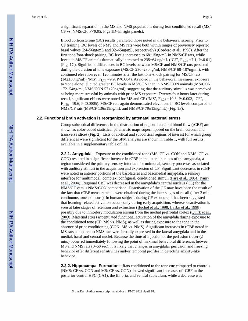
Figure 1.Psychophysiological responses of adult male rats, with previous exposure to maternal stress(MS) or no maternal stress (NMS) during a conditioned fear (CF) paradigm. TRAINING(Top): Behavioral responses of (A) NMS/CF (n=9) and MS/CF (n=14) rats during CFtraining. (B) NMS/CON (n=8) and MS/CON (n=11) controls exposed to tone withoutfootshock. The left panel depicts percent behavioral immobility (Freeze) over time, witheach point representing the group average (± SEM) over 30 sec. The right panel depicts ahistogram plotting the number of 30 sec. intervals displaying a specified percent of freezingbehavior. Also shown are blood corticosterone levels during (C) CF training for NMS/CF(n=8), NMS/CON (n=10), MS/CF (n=7), and MS/CON (n=7) rats. RECALL (Bottom): (D)
Sadler et al. Page 14
Brain Res. Author manuscript; available in PMC 2012 April 18.
NIH
-PA Author Manuscript
NIH
-PA Author Manuscript
NIH
-PA Author Manuscript

Behavioral results and (E) corticosterone results during recall, i.e. reexposure to the tonealone 24 hours after CF training. The horizontal arrow indicates the time and duration of thetone exposure. The vertical dashed bar represents the time of radiotracer infusion. *Comparison of the specified group average with each of the comparison groups, P<0.05.
Sadler et al. Page 15
Brain Res. Author manuscript; available in PMC 2012 April 18.
NIH
-PA Author Manuscript
NIH
-PA Author Manuscript
NIH
-PA Author Manuscript

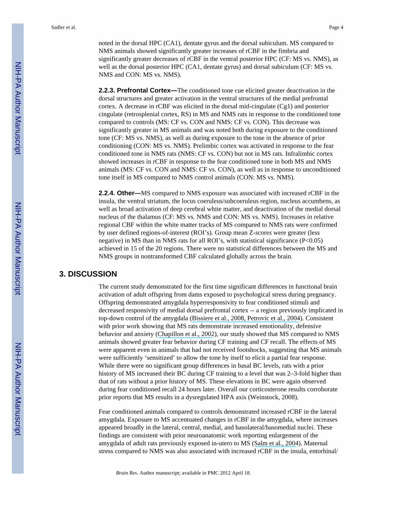
Figure 2.Changes in regional cerebral blood flow related tissue radioactivity in rats during recall to aconditioned tone 24-hours after CF training. Evaluation is made of the effects of MS and CFon functional brain activation. Depicted is a selection of representative coronal slices(anterior–posterior coordinates relative to the bregma), a transverse and a sagital brainsection. Color-coded overlays show statistically significant positive (red) and negative (blue)differences (P < 0.05 at the voxel level, cluster >100 contiguous voxels). NMS/CF = NoMaternal Stress / Conditioned Fear (tone + footshock), n=9; NMS/CON = No MaternalStress / Control (tone only), n=8; MS/CF = Maternal Stress / Conditioned Fear), n=14; andMS/CON = Maternal Stress / Control), n=11. Color bar represents statistical differences (P-
Sadler et al. Page 16
Brain Res. Author manuscript; available in PMC 2012 April 18.
NIH
-PA Author Manuscript
NIH
-PA Author Manuscript
NIH
-PA Author Manuscript
values) as determined by the statistical parametric mapping (SPM). The bilateral nature ofmany of the significant changes adds to the significance of the findings, something notaccounted for by the SPM analysis. Line drawings have been adapted from those from thePaxinos and Watson (2007) rat brain atlas (Paxinos and Watson, 2007). CORTEX: Au,auditory; aCG1, anterior dorsal cingulate, Cg2, ventral cingulate; I, insula; IL, infralimbic;M1, primary motor; Pir, piriform; PrL, prelimbic; PtA, parietal association area; RS,retrosplenial; S1BF, primary somatosensory barrel field; S1FL, primary somatosensoryforelimb area; V1, V2, primary, secondary visual. SUBCORTEX: aca, anterior commissure;ACo, anterior cortical amygdala; AV, anterior ventral thalamic n.; BL, BM, basolateral,basomedial amygdala; Ce, central n. of the amygdala; cp, cerebral peduncle; CPu, caudateputamen; dcw, deep cerebral white matter; DG, dentate gyrus; ec, external capsule; IC,inferior colliculus; La, lateral amygdala; LC, locus coeruleus n.; LSI, lateral septalintermediate n.; LV, lateral ventricle; MD, medial dorsal thalamic n.; MS, medial septal n.;PH, posterior hypothalamus; Pn, Pons; Re, reuinens thalamic n.; RLi, rostral linear n. of theraphe; Sim, simple lobule; SubC, subcoeruleus; vmCPu, ventral medial caudate putamen;vHPC, ventral hippocampus; VA, ventral anterior thalamic n.; VPM, ventral posteromedialthalamic n.; VS, ventral subiculum; 5, trigeminal n.
Sadler et al. Page 17
Brain Res. Author manuscript; available in PMC 2012 April 18.
NIH
-PA Author Manuscript
NIH
-PA Author Manuscript
NIH
-PA Author Manuscript
NIH
-PA Author Manuscript
NIH
-PA Author Manuscript
NIH
-PA Author Manuscript
Sadler et al. Page 18
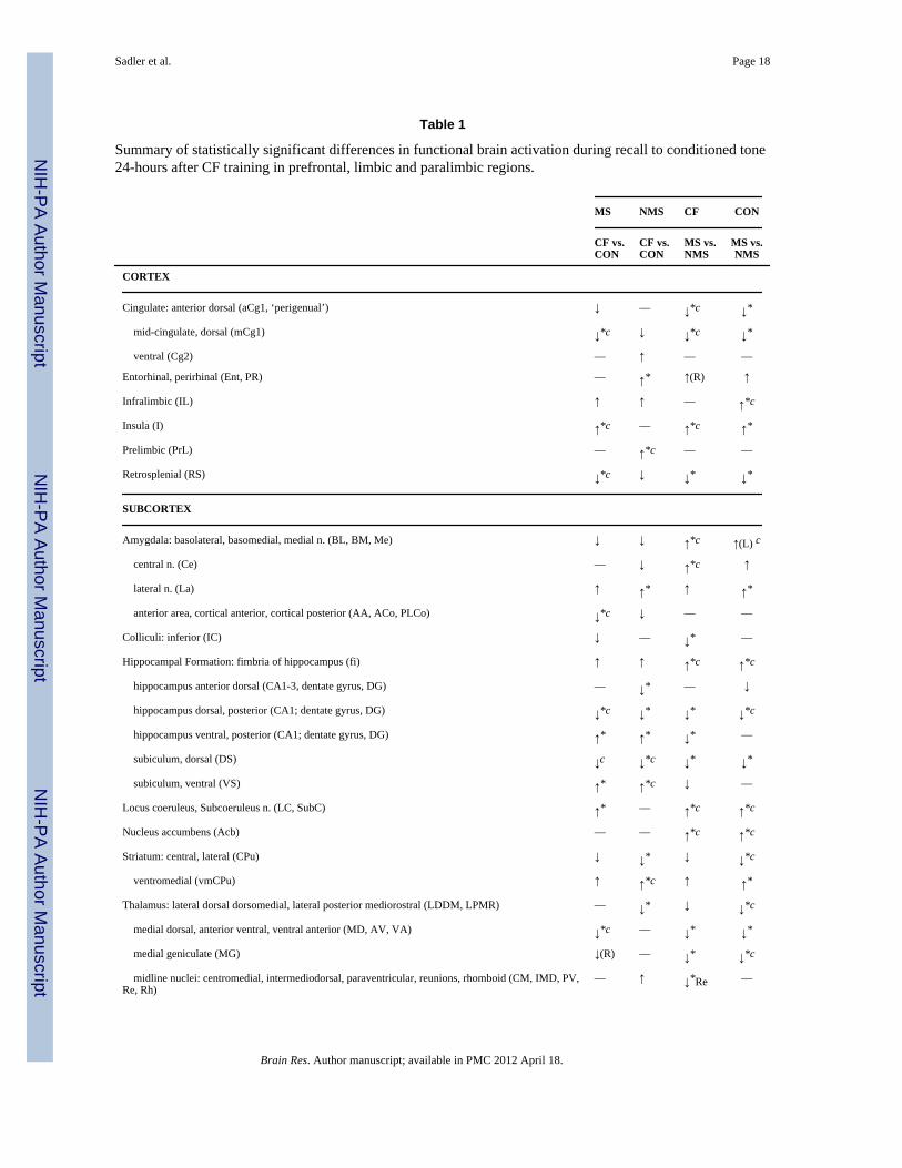
Table 1
Summary of statistically significant differences in functional brain activation during recall to conditioned tone24-hours after CF training in prefrontal, limbic and paralimbic regions.
MS NMS CF CON
CF vs.CON
CF vs.CON
MS vs.NMS
MS vs.NMS
CORTEX
Cingulate: anterior dorsal (aCg1, ‘perigenual’) ↓ –– ↓*c ↓*
mid-cingulate, dorsal (mCg1) ↓*c ↓ ↓*c ↓*
ventral (Cg2) –– ↑ –– ––
Entorhinal, perirhinal (Ent, PR) –– ↑* ↑(R) ↑
Infralimbic (IL) ↑ ↑ –– ↑*c
Insula (I) ↑*c –– ↑*c ↑*
Prelimbic (PrL) –– ↑*c –– ––
Retrosplenial (RS) ↓*c ↓ ↓* ↓*
SUBCORTEX
Amygdala: basolateral, basomedial, medial n. (BL, BM, Me) ↓ ↓ ↑*c ↑(L) c
central n. (Ce) –– ↓ ↑*c ↑
lateral n. (La) ↑ ↑* ↑ ↑*
anterior area, cortical anterior, cortical posterior (AA, ACo, PLCo) ↓*c ↓ –– ––
Colliculi: inferior (IC) ↓ –– ↓* ––
Hippocampal Formation: fimbria of hippocampus (fi) ↑ ↑ ↑*c ↑*c
hippocampus anterior dorsal (CA1-3, dentate gyrus, DG) –– ↓* –– ↓
hippocampus dorsal, posterior (CA1; dentate gyrus, DG) ↓*c ↓* ↓* ↓*c
hippocampus ventral, posterior (CA1; dentate gyrus, DG) ↑* ↑* ↓* ––
subiculum, dorsal (DS) ↓c ↓*c ↓* ↓*
subiculum, ventral (VS) ↑* ↑*c ↓ ––
Locus coeruleus, Subcoeruleus n. (LC, SubC) ↑* –– ↑*c ↑*c
Nucleus accumbens (Acb) –– –– ↑*c ↑*c
Striatum: central, lateral (CPu) ↓ ↓* ↓ ↓*c
ventromedial (vmCPu) ↑ ↑*c ↑ ↑*
Thalamus: lateral dorsal dorsomedial, lateral posterior mediorostral (LDDM, LPMR) –– ↓* ↓ ↓*c
medial dorsal, anterior ventral, ventral anterior (MD, AV, VA) ↓*c –– ↓* ↓*
medial geniculate (MG) ↓(R) –– ↓* ↓*c
midline nuclei: centromedial, intermediodorsal, paraventricular, reunions, rhomboid (CM, IMD, PV,Re, Rh)
–– ↑ ↓*Re ––
Brain Res. Author manuscript; available in PMC 2012 April 18.

NIH
-PA Author Manuscript
NIH
-PA Author Manuscript
NIH
-PA Author Manuscript
Sadler et al. Page 19
MS NMS CF CON
CF vs.CON
CF vs.CON
MS vs.NMS
MS vs.NMS
posterior (Po) –– ↓* ↓* ↓*c
ventral posteromedial, ventral posterolateral n. (VPM, VPL) –– ↓* –– ↓*
White matter (anterior commissures, corpus callosum, internal and external capsule, cerebral peduncle,deep cerebral white matter, forceps major and minor; ac, cc, ic, ec, cp, dcw, fmj, fmi)
↑*c –– ↑*c ↑*c
Significant increases (↑) or decreases (↓) are bilateral across the cerebral hemispheres, except as indicated (L=left only, R=right only). Significanceis shown at the voxel level for clusters of greater than 100 contiguous voxels (*P<0.005, and P<0.05), as well as significance at the cluster level
after correction for multiple comparisons (c P<0.005). Landmarks indicated are per the Paxinos and Watson Rat Brain Atlas (Paxinos and Watson,2007). A full table of significant differences can be found in the supplementary table online.
Brain Res. Author manuscript; available in PMC 2012 April 18.