Angiotensin II bi-directionally regulates cyclooxygenase-2 expression in intestinal epithelial cells
16
Angiotensin II bi-directionally regulates cyclooxygenase-2 expression in intestinal epithelial cells Tatsuo Tani 1 , Rie Ayuzawa 2 , Tetsuo Takagi 1 , Tsutomu Kanehira 1 , Dharmendra Kumar Maurya 2 , and Masaaki Tamura 1,2,* 1 Department of Biochemistry, Vanderbilt University, School of Medicine, Nashville, TN 37232 2 Department of Anatomy & Physiology, Kansas State University College of Veterinary Medicine, Manhattan, KS 66506 Abstract We previously demonstrated that angiotensin II (Ang II) receptor signaling is involved in azoxymethane-induced mouse colon tumorigenesis. To clarify the role of Ang II in COX-2 expression in the intestinal epithelium, the receptor subtype-specific effect on COX-2 expression in a rat intestinal epithelial cell line (RIE-1) has been investigated. Ang II dose- and time-dependently increased the expression of COX-2, but not COX-1 mRNA and protein. This stimulation was completely blocked by the AT 1 receptor antagonist but not the AT 2 receptor antagonist. Ang II and lipopolysaccharide (LPS) additively induced COX-2 protein in RIE-1 cells, whereas the LPS-induced COX-2 expression was significantly attenuated by low concentrations of Ang II or the AT 2 agonistic peptide CGP-42112A only in AT 2 over-expressed cells. These data indicate that Ang II bi- directionally regulates COX-2 expression via both AT 1 and AT 2 receptors. Control of COX-2 expression through the Ang II signaling may have significance in cytokine-induced COX-2 induction and colon tumorigenesis. Keywords AT 1 receptor; AT 2 receptor; lipopolysaccharide; cyclooxygenase-2; intestinal epithelium Introduction Prostaglandins (PGs) play an important role in a broad range of physiological processes. Alterations in its production are implicated in pathophysiological processes, including angiogenesis, cancer, acute and chronic inflammation, etc. (1,2). Cyclooxygenase (COX) is the rate limiting enzyme catalyzing the conversion of arachidonic acid into prostaglandins and others eicosanoids, critically implicated in a variety of physiological and pathophysiological processes, including inflammation, vascular and renal homeostasis, and immune responses (3). Since the early 1990s, the existence of two isoforms of cyclooxygenases (COX-1 and COX-2) has been appreciated. COX-1 and COX-2 are recognized as constitutive and inducible COX isoenzymes, respectively. Conventional findings show that the therapeutics effects of non-steroidal anti-inflammatory drugs (NSAIDs such as aspirin, ibuprofen) are mostly dependent on COX-2 inhibition; therefore, agents that selectively inhibit COX-2 over COX-1 are desirable for treatment of inflammation (4). Increased expression of COX-2 and * Correspondence: Masaaki Tamura, D.V.M., Ph.D., Current address; 210 Coles Hall, Department of Anatomy & Physiology, Kansas State University College of Veterinary Medicine, Manhattan, KS 66506, Phone: (785) 532-4825, Fax: (785) 532-0704, [email protected]. NIH Public Access Author Manuscript Mol Cell Biochem. Author manuscript; available in PMC 2010 October 5. Published in final edited form as: Mol Cell Biochem. 2008 August ; 315(1-2): 185–193. doi:10.1007/s11010-008-9806-5. NIH-PA Author Manuscript NIH-PA Author Manuscript NIH-PA Author Manuscript
-
Upload
independent -
Category
Documents
-
view
1 -
download
0
Transcript of Angiotensin II bi-directionally regulates cyclooxygenase-2 expression in intestinal epithelial cells
Angiotensin II bi-directionally regulates cyclooxygenase-2expression in intestinal epithelial cells
Tatsuo Tani1, Rie Ayuzawa2, Tetsuo Takagi1, Tsutomu Kanehira1, Dharmendra KumarMaurya2, and Masaaki Tamura1,2,*1Department of Biochemistry, Vanderbilt University, School of Medicine, Nashville, TN 372322Department of Anatomy & Physiology, Kansas State University College of Veterinary Medicine,Manhattan, KS 66506
AbstractWe previously demonstrated that angiotensin II (Ang II) receptor signaling is involved inazoxymethane-induced mouse colon tumorigenesis. To clarify the role of Ang II in COX-2expression in the intestinal epithelium, the receptor subtype-specific effect on COX-2 expression ina rat intestinal epithelial cell line (RIE-1) has been investigated. Ang II dose- and time-dependentlyincreased the expression of COX-2, but not COX-1 mRNA and protein. This stimulation wascompletely blocked by the AT1 receptor antagonist but not the AT2 receptor antagonist. Ang II andlipopolysaccharide (LPS) additively induced COX-2 protein in RIE-1 cells, whereas the LPS-inducedCOX-2 expression was significantly attenuated by low concentrations of Ang II or the AT2 agonisticpeptide CGP-42112A only in AT2 over-expressed cells. These data indicate that Ang II bi-directionally regulates COX-2 expression via both AT1 and AT2 receptors. Control of COX-2expression through the Ang II signaling may have significance in cytokine-induced COX-2 inductionand colon tumorigenesis.
KeywordsAT1 receptor; AT2 receptor; lipopolysaccharide; cyclooxygenase-2; intestinal epithelium
IntroductionProstaglandins (PGs) play an important role in a broad range of physiological processes.Alterations in its production are implicated in pathophysiological processes, includingangiogenesis, cancer, acute and chronic inflammation, etc. (1,2). Cyclooxygenase (COX) isthe rate limiting enzyme catalyzing the conversion of arachidonic acid into prostaglandins andothers eicosanoids, critically implicated in a variety of physiological and pathophysiologicalprocesses, including inflammation, vascular and renal homeostasis, and immune responses(3). Since the early 1990s, the existence of two isoforms of cyclooxygenases (COX-1 andCOX-2) has been appreciated. COX-1 and COX-2 are recognized as constitutive and inducibleCOX isoenzymes, respectively. Conventional findings show that the therapeutics effects ofnon-steroidal anti-inflammatory drugs (NSAIDs such as aspirin, ibuprofen) are mostlydependent on COX-2 inhibition; therefore, agents that selectively inhibit COX-2 over COX-1are desirable for treatment of inflammation (4). Increased expression of COX-2 and
*Correspondence: Masaaki Tamura, D.V.M., Ph.D., Current address; 210 Coles Hall, Department of Anatomy & Physiology, KansasState University College of Veterinary Medicine, Manhattan, KS 66506, Phone: (785) 532-4825, Fax: (785) 532-0704,[email protected].
NIH Public AccessAuthor ManuscriptMol Cell Biochem. Author manuscript; available in PMC 2010 October 5.
Published in final edited form as:Mol Cell Biochem. 2008 August ; 315(1-2): 185–193. doi:10.1007/s11010-008-9806-5.
NIH
-PA Author Manuscript
NIH
-PA Author Manuscript
NIH
-PA Author Manuscript

prostaglandins has been widely observed in human (5) and rodent intestinal tumors (6,7)] whencompared with normal adjacent mucosa, whereas COX-1 expression is not differentiallyexpressed. Among the consequences of elevated COX-2 expression and increasedprostaglandin production are stimulation of angiogenesis, inhibition of apoptosis, andstimulation of cellular proliferation, all of which favor tumorigenesis (8).
Colorectal cancer is a major cause of death in developed countries, even though its prognosishas improved yearly due to advances in diagnostic and surgical techniques. Many reportsindicate that the intake of NSAIDs decreases the risk of developing colorectal cancer in man(9) and in model animals (10,11). Inhibitors of COX-2 activity such as aspirin, NSAIDs, andspecific COX-2 antagonists significantly reduce the occurrence of colorectal carcinoma in bothanimal and human studies (12). Studies on the mechanism by which NSAIDs decrease the riskof colorectal cancer suggests that inhibition of prostaglandin synthesis by cyclooxygenase-2(COX-2) is the major mechanism in this chemoprevention (13,14). Therefore, the idealprocedure in the prevention of colorectal cancer is to keep prostaglandin levels low. However,few studies have reported on the endogenous negative regulation of prostaglandin productionexcept with the use of nonspecific drugs such as dexamethasone (15)].
It has been suggested that angiotensin-converting enzyme inhibitors which are commonlyemployed in the treatment of human clinical hypertension also attenuate tumor growth inexperimental animals (16–19)] and may reduce the risk of several human cancers (20).Angiotensin-converting enzyme inhibitors block the formation of angiotensin II (Ang II), anoctapeptide which exerts the many diverse effects of the renin-angiotensin system through itsreceptors (21). This suggests that Ang II may have a modulating role in neoplasia. We recentlydemonstrated that Ang II type 2 receptor (AT2)-mediated signaling is involved inazoxymethane-induced mouse colon tumorigenesis (22). Ang II, the effector of the renin-angiotensin system, has been shown to stimulate the release of prostaglandins in a variety ofcells, including intestinal epithelial cells (23,24)]. Ang II-dependent increase of prostaglandinproduction has shown to be mediated through its type 1 receptor (AT1) via the stimulation ofCOX-2 induction in intestinal cells (25). Although AT1 receptor is believed to mediate mostof the cardiovascular actions of Ang II (26), expression of AT2 in the intestinal tissue is alsonoted (27–29). Since AT2 often counteracts the AT1-mediated Ang II signaling (30,31), ifAT2 signaling attenuates COX-2 induction, this might be an endogenous bidirectionalregulation mechanism for COX-2 induction and resultant prostaglandin formation. This typeof negative regulation is potentially an important target for the chemoprevention procedure ofcolorectal cancer.
In this study, we investigated whether Ang II regulates COX-2 expression in a rat intestinalcell line (RIE-1). We further examined the Ang II receptor subtype-specific role in thispathway.
Materials and MethodsChemicals and reagents
Ang II, (Sar1, Ile8)-Ang II, AT2 receptor antagonist PD123319, AT2 receptor agonistCGP-42112A and lipopolysaccharides (LPS) from Escherichia coli were purchased fromSigma-Aldrich (St. Louis, MO). The AT1 receptor antagonist losartan was obtained from thepharmacy at Vanderbilt University Medical Center. The anti-murine COX-2 polyclonalantibody and anti-mouse COX-1 monoclonal antibody were purchased from Cayman Chemical(Ann Arbor, MI). The actin monoclonal antibody was from Santa Cruz Biotechnology, Inc.(Santa Cruz, CA).
Tani et al. Page 2
Mol Cell Biochem. Author manuscript; available in PMC 2010 October 5.
NIH
-PA Author Manuscript
NIH
-PA Author Manuscript
NIH
-PA Author Manuscript

Cell cultureRat intestinal epithelial (RIE-1) cells were grown in Dulbecco's modified Eagle medium(DMEM) supplemented with 10% fetal bovine serum, 100,000 U/L penicillin G, and 100,000µg/L streptomycin sulfate. For all experiments 90–95% confluent cultures were tenderedquiescent by incubation in serum-free DMEM for 72 h before Ang II stimulation.
RNA extraction and RT-PCRTotal cellular RNA was extracted from confluent RIE-1 cells cultured in serum-free mediumby TRIzol (Invitrogen, Carlsbad, CA) according to the manufacturer's protocol and wasquantified by measurement of absorbance at 260 nm in a spectrophotometer. Two µg of totalRNA was subjected to first-strand cDNA synthesis using the oligo-dT primer and OminiscriptRT kit (Qiagen, Valencia, CA). The primer sets for the AT1 receptor were 5'-GGA AACAGCTTG GTG GTG-3' (sense) and 5'-BCA CAA TCG CCA TAA TTA TCC-3' (antisense),for the AT2 receptor were 5'-CAC CAG CAG AAA CAC CAC-3' (sense) and 5'CCA AACAAG GGG AAC TAC3' (antisense), for COX-2 were 5'-TTC ACC AGA CAG ATT GCTGGC-3' (sense) and 5'-AGT CTG GAG TGG GAG CCA CTT G-3' (antisense), and forGAPDH were 5'-CGC CTG GTC ACC AGG GCT GC-3' (sense) and 5'-CTT ACT CCT TGGAGG CCA TGT-3' (antisense). GAPDH was used as a control to demonstrate equal total RNA.The PCR cycle consisted of 94°C for 1 min, 60°C for 1 min (GAPDH 58°C, COX-2 62°C),72°C for 1 min; these were repeated for 19 cycles for GAPDH, 25 cycles for AT1, 36 cyclesfor AT2 and for COX-2.
Western blot analysisTotal cellular protein was extracted by lysing the cells in 250 µl of lysis buffer (0.25 M sucrose,30 mM Tris, 1% Triton X-100, 0.4% SDS, 1 mM EDTA, one tablet Complete (Roche AppliedScience, Indianapolis, IN) in 10 ml extraction solution. Protein concentration was determinedby the bicinchoninic acid protein assay (Pierce, Rockford, IL). Sixty microgram of proteinunderwent electrophoresis (SDS-PAGE) and were transferred to a nitrocellulose membrane,Protran (Schleicher & Schuell, Keene, NH). Proteins were detected using anti-COX-2 and -COX-1 antibodies, respectively. An actin monoclonal antibody was used as a control todemonstrate equal loading and transfer. ECL (Amersham Biosciences, Pittsburgh, PA) wasused to detect the signals. Intensity of each protein band was quantified using Fluor-S multi-imager (Bio-Rad, Hercules, CA) and Quantity One software (Bio-Rad).
Preparation of adenoviral vectorsOverexpression of AT2 in RIE-1 cells was achieved with an adenovirus encoding mouseAT2 cDNA (Ad-AT2). To generate recombinant replication-deficient adenovirus expressingAT2 receptor, EcoRI-XbaI fragment of mouse AT2 receptor cDNA construct (generous gift ofDr. Inagami, Vanderbilt University, Nashville, TN) was subcloned into a recombinantadenovirus shuttle vector pACCMVpLpA which has deletions in the E1A, E1B and E3 genes.For preparation of virus stocks, 293 cells were infected at multiplicity of infection (MOI) 1–5and harvested after cytopathic effect became visible (72–96 h). Cells were harvested and lysedin 10% FBS containing DMEM, and virus aliquots were stored at −80°C. Titers weredetermined by either plaque assay on 293 cells for plaque forming units/ml or spectrometerreading at 260 nm for particles/ml. Recombinant replication-deficient adenovirus encoding β-galactosidase (Ad-LacZ), which contains E. Coli LacZ gene, human cytomegalovirus promoterand SV40 polyadenylation signal (pHCMVsp1LacZ, generous gift of Dr. Myers, VanderbiltUniversity, Nashville, TN) (32)was used as control vector.
Tani et al. Page 3
Mol Cell Biochem. Author manuscript; available in PMC 2010 October 5.
NIH
-PA Author Manuscript
NIH
-PA Author Manuscript
NIH
-PA Author Manuscript

Virus InfectionFor viral infections, 2 × 10⁶ cells were plated in 60-mm tissue culture dishes and incubate for48–72 h (90~95% confluent). Cells were infected by incubating with the adenoviral vectors atMOI of 50 in 200 µl of DMEM for 6 h at 37°C. After the infection, cells were washed andincubated with serum-free DMEM for 92 h, and then stimulated with either Ang II or bothreceptor antagonist and Ang II.
Radioligand-receptor-binding assayThe radioligand-receptor-binding assay was performed by using transfected cultured cells and[125I](Sar1,Ile8)-Ang II in the presence of PD123319 for AT1 or [125I]CGP-42112A for AT2.The [125I]-labeled peptides were separately prepared from (Sar1,Ile8)-Angi II or CGP-42112Aand [125I]Na by the lactoperoxidase method. Infected cells were incubated with 0.5 nMradiolabeled peptide with or without 1 µM unlabeled peptide for 2 h at 24°C in the presenceof 0.5 mg/ml bovine serum albumin. Unbound labeled ligand was thoroughly washed out withHBSS. Cells were solubilized with 0.5 N NaOH and the remaining radioactivity was counted.Specific binding was estimated by subtracting the non-specific binding obtained in the presenceof 1 µM unlabeled ligand from the total binding. An aliquot of the solubilized cells wassubjected to the bicinchoninic acid protein assay. Specific binding was normalized by theprotein quantity per dish.
Expression of COX-2 in AT2 transfected RIE-1 cellIn this study cells were transfected with AT2 cDNA using the AT2 adenoviral vector asdescribed elsewhere. Briefly, the RIE-1 cells (3 × 10⁵/well) were plated in a 6-well tissueculture plate and incubated for 48 h (90~95% confluent). Cells were infected with anadenovirus vector encoding mouse AT2 (Ad-AT2) or an adenovirus encoding LacZ (Ad-LacZ)with the adenoviral vectors MOI of 50 in 200 µl of DMEM for 6 h at 37°C. After washing withPBS the cells were incubated with 10% FBS-containing DMEM for 18 h and then serum freeDMEM for 48–96 h. Cells were stimulated with various stimuli such as Ang II, CGP-42112Aor LPS (5 µg/ml) for 6–8 h.. The AT1-specific antagonist losartan (10−6 M) or AT2-specificantagonist PD123319 (10−6 M) were added 30 min prior to the addition of stimuli. Afterstimulation cells were harvested and subjected to Western blot analysis for COX-2, GAPDHor actin as described above.
Statistical analysisData obtained from the RT-PCR and Western blot analysis were presented as means ±S.E(standard error). Statistical significance between groups were evaluated by one-way ANOVAwith the Student-Newman-Keuls test. A value of P < 0.05 was considered significant.
ResultsBoth Ang II receptor subtypes were detected in RIE-1 cells
To evaluate the status of Ang II receptor expression in RIE-1-cells, the expression levels ofboth mRNA and functional proteins of the two receptor subtypes were determined by RT-PCRand radioligand receptor-binding assay, respectively (Fig. 1). Expression level of AT1 mRNAwas abundant whereas the level of AT2 mRNA was approximately 10 % of the AT1 expressionlevels. Expression levels of the functional AT1 and AT2 receptor proteins were revealed to be226 and 12 fmol/mg protein, respectively. These data indicate that the rat intestinal epithelialcell line RIE-1 cells express a large amount of AT1 but a small amount of AT2. Since theexpression level of AT2 in adult tissues is known to be small but it is inducible (33), this cellline appears to be an appropriate cell line to study the effect of Ang II on cyclooxygenaseexpression.
Tani et al. Page 4
Mol Cell Biochem. Author manuscript; available in PMC 2010 October 5.
NIH
-PA Author Manuscript
NIH
-PA Author Manuscript
NIH
-PA Author Manuscript

Ang II stimulated COX-2 but not COX-1 expression through AT1 receptorAng II increased COX-2 protein expression in RIE-1 cells in a concentration dependent manneras evaluated by Western blot analysis (Fig 2 A). Ang II (10−9 M) time-dependently increasedCOX-2 mRNA and protein level as shown in Fig. 2B and 2C, respectively, but not COX-1protein level. The COX-2 mRNA response was observed as early as 30 min after Ang IItreatment and the maximal increase was observed 1h after treatment. Ang II-induced COX-2protein expression was first seen 2 h after treatment and the maximal response was observedat 4–6 h after the treatment and lasted until 24 h after treatment (Fig. 2 C and D).
The subtype–specific role of Ang II receptors in COX-2-induction was clarified by utilizingreceptor-specific antagonists. Ang II-dependent induction of COX-2 was completely blockedby the AT1-specific antagonist losartan (10−6 M) but not by the AT2-specific antagonistPD123319 (Fig.3). This result clearly indicates that Ang II stimulates COX-2 induction viaAT1 because there is no expression of COX-2 in the presence of the AT1-specific antagonistlosartan or both AT1 and AT2-specific antagonists (Fig.3).
Ang II and LPS additively stimulated COX-2 expressionAlthough we obtained clear-cut results indicating that the Ang II stimulates COX-2 inductionthrough the AT1 in intestinal epithelial cells, the pathophysiological significance of Ang II inCOX- 2 induction is still uncertain. In order to address this issue, the effect of Ang II on atypical COX-2 inducer, LPS-stimulated COX-2 induction was studied. Ang II (10−8 M) andLPS (5 µg/ml) additively increased COX-2 expression in RIE-1 cells and this additive effectwas completely blocked by the AT1-specific antagonist losartan (Fig.4). These results suggestthat Ang II-dependent COX-2 induction in intestinal epithelial cells is likely to havepathophysiological significance.
Ang II attenuated LPS-induced COX-2 expression through over-expressed AT2 receptorSince the AT2 protein expression level in this cultured cell is smaller than those reported (27,34) the AT2 protein level may not be sufficient to exhibit the AT2-mediated Ang II effect inCOX-2 expression. In this study, the AT2 was over-expressed by transfecting cells with AT2cDNA using the AT2 adenoviral vector. In this study the most efficient AT2 receptor expressionwas observed at 4 days after transfection, and the expression of AT2 was approximately 22%of the AT1 expression level when 50 MOI vector was transfected (Fig.5). Accordingly, co-stimulation of the cells with LPS and Ang II was performed 4 days after transfection with 50MOI AT2 vector. Although Ang II (10−8 M) and LPS (5 µg/ml) additively increased COX-2expression in both untransfected (Fig.4) and LacZ transfected RIE-1 cells (data not shown),low concentrations of Ang II and AT2 agonist CGP-42112A significantly attenuated LPS-induced COX-2 expression when AT2 receptor was transfected (Fig.6). The attenuation effectwas more pronounced when lower concentration of LPS was used for the COX-2 induction.In addition, Ang II-dependent attenuation was pronounced in the presence of the AT1 receptorantagonist losartan (1 µg/ml, data not shown). These results suggest that Ang II attenuates LPS-induced COX-2 expression through AT2 mediated signaling. This implies that Ang II bi-directionally regulates COX-2 expression (AT1 stimulates, but AT2 attenuates COX-2induction).
DiscussionIt has been suggested that Ang II signaling may play important roles in tumorigenesis in severaltissues (17,20,35–39). However, the specific role of the Ang II receptors in tumorigenesis hasnot been elucidated. We have previously demonstrated a pro-oncogenic role of the AT2 receptorin chemical carcinogen azoxymethane–induced colon tumorigenesis (37)] and tobacco-specific nitrosamine–induced lung tumorigenesis (39). In the present study, we used cultured
Tani et al. Page 5
Mol Cell Biochem. Author manuscript; available in PMC 2010 October 5.
NIH
-PA Author Manuscript
NIH
-PA Author Manuscript
NIH
-PA Author Manuscript

normal intestinal epithelial cells to clarify the roles of Ang II and its receptors incyclooxygenase-induction in intestine. Cyclooxygenases are the rate-limiting enzymes for theprostaglandin formation and have been reported to be associated with human colon cancerdevelopment (14,40,41) and other chronic bowel diseases (42). Elucidation of the role of AngII and its receptors in colon tumorigenesis-associated cyclooxygenases are important inunderstanding the insight of upstream regulation of this enzyme.
First, we examined the Ang II receptor expression status of the rat intestinal epithelial cell line,which was utilized for the present study, using RT-PCR and ligand-receptor-binding assay.The present study demonstrated that the RIE-1 cell line expresses both AT1 and AT2 receptormRNA’s (Fig 1A). However, the proportion of the AT1 protein was significantly larger thanthat of the AT2 protein (Fig. 1B). Proportion of the AT2 receptor expression levels seemssmaller than the previous measurement of the receptor expression in intestinal mucosa invivo (27). However this result seems reasonable, since AT2 expression is suppressed duringcell culture but is inducible by removing serum from the culture medium (34). Indeed, AT2protein expression in RIE-1 cells was noticeably increased by serum removal (data not shown).Therefore, the present study with cultured cells of RIE-1 should be a good model system toevaluate the role of Ang II in COX expression in intestinal epithelial cells.
In view of that, we evaluated the effect of Ang II on the expression of the COX-2 expressionin the rat intestinal epithelial cell line using RT-PCR and Western blot analysis. The resultsclearly demonstrated that Ang II, dose- and time-dependently stimulated COX-2 but notCOX-1 expression in the RIE-1 cell line (Fig. 2). The present study also indicates that the AngII AT1-mediated signal plays the primary role in COX-2 induction. These results are in goodagreement with a recent report in which Ang II stimulates COX-2 expression in non-transformed rat intestinal cell line (25). Slice et al have shown that Ang II potently stimulatedexpression of COX-2 mRNA and protein as an immediate-early gene response through theAng II type 1 receptor.
Although the present study also strongly suggests that AT1 plays a critical role in Ang II-dependent COX-2-induction in intestinal epithelium, involvement of another Ang II receptorsubtype in this reaction is unclear since the RIE-1 cell line expresses only a limited amount ofAT2 (Fig. 1). Therefore, the effect of Ang II on COX-2 expression was re-evaluated in theRIE-1 cell line expressing higher levels of AT2. As demonstrated in the Fig. 5, the AT2transfected cells express much higher levels of the AT2 protein in the cells. Although Ang IIalone did not alter COX-2 expression in these cells, Ang II significantly attenuated the effectof a most powerful stimulator LPS-induced COX-2 expression (Fig. 6) and this attenuationwas also mimicked by AT2 agonist CGP-42112A. These results clearly demonstrated that thepresence of the AT2 receptor is potentially important in the regulation of COX-2 expression.This notion is potentially important, since tumor tissue consists of a variety of cell types suchas infiltrated lymphocytes, macrophages, fibroblasts and endothelial cells. These cells seem toplay an important role in tumor cell growth, invasion, metastasis, angiogenesis and evasion ofthe host immune system (43). Prostaglandins and various cytokines produced either in tumorcells or stromal cells are involved in many of these biological reactions (14,43–45).
Although a number of reports have described the role of Ang II in intestinal sodium and waterabsorption function (46,47), only a few reports describe subtype-specific Ang II receptorexpression in the intestine (27,29,47). AT2 receptor has been shown to be involved inabsorption/secretion of water and electrolytes (47,48),and to induce mucosal release of nitricoxide (29). However, this receptor does not influence smooth muscular contractions (49,50).Since Ang II has similar affinity to both of its receptor subtypes it is suggested in most casesthe AT1 receptor counteracts AT2 receptor-mediated effects. Johansson et al. have shown suchan antagonistic relationship, in which activation of AT1 receptor contributes to inhibition and
Tani et al. Page 6
Mol Cell Biochem. Author manuscript; available in PMC 2010 October 5.
NIH
-PA Author Manuscript
NIH
-PA Author Manuscript
NIH
-PA Author Manuscript

AT2 receptor stimulates duodenal mucosal alkaline secretion (51). In terms of the potentialmechanism by which AT2 receptor signaling antagonizes AT1 receptor signaling, it has beenreported that AT2 receptor signaling stimulates tyrosine phosphatase, thus antagonizes AT1receptor-mediated actions (52–54). It is also reported that the AT2 receptor directly interactswith the AT1 receptor, thus counteracting its specific function (22,55). Accordingly, the overallresponse to Ang II in COX-2 induction is the AT1 receptor-mediated stimulation as shown infigures 2 and 3; this response may be modulated in part by AT2 receptor-mediated signalingvia regulation of de-phosphorylation of AT1 receptor-dependent phosphoproteins or directinteraction of both receptor proteins.
On the other hand, it is difficult to speculate a precise mechanism by which AT2 receptorsignaling attenuated LPS-induced COX-2 expression. However, since LPS is shown tostimulate multiple kinase activities and regulate multiple gene expression (56–58), AT2receptor over-expression may have modulated LPS-dependent phosphoproteins via proteintyrosine phosphatase activation and hence attenuated LPS-dependent COX-2 proteinexpression. In addition, AT2 receptor over-expression has shown to down regulate AT1receptor expression (22) and this may also be a part of a potential explanation for the AT2receptor-dependent attenuation of COX-2 protein expression. However, clarification of thismechanism needs to be evaluated by another study.
In summary, the present study demonstrated a bidirectional regulatory function of the Ang IIreceptors in cyclooxygenase-2 expression in non-transformed rat intestinal epithelial cell line.Since pharmacological control of the Ang II receptor function in vivo is a viable therapeutictechnique (59), it is feasible to determine whether AT1 receptor blockers attenuate colorectaltumorigenesis. Increasing AT2 expression in intestinal epithelium, which attenuates COX-2expression, may also be a potential route for a chemoprevention of colorectal cancer. This maylead to the development of a potential chemoprevention procedure for human colorectal cancer.However, the importance of the Ang II receptor function in human colorectal cancer awaitsfurther study. To the best of our knowledge, the present study is the first to demonstrate thatthe Ang II AT2 possesses a modulator function in COX-2 expression in intestinal cells.
AcknowledgmentsThe authors thank Dr. Raymond DuBois (Department of Medicine, Vanderbilt University) for sharing rat intestinalepithelial cell line. We are grateful to Ms. Lara. Pickel (Department of Engineering/Anatomy & Physiology, KansasState University) for critical reading and constructive comments during the preparation of the manuscript. This workwas supported in part by NIH grants RO3 CA091428, P50 CA90949, P30 CA68485, Kansas State University (KSU)Provost’s Fund and KSU College of Veterinary Medicine Dean’s Fund.
References1. De Witt DL. Prostaglandin endoperoxide synthase: regulation of enzyme expression. Biochim Biophys
Acta 1991;1083:121–134. [PubMed: 1903657]2. Masferrer KML, Jaime L.; Koki, Alane T.; Zweifel, Ben S.; Settle, Steven L.; Woerner, B Mark;
Edwards, Dorothy A.; Flickinger, Amy G.; Moore, Rosalyn J.; Seibert, Karen. Antiangiogenic andAntitumor Activities of Cyclooxygenase-2 Inhibitors. Cancer Research 2000;60:1306–1311.[PubMed: 10728691]
3. Cuccurullo CFM, Mezzetti A, Cipollone F. COX-2 expression in atherosclerosis: the good, the bad orthe ugly? Curr Med Chem 2007;14:1595–1605. [PubMed: 17584067]
4. Iezzi AFC, Mezzetti A, Cipollone F. COX-2: friend or foe? Curr Pharm Des 2007;13:1715–1721.[PubMed: 17584101]
5. Eberhart CECR, Radhik A, Giardiello FM, Ferrenbach S, DuBois RN. Up-regulation ofcyclooxygenase 2 gene expression in human colorectal adenomas and adenocarcinomas.Gastroenterology 1994;107:1183–1188. [PubMed: 7926468]
Tani et al. Page 7
Mol Cell Biochem. Author manuscript; available in PMC 2010 October 5.
NIH
-PA Author Manuscript
NIH
-PA Author Manuscript
NIH
-PA Author Manuscript

6. Williams CSLC, Radhika A, Zhang T, Lamps LW, Nanney LB, Beauchamp RD, DuBois RN. Elevatedcyclooxygenase-2 levels in Min mouse adenomas. Gastroenterology 1996;111:1134–1140. [PubMed:8831610]
7. Shao JSH, Aramandla R, Pereira MA, Lubet RA, Hawk E, Grogan L, Kirsch IR, Washington MK,Beauchamp RD, et al. Coordinate regulation of cyclooxygenase-2 and TGF-beta1 in replication error-positive colon cancer and azoxymethane-induced rat colonic tumors. Carcinogenesis 1999;20:185–191. [PubMed: 10069452]
8. Jianguo Du BJ, John Barnard. Differential Regulation of Cyclooxygenase-2 in Nontransformed andRas-Transformed Intestinal Epithelial Cells. Neoplasia 2005;7:761–770. [PubMed: 16207478]
9. Thun MJ, Namboodiri MM, Heath CW Jr. Aspirin use and reduced risk of fatal colon cancer. N EnglJ Med 1991;325:1593–1596. [PubMed: 1669840]
10. Kudo T, Narisawa T, Abo S. Antitumor activity of indomethacin on methylazoxymethanol-inducedlarge bowel tumors in rats. Gann 1980;71:260–264. [PubMed: 7202921]
11. Jacoby RF, Marshall DJ, Newton MA, Novakovic K, Tutsch K, Cole CE, Lubet RA, Kelloff GJ,Verma A, Moser AR, Dove WF. Chemoprevention of spontaneous intestinal adenomas in the ApcMin mouse model by the nonsteroidal anti-inflammatory drug piroxicam. Cancer Res 1996;56:710–714. [PubMed: 8631000]
12. DuBois RN. Cyclooxygenase-2 and colorectal cancer. Prog Exp Tumor Res 2003;37:124–137.[PubMed: 12795052]
13. DuBois RN, Shao J, Tsujii M, Sheng H, Beauchamp RD. G1 delay in cells overexpressingprostaglandin endoperoxide synthase-2. Cancer Res 1996;56:733–737. [PubMed: 8631005]
14. Sheng H, Shao J, Kirkland SC, Isakson P, Coffey RJ, Morrow J, Beauchamp RD, DuBois RN.Inhibition of human colon cancer cell growth by selective inhibition of cyclooxygenase-2. J ClinInvest 1997;99:2254–2259. [PubMed: 9151799]
15. Nanayama T, Hara S, Inoue H, Yokoyama C, Tanabe T. Regulation of two isozymes of prostaglandinendoperoxide synthase and thromboxane synthase in human monoblastoid cell line U937.Prostaglandins 1995;49:371–382. [PubMed: 7480805]
16. Volpert OV, Ward WF, Lingen MW, Chesler L, Solt DB, Johnson MD, Molteni A, Polverini PJ,Bouck NP. Captopril inhibits angiogenesis and slows the growth of experimental tumors in rats. JClin Invest 1996;98:671–679. [PubMed: 8698858]
17. Hii SI, Nicol DL, Gotley DC, Thompson LC, Green MK, Jonsson JR. Captopril inhibits tumourgrowth in a xenograft model of human renal cell carcinoma. Br J Cancer 1998;77:880–883. [PubMed:9528828]
18. Maluccio M, Sharma V, Lagman M, Konijn G, Suthanthiran M. Angiotensin II receptor blockade: anovel strategy to prevent immunosuppressant-associated cancer progression. Transplant Proc2001;33:1820–1821. [PubMed: 11267528]
19. Fujita M, Hayashi I, Yamashina S, Fukamizu A, Itoman M, Majima M. Angiotensin type 1a receptorsignaling-dependent induction of vascular endothelial growth factor in stroma is relevant to tumor-associated angiogenesis and tumor growth. Carcinogenesis 2005;26:271–279. [PubMed: 15637093]
20. Lever AF, Hole DJ, Gillis CR, McCallum IR, McInnes GT, MacKinnon PL, Meredith PA, MurrayLS, Reid JL, Robertson JW. Do inhibitors of angiotensin-I-converting enzyme protect against riskof cancer? Lancet 1998;352:179–184. [PubMed: 9683206]
21. Burrell LM, Johnston CI. Angiotensin converting enzyme inhibitors in perspective: an update. AustFam Physician 1993;22:1243–1247. 1251, 1255. [PubMed: 8373316]
22. Jin XQ, Fukuda N, Su JZ, Lai YM, Suzuki R, Tahira Y, Takagi H, Ikeda Y, Kanmatsuse K, MiyazakiH. Angiotensin II type 2 receptor gene transfer downregulates angiotensin II type 1a receptor invascular smooth muscle cells. Hypertension 2002;39:1021–1027. [PubMed: 12019286]
23. Segawa M, Nakao S, Ogata Y, Sugiya H, Furuyama S. Angiotensin II induces prostaglandin E(2)release in human gingival fibroblasts. Life Sci 2003;72:795–803. [PubMed: 12479978]
24. Jaimes EA, Tian RX, Pearse D, Raij L. Up-regulation of glomerular COX-2 by angiotensin II: roleof reactive oxygen species. Kidney Int 2005;68:2143–2153. [PubMed: 16221213]
25. Slice LW, Chiu T, Rozengurt E. Angiotensin II and epidermal growth factor induce cyclooxygenase-2expression in intestinal epithelial cells through small GTPases using distinct signaling pathways. JBiol Chem 2005;280:1582–1593. [PubMed: 15525649]
Tani et al. Page 8
Mol Cell Biochem. Author manuscript; available in PMC 2010 October 5.
NIH
-PA Author Manuscript
NIH
-PA Author Manuscript
NIH
-PA Author Manuscript
26. Gottlieb SSDK, Fleck E, Kostis J, Levine B, LeJemtel T, DeKock M. Hemodynamic andneurohumoral effects of the angiotensin II antagonist losartan in patients with congestive heart failure.Circulation 1993;88:1602–1609. [PubMed: 8403307]
27. Sechi LA, Valentin JP, Griffin CA, Schambelan M. Autoradiographic characterization of angiotensinII receptor subtypes in rat intestine. Am J Physiol 1993;265:G21–G27. [PubMed: 8338170]
28. Hirasawa K, Sato Y, Hosoda Y, Yamamoto T, Hanai H. Immunohistochemical localization ofangiotensin II receptor and local renin-angiotensin system in human colonic mucosa. J HistochemCytochem 2002;50:275–282. [PubMed: 11799146]
29. Ewert S, Laesser M, Johansson B, Holm M, Aneman A, Fandriks L. The angiotensin II receptor type2 agonist CGP 42112A stimulates NO production in the porcine jejunal mucosa. BMC Pharmacol2003;3:2–9. [PubMed: 12689346]
30. Munzenmaier DH, Greene AS. Opposing actions of angiotensin II on microvascular growth andarterial blood pressure. Hypertension 1996;27:760–765. [PubMed: 8613237]
31. Horiuchi M, Hayashida W, Akishita M, Tamura K, Daviet L, Lehtonen JY, Dzau VJ. Stimulation ofdifferent subtypes of angiotensin II receptors, AT1 and AT2 receptors, regulates STAT activationby negative crosstalk. Circ Res 1999;84:876–882. [PubMed: 10222333]
32. French BA, Mazur W, Geske RS, Bolli R. Direct in vivo gene transfer into porcine myocardium usingreplication-deficient adenoviral vectors. Circulation 1994;90:2414–2424. [PubMed: 7525108]
33. Kambayashi Y, Bardhan S, Takahashi K, Tsuzuki S, Inui H, Hamakubo T, Inagami T. Molecularcloning of a novel angiotensin II receptor isoform involved in phosphotyrosine phosphataseinhibition. J Biol Chem 1993;268:24543–24546. [PubMed: 8227011]
34. Ichiki T, Kambayashi Y, Inagami T. Differential inducibility of angiotensin II AT2 receptor betweenSHR and WKY vascular smooth muscle cells. Kidney Int Suppl 1996;55:S14–S17. [PubMed:8743504]
35. Reddy MK, Baskaran K, Molteni A. Inhibitors of angiotensin-converting enzyme modulate mitosisand gene expression in pancreatic cancer cells. Proc Soc Exp Biol Med 1995;210:221–226. [PubMed:8539259]
36. Yoshiji H, Kuriyama S, Kawata M, Yoshii J, Ikenaka Y, Noguchi R, Nakatani T, Tsujinoue H, FukuiH. The angiotensin-I-converting enzyme inhibitor perindopril suppresses tumor growth andangiogenesis: possible role of the vascular endothelial growth factor. Clin Cancer Res 2001;7:1073–1078. [PubMed: 11309359]
37. Takagi T, Nakano Y, Takekoshi S, Inagami T, Tamura M. Hemizygous mice for the angiotensin IItype 2 receptor gene have attenuated susceptibility to azoxymethane-induced colon tumorigenesis.Carcinogenesis 2002;23:1235–1241. [PubMed: 12117783]
38. Egami K, Murohara T, Shimada T, Sasaki K, Shintani S, Sugaya T, Ishii M, Akagi T, Ikeda H,Matsuishi T, Imaizumi T. Role of host angiotensin II type 1 receptor in tumor angiogenesis andgrowth. J Clin Invest 2003;112:67–75. [PubMed: 12840060]
39. Tani T, Takagi T, Nakano Y, Howard EF, Tamura M. Angiotensin II type 2 receptor gene deficiencyattenuates susceptibility to tobacco-specific nitrosamine-induced lung tumorigenesis: involvementof transforming growth factor-beta-dependent cell growth attenuation. Cancer Res 2005;65:7660–7665. [PubMed: 16140932]
40. Mann JR, DuBois RN. Cyclooxygenase-2 and gastrointestinal cancer. Cancer J 2004;10:145–152.[PubMed: 15285921]
41. Kohno H, Suzuki R, Sugie S, Tanaka T. Suppression of colitis-related mouse colon carcinogenesisby a COX-2 inhibitor and PPAR ligands. BMC Cancer 2005;5:46–58. [PubMed: 15892897]
42. Hendel J, Nielsen OH. Expression of cyclooxygenase-2 mRNA in active inflammatory bowel disease.Am J Gastroenterol 1997;92:1170–1173. [PubMed: 9219792]
43. Hanahan D, Weinberg RA. The hallmarks of cancer. Cell 2000;100:57–70. [PubMed: 10647931]44. Tsujii M, DuBois RN. Alterations in cellular adhesion and apoptosis in epithelial cells overexpressing
prostaglandin endoperoxide synthase 2. Cell 1995;83:493–501. [PubMed: 8521479]45. Tsujii M, Kawano S, Tsuji S, Sawaoka H, Hori M, DuBois RN. Cyclooxygenase regulates
angiogenesis induced by colon cancer cells. Cell 1998;93:705–716. [PubMed: 9630216]46. Levens NR. Control of intestinal absorption by the renin-angiotensin system. Am J Physiol
1985;249:G3–G15. [PubMed: 3893156]
Tani et al. Page 9
Mol Cell Biochem. Author manuscript; available in PMC 2010 October 5.
NIH
-PA Author Manuscript
NIH
-PA Author Manuscript
NIH
-PA Author Manuscript

47. Jin XH, Wang ZQ, Siragy HM, Guerrant RL, Carey RM. Regulation of jejunal sodium and waterabsorption by angiotensin subtype receptors. Am J Physiol 1998;275:R515–R523. [PubMed:9688688]
48. Ewert S, Sjoberg T, Johansson B, Duvetorp A, Holm M, Fandriks L. Dynamic expression of theangiotensin II type 2 receptor and duodenal mucosal alkaline secretion in the Sprague-Dawley rat.Exp Physiol 2006;91:191–199. [PubMed: 16263801]
49. Schinke M, Doods HN, Ganten D, Wienen W, Entzeroth M. Characterization of rat intestinalangiotensin II receptors. Eur J Pharmacol 1991;204:165–170. [PubMed: 1806384]
50. Ewert S, Spak E, Olbers T, Johnsson E, Edebo A, Fandriks L. Angiotensin II induced contraction ofrat and human small intestinal wall musculature in vitro. Acta Physiol (Oxf) 2006;188:33–40.[PubMed: 16911251]
51. Johansson BHM, Ewert S, Casselbrant A, Pettersson A, Fandriks L. Angiotensin II type 2 receptor-mediated duodenal mucosal alkaline secretion in the rat. Am J Physiol Gastrointest Liver Physiol2001;280:G1254–G1260. [PubMed: 11352819]
52. Tsuzuki S, Matoba T, Eguchi S, Inagami T. Angiotensin II type 2 receptor inhibits cell proliferationand activates tyrosine phosphatase. Hypertension 1996;28:916–918. [PubMed: 8901845]
53. Horiuchi M, Akishita M, Dzau VJ. Recent progress in angiotensin II type 2 receptor research in thecardiovascular system. Hypertension 1999;33:613–621. [PubMed: 10024316]
54. Feng YH, Sun Y, Douglas JG. Gbeta gamma -independent constitutive association of Galpha s withSHP-1 and angiotensin II receptor AT2 is essential in AT2-mediated ITIM-independent activationof SHP-1. Proc Natl Acad Sci U S A 2002;99:12049–12054. [PubMed: 12221292]
55. Kumar V, Knowle D, Gavini N, Pulakat L. Identification of the region of AT2 receptor needed forinhibition of the AT1 receptor-mediated inositol 1,4,5-triphosphate generation. FEBS Lett2002;532:379–386. [PubMed: 12482596]
56. Han J, Lee JD, Bibbs L, Ulevitch RJ. A MAP kinase targeted by endotoxin and hyperosmolarity inmammalian cells. Science 1994;265:808–811. [PubMed: 7914033]
57. Suzuki T, Hashimoto S, Toyoda N, Nagai S, Yamazaki N, Dong HY, Sakai J, Yamashita T, NukiwaT, Matsushima K. Comprehensive gene expression profile of LPS-stimulated human monocytes bySAGE. Blood 2000;96:2584–2591. [PubMed: 11001915]
58. Hirasawa N, Torigoe M, Ohgawara R, Murakami A, Ohuchi K. Involvement of MAP kinases inlipopolysaccharide-induced histamine production in RAW 264 cells. Life Sci 2006;80:36–42.[PubMed: 16978663]
59. Levy BI, Benessiano J, Henrion D, Caputo L, Heymes C, Duriez M, Poitevin P, Samuel JL. Chronicblockade of AT2-subtype receptors prevents the effect of angiotensin II on the rat vascular structure.J Clin Invest 1996;98:418–425. [PubMed: 8755652]
Tani et al. Page 10
Mol Cell Biochem. Author manuscript; available in PMC 2010 October 5.
NIH
-PA Author Manuscript
NIH
-PA Author Manuscript
NIH
-PA Author Manuscript

Fig. 1.Detection of Ang II type 1 (AT1) and type 2 (AT2) receptors in RIE-1 cell line. A, Total RNAwas isolated from confluent RIE-1 cells cultured in serum-free medium as described in theMaterials and Methods section. The expression of specific mRNA was detected by RT-PCR.B, Receptor protein expression was determined by ligand receptor–binding assay as describedin the Materials and Method section using cultured whole intact cells and [125I](Sar1,Ile8)-AngII. Data are representative of triplicate determinations.
Tani et al. Page 11
Mol Cell Biochem. Author manuscript; available in PMC 2010 October 5.
NIH
-PA Author Manuscript
NIH
-PA Author Manuscript
NIH
-PA Author Manuscript

Fig. 2.Effect of Ang II on COX expression in RIE-1 cells. A. Concentration dependent expression ofCOX-2 protein in RIE-1 cells by Ang II. B. Time dependent expression of COX-2 mRNA inRIE-1 cells by Ang II (10−9 M). C. Time dependent expression of COX-2 and COX-1 proteinby Ang II (10−8 M). The whole homogenates were subjected to SDS-polyacrylamide gelelectrophoresis followed by Western blotting with anti-murine COX-1 and COX-2 antibodies.D. Quantification of COX-2 protein in panel C was carried out by scanning densitometry andaverages of the expression levels (n=5) normalized by actin levels are displayed in thehistogram. *, P≤0.05 as compared to the level of time 0h.
Tani et al. Page 12
Mol Cell Biochem. Author manuscript; available in PMC 2010 October 5.
NIH
-PA Author Manuscript
NIH
-PA Author Manuscript
NIH
-PA Author Manuscript
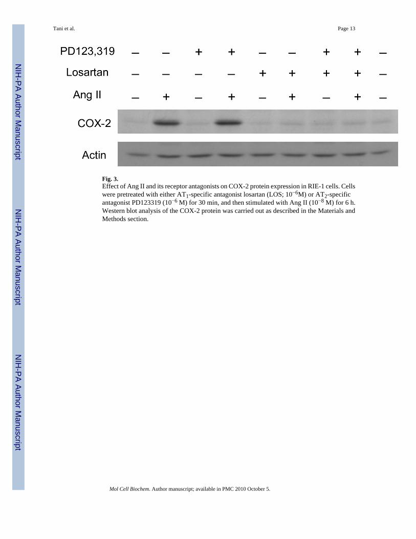
Fig. 3.Effect of Ang II and its receptor antagonists on COX-2 protein expression in RIE-1 cells. Cellswere pretreated with either AT1-specific antagonist losartan (LOS; 10−6M) or AT2-specificantagonist PD123319 (10−6 M) for 30 min, and then stimulated with Ang II (10−8 M) for 6 h.Western blot analysis of the COX-2 protein was carried out as described in the Materials andMethods section.
Tani et al. Page 13
Mol Cell Biochem. Author manuscript; available in PMC 2010 October 5.
NIH
-PA Author Manuscript
NIH
-PA Author Manuscript
NIH
-PA Author Manuscript

Fig. 4.Effect of Ang II and LPS on COX-2 protein expression in RIE-1 cells. Cells were treated witheither Ang II (10−8 M), LPS (5 µg/ml) or both for 6 h in the presence or absence of AT1-specificantagonist losartan (10−6M) or AT2-specific antagonist PD123319 (10−6 M). Western blotanalysis of the COX-2 protein was carried out as described in the Materials and Methodssection. Quantification of COX-2 protein in panel B was carried out by scanning densitometryand averages of the three expression levels normalized by actin levels are displayed in thehistogram. a, P≤0.05 as compared to the level in the untreated control. b, P≤0.05 as comparedto the level in Ang II and LPS treated cells.
Tani et al. Page 14
Mol Cell Biochem. Author manuscript; available in PMC 2010 October 5.
NIH
-PA Author Manuscript
NIH
-PA Author Manuscript
NIH
-PA Author Manuscript

Fig. 5.Expression levels of Ang II receptors in the RIE-1 cells transfected with AT2 cDNA wereassessed by ligand receptor-binding assay as described in the Materials and Methods section.AT2 protein expression was markedly increased by AT2 cDNA transfection, whereas AT1expression was not significantly altered as compared to LacZ transfected cells. The averagesof three separate experiments are displayed in the histogram.*, P≤0.05 as compared to the levelof LacZ transfected cells.
Tani et al. Page 15
Mol Cell Biochem. Author manuscript; available in PMC 2010 October 5.
NIH
-PA Author Manuscript
NIH
-PA Author Manuscript
NIH
-PA Author Manuscript
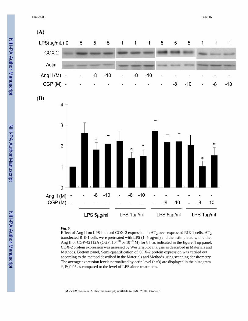
Fig. 6.Effect of Ang II on LPS-induced COX-2 expression in AT2 over-expressed RIE-1 cells. AT2transfected RIE-1 cells were pretreated with LPS (1–5 µg/ml) and then stimulated with eitherAng II or CGP-42112A (CGP, 10−10 or 10−8 M) for 8 h as indicated in the figure. Top panel,COX-2 protein expression was assessed by Western blot analysis as described in Materials andMethods. Bottom panel, Semi-quantification of COX-2 protein expression was carried outaccording to the method described in the Materials and Methods using scanning densitometry.The average expression levels normalized by actin level (n=3) are displayed in the histogram.*, P≤0.05 as compared to the level of LPS alone treatments.
Tani et al. Page 16
Mol Cell Biochem. Author manuscript; available in PMC 2010 October 5.
NIH
-PA Author Manuscript
NIH
-PA Author Manuscript
NIH
-PA Author Manuscript