Ang Negatibong Imahen ng Maynila sa Nobelang Inferno ni Dan Brown
13
Cell, Vol. 119, 19–31, October 1, 2004, Copyright 2004 by Cell Press Ca V 1.2 Calcium Channel Dysfunction Causes a Multisystem Disorder Including Arrhythmia and Autism ultimate signaling molecule for organisms ranging from prokaryotes to humans. In higher organisms, Ca 2 medi- ates processes as diverse as synaptic transmission, muscle contraction, insulin secretion, fertilization, and gene expression (Berridge et al., 2003; Brini and Carafoli, Igor Splawski, 1, * Katherine W. Timothy, 2 Leah M. Sharpe, 1 Niels Decher, 3 Pradeep Kumar, 3 Raffaella Bloise, 4 Carlo Napolitano, 4 Peter J. Schwartz, 5,6 Robert M. Joseph, 7 Karen Condouris, 7 Helen Tager-Flusberg, 7 Silvia G. Priori, 4,5 Michael C. Sanguinetti, 3 2000; Ren et al., 2001). Because Ca 2 cannot be metabo- lized, cells have evolved complex mechanisms for regu- and Mark T. Keating 1 1 Department of Cardiology lating intracellular Ca 2 levels, which are 10,000-fold lower than extracellular levels. Many proteins have been Children’s Hospital Departments of Pediatrics and Cell Biology adapted to bind and transport Ca 2 , in some cases to reduce Ca 2 levels and in others to trigger second-mes- Harvard Medical School and Howard Hughes Medical Institute senger pathways. Excitable cells contain voltage- dependent calcium channels that can dramatically in- Boston, Massachusetts 02115 2 Department of Human Genetics and crease cytosolic Ca 2 . In heart and brain, the L-type calcium channel Ca V 1.2 (CACNA1C, 1C , 1 1.2) mediates 3 Department of Physiology and Nora Eccles Harrison Cardiovascular Research and Training Institute this process (Catterall, 2000; Mikami et al., 1989; Schultz et al., 1993). By contrast with physiology, the role of University of Utah Salt Lake City, Utah 84112 Ca 2 signaling in development is poorly understood. 4 Department of Molecular Cardiology The importance and ubiquity of Ca 2 as an intracellular IRCCS Fondazione Salvatore Maugeri signaling molecule suggest that altered channel function 5 Department of Cardiology could give rise to widespread cellular and organ defects. University of Pavia Previously characterized calcium channel disorders, 6 IRCCS Policlinico San Matteo however, have been marked by dysfunction of a distinct 27100 Pavia organ system, particularly the membrane excitability of Italy neurons and skeletal muscle. For example, calcium 7 Department of Anatomy and Neurobiology channel syndromes like hypokalemic periodic paralysis Boston University School of Medicine and malignant hyperthermia affect skeletal muscle Boston, Massachusetts 02118 (Monnier et al., 1997; Ptacek et al., 1994), episodic ataxia affects the cerebellum (Ophoff et al., 1996), familial hemi- plegic migraine affects vascular smooth muscle (Ophoff Summary et al., 1996), and stationary night blindness affects retina (Bech-Hansen et al., 1998; Strom et al., 1998). None of Ca V 1.2, the cardiac L-type calcium channel, is impor- these disorders has illustrated the full extent of Ca 2 tant for excitation and contraction of the heart. Its role signaling in human development and physiology. in other tissues is unclear. Here we present Timothy Cardiac arrhythmias cause sudden loss of conscious- syndrome, a novel disorder characterized by multior- ness and sudden death in approximately 1 million Euro- gan dysfunction including lethal arrhythmias, webbing peans and North Americans every year (Priori et al., of fingers and toes, congenital heart disease, immune 2002; Zheng et al., 2001). Over the last decade, we and deficiency, intermittent hypoglycemia, cognitive ab- others have identified arrhythmia susceptibility genes normalities, and autism. In every case, Timothy syn- by studying familial syndromes (Antzelevitch, 2003; drome results from the identical, de novo Ca V 1.2 mis- Keating and Sanguinetti, 2001). These genes include sense mutation G406R. Ca V 1.2 is expressed in all SCN5A, KVLQT1, and HERG, which encode important affected tissues. Functional expression reveals that cardiac sodium and potassium channels. In the vast G406R produces maintained inward Ca 2 currents by majority of arrhythmia syndromes, individuals appear causing nearly complete loss of voltage-dependent normal except for subtle electrocardiographic abnor- channel inactivation. This likely induces intracellular malities. Ca 2 overload in multiple cell types. In the heart, pro- Here, we describe the phenotypic characterization of longed Ca 2 current delays cardiomyocyte repolariza- Timothy syndrome (TS), an arrhythmia disorder associ- tion and increases risk of arrhythmia, the ultimate ated with dysfunction in multiple organ systems, includ- cause of death in this disorder. These discoveries es- ing congenital heart disease, syndactyly, immune defi- tablish the importance of Ca V 1.2 in human physiology ciency, and autism. We show that this disorder results and development and implicate Ca 2 signaling in from a recurrent, de novo missense mutation in the autism. Ca V 1.2 L-type calcium channel gene. The Ca V 1.2 gene is expressed in multiple tissues. We demonstrate through Introduction functional expression in heterologous systems that the disease-associated mutation causes abnormal Ca 2 “Ja, Kalzium, das ist alles!” So stated Nobel laureate current. This gain-of-function mechanism is mediated Otto Loewi in 1959, and it is now clear that Ca 2 is the through failed channel inactivation, suggesting that cal- cium channel blockers may be useful for treating this and related disorders. *Correspondence: [email protected]
Transcript of Ang Negatibong Imahen ng Maynila sa Nobelang Inferno ni Dan Brown
Cell, Vol. 119, 19–31, October 1, 2004, Copyright 2004 by Cell Press
CaV1.2 Calcium Channel DysfunctionCauses a Multisystem DisorderIncluding Arrhythmia and Autism
ultimate signaling molecule for organisms ranging fromprokaryotes to humans. In higher organisms, Ca2� medi-ates processes as diverse as synaptic transmission,muscle contraction, insulin secretion, fertilization, andgene expression (Berridge et al., 2003; Brini and Carafoli,
Igor Splawski,1,* Katherine W. Timothy,2
Leah M. Sharpe,1 Niels Decher,3 Pradeep Kumar,3
Raffaella Bloise,4 Carlo Napolitano,4
Peter J. Schwartz,5,6 Robert M. Joseph,7
Karen Condouris,7 Helen Tager-Flusberg,7
Silvia G. Priori,4,5 Michael C. Sanguinetti,3 2000; Ren et al., 2001). Because Ca2� cannot be metabo-lized, cells have evolved complex mechanisms for regu-and Mark T. Keating1
1Department of Cardiology lating intracellular Ca2� levels, which are 10,000-foldlower than extracellular levels. Many proteins have beenChildren’s Hospital
Departments of Pediatrics and Cell Biology adapted to bind and transport Ca2�, in some cases toreduce Ca2� levels and in others to trigger second-mes-Harvard Medical School and
Howard Hughes Medical Institute senger pathways. Excitable cells contain voltage-dependent calcium channels that can dramatically in-Boston, Massachusetts 02115
2 Department of Human Genetics and crease cytosolic Ca2�. In heart and brain, the L-typecalcium channel CaV1.2 (CACNA1C, �1C, �11.2) mediates3 Department of Physiology and Nora Eccles Harrison
Cardiovascular Research and Training Institute this process (Catterall, 2000; Mikami et al., 1989; Schultzet al., 1993). By contrast with physiology, the role ofUniversity of Utah
Salt Lake City, Utah 84112 Ca2� signaling in development is poorly understood.4 Department of Molecular Cardiology The importance and ubiquity of Ca2� as an intracellularIRCCS Fondazione Salvatore Maugeri signaling molecule suggest that altered channel function5 Department of Cardiology could give rise to widespread cellular and organ defects.University of Pavia Previously characterized calcium channel disorders,6 IRCCS Policlinico San Matteo however, have been marked by dysfunction of a distinct27100 Pavia organ system, particularly the membrane excitability ofItaly neurons and skeletal muscle. For example, calcium7 Department of Anatomy and Neurobiology channel syndromes like hypokalemic periodic paralysisBoston University School of Medicine and malignant hyperthermia affect skeletal muscleBoston, Massachusetts 02118 (Monnier et al., 1997; Ptacek et al., 1994), episodic ataxia
affects the cerebellum (Ophoff et al., 1996), familial hemi-plegic migraine affects vascular smooth muscle (Ophoff
Summary et al., 1996), and stationary night blindness affects retina(Bech-Hansen et al., 1998; Strom et al., 1998). None of
CaV1.2, the cardiac L-type calcium channel, is impor- these disorders has illustrated the full extent of Ca2�
tant for excitation and contraction of the heart. Its role signaling in human development and physiology.in other tissues is unclear. Here we present Timothy Cardiac arrhythmias cause sudden loss of conscious-syndrome, a novel disorder characterized by multior- ness and sudden death in approximately 1 million Euro-gan dysfunction including lethal arrhythmias, webbing peans and North Americans every year (Priori et al.,of fingers and toes, congenital heart disease, immune 2002; Zheng et al., 2001). Over the last decade, we anddeficiency, intermittent hypoglycemia, cognitive ab- others have identified arrhythmia susceptibility genesnormalities, and autism. In every case, Timothy syn- by studying familial syndromes (Antzelevitch, 2003;drome results from the identical, de novo CaV1.2 mis- Keating and Sanguinetti, 2001). These genes includesense mutation G406R. CaV1.2 is expressed in all SCN5A, KVLQT1, and HERG, which encode importantaffected tissues. Functional expression reveals that cardiac sodium and potassium channels. In the vastG406R produces maintained inward Ca2� currents by majority of arrhythmia syndromes, individuals appearcausing nearly complete loss of voltage-dependent normal except for subtle electrocardiographic abnor-channel inactivation. This likely induces intracellular malities.Ca2� overload in multiple cell types. In the heart, pro- Here, we describe the phenotypic characterization oflonged Ca2� current delays cardiomyocyte repolariza- Timothy syndrome (TS), an arrhythmia disorder associ-tion and increases risk of arrhythmia, the ultimate ated with dysfunction in multiple organ systems, includ-cause of death in this disorder. These discoveries es- ing congenital heart disease, syndactyly, immune defi-tablish the importance of CaV1.2 in human physiology ciency, and autism. We show that this disorder resultsand development and implicate Ca2� signaling in from a recurrent, de novo missense mutation in theautism. CaV1.2 L-type calcium channel gene. The CaV1.2 gene is
expressed in multiple tissues. We demonstrate throughIntroduction functional expression in heterologous systems that the
disease-associated mutation causes abnormal Ca2�
“Ja, Kalzium, das ist alles!” So stated Nobel laureate current. This gain-of-function mechanism is mediatedOtto Loewi in 1959, and it is now clear that Ca2� is the through failed channel inactivation, suggesting that cal-
cium channel blockers may be useful for treating thisand related disorders.*Correspondence: [email protected]
Cell20
Results syndrome. By contrast, the complexity of the TS pheno-type suggested a second possibility, a contiguous genedeletion syndrome or a chromosomal rearrangement.Timothy Syndrome, a Multisystem DisorderKaryotypic analysis of metaphase chromosomes fromIn 1992, cases of a novel arrhythmia syndrome associ-11 children failed to reveal defects (data not shown). Thisated with syndactyly (webbing of fingers and toes) wereanalysis, however, would not exclude small deletions.described (Marks et al., 1995; Reichenbach et al., 1992).
A consistent feature of TS was severe QT intervalWe named this disorder Timothy syndrome (TS). Be-prolongation. Therefore, to define the genetic basis ofcause therapy is extending lives of affected children,this disorder, we screened known long QT syndromewe have observed that TS manifests major phenotypicgenes, including HERG, KVLQT1, KCNE1, KCNE2, SCN5A,abnormalities of multiple organ systems, includingand KCNJ2, by single-strand conformation polymor-heart, skin, eyes, teeth, immune system, and brain (Fig-phism (SSCP) and/or DNA sequence analyses. We alsoure 1, Table 1).examined several other genes encoding channel orThe inheritance pattern of TS was sporadic in all butchannel-associated subunits, including FKBP1A, KCNA4,one family. In that family, two of three siblings wereKCNA5, KCND3, KCNE3, KCNIP2, KCNJ4, KCNJ9,affected. None of the parents in any of the families wasKCNJ10, KCNJ12, NCX1, SCN1B, DNAJB1, and TRPC3.affected. Ten of 17 children with TS died. The averageThe transcription factors TFAP2B, FOG2, NKX2.5, andage of death was 2.5 years. All affected individuals hadGATA4 were tested because they have been implicatedsevere prolongation of the QT interval on electrocardio-in one or more phenotypic features of TS. No mutationsgram, syndactyly, and abnormal teeth and were bald atwere identified.birth. Arrhythmias were the most serious aspect of this
The abnormal electrocardiographic morphology ob-disorder, as 12 of 17 children had life-threatening epi-served in TS patients was similar to that of individualssodes. Arrhythmic death in two children was triggeredwith long QT syndrome caused by gain-of-function mu-by sepsis and in two other children by episodic hypogly-tations in SCN5A, the cardiac sodium channel (Zhangcemia. Individuals with this syndrome also had congeni-et al., 2000). The cardiac L-type calcium channel CaV1.2,tal heart disease including patent ductus arteriosus, pa-like SCN5A, mediates an inward depolarizing current intent foramen ovale, ventricular septal defects, andcardiomyocytes. Thus, in 1996, we examined the CaV1.2tetralogy of Fallot. Some children had dysmorphic facialgene as a candidate. SSCP analyses of 47 exons andfeatures, including a flat nasal bridge, small upper jaw,promoter regions did not show mutations (Table 2).low-set ears, or small and misplaced teeth. EpisodicHowever, new alternatively spliced forms of the CaV1.2serum hypocalcemia was described in four individuals.gene were subsequently identified (Abernethy and Sol-Many of the surviving children showed developmentaldatov, 2002). Analysis of one CaV1.2 splice variant re-delays consistent with language, motor, and generalizedvealed a G1216A transition in exon 8A in all 13 individualscognitive impairment. Children had a history of languagefor whom DNA samples were available (Figures 2A anddelay and delay in other motor skills. Many did not pro-2B). This transition caused a substitution of glycine withduce speech sounds (babbling) during infancy. Testingarginine at residue 406 (G406R). This amino acid is com-revealed significant problems in language skills includ-pletely conserved in other voltage-dependent calciuming articulation, reception, and expression. Childrenchannels of multiple species, ranging from worms towere impaired in all areas of adaptive function, includinghumans (Figure 2C). G406 is located at the C-terminalcommunication, socialization, and daily living skills. Fiveend of the sixth transmembrane segment of domain I (DI/children were formally evaluated for autism. Three metS6, Figure 2D). The G406R mutation was not identified inthe criteria for this disorder, one met criteria for autism180 ethnically matched control samples (360 chromo-spectrum disorders, and one had severe delays in lan-somes, p � 1.8 � 10�20).guage development. We could not evaluate additional
Mutational analysis of additional family members, in-children because they were deceased or unavailable.
cluding parents, failed to reveal mutations at this site.However, the association between autism spectrum dis-
Thus, the phenotype in all probands resulted from theorders and Timothy syndrome was significant (p � 1.2 � same de novo mutation. However, in one family, two10�8). Taken together, the diversity of these phenotypic siblings had the syndrome (Figure 2B). Both parentsabnormalities indicates a complex physiological and de- were phenotypically unaffected. The probability of thevelopmental disorder. mutation event happening twice in the same family was
minimal. To explain this apparent paradox, we hypothe-Recurrent De Novo Missense Mutation sized that one parent was mosaic for the mutation. Mo-in CaV1.2 Causes TS saicism is defined by the presence of two or more geneti-The TS phenotype suggested two possible genotypes. cally different cell types in the same organism. To testThe severity of arrhythmias in this disorder suggested this hypothesis, we sequenced DNA samples from thea recessive gene knockout similar to Jervell and Lange- father’s sperm and blood, but the mutation was notNielsen syndrome, which is caused by homozygous observed. In the mother, we sequenced DNA samplesloss-of-function mutations of KVLQT1 or KCNE1 potas- from blood and oral mucosa. Although the blood DNAsium channel genes (Neyroud et al., 1997; Schulze-Bahr contained only wild-type sequences, we were able toet al., 1997; Splawski et al., 1997a; Tyson et al., 1997). detect a small peak for the missense mutation in DNAConsistent with this thesis, all parents were unaffected, from the oral mucosa (Figure 2B). These findings indi-and one of the families had two children with TS. How- cate that the mother is mosaic and transmitted this mu-ever, none of the parents from any family were related, tation to her two affected children. To further test this
hypothesis, we performed genotypic analysis in thisand consanguinity would be expected in a rare recessive

Calcium Channel Mutation in Arrhythmia and Autism21
Figure 1. Timothy Syndrome Is Characterized by Multisystem Dysfunction and Developmental Defects
(A–C) TS individuals exhibiting dysmorphic facial features including round face, flat nasal bridge, receding upper jaw, and thin upper lip.(D and E) Webbing of the toes and fingers (syndactyly).(F) Left panel electrocardiogram shows severe QT interval prolongation causing 2:1 atrioventricular block seen as two atrial beats (P-waves)for each ventricular beat (QRS complex). Right panel electrocardiogram shows alternating T-wave polarity (arrows), indicating severe cardiacrepolarization abnormality.(G) Ventricular tachycardia recorded from a TS patient by an implanted automatic defibrillator.
Cell22
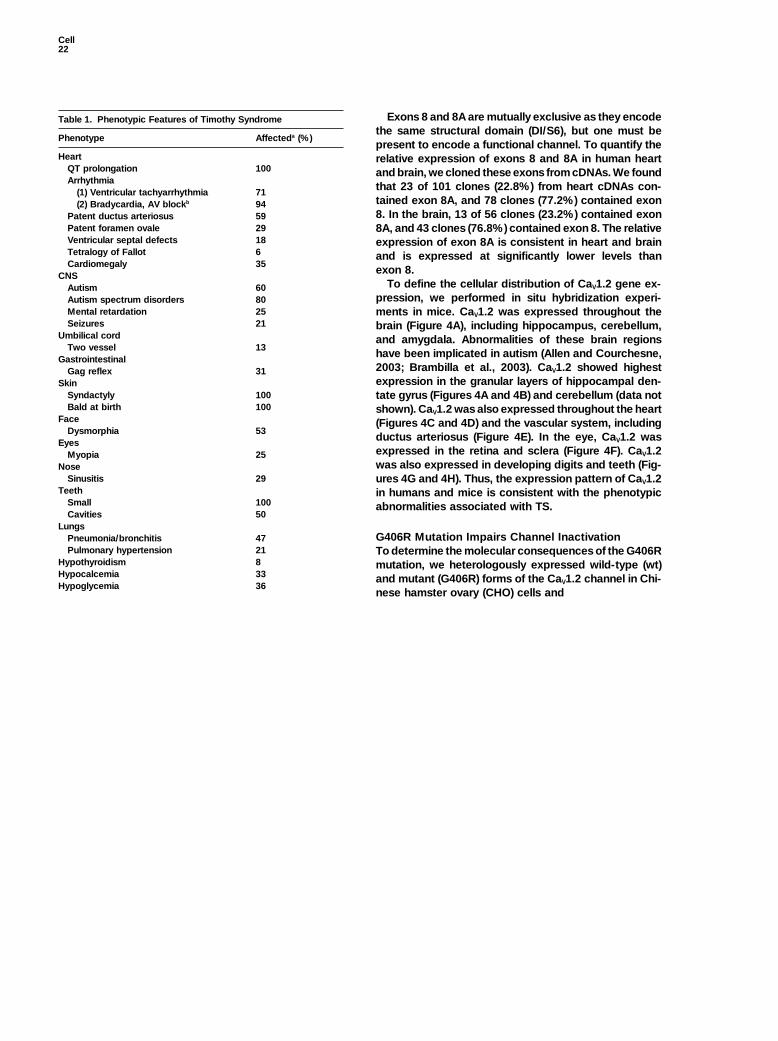
Exons 8 and 8A are mutually exclusive as they encodeTable 1. Phenotypic Features of Timothy Syndromethe same structural domain (DI/S6), but one must be
Phenotype Affecteda (%)present to encode a functional channel. To quantify the
Heart relative expression of exons 8 and 8A in human heartQT prolongation 100 and brain, we cloned these exons from cDNAs. We foundArrhythmia
that 23 of 101 clones (22.8%) from heart cDNAs con-(1) Ventricular tachyarrhythmia 71tained exon 8A, and 78 clones (77.2%) contained exon(2) Bradycardia, AV blockb 948. In the brain, 13 of 56 clones (23.2%) contained exonPatent ductus arteriosus 59
Patent foramen ovale 29 8A, and 43 clones (76.8%) contained exon 8. The relativeVentricular septal defects 18 expression of exon 8A is consistent in heart and brainTetralogy of Fallot 6 and is expressed at significantly lower levels thanCardiomegaly 35
exon 8.CNSTo define the cellular distribution of CaV1.2 gene ex-Autism 60
pression, we performed in situ hybridization experi-Autism spectrum disorders 80Mental retardation 25 ments in mice. CaV1.2 was expressed throughout theSeizures 21 brain (Figure 4A), including hippocampus, cerebellum,
Umbilical cord and amygdala. Abnormalities of these brain regionsTwo vessel 13
have been implicated in autism (Allen and Courchesne,Gastrointestinal2003; Brambilla et al., 2003). CaV1.2 showed highestGag reflex 31expression in the granular layers of hippocampal den-Skin
Syndactyly 100 tate gyrus (Figures 4A and 4B) and cerebellum (data notBald at birth 100 shown). CaV1.2 was also expressed throughout the heart
Face (Figures 4C and 4D) and the vascular system, includingDysmorphia 53
ductus arteriosus (Figure 4E). In the eye, CaV1.2 wasEyesexpressed in the retina and sclera (Figure 4F). CaV1.2Myopia 25was also expressed in developing digits and teeth (Fig-Nose
Sinusitis 29 ures 4G and 4H). Thus, the expression pattern of CaV1.2Teeth in humans and mice is consistent with the phenotypic
Small 100 abnormalities associated with TS.Cavities 50
LungsG406R Mutation Impairs Channel InactivationPneumonia/bronchitis 47
Pulmonary hypertension 21 To determine the molecular consequences of the G406RHypothyroidism 8 mutation, we heterologously expressed wild-type (wt)Hypocalcemia 33 and mutant (G406R) forms of the CaV1.2 channel in Chi-Hypoglycemia 36
nese hamster ovary (CHO) cells and Xenopus oocytes.Hypothermia 33The biophysical properties of the channel were firstMuscoskeletalcharacterized by standard whole-cell patch-clamp tech-Hypotonia 40
Immunodeficiency/recurrent infections 43 niques using Ca2� (15 mM) as a charge carrier in CHOcells cotransfected with CaV1.2 and its accessory sub-a Nine affected individuals were male, and eight were female.units, CaV�2b and CaV�2�1. The most striking differenceb Atrioventricular block.between wt and G406R channels was the extent of inac-tivation. Inactivation of wt channel current was nearlycomplete in 300 ms (Figure 5A). In contrast, G406R chan-family. We found that the G406R mutation resided on
the maternal chromosome. Thus, in one case, G406R nels only partially inactivated during the same time pe-riod (Figure 5B). Next, we assessed the voltage depen-arose de novo in a parent during development, leading
to mosaicism. Taken together, these data indicate that dence of Ca2� current inactivation. Wild-type channelinactivation was complete at �20 mV and slightly de-a recurrent, de novo G406R mutation of the CaV1.2 gene
causes TS. creased at more positive potentials, as expected forpartial relief of the Ca2�-dependent component of inacti-vation (Lee et al., 1985) (Figure 5C). In contrast, theCav1.2 Is Widely Expressed
Previous studies indicate that the CaV1.2 gene was ex- maximum attained inactivation was only 56% for G406Rchannels. Relief of inactivation was greater for G406Rpressed in heart, brain, smooth muscle, and pituitary
and adrenal glands (Ertel et al., 2000). However, the TS compared to wt channels at potentials ��30 mV (Figure5C). This observation suggests that mutant channelsphenotype suggested a broader expression pattern of
the alternatively spliced form of CaV1.2 containing exon have lost voltage-dependent Ca2�current inactivation.The time constant for inactivation () was a U-shaped8A. To determine the pattern of expression in humans,
we used exon 8A as a probe for Northern and dot blot function of voltage for wt channels but increased withmembrane voltage for mutant channels (Figure 5D). Theanalyses (Figure 3). This exon was highly expressed in
adult heart, and the mRNA was �9 kb (Figure 3A). mRNA current amplitudes measured at the peak of the current-voltage (I/V) relationship were similar (p � 0.32) andcontaining exon 8A was also expressed in multiple adult
and fetal tissues, including brain, gastrointestinal sys- averaged 70 12 pA for wt (n � 11) and 94 20 pAfor G406R (n � 9). The shape of the normalized I/Vtem, lungs, immune system, smooth muscle, and testis.
These data indicate that exon 8A of the CaV1.2 gene is relationship (Figure 5E) was only slightly altered by themutant channels, consistent with a mere �3 mV shift inwidely expressed in humans.
Calcium Channel Mutation in Arrhythmia and Autism23
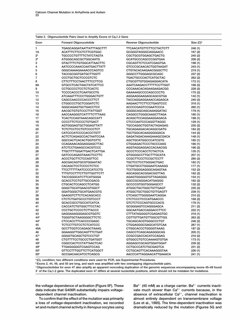
Table 2. Oligonucleotide Pairs Used to Amplify Exons of CaV1.2 Gene
Exon Forward Oligonucelotide Reverse Oligonucleotide Size (C)a
1 TGAGCAGGATAATTATTTAGCTTT TTCAACATGTTCTTCCTACTCTT 246 (1)1A ACATTTCTTCCTCTTCGTGGC GCGGGTAGGGCAGGAACC 167 (2)2b TGCCCCTGTTTTCTATCTAGTA CGCTGCGTGGAGCTGACTG 245 (2)2b ATGGGCAGCGCTGGCAATG GCATGCCCAGCCCGGTGAA 208 (2)3 GTACTTTCTGTGGCATTAACTTC CGCAGTTCTCCATCGAGTGA 198 (1)4 AATCCCCAAACCAATGACTTATT GTCCCGCAACACTGGTAAGAT 243 (1)5 GGGCAAAAGAAAACCCAGTCC CTGTACACAAGAACGGGCTTC 219 (1)6 TACCGCGGTGATGCTTGGTT GGGCCTTGGGAGCGCAGC 237 (2)7 CCCTGCTGCTCCCGTCTC TGACTGCCCACTCATGCTAC 262 (2)8 CTTCTTTCCTAACTTTCCTTCG CTGCGTTGTGGAGAGGACATA 172 (1)8A GTGCCTCACTAACTATCATTCC AAATCAAGACCTTTTTCCTTGGT 168 (2)9 CCTGCCCCTCCTCTCACTC CCCAAACACAGAAGAAGACGG 228 (2)10 TCCCCACCCTCAATGCCTG GAAAAAGCCCCAGCCCCTG 175 (2)11 ATCAAATTTCCCTGGGACTGTT AGGAAGGAAGAGCAGCGTGA 142 (1)12 CAGCCAACCCCACCCTTCT TACCAGGAGGAAACCAGAGCA 240 (2)13 CTGGCCCTGCTCGGATCTC AAGAACTTCTTCCTGAGTCCC 318 (1)14 GGGCAGAGTGCTGACCTCC ACCCCGAGTCCGAATCCCA 285 (1)15 AACGCTGTGTCCCTTATTGGT GGGGCAGCAGCAAAGGATAC 179 (1)16 AACCAAGGGTCATTTTCTTTAAG CAGGCCTGGGCAAGCTTAGA 180 (1)17 TCACTCCAGTAAACAGCCATT ACAGCTCCAGGAAGGAGACA 198 (1)18 CCCCTTCTCCCCTGTGACT CTCCCAATCCCAGGTTAGGC 129 (1)19 AGTGGGAGTGCTGGAGTTATT TGCCAGGCTGGTACTAGAGAG 235 (2)20 TCTCTCCTCCTGTCCCCTCT TGCAGAAGACACAGGCGATG 184 (2)21 CATCCCATCCCCACCCTGTT TGCTGGCACAAGGGGGACA 144 (2)22 ACTCTCAGAGCCACTAATCCAA GAGATAGACAAAGAAAGCGACA 148 (1)23 GGCCACTCACACTGGTGTTC AGCTGCATGGCCCATCGTG 185 (1)24 CCAGAAACAGGAGGAGCTTAC CTGGAGACTCCCTACCCAAG 166 (1)25 ATCTCCTGAAGCCACGTCCC AGGTAGGAGGACCACAACGG 186 (2)26 TTGCTTTTGGATTGACTCATTGA GCCCTCCCACCTCTACTCA 135 (1)27 GATCCCAGAGTAAACTCCTTC GTGGGGCCTTGCTTTGCATA 207 (1)28 ACCCTGCTTCTCCAGTTCC CCGCTTCCCTCCGCTCCTT 280 (2)29 AGCGACAGTGCGTGGAATAC TGCTCCTCCTGGGACTGAC 182 (1)30 CACAGCTCCTCCCCTCTCC CTGATGCCTGGGAATCAAAGG 177 (1)31 TTCCTTTTTAATCCCCATCCTG TCCTGGGGAGGGCAGAGTAA 152 (2)32 TTTGTCCTTTCTTGTTGGTTCTT AGCAGGCACGGACGGTTAG 182 (2)33 TACCGGGCATCTTCATGGGA AGAGTGGGGGTGTTAGAGGG 154 (1)34 GCACCTCCTGTTGCCGACG GGCCGCAGGACATGAGGG 182 (2)35 CTGCACTCCAGCCTCATGG GCCCCGTGGTGGGAGCCT 132 (2)36 GGGCTGCATGAACGTGGCT ATGGCTGCTGGCTGTTGAGT 235 (2)37 GGATGGGCTGCATGAACGTG ATGGCTGCTGGCTGTTGAGTT 239 (1)38 GAAGGTCTTCTCACAGCACC CTCAGCTTGGGGAATCAGGA 210 (2)39 CTCTCTGATGCCCTGTCCCT CTCTCCCTCCCATGAACCC 168 (2)40 GCACCGCCTGCCATCATCA GTCTCTCCCAGTGCCCACG 178 (1)41 CACCATCTGTGGCTTCCTAC GCGGGAGTCCAGGGAGCA 211 (2)42 CCCTGCTCCCCTCTTACCC GGCAATGACCAGGACCTTCC 222 (1)43 GAGGAAAGGGAGCGTGGTC CTTTATAGGGGTCGAGAGTGC 218 (1)44b TGGGTGCTAAGGGGCTTCTC CGTTGTTGATGTTGGCGTTGG 263 (2)44b CTCCACCTTCACCCCGAGC TGCAGCACGTGGGCCCTGT 246 (2)45 TCTCCTTGTCCTCTCATCCC CTGGAGGGCGAGCATGTCAA 267 (1)45A GCCTTGGTCCAGAGCTAAAG CTGGCACCCTGGGGTAAAG 187 (2)46 GGAAGGTTGGCAGTTTCTGAT CAGCCTCAGCAGAGGGCAG 205 (1)47c GGGGTGCAGCTGTCCCTGT CCGCCGACCACATCCAGAG 216 (2)48 CTGTTTTCCTGCCCTGATGGT GTGGCCTGTCCAAAAGTGTGA 173 (1)49b CGGCCACTCCTATTAACTCAC AGGAGCCGCAGTGGATGGAT 239 (2)49b TTGAGGGGGTCGAGTCCAG CCTGCCCATCTGCGAGTCA 221 (2)50b GTTCCTTTGGTTCTTCATGGCT CCTGCAGTTCACAAAGGGTAA 226 (1)50b GCCGACAACATCCTCAGCG AACCCATTAGGAACATTGAAACA 241 (1)
a (C), condition; two different conditions were used for PCR, see Experimental Procedures.b Exons 2, 44, 49, and 50 are long, and each was amplified with two overlapping oligonucleotide pairs.c Oligonucleotides for exon 47 also amplify an apparent noncoding duplication of the genomic sequences encompassing exons 45–49 found3� of the CaV1.2 gene. The duplicated exon 47 differs at several nucleotide positions, which should not be mistaken for mutations.
the voltage dependence of activation (Figure 5F). These Ba2� (40 mM) as a charge carrier. Ba2� currents inacti-vate much slower than Ca2� currents because, in thedata indicate that G406R substantially impairs voltage-
dependent channel inactivation. absence of extracellular Ca2�, channel inactivation isalmost entirely dependent on transmembrane voltageTo confirm that the effect of the mutation was primarily
a loss of voltage-dependent inactivation, we recorded (Lee et al., 1985). The time-dependent inactivation wasdramatically reduced by the mutation (Figures 5G andwt and mutant channel activity in Xenopus oocytes using

Cell24
Figure 2. Identical De Novo Cav1.2 Missense Mutation Causes Timothy Syndrome
(A) TS pedigree showing sporadic occurrence of the disease phenotype and de novo G1216A missense mutation. This mutation leads to thesubstitution of glycine 406 with arginine (G406R). Circles and squares indicate females and males, respectively. Filled and empty symbolsdenote affected and unaffected individuals. Sequence tracings were derived from blood DNA samples unless otherwise indicated.(B) TS family with two affected children. A small mutant peak (green, arrow) in the mother’s sequence from oral mucosa DNA is apparent.This peak is not seen in the sequence of her blood DNA, indicating mosaicism. Germline mosaicism explains the presence of two affectedchildren in this family. The individual with a slash is deceased.(C) Amino acid sequence alignment showing conservation of glycine 406 from multiple species. Bracket indicates the end of the sixthtransmembrane segment of domain I (DI/S6).(D) Predicted topology of CaV1.2, showing the location of the mutation.
5H). Next, we determined the voltage dependence of 50% of the current is inhibited) for block of peak currentby nisoldipine was 74 7 nM for wt channels (two toinactivation. Whereas wt channels inactivated �90%
after a conditioning pulse to �30 mV, G406R channels six cells per concentration). The IC50 for G406R channelswas 267 5 nM for peak currents and 136 11 nM (threeinactivated �20% at the same potential (Figure 5I).
These data demonstrate that the G406R mutation pro- to seven cells per concentration) for current measured atthe end of a 1 s pulse. These data indicate that mutantduces maintained inward Ca2� currents by causing
nearly complete loss of voltage-dependent CaV1.2 chan- channels remain sensitive to dihydropyridines and sug-gest that these drugs or other calcium channel blockersnel inactivation.
Block of L-type calcium channels by dihydropyridines may be useful to treat TS.is enhanced by inactivation (Bean, 1984; Sanguinettiand Kass, 1984). To determine if mutant channels with G406R Prolongs Simulated Action Potentials
A prominent feature of TS is prolongation of the QTdefective inactivation were still affected by these drugs,we measured nisoldipine block of Ba2� currents in Xeno- interval and lethal arrhythmias. An important function of
CaV1.2 channels is mediating the plateau phase of thepus oocytes. The IC50 (the drug concentration at which

Calcium Channel Mutation in Arrhythmia and Autism25
Figure 3. The Cav1.2 Gene Is Widely Expressed
(A) Human Northern blot analyses show expression of Ca V1.2 mRNA containing exon 8A in brain, heart, bladder, prostate, uterus, stomach,and other tissues.(B) mRNA dot blot demonstrates expression of Ca V1.2 mRNA containing exon 8A in multiple tissues, including many regions of the brain.
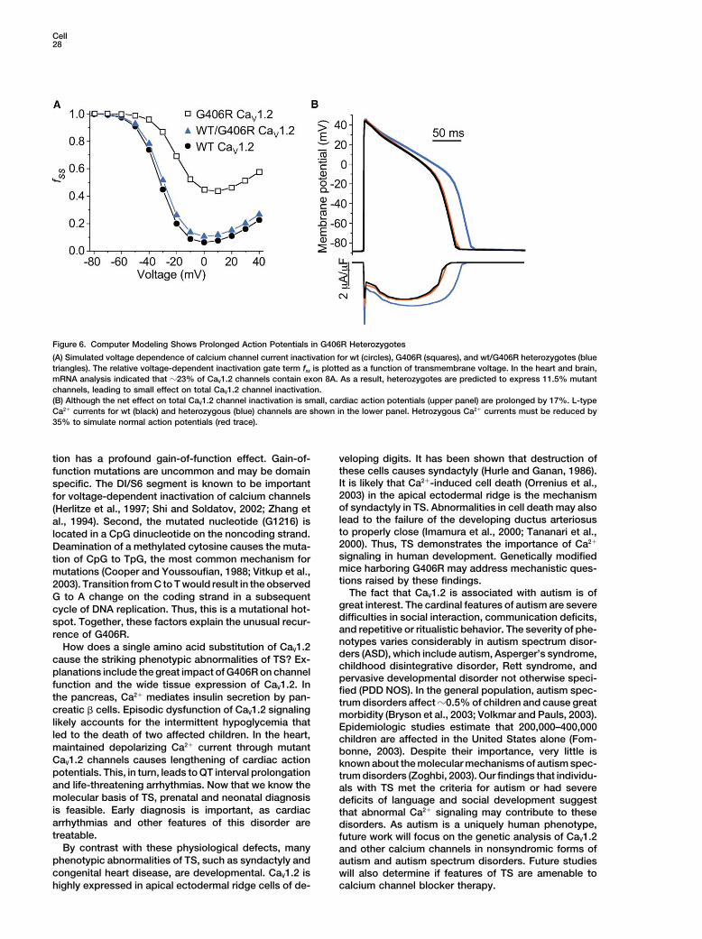
cardiac action potential. We predicted that a slowed Ca 2� current that prolonged action potential duration by17% (Figure 6B, blue traces). Simulations also indicatedrate of channel inactivation would prolong the inward
(depolarizing) Ca 2� current during the plateau phase and that 35% reduction of the abnormal L-type Ca 2� currentcould restore normal action potential duration (Figuredelay cardiomyocyte repolarization. We determined
that, in the heart, 23% of Ca V1.2 channels contained 6B, red traces). These data indicate that G406R mutationcauses substantial prolongation of cardiac action poten-exon 8A. Thus, in the heterozygous state, only 11.5%
of CaV1.2 channels carry the G406R mutation. To simu- tials, consistent with the QT interval prolongation andincreased risk of arrhythmia in TS.late the effect of the TS mutation, we assumed these
ratios in a dynamic model of a mammalian ventricularmyocyte (Faber and Rudy, 2000). We altered the voltage Discussiondependence of L-type calcium channel inactivation tomimic the expected biophysical effects of the mutation We conclude that the G406R mutation of the Ca V1.2
L-type calcium channel causes the diverse physiologicalin heterozygous condition (Figure 6A, blue triangles).The net effect on total Ca V1.2 channel inactivation was and developmental defects in TS. Several lines of evi-
dence support this conclusion. First, we identified ansmall. However, this resulted in a maintained inward


Cell28
Figure 6. Computer Modeling Shows Prolonged Action Potentials in G406R Heterozygotes
(A) Simulated voltage dependence of calcium channel current inactivation for wt (circles), G406R (squares), and wt/G406R heterozygotes (bluetriangles). The relative voltage-dependent inactivation gate term fss is plotted as a function of transmembrane voltage. In the heart and brain,mRNA analysis indicated that �23% of CaV1.2 channels contain exon 8A. As a result, heterozygotes are predicted to express 11.5% mutantchannels, leading to small effect on total CaV1.2 channel inactivation.(B) Although the net effect on total CaV1.2 channel inactivation is small, cardiac action potentials (upper panel) are prolonged by 17%. L-typeCa2� currents for wt (black) and heterozygous (blue) channels are shown in the lower panel. Hetrozygous Ca2� currents must be reduced by35% to simulate normal action potentials (red trace).
tion has a profound gain-of-function effect. Gain-of- veloping digits. It has been shown that destruction ofthese cells causes syndactyly (Hurle and Ganan, 1986).function mutations are uncommon and may be domainIt is likely that Ca2�-induced cell death (Orrenius et al.,specific. The DI/S6 segment is known to be important2003) in the apical ectodermal ridge is the mechanismfor voltage-dependent inactivation of calcium channelsof syndactyly in TS. Abnormalities in cell death may also(Herlitze et al., 1997; Shi and Soldatov, 2002; Zhang etlead to the failure of the developing ductus arteriosusal., 1994). Second, the mutated nucleotide (G1216) isto properly close (Imamura et al., 2000; Tananari et al.,located in a CpG dinucleotide on the noncoding strand.2000). Thus, TS demonstrates the importance of Ca2�
Deamination of a methylated cytosine causes the muta-signaling in human development. Genetically modifiedtion of CpG to TpG, the most common mechanism formice harboring G406R may address mechanistic ques-mutations (Cooper and Youssoufian, 1988; Vitkup et al.,tions raised by these findings.2003). Transition from C to T would result in the observed
The fact that CaV1.2 is associated with autism is ofG to A change on the coding strand in a subsequentgreat interest. The cardinal features of autism are severecycle of DNA replication. Thus, this is a mutational hot-difficulties in social interaction, communication deficits,spot. Together, these factors explain the unusual recur-and repetitive or ritualistic behavior. The severity of phe-rence of G406R.notypes varies considerably in autism spectrum disor-How does a single amino acid substitution of CaV1.2ders (ASD), which include autism, Asperger’s syndrome,cause the striking phenotypic abnormalities of TS? Ex-childhood disintegrative disorder, Rett syndrome, and
planations include the great impact of G406R on channelpervasive developmental disorder not otherwise speci-
function and the wide tissue expression of CaV1.2. In fied (PDD NOS). In the general population, autism spec-the pancreas, Ca2� mediates insulin secretion by pan- trum disorders affect �0.5% of children and cause greatcreatic � cells. Episodic dysfunction of CaV1.2 signaling morbidity (Bryson et al., 2003; Volkmar and Pauls, 2003).likely accounts for the intermittent hypoglycemia that Epidemiologic studies estimate that 200,000–400,000led to the death of two affected children. In the heart, children are affected in the United States alone (Fom-maintained depolarizing Ca2� current through mutant bonne, 2003). Despite their importance, very little isCaV1.2 channels causes lengthening of cardiac action known about the molecular mechanisms of autism spec-potentials. This, in turn, leads to QT interval prolongation trum disorders (Zoghbi, 2003). Our findings that individu-and life-threatening arrhythmias. Now that we know the als with TS met the criteria for autism or had severemolecular basis of TS, prenatal and neonatal diagnosis deficits of language and social development suggestis feasible. Early diagnosis is important, as cardiac that abnormal Ca2� signaling may contribute to thesearrhythmias and other features of this disorder are disorders. As autism is a uniquely human phenotype,treatable. future work will focus on the genetic analysis of CaV1.2
By contrast with these physiological defects, many and other calcium channels in nonsyndromic forms ofphenotypic abnormalities of TS, such as syndactyly and autism and autism spectrum disorders. Future studiescongenital heart disease, are developmental. CaV1.2 is will also determine if features of TS are amenable to
calcium channel blocker therapy.highly expressed in apical ectodermal ridge cells of de-

Calcium Channel Mutation in Arrhythmia and Autism29
Experimental Procedures (Promega). Transformed colonies were screened by PCR for thepresence of exons 8 or 8A using the original oligonucleotide pair.To identify colonies carrying only exon 8A, the same colony collec-Subject Ascertainment and Phenotypic Analysis
Informed consent or assent was obtained from all individuals or their tion was then tested using a forward oligonucleotide specific toexon 8A (8AF5�-CTGGGTCAATGATGCCGTAG) and the 9R reverseguardians according to standards established by local institutional
review boards. Phenotypic analyses included history, physical ex- oligonucleotide. DNA from 30 of 157 clones was sequenced to con-firm the results. To control for the cloning efficiency of the fragmentsamination, electrocardiography, and echocardiography. Five chil-
dren were evaluated for behavioral phenotypes and cognitive devel- carrying each exon, we cloned PCR fragments amplified from atemplate mixture of cDNA containing exon 8 and cDNA containingopment. These tests included the Diagnostic Criteria for Autistic
Disorder, DSM-IV, Autism Screening Questionnaire, Autism Diag- 8A in a 1:1 ratio and screened colonies as detailed above. No differ-ence in cloning efficiency was observed.nostic Interview—Revised, the Child Behavior Checklist, Vineland
Adaptive Behavior Scales, and the Autism Diagnostic Observation Nonradioactive in situ hybridization was performed as described(Berger and Hediger, 2001), using a digoxigenin (DIG)-labeled �800Schedule (Berument et al., 1999; Lord et al., 1994, 2000; Task Force
on DSM-IV, 2000). Cognitive and language tests included Differential nucleotide cRNA probe from the C-terminal region of the mouseCaV1.2 gene. The probe was derived from a PCR fragment amplifiedAbility Scales, Clinical Evaluation of Language Fundamentals—III,
Goldman-Fristoe Test of Articulation, and NEPSY (Korkman et al., from mouse heart Marathon-Ready cDNA (BD Biosciences Clon-tech) using MF5�-AGGCTGGCTTGCGCACCTT and MR5�-GAGA1998).GATGTCTCCCCCTTGA oligonucleotides. Frozen sections (10 m)were cut in a cryostat and captured onto Superfrost Plus microscopeStatistical Analysisslides (Fisher). Sections were then fixed, acetylated, and hybridizedFisher’s exact test and data from the study with highest prevalenceto the probe (approximate concentration 100 ng/ml) over three nights atof autism and autism spectrum disorders (60 of 8,896 individuals)70�C. Hybridized probe was visualized using alkaline phosphatase-were used to determine the p value for the association of theseconjugated anti-DIG Fab fragments (Roche) and 5-bromo-4-chloro-disorders and TS (Bertrand et al., 2001). This comparison gave the3-indolyl-phosphate/nitro-blue tetrazolium (BCIP/NBT) substratemost conservative estimate of the p value. Fisher’s exact test was(Kierkegard and Perry Laboratories). Sections were rinsed severalalso used to assess the p value for the association of G406R withtimes in 100 mM Tris, 150 mM NaCl, and 20 mM EDTA (pH 9.5) andthe TS phenotype.coverslipped with glycerol gelatin (Sigma). Control sections wereincubated with identical concentration of the sense probe transcript.Genotypic and Sequence AnalysesDigital images for antisense and sense probes for each section wereGenomic DNA from peripheral blood lymphocytes or cell lines de-captured using identical microscope settings.rived from Epstein-Barr virus-transformed lymphocytes was pre-
pared using Puregene DNA isolation kit (Gentra Systems). GenomicDNA Constructs for Functional ExpressionDNA from buccal swabs and sperm was prepared using QIAampFull-length human wt CaV1.2 cDNAs (accession number Z34815),DNA Mini Kit (Qiagen). Oligonucleotides to all known exons of thecloned in a Xenopus (pBluescript) and mammalian (pcDNA3) expres-CaV1.2 gene were designed to genomic sequences found in thesion vector systems, were a generous gift from Dr. N. Soldatov. TheCelera database using Oligo6.6 (Molecular Biology Insights, TableG406R mutation was introduced by site-directed mutagenesis using2). PCR amplification of DNA samples and mutational analyses wereQuikChange (Stratagene) into the Xenopus expression clone. Sub-carried out as previously described (Splawski et al., 1997b). Twosequently, an AfeI/SgrAI fragment containing the introduced muta-different conditions were used for PCR: (1) 94�C for 2 min, 35 cyclestion was cloned into the AfeI and SgrAI sites of the wt pcDNA3of 10 s at 94�C, 20 s at 58�C, and 20 s at 72�C, followed by 5 minclone to obtain the mammalian G406R expression construct.extension at 72�C; and (2) same as (1), but annealing was done at
CaV�2b is the � subunit splice variant associated with CaV1.2 in54�C, and the PCR reaction had a final concentration of 10% glycerolthe human heart (Colecraft et al., 2002). We obtained a cDNA cloneand 4% formamide (Table 2).containing the 5� end sequence of CaV�2b from RZPD (cloneOligonucleotides OriF5�-TACACTAATCATCATAGGGTCAT andDKFZp313F1242, RZPD, Germany) and purchased the splice variantOri2R5�-TAGCGATTCCCAGTTTAGGTAC were used to amplify aCaV�2a (the 3� sequences of CaV�2a and CaV�2b are identical) fromfragment of 1122 nucleotides containing part of exon 8A and adja-Genecopoeia (clone GC-T4617, Genecopoeia). EcoRI/PshAI frag-cent intron 8A sequences. PCR products were obtained with Pfument from DKFZp313F1242 and PshAI/NotI fragment from GC-Ultra HF DNA polymerase (Stratagene), purified, and sequenced.T4617 were cloned into the EcoRI and NotI sites of pcDNA3.1 (In-An intronic polymorphism (C to G) was identified 344 nucleotidesvitrogen) to obtain the full-length cDNA clone for human CaV�2bdownstream from exon 8A. PCR fragments were then cloned using(accession number AAG01473). The rabbit CaV�2b clone (accessionthe PCR-Script Amp Cloning Kit (Stratagene) to separate the prod-number CAA45575, amino acid sequence 96% identical to humanucts derived from individual chromosomes. DNA from several clonesCaV�2b) for expression in Xenopus oocytes was a kind gift from Dr.for each individual was sequenced to determine the parent chromo-N. Dascal.some on which the mutation arose.
CaV�2�1 is the �2� subunit associated with CaV1.2 in the heart(Arikkath and Campbell, 2003). Full-length clone for the humanmRNA Expression and cDNA AnalysesCaV�2�1 subunit (accession number NP_000713) was obtained byBlot analyses were performed using human 12-lane multiple-tissueligation of a NotI/Bpu10I fragment from IMAGE clone 2006073 andNorthern blots I and III (BD Biosciences Clontech). mRNA dot blota Bpu10I/XbaI fragment from a PCR product, amplified from humananalysis was performed using the Multiple Tissue Expression Humanheart Marathon-Ready cDNA (BD Biosciences Clontech) using oli-Array 3 (BD Biosciences Clontech). A 115 base pair PCR fragment,gonucleotides A2D1F5�-TGAATGTAGCTTCATTTAACAGCA-3� andamplified using HF5�-TGGGTCAATGATGCCGTAGG and HR5�-GAAA2D1XbaR5�-GCTCTAGATTTGGCAGGGTCTGGAGTTTAAC-3� (XbaIAACTCTCCGCTAAGCACA oligonucleotides, was used as a probesite underlined), into the NotI and XbaI sites of pcDNA3.1 (In-for exon 8A containing mRNAs. The fragment was labeled with thevitrogen). The rabbit CaV�2�1 clone (accession number AAA81562,Prime-It II labeling kit (Stratagene) using the reverse (HR) oligonucle-amino acid sequence 96% identical to human CaV�2�1) for expres-otide instead of the provided random 9-mers. Hybridization andsion in Xenopus oocytes was a kind gift from Dr. N. Dascal.washing conditions followed manufacturer’s suggestions. The blots
The full-length clones for all eight expression constructs de-were exposed to film for 3 days.scribed above were sequenced in forward and reverse direction andHuman heart marathon-ready cDNA (BD Biosciences Clontech)compared to genomic DNA to ensure that no unintended mutationsand human heart and brain race-ready cDNAs (Ambion) were ana-were present or introduced.lyzed to estimate the ratio of CaV1.2 transcripts containing exon 8A
to transcripts containing exon 8. Briefly, PCR products amplifiedusing the 7F and 9R oligonucleotides (forward from exon 7, 7F5�- Transfection and Solutions for CHO Cells
CHO cells were cultured in Ham’s F-12 Media and transiently trans-TCACGGTGTTCCAGTGCATC and reverse from exon 9, 9R5�-CAGGTAGCCTTTGAGATCCTC) were ligated into pGEM-T Easy Vector fected using Lipofectamine 2000 (GIBCO). Cells were transfected

Cell30
for 18 hr in 35 mm dishes containing 18 l Lipofectamine, 242 l W. Reynolds Foundation (M.T.K. and I.S.), Fondazione Telethon, andFondazione Cariplo (S.G.P.) is gratefully acknowledged.Optimem (GIBCO), 0.86 g enhanced green fluorescent protein (Mo-
lecular Probes), 4 g of either wt or mutant human CaV1.2, andReceived: July 9, 20041 g each of human CaV�2b and human CaV�2�1 subunit cDNAs. TheRevised: August 9, 2004extracellular solution contained the following, in mM: 130 NMDG,Accepted: August 17, 200415 CaCl2, 5 KCl, and 10 HEPES (pH 7.4 with HCl, 22�C–25�C). ThePublished: September 30, 2004intracellular pipette solution contained the following, in mM: 120 Cs
methanesulfonate, 5 CaCl2, 2 MgCl2, 10 EGTA, 2 MgATP, and 10ReferencesHEPES (pH 7.3 with CsOH). This solution results in an [Ca2�]i of
�110 nM as calculated with WinMaxc (Bers et al., 1994).Abernethy, D.R., and Soldatov, N.M. (2002). Structure-functional di-versity of human L-type Ca2� channel: perspectives for new phar-
Injection and Solutions for Oocytesmacological targets. J. Pharmacol. Exp. Ther. 300, 724–728.
Isolation and injection of Xenopus laevis oocytes and synthesis ofAllen, G., and Courchesne, E. (2003). Differential effects of develop-capped polyA cRNA from linearized cDNA templates were per-mental cerebellar abnormality on cognitive and motor functions informed as described (Goldin, 1991). Oocytes were coinjected withthe cerebellum: an fMRI study of autism. Am. J. Psychiatry 160,cRNAs encoding wt or mutant human CaV1.2 subunit (11 ng) plus262–273.rabbit CaV�2b (2.7 ng) and rabbit CaV�2�1 (2.7 ng) subunits. The extra-Antzelevitch, C. (2003). Molecular genetics of arrhythmias and car-cellular solution contained the following, in mM: 40 Ba(OH)2, 50diovascular conditions associated with arrhythmias. J. Cardiovasc.NaOH, 1 KOH, 5 HEPES (pH 7.4 with methanesulfonic acid, 22�C–Electrophysiol. 14, 1259–1272.25�C). Niflumic acid (300 M) was added to the solution to block
intracellular Ca2�-activated Cl� currents (White and Aylwin, 1990). APA (American Psychiatric Association) (2000). Diagnostic and Sta-Recording pipettes contained 3 M KCl and had resistances of tistical Manual of Mental Disorders DSM-IV-TR, Fourth Edition
(Washington, D.C.: American Psychiatric Association).0.5–1 M�.
Arikkath, J., and Campbell, K.P. (2003). Auxiliary subunits: essentialcomponents of the voltage-gated calcium channel complex. Curr.Voltage Clamp and Data AnalysisOpin. Neurobiol. 13, 298–307.Whole-cell Ca2� currents in fluorescent CHO cells were recordedBean, B.P. (1984). Nitrendipine block of cardiac calcium channels:using standard techniques (Hamill et al., 1981) and an Axopatch 200high-affinity binding to the inactivated state. Proc. Natl. Acad. Sci.patch-clamp amplifier (Axon Instruments) 2–3 days after transfec-USA 81, 6388–6392.tion with cDNA. Voltage dependence of Ca2� current inactivation in
CHO cells was determined with a two-pulse protocol. The relative Bech-Hansen, N.T., Naylor, M.J., Maybaum, T.A., Pearce, W.G.,magnitude of inward current elicited during the second pulse (to �30 Koop, B., Fishman, G.A., Mets, M., Musarella, M.A., and Boycott,mV) was plotted as a function of the variable voltage of the first K.M. (1998). Loss-of-function mutations in a calcium-channelpulse (0.8 s). Ba2� currents through calcium channels were recorded alpha1-subunit gene in Xp11.23 cause incomplete X-linked congeni-
tal stationary night blindness. Nat. Genet. 19, 264–267.from oocytes using standard two microelectrode voltage clamptechniques (Stuhmer, 1992) 2–10 days after injection of cRNA. Volt- Berger, U.V., and Hediger, M.A. (2001). Differential distribution ofage dependence of Ba2� current inactivation in oocytes was deter- the glutamate transporters GLT-1 and GLAST in tanycytes of themined with a two-pulse protocol. The relative magnitude of inward third ventricle. J. Comp. Neurol. 433, 101–114.current elicited during the second pulse (to �10 mV) is plotted as Berridge, M.J., Bootman, M.D., and Roderick, H.L. (2003). Calciuma function of the variable voltage of the first pulse (2 s). Data acquisi- signalling: dynamics, homeostasis and remodelling. Nat. Rev. Mol.tion and analyses were performed using pCLAMP8 (Axon Instru- Cell Biol. 4, 517–529.ments). Currents were filtered at 2 kHz and digitized at 10 kHz.
Bers, D.M., Patton, C.W., and Nuccitelli, R. (1994). A practical guideData from CHO and Xenopus oocyte expression were fitted to a
to the preparation of Ca2� buffers. Methods Cell Biol. 40, 3–29.Boltzmann function to obtain half point (V1/2) and slope factor (k) for
Bertrand, J., Mars, A., Boyle, C., Bove, F., Yeargin-Allsopp, M., andthe voltage dependence of CaV1.2 inactivation. Data are presentedDecoufle, P. (2001). Prevalence of autism in a United States popula-as mean SEM.tion: the Brick Township, New Jersey, investigation. Pediatrics108, 1155–1161.
Action Potential Modeling Berument, S.K., Rutter, M., Lord, C., Pickles, A., and Bailey, A. (1999).A dynamic model of mammalian ventricular myocytes (Faber and Autism screening questionnaire: diagnostic validity. Br. J. PsychiatryRudy, 2000) was used to simulate the effect of the TS mutation in 175, 444–451.heterozygotes. For these simulations, the stimulation rate was set
Brambilla, P., Hardan, A., di Nemi, S.U., Perez, J., Soares, J.C., andat 60 per min. Action potential waveforms and L-type Ca2� currentsBarale, F. (2003). Brain anatomy and development in autism: review
were computed after 100 stimulations. Channel properties wereof structural MRI studies. Brain Res. Bull. 61, 557–569.
modeled by altering the relative voltage-dependent inactivation gateBrini, M., and Carafoli, E. (2000). Calcium signalling: a historicalterm fss to a heterozygous condition in which the exon 8A containingaccount, recent developments and future perspectives. Cell. Mol.CaV1.2 protein represents 23% (11.5% wt and 11.5% G406R) of theLife Sci. 57, 354–370.total CaV1.2 protein. Thus, the shifts in the inactivation curves causedBryson, S.E., Rogers, S.J., and Fombonne, E. (2003). Autism spec-by the G406R mutation (as measured in CHO cells) were reduced,trum disorders: early detection, intervention, education, and psy-assuming only 0.115 of the total channels were mutated (V1/2 waschopharmacological management. Can. J. Psychiatry 48, 506–516.shifted by �1.2 mV, and the minimal value for fss was set at 0.106).Catterall, W.A. (2000). Structure and regulation of voltage-gatedCa2� channels. Annu. Rev. Cell Dev. Biol. 16, 521–555.AcknowledgmentsColecraft, H.M., Alseikhan, B., Takahashi, S.X., Chaudhuri, D., Mitt-man, S., Yegnasubramanian, V., Alvania, R.S., Johns, D.C., Marban,We are grateful to all of the individuals with TS and their familiesE., and Yue, D.T. (2002). Novel functional properties of Ca(2�) chan-for donated time and samples. We would also like to express ournel beta subunits revealed by their expression in adult rat heartgratitude to C. Badame, K. Braegger, S. Etheridge, T. Carson, D.cells. J. Physiol. 541, 435–452.Goldman, T. Klitzner, J. Skinner, A. Moss, H. Stalker, G.M. Vincent,Cooper, D.N., and Youssoufian, H. (1988). The CpG dinucleotideM. Marks, J. Towbin, M. Pun, C-L. Lien, GCRC Children’s Hospitaland human genetic disease. Hum. Genet. 78, 151–155.Boston, SADS Foundation, and the UK SADS Foundation. We thank
N. Soldatov and N. Dascal for expression constructs and D. Clap- Ertel, E.A., Campbell, K.P., Harpold, M.M., Hofmann, F., Mori, Y.,ham, S. Orkin, L. Kunkel, K. Thomas, and F. Engel for critically Perez-Reyes, E., Schwartz, A., Snutch, T.P., Tanabe, T., Birnbaumer,reviewing the manuscript. Funding from NIH (HL46401 and HL52338 L., et al. (2000). Nomenclature of voltage-gated calcium channels.
Neuron 25, 533–535.for M.T.K. and M.C.S., DC03610 and MH66398 for H.T.-F.), Donald

Calcium Channel Mutation in Arrhythmia and Autism31
Faber, G.M., and Rudy, Y. (2000). Action potential and contractility cinski, H., McManis, P.G., Santiago, L., Moore, M., Fouad, G., etal. (1994). Dihydropyridine receptor mutations cause hypokalemicchanges in [Na(�)](i) overloaded cardiac myocytes: a simulation
study. Biophys. J. 78, 2392–2404. periodic paralysis. Cell 77, 863–868.
Reichenbach, H., Meister, E.M., and Theile, H. (1992). The heart-Fombonne, E. (2003). Epidemiological surveys of autism and otherhand syndrome. A new variant of disorders of heart condition andpervasive developmental disorders: an update. J. Autism Dev. Dis-syndactylia including osseous changes in hands and feet. Kinder-ord. 33, 365–382.arztl. Prax. 60, 54–56.Goldin, A.L. (1991). Expression of ion channels by injection of mRNARen, D., Navarro, B., Perez, G., Jackson, A.C., Hsu, S., Shi, Q., Tilly,into Xenopus oocytes. Methods Cell Biol. 36, 487–509.J.L., and Clapham, D.E. (2001). A sperm ion channel required forHamill, O.P., Marty, A., Neher, E., Sakmann, B., and Sigworth, F.J.sperm motility and male fertility. Nature 413, 603–609.(1981). Improved patch-clamp techniques for high-resolution cur-Sanguinetti, M.C., and Kass, R.S. (1984). Voltage-dependent blockrent recording from cells and cell-free membrane patches. Pflugersof calcium channel current in the calf cardiac Purkinje fiber by dihy-Arch. 391, 85–100.dropyridine calcium channel antagonists. Circ. Res. 55, 336–348.Herlitze, S., Hockerman, G.H., Scheuer, T., and Catterall, W.A. (1997).Schultz, D., Mikala, G., Yatani, A., Engle, D.B., Iles, D.E., Segers, B.,Molecular determinants of inactivation and G protein modulation inSinke, R.J., Weghuis, D.O., Klockner, U., Wakamori, M., et al. (1993).the intracellular loop connecting domains I and II of the calciumCloning, chromosomal localization, and functional expression of thechannel alpha1A subunit. Proc. Natl. Acad. Sci. USA 94, 1512–1516.alpha 1 subunit of the L-type voltage-dependent calcium channelHurle, J.M., and Ganan, Y. (1986). Interdigital tissue chondrogenesisfrom normal human heart. Proc. Natl. Acad. Sci. USA 90, 6228–6232.induced by surgical removal of the ectoderm in the embryonic chickSchulze-Bahr, E., Wang, Q., Wedekind, H., Haverkamp, W., Chen,leg bud. J. Embryol. Exp. Morphol. 94, 231–244.Q., Sun, Y., Rubie, C., Hordt, M., Towbin, J.A., Borggrefe, M., et al.Imamura, S., Nishikawa, T., Hiratsuka, E., Takao, A., and Matsuoka,(1997). KCNE1 mutations cause Jervell and Lange-Nielsen syn-R. (2000). Behavior of smooth muscle cells during arterial ductaldrome. Nat. Genet. 17, 267–268.closure at birth. J. Histochem. Cytochem. 48, 35–44.Shi, C., and Soldatov, N.M. (2002). Molecular determinants of volt-Keating, M.T., and Sanguinetti, M.C. (2001). Molecular and cellularage-dependent slow inactivation of the Ca2� channel. J. Biol.mechanisms of cardiac arrhythmias. Cell 104, 569–580.Chem. 277, 6813–6821.
Korkman, M., Kirk, U., and Kemp, S. (1998). NEPSY: A Develop-Splawski, I., Timothy, K.W., Vincent, G.M., Atkinson, D.L., and Keat-mental Neuropsychological Assessment (San Antonio, TX: The Psy-ing, M.T. (1997a). Molecular basis of the long-QT syndrome associ-chological Corporation).ated with deafness. N. Engl. J. Med. 336, 1562–1567.
Lee, K.S., Marban, E., and Tsien, R.W. (1985). Inactivation of calciumSplawski, I., Tristani-Firouzi, M., Lehmann, M.H., Sanguinetti, M.C.,channels in mammalian heart cells: joint dependence on membraneand Keating, M.T. (1997b). Mutations in the hminK gene cause longpotential and intracellular calcium. J. Physiol. 364, 395–411.QT syndrome and suppress IKs function. Nat. Genet. 17, 338–340.
Lord, C., Rutter, M., and Le Couteur, A. (1994). Autism DiagnosticStrom, T.M., Nyakatura, G., Apfelstedt-Sylla, E., Hellebrand, H., Lo-Interview-Revised: a revised version of a diagnostic interview forrenz, B., Weber, B.H., Wutz, K., Gutwillinger, N., Ruther, K., Drescher,caregivers of individuals with possible pervasive developmental dis-B., et al. (1998). An L-type calcium-channel gene mutated in incom-orders. J. Autism Dev. Disord. 24, 659–685.plete X-linked congenital stationary night blindness. Nat. Genet.
Lord, C., Risi, S., Lambrecht, L., Cook, E.H., Jr., Leventhal, B.L., 19, 260–263.DiLavore, P.C., Pickles, A., and Rutter, M. (2000). The autism diag-
Stuhmer, W. (1992). Electrophysiological recording from Xenopusnostic observation schedule-generic: a standard measure of social
oocytes. Methods Enzymol. 207, 319–339.and communication deficits associated with the spectrum of autism.
Tananari, Y., Maeno, Y., Takagishi, T., Sasaguri, Y., Morimatsu, M.,J. Autism Dev. Disord. 30, 205–223.and Kato, H. (2000). Role of apoptosis in the closure of neonatal
Marks, M.L., Whisler, S.L., Clericuzio, C., and Keating, M. (1995). Aductus arteriosus. Jpn. Circ. J. 64, 684–688.
new form of long QT syndrome associated with syndactyly. J. Am.Tyson, J., Tranebjaerg, L., Bellman, S., Wren, C., Taylor, J.F., Bathen,Coll. Cardiol. 25, 59–64.J., Aslaksen, B., Sorland, S.J., Lund, O., Malcolm, S., et al. (1997).
Mikami, A., Imoto, K., Tanabe, T., Niidome, T., Mori, Y., Takeshima,IsK and KvLQT1: mutation in either of the two subunits of the slow
H., Narumiya, S., and Numa, S. (1989). Primary structure and func-component of the delayed rectifier potassium channel can cause
tional expression of the cardiac dihydropyridine-sensitive calciumJervell and Lange-Nielsen syndrome. Hum. Mol. Genet. 6, 2179–
channel. Nature 340, 230–233.2185.
Monnier, N., Procaccio, V., Stieglitz, P., and Lunardi, J. (1997). Malig-Vitkup, D., Sander, C., and Church, G.M. (2003). The amino-acid
nant-hyperthermia susceptibility is associated with a mutation ofmutational spectrum of human genetic disease. Genome Biol. 4,
the alpha 1-subunit of the human dihydropyridine-sensitive L-typeR72. Published online October 30, 2003. 10.1186/gb-2003-4-11-r72
voltage-dependent calcium-channel receptor in skeletal muscle.Volkmar, F.R., and Pauls, D. (2003). Autism. Lancet 362, 1133–1141.Am. J. Hum. Genet. 60, 1316–1325.White, M.M., and Aylwin, M. (1990). Niflumic and flufenamic acidsNeyroud, N., Tesson, F., Denjoy, I., Leibovici, M., Donger, C., Bar-are potent reversible blockers of Ca2(�)-activated Cl- channels inhanin, J., Faure, S., Gary, F., Coumel, P., Petit, C., et al. (1997). AXenopus oocytes. Mol. Pharmacol. 37, 720–724.novel mutation in the potassium channel gene KVLQT1 causes theZhang, J.F., Ellinor, P.T., Aldrich, R.W., and Tsien, R.W. (1994). Mo-Jervell and Lange-Nielsen cardioauditory syndrome. Nat. Genet.lecular determinants of voltage-dependent inactivation in calcium15, 186–189.channels. Nature 372, 97–100.Ophoff, R.A., Terwindt, G.M., Vergouwe, M.N., van Eijk, R., Oefner,Zhang, L., Timothy, K.W., Vincent, G.M., Lehmann, M.H., Fox, J.,P.J., Hoffman, S.M., Lamerdin, J.E., Mohrenweiser, H.W., Bulman,Giuli, L.C., Shen, J., Splawski, I., Priori, S.G., Compton, S.J., et al.D.E., Ferrari, M., et al. (1996). Familial hemiplegic migraine and epi-(2000). Spectrum of ST-T-wave patterns and repolarization parame-sodic ataxia type-2 are caused by mutations in the Ca2� channelters in congenital long-QT syndrome: ECG findings identify geno-gene CACNL1A4. Cell 87, 543–552.types. Circulation 102, 2849–2855.Orrenius, S., Zhivotovsky, B., and Nicotera, P. (2003). RegulationZheng, Z.J., Croft, J.B., Giles, W.H., and Mensah, G.A. (2001). Sud-of cell death: the calcium-apoptosis link. Nat. Rev. Mol. Cell Biol.den cardiac death in the United States, 1989 to 1998. Circulation4, 552–565.104, 2158–2163.Priori, S.G., Aliot, E., Blomstrom-Lundqvist, C., Bossaert, L., Breith-Zoghbi, H.Y. (2003). Postnatal neurodevelopmental disorders: meet-ardt, G., Brugada, P., Camm, J.A., Cappato, R., Cobbe, S.M., Di,ing at the synapse? Science 302, 826–830.M.C., et al. (2002). Task Force on Sudden Cardiac Death, European
Society of Cardiology. Europace 4, 3–18.
Ptacek, L.J., Tawil, R., Griggs, R.C., Engel, A.G., Layzer, R.B., Kwie-