Analytical Methods for Lipid Oxidation and Antioxidant ... - MDPI
19
Antioxidants 2021, 10, 1587. https://doi.org/10.3390/antiox10101587 www.mdpi.com/journal/antioxidants Review Analytical Methods for Lipid Oxidation and Antioxidant Capacity in Food Systems Edirisingha Dewage Nalaka Sandun Abeyrathne 1,2 , Kichang Nam 2 and Dong Uk Ahn 3, * 1 Department of Animal Science, Uva Wellassa University, Badulla 90000, Sri Lanka; [email protected] 2 Department of Animal Science & Technology, Sunchon National University, Suncheon 57922, Korea; [email protected] 3 Department of Animal Science, Iowa State University, Ames, IA 50011, USA * Correspondence: [email protected] Abstract: Lipid oxidation is the most crucial quality parameter in foods. Many methods were devel- oped to determine the level of oxidation and antioxidant activity. This review compares the methods used to determine lipid oxidation and antioxidant capacity in foods. Lipid oxidation methods de- veloped are based on the direct or indirect measurement of produced primary or secondary oxida- tion substances. Peroxide values and conjugated diene methods determine the primary oxidative products of lipid oxidation and are commonly used for plant oils and high-fat products. 2-Thiobar- bituric acid-reactive substances and chromatographic methods are used to determine the secondary products of oxidation and are suitable for meat and meat-based products. The fluorometric and sensory analyses are indirect methods. The antioxidant capacity of additives is determined indi- rectly using the lipid oxidation methods mentioned above or directly based on the free-radical scav- enging activity of the antioxidant compounds. Each lipid oxidation and antioxidant capacity meth- ods use different approaches, and one method cannot be used for all foods. Therefore, selecting proper methods for specific foods is essential for accurately evaluating lipid oxidation or antioxi- dant capacity. Keywords: lipid oxidation; primary oxidation products; secondary oxidation products; antioxidant capacity 1. Introduction Lipids are significant nutrients for humans and help many functional and regulatory activities in the human body, such as signal transduction, myelination, and synaptic plas- ticity. Lipids are also involved in the structural developments of the human body [1,2]. In food, lipid content and fatty acid composition are the two critical congenital parameters to the susceptibility of food to oxidative changes. Lipid content and the fatty acid compo- sition of fat of farm animals varies significantly depending on animal species and the diet [3–5]. Fish is considered as one of the primary sources for ω-3 fatty acids, especially eicosa- pentaenoic acid (EPA) and docosahexaenoic acid (DHA) [6] and is highly susceptible to oxidative changes. Eggs are also rich in ω-3 fatty acids, and these fatty acids play a signif- icant role in controlling cardiovascular diseases, inflammation, immune functions, and mental health disorders [7,8]. Not only animals but also plants contain fats and oils, which are used in cooking or preparing foods. Plant oils are rich in fatty acids, such as linoleic, linolenic, oleic, palmitic, and stearic acids, and some plant oils have industrial applica- tions, such as coatings, inks, plasticizers, lubricants, and paints [9,10]. Plants also contain various bioactive compounds, including p-cymene, thymol, eugenol, carvacrol, isothiocy- anate, cinnamaldehyde, cumin aldehyde, linalool, 1,8-cineol, α-pinene, α-terpineol, γ-ter- pinene, citral, and methyl chavicol, that are used for therapeutic, cosmetic, aromatic, Citation: Abeyrathne, E.D.N.S.; Nam, K.; Ahn, D.U. Analytical Methods for Lipid Oxidation and Antioxidant Capacity in Food Systems. Antioxidants 2021, 10, 1587. https://doi.org/10.3390/antiox 10101587 Academic Editor: Per Ertbjerg Received: 13 September 2021 Accepted: 7 October 2021 Published: 9 October 2021 Publisher’s Note: MDPI stays neu- tral with regard to jurisdictional claims in published maps and institu- tional affiliations. Copyright: © 2021 by the authors. Li- censee MDPI, Basel, Switzerland. This article is an open access article distributed under the terms and con- ditions of the Creative Commons At- tribution (CC BY) license (https://cre- ativecommons.org/licenses/by/4.0/).
-
Upload
khangminh22 -
Category
Documents
-
view
0 -
download
0
Transcript of Analytical Methods for Lipid Oxidation and Antioxidant ... - MDPI
Antioxidants 2021, 10, 1587. https://doi.org/10.3390/antiox10101587 www.mdpi.com/journal/antioxidants
Review
Analytical Methods for Lipid Oxidation and Antioxidant Capacity in Food Systems Edirisingha Dewage Nalaka Sandun Abeyrathne 1,2, Kichang Nam 2 and Dong Uk Ahn 3,*
1 Department of Animal Science, Uva Wellassa University, Badulla 90000, Sri Lanka; [email protected] 2 Department of Animal Science & Technology, Sunchon National University, Suncheon 57922, Korea;
[email protected] 3 Department of Animal Science, Iowa State University, Ames, IA 50011, USA * Correspondence: [email protected]
Abstract: Lipid oxidation is the most crucial quality parameter in foods. Many methods were devel-oped to determine the level of oxidation and antioxidant activity. This review compares the methods used to determine lipid oxidation and antioxidant capacity in foods. Lipid oxidation methods de-veloped are based on the direct or indirect measurement of produced primary or secondary oxida-tion substances. Peroxide values and conjugated diene methods determine the primary oxidative products of lipid oxidation and are commonly used for plant oils and high-fat products. 2-Thiobar-bituric acid-reactive substances and chromatographic methods are used to determine the secondary products of oxidation and are suitable for meat and meat-based products. The fluorometric and sensory analyses are indirect methods. The antioxidant capacity of additives is determined indi-rectly using the lipid oxidation methods mentioned above or directly based on the free-radical scav-enging activity of the antioxidant compounds. Each lipid oxidation and antioxidant capacity meth-ods use different approaches, and one method cannot be used for all foods. Therefore, selecting proper methods for specific foods is essential for accurately evaluating lipid oxidation or antioxi-dant capacity.
Keywords: lipid oxidation; primary oxidation products; secondary oxidation products; antioxidant capacity
1. Introduction Lipids are significant nutrients for humans and help many functional and regulatory
activities in the human body, such as signal transduction, myelination, and synaptic plas-ticity. Lipids are also involved in the structural developments of the human body [1,2]. In food, lipid content and fatty acid composition are the two critical congenital parameters to the susceptibility of food to oxidative changes. Lipid content and the fatty acid compo-sition of fat of farm animals varies significantly depending on animal species and the diet [3–5]. Fish is considered as one of the primary sources for ω-3 fatty acids, especially eicosa-pentaenoic acid (EPA) and docosahexaenoic acid (DHA) [6] and is highly susceptible to oxidative changes. Eggs are also rich in ω-3 fatty acids, and these fatty acids play a signif-icant role in controlling cardiovascular diseases, inflammation, immune functions, and mental health disorders [7,8]. Not only animals but also plants contain fats and oils, which are used in cooking or preparing foods. Plant oils are rich in fatty acids, such as linoleic, linolenic, oleic, palmitic, and stearic acids, and some plant oils have industrial applica-tions, such as coatings, inks, plasticizers, lubricants, and paints [9,10]. Plants also contain various bioactive compounds, including p-cymene, thymol, eugenol, carvacrol, isothiocy-anate, cinnamaldehyde, cumin aldehyde, linalool, 1,8-cineol, α-pinene, α-terpineol, γ-ter-pinene, citral, and methyl chavicol, that are used for therapeutic, cosmetic, aromatic,
Citation: Abeyrathne, E.D.N.S.;
Nam, K.; Ahn, D.U. Analytical
Methods for Lipid Oxidation and
Antioxidant Capacity in Food
Systems. Antioxidants 2021, 10, 1587.
https://doi.org/10.3390/antiox
10101587
Academic Editor: Per Ertbjerg
Received: 13 September 2021
Accepted: 7 October 2021
Published: 9 October 2021
Publisher’s Note: MDPI stays neu-
tral with regard to jurisdictional
claims in published maps and institu-
tional affiliations.
Copyright: © 2021 by the authors. Li-
censee MDPI, Basel, Switzerland.
This article is an open access article
distributed under the terms and con-
ditions of the Creative Commons At-
tribution (CC BY) license (https://cre-
ativecommons.org/licenses/by/4.0/).
Antioxidants 2021, 10, 1587 2 of 19
fragrant, and spiritual purposes [9,11,12]. These bioactive compounds produced from plant oils are also known to have antioxidant properties [12].
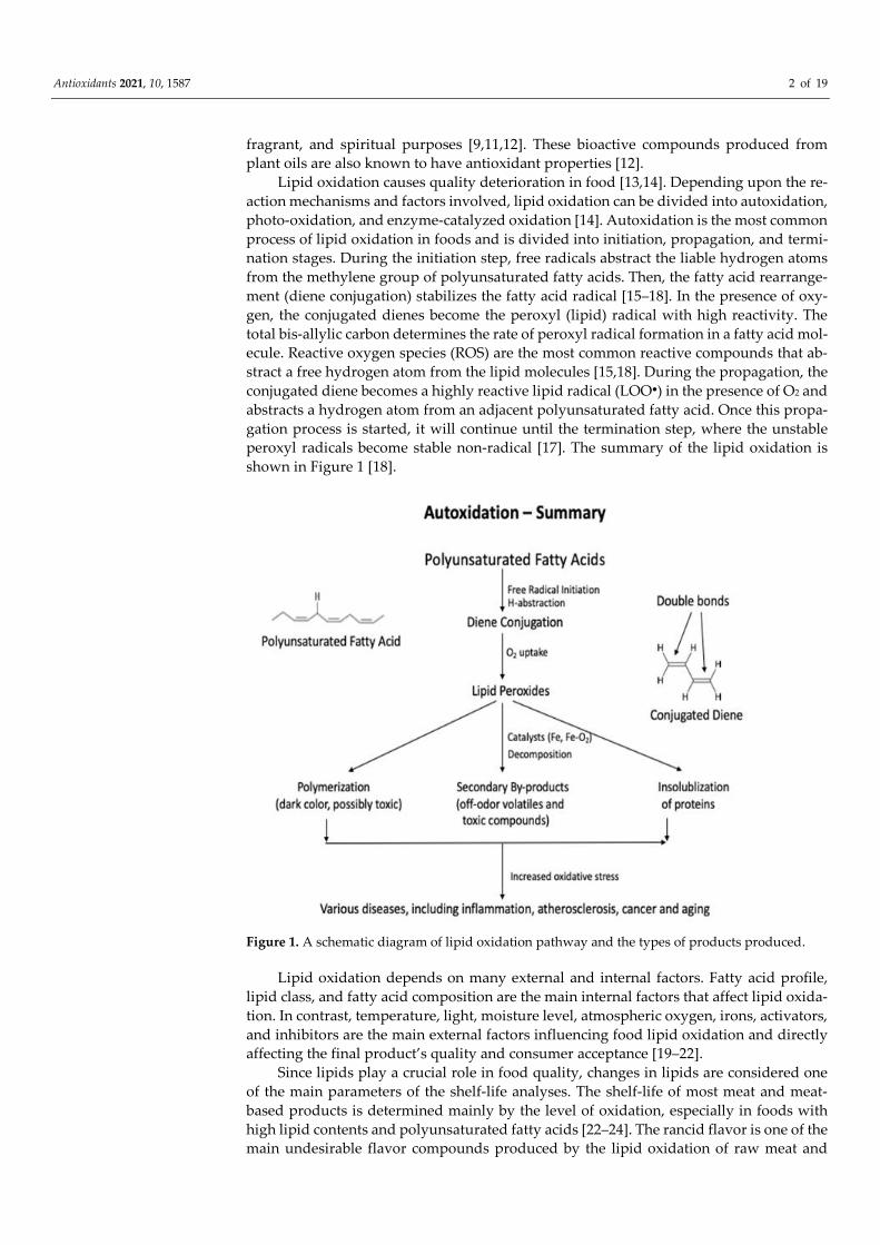
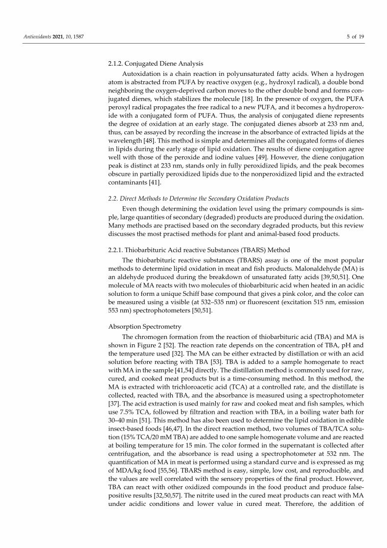
Lipid oxidation causes quality deterioration in food [13,14]. Depending upon the re-action mechanisms and factors involved, lipid oxidation can be divided into autoxidation, photo-oxidation, and enzyme-catalyzed oxidation [14]. Autoxidation is the most common process of lipid oxidation in foods and is divided into initiation, propagation, and termi-nation stages. During the initiation step, free radicals abstract the liable hydrogen atoms from the methylene group of polyunsaturated fatty acids. Then, the fatty acid rearrange-ment (diene conjugation) stabilizes the fatty acid radical [15–18]. In the presence of oxy-gen, the conjugated dienes become the peroxyl (lipid) radical with high reactivity. The total bis-allylic carbon determines the rate of peroxyl radical formation in a fatty acid mol-ecule. Reactive oxygen species (ROS) are the most common reactive compounds that ab-stract a free hydrogen atom from the lipid molecules [15,18]. During the propagation, the conjugated diene becomes a highly reactive lipid radical (LOO●) in the presence of O2 and abstracts a hydrogen atom from an adjacent polyunsaturated fatty acid. Once this propa-gation process is started, it will continue until the termination step, where the unstable peroxyl radicals become stable non-radical [17]. The summary of the lipid oxidation is shown in Figure 1 [18].
Figure 1. A schematic diagram of lipid oxidation pathway and the types of products produced.
Lipid oxidation depends on many external and internal factors. Fatty acid profile, lipid class, and fatty acid composition are the main internal factors that affect lipid oxida-tion. In contrast, temperature, light, moisture level, atmospheric oxygen, irons, activators, and inhibitors are the main external factors influencing food lipid oxidation and directly affecting the final product’s quality and consumer acceptance [19–22].
Since lipids play a crucial role in food quality, changes in lipids are considered one of the main parameters of the shelf-life analyses. The shelf-life of most meat and meat-based products is determined mainly by the level of oxidation, especially in foods with high lipid contents and polyunsaturated fatty acids [22–24]. The rancid flavor is one of the main undesirable flavor compounds produced by the lipid oxidation of raw meat and
Antioxidants 2021, 10, 1587 3 of 19
reduces the sensory qualities of the final products. Warmed-over flavors are the secondary lipid oxidation products developed in oxidized cooked meat, and aldehydes, such as hex-anal, pentanal, pentanol, and nonanal compounds, are responsible for the off-flavors [18,25,26]. Some lipid oxidation products, such as malondialdehyde, acrolein, 4-hydroxy-trans-nonenal, 4-hydroxy-trans-hexanal, and crotonaldehyde-like compounds, generate not only off-flavor but also cause diseases, such as inflammation, ageing, cancer, and ath-erosclerosis, in humans [18,22,27]. In addition, they can interfere with the signaling path-ways, leading to significant biomolecular damages [28,29]. Hydroperoxides, carbonyls, aldehydes, alcohols, furans, keto-cholesterols, epoxy-cholesterols, and oxysterols are the biomarkers produced from the enzymatic and nonenzymatic hydrolysis of lipids and are directly used to detect the physiological and pathological conditions of animals [29]. Sev-eral methods, such as antioxidants, vacuum packing, and modified atmospheric packing, have been developed to control lipid oxidation in food products [23].
Many methods are used to determine the level of oxidation in foods by detecting lipid oxidation products. The oxidation products are categorized into primary and sec-ondary products. Critical criteria for detecting lipid oxidation include accuracy, precision, sensitivity, and detection limits [30,31]. Either direct or indirect methods determine lipid oxidation in foods. The direct methods determine lipid oxidation products, while the in-direct methods use the consequences (sensory, disease conditions, etc.) of lipid oxidation or oxidation products. Peroxides and hydroperoxides are the primary oxidation com-pounds, and aldehydes, ketones, epoxides, alcohols, hydroxy compounds, oligomers, and polymers are the secondary compounds commonly used to determine the degree of lipid oxidation [31,32].
Antioxidants are considered a group of compounds that can counteract oxidation by acting as reducing agents, free radical scavengers, quenchers of radical species, or inacti-vators prooxidants, such as metals [33]. Plants contain many natural antioxidants com-pounds, including polyphenols (anthocyanins, flavonols, flavones), carotenoids, tannins, lignin, phenolic acids, and vitamins. However, these antioxidants can lose their activities under adverse conditions, such as high temperature, extreme pH, and strong lights [34]. The antioxidant capacity of certain compounds can be measured directly or indirectly. The direct method determines the ability of antioxidants to intervene or stop the lipid oxidation process in meat homogenates, meat products, oil emulsion, and liposome sys-tems in the presence of antioxidant compounds. This review discusses the methods used to determine lipid oxidation and antioxidant capacity in food products and their princi-ples and constraints.
2. The Analytical Methods to Determine Lipid Oxidation in Food Lipid oxidation is considered one of the main reasons for losing the sensory and nu-
tritional quality in many animals and plant-based foods, and various internal and external factors influence lipid oxidation in food products [18,33,35]. Methods used to determine lipid oxidation can be categorized into direct and indirect. In the direct method, the oxi-dation level is measured using lipid oxidation’s primary or secondary products. The most common methods determining primary oxidation products include peroxide value, io-dine value, and diene conjugation measurement [32,36]. Malonaldehyde (MA), a second-ary oxidation product, is the most used indicator of lipid oxidation [37,38]. The 2-thiobar-bituric acid reactive substances (TBARS) methods mostly use the reaction of MA with thi-obarbituric acid after distillation or acid extraction and determine the reaction products using the absorption or fluorescence spectrometer. However, HPLC and gas chromatog-raphy are also used to directly determine the amount of MA or other secondary lipid ox-idation products [32,39]. The indirect measurements of lipid oxidation in foods use the fluorometric method and sensory evaluation.
Antioxidants 2021, 10, 1587 4 of 19
2.1. Methods Used to Detect the Primary Oxidation Products The three most used methods for the primary oxidation products in foods are iodo-
metric, ferric thiocyanate, and diene conjugation methods, and they are the direct meth-ods that determine the amount of hydroperoxides formed by oxidation in foods [40,41]. The peroxide value (PV) analysis is performed using titration or spectrophotometer, and the diene conjugation method determines all the conjugated fatty acids, including lipid peroxides, in the food products after extracting lipids from them. All three methods are used to determine the early stage of lipid oxidation, and their sensitivities are lower than those of the measurements of the secondary products. The values of the primary products can indicate the potentials of lipid oxidation at an early stage when the products are ex-posed to catalysts or further processed and, thus, may disagree with the sensory quality of the food.
2.1.1. Peroxide Values: Iodometric and Ferric Thiocyanate Assays The iodometric assay uses the 2H+ that proceeds, according to the following equa-
tions:
ROOH + 2H+ + 2I- ROH + H2O + I2, (1)
I- + I2 ⇆ I3-. (2)
The liberated iodine (I2) is titrated with iodide Equation (1), or the reaction product of I- + I2, triiodide anion (I3-) is measured using a spectrophotometer at 353 nm Equation (2). This method requires the extraction of lipids from the sample using Folch’s solution (chloroform: methanol = 2:1). A precaution is needed to minimize oxygen in the reaction solution because molecular oxygen interferes with the reaction and increases the back-ground, lowering the method’s sensitivity. Thus, significant efforts should be directed to minimize oxygen in the reaction solution [42]. Under anaerobic conditions, iodometric assay displays high sensitivity and exact stoichiometry and can be carried out with mini-mum apparatus. Another precaution needed is eliminating any substances that promote the decomposition of hydroperoxides (e.g., transition metal ions) or that react with iodine (e.g., acetone).
The ferric thiocyanate assay also uses lipid extraction before analysis. The principle is the oxidation of ferrous iron (Fe2+) to ferric iron (Fe3+) by one-electron reduction of hy-droperoxides (LOOH), followed by the homolytic cleavage of LOOH, to produce lipid alkoxyl radical (LO●), which is highly reactive, and further react with ferrous ion, solvent molecules, and LOOH Equations (3) and (4) [43].
LOOH + Fe2+ --- LO. + OH- + Fe3+, (3)
LO● + Fe2+ + H+ -- LOH + Fe3+. (4)
The ferric irons (Fe3+) formed Equations (3) and (4) make complexes with thiocyanate, and the absorbance at 500 nm determines the amount of hydroperoxide present in the sample [44]. The final amount of Fe2+ oxidized depends on the nature of the solvent and the amount of LOOH present. However, the amount of ferrous iron oxidized depends on the amount of LOOH present in the sample when the solvent conditions are the same. The ferric thiocyanate method is simpler than the iodometric method, and the ferrous iron has lower sensitivity to oxygen than the iodide [45]. This method also has been successfully used to determine lipid oxidation levels in insect-based foods [46,47]. The method is easy, rapid, and sensitive and is responsive to mono- and polyunsaturated fatty acids (PUFA) hydroperoxides.
Antioxidants 2021, 10, 1587 5 of 19
2.1.2. Conjugated Diene Analysis Autoxidation is a chain reaction in polyunsaturated fatty acids. When a hydrogen
atom is abstracted from PUFA by reactive oxygen (e.g., hydroxyl radical), a double bond neighboring the oxygen-deprived carbon moves to the other double bond and forms con-jugated dienes, which stabilizes the molecule [18]. In the presence of oxygen, the PUFA peroxyl radical propagates the free radical to a new PUFA, and it becomes a hydroperox-ide with a conjugated form of PUFA. Thus, the analysis of conjugated diene represents the degree of oxidation at an early stage. The conjugated dienes absorb at 233 nm and, thus, can be assayed by recording the increase in the absorbance of extracted lipids at the wavelength [48]. This method is simple and determines all the conjugated forms of dienes in lipids during the early stage of lipid oxidation. The results of diene conjugation agree well with those of the peroxide and iodine values [49]. However, the diene conjugation peak is distinct at 233 nm, stands only in fully peroxidized lipids, and the peak becomes obscure in partially peroxidized lipids due to the nonperoxidized lipid and the extracted contaminants [41].
2.2. Direct Methods to Determine the Secondary Oxidation Products Even though determining the oxidation level using the primary compounds is sim-
ple, large quantities of secondary (degraded) products are produced during the oxidation. Many methods are practised based on the secondary degraded products, but this review discusses the most practised methods for plant and animal-based food products.
2.2.1. Thiobarbituric Acid reactive Substances (TBARS) Method The thiobarbituric reactive substances (TBARS) assay is one of the most popular
methods to determine lipid oxidation in meat and fish products. Malonaldehyde (MA) is an aldehyde produced during the breakdown of unsaturated fatty acids [39,50,51]. One molecule of MA reacts with two molecules of thiobarbituric acid when heated in an acidic solution to form a unique Schiff base compound that gives a pink color, and the color can be measured using a visible (at 532–535 nm) or fluorescent (excitation 515 nm, emission 553 nm) spectrophotometers [50,51].
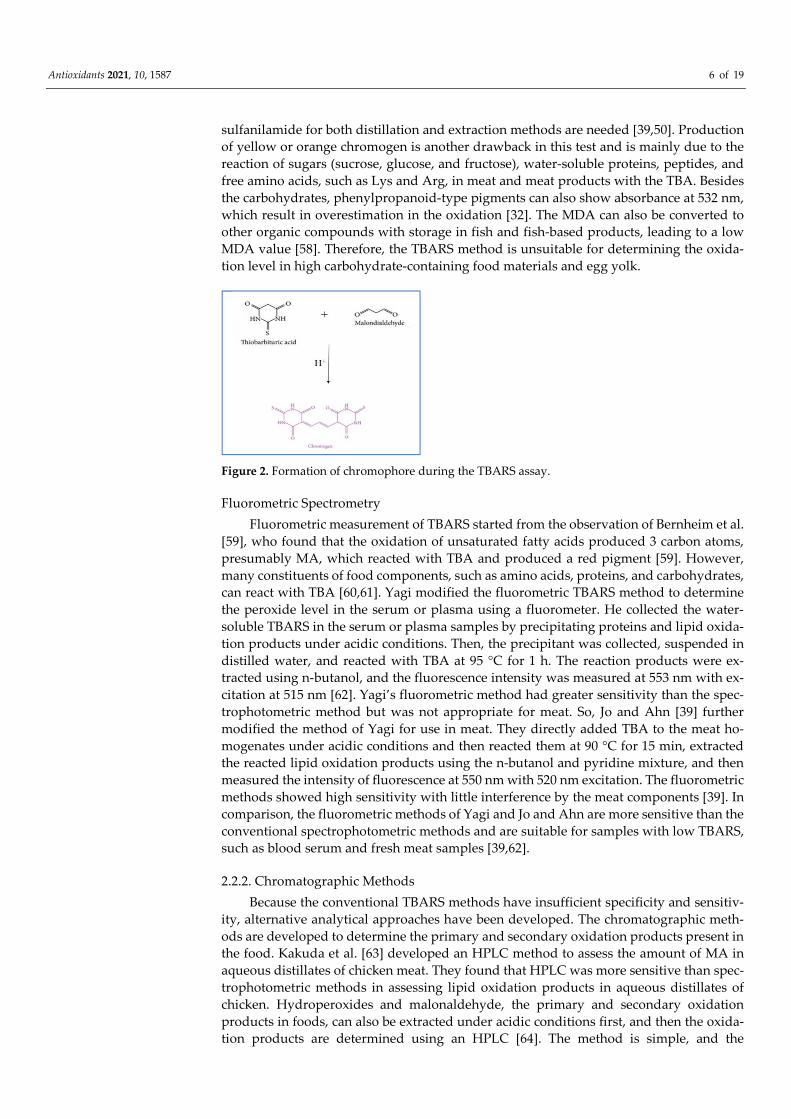
Absorption Spectrometry The chromogen formation from the reaction of thiobarbituric acid (TBA) and MA is
shown in Figure 2 [52]. The reaction rate depends on the concentration of TBA, pH and the temperature used [32]. The MA can be either extracted by distillation or with an acid solution before reacting with TBA [53]. TBA is added to a sample homogenate to react with MA in the sample [41,54] directly. The distillation method is commonly used for raw, cured, and cooked meat products but is a time-consuming method. In this method, the MA is extracted with trichloroacetic acid (TCA) at a controlled rate, and the distillate is collected, reacted with TBA, and the absorbance is measured using a spectrophotometer [37]. The acid extraction is used mainly for raw and cooked meat and fish samples, which use 7.5% TCA, followed by filtration and reaction with TBA, in a boiling water bath for 30–40 min [51]. This method has also been used to determine the lipid oxidation in edible insect-based foods [46,47]. In the direct reaction method, two volumes of TBA/TCA solu-tion (15% TCA/20 mM TBA) are added to one sample homogenate volume and are reacted at boiling temperature for 15 min. The color formed in the supernatant is collected after centrifugation, and the absorbance is read using a spectrophotometer at 532 nm. The quantification of MA in meat is performed using a standard curve and is expressed as mg of MDA/kg food [55,56]. TBARS method is easy, simple, low cost, and reproducible, and the values are well correlated with the sensory properties of the final product. However, TBA can react with other oxidized compounds in the food product and produce false-positive results [32,50,57]. The nitrite used in the cured meat products can react with MA under acidic conditions and lower value in cured meat. Therefore, the addition of
Antioxidants 2021, 10, 1587 6 of 19
sulfanilamide for both distillation and extraction methods are needed [39,50]. Production of yellow or orange chromogen is another drawback in this test and is mainly due to the reaction of sugars (sucrose, glucose, and fructose), water-soluble proteins, peptides, and free amino acids, such as Lys and Arg, in meat and meat products with the TBA. Besides the carbohydrates, phenylpropanoid-type pigments can also show absorbance at 532 nm, which result in overestimation in the oxidation [32]. The MDA can also be converted to other organic compounds with storage in fish and fish-based products, leading to a low MDA value [58]. Therefore, the TBARS method is unsuitable for determining the oxida-tion level in high carbohydrate-containing food materials and egg yolk.
Figure 2. Formation of chromophore during the TBARS assay.
Fluorometric Spectrometry Fluorometric measurement of TBARS started from the observation of Bernheim et al.
[59], who found that the oxidation of unsaturated fatty acids produced 3 carbon atoms, presumably MA, which reacted with TBA and produced a red pigment [59]. However, many constituents of food components, such as amino acids, proteins, and carbohydrates, can react with TBA [60,61]. Yagi modified the fluorometric TBARS method to determine the peroxide level in the serum or plasma using a fluorometer. He collected the water-soluble TBARS in the serum or plasma samples by precipitating proteins and lipid oxida-tion products under acidic conditions. Then, the precipitant was collected, suspended in distilled water, and reacted with TBA at 95 °C for 1 h. The reaction products were ex-tracted using n-butanol, and the fluorescence intensity was measured at 553 nm with ex-citation at 515 nm [62]. Yagi’s fluorometric method had greater sensitivity than the spec-trophotometric method but was not appropriate for meat. So, Jo and Ahn [39] further modified the method of Yagi for use in meat. They directly added TBA to the meat ho-mogenates under acidic conditions and then reacted them at 90 °C for 15 min, extracted the reacted lipid oxidation products using the n-butanol and pyridine mixture, and then measured the intensity of fluorescence at 550 nm with 520 nm excitation. The fluorometric methods showed high sensitivity with little interference by the meat components [39]. In comparison, the fluorometric methods of Yagi and Jo and Ahn are more sensitive than the conventional spectrophotometric methods and are suitable for samples with low TBARS, such as blood serum and fresh meat samples [39,62].
2.2.2. Chromatographic Methods Because the conventional TBARS methods have insufficient specificity and sensitiv-
ity, alternative analytical approaches have been developed. The chromatographic meth-ods are developed to determine the primary and secondary oxidation products present in the food. Kakuda et al. [63] developed an HPLC method to assess the amount of MA in aqueous distillates of chicken meat. They found that HPLC was more sensitive than spec-trophotometric methods in assessing lipid oxidation products in aqueous distillates of chicken. Hydroperoxides and malonaldehyde, the primary and secondary oxidation products in foods, can also be extracted under acidic conditions first, and then the oxida-tion products are determined using an HPLC [64]. The method is simple, and the
Antioxidants 2021, 10, 1587 7 of 19
sensitivity is high compared with other methods but is time-consuming. Product identifi-cation and quantification can be easily made if the HPLC unit is coupled with a mass spectrometer [32,50,64,65]. Recently, a comprehensive two-dimensional liquid chroma-tography (LC × LC) was developed to determine lipid oxidation products using a light-scattering detector (ELSD). This method can determine both polar and non-polar lipid oxidation products. However, sample preparation for the HPLC method is complicated [65], analysis time per sample is long, and the equipment cost is high. Therefore, the in-dustrial applications of this method are limited.
Gas chromatography equipped with mass spectrometry is another method used to determine the lipid oxidation products in foods. Specific marker compounds, such as hex-anal and total aldehydes, were the most successfully used volatile compounds to deter-mine the degree of oxidation in various foods, including meat, milk, dried, and plant-based foods, using a GC-MS [55]. In addition, the typical end products of lipid oxidation, such as aldehydes, ketones, hydrocarbons, and alcohols, that reacted with TBA are used to determine the degree of oxidation using a GC-MS [56]. Ahn et al. [66] showed that the amounts of propanal, pentanal, hexanal, 1-pentanol, and total volatiles were correlated highly (p < 0.01) with the TBARS values of cooked meat, but hexanal and total volatiles represented the degree of lipid oxidation better than any individual volatile compounds in cooked meat. GC method is also a sensitive method, but equipment cost is high, and analysis is time-consuming [32,66,67]. Thus, conventional spectrophotometric TBARS methods are still preferred over chromatographic methods because of their simplicity.
2.3. Indirect Methods Used to Detect the Secondary Oxidation Products 2.3.1. Fluorometric Method
The fluorometric method is an indirect method widely used to determine the level of lipid oxidation in raw fish, meat-based products, and animal by-products, such as blood [38,58]. It can also be used on plant oils rich in polyunsaturated fatty acids (PUFA) [68,69]. In addition, the fluorometric method has been used to determine lipid oxidation in low-moisture foods, such as cereals [70]. In the fluorometric method, MA is converted to tet-ramethoxy-propane, which eliminates the false fluorescence readings by vitamin A, nico-tinamide adenine dinucleotide (NADH), proteins, and amino acids. Thus, this method is more suitable for protein-rich foods with more ionic and covalent bonds, such as fish [71]. The most common method includes lipid extraction using a chloroform-methanol-water solvent. The collected organic layer is mixed with sodium sulfate and filtered. Then the mixture is dried with nitrogen gas at a temperature not exceeding 35 °C. The MA equiva-lent in lipid extracts is measured by reacting with TBA. BHT is added to the extraction solvent, and the secondary reaction products of lipid oxidation are measured by fluores-cence excitation (360 nm) and emission (440 nm) of the aqueous and organic layers that contain chloroform and methanol (2:1) [68,69]. The fluorescence produced is reliable and sensitive to detecting lipid and water-soluble hydroperoxides in meat and fish samples [72]. The sensitivity of this method is higher than the absorption spectrophotometric methods (TBARS method) and is reproducible, simple, and needs a tiny amount of sample to detect the level of oxidation in meat and meat-based products.
Lipofuscin is commonly used as a fluorescent marker, but some other markers, such as 1,4-dihydropyridines and 1-palmitoyl-2-((2-(4-(6-phenyltrans-1,3,5-hexatrienyl)-phe-nyl)-ethyl)-carbonyl)-sn-glycero-3-phosphocholine (DPH-PC), diphenyl-1-pyrenylphosphine (DPPP), are also used as a new type of fluorescent compounds in food. These markers are dissolved in a chloroform/ methanol mixture, and the solvents are re-moved using nitrogen. The dried sample is mixed with phosphate buffer, and the reaction progress is observed at 430 nm (excitation at 354 nm) at 37 °C using a fluorimeter [72]. With these markers, image processing is introduced as a novel method to determine the level of oxidation in foods [71,72].
2.3.2. Sensory Analysis
Antioxidants 2021, 10, 1587 8 of 19
Sensory evaluation evokes, measures, analyzes, and interprets human responses to the properties of food materials. A trained or untrained panelist can determine the sensory attributes, such as smell, flavor, and taste of the food product [73]. Sensory evaluation is considered a critical method since it directly interprets oxidation levels in a food product [74–76]. In a practical situation, this evaluation is performed by field panels, consumer panels, free choice profiling panels, quantitative description analysis panels, and expert panels, especially with low-moisture foods, such as cereals [70,73,77]. Sensory evaluation is cost-effective and simple and can be applied to all plant- and animal-based foods in liquid, semi-solid, and solid forms. However, the sensory analysis shows high regional variations and low repeatability because of the personal preference and experience, the cultural backgrounds, the age and sex of panelists, and the time of the evaluation per-formed. Therefore, a sensory, along with the chemical and instrumental, analysis is rec-ommended. As discussed above, many methods are available to determine the degree of lipid oxidation in food, but one method cannot fit all food products. Thus, selecting a proper and effective method for a specific food is essential.
2.4. Methods Used to Detect the Primary and Secondary Oxidation Products p-Anisidine test (p-AV) and total oxidation index (TOTOX) are two common meth-
ods used in determining lipid oxidation in food products, especially edible oils. Both p-anisidine and TOTOX tests determine both the primary and secondary oxidation products in oil [78]. However, the anisidine test uses aldehydes, such as 2-alkenals and 2,4-alka-dineals, as the main oxidation markers, and the level of anisidine produced is measured using the UV-spectrophotometer at 350 nm [78,79].
TOTOX indicates the overall oxidation states, and it can give a good explanation of the final quality of the edible oil. It is calculated using the following equation:
TOTOX= p-Anisidine value + 2 Peroxide value.
Both p-AV and TOTOX methods are simple, require less technical knowledge, and are commonly used to determine the quality of the EPA/DHA-containing oils and finished products [79,80]. However, p-AV is not suitable for determining the oxidation in omega-3-rich oils with intense color or specific flavorings added to the final oil [80]. Table 1 shows the summary of lipid oxidation methods with their principles, applications, advantage, and disadvantages of each method.
Table 1. Summary of the common methods used in lipid oxidation.
Lipid Oxidation Analysis Method
Principle of the Method Possible Applica-tions
Advantages of the Method
Disadvantages of the Method
References
Peroxide value (PV): Iodometric
and ferric thiocya-nate
Oxidation of iodide by hy-droperoxides or by oxida-
tion of Fe2+ to Fe3+. Use a spectrophotometer
to obtain the final reading.
Plant oils and liq-uid food prod-ucts, edible in-
sects.
Simple and cheap. Direct readings.
Under anaerobic con-ditions, the sensitivity
is high.
Depend on the titration skills of the person.
Only applicable to liquid-based products.
[42–47]
Conjugated diene analysis
Isomeric hydroperoxides will make conjugated
dienes with the removal of oxygen and determine 1,4-
dienes produced. Measured at 233 nm.
Suitable for PUFA-containing
foods.
Gives actual values of LDL oxidation during the early stages of oxi-
dation. Simple and cheap.
Depend on the composi-tion and size of the lipo-
proteins. Small, conjugated dienes
are difficult to detect.
[18,41,48,49]
TBARS assay
Detect the production of chromogen due to the re-action of MA and TBA. Read absorption at 532
nm. HPLC, GC-MS, and fluorometer are also used.
Meat and meat-based products. Fish and fish-
based products. Can be used in
cured meat
Simple and fast detection. Easy to detect and low cost.
Reproducible and
MA and TBA can react with other organic com-pounds present in food. Absorption spectropho-
tometer is not suitable for detection at low levels.
[32;37,39,50–58]
Antioxidants 2021, 10, 1587 9 of 19
products, edible insects.
correlate well with sensory attributes.
Chromatography methods
By using the HPLC or GC. Determine the specific compounds produced.
All types of raw and processed
foods. Oxidative stress-related dis-
eases.
Sensitive and accurate. Identification and
quantification can be made.
The cost of the equipment is high. The complexity of the method and the fact
that it is time-consuming.
[32,50,55,56,64–67]
Fluorometric method
Use different fluorescent porphyrins to interact
with MDA produced dur-ing oxidation.
Animal-based products. Can be
used to determine the changes in hu-
man se-rum/plasma. Low moisture foods.
Fast and accurate. Non-destructive
Sensitive. Image pro-duced can be further used. Can be used to detect unstable oxi-dized compounds.
High cost of the equip-ment used.
Complexity of the method.
[38,58,68–72]
Sensory analysis
Use trained or untrained human panelist to deter-
mine the level of oxidation through sensory attrib-
utes, such as odor, taste, and acceptability.
All animals and plant-based
foods, cereals.
Gives the overall qual-ity of the food. Direct
interpretation. Can be used for liquid, semi-solid, and solid
foods.
Depends on the individ-ual participants and time
variations. Depends on the region.
Reproducibility difficult. Ethical clearance is
needed.
[73–77]
p-Anisidine test
Determine the level of anisidine produced from the secondary aldehydes
produced.
Oil and oil-based products.
Simple, less technical knowledge needed.
Problems in omega-3-rich oils that contain intense colors or containing spe-
cific flavorings.
[78–80]
Total oxidation in-dex (TOTOX)
Determine the total oxi-dizied products.
Oil and oil-based products.
Simple calculation. Similar problem associ-ated with p-anisidine test.
[78–80]
3. Methods for Antioxidant Capacity Measurement 3.1. Direct Measurement of Antioxidants
The direct measurement of antioxidants uses model systems and determines the an-tioxidant capacity using DPPH radical scavenging assay, ABTS radical scavenging assay, total phenolic content analysis ferric reducing antioxidant power (FRAP) assay, CUPRAC assay, and electrochemical methods.
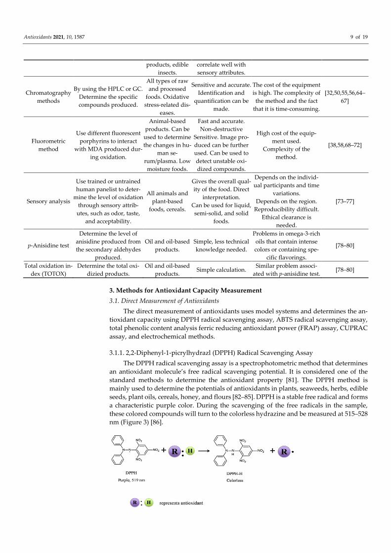
3.1.1. 2,2-Diphenyl-1-picrylhydrazl (DPPH) Radical Scavenging Assay The DPPH radical scavenging assay is a spectrophotometric method that determines
an antioxidant molecule’s free radical scavenging potential. It is considered one of the standard methods to determine the antioxidant property [81]. The DPPH method is mainly used to determine the potentials of antioxidants in plants, seaweeds, herbs, edible seeds, plant oils, cereals, honey, and flours [82–85]. DPPH is a stable free radical and forms a characteristic purple color. During the scavenging of the free radicals in the sample, these colored compounds will turn to the colorless hydrazine and be measured at 515–528 nm (Figure 3) [86].
Antioxidants 2021, 10, 1587 10 of 19
Figure 3. Changes in the DPPH radical-scavenging activity.
There are many variations, mainly the concentration of DPPH solution (22.5–250 µM) and the incubation time (5–60 min), among the methods used to determine the DPPH scavenging assay [87]. The level of radical scavenging ability is expressed in ascorbic acid equivalent. During the reaction, the initial electron transfer occurs very quickly, but hy-drogen transfer takes time. This rate depends on the solvent (ethanol or methanol) used on the acceptance of H-bonds. However, the stability of DPPH● is higher in methanol than ethanol; thus, methanol is commonly used as a solvent in DPPH assay [83].
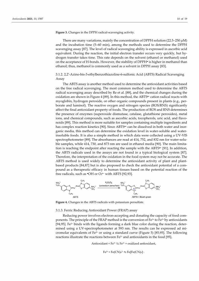
3.1.2. 2,2′-Azino-bis-3-ethylbenzothiazoline-6-sulfonic Acid (ABTS) Radical Scavenging Assay
The ABTS assay is another method used to determine the antioxidant activities based on the free radical scavenging. The most common method used to determine the ABTS radical scavenging assay described by Re et al. [88], and the chemical changes during the oxidation are shown in Figure 4 [89]. In this method, the ABTS●+ cation radical reacts with myoglobin, hydrogen peroxide, or other organic compounds present in plants (e.g., per-borate and luminol). The reactive oxygen and nitrogen species (ROS/RNS) significantly affect the final antioxidant property of foods. The production of ROS and RNS determines the presence of enzymes (superoxide dismutase, catalase, glutathione peroxides), metal ions, and chemical compounds, such as ascorbic acids, tocopherols, uric acid, and flavo-noids [89]. This method is more suitable for samples containing multiple ingredients and has complex reaction kinetics [90]. Since ABTS●+ can be dissolved in both water and inor-ganic media, this method can determine the oxidation level in water-soluble and water-insoluble foods. It is also a simple method in which data were collected using a UV-VIS spectrophotometer [89]. The absorbances are read at 414, 752, and 832 nm for water-solu-ble samples, while 414, 730, and 873 nm are used in ethanol media [90]. The main limita-tion is reaching the endpoint after reacting the sample with the ABTS●+ [91]. In addition, the ABTS radicals used in the assays are not found in a typical biological system [87]. Therefore, the interpretation of the oxidation in the food system may not be accurate. The ABTS method is used widely to determine the antioxidant activity of plant and plant-based products [84,87] but is also proposed to check the antioxidant potential of a com-pound as a therapeutic efficacy in human tissues based on the potential reaction of the free radicals, such as ●OH or O2•− with ABTS [92,93].
Figure 4. Changes in the ABTS radicals with potassium persulfate.
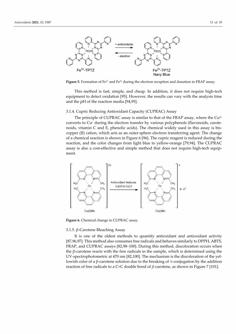
3.1.3. Ferric Reducing Antioxidant Power (FRAP) assay Reducing power involves electron-accepting and donating the capacity of food com-
ponents. The principle of the FRAP method is the conversion of Fe3+ to Fe2+ by antioxidants [94,95]. Fe2+ binds with the ligands forming a dark blue color during the reaction, deter-mined using a UV-spectrophotometer at 593 nm. The results can be expressed ad mi-cromolar equivalents of Fe2+ or using a standard curve (Figure 5) [85,95]. The following reactions illustrate the reactions between Fe2+ and antioxidants in the food [95].
Antioxidant + Fe3+ ⇆ Fe2+ + oxidized antioxidant,
Fe2+ + Fe(CN)63- ⇆ Fe[Fe(CN)6]-.
Antioxidants 2021, 10, 1587 11 of 19
Figure 5. Formation of Fe3+ and Fe2+ during the electron reception and donation in FRAP assay.
This method is fast, simple, and cheap. In addition, it does not require high-tech equipment to detect oxidation [95]. However, the results can vary with the analysis time and the pH of the reaction media [94,95].
3.1.4. Cupric Reducing Antioxidant Capacity (CUPRAC) Assay The principle of CUPRAC assay is similar to that of the FRAP assay, where the Cu2+
converts to Cu+ during the electron transfer by various polyphenols (flavonoids, carote-noids, vitamin C and E, phenolic acids). The chemical widely used in this assay is bis-copper (II) cation, which acts as an outer-sphere electron transferring agent. The change of a chemical reaction is shown in Figure 6 [96]. The cupric reagent is reduced during the reaction, and the color changes from light blue to yellow-orange [79,94]. The CUPRAC assay is also a cost-effective and simple method that does not require high-tech equip-ment.
Figure 6. Chemical change in CUPRAC assay.
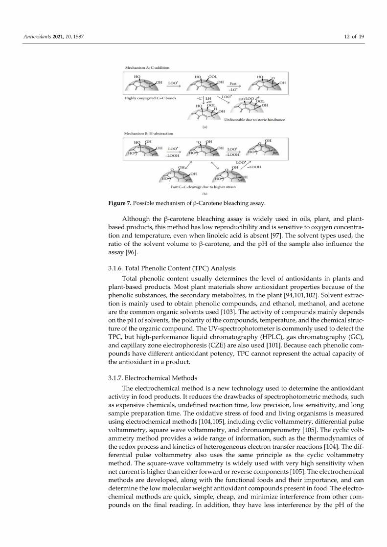
3.1.5. β-Carotene Bleaching Assay It is one of the oldest methods to quantify antioxidant and antioxidant activity
[87,96,97]. This method also consumes free radicals and behaves similarly to DPPH, ABTS, FRAP, and CUPRAC assays [82,98–100]. During this method, discoloration occurs when the β-carotene reacts with the free radicals in the sample, which is determined using the UV-spectrophotometric at 470 nm [82,100]. The mechanism is the discoloration of the yel-lowish color of a β-carotene solution due to the breaking of π-conjugation by the addition reaction of free radicals to a C=C double bond of β-carotene, as shown in Figure 7 [101].
Antioxidants 2021, 10, 1587 12 of 19
Figure 7. Possible mechanism of β-Carotene bleaching assay.
Although the β-carotene bleaching assay is widely used in oils, plant, and plant-based products, this method has low reproducibility and is sensitive to oxygen concentra-tion and temperature, even when linoleic acid is absent [97]. The solvent types used, the ratio of the solvent volume to β-carotene, and the pH of the sample also influence the assay [96].
3.1.6. Total Phenolic Content (TPC) Analysis Total phenolic content usually determines the level of antioxidants in plants and
plant-based products. Most plant materials show antioxidant properties because of the phenolic substances, the secondary metabolites, in the plant [94,101,102]. Solvent extrac-tion is mainly used to obtain phenolic compounds, and ethanol, methanol, and acetone are the common organic solvents used [103]. The activity of compounds mainly depends on the pH of solvents, the polarity of the compounds, temperature, and the chemical struc-ture of the organic compound. The UV-spectrophotometer is commonly used to detect the TPC, but high-performance liquid chromatography (HPLC), gas chromatography (GC), and capillary zone electrophoresis (CZE) are also used [101]. Because each phenolic com-pounds have different antioxidant potency, TPC cannot represent the actual capacity of the antioxidant in a product.
3.1.7. Electrochemical Methods The electrochemical method is a new technology used to determine the antioxidant
activity in food products. It reduces the drawbacks of spectrophotometric methods, such as expensive chemicals, undefined reaction time, low precision, low sensitivity, and long sample preparation time. The oxidative stress of food and living organisms is measured using electrochemical methods [104,105], including cyclic voltammetry, differential pulse voltammetry, square wave voltammetry, and chronoamperometry [105]. The cyclic volt-ammetry method provides a wide range of information, such as the thermodynamics of the redox process and kinetics of heterogeneous electron transfer reactions [104]. The dif-ferential pulse voltammetry also uses the same principle as the cyclic voltammetry method. The square-wave voltammetry is widely used with very high sensitivity when net current is higher than either forward or reverse components [105]. The electrochemical methods are developed, along with the functional foods and their importance, and can determine the low molecular weight antioxidant compounds present in food. The electro-chemical methods are quick, simple, cheap, and minimize interference from other com-pounds on the final reading. In addition, they have less interference by the pH of the
Antioxidants 2021, 10, 1587 13 of 19
solvents, solvent polarity, and concentrations [106]. Other than that, the electrochemical method allows a high number of parameters to be obtained in a single experiment [104,105]. However, applications are limited due to the electrochemical sensors used. Chemicals, such as NaCl or KCl, are mixed with the solvents and used for electrochemical analysis. These ions can interfere with the oxidation process. In addition, some antioxi-dants, such as polyphenols, present in food can produce a passivating film that can reduce the conductivity of the electrodes used [106]. The electrochemical methods show similar results to DPPH, FRAP, and ABTS radical scavenging activities; therefore, the results’ ac-curacy is high [107]. The electrochemical methods are widely used in plant-based prod-ucts [107,108] and less applied in animal-based products.
3.2. Indirect Measurement of Antioxidant Capacity The indirect methods determine the effect of an antioxidant on the prevention of ox-
idation in foods using TBARS, fluorometric, peroxide value, diene conjugation, iodine value, and HPLC and GC methods, as discussed above. The indirect methods use meat homogenates, meat products, oil emulsion, and liposome systems, and the results of anti-oxidant capacity obtained by the indirect methods often do not agree with those of the direct assays, especially in meat systems. Thus, the indirect measurement of antioxidant capacity is recommended for antioxidants’ practical and tangible effect in meat-based food systems.
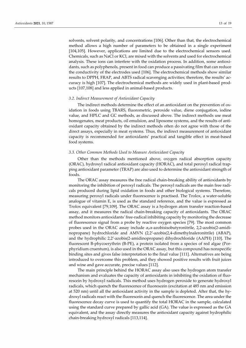
3.3. Other Common Methods Used to Measure Antioxidant Capacity Other than the methods mentioned above, oxygen radical absorption capacity
(ORAC), hydroxyl radical antioxidant capacity (HORAC), and total peroxyl radical trap-ping antioxidant parameter (TRAP) are also used to determine the antioxidant strength of foods.
The ORAC assay measures the free radical chain-breaking ability of antioxidants by monitoring the inhibition of peroxyl radicals. The peroxyl radicals are the main free radi-cals produced during lipid oxidation in foods and other biological systems. Therefore, measuring peroxyl radicals under fluorescence is practised. The Trolox, a water-soluble analogue of vitamin E, is used as the standard reference, and the value is expressed as Trolox equivalent [79,109]. The ORAC assay is a hydrogen atom transfer reaction-based assay, and it measures the radical chain-breaking capacity of antioxidants. The ORAC method monitors antioxidants’ free-radical inhibiting capacity by monitoring the decrease of fluorescence signal from a probe by reactive oxygen species [79]. The most common probes used in the ORAC assay include α,α-azobisisobutyronitrile, 2,2-azobis(2-amidi-nopropane) hydrochloride and AMVN (2,2′-azobis(2,4-dimethylnaleronitrile) (ABAP), and the hydrophilic 2,2′-azobis(2-amidinopropane) dihydrochloride (AAPH) [110]. The fluorescent B-phycoerythrin (B-PE), a protein isolated from a species of red algae (Por-phyridium cruentum), is also used in the ORAC assay, but this compound has nonspecific binding sites and gives false interpretation to the final value [111]. Alternatives are being introduced to overcome this problem, and they showed positive results with fruit juices and wine and gave accurate, precise values [112].
The main principle behind the HORAC assay also uses the hydrogen atom transfer mechanism and evaluates the capacity of antioxidants in inhibiting the oxidation of fluo-rescein by hydroxyl radicals. This method uses hydrogen peroxide to generate hydroxyl radicals, which quench the fluorescence of fluorescein (excitation at 485 nm and emission at 520 nm) until all the antioxidant activity in the sample is depleted. After that, the hy-droxyl radicals react with the fluorescein and quench the fluorescence. The area under the fluorescence decay curve is used to quantify the total HORAC in the sample, calculated using the standard curve prepared by gallic acid (GA). The value is expressed as the GA equivalent, and the assay directly measures the antioxidant capacity against hydrophilic chain-breaking hydroxyl radicals [113,114].
Antioxidants 2021, 10, 1587 14 of 19
The TRAP is another method that determines the total free-radical trapping capacity of antioxidants in foods. The principle of this method is determining the peroxyl radicals produced during thermal decomposition are reacted with the luminol-enhanced chemi-luminescence. The TRAP assay generates peroxyl radicals using 2,2′-azobis (2-amidi-nopropane) dihydrochloride (AAPH), and the oxidation reactions are monitored by oxy-gen consumption using an oxygen electrode. The idea is that the length of the lag phase before increased oxygen consumption is proportional to the antioxidant capacity of the sample. The TRAP values calculated from the length of the lag-phase caused by the anti-oxidant capacity are expressed as the Trolox equivalents [109,113,115]. The summary of the methods used to determine the antioxidant capacity in foods is listed in Table 2.
Table 2. Summary of the methods used to measure the antioxidant capacity of foods.
Antioxidant Method Principle Uses/Applications Advantages Disadvantages References
DPPH assay
Determine the free radi-cals produced during the
oxidation. Reduction of purple
color is measured using a spectrophotometer (515–
528 nm).
Can be used to de-tect the oxidation in
plants, seaweeds, herbs, edible seed,
and plant oils.
Simple, fast, and Cheap.
Depends on the solvent used. The presence of particles will interfere
with the results.
[81–87]
ABTS assay Determine the free radi-
cals produced during the oxidation and reduction.
Plant and plant-based products.
Water-soluble and insol-uble compounds can be
analyzed. Simple and fast.
Depends on the en-zymes, metal ions, en-
ergy provided, and chemicals in the food.
Not suitable in biological systems.
[84,88–93]
FRAP assay
Reducing power involves electron-accepting and
donating Fe3+ → Fe2+.
Plants and plant-based products.
Simple, fast, and cheap. No enzyme involvement.
Varies with analyzing time.
[94–96]
CUPRAC assay
Reducing power involves electron-accepting and
donating Cu2+ → Cu+.
Plants and plant-based products.
Simple and cost-effective. No need for high-tech
equipment.
Not suitable to combine with TPC, ABTS, FRAP,
or DPPH assays. [79,94,96]
β-carotene assay Determine the free radi-
cals produced during the oxidation and reduction.
Plants and plant-based products.
Plant oils. Simple and fast.
Sensitive to oxygen and temperature when lino-
leic acid is absent. Low reproducibility. De-pend on solvent type, ra-
tio, pH of the sample.
[82,87,96–101]
TPC assay Determine the levels of
antioxidants. Plants and plant-based products.
Simple, fast, and Cheap.
Activity depends on pH, the polarity of solvent and temperature. De-pends on external fac-
tors.
[94,101–103]
Electrochemical as-say
Based on thermodynam-ics of the redox process
and kinetics of heteroge-neous electron-transfer
reactions.
Animal-based and plant-based prod-
ucts. Functional foods.
Reduce the problems in spectrophotometer meth-ods. Short sample prepa-ration time. Low reaction
time. Sensitive and re-duce the interference.
Cost of the equipment used. Complexity of the method. Some polyphe-nols can produce passiv-ating films. Chemicals,
such as NaCl/KCl, inter-fere with the electrolytes.
[104–108]
Antioxidants 2021, 10, 1587 15 of 19
Oxygen radical ab-sorption capacity
(ORAC)
Based on the breaking of peroxyl radical chains re-
action by antioxidants. Monitor the inhibition of
peroxyl radicals.
Biological fluids and functional
foods. Plants and plant-based liquid
products.
Accurate, precise values can be obtained. Can be used to detect both hy-
drophilic and hydropho-bic antioxidants.
Can easily bind with other compounds and
give false values. Techni-cally demanding
method.
[79,109–112]
Hydroxyl radical antioxidant capac-
ity (HORAC)
Based on the breaking of hydroxyl radical chains
reaction by antioxidants.
Biological fluids and functional
foods. Plants and plant-based liquid
products.
Direct measurement of antioxidant capacity.
Sensitive.
Time dependent. Indirect calculation, technically
demanding. [113,114]
Total peroxyl radi-cal trapping antiox-
idant parameter (TRAP)
Based on the quenching of chemiluminescence.
Used in plasma and cerebrospinal flu-
ids. Suitable for liquid foods.
Time-dependent. Indirect calculation. Complex technology is needed. Comparisons between
labs are difficult.
[109,113,115]
4. Summary Lipid oxidation is a crucial quality problem in many processed foods. Therefore, de-
termining the level of lipid oxidation is essential to monitor the quality and assure con-sumer acceptability of foods. Numerous methods have been developed to determine lipid oxidation in foods and biological materials, and each of the methods uses different ap-proaches, indicators, or targets for the analysis. Food products have different composi-tional and physicochemical characteristics and the stage of lipid oxidation at the time of measurement. Therefore, some methods can work better than others, depending on the food, oxidation stage, and lipid oxidation level. For more accurate information about the oxidation status and antioxidants’ capacity, selecting the most appropriate analytical methods for each food product or antioxidant would be very important. Among the many methods practised in determining lipid oxidation, peroxide, iodine values, and diene con-jugate are the direct methods used for detecting the primary substances produced by ox-idation. These methods are practical for oils, high-fat foods, and raw meat products, and their sensitivity is generally low. The TBARS and chromatographic methods determine the levels of secondary products of lipid oxidation. The TBARS assays are the most widely used method to determine lipid oxidation in animal-based foods and are also simple and effective. However, TBARS methods can have a low expression level in carbohydrate-rich products because MA can react with sugar, generate a yellowish color instead of pink, and are unsuitable for oils or phospholipids-rich products. Fluorometric method and sensory evaluation are indirect detection methods of lipid oxidation in food products. Sensory evaluation is a suitable method, but its repeatability is low, and person to person varia-tions cannot be controlled. p-Anisidine and TOTOX methods determine primary and sec-ondary oxidation products in foods and are suitable for edible oils.
Antioxidant capacity in food decides the storage stability of foods and can be deter-mined by direct and indirect methods. The direct measurement of antioxidant capacities, such as ABTS, TPC, DPPH, FRAP, CUPRAC, and β-carotene methods, are widely used in plant-based foods, and they are simple and effective. The indirect methods for antioxidant capacity measurement include TBARS assay, peroxide value, iodine value, and conju-gated diene methods. Methods, such as ORAC, HORAC, and TRAP methods, use indirect calculation for antioxidant capacity. Although the antioxidant capacity can be measured either by direct or indirect methods, they sometimes do not agree well on the food prod-ucts. The analysis of lipid oxidation and antioxidant capacity using multiple methods sharing similar principles would not improve the accurate evaluation of oxidation status nor the antioxidant potential of antioxidant compounds or foods. Thus, selecting a suita-ble method for a specific food or using one or two methods with different mechanisms will be more economical and informative in evaluating the degree of oxidation or antiox-idant capacity than using several analytical methods with similar analytical principles.
Antioxidants 2021, 10, 1587 16 of 19
Funding: This research received no external funding.
Conflicts of Interest: The authors declare no conflict of interest.
References 1. Goodman, B.E. Insights into digestion and absorption of major nutrients in humans. Adv. Physiol. Educ. 2010, 34, 44–53. 2. Zheng, L.; Faith, M.; Giuffrida, F.; O’Neill, B.V.; Schneider, N. Dietary polar lipids and cognitive development: A narrative
review. Adv. Nutr. 2019, 10, 1163–1176. 3. Baggio, S.R.; Vicente, E.; Bragagnolo, N. Cholesterol oxides, cholesterol, total lipid and fatty acid composition in turkey meat. J.
Agric. Food Chem. 2002, 50, 5981–5986. 4. Gao, F.; Ben-Amotz, D.; Yang, Z.; Han, L.; Liu, X. Complementarity of FT-IR and Raman spectroscopies for the species discrim-
ination of meat and bone meals related to lipid molecular profiles. Food Chem. 2021, 345, 128754. 5. Borys, B.; Oprządek, J.; Borys, A.; Przegalińska-Gorączkowska, M. Lipid profile of intramuscular fat in lamb meat. Anim. Sci.
Papers Rep. 2012, 30, 45–56. 6. Prato, E.; Biandolino, F. Total lipid content and fatty acid composition of commercially important fish species from the Medi-
terranean, Mar Grande Sea. Food Chem. 2012, 131, 1233–1239. 7. Xiao, N.; Zhao, Y.; Yao, Y.; Wu, N.; Xu, M.; Du, H.; Tu, Y. Biological activities of egg yolk lipids. J. Agric. Food Chem. 2020, 68,
1948–1957. 8. Zhao, Y.C.; Shi, H.H.; Wang, C.C.; Yang, J.Y.; Xue, C.H.; Jiang, X.M.; Chen, G.D.; Zhang, T.T.; Wang, Y.U. The enrichment of
eggs with docosahexaenoic acid and eicosapentaenoic acid through supplementation of the laying hen diet. Food Chem. 2021, 346, 12895.
9. Gonçalves, S.; Romano, A. Aromatic oils from forest and their application. In Non-Timber Forest Products; Husen, A., Bachheti, R.K., Bachheti, A., Eds.; Springer: Cham, Switzerland, 2021. https://doi.org/10.1007/978-3-030-73077-2_2.
10. Adekunle, K.F. A Review of vegetable oil-based polymers: Synthesis and applications. Open J. Polymer Chem. 2015, 5, 34–40. 11. Ni, Z.J.; Wang, X.; Shen, Y.; Thakur, K.; Han, J.; Zhang, J.G.; Hu, F.; Wei, Z.J. Recent updates on the chemistry, bioactivities,
mode of action, and industrial applications of plant essential oils. Trends Food Sci. Technol. 2021, 110, 78–89. 12. Mikołajczak, N.; Tanska, M.; Ogrodowska, D. Phenolic compounds in plant oils: A review of composition, analytical methods,
and effect on oxidative stability. Trends Food Sci. Technol. 2021, 113, 110–138. 13. Laguerre, M.; Bily, A.; Birtic, S. Oxidation of lipids in food. In Lipids and Edible Oils—Properties, Processing, and Applications;
Galanakis, C.M., Ed.; Academic Press: Cambridge, MA, USA, 2020; pp. 243–287. 14. Ahmed, M.; Pickova, J.; Ahmad, T.; Liaquat, M.; Farid, A.; Jahangir, M. Oxidation of lipids in foods. Sarhad J. Agric. 2016, 32,
230–238. 15. Min, B.; Ahn, D.U. Mechanism of lipid peroxidation in meat and meat products—A review. Food Sci. Biotechnol. 2005, 14, 152–
163. 16. Guéraud, F.; Atalay, M.; Bresgen, N.; Cipak, A.; Eckl, P.M.; Huc, L.; Jouanin, I.; Siems, W.; Uchida, K. Chemistry and biochem-
istry of lipid peroxidation products. Free Rad. Res. 2010, 44, 1098–1124. 17. Ayala, A.; Munoz, M.F.; Argüelles, S. Lipid peroxidation: Production, metabolism, and signaling mechanisms of malondialde-
hyde and 4-hydroxy-2-nominal. Oxid. Med. Cell Longev. 2014, Article ID 360438, 31 pages, http://dx.doi:10.1155/2014/360438. 18. Huang, X.; Ahn, D.U. Lipid oxidation and its implications to meat quality and human health. Food Sci. Biotechnol. 2019, 28, 1275–1285. 19. Grabowski, R.; Ilowska, J.; Chrobak, J.; Szmatola, M.; Drabik, J.; Trzos, M. Methods of the modification of vegetable oils in order
to obtain base oils. Tribologia 2018, 6, 23–29. 20. Shahidi, F.; Zhong, Y. Lipid oxidation and improving the oxidative stability. Chem. Soc. Rev. 2010, 39, 4067–4079. 21. Gong, Y.; Parker, R.S.; Richards, M.P. Factors affecting lipid oxidation in breast and thigh muscle from chicken, turkey and
duck. J. Food Biochem. 2010, 34, 869–885. 22. Wu, H.; Yin, J.; Zhang, J.; Richards, M. Factors affecting lipid oxidation due to pig and turkey hemolysate. J. Agric. Food Chem.
2017, 65, 8011–8017. 23. Domínguez, R.; Pateiro, M.; Gagaoua, M.; Barba, F.J.; Zhang, W.; Lorenzo, J.M. A comprehensive review on lipid oxidation in
meat and meat products. Antioxidants 2019, 8, 2–31. 24. Amaral, A.B.; da Silva, M.V.; da Silva Lannes, S.C. Lipid oxidation in meat: mechanisms and protective factors—A review. Food
Sci. Technol. 2018, 38, 1–15. 25. Wazir, H.; Chay, S.Y.; Zarei, M.; Hussin, F.S.; Mustapha, N.A.; Abdullah, W.Z.W.; Saari, N. Effects of storage time and temper-
ature on lipid oxidation and protein co-oxidation of low-moisture shredded meat products. Antioxidants 2019, 8, 486, https://doi.org/10.3390/antiox8100486.
26. Langu, N.S.; Afolayan, A.J.; Thomas, R.S.; Idamokoro, E.M. Consumer exposure to warmed-over flavour and their attitudes towards the use of natural antioxidants as preservatives in meat and meat products. Br. Food J. 2020, 122, 2927–2937.
27. Tikk, K.; Haugen, J.-E.; Andersen, H.J.; Aaslyng, M.D. Monitoring of warmed-over flavor in pork using the electronic nose–correlation to sensory attributes and secondary lipid oxidation products. Meat Sci. 2008, 80, 1254–1263.
28. Vieira, S.A.; Zhang, G.; Decker, E.A. Biological implications of lipid oxidation products. J. Am. Oil Chem. Soc. 2017, 94, 339–351. 29. Papuc, C.; Goran, G.V.; Predescu, C.N.; Nicorescu, V. Mechanisms of oxidative processes in meat and toxicity induced by post-
prandial degradation products: A review. Comprehen. Rev. Food Sci. Food Saf. 2017, 16, 96-123.
Antioxidants 2021, 10, 1587 17 of 19
30. Mas-Bargues, C.; Escrivá, C.; Dormant, M.; Borrás, C.; Viña, J. Lipid peroxidation as measured by chromatographic determina-tion of malondialdehyde. Human plasma reference values in health and disease. Arch. Biochem. Biophys. 2021, 709, 108941, doi.org/10.1016/j.abb.2021.108941.
31. Kinter, M. Analytical technologies for lipid oxidation products analysis. J. Chromatogr. B. 1995, 671, 223–236. 32. Kunyaboon, S.; Thumanu, K.; Park, J.W.; Khongla, C.; Yongsawatdigul, J. Evaluation of lipid oxidation, volatile compounds
and vibrational spectroscopy of silver carp (Hypophthalmichthys molitrix) during ice storage as related to the quality of its washed mince. Foods 2021, 10, 495
33. Loi, M.; Paciolla, C. Plant antioxidants for food safety and quality: Exploring new trends in research. Antioxidants 2021, 10, 972. 34. Maqsood, S.; Benjakul, S. Comparative studies of four different phenolic compounds on in vitro antioxidative activity and the
preventive effect on lipid oxidation of fish oil emulsion and fish mince. Food Chem. 2010, 119, 123–132. 35. Irwin, J.W.; Hedges, N. Measuring lipid oxidation. In Understanding and Measuring the Shelf-Life of Food, 1st ed.; Steele, K., Ed.;
Woodhead Publishing: Cambridge, MA, USA, 2016; pp. 289–316. 36. Knothe, G. Structure indices in FA chemistry. How relevant is the iodine value? JAOCS 2002, 79, 847–854. 37. Tarladgis, B.G.; Watts, B.M.; Younathan, M.T.; Dugan, L., Jr. A distillation method for the quantitative determination of malo-
naldehyde in rancid foods. J. Am. Oil Chem. Soc. 1960, 37, 44–48. 38. Reitznerová, A.; Šuleková, M.; Nagy, J.; Marcinčák, S.; Semjon, B.; Čertík, M.; Klempová, T. Lipid peroxidation process in meat
and meat products: A comparison study of malondialdehyde determination between modified 2-thiobarbituric acid spectro-photometric method and reverse-phase high-performance liquid chromatography. Molecules 2017, 22, 1988, doi:10.3390/mole-cules22111988.
39. Jo, C.; Ahn, D.U. Fluorometric analysis of 2-thiobarbituric acid reactive substances in turkey. Poultry Sci. 1998, 77, 475–480. 40. Barthel, G.; Grosch, W. Peroxide value determination—Comparison of some methods. JAOCS 1974, 51, 540–544. 41. Buege, J.A.; Aust, S.D. Microsomal lipid peroxidation. Method Enzymol. 1978, 52, 302–310. 42. Pryor, W.A.; Castle, L. Chemical methods for the detection of lipid hydroperoxides. Method Enzymol. 1984, 5, 293–299. 43. Labeque, R.; Marnett, L.J. 10-Hydroperoxy-8,12-octadecadienoic acid: A diagnostic probe of alkoxyl radical generation in metal-
hydroperoxide reactions. J. Am. Chem. Soc. 1987, 109, 2828–2829. 44. Wagner, C.D.; Clever, H.L.; Peters, E.D. Evaluation of the ferrous thiocyanate colorimetric method. Anal. Chem. 1947, 19, 980–982. 45. Mihalević, B.; Katušin-Ražem, B.; Ražem, D. The revaluation of the ferric thiocyanate assay for lipid hydroperoxides with spe-
cial considerations of the mechanistic aspects of the response. Free Radic. Biol. Med. 1996, 21, 53–63. 46. Ojha, S.; Bußler, S.; Psarianos, M.; Rossi, G.; Schlüter, O.K. Edible insect processing pathways and implementation of emerging
technologies. J. Insects Food Feed. 2021, 7, 877–890 47. Tenyang, N.; Tiencheu, B.; Mamat, A.; Mawamba, L.A.; Ponka, R. Effect of cooking methods on the nutritive value and lipid
oxidation of two Cricket species consumed in Cameroon. Eur. J. Nutr. Food Saf. 2021, 13, 11–23 48. Recknagel, R.O.; Glende, E.A., Jr. Spectrophotometric detection of lipid conjugated dienes. Methods Enzymol. 1984, 105, 331–337. 49. Corongiu, F.P.; Milia, A. An improved and simple method for determining diene conjugation in autoxidized polyunsaturated
fatty acids. Chem. Biol. Interact. 1983, 44, 289–297. 50. Jung, S.; Nam, K.C.; Jo, C. Detection of malondialdehyde in processed meat products without interference from the ingredients.
Food Chem. 2016, 209, 90–94. 51. Zeb, A.; Ullah, F. A simple spectrophotometric method for the determination of thiobarbituric acid reactive substances in fried
fast foods. J. Anal. Methods Chem. 2016, Article ID 9412767, 5 pages, http://dx.http://dx.doi.org/10.1155/2016/9412767. 52. Antolovich, M.; Prenzler, P.D.; Patsalides, E.; McDonald, S.; Robards, K. Methods for testing antioxidant activity. Analyst 2002,
127, 183–198 53. Salih, A.; Smith, D.; Price, J.; Dawson, L. Modified extraction 2-thiobarbituric acid method for measuring lipid oxidation in
poultry. Poultry Sci. 1987, 66, 1483–1488. 54. Ahn, D.U.; Sell, J.L.; Jeffery, M.; Jo, C.; Chen, X.; Wu, C.; Lee, J.I. Dietary vitamin E affects lipid oxidation and total volatiles of
irradiated raw turkey meat. J. Food Sci. 1997, 62, 954–959. 55. Ahn, D.U.; Olson, D.G.; Jo, C.; Chen, X.; Wu, C.; Lee, J.I. Effect of muscle type, packaging, and irradiation on lipid oxidation,
volatile production and color in raw pork patties. Meat Sci. 1998, 49, 27–39. 56. Schaich, K.M. Analysis of lipid and protein oxidation in fats, oils and foods. In Oxidative Stability and Shelf Life of Foods Containing
Oils and Fats, 1st ed.; Hu, M., Jaconsen, C., Eds.; AOCS Press: Cambridge, MA, USA, 2016; pp. 1–131. 57. Hodges, D.M.; DeLong, J.M.; Forney, C.F.; Prange, R.K. Improving the thiobarbituric acid-reactive-substances assay for esti-
mating lipid peroxidation in plant tissues containing anthocyanin and other interfering compounds. Planta 1999, 207, 604–611. 58. Aubourg, S.P. Recent advances in assessment of marine lipid oxidation by using fluorescence. J. Am. Chem. Soc. 1999, 76, 409–419. 59. Bernheim, F.; Bernheim, M.L.C.; Wilbur, K.M. The reaction between thiobarbituric acid and the oxidation products of certain
lipids. J. Biol. Chem. 1948, 174, 257–264. 60. Kosugi, H.; Kikugawa, K. Potential thiobarbiuturic acid-reactive substances in peroxidized lipids. Free Radic. Biol. Med. 1989, 7,
205–208. 61. Pérez-Palacios, T.; Estévez, M. Chapter 13—Analysis of lipids and lipid oxidation products. In Meat Quality Analysis—Advanced
Evaluation Methods, Techniques, and Technologies; Biswas, A.K., Mandal, P.K., Eds.; Academic Press: Cambridge, MA, USA, 2020; pp. 217–239.
62. Yagi, K. Lipid Peroxides and human diseases. Chem. Phys. Lipids 1987, 45, 337–351.
Antioxidants 2021, 10, 1587 18 of 19
63. Kakuda, Y.; Stanlry, D.W.; van deVoort, F.R. Determination of TBA number by high performance liquid chromatography. J. Am. Oil Chem. Soc. 1981, 58, 773–775.
64. Kato, S.; Shimizu, N.; Hanzawa, Y.; Otoki, Y.; Ito, J.; Kimura, F.; Takekoshi, S.; Sakaino, M.; Sano, T.; Eitsuka, T.; et al. Determi-nation of triacylglycerol oxidation mechanisms in canola oil using liquid chromatography-tandem mass spectrometry. NPJ Sci. Food 2018, , 2(1), doi:10.1038/s41538-017-0009-x.
65. Steinhorst-Slikkerveer, L.; Louter, A.; Janssen, H.G.; Bauer-Plank, C. Analysis of nonvolatile lipid oxidation products in vege-table oils by normal-phase high-performance liquid chromatography with mass spectrometric detection. J. Am. Oil Chem. Soc. 2000, 8, 837–845.
66. Ahn, D.U.; Olson, D.G.; Jo, C.; Love, J.; Jin, S.K. Volatile production and lipid oxidation of irradiated coocked sausages with different packaging during storage. J. Food Sci. 1999, 64, 226–229.
67. Lazaridi, E.; Janssen, H.-G.; Vincken, J.-P.; Pirok, B.; Hennbelle, M. A comprehensive two-dimensional liquid chromatography method for the simultaneous separation of lipid species and their oxidation products. J. Chromatogr. A 2021, 1644, 462106.
68. Pikul, J.; Leszczynski, D.E.; Bechtel, P.J.; Kummerow, F.A. Effects of frozen storage and cooking on lipid oxidation in chicken meat. J. Food Sci. 1984, 49, 838–843.
69. Tappel. A.L. Vitamin E and free radical peroxidation of lipids. Ann. N. Y. Acad. Sci. 1972, 203, 12–28. 70. Barden, L.; Decker, E.A. Lipid oxidation in low-moisture foods: A review. Crit. Rev. Food Sci. Nutr. 2016, 56, 2467–2482 71. Hassoun, A.; Karoui, R. Front-face fluorescence spectroscopy coupled with chemometric tools for monitoring fish freshness
stored under different refrigerated conditions. Food Control 2015, 54, 240–249. 72. Cropotova, J.; Mozuraityte, R.; Standal, I.B.; Rustad, T. Assessment of lipid oxidation in Atlantic mackerel (Scomber scombrus)
subjected to different antioxidant and sous-vide cooking treatments by conventional and fluorescence microscopy methods. Food Control 2019, 104, 1–8.
73. Fruhwirth, G.O.; Wenzl, T.; El-Toukhy, R.; Wagner, F.S.; Hermetter, A. Fluorescence screening of antioxidant capacity in pump-kin seed oils and other natural oils. Eur. J. Lipid Sci. Technol. 2003, 105, 266–274.
74. Mihafu, F.D.; Issa, J.Y.; Kamiyango, M.W. Implication of sensory evaluation and quality assessment in food product develop-ment: A review. Curr. Res. Nutr. Food Sci. 2020, 8(3), 690-702.
75. Clarke, H.J.; O’Sullivan, M.G.; Kerry, J.P.; Kilcawley, K.N. Correlating volatile lipid oxidation compounds with consumer sen-sory data in dairy-based powders during storage. Antioxidants 2020, 9, 338, https://doi.org/10.3390/antiox9040338.
76. Fan, Y.; Odabasi, A.; Sims, C.; Gao, Z.; Sarnoski, P. Utilization of descriptive sensory analysis and volatile analysis to determine quality indicators of aquacultured whiteleg shrimp (Litopanaeus vannemei) during Refrigerated Storage. J. Aquatic Food Prod. Technol. 2020, 29, 722–735.
77. Paradiso, V.M.; Summo, C.; Pasqualone, A.; Caponio, F. Evaluation of different natural antioxidants as affectivg volatile lipid oxidation products related to off-flavours in corn flakes. Food Chem. 2009, 113, 543–549
78. Cong, S.; Dong, W.; Zhao, J.; Hu, R.; Long, Y.; Chi, X. Characterization of the Lipid Oxidation Process of Robusta Green Coffee Beans and Shelf-Life Prediction during Accelerated Storage. Molecules 2020, 25, 1157, doi:10.3390/molecules25051157
79. Shahidi, F.; Zhong, Y. Measurement of antioxidant activity. J. Funct. Foods 2015, 18, 757–781. 80. Ismail, A.; Bannenberg, G.; Rice, H.B.; Schutt, E.; MacKay, D. Oxidation in EPA- and DHA-rich oils: an overview. Lipid Technol.
2016, 28, 3–4 81. Ritter, J.C.S.; Budge, S.M. Key lipid oxidation products can be used to predict sensory quality of fish oils with different levels
of EPA and DHA. Lipids 2012, 47, 1169–1179. 82. Mishra, K.; Ojha, H.; Chaudhury, N.K. Estimation of antiradical properties of antioxidants using DPPH● assay: A critical review
and results. Food Chem. 2012, 130, 1036–1043. 83. Lu, Y.; Khoo, T.J.; Wiart, C. Antioxidant activity determination of citronellal and crude extracts of Cymbopogon citratus by 3
different methods. Pharmacol. Pharm. 2014, 5, 395–400. 84. Mareček, V.; Mikyška, A.; Hampel, D.; Čejka, P.; Neuwirthová, J.; Malachová, A.; Cerkal, R. ABTS and DPPH methods as a tool
for studying antioxidant capacity of spring barley and malt. J. Cereal Sci. 2017, 73, 40–45. 85. Moniruzzaman, M.; Khalil, M.I.; Sulaiman, S.A.; Gan, S.H. Advances in the analytical methods for determining the antioxidant
properties of honey: A review. Afr. J. Tradit. Complement Altern. Med. 2012, 9, 36–42. 86. Pyrzynska, K.; Pękal, A. application of free radical diphenylpicrylhdrazyl (DPPH) to estimate the antioxidant capacity of food
samples. Anal. Methods 2013, 5, 4288–4295. 87. Liang, N.; Kitts, D.D. Antioxidant property of coffee components: Assessment of methods that define mechanisms of action.
Molecules 2014, 19, 19180–19208. 88. Re, R.; Pellegrini, N.; Proteggente, A.; Pannala, A.; Yang, M.; Rice-Evans, C. Antioxidant activity applying an improved ABTS
radical cation decolorization assay. Free Rad. Biol. Med. 1999, 26, 1231–1237. 89. Xiao, F.; Xu, T.; Lu, B.; Liu, T. Guidelines for antioxidant assays for food components. Food Front. 2020, 1, 60-69. 90. GregórioInês, B.J.R.; RamosLuís, I.; Magalhães, L.M.; Silva, E.M.P.; Reis, S.; Segundo, M.A. Microplate ORAC-pyranine spec-
trophotometric assay for high-throughput assessment of antioxidant capacity. Microchem. J. 2020, 158, 105156. 91. Arnao, M.B. Some methodological problems in the determination of antioxidant activity using chromogen radicals: a practical
case. Trends Food Sci. Technol. 2000, 11, 419–421. 92. Ilyasov, I.; Beloborodov, V.L.; Selivanova, I.A.; Terekhev, R. ABTS/PP decolorization assay of antioxidant capacity reaction
pathways. Int. J. Mol. Sci. 2020, 21, 1131. https://doi.org/10.3390/ijms21031131.

Antioxidants 2021, 10, 1587 19 of 19
93. Mantle, D.; Anderton, J.G.; Falkous, G.; Barnes, M.; Jones, P.; Perry, E.K. Comparison of methods for determination of total antioxidant status: application to analysis of medicinal plants essential oils. Comp. Biochem. Physiol. Part B 1998, 121, 385–391.
94. Nimse, S.B.; Pal, D. Free radicals, natural antioxidants, and their reaction mechanisms. Royal Soc. Chem. 2015, 5, 27986–28006. 95. Gulcin, I. Antioxidants and antioxidant methods: an updated overview. Arch. Toxicol. 2020, 94, 651–715. 96. Özyürek, M.; Güçlü, K.; Tütem, E.; Başkan, K.S.; Erçağ, E.; Ҫelik, E.; Baki, S.; Yıldız, L.; Karaman, Ş.; Apak, R. A comprehensive
review of CUPRAC methodology. Anal. Methods 2011, 3, 2439-2453. 97. Dawidowicz, A.; Olszowy, M. Influence of some experimental variables and matrix components in the determination of anti-
oxidant properties by β-carotene bleaching assay: experiments with BHT used as standard antioxidant. Eur. Food Res. Technol. 2010, 231, 835–840.
98. Prieto, M.A.; Rodriguez-Amado, I.; Vázquez, J.A.; Murado, M.A. β-Carotene assay revisited. Application to characterize and quantify antioxidant and prooxidant activities in a microplate. J. Agric. Food Chem. 2012, 60, 8983–8993.
99. Olszowy, M.; Dawidowicz, A.L. Essential oils as antioxidants: their evaluation by DPPH, ABTS, FRAP, CUPRAC and β-carotene bleaching methods. Monatsh Chem. 2016, 147, 2083–2091.
100. Barros, L.; Ferreira, M.J.; Queirós, B.; Ferreira, I.C.F.R.; Baptista, P. Total phenols, ascorbic acid, β -carotene and lycopene in Portuguese wild edible mushrooms and their antioxidant activities. Food Chem. 2007, 103, 413–419.
101. Ueno, H.; Yamakura, S.; Arastoo, R.S.; Oshima, T.; Kokubo, K. Systematic evaluation and mechanistic investigation of antioxi-dant activity of fullerenols using 훽-carotene. J. Nanomat. 2014, Article ID 802596, 7 pages, https://doi.org/10.1155/2014/802596.
102. Tian, W.; Chen, G.; Zhang, G.; Wang, D.; Tilley, M.; Li, Y. Rapid determination of total phenolic content of whole wheat flour using near-infrared spectroscopy and chemometrics. Food Chem. 2021, 344, 128633.
103. Fu, R.; Zhang, Y.; Guo, Y.; Liu, F.; Chen, F. Determination of phenolic contents and antioxidant activities of extracts of Jatropha curcas L. seed shell, a by-product, a new source of natural antioxidant. Indust. Crops Prod. 2014, 58, 265–270.
104. Lampakis, D.; Skenderidis, P.; Leontopoulos, S. Technologies and Extraction Methods of Polyphenolic Compounds Derived from Pomegranate (Punica granatum) Peels. A Mini Review. Processes 2021, 9, 236. https://doi.org/10.3390/pr9020236.
105. Pisoschi, A.M.; Cimpeanu, C.; Predoi, G. Electrochemical methods for total antioxidant capacity and its main contributor deter-mination: A review. Open Chem. 2015, 13, 824–856.
106. Hoyos-Arbeláez, J.; Vázquez, M.; Contreras-Calderón, J. Electrochemical methods as a tool for determining the antioxidant capacity of food and beverages: A review. Food Chem. 2017, 221, 1371–1381.
107. Haque, M.A.; Morozova, K.; Ferrentino, G.; Scampicchio, M. Electrochemical methods to evaluate the antioxidant activity and capacity of foods: A review. Electroanalysis 2021, 33, 1–18.
108. Barros, L.; Cabrita, L.; Boas, M.V.; Carvalho, A.M.; Ferreira, I.C.F.R. Chemical, biochemical and electrochemical assays to eval-uate phytochemicals and antioxidant activity of wild plants. Food Chem. 2011, 127, 1600–1608.
109. Pisoschi, A.M.; Negulescu, G.P. Methods for total antioxidant activity determination: A review. Biochem. Anal. Biochem. 2011, 1, 1000106.
110. Becker, E.M.; Nissen, L.R.; Skibsted, L.H. Antioxidant evaluation protocols. Food quality or health effects. Eur. Food Res. Technol. 2004, 219, 561–571.
111. Prior, R.L.; Wu, X.; Schaich, K. Standardized methods for the determination of antioxidant capacity and phenolics in foods and dietary supplements. J. Agri. Food Chem. 2005, 53, 4290–4302.
112. Guclu, K.; Kibrislioglu, G.; Ozyurek, M.; Apak, R. Development of a fluorescent probe for measurement of peroxyl radical scavenging activity in biological samples. J. Agric. Food Chem. 2014, 62, 1839–1845.
113. Denev, P.; Ciz, M.; Ambrozova, G.; Lojek, A.; Yanakieva, I.; Kratchanova, M. Solid-phase extraction of berries' anthocyanins and evaluation of their antioxidative properties. Food Chem. 2010, 123, 1055–1061.
114. Ou, B.; Hampsch-Woodill, M.; Flanagan, J.; Deemer, E.K.; Prior, R.L.; Huang, D. Novel fluorometric assay for hydroxyl radical prevention capacity using fluorescein as the probe. J. Agric. Food Chem. 2002, 50, 2772–2777.
115. Uotila, J.T.; Kirkkola, A.-L.; Rorarius, M.; Tuimala, R.J.; Metsä-Ketelä, T. The total peroxyl radical-trapping ability of plasma and cerebrospinal fluid in normal andd preeclamptic parturients. Free Radic. Bio. Med. 1994, 16, 581–590.