Analytical Imaging Studies Clarifying the Process of the Darkening of Vermilion in Paintings
9
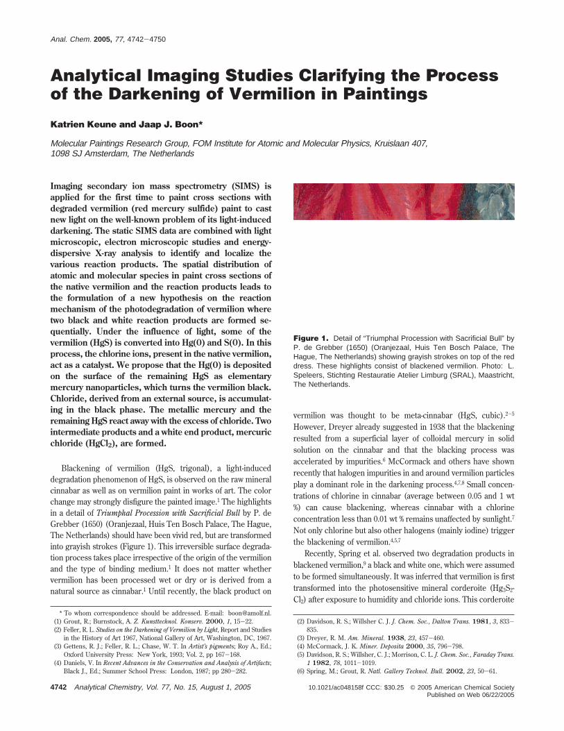
Analytical Imaging Studies Clarifying the Process of the Darkening of Vermilion in Paintings Katrien Keune and Jaap J. Boon* Molecular Paintings Research Group, FOM Institute for Atomic and Molecular Physics, Kruislaan 407, 1098 SJ Amsterdam, The Netherlands Imaging secondary ion mass spectrometry (SIMS) is applied for the first time to paint cross sections with degraded vermilion (red mercury sulfide) paint to cast new light on the well-known problem of its light-induced darkening. The static SIMS data are combined with light microscopic, electron microscopic studies and energy- dispersive X-ray analysis to identify and localize the various reaction products. The spatial distribution of atomic and molecular species in paint cross sections of the native vermilion and the reaction products leads to the formulation of a new hypothesis on the reaction mechanism of the photodegradation of vermilion where two black and white reaction products are formed se- quentially. Under the influence of light, some of the vermilion (HgS) is converted into Hg(0) and S(0). In this process, the chlorine ions, present in the native vermilion, act as a catalyst. We propose that the Hg(0) is deposited on the surface of the remaining HgS as elementary mercury nanoparticles, which turns the vermilion black. Chloride, derived from an external source, is accumulat- ing in the black phase. The metallic mercury and the remaining HgS react away with the excess of chloride. Two intermediate products and a white end product, mercuric chloride (HgCl 2 ), are formed. Blackening of vermilion (HgS, trigonal), a light-induced degradation phenomenon of HgS, is observed on the raw mineral cinnabar as well as on vermilion paint in works of art. The color change may strongly disfigure the painted image. 1 The highlights in a detail of Triumphal Procession with Sacrificial Bull by P. de Grebber (1650) (Oranjezaal, Huis Ten Bosch Palace, The Hague, The Netherlands) should have been vivid red, but are transformed into grayish strokes (Figure 1). This irreversible surface degrada- tion process takes place irrespective of the origin of the vermilion and the type of binding medium. 1 It does not matter whether vermilion has been processed wet or dry or is derived from a natural source as cinnabar. 1 Until recently, the black product on vermilion was thought to be meta-cinnabar (HgS, cubic). 2-5 However, Dreyer already suggested in 1938 that the blackening resulted from a superficial layer of colloidal mercury in solid solution on the cinnabar and that the blacking process was accelerated by impurities. 6 McCormack and others have shown recently that halogen impurities in and around vermilion particles play a dominant role in the darkening process. 4,7,8 Small concen- trations of chlorine in cinnabar (average between 0.05 and 1 wt %) can cause blackening, whereas cinnabar with a chlorine concentration less than 0.01 wt % remains unaffected by sunlight. 7 Not only chlorine but also other halogens (mainly iodine) trigger the blackening of vermilion. 4,5,7 Recently, Spring et al. observed two degradation products in blackened vermilion, 9 a black and white one, which were assumed to be formed simultaneously. It was inferred that vermilion is first transformed into the photosensitive mineral corderoite (Hg 3 S 2 - Cl 2 ) after exposure to humidity and chloride ions. This corderoite * To whom correspondence should be addressed. E-mail: [email protected]. (1) Grout, R.; Burnstock, A. Z. Kunsttechnol. Konserv. 2000, 1, 15-22. (2) Feller, R. L. Studies on the Darkening of Vermilion by Light, Report and Studies in the History of Art 1967, National Gallery of Art, Washington, DC, 1967. (3) Gettens, R. J.; Feller, R. L.; Chase, W. T. In Artist’s pigments; Roy A., Ed.; Oxford University Press: New York, 1993; Vol. 2, pp 167-168. (4) Daniels, V. In Recent Advances in the Conservation and Analysis of Artifacts; Black J., Ed.; Summer School Press: London, 1987; pp 280-282. (2) Davidson, R. S.; Willsher C. J. J. Chem. Soc., Dalton Trans. 1981, 3, 833- 835. (3) Dreyer, R. M. Am. Mineral. 1938, 23, 457-460. (4) McCormack, J. K. Miner. Deposita 2000, 35, 796-798. (5) Davidson, R. S.; Willsher, C. J.; Morrison, C. L. J. Chem. Soc., Faraday Trans. 1 1982, 78, 1011-1019. (6) Spring, M.; Grout, R. Natl. Gallery Technol. Bull. 2002, 23, 50-61. Figure 1. Detail of “Triumphal Procession with Sacrificial Bull” by P. de Grebber (1650) (Oranjezaal, Huis Ten Bosch Palace, The Hague, The Netherlands) showing grayish strokes on top of the red dress. These highlights consist of blackened vermilion. Photo: L. Speleers, Stichting Restauratie Atelier Limburg (SRAL), Maastricht, The Netherlands. Anal. Chem. 2005, 77, 4742-4750 4742 Analytical Chemistry, Vol. 77, No. 15, August 1, 2005 10.1021/ac048158f CCC: $30.25 © 2005 American Chemical Society Published on Web 06/22/2005
Transcript of Analytical Imaging Studies Clarifying the Process of the Darkening of Vermilion in Paintings
Analytical Imaging Studies Clarifying the Processof the Darkening of Vermilion in Paintings
Katrien Keune and Jaap J. Boon*
Molecular Paintings Research Group, FOM Institute for Atomic and Molecular Physics, Kruislaan 407,1098 SJ Amsterdam, The Netherlands
Imaging secondary ion mass spectrometry (SIMS) isapplied for the first time to paint cross sections withdegraded vermilion (red mercury sulfide) paint to castnew light on the well-known problem of its light-induceddarkening. The static SIMS data are combined with lightmicroscopic, electron microscopic studies and energy-dispersive X-ray analysis to identify and localize thevarious reaction products. The spatial distribution ofatomic and molecular species in paint cross sections ofthe native vermilion and the reaction products leads tothe formulation of a new hypothesis on the reactionmechanism of the photodegradation of vermilion wheretwo black and white reaction products are formed se-quentially. Under the influence of light, some of thevermilion (HgS) is converted into Hg(0) and S(0). In thisprocess, the chlorine ions, present in the native vermilion,act as a catalyst. We propose that the Hg(0) is depositedon the surface of the remaining HgS as elementarymercury nanoparticles, which turns the vermilion black.Chloride, derived from an external source, is accumulat-ing in the black phase. The metallic mercury and theremaining HgS react away with the excess of chloride. Twointermediate products and a white end product, mercuricchloride (HgCl2), are formed.
Blackening of vermilion (HgS, trigonal), a light-induceddegradation phenomenon of HgS, is observed on the raw mineralcinnabar as well as on vermilion paint in works of art. The colorchange may strongly disfigure the painted image.1 The highlightsin a detail of Triumphal Procession with Sacrificial Bull by P. deGrebber (1650) (Oranjezaal, Huis Ten Bosch Palace, The Hague,The Netherlands) should have been vivid red, but are transformedinto grayish strokes (Figure 1). This irreversible surface degrada-tion process takes place irrespective of the origin of the vermilionand the type of binding medium.1 It does not matter whethervermilion has been processed wet or dry or is derived from anatural source as cinnabar.1 Until recently, the black product on
vermilion was thought to be meta-cinnabar (HgS, cubic).2-5
However, Dreyer already suggested in 1938 that the blackeningresulted from a superficial layer of colloidal mercury in solidsolution on the cinnabar and that the blacking process wasaccelerated by impurities.6 McCormack and others have shownrecently that halogen impurities in and around vermilion particlesplay a dominant role in the darkening process.4,7,8 Small concen-trations of chlorine in cinnabar (average between 0.05 and 1 wt%) can cause blackening, whereas cinnabar with a chlorineconcentration less than 0.01 wt % remains unaffected by sunlight.7
Not only chlorine but also other halogens (mainly iodine) triggerthe blackening of vermilion.4,5,7
Recently, Spring et al. observed two degradation products inblackened vermilion,9 a black and white one, which were assumedto be formed simultaneously. It was inferred that vermilion is firsttransformed into the photosensitive mineral corderoite (Hg3S2-Cl2) after exposure to humidity and chloride ions. This corderoite
* To whom correspondence should be addressed. E-mail: [email protected].(1) Grout, R.; Burnstock, A. Z. Kunsttechnol. Konserv. 2000, 1, 15-22.(2) Feller, R. L. Studies on the Darkening of Vermilion by Light, Report and Studies
in the History of Art 1967, National Gallery of Art, Washington, DC, 1967.(3) Gettens, R. J.; Feller, R. L.; Chase, W. T. In Artist’s pigments; Roy A., Ed.;
Oxford University Press: New York, 1993; Vol. 2, pp 167-168.(4) Daniels, V. In Recent Advances in the Conservation and Analysis of Artifacts;
Black J., Ed.; Summer School Press: London, 1987; pp 280-282.
(2) Davidson, R. S.; Willsher C. J. J. Chem. Soc., Dalton Trans. 1981, 3, 833-835.
(3) Dreyer, R. M. Am. Mineral. 1938, 23, 457-460.(4) McCormack, J. K. Miner. Deposita 2000, 35, 796-798.(5) Davidson, R. S.; Willsher, C. J.; Morrison, C. L. J. Chem. Soc., Faraday Trans.
1 1982, 78, 1011-1019.(6) Spring, M.; Grout, R. Natl. Gallery Technol. Bull. 2002, 23, 50-61.
Figure 1. Detail of “Triumphal Procession with Sacrificial Bull” byP. de Grebber (1650) (Oranjezaal, Huis Ten Bosch Palace, TheHague, The Netherlands) showing grayish strokes on top of the reddress. These highlights consist of blackened vermilion. Photo: L.Speleers, Stichting Restauratie Atelier Limburg (SRAL), Maastricht,The Netherlands.
Anal. Chem. 2005, 77, 4742-4750
4742 Analytical Chemistry, Vol. 77, No. 15, August 1, 2005 10.1021/ac048158f CCC: $30.25 © 2005 American Chemical SocietyPublished on Web 06/22/2005

subsequently degrades into the black meta-cinnabar, the whitecalomel (Hg2Cl2), and elemental sulfur by a light-induced reaction.The chloride was postulated to originate from external sources.
In general, the degraded vermilion layers in paint crosssections are extremely thin (less than a few micrometers), andmost analytical techniques do not have sufficient spatial resolutionand sensitivity to study the changes within the paint layer.4,9 Theavailability of a paint cross section taken from an (17th century?)overpaint on the painting Portrait of a Lady by Peter Paul Rubens(ca. 1625) (Mauritshuis, The Hague; sample number MH251/26) with an extraordinary thick layer of partially degraded,blackened vermilion particles made it possible to investigate thedegradation process in more detail (Figures 2A and 3A). In thispaper, a second paint cross section from an original layer inMinerva and Hercules Opening the Doors for Victory by Christiaanvan Couwenberg (1651), (Oranjezaal Huis ten Bosch Palace, TheHague; sample number HSTB 34/2), derived from a collectiondifferent from the first sample, is examined as well.
An important advantage of investigating the blackening processof vermilion in paint cross sections is the clear visualization of agradient in the deterioration from the light-exposed top to thelight-shielded bottom. In addition, the pigment particles areembedded in an organic binding medium layer, so the componentspossibly released during the blackening of vermilion are embed-ded and fixed in position so they can be investigated bymicroscopy.
The analytical data obtained from light microscopy, scanningelectron microscopy combined with energy-dispersive X-ray
analysis (SEM/EDX), and secondary ion mass spectrometry(SIMS) presented here lead to new insights in the mechanism ofthe blackening of vermilion. In contrast to work presented earlier,9
we observe that a black reaction product is formed first, which isspatially separated and thus followed by a white compound. Weprove that meta-cinnabar cannot be formed, and with the exclusiveresults from secondary ion mass spectrometry, we could identifyanother white reaction product that was never before describedin paintings.
EXPERIMENTAL SECTIONSamples. The paint sample MH251/26 was embedded in
polyester resin Polypol using the Easysection system. SampleHSTB 34/2 was embedded in Technovit 2000LC (HeraeusKulzer). Both paint cross sections were dry polished withMicromesh polishing cloths (final step 12 000 mesh) (ScientificInstruments Services Inc.).
The following reference materials were used: mercury sulfide(HgS) (Merck), mercuric chloride (HgCl2) (Merck), and mercur-ous chloride ((HgCl)2) (Fluka).
Instruments. The static SIMS experiments were performedon a Physical Electronics (Eden Prairie, MN) TRIFT-II time-of-flight SIMS. The surface of the sample was scanned with a 15-keV primary ion beam from an 115In+ liquid metal ion gun. Thepulsed beam was nonbunched with a pulse width of 20 ns, acurrent of 600 pA, and a spot size of ∼120 nm. The primary beamwas rastered over a 50 × 50 µm sample area, divided into 256 ×256 pixels. The surface of the sample was charge compensated
Figure 2. Light microscopic image under white light (A) and UV illumination (B). The image illustrates the partial photodegradation of a vermilionparticle. The BSE image (C) shows the structural difference between the red and the black areas. The horizontal lines are due to polishing; thetext refers to the particle marked X. EDX spectra of spot analyses (D) in black (S1), red (S2), and white (S3) area demonstrate the differentelementary composition of the native material and the reaction products.
Analytical Chemistry, Vol. 77, No. 15, August 1, 2005 4743

with electrons pulsed between the primary ion beam pulses. Toprevent large variations in the extraction field over the largeinsulation surface area of the paint cross section, a nonmagneticstainless steel plate with slits (1 mm) was placed on top of thesample. The paint cross section (150 × 50 × 3 mm) was rinsedin hexane to eliminate contamination of airborne silicones.
A 15-keV 115In+ primary ion beam was used for the referencesamples, with primary ion pulses that were compressed (bunched)to ∼1 ns to obtain a better mass resolution. These experimentswere performed with 600 pA beam current. All these measure-ments were charge compensated with electrons pulsed betweenthe primary ion beam pulses.
The oil immersion reflected light microscopic images wereobtained on a Leica DMRX microscope (Leica, Wetzlar, Germany).White light was provided by a 100-W halogen lamp, and an OsramHBO 50-W lamp and Leica filter D (excitation 360-425 nm,emission >460 nm) were used for fluorescence microscopy. Thesurface of the paint cross section was covered with immersionoil (Catalog No. 11 513 787, Leica). Images were recorded with aNikon digital still camera DXM1200 (Nikon Instech Co., Ltd.).
Scanning electron microscopy studies in combination withenergy-dispersive X-ray analysis were performed on a XL30 SFEGhigh-vacuum electron microscope (FEI, Eindhoven, The Nether-lands) with EDX system (spot analysis and elemental mappingfacilities) from EDAX (Tilburg, The Netherlands). Backscatteredelectron images of the cross sections were taken at 20-kVaccelerating voltage at a 5-mm eucentric working distance and aspot size of 3, which corresponds to a beam diameter of 2.2 nmwith current density of ∼130 pA. EDX analysis was performed ata spot size setting of 4 (beam diameter 2.5 nm and current density550 pA) to obtain a higher count rate and at an acceleration voltageof 20 kV. EDX mapping parameters were as follows: 256 × 200matrix, 1028 frames, 100-µs dwell time, and 50-µs amplitude time.Samples were carbon coated to improve surface conduction in a
CC7650 Polaron Carbon Coater with carbon fiber (QuorumTechnologies, East Sussex, U.K.).
RESULTSLight Microscopy. Light microscopic images of a highly
magnified area of discolored vermilion in the paint cross sectionMH251/26 are shown in Figures 2A, 3A, and 4A (normal light)and 2B and 3B (UV light). The area of the panel painting of thesample MH251/26 is overpainted, and the paint buildup consistsnow of at least six layers. The original paint layers consist of achalk ground, a lead white, and charcoal imprimatura and finallyseveral layers of vermilion and red lake. A bone black layer blocksout this original red paint. On top, a thin organic intermediatelayer is applied with as final layer the now degraded vermilionwith red lake (paint cross section not shown). The total thicknessof the vermilion layer is 25 µm of which 15 µm is discolored,containing both the black and white degradation products. Thevermilion particles are coarse grained, and the individual particlesare partially degraded in some instances. These latter particlesconsist of a black upper part and a red lower part (in UV light,the colors remain black and red). The white product is positionedon top and around the black product and is transparent underUV illumination.
A magnified area of the darkened vermilion in paint crosssection HSTB 34/2 is shown in Figure 5A and B (normal and UVlight). There are three layers in the area of the panel paintingwhere the sample was removed: a grayish lead white ground, areddish-brown iron oxide paint layer, and a red paint layer with amixture of vermilion and red lake on top (paint cross section notshown). The degraded top layer with black and white products isvery thin (4 µm), while the total thickness of the vermilion andred lake layer is 50 µm. The intact layer consists mainly of finelydispersed vermilion particles with a few larger ones. Red lake is
Figure 3. Light microscopic images under white light (A) and UV light (B) illumination and BSE (C) of the partially degraded vermilion paintin paint cross section MH251/26. The images correspond with the area measured with EDX imaging. X-ray maps of intact (R) and blackened(B) vermilion layer reveals the spatial distribution of mercury (Hg L) (D), sulfur (S K) (E), and chlorine (Cl K) (F).
4744 Analytical Chemistry, Vol. 77, No. 15, August 1, 2005

present in the vermilion layer and is recognizable by the pinkishfluoresce in the UV image (Figure 5B).
SEM/EDX. The backscattered electron image (BSE-image)illustrates the presence of a structural change in the partiallydegraded vermilion particle of MH251/26 (Figure 2C). Thestructure of the red vermilion particle differs from the structureof the black product. The red part of particle X (indicated in Figure2C) is flat and homogeneous. The horizontal lines in the particlesare polishing marks. The black part of the particle X is irregularand broken up. Spots with a higher BSE intensity and a sizesmaller than 100 nm are visible in the degraded black phase(Figure 2C and Supporting Information Figure 1). The white areasdisplay the same BSE intensity as the black and the red areas.
The two elements in vermilionsmercury and sulfursaredetected with EDX spot analysis in the red (S2), black (S1), andwhite (S3) particles (Figure 2D). EDX spot analyses of the red(S2), black (S1), and the white (S3) phases demonstrate that thereis relatively less sulfur in the degraded particles (Figure 2D),considering the sulfur/mercury ratio. This sulfur/mercury ratiois only indicative because the X-ray intensity peaks of the mercuryM-shell and the sulfur K-shell are partially overlapping. EDX spotanalysis from the black area (S1) and white area (S3) showsrelatively high chlorine counts; the chlorine concentration in thewhite particle is much higher than in the black particle (taking
the mercury peak as reference). The EDX spectrum of the whitearea reveals that the intensities of carbon, oxygen, and aluminumare high compared to the black and red areas. However, theseelements are representative for the organic binding medium andare only detected due to the fact that the interaction volume ofthe beam is larger than the volume of the particle. The sulfur-to-mercury ratio in the binding medium of both the degraded andintact parts is higher than the red particles (spectrum not shown),which indicates that sulfur, besides being a constituent of HgS,is also present in another form in the binding medium.
Elemental X-ray maps of the affected vermilion layer inMH251/26 visualize the distribution of mercury, sulfur, andchlorine (Figure 3). The dotted line in the elemental imagesindicates the compositional transition between red (R) and black(B) particles (Figure 3). The elemental X-ray map of mercuryillustrates that mercury is only present inside the pigment particlesand has not diffused into the binding medium. Sulfur is presentin high relative intensity in the mercury containing particles ofthe intact layer (R), whereas lower sulfur X-ray counts areobserved in and around the degraded particles (B). The chlorineis only detected in high counts in the degraded vermilion particles.
SIMS. SIMS spectra were acquired in the negative ion mode.Chloride and sulfide dominate in the SIMS spectra of MH251/26and HSTB 34/2. The spatial distribution of the sulfide and chloride
Figure 4. Light microscopic image (A), representing the scanned SIMS area of the partially degraded vermilion paint of MH251/26. The arrowindicates the direction of the line scan. SIMS images of the sulfide (B) and chloride (C) represent the distribution of these ions over the partiallydegraded vermilion paint layer. The line scan (D) illustrates the distribution of chlorine and sulfur from top to bottom in the paint cross section.
Analytical Chemistry, Vol. 77, No. 15, August 1, 2005 4745

ions in sample MH251/26 is plotted in Figure 4B and 4C,respectively. Figure 4A represents the light microscopic imageof the scanned SIMS area. Three horizontal lines in the imagesin Figure 4 outline the blackened layer (B) and the red vermilionlayer (R). The intact red vermilion particles contain sulfide andchloride (see layer R in Figure 4B and C). The chloride is presentin higher amounts in the degraded layer (B) and corresponds tothe black and white particles. Line scans drawn from the top tothe bottom of the layer (see white arrows in Figure 4A-C)visualize the distribution of the secondary ion yields. The second-ary ion yields of both chloride and sulfide ions are depicted inFigure 4D. The line scan, with a resolution of 1 µm, first crossesa degraded vermilion particle (3 µm) and then scans an intactvermilion particle (5 µm). The chloride ion yield is higher fordegraded vermilion compared to the intact vermilion. The sulfideion yield is relatively diminished in the degraded particlescompared to the intact vermilion. The same phenomenon isobserved in paint cross section HSTB 34/2 (Figure 5C). The linescan crosses the degraded vermilion layer (4 µm) and two intact
vermilion particles (6 µm) (Figure 5A-C). The chloride andsulfide ion distribution are displayed in Figure 5D and E.
Similar negatively charged mercury-halogen cluster ion pat-terns were detected in the paint cross sections MH251/26 andHSTB34/2. The corresponding molecules were deduced on thebasis of their molecular weight distribution and isotopic pattern.The SIMS spectra of HSTB 34/2 show the cluster ions from[HgnSnCl]- (n ) 1-6), [HgnSn-1Cl3]- (n ) 1-4), and [HgnSn+1Cl2]-
(n ) 1-3). The spectra of sample MH251/26 show the same typesof cluster ions, but only for n ) 1-2 (full spectra not shown; apartial negative ion spectrum of HSTB 34/2 with n ) 3 isdiscussed in the next paragraph). The ions are interpreted aschloride adduct ions from (HgS)n [(HgS)n‚Cl- (n ) 1-6)], (HgS)n-(HgCl2) [(HgS)n(HgCl2)‚Cl- (n ) 0-3)], and (HgS)n(SCl)[(HgS)n(SCl)‚Cl- (n ) 1-3)], respectively. Other types of clusterions detected with lower ion yields in the paint sample are(HgCl)2‚Cl- and HgnSyCl- (y ) n + 1, n + 2, or n + 3). (HgS)n‚Cl-,(HgS)n(SCl)‚Cl-, and HgnSyCl- are detected in the intact anddegraded vermilion particles of HSTB 34/2, whereas (HgS)n(Hg-
Figure 5. Distribution of sulfide and chloride and the molecular distribution in a partially degraded vermilion paint. The arrow in the lightmicroscopic image (A) and UV light image (B), which represents the scanned SIMS area, indicates the direction of the line scan (C). Thenegative SIMS image of mercury-halogen cluster ions elucidates the position of S- (D), Cl- (E), (HgS)Cl- (F), (HgCl2)Cl- (G), (HgS)(SCl)Cl-
(H), and (HgCl)2Cl- (I) in the partially degraded vermilion paint. A contour, based on the light microscopic image and total negative ion image,outlines four red vermilion particles and the boundary between the intact and degraded paint.
4746 Analytical Chemistry, Vol. 77, No. 15, August 1, 2005

Cl2)‚Cl- and (HgCl)2‚Cl- are only detected in the degraded layer.The SIMS images in Figure 5F-I illustrate this distribution of(HgS)‚Cl-, (HgCl2)‚Cl-, (HgS)(SCl)‚Cl-, and (HgCl)2‚Cl- in HSTB34/2. A contour drawn on the basis of the light microscopic imageand the total negative ion image outlines five red vermilionparticles and the boundary in blue between the intact anddegraded paint.
Figure 6A-F represents partial negative ion mass spectra ofthe reference materials HgS, (HgCl)2, and HgCl2 and theirmixtures HgS + (HgCl)2 and HgS + HgCl2. The referencematerials, HgCl2 and (HgCl)2, are two separate white mercurychloride complexes that are suspected to be present in thedegraded vermilion layer of paint cross section HSTB 34/2. Partof the negative ion mass spectrum of the degraded vermilion paintin paint cross section HSTB 34/2 is depicted in Figure 6G. Thespectrum is acquired after selecting the region of interest in thetotal area analyzed (50 µm2). The SIMS spectrum of the intactvermilion is represented by Figure 6H. Figure 6I represents thecalculated isotopic distributions of the ion clusters (Hg3S3)Cl-,(Hg3S2Cl2)Cl-, (Hg3S5)Cl-, and (Hg3S6)Cl-. These ions werechosen for calculation because they match the mass window ofthe relevant cluster ion peak patterns from mercury-halogencluster ions with three mercury atoms in the regions of interest.
The SIMS spectra of the reference materials and their mixturessupport the identification of the new products present in thedegraded vermilion paint layer of HSTB 34/2. Vertical dotted linespasted over the mass spectra in Figure 6 divide these into fourmass windows corresponding to the four columns in the Table 1.Table 1 gives an overview of the interpreted mercury-halogencluster ion patterns in four mass windows deduced from thespectra of the various reference materials and the paint crosssection present (Figure 6 and Table 1).
In the mass window from 720 to 750 amu, (HgS)3Cl- (m/z726-743) is detected in the mass spectrum of intact and degradedvermilion in HSTB 34/2 (Figure 6G and H). The reference spectraof both mixtures (HgS + HgCl2 and HgS + (HgCl)2) show thesame cluster ions (Figure 6E, F), while the spectrum of pure HgSshows a (HgS)3S- cluster ion without a chloride ion attached(Figure 6A). In the mass window from 750 to 790 amu, peaks at760-781 amu assigned to (Hg3S2Cl2)Cl- are dominant in the massspectrum of the degraded vermilion (Figure 6G). The ion cluster(Hg3S2Cl2)Cl- is only found in the spectrum of the mixture of HgS+ HgCl2 (Figure 6F). A relative small amount of (Hg3S4)Cl- and(Hg3SCl3)Cl- in this mass range is detected in the degradedvermilion (these cluster ions partially overlap with the cluster ionsof (Hg3S2Cl2)Cl-). In the intact vermilion, relatively minor con-tributions from (Hg3S2Cl2)Cl- and (Hg3S4)Cl- are detected (Figure6H). The mass window from 790 to 820 amu shows (Hg3S5)Cl-
and (Hg3S4Cl)Cl- in the mass spectrum of the intact vermilion ofHSTB 34/2. The latter cluster ion is found in low yields in thespectrum of the mixture [HgS + HgCl2]. In the mass window from820 to 860 amu, the cluster (Hg3S6)Cl- is detected in low yieldsin spectra of the paint cross section as well as in the mixture HgS+ HgCl2 (Figure 6F and H and Table 1).
Figure 6B represents the SIMS spectrum of the referencematerial HgS with an excess of chloride ions added as KCl. Weconclude that the mercury-halogen cluster ions detected in thepaint cross section are not formed by gas-phase ion chemistryjust above the surface after SIMS of HgS and KCl, because
Figure 6. Part of the negative ion mass spectrum in the mass range720-860 amu of pure HgS (A), HgS + Cl- ions (B), pure (HgCl)2
(C), pure HgCl2 (D) mixture of HgS + (HgCl)2 (E), mixture of HgS +HgCl2 (F), degraded vermilion layer in paint cross section HSTB 34/2(G), intact vermilion layer in paint cross section HSTB 34/2 (H), andcalculated isotopic distribution of (Hg3S3)Cl-, (Hg3S2Cl2)Cl-, (Hg3S5)Cl-,and (Hg3S6)Cl- (I). The vertical dotted lines divide the spectra intofour mass ranges, which correspond with the columns in Table 1.
Table 1. Overview of the Various Mercury-HalogenCluster Ions Detected in the Negative Ion MassSpectra in the Mass Range of 720-860 AmuOriginating from the Reference Materials, TheirMixtures, and the Degraded and Intact Vermilion Layerin HSTB 34/2
mass range,a amu
sample 720-750 750-790 790-820 820-860
HgS (HgS)3S- Hg3S5- Hg3S6
- Hg3S7-
HgS + Cl- Hg3S6-
HgCl2Hg2Cl2
(Hg3Cl4)Cl-(Hg3Cl4)Cl-
(Hg3Cl5)Cl-(Hg3Cl5)Cl-
(Hg3Cl6)Cl-(Hg3Cl6)Cl-
HgS + HgCl2 (HgS)3Cl- (Hg3S2Cl2)Cl- (Hg3S2Cl3)Cl- (Hg3S6)Cl-(Hg3S3Cl2)Cl-(Hg3S4Cl)Cl-
HgS + Hg2Cl2 (HgS)3Cl- (Hg3Cl4)Cl- (Hg3Cl5)Cl- (Hg3Cl6)Cl-degraded layerHSTB-34/2
(HgS)3Cl- (Hg3S2Cl2)Cl-(Hg3S4)Cl-(Hg3SCl3)Cl-
intact layerHSTB 34/2
(HgS)3Cl- (Hg3S2Cl2)Cl-(Hg3S4)Cl-
(Hg3S5)Cl- (Hg3S6)Cl-
a The four mass ranges correspond to the mass ranges indicated inFigure 6.
Analytical Chemistry, Vol. 77, No. 15, August 1, 2005 4747

(HgS)n(HgCl2)‚Cl- and (HgCl)2‚Cl- are not observed in thisspectrum. The exact identification of the composition of themercury-halogen cluster ions in Figure 6B is difficult as the yieldsand thus the resolution is low.
DISCUSSIONThe two paint cross sections investigated that originate from
paintings made by two different painters and that were kept underdifferent conditions give similar analytical information on the colorchanges in vermilion. The analytical imaging studies on thedarkened vermilion oil paint layer in sample MH251/26 demon-strate the degradation phenomena in detail. As light is a require-ment for the degradation process, it is concluded that the lightmust have penetrated the whole affected layer as all particles insample MH251/26 are homogeneously transformed. On the basisof the transition of the red into the black materials observed inthe partially degraded particles and the position of the whitematerials around the black materials, we propose that the blackmaterial is formed first. In a later stage, the white product isformed from the black reaction product.
Both the SIMS and EDX techniques show relatively highamounts of chloride and relatively low amounts of sulfide in thedegraded particles. The relative amount of mercury in the intactand degraded particles is considered to be constant, since thebackscattered electron intensity in the BSE image is the samefor all phases. The EDX images lead us to conclude that sulfurleaches from the particles in the degraded layer into the environ-ment. However, EDX spot analyses detect sulfur in the organicbinding medium of both the degraded and the intact part. Theorganic binding medium also has been shown to contain alumi-num. Sulfur and aluminum are known constituents of the inorganicsubstrate of red lakes.10 It is therefore possible that red lake hasbeen mixed into the paint although no distinct pigment particlesare visible.
The mercury-halogen cluster anions detected in the wholevermilion layer with SIMS are charged by attachment of Cl- ions.Neutral molecules are known to be anionized by chloride to forma negatively charged complex.11,12 The ions (HgS)nCl- and(HgS)n(SCl)Cl- are found in the intact vermilion paint layer andare representative for intact HgS. (HgS)nS-, detected in the pureHgS, is not present in the SIMS spectra of the mixtures and maybe suppressed by the presence of chloride. Ions from (HgS)n(Hg-Cl2)‚Cl- (n ) 0-6) are detected and mainly present in thedegraded vermilion layer. These types of ions are also formedfrom the mixture of HgS + HgCl2 but not from the mixture HgS+ (HgCl)2. We conclude on the basis of the presence of thesetypes of ions found in the degraded vermilion layer that it isindicative for mercuric chloride (HgCl2). In addition, the ions from(HgS)n(HgCl2)‚Cl- detected in SIMS data of a mixture of HgS +HgCl2 indicate that HgS is still present in the degraded vermilionlayer, because these ions are absent in the spectra of pure HgCl2.The ions (HgCl)2‚Cl- are not only representative for (HgCl)2
(calomel), because these ions are also detected in the spectrumof HgS + HgCl2 (part of the spectrum not shown). It is concludedthat the presence of calomel in the paint sample cannot be provenwith SIMS, because this ion (HgCl)2‚Cl- is not characteristic for(HgCl)2 (calomel) and (Hg3Cl4)‚Cl- is only detected in the HgS+ (HgCl)2 reference mixture and not in the paint sample.
On the basis of our results and literature data, we proposethat the transformation of vermilion from red into black materialsis a conversion from HgS into Hg(0) and S(0) by a photochemicalreaction catalyzed by chloride (step 1 in Figure 7). The conversionof Hg(0) from photoreduced Hg(II) has been reported both insolid phase13 and aqueous media.14-19 This supports the idea of
(10) Schweppe, H.; Winter, J. In Artist’s pigments; West FitzHugh, E. Ed.; OxfordUniversity Press: New York, 1997; Vol. 3, Chapter 4.
(11) Groenewold, G. S.; Appelhaus, A. D.; Gresham, G. L.; Ingram, J. C.; Shaw,A. D. Int. J. Mass Spectrom. 1998, 178, 19-29.
(12) Delcorte, A. In ToF-SIMS: Surface Analysis by Mass Spectrometry’ Vickerman,J. C., Briggs, D., Eds.; IM Publication and Surface Spectra Limited: London,2001; p 183.
(13) Gustin, M. S.; Biester, H.; Kim, C. S.; Atmos. Environ. 2002, 36, 3241-3254.
Figure 7. Scheme of the proposed mechanisms of the degradation process of the red vermilion into a black and finally a white product.
4748 Analytical Chemistry, Vol. 77, No. 15, August 1, 2005

the light-induced conversion of Hg(II)S into Hg(0). After thephotoreduction reaction, the metallic mercury is deposited on theintact HgS surface, resulting in a black reaction product. Theremaining HgS in the degraded vermilion is identified with SIMS.The metallic mercury is present as nanoparticles, which issupported by the “hot” spots smaller than 100 nm visualized inthe BSE-image (Figure 2C and Supporting Information, Figure1). The Hg(0) precipitation on the vermilion surface has neverbeen proven directly, but has been suggested by other au-thors.6,16,20 Elementary sulfur is also detected as a product ofphotodegraded HgS.19 The sulfur leaves the paint particles possiblyin the form of volatile sulfur oxide, as sulfur has been shown tooxidize to sulfate ions via polythionic acids.21 Since the sulfur islost from the particle, it is inferred that a conversion of vermilionto meta-cinnabar is unlikely. The small relative concentration ofsulfur still present in the particles is ascribed to intact residualintact vermilion. XRD measurements performed on reconstruc-tions and other paint samples elsewhere by other authors supportthis conclusion.1,2,4,7,9 It is shown that vermilion is still present inthe XRD spectra of the blackened vermilion as is deduced fromthe decreased intensity of the XRD patterns of vermilion. No blackmeta-cinnabar was shown in the XRD spectra.
Chlorine plays a dominant role in the light-induced blackeningphenomenon, as already suggested in the literature.7,9 The muchmore sensitive surface technique of SIMS demonstrates, incontrast to the EDX results, that chloride is present inside thevermilion particles of both MH 251/26 and HSTB 34/2. Theconcentration of chloride in the red particles must be below thedetection limit of EDX. As small quantities of chloride makevermilion photosensitive,7 the small amounts of chloride detectedin the intact vermilion trigger the blackening process. Due to thefact that only small quantities of chloride are necessary to formblackened cinnabar and that other compounds besides chlorideions, such as iodide, enhance the blackening as well, it isconcluded that chloride initially acts as a catalyst and is not partof the primary reaction products. The exact minimum concentra-tion of chloride necessary to trigger the process is not known.McCormack states that cinnabar is light stable below 0.01 wt %((0.01 wt %) chloride, as determined with a wavelength-dispersivemicroprobe analyzer cinnabar is light stable and it blackens at aconcentration of 0.05 wt % and higher.7
The exact mechanism of the photodegradation of vermilioncatalyzed by chloride is still an open question. We suggest thatunder the influence of light an electron-hole pair is generatedand a photoeletrochemical process takes place, which enables anelectron transfer from chlorine anion to mercury and from sulfideback to chloride (i.e., chloride has a catalytic function). Assumingthat chlorine anions are oxidized by the positive holes of thevalence band of HgS, the valence band edge of HgS must be more
positive than the redox potential of Cl-/Cl2.16,22 The exact positionof the valence band edge of HgS is not clear, but the value of thisposition is expected to be close to the redox potential of Cl-/Cl2.22
The relatively high quantity of chloride in the degraded partis hard to explain from an original vermilion source alone. Wealso have to conclude that the chloride is not introduced via thebinding medium, as EDX did not detect chloride in the bindingmedium of the intact area. To explain the relatively high yields ofchloride ions in the degraded vermilion particles, it is necessaryto invoke additional source of chlorides. Chloride is possiblytransported by diffusion from paint layers positioned lower orintroduced from an external source.
After the first transformation, the black product is thought totrap chloride to transform subsequently into a white reactionproduct. Although the elemental composition (Hg, S, Cl) of theblack and the white phases is the same, the EDX spot analysisshows a higher relative intensity of chlorine and a fairly lowerrelative intensity of sulfur in the white product (S3) compared tothe black product (S1) (Figure 2A and D). SIMS identifies themercury chlorine complex HgCl2 in the degraded vermilion layer.Mercuric chloride (HgCl2) has never been identified in paintingsbefore. Calomel (HgCl)2 cannot be identified positively with SIMS;however, Spring and Grout had identified calomel as the whiteproduct in paintings containing degraded vermilion using Ramanmicroscopy.9
The two components in the black product, metallic mercuryand residual vermilion, react with chloride supplied by sourcesexternal to the vermilion. The residual vermilion reacts with thisexternal chloride to the light-sensitive mineral corderoite, whichdegrades under the influence of light into calomel, metallicmercury, and sulfur. Corderoite (Hg3S2Cl2) can easily be formedin a mixture of HgS and NaCl solution kept at room temperatureafter a long period7,9 and will after light exposure degrade into adark gray product containing (HgCl)2.9 The formation of corderoitecould not have taken place in the first degradation steps, becauseof the high amount of chloride required that is suggested toaccumulate after the blackening. The (HgCl)2 formed photode-grades under normal light into HgCl2 and Hg(0).23 The metallicmercury, derived from the photoreduction in the first step, thecorderoite, and the (HgCl)2, react with chloride to form mercuricchloride HgCl2. In vitro, HgCl2 can be formed from metallicmercury via the reaction with chlorine gas or by the formation ofmercury(II) sulfate.24 The white end product is found around theblack product, which means that external sources, light, and ahigher concentration of chloride are necessary to create the whiteproduct.
Figure 7 illustrates the scheme of the proposed mechanismsof the degradation process of the red vermilion into the blackand subsequently the white reaction product. The schemedescribes the degradation phenomena qualitatively to clarify theprocess. The products observed in the paint cross sections aremixed phases and consequently cannot be classified in a single
(14) Hsieh, Y. H.; Tokunaga, S.; Huang, C. P. Colloids Surf. 1991, 53, 257-274.(15) Ravichandran, M. Chemosphere 2004, 55, 319-331.(16) Pal, B.; Ikeda, S.; Ohtani, B. Inorg. Chem. 2003, 42, 1518-1524.(17) Litter, M. I. App. Catal., B 1999, 23, 89-114.(18) Stromberg, D.; Stromberg, A.; Wahlgren, U. Water, Air, Soil Pollut. 1991,
56, 681-695.(19) Okouchi, S.; Sasaki, S. Environ. Int. 1983, 9, 103-106.(20) Gaustin, M. S.; Maxy, R. A.; Rasmussen, P. In Symposium Volume of Air
Pollutance, Cary, NC, September 1998.(21) Naito, K.; Takei, S.; Okabe, T. Bull. Chem. Soc. Jpn. 1970, 43, 1360-1364.
(22) Personal communication with Professor B. Ohtani (Catalysis ResearchCenter, Hokkaido University, Japan), 2004.
(23) Chambers, C.; Holliday, A. K. In Modern Inorganic Chemistry; Butterworth& Co.: London, 1975; p 437.
(24) Holleman, A. F.; Wilberg, E. In Inorganic Chemistry; Academic Press: SanDiego, 2001; p 1310.
Analytical Chemistry, Vol. 77, No. 15, August 1, 2005 4749

category in the scheme. For example, the black product in sampleMH251/26 has a relatively higher concentration of chloridecompared to the intact red vermilion. Based on its color, this blackproduct would be classified in the black box after step 1 in thediagram, but a relative increase in chloride is taking place. Thisaccumulation in the black product is indicated in step 2, but aconversion into a white product is not observed with lightmicroscopy. Therefore, the black product would be classified inthe box after step 2, which is indicated in the scheme as gray.Furthermore, not all the degraded vermilion is converted intoHgCl2 in sample HSTB 34/2; some of the former reaction productsare still expected to be present.
The obtained knowledge implies that the quality of thevermilion used in paintings is of significant importance for thechanges of preservation of the red color. Small chloride or otherhalogen impurities in the vermilion makes the red pigmentphotosensitive. Chloride present in the medium or derived fromother external sources can drive subsequent reactions. Works ofart such as easel and mural paintings can be expected to beexposed to light and chlorides.
CONCLUSIONAnalytical studies on paint cross sections containing darkened
vermilion reveal the light-induced degradation phenomena indetail. Light microscopic images reveal two colored reactionproducts, a white product positioned above and around a blackproduct. SEM/EDX data give information about the elementaldistribution and the structural changes between the three coloredphases. SIMS detects, in contrast to EDX, traces of chloride inthe native vermilion. The additional benefit of imaging SIMSapplied for the first time to investigate this phenomenon in paintcross sections is that the molecular information from SIMS is ableto identify the white reaction product as mercuric chloride. We
propose a new hypothesis, which describes a three-step processwhere black and white reaction products are formed from eachother. Chloride acts as catalyst in the first step of the photoinducedchemical degradation of vermilion. In the formed black reactionproduct, an excess of chloride is accumulated. The black phasewill react further with the excess of chloride to form the whiteproduct, mercuric chloride. The combination of both techniques,SEM/EDX and SIMS, is essential to elucidate the familiarphenomenon of “blackening of vermilion” in detail.
ACKNOWLEDGMENTWe are grateful to Linnaea Saunders, Mauritshuis, The Hague,
The Netherlands, who provided us the paint cross section MH251/26. We also thank Lidwien Speleers, AMOLF, Amsterdam, TheNetherlands, for providing the paint cross section HSTB 34/2.The research at AMOLF is embedded in the FOM researchprogram 49 “Mass spectrometric imaging and structural analysisof biomacromolecules”. This work is part of the “De Mayerne”program funded by the Dutch Organization for Scientific Research(NWO) and the Foundation for Fundamental Research on Matter(FOM), a subsidiary of the Dutch Organization for ScientificResearch (NWO).
SUPPORTING INFORMATION AVAILABLEAdditional information concerning Figure 1: The BSE image
visualizes a black particle containing hot spots of smaller than100 nm. These hot spots are nanoparticles of metallic mercury.This material is available free of charge via the Internet at http://pubs.acs.org.
Received for review December 14, 2004. Accepted April18, 2005.
AC048158F
4750 Analytical Chemistry, Vol. 77, No. 15, August 1, 2005