Analysis on the activation of the VMO and VLL muscles during semisquat exercises with and without...
8
Analysis on the activation of the VMO and VLL muscles during semisquat exercises with and without hip adduction in individuals with patellofemoral pain syndrome Kelly Rafael Ribeiro Coqueiro a , De ´bora Bevilaqua-Grossi b , Fausto Be ´rzin c , Alcimar Barbosa Soares d , Cecı ´lia Candolo a , Vanessa Monteiro-Pedro a, * a Federal University of Sa ˜ o Carlos, Rodovia Washington Luı ´s, Km 235, Sa ˜ o Carlos-SP 13565-905, Brazil b Ribeira ˜ o Preto School of Medicine, Sa ˜ o Paulo University, SP, Brazil c Dentistry College of Piracicaba, University of Campinas, SP, Brazil d Federal University of Uberla ˆ ndia, MG, Brazil Abstract Objective: The purpose of this study was to investigate the effect of hip adduction on the activity of the Vastus Medialis Obliquus (VMO) and Vastus Lateralis Longus (VLL) muscles during semisquat exercises. Methods: Twenty female subjects, divided into two groups comprising healthy and patellofemoral pain syndrome (PFPS) subjects (ten volunteers for each group), performed three double-leg semisquat exercise trials with maximum hip adduction isometric con- traction (DLSS-HA) and three double-leg semisquat exercise trials without hip adduction (DLSS). The normalized electromyo- graphic muscle data were analysed using Repeated Measure ANOVA (p 6 0.05). Results: The electrical activity of both VMO and VLL muscles was significantly greater during DLSS-HA exercise than during DLSS (p = 0.0002) for both groups. Additionally, an independent Repeated Measure ANOVA revealed that the electric activity of the VLL muscle was significantly greater (p = 0.0149) than that of the VMO muscle during DLSS exercises only for the PFPS group. However, no differences were found during DLSS-HA exercises. Conclusions: Although there was no preferential VMO muscle activation, the association of hip adduction with squat exercise pro- moted a greater balance between the medial and lateral portions of the quadriceps femoris muscle and could be indicated for the conservatory treatment of PFPS patients. The association of isometric hip adduction with isometric semisquat exercises produced a more overall quadriceps activity and could be indicated for clinical rehabilitation or muscle strengthening programs. Ó 2005 Elsevier Ltd. All rights reserved. Keywords: Surface electromyography; Knee rehabilitation; Squat exercise; Quadriceps femoris muscle 1. Introduction Patellofemoral pain syndrome (PFPS), a disorder re- lated to peripatellar or retropatellar pain, most fre- quently occurs in athletes and women [6]. It is aggravated by a number of factors such as sports activ- ities, stairs climbing, kneeling or squatting and pro- longed sitting with the knee flexed [7,21]. Although its aetiology has not yet been clearly defined [26], it is pos- sible to relate it to various components that can result in patellar malalignment, such as an increase in the Q an- gle, patella alta, excessive subtalar pronation and exter- nal tibial rotation [24,31,36]. Also, the imbalance caused by the atrophy and dysplasia [12] of the oblique portion of the Vastus Medialis Obliquus muscle (VMO) plays an important role in patellar malalignment. Such 1050-6411/$ - see front matter Ó 2005 Elsevier Ltd. All rights reserved. doi:10.1016/j.jelekin.2005.03.001 * Corresponding Author. Tel.: +55 16 3351 8754; fax: +55 16 3361 2081. E-mail address: [email protected] (V. Monteiro-Pedro). www.elsevier.com/locate/jelekin Journal of Electromyography and Kinesiology 15 (2005) 596–603
-
Upload
independent -
Category
Documents
-
view
6 -
download
0
Transcript of Analysis on the activation of the VMO and VLL muscles during semisquat exercises with and without...
www.elsevier.com/locate/jelekin
Journal of Electromyography and Kinesiology 15 (2005) 596–603
Analysis on the activation of the VMO and VLL muscles duringsemisquat exercises with and without hip adduction in
individuals with patellofemoral pain syndrome
Kelly Rafael Ribeiro Coqueiro a, Debora Bevilaqua-Grossi b, Fausto Berzin c,Alcimar Barbosa Soares d, Cecılia Candolo a, Vanessa Monteiro-Pedro a,*
a Federal University of Sao Carlos, Rodovia Washington Luıs, Km 235, Sao Carlos-SP 13565-905, Brazilb Ribeirao Preto School of Medicine, Sao Paulo University, SP, Brazilc Dentistry College of Piracicaba, University of Campinas, SP, Brazil
d Federal University of Uberlandia, MG, Brazil
Abstract
Objective: The purpose of this study was to investigate the effect of hip adduction on the activity of the Vastus Medialis Obliquus
(VMO) and Vastus Lateralis Longus (VLL) muscles during semisquat exercises.
Methods: Twenty female subjects, divided into two groups comprising healthy and patellofemoral pain syndrome (PFPS) subjects
(ten volunteers for each group), performed three double-leg semisquat exercise trials with maximum hip adduction isometric con-
traction (DLSS-HA) and three double-leg semisquat exercise trials without hip adduction (DLSS). The normalized electromyo-
graphic muscle data were analysed using Repeated Measure ANOVA (p 6 0.05).
Results: The electrical activity of both VMO and VLL muscles was significantly greater during DLSS-HA exercise than during
DLSS (p = 0.0002) for both groups. Additionally, an independent Repeated Measure ANOVA revealed that the electric activity
of the VLL muscle was significantly greater (p = 0.0149) than that of the VMO muscle during DLSS exercises only for the PFPS
group. However, no differences were found during DLSS-HA exercises.
Conclusions: Although there was no preferential VMO muscle activation, the association of hip adduction with squat exercise pro-
moted a greater balance between the medial and lateral portions of the quadriceps femoris muscle and could be indicated for the
conservatory treatment of PFPS patients. The association of isometric hip adduction with isometric semisquat exercises produced a
more overall quadriceps activity and could be indicated for clinical rehabilitation or muscle strengthening programs.
� 2005 Elsevier Ltd. All rights reserved.
Keywords: Surface electromyography; Knee rehabilitation; Squat exercise; Quadriceps femoris muscle
1. Introduction
Patellofemoral pain syndrome (PFPS), a disorder re-
lated to peripatellar or retropatellar pain, most fre-
quently occurs in athletes and women [6]. It is
aggravated by a number of factors such as sports activ-
1050-6411/$ - see front matter � 2005 Elsevier Ltd. All rights reserved.
doi:10.1016/j.jelekin.2005.03.001
* Corresponding Author. Tel.: +55 16 3351 8754; fax: +55 16 3361
2081.
E-mail address: [email protected] (V. Monteiro-Pedro).
ities, stairs climbing, kneeling or squatting and pro-
longed sitting with the knee flexed [7,21]. Although itsaetiology has not yet been clearly defined [26], it is pos-
sible to relate it to various components that can result in
patellar malalignment, such as an increase in the Q an-
gle, patella alta, excessive subtalar pronation and exter-
nal tibial rotation [24,31,36]. Also, the imbalance caused
by the atrophy and dysplasia [12] of the oblique portion
of the Vastus Medialis Obliquus muscle (VMO) plays an
important role in patellar malalignment. Such
K.R.R. Coqueiro et al. / Journal of Electromyography and Kinesiology 15 (2005) 596–603 597
imbalance occurs because the VMO muscle cannot op-
pose to the counteracting force produced by both the
vastus lateralis (VL) muscle and the iliotibial band [9].
The result is that the combination of all those elements
contributing to patellar malalignment can seriously im-
pair the recruitment pattern of the quadriceps musclein PFPS subjects.
Therefore, treatment protocols for PFPS must aim
primarily at the functional recovery of the VMO muscle
[5]. However, it is important to find out what the correct
protocol is and, even more important, to find out if the
chosen exercises are really contributing to prompt and
quick muscle recovery. In order to do so, one of the main
techniques used by researchers worldwide is to measurethe electric activity of muscles (electromyography) dur-
ing specific exercises [17,27]. But what would the best
exercises for PFPS treatment be? Unfortunately, no
agreement has been reached on that matter. Knee exten-
sion exercises in both open kinetic chain (OKC) and
closed kinetic chain (CKC), for instance, have been sug-
gested to be used in association with tibial rotation [1,21],
hip rotation [20] and hip adduction [14,15,27]. However,it is noteworthy that hip adduction exercises can be used
to selectively strengthen the VMO muscle [14,23,27,30]
since part of it originates in the adductor magnus muscle
and part of it originates in the adductor longus muscle
[4,22]. But what kind of chain (OKC or CKC), for hip
adduction exercises or for hip adduction exercises associ-
ated with knee extension, should be performed to pro-
duce the best selective strengthening of the VMOmuscle?A number of studies can be found in the literature
concerning the activation of the VMO and VL muscles
during different exercises. Nevertheless, most of them
cannot be applied to answer that question since they
are not directly related to it, are not conclusive and do
not agree as to many aspects. Some authors have re-
ported an increase in the VMO muscle activity as com-
pared to VL activity when OKC isometric hip adductionexercises are performed by healthy subjects [14]. Others,
however [37], have not found such effect. Also, when iso-
metric adduction is associated with OKC knee extension
exercises, no increase in the VMO muscle activity as
compared to VL activity has been reported for healthy
[6,19,21] or PFPS subjects [6,21].
CKC exercises such as squat have been widely indi-
cated for knee rehabilitation [11,28] since the functionalnature of those exercises, including co-contraction of the
quadriceps femoris and hamstrings, decreases tibiofem-
oral translation and compression forces on the tibiofem-
oral joint [36], thus promoting strengthening and normal
proprioceptive input [26,33]. Gresalmer and Klein [13]
and Escamilla [10] reported that such exercises should
be performed in PFPS subjects with the knee flexed at
0–45� or 0–50�.The electric activity of the VMO and VL muscles dur-
ing hip adduction exercises with knee extension have
been studied for healthy [6,9] and PFPS [6] subjects.
The results show that those exercises increase the electric
activity of both muscles. Furthermore, it has been
shown that exercises also lead to a higher electric activ-
ity of the VMO muscle as compared to the electric activ-
ity of the lateral portions of the quadriceps muscle forhealthy [15] and PFPS subjects [29].
It is noteworthy that, although a number of works
have described the activity of VMO and, considering
anatomic descriptions and the long portion of the VL
muscle, i.e., the Vastus Lateralis Longus muscle –
VLL [17,28,30] during different exercises, very little has
been reported about the association of double-leg semi-
squat exercises with hip adduction. To this date, theauthors of this study have found only one report of
hip adduction associated with mini-squat exercises in
healthy subjects [9]. Nothing has been found concerning
the electric activity of the VMO and the VLL muscles
during double-leg semisquat exercises associated with
hip adduction in PFPS subjects.
In order to fill that gap and to shed new light on fea-
sible treatment protocols for PFPS, the authors in thisstudy decided to investigate the effects of hip adduction
on the electric activity of the VMO and VLL muscles
during double-leg semisquat exercises at 45� knee flexionin healthy and PFPS subjects.
2. Methods
2.1. Subjects
The population for this study consisted of twenty fe-
male volunteers divided into two groups:
� Ten healthy subjects (age: 21.8 ± 2.52 years old,
height: 165 ± 0.043 cm, weight: 58.38 ± 5.88 kg): sub-
jects who did not report pain in the knee area and hadnever had any significant knee or lower limb pathol-
ogy or surgery.
� Ten sedentary PFPS subjects (age: 23.2 ± 2.65 years
old, height: 158 ± 0.056 cm, weight: 50.53 ±
5.83 kg): subjects who did not report any previous
muscle-skeletal damage to the hip, knee or ankle
joints and were submitted to physical evaluation con-
sisting of specific tests for those joints.
The following aspects defined the inclusion criteria
for PFPS volunteers in this work:
� Asymptomatic for at least three months prior to the
physical evaluation [7].
� Reports of episodes of anterior or retropatellar knee
pain when performing at least two of the followingactivities: prolonged sitting, squatting, ascending or
descending stairs, running, kneeling and jumping [7].

598 K.R.R. Coqueiro et al. / Journal of Electromyography and Kinesiology 15 (2005) 596–603
� Three or more of the following signs and symptoms
had to be detected: excessive pronation of the subtalar
joint, patella alta, hyper or hypomobile patella, tight-
ening of the iliotibial band (Ober�s Test), increase of
the Q angle, external tibial rotation, squeezing patel-
lae and pain on patella palpation [7,24].
The exclusion criteria were based on:
� History of knee or lower limb surgery [7].
� History of patellar dislocation or subluxation [7].
� Clinical evidence of meniscal or ligamentous lesions
and patellar tendon pathology [7].
Table 1 shows the percentage of incidence of the signs
and symptoms used in the inclusion criteria of PFPS
volunteers.
This study was approved of by the Ethics Committee
for Human Experimentation Research of the Federal
University of Sao Carlos and all the subjects were asked
to sign a consent form.
2.2. Instrumentation
The electrical activity of the VMO and VLL muscles
was detected by active differential surface EMG elec-
trodes, supplied by Lynx Electronics Technologies (Bra-
zil). The main features of those electrodes are:
� Detection surfaces: two Ag parallel bars – 10 · 2 mmof contact area.
� Distance between electrodes: 10 mm.
� Gain: 100·.� Input impedance: higher than 100 MX.� Common mode rejection ratio (CMRR): higher than
80 dB @ 60 Hz.
Signal conditioning included a further gain of 10·and a band pass filter tuned at 20 and 500 Hz. A 12-
bit A/D card (CAD 12/26–60 K by Lynx Electronics
Technologies) was used to sample the data at 2000 Hz.
The electromyographic (EMG) signals were synchro-
nously sampled (simultaneous sample and hold) and
stored for later processing.
Table 1
Percentage of the incidence of the chosen signs and symptoms in PFPS
volunteers (n = 10)
Signs and symptoms %
External tibial rotation 30
Excessive pronation of the subtalar joint 30
Increase of the Q angle 100
Hyper or hypomobile patella 10
Squeezing patellae 60
Patella alta 20
Pain on patella palpation 40
Tight iliotibial band (Ober�s Test) 20
A knee extension table was used for the maximum
isometric voluntary contraction (MIVC) exercises and
the angular position of the knee was measured by a uni-
versal goniometer. Hip adduction exercises were per-
formed against a mechanical resistance device, as
shown in Fig. 1 [29].
2.3. Procedures
A reference line, joining the anterosuperior iliac spine
to the centre of the patella, was drawn with a pen to help
define the correct position of the surface EMG elec-
trodes. The ‘‘VMO electrode’’ was fixed approximately
4 cm superior and medial to the superomedial borderof the patella [14] at a 50–55� angle to the reference line
[22]. The ‘‘VLL electrode’’ was fixed 15 cm superior to
the superolateral border of the patella at approximately
13.6� to the reference line [3]. The electrodes were posi-
tioned, with the subjects standing, on the midline of the
belly muscle with the detection surface (two parallel
bars) perpendicular to the direction of the muscle fibres,
as suggested by De Luca [8]. The reference electrode waspositioned on the volunteer�s left wrist and held in place
by Velcro tape. Adhesive tape was used to hold the ac-
tive electrodes in place. To minimise the contact imped-
ance, the skin was shaved, swabbed with alcohol 70%
and abraded with sandpaper. The electrodes were placed
on only one of the volunteers� legs. For the PFPS group,
a leg was randomly chosen; for the healthy group, the
leg with one or no PFPS signals was selected (in the casewhere both legs presented the same number of signals, a
random choice was also made).
Prior to the experiments, the volunteers were submit-
ted to a short training in order to become used to testing
procedures. This included a warm-up session and the
stretching of the adductor, quadriceps, hamstrings and
calf muscles for one minute.
The volunteer was then stabilised on the knee exten-sion table with the ankles, knees and hips flexed at 90�.
Fig. 1. Illustration of the mechanical resistance device designed to
provide resistance for hip adduction placed over the knee joint
interline.

ig. 2. Lateral view of DLSS and DLSS-HA exercises at 45� knee
exion.
K.R.R. Coqueiro et al. / Journal of Electromyography and Kinesiology 15 (2005) 596–603 599
A thoracic belt encircling the back of the table was fas-
tened under the axillae and a pelvic belt, also fixed to the
table, was fastened around the subject�s pelvis, just un-der the anterior superior iliac spines. A resistance pad
was positioned 2.5 cm above the medial malleolus, with
the knee flexed at 90�, in order to provide maximumresistance to the extension of knee.
The average of the RMS (root mean square) values of
three MIVC knee extension exercises performed on the
knee extension table was used as benchmark to normal-
ise the EMG recordings obtained during the double-leg
semisquat exercises. To do so, the volunteers were asked
to extend their knee as hard as possible and encouraged
by continuous verbal commands (Attention! Prepare!Go! Push! Push! Relax!). The three MIVCs, lasting 6 s
each, were performed following two-minute intervals
(resting period).
In this study, double-leg semisquat (DLSS) exercise
refers to semisquat exercise on two legs performed with
the knees flexed at 45�. Double-leg semisquat exercise
associated with hip adduction (DLSS-HA) refers to
semisquat exercise on two legs performed with the kneesflexed at 45� associated with hip adduction at MIVC
against a mechanical resistance (Fig. 1).
As described earlier, the angular position of the knee
was measured by a universal goniometer. The device�srotation centre was positioned directly over the joint�srotation centre. The proximal arm was fixed over the
thigh�s lateral line and aligned with the lateral midline
of the femur, using the greater throcanter as reference.The distal arm was fixed on the lateral aspect of the
lower leg and aligned with the lateral midline of the fib-
ula, using the lateral malleolus as reference [18].
DLSS-HA exercises were performed with the
mechanical device, which was created to provide resis-
tance to hip adduction, on the knee joint interline
throughout the whole exercise. This provided a 30� an-gle of abduction during the exercises. On the other hand,DLSS exercises used that device only to establish the ini-
tial position of the knee and then removed, just before
the beginning of the exercise. This ensured that both
exercises began with the knees in the same position.
The volunteers were also instructed to maintain the
trunk in the upright position, the feet in the neutral po-
sition and the shoulders flexed at 90� (Fig. 2) for both
exercises. This arrangement aimed to ensure that themovement was as stable as possible.
DLSS exercises were executed according to the fol-
lowing sequence:
1. descend to 45� knee flexion (Fig. 2),
2. hold for 6 s and
3. ascend to the initial position.
Three sequences were executed following two-minute
intervals. During the exercise the volunteer was verbally
F
fl
encouraged with commands such as Hold it! Hold it!Relax!
DLSS-HA exercises were executed as follows:
1. descend to 45� knee flexion (Fig. 2),
2. hold and perform a hip adduction MIVC against the
mechanical resistance for 6 s and
3. ascend to the initial position.
Three sequences were executed following two-minute
intervals. During the exercise the volunteer was verbally
encouraged with commands such as Squeeze it! Squeeze
it! Relax!
The volunteers executed the exercise sequences
(DLSS and DLSS-HA) at 4-minute resting intervals.
The order in which the sequences were executed was
randomly defined.EMG data acquisition began 2 s after the volunteer
got into the ‘‘hold’’ phase for DLSS exercises, and 2 s
600 K.R.R. Coqueiro et al. / Journal of Electromyography and Kinesiology 15 (2005) 596–603
after he began hip adduction at MIVC for DLSS-HA
exercises. These data were analysed in terms of their
RMS values, normalised by the mean RMS value of
the three knee extension MIVC executed on the resis-
tance knee extension table [14]
DLSS or DLSS�HA
knee Extension MIVC� 100.
2.4. Statistical analysis
Tests of Repeated Measure Analysis of Variance were
used to evaluate the differences between groups, muscles
and exercises. The analyses were performed between sub-
jects to evaluate groups and among subjects to evaluate
muscles and exercises. Significance levels were set at
p 6 0.05.
Independent repeated measure analyses of variance
were also performed for each group. Those were definedby tests of hypotheses among subjects for the effects of
muscles and exercises.
Prior to statistical analysis, statistical normality tests
were performed and descriptive analysis was carried out.
Calculations were performed using the R statistical free
software [16].
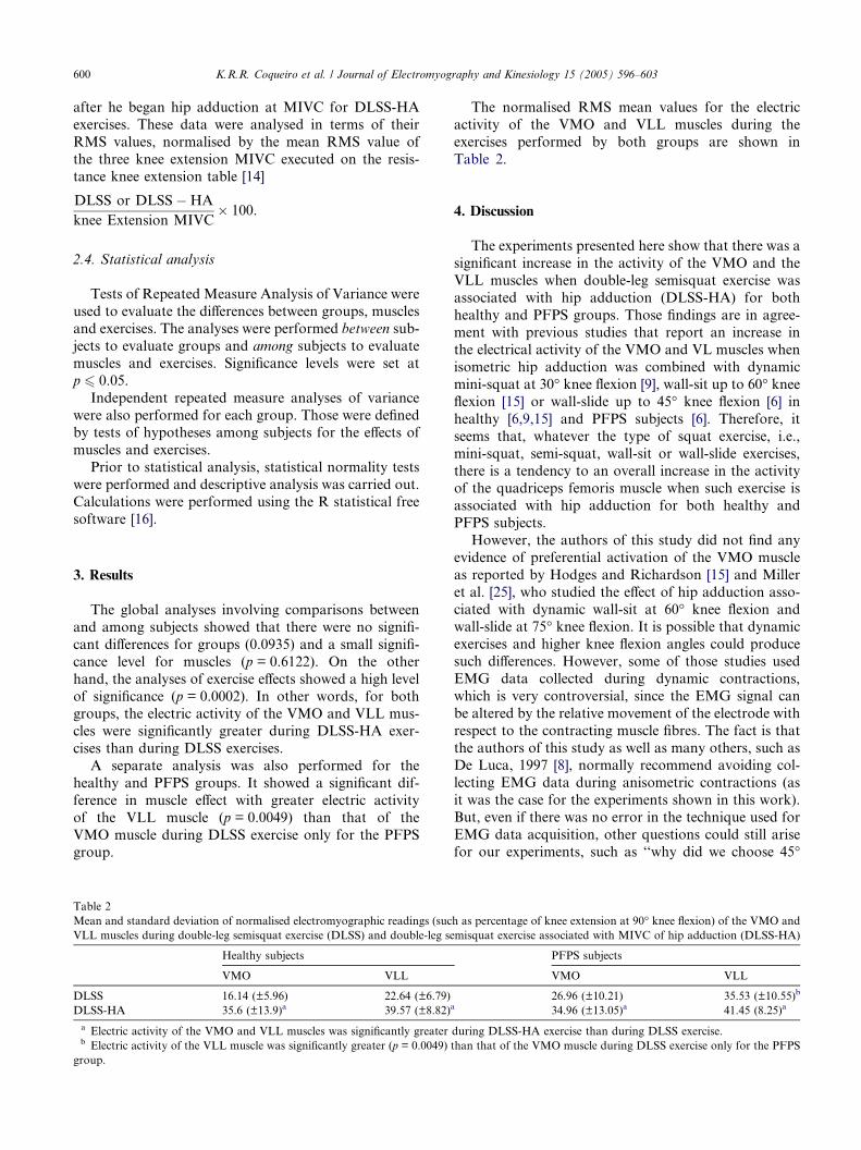
3. Results
The global analyses involving comparisons between
and among subjects showed that there were no signifi-
cant differences for groups (0.0935) and a small signifi-
cance level for muscles (p = 0.6122). On the other
hand, the analyses of exercise effects showed a high level
of significance (p = 0.0002). In other words, for bothgroups, the electric activity of the VMO and VLL mus-
cles were significantly greater during DLSS-HA exer-
cises than during DLSS exercises.
A separate analysis was also performed for the
healthy and PFPS groups. It showed a significant dif-
ference in muscle effect with greater electric activity
of the VLL muscle (p = 0.0049) than that of the
VMO muscle during DLSS exercise only for the PFPSgroup.
Table 2
Mean and standard deviation of normalised electromyographic readings (suc
VLL muscles during double-leg semisquat exercise (DLSS) and double-leg se
Healthy subjects
VMO VLL
DLSS 16.14 (±5.96) 22.64 (±6.79)
DLSS-HA 35.6 (±13.9)a 39.57 (±8.82)
a Electric activity of the VMO and VLL muscles was significantly greaterb Electric activity of the VLL muscle was significantly greater (p = 0.0049)
group.
The normalised RMS mean values for the electric
activity of the VMO and VLL muscles during the
exercises performed by both groups are shown in
Table 2.
4. Discussion
The experiments presented here show that there was a
significant increase in the activity of the VMO and the
VLL muscles when double-leg semisquat exercise was
associated with hip adduction (DLSS-HA) for both
healthy and PFPS groups. Those findings are in agree-
ment with previous studies that report an increase inthe electrical activity of the VMO and VL muscles when
isometric hip adduction was combined with dynamic
mini-squat at 30� knee flexion [9], wall-sit up to 60� kneeflexion [15] or wall-slide up to 45� knee flexion [6] in
healthy [6,9,15] and PFPS subjects [6]. Therefore, it
seems that, whatever the type of squat exercise, i.e.,
mini-squat, semi-squat, wall-sit or wall-slide exercises,
there is a tendency to an overall increase in the activityof the quadriceps femoris muscle when such exercise is
associated with hip adduction for both healthy and
PFPS subjects.
However, the authors of this study did not find any
evidence of preferential activation of the VMO muscle
as reported by Hodges and Richardson [15] and Miller
et al. [25], who studied the effect of hip adduction asso-
ciated with dynamic wall-sit at 60� knee flexion andwall-slide at 75� knee flexion. It is possible that dynamic
exercises and higher knee flexion angles could produce
such differences. However, some of those studies used
EMG data collected during dynamic contractions,
which is very controversial, since the EMG signal can
be altered by the relative movement of the electrode with
respect to the contracting muscle fibres. The fact is that
the authors of this study as well as many others, such asDe Luca, 1997 [8], normally recommend avoiding col-
lecting EMG data during anisometric contractions (as
it was the case for the experiments shown in this work).
But, even if there was no error in the technique used for
EMG data acquisition, other questions could still arise
for our experiments, such as ‘‘why did we choose 45�
h as percentage of knee extension at 90� knee flexion) of the VMO and
misquat exercise associated with MIVC of hip adduction (DLSS-HA)
PFPS subjects
VMO VLL
26.96 (±10.21) 35.53 (±10.55)b
a 34.96 (±13.05)a 41.45 (8.25)a
during DLSS-HA exercise than during DLSS exercise.
than that of the VMO muscle during DLSS exercise only for the PFPS
K.R.R. Coqueiro et al. / Journal of Electromyography and Kinesiology 15 (2005) 596–603 601
knee flexion? Why did we choose to normalise the data
using MIVC? . . .’’.Steinkamp et al. [34] suggested that CKC knee exten-
sion exercises in the functional range of motion should
be emphasised to strengthen the knee joint muscles be-
cause of its lower stress on the patellofemoral joint.Therefore, it seems that those exercises are good for
the PFPS treatment. However, what would optimum
knee flexion during squat exercises be? The answer could
be pointed by Escamilla [10], who stated �. . .because thepeak compressive forces generally occur near maximum
knee flexion, individuals with patellofemoral disorders
should avoid performing the squat at high knee flexion an-
gles�. But what would those ‘‘high knee flexion angles’’be? It certainly depends on each patient. In view of such
situation, the authors in this study elected 45� as a good
compromise for all the participant volunteers.
The normalisation of the raw data is an important
requirement if one intends to study and compare data
collected from different muscles and different individuals
in different situations. MIVC was elected as the normal-
isation unit based on the experience of many researchersworldwide and its use in similar studies concerning
PFPS and the muscles of the thigh [1,6,9,14,18,25,29].
However, we used data collected during MIVC exten-
sion of the knee at OKC to normalise CKC contrac-
tions. According to Schaub and Worrel [32], this is not
a problem either, since they have shown that muscle
recruitment patterns are very similar for OKC and
CKC exercises.Nevertheless, considering that there are differences
between the evaluated groups, especially regarding mus-
cle imbalance in PFPS subjects, widely reported in the
literature [1,7,9,20,21,23,24,29,33,35,36], we felt that a
separated statistical analysis comparing, for each group,
electric activity between the VMO and the VLL muscles
during DLSS and DLSS-HA exercises would be clini-
cally important for knee rehabilitation programs. Thisindependent analysis revealed that, the electrical activity
of the VLL muscle was significantly greater than that of
the VMO muscle during DLSS exercise for the PFPS
group and no differences were found during DLSS-HA
exercise. The greater electrical activity of the VLL mus-
cle when compared with that of the VMO muscle during
DLSQ exercise confirms the reports of imbalance
between the medial and lateral compartments of thequadriceps femoris muscle in PFPS subjects
[1,7,9,12,20,21,23,24,29,33,35,36]. However, this theory
should be carefully dealt with, since this work investi-
gated only EMG data and other tools, such as magnetic
resonance imaging (MRI) and computed tomography
(CT), should be added to explore the investigation of
muscle imbalance.
However, although there was no selective recruit-ment of the VMO muscle during DLSS-HA exercise,
considering that during DLSS exercise there was a sig-
nificant difference between the VLL and the VMO
muscles and that such difference had not been found
during DLSS-HA, we believe that DLSS-HA exercises
could promote a greater muscle balance between the
electric activity of the VMO and VLL muscles and
could benefit the treatment of PFPS patients. Thisgreater balance caused by the effect of hip adduction
on the activity of the VMO muscle could be explained
by the strong anatomical relation between the adductor
muscles and the VMO muscle. The action of adductor
muscles is essential to selectively strengthen the VMO
muscle. According to Hanten and Schulties [14],
‘‘. . .a strong VMO originating from weak adductors
would serve only to draw the adductor tendons toward
the patella . . .’’ and, on the other hand, ‘‘. . . strong hip
adductors give the VMO a stable origin from which to
contract . . .’’. Besides, another favourable factor for
the selective contraction of the VMO muscle during
DLSS-HA exercise refers to the length tension proper-
ties, as proposed by Beck and Wildermuth [2] who re-
ported that with the addition of hip adduction there is
a stretch to the VMO muscle, which would alter lengthtension properties, thus contributing to an enhanced
contraction force.
This study was designed with the primary goal of pro-
viding information regarding the effects of hip adduction
on the electric activity of the VMO and VLL muscles in
healthy and PFPS subjects. It has demonstrated that the
association of isometric hip adduction with isometric
semi-squat exercise produces a balanced activity of thequadriceps muscle group. Therefore, this exercise can
be indicated when overall quadriceps activity is desired
for clinical rehabilitation or muscle strengthening pro-
grams. Furthermore, because of the greater balance be-
tween the medial and lateral portions of the quadriceps
femoris muscle promoted by the association of semi-
squat exercise at 45� knee flexion and hip, it could also
be indicated for the conservatory treatment of PFPS.However, to confirm this statement, further EMG inves-
tigation would be necessary in order to compare the ef-
fects of muscle training programs based on DLSS-HA
exercises for both groups.
Acknowledgements
The authors thank Mrs. Lucia Batista for the illustra-
tions in this paper, CNPq-Brazil (Process No. 52419096-
8) and CAPES-Brazil for supporting this study.
References
[1] P.H. Andrade, D. Bevilaqua-Grosso, F. Berzin, I.A. Gil, V.
Monteiro-Pedro, Comparacao da atividade eletrica do musculo
vasto medial oblıquo e vasto lateral oblıquo em indivıduos com
602 K.R.R. Coqueiro et al. / Journal of Electromyography and Kinesiology 15 (2005) 596–603
disfuncao femoro-patelar, Revista de Fisioterapia da Universid-
ade de Sao Paulo 8 (2) (2001) 65–71.
[2] J.L. Beck, B.P. Wildermuth, The female athlete�s knee, Clinical
Sports Medicine 4 (1985) 345.
[3] D. Bevilaqua-Grossi, V. Monteiro-Pedro, G.C. Souza, Z. Silva,
F. Berzin, Contribution to the Anatomical Study of the Oblique
Portion of the Vastus Lateralis Muscle, Brazilian Journal of
Morphological Science 21 (1) (2004) 42–47.
[4] K. Bose, R. Kanagasuntherum, M. Osman, Vastus Medialis
Oblique: An Anatomical and Physiologic Study, Orthopedics 3
(1980) 880–883.
[5] M.J. Callagham, J.A. Oldham, The role of quadriceps exercise in
the treatment of patellofemoral pain syndrome, Sports Medicine
21 (1996) 384–391.
[6] K. Cerny, Vastus medialis oblique/ vastus lateralis muscle
activity ratios for selected exercises in persons with and without
patellofemoral pain syndrome, Physical Therapy 75 (1995) 672–
683.
[7] S.M. Cowan, K.B.L. Bennel, P. Hodges, K.M. Crossely, J.
McConnell, Delayed Onset of Electromyographic Activity of
Vastus Medialis Obliquos relative to Vastus Lateralis in subjects
with Patellofemoral Pain Syndrome, The Archives of Physical
Medicine Rehabilitation 82 (2001) 183–189.
[8] C.J. De Luca, The use of Surface Electromyography in Biome-
chanics, Journal of Applied Biomechanics 13 (1997) 135–163.
[9] J.E. Earl, B.L. Schimitz, B.L. Arnold, Activation of the VMO and
VL during dynamic mini-squat exercises with and without
isometric hip adduction, Journal of Electromyography and
Kinesiology 11 (1992) 381–386.
[10] R.F. Escamilla, Knee biomechanics of the dynamic squat
exercise, Medicine and Science in Sports and Exercise 33
(2001) 127–141.
[11] G.K. Fitzgerald, Open versus closed kinect chain exercise: issues
in rehabilitation after anterior cruciate ligament reconstructive
surgery, Physical Therapy 77 (1997) 1747–1754.
[12] T.A. Fox, Dysplasia of the quadriceps mechanism: hypoplasia of
the vastus medialis muscle as related to the hypermobile patella
syndrome, Surgical Clinics of North America 55 (1975) 199–206.
[13] R. Gresalmer, J. Klein, The biomechanics of the patellofemoral
joint, Journal of Orthopaedic and Sports Physical Therapy 28
(1998) 286–297.
[14] W.P. Hanten, S. Schulthies, Exercise effect on electromyographic
activity of the vastus medialis oblique and vastus lateralis muscles,
Physical Therapy 7 (1990) 561–565.
[15] P.W. Hodges, C.A. Richardson, The influence of isometric hip
adduction on quadriceps femoris activity, Scandinavian Journal of
Rehabilitation Medicine 25 (1993) 57–62.
[16] R. Ihaka, R.A. Gentleman, Language for Data Analysis and
Graphics, Journal of Computational and Graphical Statistics 5
(1996) 299–314.
[17] J. Insall, Current Concepts Review Patellar Pain, Journal of Bone
and Joint Surgery 64-A (1982) 147–152.
[18] J.A. Isear, J.C. Erickson, T.W. Worrel, EMG analysis of lower
extremity muscle recruitment patterns during an unloaded squat,
Medicine and Science in Sports and Exercise 29 (4) (1997) 532–
539.
[19] G.M. Karst, P.D. Jewett, Eletromyography Analysis of Exercises
Proposed for Differential Activation of Medial and Lateral
Quadriceps Femoris Muscle Components, Physical Therapy 73
(1993) 289–299.
[20] P.L. Lam, G.Y.F. Ng, Activation of the Quadriceps Muscle
during Semisquating with Different Hip and Knee Positions in
Patients with Anterior Knee Pain, Amerian Journal of Physical
Medicine and Rehabilitation 80 (2001) 804–808.
[21] J. Laprade, E. Culhan, B. Brouwer, Comparison of five
isometric exercises in the recrutment of the vastus medialis
oblique in persons with and without patellofemoral pain
syndrome, Journal of Orthopaedic and Sport Physical Therapy
27 (1998) 197–204.
[22] F.J. Lieb, J. Perry, Quadriceps functions: an anatomical and
mechanical study using amputed limbs, Journal of Bone and Joint
Surgery 53A (1968) 1535–1548.
[23] J. Mc Connell, The management of chondromalacia patellae: a
long-term solution, Australian Journal of Physiotherapy 32 (1986)
215–223.
[24] J. McConnell, J. Cook, Anterior Knee Pain. In: P. Burker, K.
Kham, Clinical Sports Medicine. 2000 (Chapter 24). Available
from:Æwww.clinicalsportsmedicine.comæ.[25] J.P. Miller, D. Sedory, R. Croce, Vastus Medialis Obliquus and
Vastus Lateralis Activity in Patients with and Without Patellofe-
moral Pain Syndrome, Journal of Sports Rehabilitation 6 (1997)
1–10.
[26] V. Monteiro-Pedro, M. Vitti, F. Berzin, D. Bevilaqua-Grosso,
Electromyographic activity of vastus medialis oblique muscle in
step-up and step-down exercises, Brazilian Journal of Morpho-
logical Science 14 (1997) 19–23.
[27] V. Monteiro-Pedro, M. Viiti, F. Berzin, D. Bevilaqua-Grosso,
The effect of free isotonic contraction exercises of the hip
adduction on vastus medialis oblique muscle: an electromyo-
graphic study, Electromyography and. Clinical Neurophysiology
39 (1999) 435–440.
[28] J.C. Ninos, J.J. Irrgang, R. Burdett, J.R. Weiss, Electromyo-
graphic analysis of the squat performed in self-selected lower
extremity neutral rotation and 30� of lower extremity turn-out
from self-selected neutral position, Journal of Orthopaedic and
Sport Physical Therapy 25 (1997) 307–315.
[29] C.V. Nunes, V. Monteiro-Pedro, Efeito do Exercıcio Isometrico
de Extensao do Joelho associado a Aducao Isometrica do
Quadril na Atividade Eletrica dos Musculos Vasto Medial
Oblıquo e Vasto Lateral Oblıquo em Indivıduos com Disfuncao
Femoro-patelar, Revista Brasileira de Fisioterapia 7 (2) (2003)
145–150.
[30] L. Reynolds, T.A. Levin, J.M. Medeiros, N.S. Adler, A.
Hallun, EMG activity of the Vastus Medialis oblique and the
vastus lateralis in their role in patellar alignment, American
Journal of Physical Medicine and Rehabilitation 62 (1983) 61–
70.
[31] M.T. Ruffin, R.B. Kinninghan, Anterior knee pain: the challenge
of patellofemoral syndrome, American Family Physician 43 (1993)
185–194.
[32] P.A. Schaub, T.W. Worrel, EMG activity of six muscles and
VMO:VL ratio determination during a maximal squat exercise,
Journal of Sports Rehabilitation 4 (1995) 203–209.
[33] H.A. Stiene, T. Brosky, M.F. Reinking, J. Nyland, M.B. Mason,
A comparison of closed kinetic chain and isokinetic joint
isolation exercise in patients with patellofemoral dysfunction,
Journal of Orthopaedic and Sports Physical Therapy 24 (1991)
136–141.
[34] L.A. Steinkamp, M.F. Dillingham, M.D. Markel, J.A. Hill, K.R.
Kaufman, Biomechanical considerations in patellofemoral joint
rehabilitation, American Journal of Sports Medicine 21 (1993)
438–444.
[35] R. Thomee, J. Augustsson, J. Karlsson, Patellofemoral Pain
Syndrome: A review of current issues, Sports Medicine 28 (4)
(1999) 245–262.
[36] E. Witvrouw, C. Sneyers, R. Lysens, J. Victor, J. Bellemans,
Reflex response times of vastus medialis oblique and vastus
lateralis in normal subjects and in subjects with patellofemoral
pain syndrome, Journal of Orthopaedic and Sports Physical
Therapy 24 (1996) 160–165.
[37] D. Zakaria, K.L. Harburn, J.F. Kramer, Preferential activation
of the Vastus Medialis oblique, Vastus lateralis and hip adductor
muscles during isometric exercises in females, Journal of Ortho-
paedic Sports Physical Therapy 26 (1) (1997) 23–28.
K.R.R. Coqueiro et al. / Journal of Electromyography and Kinesiology 15 (2005) 596–603 603
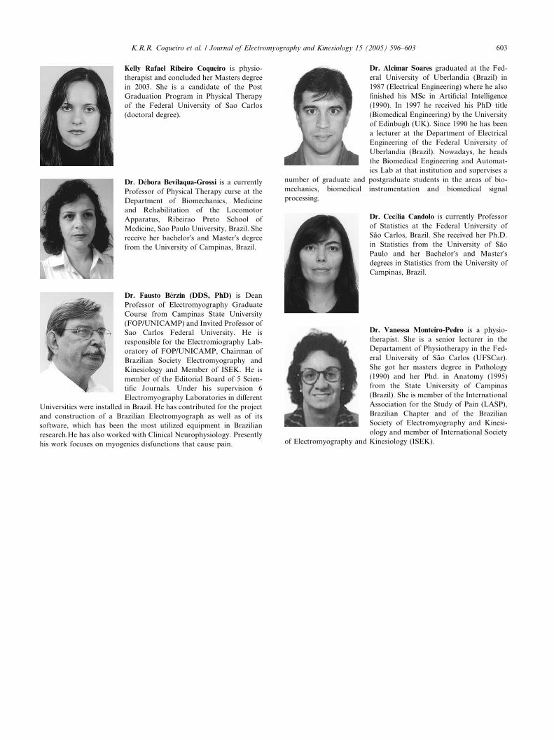
Kelly Rafael Ribeiro Coqueiro is physio-
therapist and concluded her Masters degree
in 2003. She is a candidate of the Post
Graduation Program in Physical Therapy
of the Federal University of Sao Carlos
(doctoral degree).
Dr. Debora Bevilaqua-Grossi is a currently
Professor of Physical Therapy curse at the
Department of Biomechanics, Medicine
and Rehabilitation of the Locomotor
Apparatus, Ribeirao Preto School of
Medicine, Sao Paulo University, Brazil. She
receive her bachelor�s and Master�s degree
from the University of Campinas, Brazil.
Dr. Fausto Berzin (DDS, PhD) is Dean
Professor of Electromyography Graduate
Course from Campinas State University
(FOP/UNICAMP) and Invited Professor of
Sao Carlos Federal University. He is
responsible for the Electromiography Lab-
oratory of FOP/UNICAMP, Chairman of
Brazilian Society Electromyography and
Kinesiology and Member of ISEK. He is
member of the Editorial Board of 5 Scien-
tific Journals. Under his supervision 6
Electromyography Laboratories in different
Universities were installed in Brazil. He has contributed for the project
and construction of a Brazilian Electromyograph as well as of its
software, which has been the most utilized equipment in Brazilian
research.He has also worked with Clinical Neurophysiology. Presently
his work focuses on myogenics disfunctions that cause pain.
Dr. Alcimar Soares graduated at the Fed-
eral University of Uberlandia (Brazil) in
1987 (Electrical Engineering) where he also
finished his MSc in Artificial Intelligence
(1990). In 1997 he received his PhD title
(Biomedical Engineering) by the University
of Edinbugh (UK). Since 1990 he has been
a lecturer at the Department of Electrical
Engineering of the Federal University of
Uberlandia (Brazil). Nowadays, he heads
the Biomedical Engineering and Automat-
ics Lab at that institution and supervises a
number of graduate and postgraduate students in the areas of bio-
mechanics, biomedical instrumentation and biomedical signal
processing.
Dr. Cecılia Candolo is currently Professor
of Statistics at the Federal University of
Sao Carlos, Brazil. She received her Ph.D.
in Statistics from the University of Sao
Paulo and her Bachelor�s and Master�sdegrees in Statistics from the University of
Campinas, Brazil.
Dr. Vanessa Monteiro-Pedro is a physio-
therapist. She is a senior lecturer in the
Departament of Physiotherapy in the Fed-
eral University of Sao Carlos (UFSCar).
She got her masters degree in Pathology
(1990) and her Phd. in Anatomy (1995)
from the State University of Campinas
(Brazil). She is member of the International
Association for the Study of Pain (LASP),
Brazilian Chapter and of the Brazilian
Society of Electromyography and Kinesi-
ology and member of International Society
of Electromyography and Kinesiology (ISEK).