Dynamics of chemosensitivity and chromosomal instability in recurrent glioblastoma
ORIGINAL PAPER
L. Prado á F. Lombo á A. F. BranÄ a á C. Me ndezJ. Rohr á J. A. Salas
Analysis of two chromosomal regions adjacent to genesfor a type II polyketide synthase involved in the biosynthesisof the antitumor polyketide mithramycin in Streptomyces argillaceus
Received: 13 August 1998 /Accepted: 30 October 1998
Abstract Mithramycin is an aromatic antitumourpolyketide synthesized by Streptomyces argillaceus. Twochromosomal regions located upstream and downstreamof the locus for the mithramycin type II polyketidesynthase were cloned and sequenced. Analysis of thesequence revealed the presence of eight genes encodingthree oxygenases (mtmOI, mtmOII and mtmOIII), threereductases (mtmTI, mtmTII and mtmTIII), a cyclase(mtmY) and an acyl CoA ligase (mtmL). The three ox-ygenase genes were each inactivated by gene replace-ment. Inactivation of one of them (mtmOII) generated anon-producing mutant, while inactivation of the othertwo (mtmOI and mtmOIII) did not a�ect the biosyn-thesis of mithramycin. The mtmOII gene may code foran oxygenase responsible for the introduction of oxygenatoms at early steps in the biosynthesis of mithramycinleading to 4-demethylpremithramycinone. One of thereductases may be responsible for reductive cleavage ofan intermediate from an enzyme and another for thereduction of a keto group in the side-chain of the mi-thramycin aglycon moiety. A hypothetical biosyntheticpathway showing in particular the involvement of oxy-genase MtmOII and of various other gene products inmithramycin biosynthesis is proposed.
Key words Oxygenases á Ketoreductases á Acyl CoAligase á Loading enzyme á Polyketides
Introduction
Aromatic polyketides (PKs) constitute a large family ofcompounds, many of which have clinical applications.Their chemical diversity is based on the use of di�erentstarter and extender units, the programming of thepolyketide synthase (PKS) catalysing the assembly pro-cess, and various modi®cations (oxygenations, meth-ylations, glycosylations, etc) occurring after chain as-sembly (for reviews see Hopwood and Sherman 1990;Katz and Donadio 1993; Hutchinson and Fujii 1995;Hopwood 1997). The biosynthesis of the PK moiety ofall aromatic PKs so far characterized is catalysed by atype II PKS, in a series of reactions involving the con-densation of a number of acetates, and speci®c reduc-tion, cyclisation and dehydration steps. Mithramycin(also termed aureolic acid) is an aromatic PK showingantibacterial and antitumor activity, which has clinicalapplications in the treatment of some tumours (Remers1979). Its chromophoric PK ring is derived from thecondensation of ten acetates (Montanari and Rosazza1990; Rohr et al. 1998b), with further incorporation of adisaccharide and a trisaccharide consisting of 2,6-did-eoxyhexoses. A type II PKS from Streptomycesargillaceus (a mithramycin producer) which is involvedin the biosynthesis of the mithramycin aglycon has beencharacterized (Lombo et al. 1996), and it has beendemonstrated that the biosynthesis of the PK moiety isdependent on the activity of a unique PKS (Blanco et al.1996). We have also reported (Lombo et al. 1997) theidenti®cation of two genes involved in early steps of 2,6-dideoxyhexose biosynthesis. Inactivation of these genesresults in the accumulation of premithramycinone and4-demethylpremithramycinone, tetracyclic intermediatesin the biosynthesis of mithramycin (Lombo et al. 1997;Rohr et al. 1998a). In addition, we have recently re-ported the identi®cation of two glycosyltransferases in-volved in the formation and transfer of the disaccharide(Ferna ndez et al. 1998). Here we report the cloning andsequencing of eight additional genes of the mithramycin
Mol Gen Genet (1999) 261: 216±225 Ó Springer-Verlag 1999
Communicated by W. Goebel
L. Prado á F. Lombo á A. F. BranÄ a á C. Me ndezJ. A. Salas (&)Departamento de Biologõ a Funcional e Instituto Universitario deBiotecnologõ a de Asturias (I.U.B.A-C.S.I.C)Universidad de Oviedo, 33006 Oviedo, Spaine-mail: [email protected]./Fax: +34-985-103652
J. RohrDepartment of Pharmaceutical SciencesMedical University of South Carolina171 Ashley Avenue, Charleston, SC 29425-2303, USA

gene cluster in S. argillaceus, which encode three oxy-genases, three reductases, a cyclase and an acyl CoAligase. We have also insertionally inactivated each of theoxygenase genes and describe the e�ects of these muta-tions on mithramycin biosynthesis.
Materials and methods
Microorganisms, culture conditions and vectors
Streptomyces argillaceus ATCC 12596, a mithramycin producer,was used as the source of chromosomal DNA. For sporulation onsolid medium, it was grown at 30°C on plates containing A me-dium (Ferna ndez et al. 1998). For protoplast transformation,S. argillaceus was grown in YEME medium containing 17% su-crose. For growth in liquid medium, the organisms were inoculatedinto TSB medium (trypticase soya broth, Oxoid). Escherichia coliTG1 recO1504::Tn5 (Kolodner et al. 1985) was used as the host forsubcloning. When plasmid-containing clones were grown, the me-dium was supplemented with the appropiate antibiotics: 100 lg/mlampicillin, 25 lg/ml apramycin, 50 lg/ml thiostrepton or 20 lg/mltobramycin. The phage vectors M13mp18 and M13mp19 and theplasmid pUC18 were used for sequencing and subcloning, respec-tively. The pUC-derived vectors pIJ2921 and pIJ2925 (Janssen andBibb 1993) were also used as cloning vectors. Plasmid pBSKT is apBluescript derivative containing a thiostrepton resistance cassette(F. Lombo , unpublished). pEM4 (Quiro s et al. 1998) is a bifunc-tional plasmid (Streptomyces-E. coli) containing the erythromycinresistance promoter (ermE*p). pUO9090 is a pUK21 derivativecontaining an apramycin resistance cassette (M. C. Martin, un-published).
DNA manipulations
Plasmid DNA preparations, restriction enzyme digestions, alkalinephosphatase treatments, DNA ligations and other DNA manipu-lations were performed according to standard techniques for E. coli(Sambrook et al. 1989) and Streptomyces (Hopwood et al. 1985).Preparation of S. argillaceus protoplasts, transformation and se-lection of transformants were carried out as described by Hopwoodet al. (1985).
DNA sequencing
Sequencing was performed on single-stranded templates derivedfrom various clones in either M13mp18 or M13mp19, using thedideoxynucleotide chain-termination method (Sanger et al. 1977)with [a-35S]dATP (1200 Ci/mmol; Amersham) and modi®ed T7DNA polymerase (Sequenase version 2.0, US Biochemicals). Insome clones, and in order to avoid band compression artifacts,
7-deaza-dGTP was used instead of dGTP (Mizusawa et al. 1986).Single-stranded DNA was prepared by polyethylene glycol pre-cipitation as described (Sambrook et al. 1989). Both DNA strandswere sequenced with the primers supplied in the Sequenase kit orwith internal (17mer) primers. Computer-aided database searchesand sequence analyses were carried out using the University ofWisconsin Genetics Computer Group programs package(UWGCG; Devereux et al. 1984) and the BLAST program (Alt-schul et al. 1990).
Insertional inactivation
To generate knockout mutants for the three oxygenase genes, thefollowing constructions were prepared.
For mtmOI inactivation, a 4.5-kb BamHI fragment (sites 16±20in Fig. 1) was subcloned into the BamHI site of pBSKT. Then anapramycin resistance cassette was introduced into the unique StuIsite in the BamHI fragment (site 18 in Fig. 1) as a 1.5-kb SmaI-EcoRV fragment from pUO9090, generating pLPO1. In this con-struction the apramycin resistance cassette is oriented in the samedirection as the transcriptional unit.
To inactivate mtmOII, a 5-kb EcoRI-BamHI fragment (sites 9±14 in Fig. 1) was subcloned into the same restriction sites in pBSKT(pL7aEB). In parallel, a 5.5-kb BamHI-EcoRI fragment (sites 3±9in Fig. 1) was subcloned into the same sites in pUC18 (pFL7a-E).In this construction there is a unique XhoI site (site 7 in Fig. 1)located at the 5¢ end of mtmOII. pLP7a-E was then digested withXhoI, blunted with Klenow polymerase and religated. The insert inthis construction was then rescued as a 5.5-kb HindIII-EcoRIfragment (using the HindIII site of the pUC polylinker) and sub-cloned into the same sites of pLP7aEB, generating the ®nal con-struction pLPO2. In this construct a frameshift mutation has beenintroduced at the XhoI site in mtmOII.
For inactivation of mtmOIII, a 9-kb BglII fragment (sites 4±13in Fig. 1) was subcloned into the BglII site of pIJ2925. The apra-mycin resistance cassette was subcloned, as a 1.5-kb EcoRV-EcoRIfragment from pUO9090, into the unique StuI and EcoRI siteslocated within the mtmOIII gene (sites 8±9 in Fig. 1). Then thisconstruct was rescued as a BglII fragment and subcloned into theBamHI site of pBSKT. This construction was designated aspLPDO3 and the apramycin resistance cassette is oriented in thesame direction as the transcriptional unit.
The three constructions (pLPO1, pLPO2 and pLPDO3) werethen used to transform S. argillaceus protoplasts, and transform-ants were selected for resistance to apramycin (25 lg/ml) in the caseof pLPO1 and pLPDO3, and to thiostrepton (50 lg/ml) in the caseof pLPO2. In the case of the mtmOI and mtmOIII gene replace-ments, all the transformants from this primary screen were foundto produce mithramycin, possibly owing to the integration of theplasmid into the chromosome by a single crossover. To force theoccurrence of a second crossover, one transformant from eachtransformation was grown for 72 h in TSB medium in the absenceof antibiotics, and then plated onto apramycin-containing agarplates. Surviving colonies were then simultaneously tested for
Fig. 1 Schematic representa-tion of the regions of cosAR7sequenced, which ¯ank thegenes for the mithramycin PKS.The di�erent genes identi®edand their locations with respectto the PKS genes (mtmQXPKSgenes) and sugar biosyntheticgenes (mtmDE genes) are indi-cated. B, BamHI; E, EcoRI; G,BglII; P, PstI; S, SacI; T, StuI;X, XhoI
217

thiostrepton and apramycin resistance. Several apramycin-resis-tant, thiostrepton-sensitive colonies were selected for further studyand considered to result from a double crossover. In the case of themtmOII gene replacement, one thiostrepton-resistant colony wasselected and grown for 72 h in TSB medium in the absence ofantibiotic. Then, di�erent dilutions were plated onto antibiotic-freeagar plates and colonies were replica-plated onto thiostrepton-containing plates. Colonies that could not grow on thiostreptonplates were considered to be the result of a double crossover andselected for further study.
Bioassay of mithramycin
The antibacterial activity of mithramycin was determined by bio-assay againstMicrococcus luteus ATCC 10240 as described (Vilcheset al. 1990).
Extraction and analysis of mithramycinand biosynthetic intermediates
For the analysis of mithramycin and its intermediates, the di�erentstrains (wild-type and mutant strains) were grown on plates con-taining R5 solid medium (Hopwood et al. 1985) at 30°C for 6 days.The agar from one quadrant of a plate was extracted with 5 ml ofethyl acetate and, after phase separation, the organic phase wasevaporated under vacuum and the residue suspended in a smallvolume of methanol. The samples were then analysed by high-performance liquid chromatography (HPLC) using a lBondapakC18 column and an isocratic gradient composed of 30% acetonitrileand 70% 50 mM phosphate bu�er (pH 6.8) at a 2 ml/min ¯ow rate;detection was carried out at 280 nm.
Results
Sequencing of two regions ¯ankingthe mithramycin PKS gene cluster
A 4.4-kb DNA region located to the left of the mi-thramycin PKS gene cluster (immediately downstreamof the aromatase gene mtmQ) and a 4-kb region from theright ¯ank of the PKS cluster, downstream of the mtmSgene (encoding an acyl carrier protein), were sequenced
(Fig. 1). The nucleotide sequences (EMBL AccessionNo. AJ007932) from both regions were analysed forcoding regions (orfs) using the CODONPREFERENCEprogram (Devereux et al. 1984). Four orfs (designated asmtmL, mtmOIII, mtmOII and mtmTII) were detected inthe left-hand region, transcribed in the same directionas, and away from, the aromatase gene; four orfs werealso found in the right-hand region (designated mtmTI,mtmOI, mtmY and mtmTIII) downstream of the mtmSgene (it should be mentioned that most of the sequenceof the mtmTI gene has already been published by Lo-mbo et al. 1996). Three of these orfs (mtmTI, mtmOI andmtmY) are transcribed in the same direction as, andaway from, mtmS and the fourth one, mtmTIII, istranscribed convergently to the other three. All the genesshowed the typical codon usage for Streptomyces genes,with high overall G+C content and a very high GCcontent in the third codon position.
Analysis of the deduced products
Four genes (mtmL, mtmOIII, mtmOII and mtmTII) wereidenti®ed to the left of the mithramycin PKS genecluster.
The ®rst gene (mtmL), located immediately down-stream of the mtmQ gene, overlaps with the last 8 bp ofthe 3¢ end of the preceding gene (mtmQ). The mtmL startcodon is preceded by a base sequence (AGGAG) thatshows a degree of complementarity to a region close tothe 3¢ end of the 16S rRNA of S. lividans (Bibb andCohen 1982), which could represent a ribosomal bindingsite. The MtmL protein signi®cantly resembles variousacyl CoA ligases involved in the activation of organicacids and long-chain fatty acids as CoA derivatives(Table 1). It also has a sequence (FTSGTTGPPK) thatcorresponds to a conserved AMP-binding motif presentin a number of prokaryotic and eukaryotic enzymeswhich act via an ATP-dependent covalent binding ofAMP to their substrates.
Table 1 Comparison of the products of the characterised S. argillaceus genes with their nearest homologues
Protein Number ofamino acids
Predicted Mr Homologues (% identity) Reference
MtmOI 437 46,629 JadORF6 (31.4) Yang et al. 1996UrdE (26.6) Decker and Haag 1995
MtmOII 532 56,888 SchC (31.6) Blanco et al. 1993UrdE (26.6) Decker and Haag 1995
MtmOIII 300 30,761 DpsORF8 (38.3) Grimm et al. 1994DauORFE (34) Ye et al. 1994
MtmTI 255 25,921 ChcA (38.9) Wang et al. 1996DnrE (38) Grimm et al. 1994
MtmTII 254 26,537 Ard2ORF4 (38.5) Barrasa et al. 1997DnrE (36.9) Grimm et al. 1994
MtmTIII 250 26,196 DnrE (54.5) Grimm et al. 1994DauE (54.1) Dickens et al. 1996
MtmY 258 28,233 DpsY (68.4) Lomovskaya et al. 1998ORF1 (65.7) KruÈ gel et al. 1993
MtmL 515 54,138 4CL.1 (32.8) Zhao et al. 1990FadD (31) Fulda et al. 1994
218
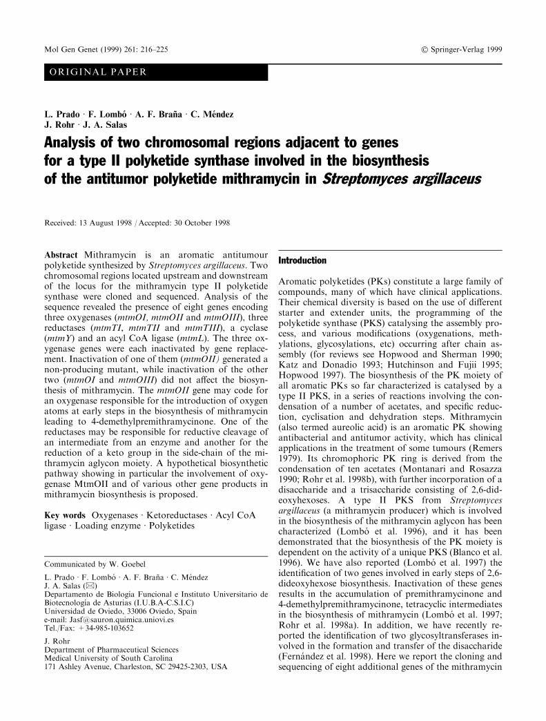
The mtmOIII gene is separated by 43 bp from themtmL gene. The MtmOIII protein showed the highestsimilarities to semiquinone monoxygenases in the data-base (Table 1, Fig. 2A). These enzymes are responsiblefor the introduction of molecular oxygen into one of theanthracycline rings, leading to the formation of a qui-none intermediate characteristic of this group. They arealso similar to TcmH (Shen and Hutchinson 1993) andto ActVaORF6 (Caballero et al. 1991), which are in-volved in tetracenomycin and actinorhodin biosynthesis,respectively. TcmH (Shen and Hutchinson 1993) andActVaORF6 (Kendrew et al. 1997) have both beenshown to be monooxygenases that appear to requiremolecular oxygen but no cofactors or metal ions foractivity. These enzymes contain a conserved histidineresidue that has been found to be important for catalyticactivity in ActVaORF6 (Kendrew et al. 1997). Interest-ingly, MtmOIII lacks this histidine residue.
The starting codon of the mtmOII gene overlaps thepreceding mtmOIII gene by 8 bp. The MtmOII proteinresembles various hydroxylases and monooxygenases,some of which are involved in antibiotic biosynthesis(Table 1). Two motifs are present in MtmOII (Fig. 2B,C) which are characteristic of ¯avin-type hydroxylases: aregion very close to the N-terminus ± the so-called babfold, which is involved in binding of the ADP moiety ofFAD (Wierenga et al. 1986), and a second which is be-lieved to be involved in the binding of the ribityl chain ofFAD (Russell and Model 1988; Eggink et al. 1990).
The mtmTII gene is the last gene of the operonidenti®ed to the left of the PKS genes and it is followedby two inverted repeat sequences, which can form astem-loop structure that could act as a transcriptionalterminator. It is separated from the preceding mtmOIIgene by 16 bp. Comparison with proteins in the data-bases showed that MtmTII resembles PK ketoreductasesinvolved in the biosynthesis of various antibiotics(Table 1).
Downstream of the mithramycin PKS genes, fourgenes (mtmTI, mtmOI, mtmY and mtmTIII) were iden-ti®ed.
Fig. 2A±C Comparison of the amino acid sequences of di�erenthydroxylases and oxygenases. A Motif present in many monoxygen-ases, including MtmOIII. DauAORFE acts in the daunomycinpathway in Streptomyces C5 sp. (Ye et al. 1994), DspORF8 in thedoxorubicin pathway in S. peucetius (Grimm et al. 1994); SnoB in thenogalamycin pathway in S. nogalater (Ylihonko et al. 1996),ActVAORF6 in the actinorhodin pathway in S. coelicolor (Caballeroet al. 1991), TcmH in the tetracenomycin C pathway in S. glaucescens(Shen and Hutchinson 1993), and JadORF7 in the jadomycinpathway in S. venezuelae (Yang et al. 1996). B, C Motifs present inmany FAD- and NADPH-dependent enzymes, and in MtmOI andMtmOII. SchC is a hydroxylase from S. halstedii (Blanco et al. 1993),TcmG is tetracenomycin hydroxylase from S. glaucescens (Decker etal. 1993), RdmE is rhodomycin hydroxylase from S. purpurascens(Niemi andMaÈ ntsaÈ laÈ 1995), JadORF6 is jadomycin hydroxylase fromS. venezuelae (Yang et al. 1996), UrdE is urdamycin hydroxylase fromS. fradiae (Decker and Haag 1995), and PcpB is pentachlorophenol-4-monooxygenase of Flavobacterium sp. (Orser et al. 1993)
219

Most of the mtmTI gene was already sequenced(Lombo et al. 1996) and its gene product also resemblesPK ketoreductases (Table 1). The mtmOI gene starts4 bp downstream of the mtmTI gene. The MtmOI pro-tein also resembles hydroxylases and oxygenases likeMtmOII (Table 1; Fig. 2B, C). mtmY is the last gene ofthis transcriptional unit and is followed by two invertedrepeat sequences able to form secondary structures thatcould act as transcriptional terminators. The startingcodon of the mtmY gene is separated by 3 bp from thestop codon of the mtmOI gene. The MtmY proteinshows similarity to two proteins involved in anthracy-cline biosynthesis (Table 1). Recently, it has been pro-posed that one of them, DpsY, together with DpsH, isinvolved in cyclisation of the second and third ringsduring the biosynthesis of daunorubicin in S. peucetius(Lomovskaya et al. 1998). Based on its similarity toDpsY we propose a role for MtmY as a cyclase in thebiosynthesis of mithramycin. The mtmTIII gene is thelast gene in this region and is transcribed convergently tomtmY. It also resembles PK ketoreductases, its closesthomologues being aklaviketone ketoreductases respon-sible for the reduction of aklaviketone to aklavinone(Table 1).
Insertional inactivation of the three oxygenase genes
To elucidate the role of the oxygenases encoded by thegenes mtmOI, mtmOII and mtmOIII in mithramycinbiosynthesis, the three genes were independently inacti-vated by gene replacement.
The mtmOI gene was inactivated by the insertion ofan apramycin resistance cassette into the unique StuI sitewithin the gene. Gene replacement was veri®ed bySouthern analysis using as probe the 4.5-kb BamHIfragment (sites 16±20 in Fig. 1): two BamHI bands (2.5and 3.4 kb) were found in the mutant M7O1 in contrastto the 4.5-kb BamHI band of the wild type strain(Fig. 3A, B). The production of mithramycin or inter-mediates by the M7O1 mutant was analysed by HPLC.A peak was found (Fig. 3C) with exactly the same mo-bility and absorption spectrum as mithramycin; its bio-logical activity was veri®ed by bioassay againstM. luteus.The material in this peak was isolated and analysed byNMR and mass spectroscopy, and found to be indis-tinguishable from mithramycin. Therefore, we concludethat mtmOI is not essential for mithramycin biosynthe-sis.
An mtmOII mutant was generated by replacing thewild-type mtmOII gene by one in which a frameshiftmutation had been introduced at the unique XhoI site.
Fig. 3A±C Insertional inactivation of mtmOI. A Schematic represen-tation of the double cross-over replacement event in the chromosomeof the wild-type S. argillaceus strain, used to construct mutant M7O1.The asterisks indicate the ends of the probe used for Southern analysis(a 4.5-kb BamHI fragment). The thin lines represent the BamHIfragments that hybridise with the probe. B, BamHI; EV, EcoRV; M,SmaI; P, PstI; T, StuI. tsr, thiostrepton resistance gene; aac,aparamycin resistance gene. B Southern analysis using the 4.5 kbBamHI fragment. Chromosomal DNA from the wild type and theM7O1 strains were digested with BamHI. C HPLC analysis of theculture supernatant from strain M7O1. The arrow indicates theposition of mithramycin
220

The occurrence of the genetic replacement was veri®edby Southern analysis, using as probe the 10.5-kb BamHIfragment (sites 3±14 in Fig. 1). The wild-type strainshowed three hybridising XhoI fragments (14, 6.1 and3 kb) while in the M7O2 mutant the 3 kb band wasabsent, as expected for the loss of the internal XhoI sitewithin mtmOII, and the 14-kb band was converted into a17-kb band (Fig. 4A, B). Growth of the M7O2 mutantresulted in formation of a black pigment in the culture,thus complicating the puri®cation and analysis of theaccumulated products. HPLC analysis of ethyl acetateextracts of young cultures of mutant M7O2 showed(Fig. 4C) the absence of mithramycin and the appear-ance of two major new peaks. One of them (peak a) hadshorter retention time than mithramycin but the char-acteristic mobility and absorption spectrum of SEK15.This is a shunt product formed by spontaneous cycli-sations of decaketide chains during the biosynthesis ofvarious polyketides (McDaniel et al. 1994). The other(peak b) did not correspond to any known mithramycinintermediate, and did not present an absorption spec-trum that would suggest it to be a premithramycinone-like intermediate (Fig. 4C). All attempts to purify thiscompound have failed so far on account of its instabil-ity. Mutant M7O2 could be complemented by plasmidpLPO2C (mtmOII cloned in pEM4), indicating that inthe M7O2 mutant only mtmOII (and not other down-stream genes) was a�ected. The tetracyclic intermediate4-demethyl-premithramycinone (Rohr et al. 1998a) isthe earliest mithramycin intermediate so far isolated in astable form. We therefore assume that the MtmOII ox-
ygenase must act before formation of 4-demethyl-pre-mithramycinone.
The mtmOIII gene was inactivated by deleting aninternal fragment of the gene and inserting an antibioticresistance cassette in the same transcriptional orienta-tion, thus generating M7DO3. Southern analysis usingthe 9-kb BglII fragment (sites 4±13 in Fig. 1) con®rmedthat the gene replacement had taken place (Fig. 5A, B).The wild-type strain only showed a 10.4-kb hybridisingBamHI band while in the M7DO3 mutant this wasreplaced by two BamHI bands (4.7 kb and 6.7 kb).HPLC analysis of the culture supernatant of mutantM7DO3 showed a peak with a retention time identical tothat of mithramycin (Fig. 5C), which also had thecharacteristic absorption spectrum of mithramycin(Fig. 5C) and was biologically active against M. luteus.Therefore, it can be concluded that the MtmOIII oxy-genase also is not essential for the biosynthesis of mi-thramycin.
Fig. 4A±C Insertional inactivation of mtmOII. A Schematic repre-sentation of the double crossover replacement event in the chromo-some of the wild-type S. argillaceus strain, used to construct mutantM7O2. The asterisks indicate the ends of the probe used for Southernanalysis (a 10-kb BamHI fragment). The thin lines represent the XhoIfragments that hybridise with the probe. B, BamHI; H, HindIII; X,XhoI. tsr, thiostrepton resistance gene. B Southern analysis using the10-kb BamHI fragment. Chromosomal DNA from the wild type andthe M7O2 strain were digested with XhoI. C HPLC analysis of theculture supernatant of strain M7O2 and absorption spectra ofcompounds present in peaks a and b
221

Discussion
The mithramycin gene cluster of S. argillaceus has beencloned and several genes have been characterized(Blanco et al. 1996; Ferna ndez et al. 1996, 1998; LomboÂet al. 1996, 1997; KuÈ nzel et al. 1997). We report here thecharacterization of the DNA regions on each side of themithramycin PKS genes, which contain eight genes.Most aromatic polyketides so far described use acetateor propionate as starter and acetate as extender units(Hopwood and Sherman 1990; Hopwood 1997). Whenused as starter, acetyl CoA is the precursor for the PKSin aromatic PKs. Although it is normally generated (andtherefore should be available) by primary metabolism,the mithramycin producer may have evolved a moree�cient system to channel acetate into the mithramycinpathway by using MtmL as an acetate loading enzyme.
The products of mtmTI, mtmTII and mtmTIII aresimilar to several reductases belonging to the short-chaindehydrogenase/reductase protein family (SDR) (JoÈ rnv-all et al. 1995). They contain two well conserved
domains. The ®rst one, the so-called alcohol dehydro-genase motif, includes a tyrosine residue followed fourresidues downstream by a lysine residue. The secondmotif is a glycine-rich sequence located at the C-terminalregion of the protein, which is involved in binding ofNAD(H) or NADP(H). All the mithramycin reductases(MtmTI, MtmTII and MtmTIII) contain the glycine-rich sequences. MtmTII and MtmTIII also contain theconserved tyrosine/lysine region, and MtmTI containsboth amino acids but not in exactly the same positions.This observation suggests that MtmTI might be inactive.However ChcA, the closest homologue to MtmTI, is areductase that has been shown to be active, participatingin the biosynthesis of the cyclohexyl moiety of the an-tifungal antibiotic ansatrienin A ± and it also lacks thetyrosine residue at the ``conserved'' position in that re-gion (Wang et al. 1996). However, both ChcA andMtmTI have a tyrosine residue seven amino acids up-stream of the lysine residue.
MtmTIII shows similarity to aklaviketone reductases.Premithramycinone (a mithramycin biosynthetic inter-mediate) has a keto group at the same relative positionas aklaviketone. However, all the mithramycin inter-mediates so far characterized (Lombo et al. 1997; Rohret al. 1998a; Ferna ndez et al. 1998) keep this keto group(C-1) unreduced, and therefore this keto group is un-likely to be the site of action of MtmTIII. Moreover, thiscarbon atom is lost through decarboxylation once thefourth ring is opened, and for this process it must not bereduced (Fig. 6).
In aromatic polyketides, the oxygens present in the®nal molecule can derive from acyl precursors, frommolecular oxygen, or from water (Udvarnoki et al.
Fig. 5A±C Insertional inactivation of mtmOIII. A Schematic repre-sentation of the double crossover replacement event in the chromo-some of the wild-type S. argillaceus strain, used to construct mutantM7DDO3. The asterisks indicate the ends of the probe used forSouthern analysis (a 9-kb BglII fragment). The thin lines represent theBamHI fragments that hybridise with the probe. B, BamHI; E, EcoRI;EV, EcoRV; G, BglII; P, PstI; T, StuI. aac, apramycin resistance gene;tsr, thiostrepton resistance gene. B Southern hybridization using the 9-kb BglII fragment. Chromosomal DNAs from the wild type and theM7DDO3 strain were digested with BamHI. C HPLC analysis of theculture supernatant of strain M7DDO3. The arrow indicates theposition of mithramycin
222

1995). In the mithramycin polyketide skeleton, some ofthe oxygens derive from molecular oxygen (Montanariand Rosazza 1990; Rohr et al. 1998b). The mtmOI,mtmOII and mtmOIII genes all encode oxygenases. In-dependent inactivation of mtmOI and mtmOIII hasshown that their products are not essential for the bio-synthesis of mithramycin. The existence of genes withinan antibiotic biosynthetic gene cluster which are notessential for its biosynthesis is not common, thoughsome examples are known. Thus, in the erythromycingene cluster, it has been shown that eryBI is not essentialfor erythromycin biosynthesis (Gaisser et al. 1998).However, inactivation of the third oxygenase in themithramycin cluster, mtmOII, generates a non-produc-ing mutant that accumulates a shunt product, SEK15,and a compound with a di�erent absorption spectrumfrom premithramycinone or mithramycin. Attempts topurify this compound have failed so far due to its in-stability. MtmOII is probably responsible for the in-
troduction of the two hydroxyl groups present in4-demethylpremithramycinone, and these two hydroxylgroups could be essential for the stability of the molecule(Fig. 6). For the mechanism of action of MtmOII wesuggest a dioxygenase mechanism in which the 4,4a-bond is initially attacked, forming a dioxetane which isfurther rearranged into a 4a,12a epoxide and a second-ary alcohol at C-4. To generate the stereochemistryfound at the C-4 of demethylpremithramycinone, pre-mithramycinone and the premithramycins, as well as atC-3 of mithramycin, the epoxide intermediate must ®rstbe opened by an enzyme which is ®nally cleaved o�through the action of a reductase (SN2-type mechanism,Fig. 6). An analogous mechanisms has been establishedfor vitamin K (Dowd et al. 1994, 1995), and was alsosuggested for the tetracenomycin A2 oxygenase (TcmG;Decker et al. 1993; Udvarnoki et al. 1995). After thebreakage of the fourth ring by an enzyme that carriesout a Baeyer-Villiger-type oxidation, and the subsequent
Fig. 6 Proposed steps in themithramycin biosynthesis
223

hydrolysis and the decarboxylation of C-1 (see Rohret al., 1998a), the 4¢-keto function (formerly C-1¢) can bereduced to the corresponding secondary alcohol throughthe action of another ketoreductase, to form the ®nalstructure of mithramycin.
On the basis of all these considerations, and the re-sults of previously published work on mithramycin bio-synthesis (Blanco et al. 1996; Lombo et al. 1996,1997;Rohr et al. 1998a; Ferna ndez et al. 1998), a model isproposed for mithramycin biosynthesis that incorporatesthe gene products described in this paper (Fig. 6).MtmOII could act by introducing the hydroxyl groups atC-4 and C-12a. Two of the three reductases found arepresumably involved in the reduction steps depicted inFig. 6, namely the reductive cleavage of the epoxide-opening enzyme and the ®nal reduction of the keto group(C-4¢) in the side chain of the mithramycinone skeleton.
Acknowledgements Work on this project was supported by grantsfrom the Plan Nacional en Biotecnologia to J.A.S. (BIO94-0037;BIO97-0771), the European Union to J.A.S. and J.R. (BIOTECHprogramme BIO4-CT96-0068) and the Medical University of SouthCarolina to J.R. We would like to thank the Lilly Co. for cosmidpKC505 and P®zer Co. for the kind gift of mithramycin.
References
Altschul SF, Gish W, Miller W, Myers EW, Lipman DJ (1990)Basic local alignment search tool. J Mol Biol 215:403±410
Barrasa MI, Tercero JA, Jimenez A (1997) The aminonucleosideantibiotic A201A is inactivated by a phosphotransferase activ-ity from Streptomyces capreolus NRRL 3817, the producingorganism. Isolation and molecular characterization of the rel-evant encoding gene and its DNA ¯anking regions. Eur JBiochem 245:54±63
Bibb MJ, Cohen SN (1982) Gene expression in Streptomyces:construction and application of promoter-probe plasmid vec-tors in Streptomyces. Mol Gen Genet 187:265±277
Blanco G, Pereda A, Brian P, Me ndez C, Chater KF, Salas JA(1993) A hydroxylase-like gene product contributes to synthesisof a polyketide spore pigment in Streptomyces halstedii. JBacteriol 175:8043±8048
Blanco G, Fu H, Me ndez C, Khosla C, Salas JA (1996) Deci-phering the biosynthetic origin of the aureolic acid group ofanti-tumor agents. Chem Biol 3:193±196
Caballero JL, MartõÂ nez E, Malpartida F, Hopwood DA (1991)Organisation and functions of the actVA region of the acti-norhodin biosynthetic gene cluster of Streptomyces coelicolor.Mol Gen Genet 230:401±412
Decker H, Haag S (1995) Cloning and characterization of apolyketide synthase gene from Streptomyces fradiae TuÈ 2717,which carries the genes for biosynthesis of the angucycline an-tibiotic urdamycin A and a gene probably involved in its oxy-genation. J Bacteriol 177:6126±6136
Decker H, Motamedi H, Hutchinson CR (1993) Nucleotide se-quences and heterologous expression of tcmG and tcmP, bio-synthetic genes for tetracenomycin C in Streptomycesglaucescens. J Bacteriol 175:3876±3886
Devereux J, Haeberli P, Smithies O (1984) A comprehensive set ofsequence analysis programs for the VAX. Nucleic Acids Res12:387±395
Dickens ML, Ye J, Strohl WR (1996) Cloning, sequencing, andanalysis of aklaviketone reductase from Streptomyces sp. strainC5. J Bacteriol 178:3384±3388
Dowd P, Hershline R, Ham SW, Naganathan S (1994) Mechanismof action of vitamin K. Nat Prod Rep 11:251±264
Dowd P, Hershline R, Ham SW, Naganathan S (1995) Vitamin Kand energy transduction: a base strength ampli®cation mecha-nism. Science 269:1684±1691
Eggink G, Engel H, Vriend G, Terpstra P, Witholt B (1990)Rubredoxin reductase of Pseudomonas oleovorans: structuralrelationship to other ¯avoprotein oxidoreductases based on oneNAD and two FAD ®ngerprints. J Mol Biol 212:135±142
Ferna ndez E, Lombo F, Me ndez C, Salas JA (1996) An ABCtransporter is essential for resistance to the antitumor agentmithramycin in the producer Streptomyces argillaceus. Mol GenGenet 251:692±698
Ferna ndez E, Weissbach U, Sa nchez Reillo C, BranÄ a AF, Me ndezC, Rohr J, Salas JA (1998) Identi®cation of two genes fromStreptomyces argillaceus encoding two glycosyltransferases in-volved in the transfer of a disaccharide during the biosynthesisof the antitumor drug mithramycin. J Bacteriol 180:4929±4937
Fulda M, Heinz E, Wolter FP (1994) The fadD gene of Escherichiacoli K12 is located close to rnd at 39.6 min of the chromosomalmap and is a new member of the AMP-binding protein family.Mol Gen Genet 242:241±249
Gaisser S, BoÈ hm GA, Doutmith M, Raynla MC, Dhillon N, Corte sJ, Leadlay PF (1998) Analysis of eryBI, eryBIII and eryBVIIfrom the erythromycin biosynthetic gene cluster inSaccharopolyspora erythraea. Mol Gen Genet 258:78±88
Grimm A, Madduri K, Ali A, Hutchinson CR (1994) Character-ization of the Streptomyces peucetius ATCC 29050 genes en-coding doxorubicin polyketide synthase. Gene 151:1±10
Hopwood DA (1997) Genetic contributions to understandingpolyketide synthases. Chem Rev 97:2465±2497
Hopwood DA, Sherman DH (1990) Molecular genetics ofpolyketides and its comparison to fatty acid biosynthesis. AnnuRev Genet 24:37±66
Hopwood DA, Bibb MJ, Chater KF, Kieser T, Bruton CJ, KieserHM, Lydiate DJ, Smith CP, Ward JM, Schrempf H (1985)Genetic manipulation of Streptomyces. A laboratory manual.John Innes Foundation, Norwich, UK
Hutchinson CR, Fujii I (1995) Polyketide synthase gene manipu-lation: a structure-function approach in engineering novel an-tibiotics. Annu Rev Microbiol 49:201±238
Janssen G, Bibb MJ (1993) Derivatives of pUC18 that have BglIIsites ¯anking a modi®ed multiple cloning site and that retain theability to identify recombinant clones by visual screening ofEscherichia coli colonies. Gene 124:133±134
JoÈ rnvall H, Persson B, Krook M, Atrian S, Gonzalez-Duarte R,Je�ery J, Ghosh D (1995). Short-chain dehydrogenases/reduc-tases (SDR). Biochemistry 34:6003±6013
Katz L, Donadio S (1993) Polyketide synthesis: prospect for hybridantibiotics. Annu Rev Microbiol 47:875±892
Kendrew SG, Hopwood DA, Marsh EN (1997) Identi®cation of amonooxygenase from Streptomyces coelicolor A3(2) involved inbiosynthesis of actinorhodin: puri®cation and characterizationof the recombinant enzyme. J Bacteriol 179:4305±4310
Kolodner R, Fishel RA, Howard M (1985) Genetic recombinationof bacterial plasmid DNA: e�ect of RecF pathway mutationson plasmid recombination in Escherichia coli. J Bacteriol163:1060±1066
KuÈ nzel E, Wohlert SE, Beninga C, Haag S, Decker H, HutchinsonCR, Blanco G, Mendez C, Salas JA, Rohr J (1997) Tetrace-nomycin M, a novel genetically engineered tetracenomycin re-sulting from a combination of mithramycin and tetracenomycinbiosynthetic genes. Chem Eur J 3:1675±1678
KruÈ gel H, Schumann G, Hanel F, Fiedler G (1993) Nucleotidesequence analysis of ®ve putative Streptomyces griseus genes,one of which complements an early function in daunorubicinbiosynthesis that is linked to a putative gene cluster involved inTDP-daunosamine formation. Mol Gen Genet 241:193±202
Lombo F, Blanco G, Ferna ndez EF, Me ndez C, Salas JA (1996)Characterization of Streptomyces argillaceus genes encoding apolyketide synthase involved in the biosynthesis of the antitu-mor mithramycin. Gene 172:87±91
Lombo F, Siems K, BranÄ a AF, Me ndez C, Bindseil K, Salas JA(1997) Cloning and insertional inactivation of Streptomyces
224

argillaceus genes involved in earliest steps of sugar biosynthesisof the antitumor polyketide mithramycin. J Bacteriol 179:3354±3357
Lomovskaya N, Doi-Katayama Y, Filippini S, Nastro C, FonsteinL, Gallo M, Colombo AL, Hutchinson CR (1998) The Strep-tomyces peucetius dpsY and dnrX genes govern early and latesteps of daunorubicin and doxorubicin biosynthesis. J Bacteriol180:2379±2386
McDaniel R, Ebert-Khosla S, Fu H, Hopwood DA, Khosla C(1994) Engineered biosynthesis of novel polyketides: in¯uenceof a downstream enzyme on the catalytic speci®city of a mini-mal aromatic polyketide synthase. Proc Natl Acad Sci USA91:11542±11546
Mizusawa S, Nishimura S, Seila F (1986) Improvement of thedideoxy chain termination method of DNA sequencing by thedideoxy-7-deazaguanosine triphosphate in place of dGTP.Nucleic Acids Res 14:1319±1324
Montanari A, Rosazza JP (1990) Biogenesis of chromomycin A3by Streptomyces griseus. J Antibiot 43:883±889
Niemi J, MaÈ ntsaÈ laÈ P (1995) Nucleotide sequences and expression ofgenes from Streptomyces purpurascens that cause the produc-tion of new anthracyclines in Streptomyces galilaeus. J Bacteriol177:2942±2945
Orser CS, Lange CC, Xun L, Zahrt TC, Schneider BJ (1993)Cloning, sequence analysis, and expression of the Flavobacte-rium pentachlorophenol-4-monooxygenase gene in Escherichiacoli. J Bacteriol 175:411±416
Quiro s LM, Aguirrezabalaga I, Olano C, Me ndez C, Salas JA(1998) Two glycosyltransferases and a glycosidase are involvedin oleandomycin modi®cation during its biosynthesis byStreptomyces antibioticus. Mol Microbiol 28:1177±1186
Remers WA (ed) (1979) The Chemistry of antitumor antibiotics,vol 1. Wiley-Interscience, New York
Rohr J, Weissbach U, Beninga C, KuÈ nzel E, Siems K, Bindseil K,Prado L, Lombo F, BranÄ a AF, Me ndez C, Salas JA (1998a)The structure of premithramycinone and demethylpre-mithramycinone, early intermediates of the aureolic acid groupantibiotic mithramycin. J Chem Soc Chem Comm 437±438
Rohr J, Me ndez C, Salas JA (1998b) The biosynthesis of aureolicacid group antibiotics. Bioorg Chem, in press
Russell M, Model P (1988) Sequence of thioredoxin reductase fromEscherichia coli: relationship to other ¯avoprotein disul®deoxidoreductases. J Biol Chem 263: 9015±9019
Sambrook J, Fritsch EF, Maniatis T (1989) Molecular cloning: alaboratory manual (2nd edn). Cold Spring Harbor LaboratoryPress, Cold Spring Harbor, N.Y.
Sanger F, Nicklen S, Coulson AR (1977) DNA sequencing withchain terminating inhibitors. Proc Natl Acad Sci USA 74:5463±5467
Shen B, Hutchinson CR (1993) Tetracenomycin F1 monooxygen-ase: oxidation of naphthacenone to a naphthacenequinonein the biosynthesis of tetracenomycin C in Streptomycesglaucescens. Biochemistry 32:6656±6663
Udvarnoki G, Wagner C, Machinek R, Rohr J (1995) Biosyntheticorigin of the oxygen atoms of tetracenomycin C. Angew ChemInt Ed 34:565±567
Vilches C, Me ndez C, Hardisson C, Salas JA (1990) Biosynthesis ofoleandomycin by Streptomyces antibioticus: in¯uence of nutri-tional conditions and development of resistance. J Gen Mi-crobiol 136:1447±1454
Wang P, Denoya CD, Morgenstern MR, Skinner DD, WallaceKK, Digate R, Patton S, Banavali N, Schuler G, Speedie MK,Reynolds KA (1996) Cloning and characterization of the geneencoding 1-cyclohexenylcarbonyl coenzyme A reductase fromStreptomyces collinus. J Bacteriol 178:6873±6881
Wierenga RK, Terpstra P, Hol WGJ (1986) Interaction of pyro-phosphate moieties with a-helices in dinucleotide binding pro-teins. J Mol Biol 24:1346±1357
Yang K, Han L, Ayer SW, Vining LC (1996) Accumulation of theangucycline antibiotic rabelomycin after disruption of an oxy-genase gene in the jadomycin B biosynthetic gene cluster ofStreptomyces venezuelae. Microbiology 142:123±132
Ye J, Dickens ML, Plater R, Li Y, Lawrence J, Strohl WR (1994)Isolation and sequence analysis of polyketide synthase genesfrom the daunomycin-producing Streptomyces sp. strain C5. JBacteriol 176:6270±6280
Ylihonko K, Tuikkanen J, Jussila S, Cong L, MaÈ ntsaÈ laÈ P (1996) Agene cluster involved in nogalamycin biosynthesis from Strep-tomyces nogalater: sequence analysis and complementation ofearly-block mutations in the anthracycline pathway. Mol GenGenet 251:113±120
Zhao Y, Kung S, Dube SK (1990) Nucleotide sequence of rice-4-coumarate:CoA ligase gene, 4-CL.1. Nucleic Acids Res 18:614
225
Copyright © 2022 FDOKUMEN