Analysis of hairless corepressor mutants to characterize molecular cooperation with the vitamin D...
32
Analysis of Hairless Corepressor Mutants to Characterize Molecular Cooperation with the Vitamin D Receptor in Promoting the Mammalian Hair Cycle Jui-Cheng Hsieh 1,* , Stephanie A. Slater 2 , G. Kerr Whitfield 1 , Jamie L. Dawson 2 , Grace Hsieh 2 , Craig Sheedy 2 , Carol A. Haussler 1 , and Mark R. Haussler 1 1 Department of Basic Medical Sciences, University of Arizona College of Medicine, Phoenix, AZ 2 Department of Biochemistry & Molecular Biophysics, University of Arizona College of Medicine, Tucson, AZ Abstract The mammalian hair cycle requires both the vitamin D receptor (VDR) and the hairless (Hr) corepressor, each of which is expressed in the hair follicle. Hr interacts directly with VDR to repress VDR-targeted transcription. Herein, we further map the VDR-interaction domain to regions in the C-terminal half of Hr that contain two LXXLL-like pairs of motifs known to mediate contact of Hr with the RAR-related orphan receptor alpha and with the thyroid hormone receptor, respectively. Site-directed mutagenesis indicates that all four hydrophobic motifs are required for VDR transrepression by Hr. Point mutation of rat Hr at conserved residues corresponding to natural mutants causing alopecia in mice (G985W and a C-terminal deletion ΔAK) and in humans (P95S, C422Y, E611G, R640Q, C642G, N988S, D1030N, A1040T, V1074M and V1154D), as well as alteration of residues in the C-terminal Jumonji C domain implicated in histone demethylation activity (C1025G/E1027G and H1143G) revealed that all Hr mutants retained VDR association, and that transrepressor activity was selectively abrogated in C642G, G985W, N988S, D1030N, V1074M, H1143G and V1154D. Four of these latter Hr mutants (C642G, N988S, D1030N and V1154D) were found to associate normally with histone deacetylase-3. Finally, we identified three regions of human VDR necessary for association with Hr, namely residues 109–111, 134–201, and 202–303. It is concluded that Hr and VDR interact via multiple protein-protein interfaces, with Hr recruiting histone deacetylases and possibly itself catalyzing histone demethylation to effect chromatin remodeling and repress the transcription of VDR target genes that control the hair cycle. Keywords calcitriol receptors; histone deacetylase; histone demethylase; human HR protein; rat hr protein; Jumonji domain The hairless protein (Hr), a 130 kDa nuclear transcription factor, is mutated in at least two forms of human alopecia: alopecia universalis congenita and atrichia with papular lesions [Ahmad et al., 1998; Ahmad et al., 1999]. Also, mouse models in which hr gene expression has been reduced or eliminated [Zarach et al., 2004] display a phenotype of hair loss, hyperproliferation of skin and dermal cysts. However, the molecular mechanisms by which Hr exerts its effects on the skin and on hair growth/maintenance are still being elucidated. * Correspondence to: Jui-Cheng Hsieh, Ph.D., Department of Basic Medical Sciences, University of Arizona College of Medicine - Phoenix, 425 N. Fifth Street, Phoenix, AZ 85004, TEL (602) 827-2133, FAX (602) 827-2130, [email protected]. NIH Public Access Author Manuscript J Cell Biochem. Author manuscript; available in PMC 2011 June 1. Published in final edited form as: J Cell Biochem. 2010 June 1; 110(3): 671–686. doi:10.1002/jcb.22578. NIH-PA Author Manuscript NIH-PA Author Manuscript NIH-PA Author Manuscript
-
Upload
independent -
Category
Documents
-
view
0 -
download
0
Transcript of Analysis of hairless corepressor mutants to characterize molecular cooperation with the vitamin D...
Analysis of Hairless Corepressor Mutants to CharacterizeMolecular Cooperation with the Vitamin D Receptor in Promotingthe Mammalian Hair Cycle
Jui-Cheng Hsieh1,*, Stephanie A. Slater2, G. Kerr Whitfield1, Jamie L. Dawson2, GraceHsieh2, Craig Sheedy2, Carol A. Haussler1, and Mark R. Haussler1
1Department of Basic Medical Sciences, University of Arizona College of Medicine, Phoenix, AZ2Department of Biochemistry & Molecular Biophysics, University of Arizona College of Medicine,Tucson, AZ
AbstractThe mammalian hair cycle requires both the vitamin D receptor (VDR) and the hairless (Hr)corepressor, each of which is expressed in the hair follicle. Hr interacts directly with VDR torepress VDR-targeted transcription. Herein, we further map the VDR-interaction domain toregions in the C-terminal half of Hr that contain two LXXLL-like pairs of motifs known tomediate contact of Hr with the RAR-related orphan receptor alpha and with the thyroid hormonereceptor, respectively. Site-directed mutagenesis indicates that all four hydrophobic motifs arerequired for VDR transrepression by Hr. Point mutation of rat Hr at conserved residuescorresponding to natural mutants causing alopecia in mice (G985W and a C-terminal deletionΔAK) and in humans (P95S, C422Y, E611G, R640Q, C642G, N988S, D1030N, A1040T,V1074M and V1154D), as well as alteration of residues in the C-terminal Jumonji C domainimplicated in histone demethylation activity (C1025G/E1027G and H1143G) revealed that all Hrmutants retained VDR association, and that transrepressor activity was selectively abrogated inC642G, G985W, N988S, D1030N, V1074M, H1143G and V1154D. Four of these latter Hrmutants (C642G, N988S, D1030N and V1154D) were found to associate normally with histonedeacetylase-3. Finally, we identified three regions of human VDR necessary for association withHr, namely residues 109–111, 134–201, and 202–303. It is concluded that Hr and VDR interactvia multiple protein-protein interfaces, with Hr recruiting histone deacetylases and possibly itselfcatalyzing histone demethylation to effect chromatin remodeling and repress the transcription ofVDR target genes that control the hair cycle.
Keywordscalcitriol receptors; histone deacetylase; histone demethylase; human HR protein; rat hr protein;Jumonji domain
The hairless protein (Hr), a 130 kDa nuclear transcription factor, is mutated in at least twoforms of human alopecia: alopecia universalis congenita and atrichia with papular lesions[Ahmad et al., 1998; Ahmad et al., 1999]. Also, mouse models in which hr gene expressionhas been reduced or eliminated [Zarach et al., 2004] display a phenotype of hair loss,hyperproliferation of skin and dermal cysts. However, the molecular mechanisms by whichHr exerts its effects on the skin and on hair growth/maintenance are still being elucidated.
*Correspondence to: Jui-Cheng Hsieh, Ph.D., Department of Basic Medical Sciences, University of Arizona College of Medicine -Phoenix, 425 N. Fifth Street, Phoenix, AZ 85004, TEL (602) 827-2133, FAX (602) 827-2130, [email protected].
NIH Public AccessAuthor ManuscriptJ Cell Biochem. Author manuscript; available in PMC 2011 June 1.
Published in final edited form as:J Cell Biochem. 2010 June 1; 110(3): 671–686. doi:10.1002/jcb.22578.
NIH
-PA Author Manuscript
NIH
-PA Author Manuscript
NIH
-PA Author Manuscript
Hr, which is also highly expressed in brain, has been shown to interact with the thyroidhormone receptor (TR) [Potter et al., 2001] and with the RAR-related orphan receptors(RORs), especially RORα [Moraitis et al., 2002]. In addition, some of the functionaldomains in Hr mediating these interactions have been mapped (Fig. 1D and Fig. 2A). Hrpossesses a nuclear localization signal (NLS) from residues 437–454 [Djabali et al., 2001]and a single zinc-finger motif (amino acids 620–645) [Cachon-Gonzalez et al., 1994] (seealso Fig. 2A), as well as four motifs of hydrophobic amino acids, two of the form LXXLL(where L = leucine and X = any amino acid residue) and two of the form ΦXXΦΦ (where Φ= any one of the hydrophobic amino acids leucine, isoleucine or valine) [Moraitis et al.,2002; Potter et al., 2001; Potter et al., 2002]. These hydrophobic motifs are also referred toas interaction domains (IDs) and are illustrated schematically in Fig. 1D. Similarhydrophobic motifs (especially of the LXXLL type) are known from various coactivatorfamilies, such as the histone acetyltransferases, and other nuclear receptor interactingproteins, including the TRIP (thyroid receptor interacting protein) and DRIP (D-receptorinteracting protein) families of nuclear receptor comodulators, where they serve as nuclearreceptor (NR) boxes [Djabali et al., 2001]. Hr has been shown to interact with the RORα andTR nuclear receptors via the LXXLL motif pair and the ΦXXΦΦ motif pair, respectively[Moraitis et al., 2002; Potter et al., 2002]. Through its interactions with TR, Hr has beenimplicated as playing a role in mammalian CNS development [Potter et al., 2001]. Similarly,Hr interactions with RORα have been shown to be important in cerebellar development[Moraitis et al., 2002]. Hr appears to function as a corepressor when interacting with TR andRORα. Consistent with a repressive role, Hr has been demonstrated to interact with histonedeacetylases (HDACs), which modify chromatin structure to silence gene transcription[Djabali and Christiano, 2004; Malloy et al., 2009; Potter et al., 2001; Potter et al., 2002;Wang et al., 2007]. Three repressor domains (RD1, RD2 and RD3) that are essential for therepressive function of Hr have been mapped (see [Potter et al., 2001], and Figs. 1D and 2A).However, with the exception of the Hr-HDAC association, protein interactions that mightunderlie the role of Hr in the mammalian hair cycle have not as yet been characterized.
The mammalian nuclear vitamin D receptor (VDR) has also been demonstrated to have animportant role in hair growth, specifically in the mammalian hair cycle. VDR-null mice[Sakai et al., 2001] and a subset of human patients with inactivating VDR mutations [Malloyet al., 2004] display a complete lack of hair (alopecia) in addition to rickets. The retinoid Xreceptor alpha (RXRα) is a third protein required for normal hair cycling, given thattemporal RXRα ablation in the skin of mice causes irreplaceable hair loss in the first haircycle [Li et al., 2000]. Since VDR and RXR function as a heterodimer in gene regulation, itmight be predicted that VDR's main physiologic ligand, 1,25-dihydroxyvitamin D3(1,25(OH)2D3), would also be involved in hair cycling. However, humans who are deficientin, or cannot synthesize, 1,25(OH)2D3 do not have alopecia [Malloy et al., 2004]. Additionalstudies also demonstrate that, whereas mutations in the C-terminal activation function-2(AF-2) domain of VDR or in regions of VDR involved in binding the 1,25(OH)2D3 ligandcause rickets, they do not elicit the alopecic phenotype [Malloy et al., 2002; Skorija et al.,2005]. These observations suggest that the mechanism by which the VDR-RXRαheterodimer facilitates normal hair cycling is functionally different from its participation inbone and calcium metabolism, where the 1,25(OH)2D3-liganded VDR-RXRα heterodimeractivates or, in some cases, represses target genes in a ligand-dependent manner.
Two specific subtypes of alopecia that have been associated with mutation or ablation of Hrin mice and humans are alopecia universalis congenita (ALUNC, OMIM #203655) andatrichia with papular lesions (APL, OMIM #209500). Animals with mutations in Hr displaya stalling of the first hair cycle, such that it cannot progress from telogen to anagen forregrowing hair after the first shedding [Sakai et al., 2001]. As indicated above, Hr has beenidentified as a corepressor of TR [Potter et al., 2001] and RORα [Moraitis et al., 2002].
Hsieh et al. Page 2
J Cell Biochem. Author manuscript; available in PMC 2011 June 1.
NIH
-PA Author Manuscript
NIH
-PA Author Manuscript
NIH
-PA Author Manuscript

However, mice in which genes encoding either TR or RORα have been ablated do notreplicate the atrichia phenotype. A TR knockout in mice causes wrinkling of the skin[Zarach et al., 2004] but not alopecia, while a RORα knockout in mice causes the“staggerer” phenotype, which includes only partial hair loss [Steinmayr et al., 1998]. On theother hand, specific mutations in the VDR gene result in a virtual phenocopy of atrichiacaused by mutations in the hr gene [Bergman et al., 2005]. These observations suggest thatVDR and Hr act in the same genetic pathway to control hair follicle cycling in mammals.
We have demonstrated previously that Hr not only interacts physically with VDR, but alsodramatically represses VDR-mediated transactivation [Hsieh et al., 2003]. In this previousstudy, domains in both Hr and VDR that mediate their interaction were mapped to generalregions of the respective proteins. For VDR, one major region appeared to reside in the N-terminal portion of the ligand binding domain corresponding to helices 3–6 and, for Hr, theC-terminal half of the macromolecule (residues 568–1207) was shown to be capable ofbinding VDR and repressing its transactivation effect on a 1,25(OH)2D3-responsive reportergene. Herein we extend the studies of VDR-Hr interactions by: a) further mapping the C-terminal half of Hr and examining the functional role of individual ID motifs by site-directed mutagenesis, b) probing the molecular effects of natural point mutations in Hrwhich confer the alopecic phenotype on the interaction of this corepressor with VDR andwith HDAC3, c) providing evidence that a Jumonji C-like domain is crucial for Hr actionand may serve as a histone demethylase in gene repression, and d) reporting that the C-terminal extension of the zinc finger DNA binding domain, as well as the hinge region ofVDR, constitute two additional docking sites for Hr on VDR.
MATERIALS AND METHODSPLASMID CONSTRUCTS
The human VDR cDNA cloned into the pSG5 vector (pSG5hVDR) has been describedpreviously [Hsieh et al., 1991]. VDR truncations (Δ403, Δ303, Δ202 and Δ134) and VDRpoint mutations E98K/E99K; R102A/K103A/R104A (RKR→AAA); K109A/R110A/K111A (KRK→AAA) and I238D were previously generated in the pSG5 vector asdescribed in Hsieh et al. [1995; 1999]. Expression vectors for wild-type rat Hr (pRK5myc-rhr), containing the coding sequence of rHr fused to a myc tag, and similar expressionplasmids expressing rHr fragments were described previously [Potter et al., 2001]. Full-length rHr point mutants 1–9 (see Fig. 2A) and twelve rHr point mutants corresponding tothe natural mutations in hHr were created in the pRK5 expression vector by site-directedmutagenesis utilizing the Quickchange XL Mutagenesis Kit (Stratagene, La Jolla, CA). Anexpression vector for human Hr (hHr) was kindly provided by Dr. A. Hillmer (RheinischeFriedrich-Wilhelms-Universität, Germany). The construction of a human TR-β1 expressionplasmid in the vector pSG5 was described previously [Thompson et al., 1999]. The reporterconstruct utilized in this study for assaying VDR signaling, p24OHaseLuc, contains 5500 bpof the natural promoter from the human 24OHase gene upstream of the firefly luciferasegene [Jin et al., 1996] and was kindly provided by Drs. J.W. Pike (University of Wisconsin)and S. Christakos (New Jersey Medical School). The reporter construct utilized for assayingTR signaling was constructed by inserting the double-stranded oligonucleotideCTGGGAGGTGACAGGAGGACACGAGCTGGGAGGTGACAGGAGGACACGAG,with a BglII overhang on the 5' end and a HindIII overhang on the 3' end, upstream of aminimal promoter in the luciferase vector pLUC-MCS (Stratagene Corp., La Jolla, CA).This oligonucleotide contains two copies of the thyroid hormone responsive element (halfsites are underlined) from the rat myosin heavy chain gene [Tsika et al., 1990]. Thefollowing plasmids were obtained from commercial sources: pRL-CMV (Promega,Madison, WI), which expresses Renilla luciferase driven by a constitutive CMV promoter,and pTZ18U (Promega, Madison, WI), which was used as carrier DNA for transfections.
Hsieh et al. Page 3
J Cell Biochem. Author manuscript; available in PMC 2011 June 1.
NIH
-PA Author Manuscript
NIH
-PA Author Manuscript
NIH
-PA Author Manuscript

GST fusion proteins were cloned as described in Hsieh et al. [2003] using parent vectorsobtained from GE Healthcare Biosciences, Piscataway, NJ.
GST PULL-DOWN ASSAYSBacteria harboring the GST protein and the pGEX-4t-2 fusion constructs were grown in 500mL cultures containing tetracycline and ampicillin. Fusion proteins GST-VDR and GST-(568–1207)Hr were extracted from the cultures and linked to glutathione-sepharose beads,according to the protocol of the manufacturer (Amersham Biosciences, Piscataway, NJ).GST glutathione-sepharose beads were prepared for use as negative controls. The variousglutathione-sepharose beads served as an immobilized protein matrix to recruit 35S-labeledproteins (rHr, hVDR or hRXRα) in the pull-down assays described below.
Expression plasmids (pSG5hVDR, pRK5myc-rhr and the indicated hVDR and rHrmutations – see figure legends for point and deletion mutants) were used as templates for invitro transcription and translation (IVTT), utilizing a TNT coupled rabbit reticulocyte lysatekit (Promega, Madison, WI) in the presence of 35S-labeled methionine (PerkinElmer LifeAnd Analytical Sciences, Inc, Waltham MA). Each of the IVTT reactions included templateDNA (0.5–2.0 µg), lysate, amino acids and RNA polymerase in a total volume of 42 µL, andwere incubated for 2 hours at 30°C. For each pull-down reaction, 25 µL of GST or 25 µL ofGST-fusion protein beads were preblocked in KETZD buffer (0.15 M KCl, 1 mM EDTA, 10mM Tris-HCl, pH 7.6, 0.3 mM ZnCl, 1 mM DTT containing 0.1% Tween-20, 150 mM KCl,1 mg/mL BSA and protease inhibitors aprotinin, leupeptin, pefabloc SC and pepstatin(protease inhibitors obtained from Roche Applied Science, Indianapolis, IN)) for one hour at4°C. Forty µL aliquots from each IVTT reaction were then incubated with the preblockedbeads in a 500 µL solution of KETZD buffer for one hour at 4°C in the absence or presenceof 10−8 M hormone. The GST or GST-fusion protein beads were then washed four timeswith 1 mL of KETZD to release any nonspecifically bound proteins. After removal of washbuffer, 40 µL of 2X final sample buffer (FSB) (4% SDS, 10% 2-mercaptoethanol, 0.125MTris-HCl and 20% glycerol) were added to each sample of beads, followed by boiling in ahot water bath for 2 minutes to elute the bound, 35S-labeled proteins. Eluates, along withIVTT input samples (1.2 µL of IVTT reaction diluted with 18.8 µL of KETZD, 15 µL ofwhich was aliquoted and mixed with 15 µL 2X FSB), were resolved by 5–15% gradientSDS polyacrylamide gel electrophoresis (SDS-PAGE). Autoradiography of Kodak XAR-5film at −80°C allowed visualization of the protein bands.
COTRANSFECTIONS OF CELLS WITH EFFECTENE TRANSFECTION REAGENTIn the transfections using COS-7 monkey kidney cells and human KERTr-1106 skin cells(keratinocytes), 24 well plates were employed at a density of 80,000 cells per well. COS-7cells were cultured in 1 mL of 10% FBS DMEM per well, and keratinocytes were grown in2 ml per well of keratinocyte serum free media (SFM) obtained from Invitrogen Corp.,Carlsbad CA. Six hours (COS-7) or 18 hours (keratinocytes) following plating, the cellswere cotransfected using Effectene transfection reagent (Qiagen, Valencia, CA) with thenatural VDRE-reporter p24OHaseLuc (250 ng), pRL-CMV (10 ng) and pSG5hVDR (250ng) along with the indicated pRK5myc-rhr or p3XFLAG-CMV-hHr constructs (125 ng). Foreach well, the amounts of each plasmid indicated above were combined with 1.33 µL ofEnhancer buffer and a volume of buffer EC to bring the mixture to a total of 100 µL perwell. The DNA, Enhancer buffer and buffer EC mixtures were vortexed for 10 seconds, and1.33 µL per well of Effectene reagent was immediately added. Each mixture was incubatedfor 10 minutes at room temperature to allow DNA and liposome complexing. During thisincubation, all wells were aspirated, washed with 0.5 mL of medium, and supplied with 0.5mL of fresh medium. Twenty-five µL of medium per well was added to each master mix,and the resulting mixture was equally distributed among six similar wells. The cells were
Hsieh et al. Page 4
J Cell Biochem. Author manuscript; available in PMC 2011 June 1.
NIH
-PA Author Manuscript
NIH
-PA Author Manuscript
NIH
-PA Author Manuscript

incubated either overnight (COS-7) or for three hours (keratinocytes) at 37°C. After theindicated incubation times, the cells were supplied with 1 mL fresh 10% FBS DMEM media(COS-7) or 1.5 mL SFM (keratinocytes) per well and treated with either ethanol or 10−8 Mof 1,25(OH)2D3. After a 24 hour incubation at 37°C, the cells were washed twice with PBSand lysed in 150 µL of PLB in a 37°C shaker for 10 minutes. The cell lysates were collectedby scraping and frozen at −80°C. A dual luciferase assay system (Promega, Madison, WI)and a Sirius Luminometer (Pforzheim, Germany) were used to sequentially measure fireflyand Renilla luciferase activities in 10 µL of COS-7 or keratinocyte cell lysate combined with50 µL of LARII reagent for the firefly reading, followed by 50 µL of Stop and Glo reagentfor the Renilla reading. The ratio of firefly to Renilla luciferase activity was calculated foreach sample. Results are expressed as the average of triplicate samples ± the standarddeviation.
COTRANSFECTIONS OF CELLS WITH TRANSIT TRANSFECTION REAGENTHuman KERTr-1106 skin cells (keratinocytes) were plated at a density of 80,000 cells perwell in 24-well plates. The cells were cultured in 2 mL of keratinocyte SFM and incubatedovernight at 37°C. The morning after the day of plating, keratinocytes were cotransfectedusing TransIT transfection reagent (Mirus, Madison, WI) with the natural VDRE-reporterp24OHaseLuc (375 ng), pRL-CMV (1 ng) and pSG5hVDR (100 ng) with the indicatedpRK5myc-rhr constructs (50 ng). For each well, 3 µL of the TransIT reagent was mixedwith 33.3 µL of SFM. The plasmids listed above were then added to the liposome mixtureand incubated for twenty minutes to allow the DNA to penetrate the liposomes. During thisincubation, all wells were aspirated, washed with 0.5 mL of PBS, and supplied with 0.5 mLof fresh SFM. DNA and liposome mixtures were added to the wells and the cells wereincubated for three hours at 37°C. Following this incubation, the cells were supplied with1.5 mL fresh SFM per well and treated with either 2 µL of ethanol or ethanol containing10−5 M of 1,25(OH)2D3 for a final concentration of 10−8 M. After a 36 hour incubation at37°C, the cells were washed twice with PBS and lysed in 150 µL of PLB in a 37°C shakerfor 10 minutes. The cell lysates were collected and assayed using a luminometer asdescribed above.
COIMMUNOPRECIPITATION ASSAYRat Hr and mutants thereof were produced in an in vitro transcription-translation system (see"GST Pull-down Assay" section above), then incubated with an HDAC3 protein containinga FLAG epitope at its N-terminus overnight at 4°C (this FLAG-HDAC3 construct was akind gift from W.-M. Yang and E. Seto, Moffitt Cancer Center and Research Institute,Tampa FL). Immunocomplexes were formed using either non-specific IgG or α-Flagantiserum, then incubated with Protein A-Sepharose beads (Sigma) for 3 h at 4°C. Beadswere washed 3 times with ice-cold IP buffer (see [Potter et al., 2002] for further details).Immunoprecipitates were fractionated by SDS-PAGE and analyzed by Western blotting.Western analysis was performed with the indicated antisera and detected by use of enhancedchemiluminescence (ECL) under conditions specified by the manufacturer (Amersham).Antisera (α-FLAG-, HDAC3-specific and preimmune IgG) were obtained from Sigma.
RESULTSTHE C-TERMINAL HALF OF RAT HR POSSESSES SEVERAL EPITOPES FORINTERACTION WITH VDR
Utilizing coimmunoprecipitation methodology, we previously observed and reported [Hsiehet al., 2003] that full-length rat Hr (residues 1–1207) interacts physically with VDR, andshowed that this interaction with VDR is concentrated in the C-terminal half (residues 568–1207) of the Hr macromolecule. Fig. 1D reiterates these previous results in the context of a
Hsieh et al. Page 5
J Cell Biochem. Author manuscript; available in PMC 2011 June 1.
NIH
-PA Author Manuscript
NIH
-PA Author Manuscript
NIH
-PA Author Manuscript

schematic representation of Hr and its relevant deletion constructs, and recapitulates thatonly the C-terminal half of Hr and its residue 750–1084 and 750–864 fragments retainsignificant VDR corepressor functional activity. As a prelude to point mutagenesis studies ofrat Hr, we repeated Hr-VDR association experiments employing the independent techniqueof pull-down of Hr deletion mutants with immobilized GST-VDR. The results in Fig. 1Bverify that whereas the N-terminal half of Hr (31–568) does not bind VDR specificallycompared to the GST alone control (Fig. 1C), the C-terminal half (568–1207) and all C-terminal subfragments tested retained specific binding to VDR. We conclude that there existmultiple regions in the C-terminal half of Hr for VDR association, and Fig. 1D illustratesthat all VDR-interacting Hr fragments identified by pull-down assay (PDA), with theexception of the 864–981 fragment, contain at least one of the four LXXLL-like interactiondomains (IDs) identified in Hr known to mediate interaction with other nuclear receptors,namely RORα [Moraitis et al., 2002] shown in green in Fig. 1D and TR [Potter et al., 2002]shown in blue in Fig. 1D. However, Hr segment 864–981 neither coimmunoprecipitates withVDR nor retains corepressor activity (Fig. 1D), which is more consistent with the lack of anuclear receptor ID. Notably, Hr fragment 980–1084 also neither coimmunoprecipitateswith VDR nor retains corepressor activity (Fig. 1D), suggesting that TR ID2 may be theleast important of the four IDs with respect to the functioning of the VDR-Hr repressorcomplex.
TESTING OF KNOWN NUCLEAR RECEPTOR INTERACTION DOMAINS FOR THEIRINVOLVEMENT IN BINDING TO VDR
In order to determine which of the four Hr ID motifs are crucial for VDR interaction andrepressor function, nine point mutants were constructed within these ID motifs, denoted Hrmutants 1–9 (Fig. 2A). Each constructed mutant contained at least one altered ID, andseveral possess dual ID mutations. Tests of the abilities of these nine ID mutants to bindimmobilized VDR are illustrated in Fig. 2B. The inputs of wild type Hr and the majority ofthe Hr ID mutants (abbreviated m1–m9) exhibited significant expression in the IVTT system(Fig. 2B, left panels); only mutants 4, 5 and 9 showed reduced expression. Hr mutant 5 waseliminated from the study because of especially poor expression (not shown in Fig. 2B).
The right panels of Fig. 2B show the results of pull-down experiments with either GST-bound beads or with beads bearing a full-length VDR protein fused to GST. Protein bandsdo not appear in the control GST pull-downs, indicating a lack of nonspecific binding toGST by wild-type Hr or any tested Hr mutants (Fig. 2B, lanes labeled "GST"). The Hr bandsobserved in the GST-VDR pull-downs of wild-type Hr and mutants 1–4 and 6–9 show thatall of these mutants retain their ability to bind immobilized VDR to some extent. Closerobservation of multiple pull-down results (Fig. 2B and data not shown) revealed that wild-type rHr and mutants 2, 8 and 9 had the strongest association to GST-VDR. Mutants 1, 3, 4and 7 had slightly weaker associations to immobilized VDR, and mutant 6 had the weakestassociation to GST-VDR relative to its IVTT expression (Fig. 2B, compare Hr m6 input toHr m6 pull-down lanes), indicating that it is the most compromised Hr mutant in its abilityto bind VDR.
As depicted in Fig. 2A, each of the nine Hr mutants are altered in only one or two of the fourIDs. The slight decrease in intensity of binding to VDR by mutants 1, 3, 4, and 7, and theeven more significant decrease in binding to VDR by mutant 6 suggest that some Hr nuclearreceptor IDs contribute to VDR association more than others. Thus, it appears that mutationof both RORα IDs (mutant 6) is more damaging to Hr-VDR interaction than mutation ofboth TR IDs (mutant 9). Taken together, however, these results indicate that all four knowninteraction domains (RORα ID1, RORα ID2, TR ID1 and TR ID2) participate to somedegree in Hr association with VDR. Evidently, the presence of remaining intact nuclear
Hsieh et al. Page 6
J Cell Biochem. Author manuscript; available in PMC 2011 June 1.
NIH
-PA Author Manuscript
NIH
-PA Author Manuscript
NIH
-PA Author Manuscript

receptor IDs can compensate for the mutations of one or two IDs to allow detectable VDRassociation.
ALL FOUR NUCLEAR RECEPTOR INTERACTION DOMAINS OF HR ARE NECESSARY TOREPRESS TRANSACTIVATION BY VDR
To assess which of the known RORα and TR nuclear receptor interaction domains of Hr areof primary importance in functional repression of VDR signaling, the nine point mutantsexamined in GST pull-downs were also tested in transfection studies using thep24OHaseLuc reporter (Fig. 2C). Both monkey kidney (COS-7) and human keratinocyte(KERTr-1106) cell lines were used in this study. The COS-7 transfection shows a significantloss of repression of VDR transactivation by all rHr point mutants, except for Hr mutant 1(Fig. 2C, left panel). The KERTr-1106 transfection also shows that all rHr point mutants,again excepting Hr mutant 1, have lost some or all of their ability to act as VDRcorepressors (Fig. 2C, right panel). Together, these results indicate that point mutation ofL586A in the N-terminal RORα ID is not sufficiently damaging to compromise therepressive function of Hr. However, mutation of two other leucines in this same RORα ID(mutant 2) does compromise the repressor function of rHr. These results therefore suggestthat, unlike the GST pull-down results in which loss of one or two ID domains can becompensated for by other intact IDs, the integrity of all four nuclear receptor interactiondomains is necessary for the rHr repression of VDR transcriptional activation to occur.
SITE-DIRECTED MUTATION OF RAT HR CDNA BASED ON ALOPECIC MOUSE MODELSFig. 3A illustrates the location of two Hr residues found to be mutated in strains of alopecicmice. The first mutation, Gly-960 to Trp, was found in homozygous offspring of miceoriginally mutagenized with N-ethyl-N-nitrosourea [Nam et al., 2006]. In homozygous mice,the G960W Hr mutation leads to a phenotype of irreversible hair loss soon after birth,wrinkled skin and long, curved nails. This glycine residue is positionally conserved, not onlyin the hairless genes from other mammals (shown are mouse, rat and human), but also in theJmJD1A, JmJD1B and JmJD1C proteins that are related to hairless (Fig. 3A, lower left). Wegenerated the corresponding mutation (G985W) in rat Hr for functional testing.
The second mutation was based on an early report of the "bald Mill Hill" mouse [Brancaz etal., 2004]. This mutation, which spontaneously arose in a mouse colony in Mill Hill, Londonin 1998, also causes irreversible loss of hair in homozygous mice. The original reportdescribed a 296 bp deletion at the 3' end of the gene that "removed the last two amino acidsof the C-terminal region of the hairless protein and a large part of the 3'-UTR" [Brancaz etal., 2004]. The terminal two amino acids of all known mammalian Hr proteins to date areeither Ala-Lys (all eutherial mammals) or Ser-Lys (the marsupial opossum (Fig. 3A, lowerright)). We therefore created a rat Hr expression plasmid by site-directed mutagenesis with apremature stop codon that terminates translation at the codon prior to Ala-Lys, thusproducing a rat Hr lacking the terminal two amino acids (designated ΔAK).
GST pull-down experiments were performed with the G985W and ΔAK rat Hr mutants (Fig.3B). As can be seen in the lanes containing the GST-VDR fusion protein, both mutantsinteract relatively normally with VDR. We then proceeded to test whether these mutationmight disrupt the ability of rHr to repress VDR and/or TR signaling. As illustrated in Fig.4A, the G985W mutant, despite its ability to interact near normally with VDR in a GSTpull-down, has largely lost its ability to repress VDR signaling when assayed using aVDRE-containing p24OHaseLuc reporter plasmid. Further, G985W rHr is also impaired inits ability to repress TR signaling as assayed using a thyroid hormone-responsive reporterconstruct, (rMHC)2pLucMCS (Fig. 4B). In contrast, the terminal Ala-Lys residues appear to
Hsieh et al. Page 7
J Cell Biochem. Author manuscript; available in PMC 2011 June 1.
NIH
-PA Author Manuscript
NIH
-PA Author Manuscript
NIH
-PA Author Manuscript

be essential for the ability of Hr to repress TR signaling (Fig. 4D), but are not required forrepressing VDR signaling (Fig. 4C), at least in the context of the present assay systems.
EVALUATION OF RAT HR POINT MUTANTS CORRESPONDING TO HUMAN HRMUTATIONS CAUSING ALOPECIA
Twelve naturally-occurring Hr missense mutations associated with human alopecia wereseparately introduced into positionally conserved residues in rHr cDNA and subjected tofunctional analysis similar to that carried out on the mouse alopecia-derived G985W andΔAK mutants shown in Fig. 3 and Fig. 4. For the sake of brevity, functional data for onlytwo of the rat Hr mutants are illustrated in Fig. 5A; rat Hr V1154D and D1030N exhibit astriking loss of their ability to repress VDR signaling.
A complete compilation of our analysis of the twelve Hr mutants appears in Table 1, alongwith a comparison to published work from another laboratory [Wang et al., 2007] (shadedportion of Table 1). Each laboratory evaluated a similar set of Hr mutations: the mutationswe studied were in the context of rat Hr, whereas the mutations probed by Wang et al. weregenerated in human Hr. In our experiments, most mutants were expressed at levels similar tothat of wild-type rat Hr in transfected COS-7 cells; however, two mutants created in rat Hr,namely A596V and E603V, appeared to be degraded in transfected cells (data not shown).Wang et al. also experienced poor expression of one human Hr mutant, P69S. Curiously,however, the equivalent rat mutant, P95S, was well expressed in our hands. Conversely, thehuman mutants corresponding to the poorly expressed rat mutants A596V and E603V,namely A576V and E583V, were well expressed in the studies of Wang et al. Theexplanation for these species differences in stability of certain expressed Hr mutants isunclear.
Both laboratories tested the ability of each mutant to repress VDR signaling using reportergene assays in transfected COS-7 cells. As is evident in Table 1, the results are verycomparable between the human Hr mutants probed by Wang et al. and the corresponding ratHr mutants tested in our laboratory. In both cases, wild-type Hr shows a robust ability torepress signaling by VDR, and this ability was shown by Wang et al. to extend to signalingby RORα as previously reported by Moraitis and colleagues [2002]). This repressive abilityof Hr was reduced or abolished by some but not all Hr mutants. A comparison of our resultswith those of Wang et al. show that certain mutants, notably rat C642G (human C622G), ratN988S (human N970S), and rat D1030N (human D1012N), lose the ability to repress VDRsignaling in the context of Hr from both species (see Fig. 5A for results with D1030N). Inthe case of rat V1154D, our results show essentially a complete loss of VDR repression (Fig.5A), whereas the corresponding human mutant (V1136D) still retained partial activity[Wang et al., 2007]. Conversely, the rat V1074 mutant was only partially impaired in itscapacity to repress VDR signaling in our experiments, but the corresponding human mutant(V1056M) displayed no VDR-repressive activity when tested by Wang et al. These minordiscrepancies could be because of species difference, or the single dose of 1,25(OH)2D3chosen for our assays, which is in contrast to the several concentrations of 1,25(OH)2D3employed by Wang et al. to construct dose-response curves.
It is interesting to note that the same Hr mutations identified above that affect VDRrepression also affect RORα repression [Wang et al., 2007]. One might therefore expect thatthese mutations would affect the ability of Hr to interact with VDR and/or RORα. However,as summarized in Table 1, both our data and the results of Wang et al. demonstrate that thestrength of interaction between a given Hr mutant and either VDR or RORα is not a reliablepredictor of the ability of that mutant to repress transactivation by VDR or RORα. Forexample, we were able to detect some degree of VDR interaction by every rat Hr mutant wetested, even those that had lost the ability to repress VDR signaling. Likewise, Wang et al.
Hsieh et al. Page 8
J Cell Biochem. Author manuscript; available in PMC 2011 June 1.
NIH
-PA Author Manuscript
NIH
-PA Author Manuscript
NIH
-PA Author Manuscript
reported some degree of VDR interaction with all human Hr mutants, including the fivewhich exhibited low to absent VDR repression. The fact also that the methodologies used bythe two laboratories to assess interaction of Hr with VDR differed (GST-pull-down vs. co-immunoprecipitation) indicates that this result is not an artifact of the specific method used,and suggests that these Hr mutants must be defective in some other activity related totranscriptional corepression.
As mentioned above, Hr has been reported to interact with histone deacetylases [Potter et al.,2001; Potter et al., 2002; Djabali and Christiano, 2004]. Therefore, CoIP experiments wereundertaken to determine whether individual Hr mutations might compromise the ability ofHr to interact with HDACs. Results in Fig. 5B indicate that all four rat Hr mutants tested(C642G, N988S, D1030N and V1154D) were able to interact with HDAC3, although theextent of interaction may be slightly reduced in the case of C642G and V1154D (notehowever that the inputs are also reduced for these two mutants). The human panel of Hrmutants tested by Wang et al. (Table 1) revealed strongly reduced HDAC1 interaction byC622G (the equivalent of rat C642G) and moderately reduced interaction by mutantsN970S, D1030N, T1022A, V1056M and V1136D. The reasons for the differences betweenthe present results with HDAC3 and the findings of Wang et al. with HDAC1, as well as therelevance of either set of results to the mechanism of VDR-Hr in the mammalian hair cycle,are discussed below.
POINT MUTAGENESIS OF THE JUMONJI DOMAIN IN HRWe next mutated three residues in the conserved Jumonji C domain of rat Hr (Fig. 6A, greyshaded residues) to determine if this alteration would influence the ability of the Hr proteinto repress VDR signaling. These three residues have been proposed to play crucial roles inhistone demethylation by other Jumonji C containing proteins (see [Trewick et al., 2005]and Discussion). As illustrated in Fig. 6B, mutation of one of these residues, a highlyconserved histidine at position 1143 near the C-terminus of the Jumonji C domain, abolishedthe ability of the Hr protein to repress VDR signaling from a reporter construct. However,mutation of the other two residues, a cysteine and a glutamate, had no discernible effect onHr activity (Fig. 6C). It should be noted that unlike H1143, neither the cysteine nor theglutamate are positionally conserved in traditional Jumonji proteins (Fig. 6A), suggestingthat loss of function in the H1143 mutant may be more informative.
A CLUSTER OF POSITIVELY-CHARGED RESIDUES C-TERMINAL TO THE VDR ZINCFINGER REGION IS IMPORTANT FOR HR ASSOCIATION
Utilizing primarily coimmunoprecipitation of full-length Hr and VDR, we previouslyidentified the helix 3–6 region of VDR as a major domain that mediates interaction with ratHr [Hsieh et al., 2003]. As summarized in Fig. 7A, this docking region for Hr on VDR isdesignated Hr Site #1, and is flanked on the N-terminal side by hVDR residues 134–202comprising the hinge domain of VDR which contains a second weaker interface for Hr(cross-hatched at the top of Fig. 7A). We performed independent GST-pull-downexperiments, utilizing the immobilized C-terminal half of rat Hr (residues 568"1207) whichpossesses all VDR-interaction domains (Fig. 1), to reexamine C-terminal truncations ofhVDR for their ability to complex with Hr. As expected, interaction with Hr wasdramatically reduced upon truncation of VDR from residue 303 to residue 201, however,there still existed a significant signal for Hr binding to hVDR truncated to residue 201 (datanot shown), confirming that a second site of Hr contact lies N-terminal of residue 202 inhVDR, likely in the unconserved “hinge” region of the receptor. Moreover, because thetruncation strategy used to map these sites does not exclude a role for other, even more N-terminal VDR domains in interacting with Hr to create the binary complex, we examined thepotential role of highly conserved clusters of charged residues located between hVDR
Hsieh et al. Page 9
J Cell Biochem. Author manuscript; available in PMC 2011 June 1.
NIH
-PA Author Manuscript
NIH
-PA Author Manuscript
NIH
-PA Author Manuscript

residues 89 (the C-terminus of the zinc finger motif) and 134 (the N-terminus of the“hinge”). As depicted in Fig. 7A, several of these clusters of charged amino acids occur justC-terminal of the zinc finger DNA binding domain (DBD) which are fully conserved inmammalian VDR proteins, and are partially conserved in the closely related nuclearreceptors, the pregnane X receptor (PXR, also called NR1I2) and the constitutive androstanereceptor (CAR, also called NR1I3).
A GST-Hr pull-down assay was employed using the following IVTT-generated hVDR pointmutants: E98K/E99K, RKR → AAA102-104, KRK → AAA109-111 and I238D (seeMethods for construction of these mutants). E98/E99 and the RKR triplet (residues102"104) VDR mutations are located in the “tandem” T-box region of VDR just C-terminalof the second zinc finger. This T-box region provides a dimerization interface for contactwith the first zinc finger of the RXR coreceptor and thus mediates VDR heterodimerizationand binding to direct repeat VDREs [Hsieh et al., 1995] with spacers of 3 residues. TheKRK triplet (residues 109–111) is positioned more C-terminally in the A-box region inhVDR that participates in DNA binding and transactivation by VDR [Hsieh et al., 1999].Although these three mutations do not lie within the 134–303 interval in the hVDR aminoacid sequence, they may still participate in interaction with Hr because they are part of theresidue 89–427 fragment of hVDR which was demonstrated by coimmunoprecipitationexperiments to associate with Hr [Hsieh et al., 2003]. Another pertinent observation is that aP160R point mutation in TR renders this nuclear receptor incapable of binding to rHr [Potteret al., 2001]; the Pro-160 residue in TR corresponds to position 128 in hVDR, N-terminal toamino acid 134 which marks the N-terminal boundary of Hr contacts in the hinge and helix3–6 domains of hVDR. Finally, the I238D hVDR point mutant was chosen because it lieswithin the 134–303 interval, although it is also required for the transactivation function ofVDR as a residue in helix #3 that generates a platform for intramolecular interaction withhelix 12/AF-2. The inputs of hVDR and the four hVDR point mutants show a strong 35Ssignal at the same apparent molecular mass as wild type VDR, indicating significantexpression of intact proteins in the IVTT system (Fig. 7B). The control GST pull-downs ofhVDR and mutants E98K/E99K, RKR→AAA, KRK→AAA and I238D show a lack ofnonspecific binding to GST (Fig. 7C, left lanes in each set). IVTT-generated wild-typehVDR, E98K/E99K, RKR→AAA, and I238D retain the ability to bind immobilized GST-Hr (Fig. 7C, right lanes in the first, second, third and fifth sets), whereas Hr binding by theKRK→AAA mutant at residues 109–111 is essentially abolished (Fig. 7C, right lane, fourthset). These results provide evidence that the hVDR A-box is yet a third potential contact sitefor Hr binding to VDR. However, neither Ileu 238 in helix 3 (Fig. 7C) nor Glu 420 in helix12 [Hsieh et al., 2003], both of which are required for VDR-mediated transactivation, arecontacts for rHr.
In summary, combined analysis of the interaction of deletion and point mutants of hVDRwith rHR, as assessed by GST pull-down and coimmunoprecipitation, indicates that rHrdoes not dock on either the VDRE recognition zinc fingers or on the helix 12/AF-2 site thatbinds LXXLL motifs in coactivators such as SRC-1 [Skorija et al., 2005]. Instead, Hrapparently associates with the 3-dimensional structure of an array of hVDR amino acids inthe central portion of the primary sequence (89–303), ranging from a KRK cluster atresidues 109–111 in the A-box in the C-terminal extension of the DNA-binding domain(Fig. 7A, CTE) through the unconserved “hinge” region (Fig. 7A, hatched region at top offigure) to helices 3–6 of the ligand binding domain (Fig. 7A, Hr site #1).
DISCUSSIONThe interaction between VDR and Hr has many implications for the control of hair cycling,but also for other differentiation and/or proliferation processes in skin and possibly other
Hsieh et al. Page 10
J Cell Biochem. Author manuscript; available in PMC 2011 June 1.
NIH
-PA Author Manuscript
NIH
-PA Author Manuscript
NIH
-PA Author Manuscript
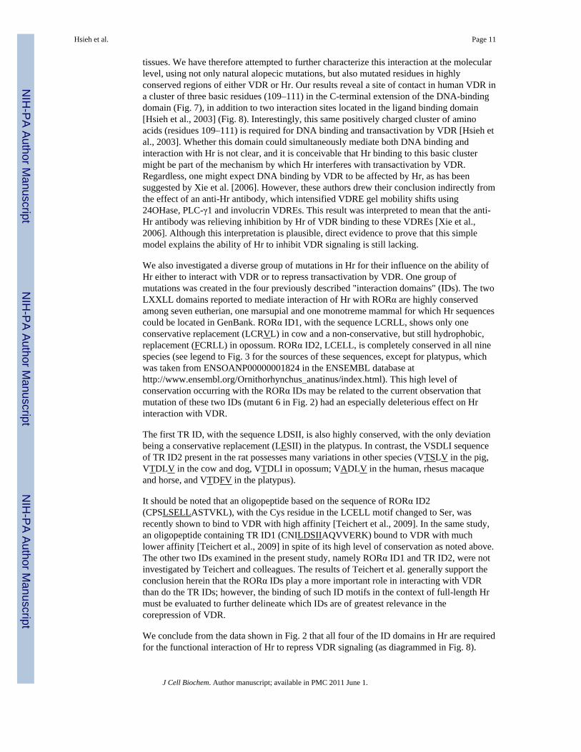
tissues. We have therefore attempted to further characterize this interaction at the molecularlevel, using not only natural alopecic mutations, but also mutated residues in highlyconserved regions of either VDR or Hr. Our results reveal a site of contact in human VDR ina cluster of three basic residues (109–111) in the C-terminal extension of the DNA-bindingdomain (Fig. 7), in addition to two interaction sites located in the ligand binding domain[Hsieh et al., 2003] (Fig. 8). Interestingly, this same positively charged cluster of aminoacids (residues 109–111) is required for DNA binding and transactivation by VDR [Hsieh etal., 2003]. Whether this domain could simultaneously mediate both DNA binding andinteraction with Hr is not clear, and it is conceivable that Hr binding to this basic clustermight be part of the mechanism by which Hr interferes with transactivation by VDR.Regardless, one might expect DNA binding by VDR to be affected by Hr, as has beensuggested by Xie et al. [2006]. However, these authors drew their conclusion indirectly fromthe effect of an anti-Hr antibody, which intensified VDRE gel mobility shifts using24OHase, PLC-γ1 and involucrin VDREs. This result was interpreted to mean that the anti-Hr antibody was relieving inhibition by Hr of VDR binding to these VDREs [Xie et al.,2006]. Although this interpretation is plausible, direct evidence to prove that this simplemodel explains the ability of Hr to inhibit VDR signaling is still lacking.
We also investigated a diverse group of mutations in Hr for their influence on the ability ofHr either to interact with VDR or to repress transactivation by VDR. One group ofmutations was created in the four previously described "interaction domains" (IDs). The twoLXXLL domains reported to mediate interaction of Hr with RORα are highly conservedamong seven eutherian, one marsupial and one monotreme mammal for which Hr sequencescould be located in GenBank. RORα ID1, with the sequence LCRLL, shows only oneconservative replacement (LCRVL) in cow and a non-conservative, but still hydrophobic,replacement (FCRLL) in opossum. RORα ID2, LCELL, is completely conserved in all ninespecies (see legend to Fig. 3 for the sources of these sequences, except for platypus, whichwas taken from ENSOANP00000001824 in the ENSEMBL database athttp://www.ensembl.org/Ornithorhynchus_anatinus/index.html). This high level ofconservation occurring with the RORα IDs may be related to the current observation thatmutation of these two IDs (mutant 6 in Fig. 2) had an especially deleterious effect on Hrinteraction with VDR.
The first TR ID, with the sequence LDSII, is also highly conserved, with the only deviationbeing a conservative replacement (LESII) in the platypus. In contrast, the VSDLI sequenceof TR ID2 present in the rat possesses many variations in other species (VTSLV in the pig,VTDLV in the cow and dog, VTDLI in opossum; VADLV in the human, rhesus macaqueand horse, and VTDFV in the platypus).
It should be noted that an oligopeptide based on the sequence of RORα ID2(CPSLSELLASTVKL), with the Cys residue in the LCELL motif changed to Ser, wasrecently shown to bind to VDR with high affinity [Teichert et al., 2009]. In the same study,an oligopeptide containing TR ID1 (CNILDSIIAQVVERK) bound to VDR with muchlower affinity [Teichert et al., 2009] in spite of its high level of conservation as noted above.The other two IDs examined in the present study, namely RORα ID1 and TR ID2, were notinvestigated by Teichert and colleagues. The results of Teichert et al. generally support theconclusion herein that the RORα IDs play a more important role in interacting with VDRthan do the TR IDs; however, the binding of such ID motifs in the context of full-length Hrmust be evaluated to further delineate which IDs are of greatest relevance in thecorepression of VDR.
We conclude from the data shown in Fig. 2 that all four of the ID domains in Hr are requiredfor the functional interaction of Hr to repress VDR signaling (as diagrammed in Fig. 8).
Hsieh et al. Page 11
J Cell Biochem. Author manuscript; available in PMC 2011 June 1.
NIH
-PA Author Manuscript
NIH
-PA Author Manuscript
NIH
-PA Author Manuscript

Nevertheless, some level of interaction between the proteins occurs when one, or as many astwo IDs are altered as indicated by the GST pull-down results in Fig. 2. The precise role ofTR ID2, however, is still questionable given its poor level of conservation among mammalsand also the fact that we have not been able to test a mutation that alters only this ID motif.How all of these interaction domains in Hr and VDR might combine in three dimensions toform a functional interface is not easily visualized in the absence of complete x-ray crystalstructures for Hr and/or full-length VDR.
Another pair of mutations tested herein were based on published alopecic Hr mutants inmice. These mutations were recreated in rat Hr, which is 93% identical to mouse Hr at theamino acid level. The current results indicate that both rat mutations, G985W and the C-terminal deletant ΔAK, render Hr unable to repress TR signaling from a myosin heavy chainTRE. However, only the G985W mutant impaired the ability of Hr to repress VDRsignaling, as the ΔAK mutant retains its capacity to repress VDR transactivation normally. Itshould be noted that, according to a more recent report on the bald Mill Hill mouse[Brancaz-Bouvier et al., 2008], the ΔAK mutation does not accurately recapitulate the defectin this alopecic mouse model. The more recent report describes the bald Mill Hill mutationas a 296 bp deletion leading to the loss of only a single residue at the C-terminus, but alsothe loss of the stop codon, leading to the introduction of 117 amino acids by read-through tothe next in-frame stop codon [Brancaz-Bouvier et al., 2008]. Thus, the bald Mill Hill mutantHr is different than the C-terminal truncation evaluated in the current experiments, althoughit may be significant that the ΔAK mutant tested herein displays a unique impairment in Hr'sability to repress TR, but not VDR, signaling, perhaps indicating that the functionalinteractions of Hr with TR vs. VDR differ qualitatively.
Twelve additional rHr mutations evaluated in the present report, as summarized in Table 1,correspond to human mutations originally reported as being alopecic (see [Wang et al.,2007] for citations on each of these alopecic alterations). All of these Hr mutations weretested by another laboratory as human Hr alterations in transfected COS-7 cells [Wang et al.,2007]. It is significant to note that all three of the mutations that severely reduced orabolished the ability of Hr to repress VDR signaling in both rat and human Hr (listed withrat/human numbering: C642/622G, N988/970S and D1030/1012N) have been positivelyidentified to cause alopecia in human patients [Aita et al., 2000;Klein et al., 2002;Kruse etal., 1999]. Two other mutations (V1074/1056M and V1154/1136D) were reported to bealopecic in humans [Zlotogorski et al., 2002; Chichon et al., 1998], but each showed severereduction in Hr repressive ability only in one species (V1074/1056M in human Hr andV1154/1136D in rat Hr). Finally, the alopecic E603/583V mutant [Paradisi et al., 2003],which was only tested in its human version, showed essentially a normal ability to repressVDR signaling, indicating that some other function of Hr must be affected in order toexplain the alopecic phenotype in this case.
One obvious candidate function of Hr that might be impaired in certain alopecic mutants isthe ability to interact with HDACs [Djabali and Christiano, 2004; Potter et al., 2001; Potteret al., 2002]. We therefore examined the ability of selected rHr mutants to interact with anHDAC. Our choice of HDAC3 was based on colocalization studies of Hr and Hr fragmentswith HDAC3 in brain tissue [Potter et al., 2002] and in mouse NIH3T3 cells [Djabali andChristiano, 2004]. HDAC3 has also been reported to specifically interact with VDR [Donget al., 2005] to mediate transcriptional repression. However, both VDR and Hr have alsobeen shown to interact with other HDACs, most notably HDACs 1 [Dong et al., 2005;Huang and Hung, 2006; Potter et al., 2002], 2 [Kim et al., 2007] and 5 [Potter et al., 2002],raising the possibility that VDR and Hr may associate with distinct HDACs depending onthe cellular, or possibly the promoter, context [Huang and Hung, 2006]. Wang et al. choseHDAC1 for their studies based on analysis in transfected COS-7 cells, which indicated that
Hsieh et al. Page 12
J Cell Biochem. Author manuscript; available in PMC 2011 June 1.
NIH
-PA Author Manuscript
NIH
-PA Author Manuscript
NIH
-PA Author Manuscript
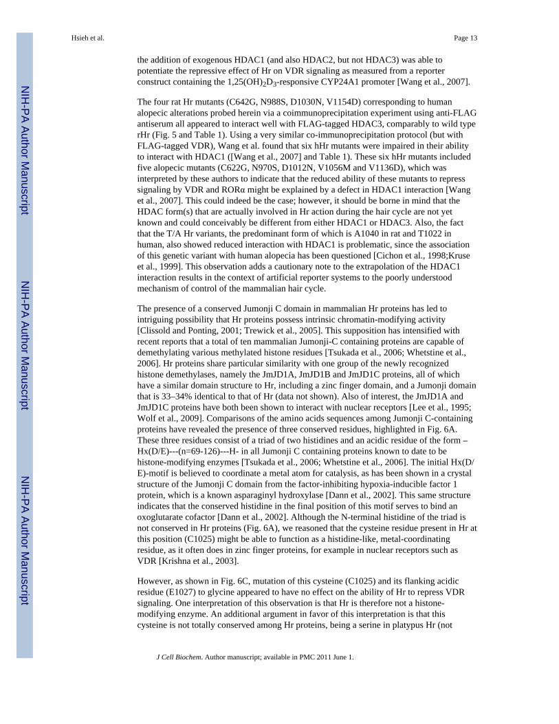
the addition of exogenous HDAC1 (and also HDAC2, but not HDAC3) was able topotentiate the repressive effect of Hr on VDR signaling as measured from a reporterconstruct containing the 1,25(OH)2D3-responsive CYP24A1 promoter [Wang et al., 2007].
The four rat Hr mutants (C642G, N988S, D1030N, V1154D) corresponding to humanalopecic alterations probed herein via a coimmunoprecipitation experiment using anti-FLAGantiserum all appeared to interact well with FLAG-tagged HDAC3, comparably to wild typerHr (Fig. 5 and Table 1). Using a very similar co-immunoprecipitation protocol (but withFLAG-tagged VDR), Wang et al. found that six hHr mutants were impaired in their abilityto interact with HDAC1 ([Wang et al., 2007] and Table 1). These six hHr mutants includedfive alopecic mutants (C622G, N970S, D1012N, V1056M and V1136D), which wasinterpreted by these authors to indicate that the reduced ability of these mutants to represssignaling by VDR and RORα might be explained by a defect in HDAC1 interaction [Wanget al., 2007]. This could indeed be the case; however, it should be borne in mind that theHDAC form(s) that are actually involved in Hr action during the hair cycle are not yetknown and could conceivably be different from either HDAC1 or HDAC3. Also, the factthat the T/A Hr variants, the predominant form of which is A1040 in rat and T1022 inhuman, also showed reduced interaction with HDAC1 is problematic, since the associationof this genetic variant with human alopecia has been questioned [Cichon et al., 1998;Kruseet al., 1999]. This observation adds a cautionary note to the extrapolation of the HDAC1interaction results in the context of artificial reporter systems to the poorly understoodmechanism of control of the mammalian hair cycle.
The presence of a conserved Jumonji C domain in mammalian Hr proteins has led tointriguing possibility that Hr proteins possess intrinsic chromatin-modifying activity[Clissold and Ponting, 2001; Trewick et al., 2005]. This supposition has intensified withrecent reports that a total of ten mammalian Jumonji-C containing proteins are capable ofdemethylating various methylated histone residues [Tsukada et al., 2006; Whetstine et al.,2006]. Hr proteins share particular similarity with one group of the newly recognizedhistone demethylases, namely the JmJD1A, JmJD1B and JmJD1C proteins, all of whichhave a similar domain structure to Hr, including a zinc finger domain, and a Jumonji domainthat is 33–34% identical to that of Hr (data not shown). Also of interest, the JmJD1A andJmJD1C proteins have both been shown to interact with nuclear receptors [Lee et al., 1995;Wolf et al., 2009]. Comparisons of the amino acids sequences among Jumonji C-containingproteins have revealed the presence of three conserved residues, highlighted in Fig. 6A.These three residues consist of a triad of two histidines and an acidic residue of the form –Hx(D/E)---(n=69-126)---H- in all Jumonji C containing proteins known to date to behistone-modifying enzymes [Tsukada et al., 2006; Whetstine et al., 2006]. The initial Hx(D/E)-motif is believed to coordinate a metal atom for catalysis, as has been shown in a crystalstructure of the Jumonji C domain from the factor-inhibiting hypoxia-inducible factor 1protein, which is a known asparaginyl hydroxylase [Dann et al., 2002]. This same structureindicates that the conserved histidine in the final position of this motif serves to bind anoxoglutarate cofactor [Dann et al., 2002]. Although the N-terminal histidine of the triad isnot conserved in Hr proteins (Fig. 6A), we reasoned that the cysteine residue present in Hr atthis position (C1025) might be able to function as a histidine-like, metal-coordinatingresidue, as it often does in zinc finger proteins, for example in nuclear receptors such asVDR [Krishna et al., 2003].
However, as shown in Fig. 6C, mutation of this cysteine (C1025) and its flanking acidicresidue (E1027) to glycine appeared to have no effect on the ability of Hr to repress VDRsignaling. One interpretation of this observation is that Hr is therefore not a histone-modifying enzyme. An additional argument in favor of this interpretation is that thiscysteine is not totally conserved among Hr proteins, being a serine in platypus Hr (not
Hsieh et al. Page 13
J Cell Biochem. Author manuscript; available in PMC 2011 June 1.
NIH
-PA Author Manuscript
NIH
-PA Author Manuscript
NIH
-PA Author Manuscript

shown). Nevertheless, mutation of the third residue in the Jumonji C catalytic triad, the C-terminal histidine 1143 (which is conserved in all Hr proteins, including platypus) exhibits acomplete loss in the ability of Hr to repress VDR signaling (Fig. 6B). This latter observationlikely has relevance to the function of Hr, and we favor the interpretation that Hr acts as acorepressor in part by catalyzing histone demethylation and/or to modify chromatin in afashion that is not revealed by functional testing using an artificial, plasmid-based reportergene construct. It is important to emphasize that five of the eight alopecic mutations inconserved residues described herein for either mouse or human Hr occur in, or closely flank,the Jumonji C domain. Clearly, this novel domain in Hr which we have highlighted for thefirst time in the present communication is crucial to the direction of the mammalian haircycle by the VDR-Hr complex.
The original paper describing the cloning of human Hr reported the existence of an Hrisoform in which exon 17 is absent (as illustrated in Fig. 8, upper right), presumably due toalternative splicing of the Hr mRNA in certain human tissues, resulting in the in-framedeletion of codons 1072 to 1126 in hHr (corresponding to 1090–1144 in rHr) [Cichon et al.,1998]. This deletion removes a C-terminal portion of the Jumonji C domain and specificallyeliminates H1125, the human equivalent of rat Hr H1143, which is the proposedoxoglutarate-binding residue that could potentially participate in enzymatic activity by Hr[Dann et al., 2002] and, most important, we have demonstrated as essential for the VDRcorepressor activity of Hr (Fig. 6B). A study of the tissue distribution of the isoform lackingexon 17 (designated isoform b), along with the full-length isoform, was reported in thissame publication, with the result that the isoform missing exon 17 was the predominant, ifnot sole, isoform found in human skin [Cichon et al., 1998]. Human Hr isoform b, lackingresidues 1072 to 1126, was tested by Wang et al. and more recently by Malloy et al. [Malloyet al., 2009], with the result that this Hr isoform is incapable of repressing signaling eitherby RORα [Wang et al., 2007] or by VDR, and is also incapable of interacting with HDAC1[Malloy et al., 2009], suggesting that perhaps this isoform is biologically inactive. Why thepredominant isoform of Hr in human skin should be inactive is unclear, although regulationof hHr mRNA splicing would be one mechanism of generating a gradient of functional Hr tocreate the cyclical nature of hair growth. Also, information about the existence of an Hrisoform b in any species other than human appears to be lacking. It should be noted,however, that none of the naturally occurring alopecic mutants described so far occur in thisdeleted part of the Hr sequence in isoform b. Thus, the significance of isoform b, if any, inthe bioactions of Hr remains unclear.
Fig. 8 illustrates our current understanding of Hr structure and function. Specific residues orsequence motifs that have a demonstrated role in Hr function are largely confined to the C-terminal two-thirds of the protein. The zinc finger domain, repression domains 2 and 3, andthe Jumonji C domain are all found in the C-terminal half of the protein, and it is significantto note that point mutations leading to alopecia in either human patients or rodent models arefound in, or immediately adjacent to, each of these domains. The C-terminus of Hr (endingin Ala-Lys) also may be important for Hr function, at least for the repression of TR signaling(Fig. 4D and Fig. 8). Our understanding of the N-terminal portion of Hr is limited to theoriginal description of repression domain 1 (RD1) [Potter et al., 2001] and of a nuclearlocalization domain [Djabali et al., 2001] at the C-terminal boundary of RD1. No alopecicmutants have been reported in either of these domains, although a polymorphic variant inRD1 (C422/397Y) has been reported [Hillmer et al., 2002]. However, testing by both Wanget al. and by our laboratory of the human and rat versions of this variant, respectively, failedto show any significant reduction in the ability to repress VDR signaling relative to wildtype (see Table 1 and [Wang et al., 2007]).
Hsieh et al. Page 14
J Cell Biochem. Author manuscript; available in PMC 2011 June 1.
NIH
-PA Author Manuscript
NIH
-PA Author Manuscript
NIH
-PA Author Manuscript

As suggested in Fig. 8, the current concept of Hr action in the hair cycle is that Hr acts inconcert with VDR and likely also with the VDR coreceptor RXRα, based on the fact thatinactivating mutations or deletions in all three of these genes give a similar alopecicphenotype [Li et al., 2000;Malloy et al., 2004;Sakai et al., 2001]. The mechanism by whichthis occurs remains to be elucidated, although, as discussed above, the 1,25(OH)2D3 ligandfor VDR appears to play no role [Malloy et al., 2004]. The involvement of HDACs issuggested not only by studies demonstrating an interaction between Hr and certain HDACs[Djabali et al., 2004;Potter et al., 2001;Potter et al., 2002], but also by the results of Wang etal. that certain alopecic mutants in Hr are deficient in their interactions with HDAC1 [Wanget al., 2007]. However, at least one alopecic mutant, E583V, is not deficient in HDACinteraction as tested by Wang et al. (Table 1). It therefore appears that other interactions maybe important for biological functioning of Hr, such as an as-yet-uncharacterized role for thewell-conserved Jumonji C domain. We theorize that the Jumonji C domain in Hr, whichcontains the highest concentration of naturally occurring alopecic mutations of any domain,and is strikingly compromised by the alteration of absolutely conserved H1143, performsrepressive modifications of chromatin such as histone demethylation. Further studies intothe structure function relationships of the Hr protein will likely shed light not only on thecontrol of the mammalian hair cycle, but also may provide general insights into themechanisms by which nuclear receptors interact with transcriptional repressors.
AcknowledgmentsThe authors are grateful to Dr. Catherine C. Thompson and her laboratory at the Kennedy Krieger Institute andDepartment of Neuroscience, John Hopkins University for performing co-IP experiments with our rat Hr mutantsand HDAC-3. This work was supported by an Arizona Biomedical Research Commission grant (ABRC0912) to J-C. Hsieh, and National Institutes of Health R01 grants DK 063930 and DK 33351 to M. R. Haussler.
Grant sponsor: Arizona Biomedical Research Commission; grant contract number 0912
Grant sponsor: NIH; grant contract numbers DK033351 and DK06393
REFERENCESAhmad W, Faiyaz ul Haque M, Brancolini V, Tsou HC, ul Haque S, Lam H, Aita VM, Owen J,
deBlaquiere M, Frank J, Cserhalmi-Friedman PB, Leask A, McGrath JA, Peacocke M, Ahmad M,Ott J, Christiano AM. Alopecia universalis associated with a mutation in the human hairless gene.Science. 1998; 279:720–724. [PubMed: 9445480]
Ahmad W, Zlotogorski A, Panteleyev AA, Lam H, Ahmad M, ul Haque MF, Abdallah HM, Dragan L,Christiano AM. Genomic organization of the human hairless gene (HR) and identification of amutation underlying congenital atrichia in an Arab Palestinian family. Genomics. 1999; 56:141–148. [PubMed: 10051399]
Aita VM, Ahmad W, Panteleyev AA, Kozlowska U, Kozlowska A, Gilliam TC, Jablonska S,Christiano AM. A novel missense mutation (C622G) in the zinc-finger domain of the humanhairless gene associated with congenital atrichia with papular lesions. Exp Dermatol. 2000; 9:157–162. [PubMed: 10772391]
Bergman R, Schein-Goldshmid R, Hochberg Z, Ben-Izhak O, Sprecher E. The alopecias associatedwith vitamin D-dependent rickets type IIA and with hairless gene mutations: a comparative clinical,histologic, and immunohistochemical study. Arch Dermatol. 2005; 141:343–351. [PubMed:15781675]
Brancaz MV, Iratni R, Morrison A, Mancini SJ, Marche P, Sundberg J, Nonchev S. A new allele of themouse hairless gene interferes with Hox/LacZ transgene regulation in hair follicle primordia. ExpMol Pathol. 2004; 76:173–181. [PubMed: 15010296]
Brancaz-Bouvier MV, Folco EJ, Salameire D, Romero Y, Iratni R, Nonchev S. The "bald Mill Hill"mutation in the mouse is associated with an abnormal, mislocalized HR bmh protein. J InvestDermatol. 2008; 128:311–321. [PubMed: 17657241]
Hsieh et al. Page 15
J Cell Biochem. Author manuscript; available in PMC 2011 June 1.
NIH
-PA Author Manuscript
NIH
-PA Author Manuscript
NIH
-PA Author Manuscript

Cachon-Gonzalez MB, Fenner S, Coffin JM, Moran C, Best S, Stoye JP. Structure and expression ofthe hairless gene of mice. Proc Natl Acad Sci USA. 1994; 91:7717–7721. [PubMed: 8052649]
Cichon S, Anker M, Vogt IR, Rohleder H, Putzstuck M, Hillmer A, Farooq SA, Al-Dhafri KS, AhmadM, Haque S, Rietschel M, Propping P, Kruse R, Nothen MM. Cloning, genomic organization,alternative transcripts and mutational analysis of the gene responsible for autosomal recessiveuniversal congenital alopecia. Hum Mol Genet. 1998; 7:1671–1679. [PubMed: 9736769]
Clissold PM, Ponting CP. JmjC: cupin metalloenzyme-like domains in jumonji, hairless andphospholipase A2beta. Trends Biochem Sci. 2001; 26:7–9. [PubMed: 11165500]
Dann, CEr; Bruick, RK.; Deisenhofer, J. Structure of factor-inhibiting hypoxia-inducible factor 1: Anasparaginyl hydroxylase involved in the hypoxic response pathway. Proc Natl Acad Sci USA.2002; 99:15351–15356. [PubMed: 12432100]
Djabali K, Aita VM, Christiano AM. Hairless is translocated to the nucleus via a novel bipartitenuclear localization signal and is associated with the nuclear matrix. J Cell Sci. 2001; 114:367–376. [PubMed: 11148138]
Djabali K, Christiano AM. Hairless contains a novel nuclear matrix targeting signal and associateswith histone deacetylase 3 in nuclear speckles. Differentiation. 2004; 72:410–418. [PubMed:15606500]
Djabali K, Zlotogorski A, Metzker A, Ben-Amitai D, Christiano AM. Interaction of hairless andthyroid hormone receptor is not involved in the pathogenesis of atrichia with papular lesions. ExpDermatol. 2004; 13:251–256. [PubMed: 15086341]
Dong X, Lutz W, Schroeder TM, Bachman LA, Westendorf JJ, Kumar R, Griffin MD. Regulation ofrelB in dendritic cells by means of modulated association of vitamin D receptor and histonedeacetylase 3 with the promoter. Proc Natl Acad Sci USA. 2005; 102:16007–16012. [PubMed:16239345]
Hillmer AM, Kruse R, Macciardi F, Heyn U, Betz RC, Ruzicka T, Propping P, Nothen MM, Cichon S.The hairless gene in androgenetic alopecia: results of a systematic mutation screening and afamily-based association approach. Br J Dermatol. 2002; 146:601–608. [PubMed: 11966690]
Hsieh J-C, Jurutka PW, Galligan MA, Terpening CM, Haussler CA, Samuels DS, Shimizu Y, ShimizuN, Haussler MR. Human vitamin D receptor is selectively phosphorylated by protein kinase C onserine 51, a residue crucial to its trans-activation function. Proc Natl Acad Sci USA. 1991;88:9315–9319. [PubMed: 1656468]
Hsieh J-C, Jurutka PW, Selznick SH, Reeder MC, Haussler CA, Whitfield GK, Haussler MR. The T-box near the zinc fingers of the human vitamin D receptor is required for heterodimeric DNAbinding and transactivation. Biochem Biophys Res Commun. 1995; 215:1–7. [PubMed: 7575575]
Hsieh J-C, Sisk JM, Jurutka PW, Haussler CA, Slater SA, Haussler MR, Thompson CC. Physical andfunctional interaction between the vitamin D receptor and hairless corepressor, two proteinsrequired for hair cycling. J Biol Chem. 2003; 278:38665–38674. [PubMed: 12847098]
Hsieh J-C, Whitfield GK, Oza AK, Dang HTL, Price JN, Galligan MA, Jurutka PW, Thompson PD,Haussler CA, Haussler MR. Characterization of unique DNA binding and transcriptionalactivation functions in the carboxyl-terminal extension of the zinc finger region in the humanvitamin D receptor. Biochemistry. 1999; 38:16347–16358. [PubMed: 10587460]
Huang Y-C, Hung W-C. 1,25-dihydroxyvitamin D3 transcriptionally represses p45Skp2 expression viathe Sp1 sites in human prostate cancer cells. J Cell Physiol. 2006; 209:363–369. [PubMed:16883603]
Jin CH, Kerner SA, Hong MH, Pike JW. Transcriptional activation and dimerization functions in thehuman vitamin D receptor. Mol Endocrinol. 1996; 10:945–957. [PubMed: 8843411]
Kim M-S, Fujiki R, Murayama A, Kitagawa H, Yamaoka K, Yamamoto Y, Mihara M, Takeyama K,Kato S. 1Alpha,25(OH)2D3-induced transrepression by vitamin D receptor through E-box-typeelements in the human parathyroid hormone gene promoter. Molecular Endocrinology. 2007;21:334–342. [PubMed: 17095575]
Klein I, Bergman R, Indelman M, Sprecher E. A novel missense mutation affecting the human hairlessthyroid receptor interacting domain 2 causes congenital atrichia. J Invest Dermatol. 2002;119:920–922. [PubMed: 12406339]
Hsieh et al. Page 16
J Cell Biochem. Author manuscript; available in PMC 2011 June 1.
NIH
-PA Author Manuscript
NIH
-PA Author Manuscript
NIH
-PA Author Manuscript

Krishna SS, Majumdar I, Grishin NV. Structural classification of zinc fingers: survey and summary.Nucleic Acids Res. 2003; 31:532–550. [PubMed: 12527760]
Kruse R, Cichon S, Anker M, Hillmer AM, Barros-Nunez P, Cantu JM, Leal E, Weinlich G, SchmuthM, Fritsch P, Ruzicka T, Propping P, Nothen MM. Novel Hairless mutations in two kindreds withautosomal recessive papular atrichia. J Invest Dermatol. 1999; 113:954–959. [PubMed: 10594736]
Lee JW, Choi H-S, Gyuris J, Brent R, Moore DD. Two classes of proteins dependent on either thepresence or absence of thyroid hormone for interaction with the thyroid hormone receptor.Molecular Endocrinology. 1995; 9:243–254. [PubMed: 7776974]
Li M, Indra AK, Warot X, Brocard J, Messaddeq N, Kato S, Metzger D, Chambon P. Skinabnormalities generated by temporally controlled RXRalpha mutations in mouse epidermis.Nature. 2000; 407:633–636. [PubMed: 11034212]
Malloy PJ, Wang J, Jensen K, Feldman D. Modulation of vitamin D receptor activity by thecorepressor hairless: differential effects of hairless isoforms. Endocrinology. 2009; 150:4950–4957. [PubMed: 19819974]
Malloy PJ, Xu R, Peng L, Clark PA, Feldman D. A novel mutation in helix 12 of the vitamin Dreceptor impairs coactivator interaction and causes hereditary 1,25-dihydroxyvitamin D-resistantrickets without alopecia. Mol Endocrinol. 2002; 16:2538–2546. [PubMed: 12403843]
Malloy PJ, Xu R, Peng L, Peleg S, Al-Ashwal A, Feldman D. Hereditary 1,25-dihydroxyvitamin Dresistant rickets due to a mutation causing multiple defects in vitamin D receptor function.Endocrinology. 2004; 145:5106–5114. [PubMed: 15308610]
Moraitis AN, Giguere V, Thompson CC. Novel mechanism of nuclear receptor corepressor interactiondictated by activation function 2 helix determinants. Mol Cell Biol. 2002; 22:6831–6841.[PubMed: 12215540]
Nam Y, Kim JK, Cha DS, Cho JW, Cho KH, Yoon S, Yoon JB, Oh YS, Suh JG, Han SS, Song CW,Yoon SK. A novel missense mutation in the mouse hairless gene causes irreversible hair loss:Genetic and molecular analyses of Hr(m1Enu). Genomics. 2006; 87:520–526. [PubMed:16455232]
Paradisi M, Chuang GS, Angelo C, Pedicelli C, Martinez-Mir A, Christiano AM. Atrichia with papularlesions resulting from a novel homozygous missense mutation in the hairless gene. Clinical andExperimental Dermatology. 2003; 28:535–558. [PubMed: 12950347]
Potter GB, Beaudoin GM 3rd, DeRenzo CL, Zarach JM, Chen SH, Thompson CC. The hairless genemutated in congenital hair loss disorders encodes a novel nuclear receptor corepressor. Genes Dev.2001; 15:2687–2701. [PubMed: 11641275]
Potter GB, Zarach JM, Sisk JM, Thompson CC. The thyroid hormone-regulated corepressor hairlessassociates with histone deacetylases in neonatal rat brain. Mol Endocrinol. 2002; 16:2547–2560.[PubMed: 12403844]
Sakai Y, Kishimoto J, Demay MB. Metabolic and cellular analysis of alopecia in vitamin D receptorknockout mice. J Clin Invest. 2001; 107:961–966. [PubMed: 11306599]
Skorija K, Cox M, Sisk JM, Dowd DR, MacDonald PN, Thompson CC, Demay MB. Ligand-independent actions of the vitamin D receptor maintain hair follicle homeostasis. Mol Endocrinol.2005; 19:855–862. [PubMed: 15591533]
Steinmayr M, Andre E, Conquet F, Rondi-Reig L, Delhaye-Bouchaud N, Auclair N, Daniel H, CrepelF, Mariani J, Sotelo C, Becker-Andre M. Staggerer phenotype in retinoid-related orphan receptoralpha-deficient mice. Proc Natl Acad Sci USA. 1998; 95:3960–3965. [PubMed: 9520475]
Teichert A, Arnold LA, Otieno S, Oda Y, Augustinaite I, Geistlinger TR, Kriwacki RW, Guy RK,Bikle DD. Quantification of the vitamin D receptor-coregulator interaction. Biochemistry. 2009;48:1454–1461. [PubMed: 19183053]
Thompson PD, Hsieh J-C, Whitfield GK, Haussler CA, Jurutka PW, Galligan MA, Tillman JB,Spindler SR, Haussler MR. The vitamin D receptor displays DNA binding and transactivation as aheterodimer with the retinoid X receptor, but not with the thyroid hormone receptor. J CellBiochem. 1999; 75:462–480. [PubMed: 10536369]
Trewick SC, McLaughlin PJ, Allshire RC. Methylation: lost in hydroxylation? EMBO Rep. 2005;6:315–320. [PubMed: 15809658]
Hsieh et al. Page 17
J Cell Biochem. Author manuscript; available in PMC 2011 June 1.
NIH
-PA Author Manuscript
NIH
-PA Author Manuscript
NIH
-PA Author Manuscript
Tsika RW, Bahl JJ, Leinwand LA, Morkin E. Thyroid hormone regulates expression of a transfectedhuman alpha- myosin heavy-chain fusion gene in fetal rat heart cells. Proc Natl Acad Sci USA.1990; 87:379–383. [PubMed: 2296592]
Tsukada Y, Fang J, Erdjument-Bromage H, Warren ME, Borchers CH, Tempst P, Zhang Y. Histonedemethylation by a family of JmjC domain-containing proteins. Nature. 2006; 439:811–816.[PubMed: 16362057]
Wang J, Malloy PJ, Feldman D. Interactions of the vitamin D receptor with the corepressor hairless:analysis of hairless mutants in atrichia with papular lesions. J Biol Chem. 2007; 282:25231–25239.[PubMed: 17609203]
Wen Y-D, Perissi V, Staszewski LM, Yang Y-M, Krones A, Glass CK, Rosenfeld MG, Seto E. Thehistone deacetylase-3 complex contains nuclear receptor corepressors. Proc Natl Acad Sci USA.2000; 97:7202–7207. [PubMed: 10860984]
Whetstine JR, Nottke A, Lan F, Huarte M, Smolikov S, Chen Z, Spooner E, Li E, Zhang G,Colaiacovo M, Shi Y. Reversal of histone lysine trimethylation by the JMJD2 family of histonedemethylases. Cell. 2006; 125:467–481. [PubMed: 16603238]
Wolf SS, Patchev VK, Obendorf M. A novel variant of the putative demethylase gene, s-JMJD1C, is acoactivator of the AR. Arch Biochem Biophys. 2009; 460:56–66. [PubMed: 17353003]
Xie Z, Chang S, Oda Y, Bikle DD. Hairless suppresses vitamin D receptor transactivation in humankeratinocytes. Endocrinology. 2006; 147:314–323. [PubMed: 16269453]
Zarach JM, Beaudoin GM 3rd, Coulombe PA, Thompson CC. The co-repressor hairless has a role inepithelial cell differentiation in the skin. Development. 2004; 131:4189–4200. [PubMed:15280217]
Zlotogorski A, Panteleyev AA, Aita VM, Christiano AM. Clinical and molecular diagnostic criteria ofcongenital atrichia with papular lesions. J Invest Dermatol. 2002; 118:887–890. [PubMed:11982770]
Hsieh et al. Page 18
J Cell Biochem. Author manuscript; available in PMC 2011 June 1.
NIH
-PA Author Manuscript
NIH
-PA Author Manuscript
NIH
-PA Author Manuscript
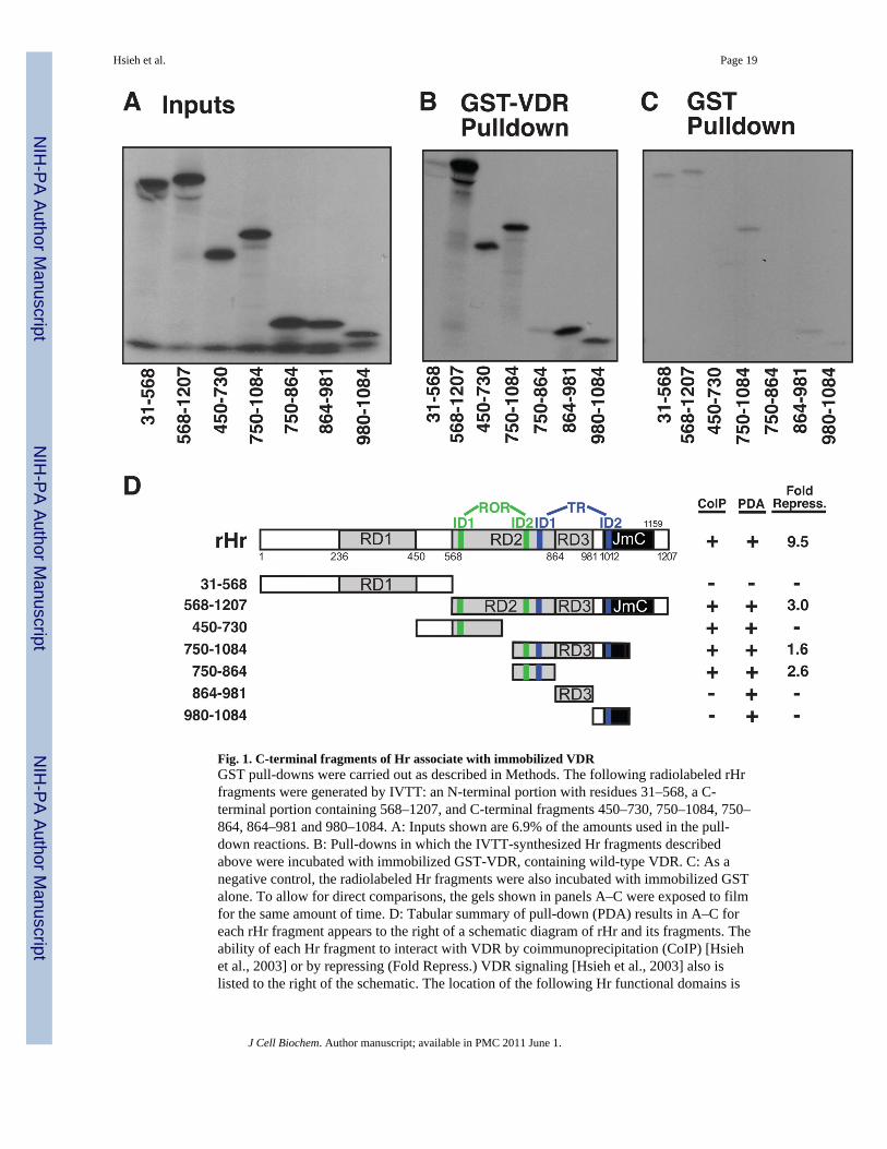
Fig. 1. C-terminal fragments of Hr associate with immobilized VDRGST pull-downs were carried out as described in Methods. The following radiolabeled rHrfragments were generated by IVTT: an N-terminal portion with residues 31–568, a C-terminal portion containing 568–1207, and C-terminal fragments 450–730, 750–1084, 750–864, 864–981 and 980–1084. A: Inputs shown are 6.9% of the amounts used in the pull-down reactions. B: Pull-downs in which the IVTT-synthesized Hr fragments describedabove were incubated with immobilized GST-VDR, containing wild-type VDR. C: As anegative control, the radiolabeled Hr fragments were also incubated with immobilized GSTalone. To allow for direct comparisons, the gels shown in panels A–C were exposed to filmfor the same amount of time. D: Tabular summary of pull-down (PDA) results in A–C foreach rHr fragment appears to the right of a schematic diagram of rHr and its fragments. Theability of each Hr fragment to interact with VDR by coimmunoprecipitation (CoIP) [Hsiehet al., 2003] or by repressing (Fold Repress.) VDR signaling [Hsieh et al., 2003] also islisted to the right of the schematic. The location of the following Hr functional domains is
Hsieh et al. Page 19
J Cell Biochem. Author manuscript; available in PMC 2011 June 1.
NIH
-PA Author Manuscript
NIH
-PA Author Manuscript
NIH
-PA Author Manuscript
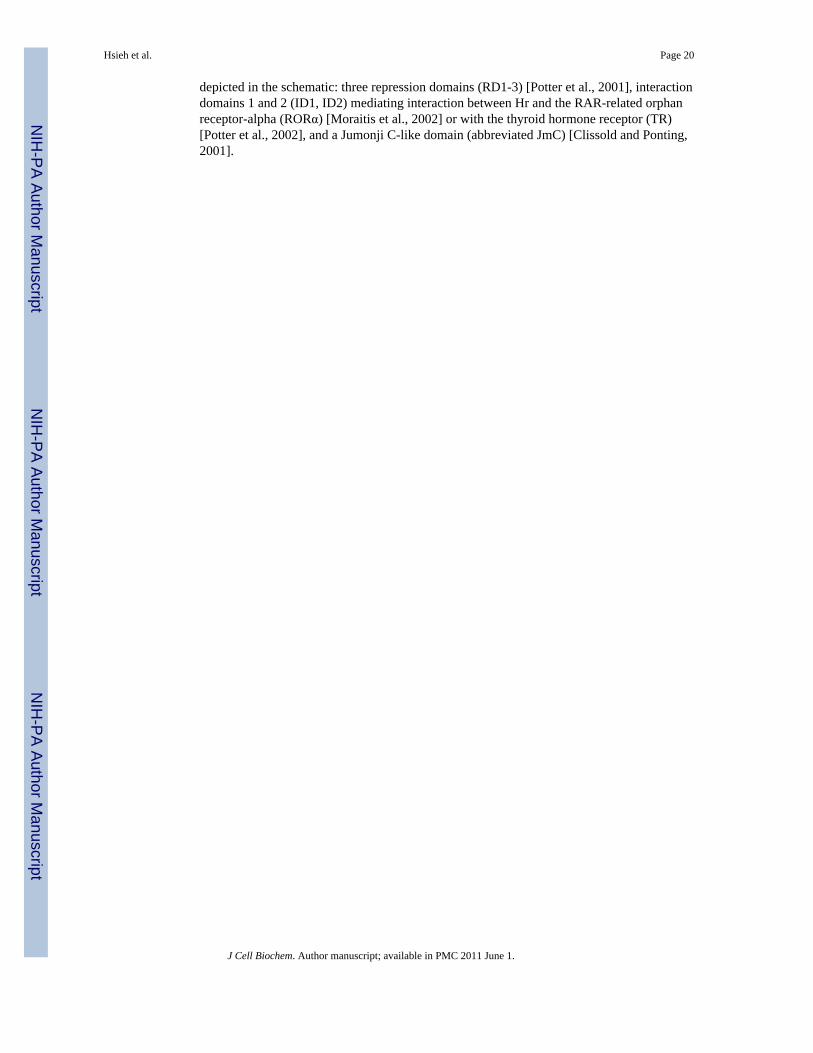
depicted in the schematic: three repression domains (RD1-3) [Potter et al., 2001], interactiondomains 1 and 2 (ID1, ID2) mediating interaction between Hr and the RAR-related orphanreceptor-alpha (RORα) [Moraitis et al., 2002] or with the thyroid hormone receptor (TR)[Potter et al., 2002], and a Jumonji C-like domain (abbreviated JmC) [Clissold and Ponting,2001].
Hsieh et al. Page 20
J Cell Biochem. Author manuscript; available in PMC 2011 June 1.
NIH
-PA Author Manuscript
NIH
-PA Author Manuscript
NIH
-PA Author Manuscript

Fig. 2. Functional analysis of rat Hr point mutations in the interaction domains for RORα andTRA: Various combinations of ID point mutations were used to create nine rHr mutants forfunctional testing, designated mutant 1 through mutant 9. Altered amino acids (all toalanine) are indicated by asterisks (*) and color-coded to match their correspondinginteraction domains. Functional domains, in addition to those shown in Fig. 1D, are anuclear localization domain [Djabali et al., 2001] and a zinc-finger domain [Cachon-Gonzalez et al., 1994]. B: GST pull-down results for the association of nuclear receptorbinding domain mutants of Hr with immobilized VDR. The left panels of B show 4.5% ofthe inputs of 35S-labeled wild type (wt) or mutant (m1-m9) Hr proteins produced in an in
Hsieh et al. Page 21
J Cell Biochem. Author manuscript; available in PMC 2011 June 1.
NIH
-PA Author Manuscript
NIH
-PA Author Manuscript
NIH
-PA Author Manuscript
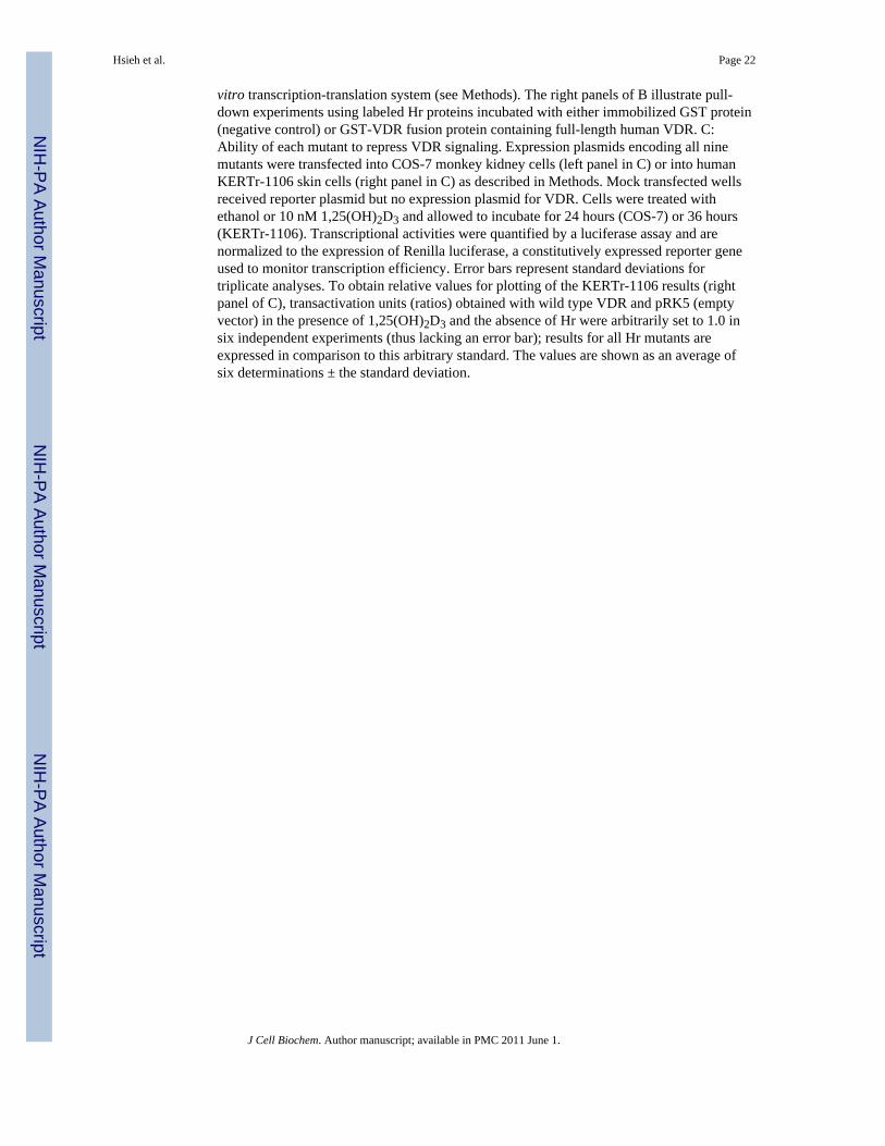
vitro transcription-translation system (see Methods). The right panels of B illustrate pull-down experiments using labeled Hr proteins incubated with either immobilized GST protein(negative control) or GST-VDR fusion protein containing full-length human VDR. C:Ability of each mutant to repress VDR signaling. Expression plasmids encoding all ninemutants were transfected into COS-7 monkey kidney cells (left panel in C) or into humanKERTr-1106 skin cells (right panel in C) as described in Methods. Mock transfected wellsreceived reporter plasmid but no expression plasmid for VDR. Cells were treated withethanol or 10 nM 1,25(OH)2D3 and allowed to incubate for 24 hours (COS-7) or 36 hours(KERTr-1106). Transcriptional activities were quantified by a luciferase assay and arenormalized to the expression of Renilla luciferase, a constitutively expressed reporter geneused to monitor transcription efficiency. Error bars represent standard deviations fortriplicate analyses. To obtain relative values for plotting of the KERTr-1106 results (rightpanel of C), transactivation units (ratios) obtained with wild type VDR and pRK5 (emptyvector) in the presence of 1,25(OH)2D3 and the absence of Hr were arbitrarily set to 1.0 insix independent experiments (thus lacking an error bar); results for all Hr mutants areexpressed in comparison to this arbitrary standard. The values are shown as an average ofsix determinations ± the standard deviation.
Hsieh et al. Page 22
J Cell Biochem. Author manuscript; available in PMC 2011 June 1.
NIH
-PA Author Manuscript
NIH
-PA Author Manuscript
NIH
-PA Author Manuscript
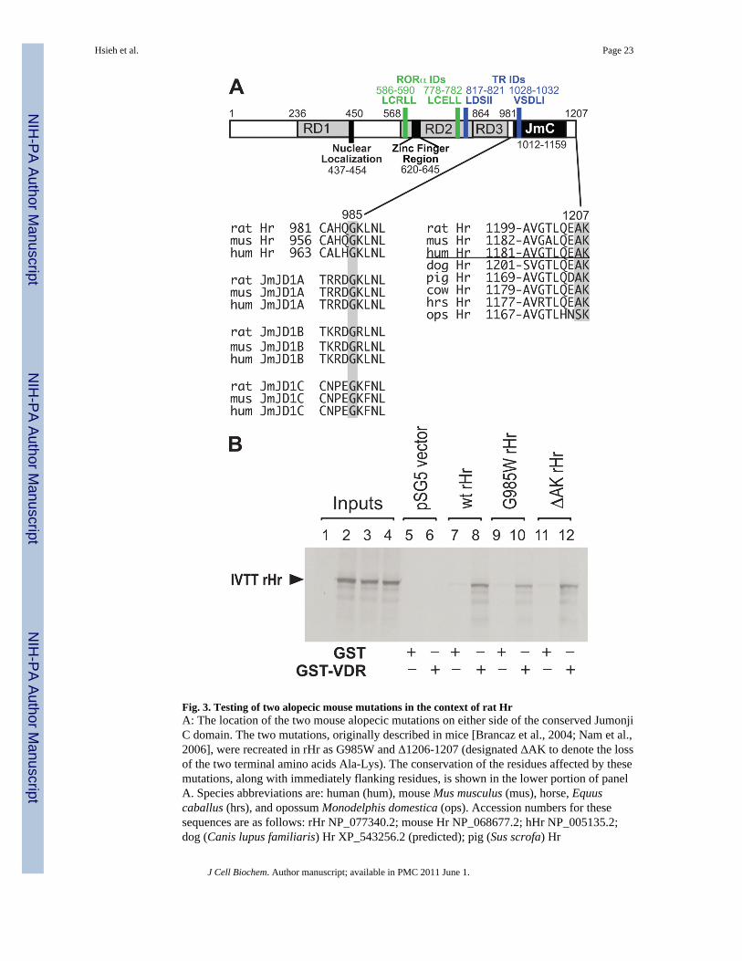
Fig. 3. Testing of two alopecic mouse mutations in the context of rat HrA: The location of the two mouse alopecic mutations on either side of the conserved JumonjiC domain. The two mutations, originally described in mice [Brancaz et al., 2004; Nam et al.,2006], were recreated in rHr as G985W and Δ1206-1207 (designated ΔAK to denote the lossof the two terminal amino acids Ala-Lys). The conservation of the residues affected by thesemutations, along with immediately flanking residues, is shown in the lower portion of panelA. Species abbreviations are: human (hum), mouse Mus musculus (mus), horse, Equuscaballus (hrs), and opossum Monodelphis domestica (ops). Accession numbers for thesesequences are as follows: rHr NP_077340.2; mouse Hr NP_068677.2; hHr NP_005135.2;dog (Canis lupus familiaris) Hr XP_543256.2 (predicted); pig (Sus scrofa) Hr
Hsieh et al. Page 23
J Cell Biochem. Author manuscript; available in PMC 2011 June 1.
NIH
-PA Author Manuscript
NIH
-PA Author Manuscript
NIH
-PA Author Manuscript

NP_001077399.1; cow (Bos taurus) Hr NP_001096005.1; horse (Equus caballus) HrXP_001490941.1; opossum Hr XP_001381979.1 (predicted); rat JmJD1a, also known asJmjC-containing histone demethylase 2A (JHDM2A) or as testis-specific gene A (TSGA),NP_786940; mouse JmJD1a, NP_766589; human JmJD1a, NP_060903.2; rat JmJD1b, alsoknown as nuclear protein 5qNCA or as JHDM2B, XP_001061636 (predicted); mouseJmJD1b, NP_001074725; Human JmJD1b, NP_057688; rat JmJD1c, also called thyroidreceptor interacting protein 8 (TRIP8) or JHDM2C, XP_001080424 (predicted); MouseJmJD1c XP_980927 (predicted); and human JmJD1c NP_004232. B: GST-VDR pull-downanalysis of the G985W and ΔAK rHr mutants using radiolabeled rHr proteins generated inan in vitro transcription-translation system as described in Methods. Beads contained eitherGST alone or full-length hVDR fused to GST (GST-VDR). Input lanes 1–4 correspond to,and represent 5% of the amount used for respective pull-down lanes 5/6, 7/8, 9/10, and11/12.
Hsieh et al. Page 24
J Cell Biochem. Author manuscript; available in PMC 2011 June 1.
NIH
-PA Author Manuscript
NIH
-PA Author Manuscript
NIH
-PA Author Manuscript

Fig. 4. Ability of the G985W and ΔAK rat Hr mutants to repress VDR- and TR-mediatedtransactivation of reporter genesExpression plasmids for wild-type and mutant rHrs were cotransfected into COS-7 cellsalong with, if indicated, a reporter plasmid for VDR (the p24OHaseLuc vector) and thepSG5-hVDR expression plasmid (panels A and C) or a reporter plasmid for TR,(rMHC)2pLucMCS, and the pSG5-hTRB1 expression plasmid for human TRβ1 (B, D).Transfected cultures were treated ± 10 nM 1,25(OH)2D3 or ± 10 nM tri-iodothyronine (T3)as indicated. Luciferase assays were performed as described in Methods and the resultsshown represent the average of six replicates ± the standard deviation.
Hsieh et al. Page 25
J Cell Biochem. Author manuscript; available in PMC 2011 June 1.
NIH
-PA Author Manuscript
NIH
-PA Author Manuscript
NIH
-PA Author Manuscript

Fig. 5. Functional characterization of selected rat Hr mutants which recapitulate naturallyoccurring point mutations in human Hr that confer alopeciaA: Loss of VDR corepressor activity by rHr mutants D1030N and V1154D. COS-7 cellswere transfected with the natural VDRE-reporter plasmid p24OHaseLuc, pSG5-hVDR andan expression plasmid for the indicated rHr mutant as described in Methods. Cells weretreated for 24 hrs ± 10 nM 1,25(OH)2D3 as indicated, and transcriptional activity wasquantified via luciferase assay as described in Methods and the legend to Fig. 2C. Error barsrepresent standard deviations for triplicate wells. B: Radiolabeled rHr mutants wereproduced in an in vitro transcription-translation system, incubated with FLAG-taggedHDAC3 [Wen et al., 2000], immunoprecipitated with the indicated antisera, and subjected toSDS-PAGE and autoradiography as described in Methods. A small aliquot of radiolabeledinput was electrophoresed in lanes indicated by “In”. The appearance of a labeled rHr bandin the α-Flag lane, but not the non-specific IgG lane, is taken as evidence of a directinteraction between the rHr mutant and HDAC3.
Hsieh et al. Page 26
J Cell Biochem. Author manuscript; available in PMC 2011 June 1.
NIH
-PA Author Manuscript
NIH
-PA Author Manuscript
NIH
-PA Author Manuscript

Fig. 6. Mutation of conserved residues in the Jumonji C domain of rat Hr and the functionalconsequence of these alterations on the VDR corepressor activity of HrA: Conservation of the three mutated residues which are postulated to confer enzymaticactivity, such as histone demethylase, onto the Hr protein (see text). Sources for sequencedata are given in the legend to Fig. 3. B: Effect of mutating residue H1143 to glycine in ratHr. Cells were transfected, and cell lysates were assayed for luciferase activity as describedin Methods and the legend to Fig. 2C; "No rHr" wells received reporter plasmid and thepSG5-hVDR expression plasmid, but no expression plasmid for rHr. C: Similar to panel B,except that the double mutant C1025 and E1027 (each to glycine) rHr was investigated.
Hsieh et al. Page 27
J Cell Biochem. Author manuscript; available in PMC 2011 June 1.
NIH
-PA Author Manuscript
NIH
-PA Author Manuscript
NIH
-PA Author Manuscript
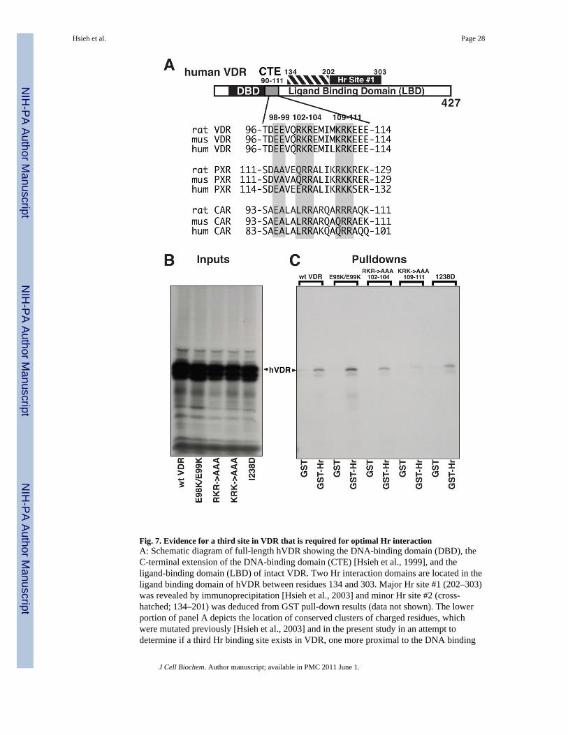
Fig. 7. Evidence for a third site in VDR that is required for optimal Hr interactionA: Schematic diagram of full-length hVDR showing the DNA-binding domain (DBD), theC-terminal extension of the DNA-binding domain (CTE) [Hsieh et al., 1999], and theligand-binding domain (LBD) of intact VDR. Two Hr interaction domains are located in theligand binding domain of hVDR between residues 134 and 303. Major Hr site #1 (202–303)was revealed by immunoprecipitation [Hsieh et al., 2003] and minor Hr site #2 (cross-hatched; 134–201) was deduced from GST pull-down results (data not shown). The lowerportion of panel A depicts the location of conserved clusters of charged residues, whichwere mutated previously [Hsieh et al., 2003] and in the present study in an attempt todetermine if a third Hr binding site exists in VDR, one more proximal to the DNA binding
Hsieh et al. Page 28
J Cell Biochem. Author manuscript; available in PMC 2011 June 1.
NIH
-PA Author Manuscript
NIH
-PA Author Manuscript
NIH
-PA Author Manuscript

domain. In the first mutant, glutamate residues at positions 98 and 99 in human VDR werechanged to lysines to produce E98K/E99K. Next, clusters of basic residues at positions 102–104 and at 109–111 were altered to alanines to produce RKR->AAA 102–104 and KRK->AAA 109–111, respectively. The lower portion of panel A illustrates conservation of theseresidues among VDRs (NR1I1) and also among the related PXR (NR1I2) and CAR (NR1I3)receptors. Sequence sources are as follows: rat VDR NP_058754.1; mouse VDRNP_033530.2; human VDR NP_000367.1; rat PXR NP_443212.1; mouse PXRNP_035066.1; human PXR NP_003880.3; rat CAR NP_075230.1; mouse CARNP_033933.2; human CAR NP_001070950.1. Human VDR proteins containing mutationsin each of the charged clusters were separately expressed in an in vitro transcription-translation system along with wild-type VDR (see Methods). B and C: A representative GSTpull-down experiment designed to assess the ability of these mutant proteins to bindimmobilized rat Hr. B: Display of 4.5% of the inputs of 35S-labeled wild type (wt) or mutantVDRs used in the pull-down reactions. C: Autoradiograph of a gel containing point-mutatedhVDR pull-down reactions using either immobilized GST protein (negative control) orGST-Hr fusion protein containing full-length rat Hr.
Hsieh et al. Page 29
J Cell Biochem. Author manuscript; available in PMC 2011 June 1.
NIH
-PA Author Manuscript
NIH
-PA Author Manuscript
NIH
-PA Author Manuscript

Fig. 8. Integrated model for the functional interactions of VDR and Hr in controlling geneexpression to drive the mammalian hair cycleFunctional domains in VDR and Hr are defined in the legends to Fig. 1D, Fig. 2A, and Fig.7, and in the text. The upper portion depicts schematically the interaction interface betweenVDR and Hr, which appears to consist of three domains in VDR (denoted in red as Hr sites#1 – #3) and four domains in Hr, previously described [Potter et al., 2001;Moraitis et al.,2002;Potter et al., 2002] as LXXLL-like nuclear receptor interaction domains for eitherRORα (shaded in green: LCRLL and LCELL) or TR (shaded in blue: LDSII and VSDLI).The central portion illustrates the position in Hr of a set of naturally occurring mutationswhich confer the alopecic phenotype and were evaluated experimentally in the present studyand/or by Wang and coworkers [Wang et al., 2007]. Residue numbers shown in black are forrat Hr; human Hr numbering is shown in magenta underneath the rat numbering. Mutatedamino acids in human patients that display alopecia with papular lesions (APL, OMIM#209500) are designated with an asterisk; the corresponding residue in rHr that wasevaluated in the current study is shown above the human residue number. A single alopecicHr mutation from mice, G985W, is represented by the designation G985 with thecorresponding human Hr residue number shown in parentheses below. The region of hHrthat is encoded by exon 17 and that is missing in a human variant of Hr (isoform b) [Cichonet al., 1998] is shown at upper right with both rat (black) and human (magenta) residuenumbering (note that is this isoform is only found in the human). Potential functionalinteractions between these residues and either HDACs or other unknown nuclear factors aredepicted as large arrows in the lower portion and are discussed in the text. Residues 865 to
Hsieh et al. Page 30
J Cell Biochem. Author manuscript; available in PMC 2011 June 1.
NIH
-PA Author Manuscript
NIH
-PA Author Manuscript
NIH
-PA Author Manuscript

1204 in rat Hr show high homology to the Jumonji domain-containing protein JmJD1 (34%homology to rat JmJD1B and 33% each to rat JmJD1A and JmJD1C; for accession numbersused in these comparisons see legend to Fig. 3); this region of rat Hr is shaded yellow. Mostof the alopecia conferring mutants occur in this region of Hr, as discussed in the text, andlead us to propose that the C-terminal JmJD1-homologous domain in Hr is enzymaticallyactive in histone demethylation, yielding chromatin modifications that repress the expressionof genes to control the mammalian hair cycle. The proposed histone demethylase activity ofHr likely operates in concert with histone deacetylases recruited by the JmJD1-homologousdomain as well as other repressive domains of Hr to complete a constellation of chromatinrepressive alterations required to silence RXR-VDR-Hr target genes. Potential interactionsbetween these residues or regions and either HDACs or other unknown nuclear factors arediscussed in the text.
Hsieh et al. Page 31
J Cell Biochem. Author manuscript; available in PMC 2011 June 1.
NIH
-PA Author Manuscript
NIH
-PA Author Manuscript
NIH
-PA Author Manuscript

NIH
-PA Author Manuscript
NIH
-PA Author Manuscript
NIH
-PA Author Manuscript
Hsieh et al. Page 32
Tabl
e 1
Sum
mar
y of
eva
luat
ion
of ra
t Hr m
utat
ions
in th
e cu
rren
t stu
dy c
ompa
red
to p
revi
ousl
y pu
blis
hed
(Wan
g et
al.,
200
7) re
sults
with
hum
an H
r (sh
aded
ingr
ey)
Rat
Hr
Mut
ants
VD
RR
epre
ssio
n(r
epor
ter)
VD
RA
ssoc
iatio
n(G
ST)
HD
AC
3A
ssoc
iatio
n(C
oIP)
Hum
anH
rM
utan
ts*
VD
RR
epre
ssio
n(r
epor
ter)
RO
Rα
Rep
ress
ion
(rep
orte
r)
VD
RA
ssoc
iatio
n(C
oIP)
HD
AC
1A
ssoc
iatio
n(C
oIP)
wtV
DR
++++
yes
++++
wtV
DR
++++
++++
+++
++
P95S
+++
yes
Not
test
edP6
9S(d
egra
ded)
(deg
rade
d)(d
egra
ded)
(deg
rade
d)
C42
2Y++
+ye
sN
ot te
sted
C39
7Y++
+++
+++
A59
6V(d
egra
ded)
(deg
rade
d)(d
egra
ded)
A57
6V++
+++
++++
E603
V(d
egra
ded)
(deg
rade
d)(d
egra
ded)
E583
V†
++++
++++
+++
++
E611
G++
++ye
sN
ot te
sted
E591
G++
++++
+++
++
R64
0Q++
yes
Not
test
edR
620Q
++++
+++
++++
C64
2G−
yes
+++
C62
2G†
+/−
−++
++/−
N98
8S−
yes
++++
N97
0S†
−+/−
+++
+
D10
30N
−ye
s++
+D
1012
N†
−−
+++
A10
40T
++ye
sN
ot te
sted
T102
2A++
−++
++
V10
74M
++ye
sN
ot te
sted
V10
56M
†−
++++
+++
V11
54D
−ye
s++
++V
1136
D†
+++
+++
+
* Wan
g et
al.
(Wan
g et
al.,
200
7) h
uman
Hr m
utan
ts
† Hr m
utan
ts re
porte
d to
cau
se a
lope
cia
in h
uman
s
J Cell Biochem. Author manuscript; available in PMC 2011 June 1.