Analysis of cell-cycle specific localization of the Rdi1p RhoGDI and the structural determinants...
11
RESEARCH ARTICLE Tamara J. Richman Kurt A. Toenjes Sergio E. Morales Karen C. Cole Ben T. Wasserman Chad M. Taylor Jacob A. Koster Matthew F. Whelihan Douglas I. Johnson Analysis of cell-cycle specific localization of the Rdi1p RhoGDI and the structural determinants required for Cdc42p membrane localization and clustering at sites of polarized growth Received: 20 February 2004 / Revised: 23 March 2004 / Accepted: 27 March 2004 / Published online: 17 April 2004 ȑ Springer-Verlag 2004 Abstract The Cdc42p GTPase regulates multiple signal transduction pathways through its interactions with downstream effectors. Specific functional domains within Cdc42p are required for guanine-nucleotide binding, interactions with downstream effectors, and membrane localization. However, little is known about how Cdc42p is clustered at polarized growth sites or is extracted from membranes by Rho guanine-nucleotide dissociation inhibitors (RhoGDIs) at specific times in the cell cycle. To address these points, localization studies were performed in Saccharomyces cerevisiae using green fluorescent protein (GFP)-tagged Cdc42p and the RhoGDI Rdi1p. GFP-Rdi1p localized to polarized growth sites at specific times of the cell cycle but not to other sites of Cdc42p localization. Overex- pression of Rdi1p led to loss of GFP-Cdc42p from internal and plasma membranes. This effect was medi- ated through the Cdc42p Rho-insert domain, which was also implicated in interactions with the Bni1p scaffold protein. These data suggested that Rdi1p functions in cell cycle-specific Cdc42p membrane detachment. Additional genetic and time-lapse microscopy analyses implicated nucleotide binding in the clustering of Cdc42p. Taken together, these results provide insight into the complicated nature of the relationships between Cdc42p localization, nucleotide binding, and protein– protein interactions. Keywords RhoGDI Cell polarity Cdc42p Saccharomyces cerevisiae Green fluorescent protein Introduction The Rho-type GTPase Cdc42p mediates various signal transduction pathways that regulate cell polarity, actin rearrangements, and cell cycle progression in most, if not all, eukaryotic cells (Etienne-Manneville and Hall 2002; Johnson 1999). Three inherent Cdc42p properties control these various functions: guanine-nucleotide binding and hydrolysis, interactions with regulators and downstream effectors, and intracellular targeting. Each of these properties is associated with specific structural domains within Cdc42p (Fig. 1). Guanine-nucleotide binding and hydrolysis depends primarily on three do- mains (Vetter and Wittinghofer 2001): (1) residues 5–20, which constitute the conserved P-loop that interacts with the b,c-phosphates of the bound nucleotide, (2) resi- dues 53–62, known as the switch II domain, which make contacts with the c-phosphate and thus functions in GTPase-activating protein (GAP)-mediated GTP hydrolysis, and (3) residues 111–118, which interact with the guanine ring. Interactions with downstream effectors occur primarily through the switch I effector domain (residues 26–50), although other residues mediate inter- actions with some effectors and regulators. The Rho- insert domain (residues 122–135), a ca. 13-amino-acid region unique to Rho-type GTPases, is implicated in the activation of various downstream effectors (Diebold and Bokoch 2001; Joneson and Bar-Sagi 1998; McCallum et al. 1996; Walker and Brown 2002; Walker et al. 2000), in cellular transformation (Wu et al. 1998), and in pro- viding stability to the nearby guanine-nucleotide binding Communicated by S. Hohmann T. J. Richman K. A. Toenjes S. E. Morales K. C. Cole B. T. Wasserman C. M. Taylor J. A. Koster M. F. Whelihan D. I. Johnson (&) Department of Microbiology and Molecular Genetics and the Markey Center for Molecular Genetics, University of Vermont, 95 Carrigan Dr., 202 Stafford Hall, Burlington, VT 05405, USA E-mail: [email protected] Tel.: +1-802-6568203 Fax: +1-802-6568749 Present address: T. J. Richman Department of Biochemistry and Molecular Pharmacology, University of Massachusetts Medical School, 364 Plantation St., FL 8, Worcester, MA 01605-2324, USA Curr Genet (2004) 45: 339–349 DOI 10.1007/s00294-004-0505-9
-
Upload
independent -
Category
Documents
-
view
3 -
download
0
Transcript of Analysis of cell-cycle specific localization of the Rdi1p RhoGDI and the structural determinants...
RESEARCH ARTICLE
Tamara J. Richman Æ Kurt A. ToenjesSergio E. Morales Æ Karen C. Cole Æ Ben T. Wasserman
Chad M. Taylor Æ Jacob A. Koster
Matthew F. Whelihan Æ Douglas I. Johnson
Analysis of cell-cycle specific localization of the Rdi1p RhoGDIand the structural determinants required for Cdc42p membranelocalization and clustering at sites of polarized growth
Received: 20 February 2004 / Revised: 23 March 2004 /Accepted: 27 March 2004 / Published online: 17 April 2004� Springer-Verlag 2004
Abstract The Cdc42p GTPase regulates multiple signaltransduction pathways through its interactions withdownstream effectors. Specific functional domainswithin Cdc42p are required for guanine-nucleotidebinding, interactions with downstream effectors, andmembrane localization. However, little is known abouthow Cdc42p is clustered at polarized growth sites or isextracted from membranes by Rho guanine-nucleotidedissociation inhibitors (RhoGDIs) at specific times inthe cell cycle. To address these points, localizationstudies were performed in Saccharomyces cerevisiaeusing green fluorescent protein (GFP)-tagged Cdc42pand the RhoGDI Rdi1p. GFP-Rdi1p localized topolarized growth sites at specific times of the cell cyclebut not to other sites of Cdc42p localization. Overex-pression of Rdi1p led to loss of GFP-Cdc42p frominternal and plasma membranes. This effect was medi-ated through the Cdc42p Rho-insert domain, which wasalso implicated in interactions with the Bni1p scaffoldprotein. These data suggested that Rdi1p functions incell cycle-specific Cdc42p membrane detachment.Additional genetic and time-lapse microscopy analysesimplicated nucleotide binding in the clustering ofCdc42p. Taken together, these results provide insightinto the complicated nature of the relationships between
Cdc42p localization, nucleotide binding, and protein–protein interactions.
Keywords RhoGDI Æ Cell polarity Æ Cdc42p ÆSaccharomyces cerevisiae Æ Green fluorescent protein
Introduction
The Rho-type GTPase Cdc42p mediates various signaltransduction pathways that regulate cell polarity, actinrearrangements, and cell cycle progression in most, ifnot all, eukaryotic cells (Etienne-Manneville and Hall2002; Johnson 1999). Three inherent Cdc42p propertiescontrol these various functions: guanine-nucleotidebinding and hydrolysis, interactions with regulators anddownstream effectors, and intracellular targeting. Eachof these properties is associated with specific structuraldomains within Cdc42p (Fig. 1). Guanine-nucleotidebinding and hydrolysis depends primarily on three do-mains (Vetter and Wittinghofer 2001): (1) residues 5–20,which constitute the conserved P-loop that interacts withthe b,c-phosphates of the bound nucleotide, (2) resi-dues 53–62, known as the switch II domain, which makecontacts with the c-phosphate and thus functions inGTPase-activating protein (GAP)-mediated GTPhydrolysis, and (3) residues 111–118, which interact withthe guanine ring. Interactions with downstream effectorsoccur primarily through the switch I effector domain(residues 26–50), although other residues mediate inter-actions with some effectors and regulators. The Rho-insert domain (residues 122–135), a ca. 13-amino-acidregion unique to Rho-type GTPases, is implicated in theactivation of various downstream effectors (Diebold andBokoch 2001; Joneson and Bar-Sagi 1998; McCallumet al. 1996; Walker and Brown 2002; Walker et al. 2000),in cellular transformation (Wu et al. 1998), and in pro-viding stability to the nearby guanine-nucleotide binding
Communicated by S. Hohmann
T. J. Richman Æ K. A. Toenjes Æ S. E. Morales Æ K. C. ColeB. T. Wasserman Æ C. M. Taylor Æ J. A. KosterM. F. Whelihan Æ D. I. Johnson (&)Department of Microbiology and Molecular Genetics and theMarkey Center for Molecular Genetics, University of Vermont,95 Carrigan Dr., 202 Stafford Hall, Burlington, VT 05405, USAE-mail: [email protected].: +1-802-6568203Fax: +1-802-6568749
Present address: T. J. RichmanDepartment of Biochemistry and Molecular Pharmacology,University of Massachusetts Medical School, 364 Plantation St.,FL 8, Worcester, MA 01605-2324, USA
Curr Genet (2004) 45: 339–349DOI 10.1007/s00294-004-0505-9

domain that coordinates guanine ring binding (Hoffmanet al. 2000).
Localization and attachment of Cdc42p to mem-branes is essential for its function (Ziman et al. 1991,1993) and depends on the carboxyl-terminal membranelocalization domain (residues 183–191), encompassingthe geranylgeranylated 188Cys residue and the183KKSKK polylysine domain (Davis et al. 1998;Finegold et al. 1991; Ziman et al. 1993). Functionalgreen fluorescent protein (GFP)-Cdc42p fusions in thebudding yeast Saccharomyces cerevisiae and the fissionyeast Schizosacharomyces pombe have been localized tothe entire plasma membrane and internal membranes,including the nuclear and vacuole membranes (Merlaand Johnson 2000; Richman et al. 2002). In Sac. ce-revisiae, GFP-Cdc42p also clustered at polarizedgrowth sites at two distinct stages of the cell cycle: theincipient bud site in G1/S and the mother-bud neckregion at cytokinesis (Richman et al. 1999, 2002).Mutational analyses indicated that Cdc42p has essen-tial signaling functions at both of these times in the cellcycle (Johnson 1999; Richman and Johnson 2000;Richman et al. 1999), correlating clustering to func-tional activity. The carboxyl-terminal localization do-main was sufficient for membrane localization but notfor clustering (Richman et al. 2002), indicating thatresidues amino-terminal to the localization domainwere involved in clustering.
The involvement of protein–protein interactions ornucleotide binding in regulating Cdc42p localization hasnot been definitively shown. However, nucleotide bind-ing was implicated in clustering through analysis ofconstitutively active, GTP-bound GFP-Cdc42G12Vp,which clustered at multiple sites and persisted at thosesites longer than wild-type GFP-Cdc42p (Richman et al.2002). Furthermore, the guanine-nucleotide exchangefactor (GEF) Cdc24p localized to the same polarizedgrowth sites as Cdc42p (Butty et al. 2002; Nern andArkowitz 1999; Richman et al. 2002; Toenjes et al.1999), suggesting a possible relationship betweenCdc42p activation to a GTP-bound state and clustering.Cdc42p clustering was not affected in strains deleted forthe downstream effectors Cla4p, Bni1p, Gic1p, andGic2p, but the Bni1p-forming homologue and the fila-ment-forming septin proteins were implicated in thetiming and positioning, respectively, of mother-bud neckclustering (Richman et al. 2002).
Mammalian Rho guanine-nucleotide dissociationinhibitors (RhoGDIs) have been shown to stimulate therelease of Rho-type GTPases from cellular membranes(Hori et al. 1991; Nomanbhoy and Cerione 1996; Wuet al. 1997) and to block GDP dissociation and inhibitGTP hydrolysis by antagonizing the actions of GEFsand GAPs (Hart et al. 1992). Stimulation of membranerelease requires the binding of RhoGDI to the geranyl-geranylated carboxyl-terminus of Cdc42p and interac-tions between an ‘‘acidic patch’’ in RhoGDI and thepolylysine region (Hoffman et al. 2000; Nomanbhoyet al. 1999). The ability of RhoGDI to block GDP dis-sociation and inhibit GTP hydrolysis is thought to be aconsequence of interactions between RhoGDI amino-terminal residues and the Cdc42p switch I and II regions(Hoffman et al. 2000). Thus, RhoGDIs provide a linkbetween nucleotide binding and membrane localization.Sac. cerevisiae has a single RhoGDI, Rdi1p; and previ-ous studies showed that deletion of RDI1 had no effecton cellular morphogenesis, but overexpression of Rdi1pled to lethality with an increased amount of Cdc42p incytosolic fractions (Koch et al. 1997; Masuda et al.1994). These data suggest that Rdi1p is not importantfor Cdc42p membrane localization and attachment,which is essential for Cdc42p function. However, thesedata are consistent with Rdi1p activity being importantfor regulating the extraction of Cdc42p from mem-branes, a controlled process that can have a negativeimpact on Cdc42p essential functions and cell viabilitywhen exacerbated by Rdi1p overexpression.
The objectives of this study were to examine the roleof the Rdi1p RhoGDI in Cdc42p membrane localizationand to look more closely at the potential roles of pro-tein–protein interactions and nucleotide binding inCdc42p clustering at polarized growth sites. GFP-Rdi1plocalized to sites of Cdc42p localization only at specifictimes in the cell cycle, i.e., the tips of small buds in G1/Sand the mother-bud neck region during cytokinesis.Overexpression of the Rdi1p RhoGDI led to a decreasein Cdc42p membrane localization, but did not affectclustering, suggesting that Rdi1p functions in membraneextraction but not membrane attachment. The Rdi1p-dependent release of Cdc42p from membranes requiredthe Cdc42p Rho-insert domain, which was also impli-cated in Cdc42p–Bni1p interactions. Localizationpatterns of truncated GFP-Cdc42p and the temporalco-localization of Cdc42p and its guanine-nucleotideexchange factor Cdc24p also supported a model inwhich nucleotide binding regulated clustering. Collec-tively, these studies shed light on the unique and over-lapping functions of Cdc42p domains.
Materials and methods
Reagents, media, and strains
Enzymes, polymerase chain reaction (PCR) kits, andother reagents were obtained from standard commercial
Fig. 1 Cdc42p functional domains. Black boxes represent resi-dues 5–20, 53–62, and 111–118, implicated in nucleotide bindingand hydrolysis. The striped box represents residues 26–51, definingthe switch I effector domain, and the gray box representsresidues 122–135, defining the Rho-insert domain. The stippledbox represents the 183KKSKKCTIL membrane localizationdomain
340
sources and used as specified by the suppliers. Oligo-nucleotide primers for sequencing and PCR were ob-tained from Qiagen Operon (Alameda, Calif.) and areavailable upon request. Growth media, maintenance ofbacterial and yeast strains, and yeast transformationswere described by Sambrook et al. (1989) and Shermanet al. (1986). Selection of transformants was on syntheticcomplete (SC) dropout medium lacking specified aminoacid(s) and containing 2% glucose as a carbon source.For galactose induction, cells were grown in mediumcontaining 2% raffinose with 2% galactose instead of2% glucose. The yeast strains used are listed in Table 1.
DNA and protein analysis
Recombinant DNA manipulations (Sambrook et al.1989) and plasmid isolation from Escherichia coli(Birnboim and Doly 1979) were performed as describedby Davis et al. (1998). Automated DNA sequencing atthe Vermont Cancer Center DNA Sequencing Facilitywas used to sequence all gene constructs. Site-directedmutagenesis was performed with the QuikChange site-directed mutagenesis kit (Stratagene, La Jolla, Calif.A).The plasmids used are listed in Table 2. Plasmidp415MET(GFP-RDI1) was constructed by inserting aBamHI-linked ca. 600-bp PCR-generated RDI1 frag-ment from pRS316(RDI1) into BamHI-cleavedp415MET(GFP). Using the QuikChange kit, the T17Nand R133G mutations were incorporated separately intop415MET(GFP-CDC42) and the R133G mutation was
incorporated into pRS315(CDC42) and pRS306(CDC42). The cycling parameters for the mutagenesiswere 12 cycles of 95�C for 30 s, 55�C for 30 s, and68�C for 17 min. Primer sequences are available uponrequest.
The starting template for the creation of amino-ter-minal truncations was p415MET(GFP-CDC42), whichhas a BamHI restriction site immediately 5¢ of theCDC42 ORF (Fig. 4). A second BamHI restriction sitewas incorporated at the desired site in the CDC42 ORFand the resulting plasmid was digested with BamHI toremove the intervening coding region and then religated,ensuring that the integrity of the GFP-Cdc42p fusionremained intact and the designated coding region wasremoved. The same starting template was used for thecreation of internal cdc42 deletions, but the BamHI siteimmediately 5¢ of the CDC42 ORF was first destroyedand new BamHI restriction sites were then incorporatedflanking the sequences to be removed. The resultingplasmid was digested with BamHI to remove the inter-vening coding region and the gapped plasmid was reli-gated with the GFP-Cdc42p fusion remaining intact.
Expression and molecular weights of all truncatedproteins were verified by immunoblotting methods.Protein was isolated from wild-type strain TRY11-7Dexpressing the various GFP-Cdc42p constructs andimmunoblot analysis was performed using anti-GFPantibody (Roche, Indianapolis, Ind.) as described byToenjes et al. (1999). Detection of GTP-bound wild-typeand mutant GFP-Cdc42p constructs was through theEZ-Detect Cdc42 activation kit (Pierce Biotechnology,
Table 1 Yeast strains used in this study. To create strain WRY1,the integrating plasmid pRS306 (cdc42R133G), which was linearizedwithin the URA3 gene, was transformed into CDC42/Dcdc42;TRP1 heterozygous diploid DJD6-11; and stable Ura+
transformants had cdc42R133G, under the control of the endoge-
nous CDC42 promoter, integrated at the ura3 locus. WRY1-8A is aspore of WRY1. TRY4-2A, TRY4-7B and TRY4-3D were gener-ated from tetrad dissection of SY3032 (Stevenson et al. 1995).TRY4-3D-H was generated by integrating a ura3:HIS3 fragmentinto TRY4-3D
Strain Genotype Reference or source
DJTD2-16A MATa cdc42-1 his4 leu2 ura3 trp1 Johnson and Pringle (1990)Y147 MATa cdc24-4 his3 leu2 ura3 Bender and Pringle (1989)DJD6-11 MATa/MATa his3D200/+ his4/+ ade2-101/+ leu2/+ Miller and Johnson (1997)
lys2-801/lys2-801 ura3-52/ura3-52 trp1-D1/trp1-D101 can1/+ cdc42D;TRP1/+WRY1 MATa/MATa his3D200/+ his4/+ ade2-101/+ leu2/+ lys2-801/lys2-801 trp1-D1/
trp1-D101 can1/+ ura3-52:cdc42R133G:URA3/ura3-52 cdc42D;TRP1/+This study
WRY1-8A MATa lys2-801 trp1-D1 ura3-52:cdc42R133G:URA3 cdc42D;TRP1 This studyTRY11-7D MATa his leu2 ura3 trp1 Richman et al. (1999)W303-1A MATa his3-11,5 ade2-101 leu2-3,112 trp1-D1 ura3-1 can1-100 R. RothsteinEGY48-p1840 MATa his3 ura3 trp1 integrated lexAop-LEU2 integrated lexAop-lacZ Gyuris et al. (1993)MJ398 MATa his3 leu2 ura3 trp1 ade2 gic1;URA3 gic2:his;URA3 Brown et al. (1997)RAK21 MATa ade2 his3 leu2 trp1 ura3 can1 bar1;HIS3 Ziman et al. (1993)RG13340 MAThis3 leu2 lys2 ura3 bem1;G418 Research GeneticsTRY4-2A MATa his3 leu2 ura3 trp1 rga1;URA3 This studyTRY4-7B MATa his3 leu2 ura3 trp1 bem3;LEU2 This studyTRY4-3D MATa his3 ade1 leu2 ura3 trp1 bem3;LEU2 rga1;URA3 This studyTRY4-3D-H MATa his3 ade1 leu2 ura3 trp1 bem3;LEU2 rga1;ura3;HIS3 This studyOHNY-TM1 MATa his3 ade2 leu2 ura3 rdi1;HIS3 Masuda et al. (1994)ABY973 MATa/MATa his3-D200/his3-D200 leu2-3,112/leu2-3,112 lys2-801/lys2-801
ura3-52/ura3-52 trp1-1(am)/trp1-1(am) tpm2;HIS3/tpm2;HIS3A. Bretscher
ABY971 MATa/MATa his3-D200/his3-D200 leu2-3,112/leu2-3,112 lys2-801/lys2-801ura3-52/ura3–52 trp1-1(am)/trp1-1(am) tpm1-2ts;LEU2/tpm1-2ts;LEU2 tpm2;HIS3/tpm2;HIS
A. Bretscher
341

Rockford, Ill.), which is based on the ability of GTPcS-bound Cdc42p to preferentially interact with ap21-binding domain (PBD) peptide conjugated to glu-tathione-S-transferase (GST) and bound to glutathione-agarose beads (Benard et al. 1999). GTPcS-boundGFP-Cdc42p was observed by immunoblotting usinganti-GFP antibodies (1:500 dilution).
Random mutagenesis and two-hybrid analysis
A PCR-based mutagenesis protocol (Miller and Johnson1997) was performed with Taq polymerase to producerandom mutations in CDC42 that inhibited Bni1pinteractions. A 2,279-bp fragment, containing theCDC42 coding sequence plus the ADH promoter, LexA-binding domain, and ADH terminator non-CDC42flanking regions, was obtained by mutagenic PCR frompEG202(CDC42). The pool of mutagenized fragmentswas co-transformed with pEG202(CDC42), which hadbeen cut with EcoRI, into the two-hybrid strain EGY48-p1840. Through gap repair, the mutagenized fragments,designated cdc42PCRMUT, recombined with the gappedplasmid, reconstituting pEG202(cdc42PCRMUT). Theresulting transformants were mated with W303-1Acontaining pJG4-5(BNI1), selected on SC-His-Trpmedium containing 2% galactose with 2% raffinose at23�C, and screened for interactions using two-hybrid
lifts as described by Davis et al. (1998). Plasmids wererecovered from diploids showing reduced two-hybridinteractions and were subjected to PCR to verify that theplasmid contained a cdc42 insert. Such plasmids werethen retransformed into EGY48-p1840 and mated withW303-1A containing pJG4-5(BNI1) to verify the inter-action phenotype. The different cdc42PCRMUT insertswere sequenced to determine what mutations wereincorporated. Using the same mating and two-hybridprocedure, the confirmed pEG202(cdc42PCRMUT) plas-mids were tested with pJG4-5(CLA4), pRL222(STE20),pJG4-5(GIC1), pJG4-5(GIC2), and pRL222(STE20).Strains containing pRL222(STE20) and the variouspEG202(CDC42) constructs were selected on SC-His-Leu containing 2% glucose.
Photomicroscopy
Cells were grown in the appropriate liquid media to mid-log phase, collected by centrifugation, sonicated, andexamined morphologically. Methods for preparing andstaining cells with FM4-64 were described by Murrayand Johnson (2001). Cells containing the various GFP-Cdc42p constructs were grown in SC-Ura-Met or SC-Leu-Met medium as appropriate for expression from themethionine-repressible promoter. Time-lapse photomi-croscopy using phase-contrast optics and an OmegaXF100 optical filter cube to visualize GFP fluorescencewas performed on an E400 Nikon microscope (OmegaOptical, Brattleboro, Vt.), as described by Richmanet al. (2002). Digital cell images were obtained andanalyzed as described by Merla and Johnson (2001).Where indicated, cells from the same culture but differ-ent fields were assembled into collages using AdobePhotoshop ver. 7.0.
Results
Localization of GFP-Rdi1p to tips of enlarging budsand mother-bud neck regions
Based on the multiple functions of RhoGDIs in regu-lating Cdc42p localization and activation, it is positedthat Rdi1p either co-localizes with Cdc42p at all loca-tions throughout the cell cycle or only co-localizes whenit is necessary to extract Cdc42p from membranes (i.e.,inactivate Cdc42p). To examine this question, a GFP-Rdi1p fusion protein was localized. In an asynchronousculture of cells, GFP-Rdi1p had a general cytoplasmiclocalization, which was in agreement with previouslypublished immunofluorescence localization of a hemag-glutinin (HA)-tagged Rdi1p (Koch et al. 1997). How-ever, GFP-Rdi1p was also observed at the tips of smallbuds during S phase and as a band at the mother-budneck region during cytokinesis (Fig. 2a), times in the cellcycle when Cdc42p would be predicted to be active.GFP-Rdi1p was not consistently observed at other sites
Table 2 Plasmids used in this study. Plasmids containing Cdc42ptruncations and internal deletions (Fig. 4) are not listed but werederived from plasmid p415MET(GFP-CDC42); see Materials andmethods
Plasmid Reference or Source
p415MET(GFP) Toenjes et al. (1999)p415MET(GFP-CDC42) Richman and Johnson (2000)p415MET(GFP-cdc42T17N) This studyp415MET(GFP-cdc42R133G) This studyp415MET(GFP-cdc42C188S) Richman et al. (2002)p415MET(GFP-KKSKKCTIL) Richman et al. (2002)p415MET(GFP-CTIL) Richman et al. (2002)p415MET(YFP-CDC24) Richman et al. (2002)p416MET(GFP-CDC42) Richman and Johnson (2000)p416MET(CFP-CDC42) Richman et al. (2002)pKT10(RDI1) Masuda et al. (1994)pRS316(RDI1) This studyp415MET(GFP-RDI1) This studypRS315(CDC42) Ziman et al. (1991)pRS315(CDC42R133G) This studypRS306(CDC42) Ziman et al. (1991)pRS306(CDC42R133G) This studypRS315(p GAL-CDC42) Ziman et al. (1991)pRS315(CDC24) Ziman and Johnson (1994)pEG202(CDC42) Stevenson et al. (1995)pEG202(cdc42C188S) Stevenson et al. (1995)pEG202(cdc42D118A, C188S) Stevenson et al. (1995)pEG202(cdc42G12V, C188S) Stevenson et al. (1995)pEG202(cdc42R133G, C188S) This studypJG4-5(BNI1) Evangelista et al. (1997)pJG4-5(CLA4) Cvrckova et al. (1995)pRL222(STE20) M. WhitewaypJG4-5(GIC1) Chen et al. (1997)pJG4-5(GIC2) Chen et al. (1997)
342

of Cdc42p localization, such as around the periphery ofthe cell, at internal membranes, or at sites of incipientbud formation. These data indicate that Rdi1p localizesto sites of Cdc42p localization only at specific times inthe cell cycle.
Stimulation of GFP-Cdc42p release from membranesby the Rdi1p RhoGDI required the Rho-insert domain
Previous studies have shown that Rdi1p overexpressionleads to lethality, with an increased amount of Cdc42pin cytosolic fractions (Koch et al. 1997; Masuda et al.1994). The nature of the lethality was not determinedbut it is consistent with a model in which excess Rdi1pimproperly removed Cdc42p from membranes, apotentially lethal event. To analyze the effects of Rdi1poverexpression on Cdc42p membrane localization andclustering, a plasmid containing RDI1 under a galactose-inducible promoter was transformed into wild-typestrain TRY11-7D along with a GFP-CDC42 vector.Under non-inducing (glucose) growth conditions, GFP-Cdc42p membrane localization and clustering werecomparable with wild-type cells without the RDI vector(Fig. 2b). After galactose induction of Rdi1p expressionfor up to 24 h, ca. 50% of the cells showed diffusecytosolic fluorescence and no membrane localization orclustering (Fig. 2b). These results further supported themodel in which excess Rdi1p can stimulate the removalof Cdc42p from membranes. Although overexpressionof Rdi1p has been shown to be lethal, rdi1D cells did notexhibit any defects in cell viability or morphology(Masuda et al. 1994), suggesting that Cdc42p localiza-tion would not be affected by the loss of Rdi1p. Con-sistent with this assertion, GFP-Cdc42p localized tomembranes and clustered normally at polarized growthsites in rdi1D cells (data not shown).
Cdc42p Rho-insert domain is necessary for Rdi1pfunction
To define the Cdc42p region required for Rdi1p action,the effects of Rdi1p overexpression on the localization oftruncated Cdc42 proteins were observed. Rdi1p over-expression led to the removal of GFP-Cdc42119–191pfrom membranes (see Fig. 4a for Cdc42p truncationmutants), with ca. 44% of cells showing only cytoplas-mic fluorescence and no membrane fluorescence uponinduction with galactose for 7.5 h (Fig. 2c). However,GFP-Cdc42135–191p (Fig. 2c) and GFP-Cdc42160–191p(see Figs. 4, 5; data not shown) were still observedaround the periphery of cells. These results suggestedthat residues 119–135, which define the Rho-insert do-main, were necessary for Rdi1p-dependent stimulationof Cdc42p removal from membranes. However, basedon the structural characterization of the mammalianCdc42p–RhoGDI complex (Hoffman et al. 2000),RhoGDI does not bind to the Rho-insert domain.
Therefore, the Rho-insert domain effects on RhoGDIfunction are most probably indirect and may require theinvolvement of another binding partner.
Fig. 2 a–c Overexpression of Rdi1p leads to accumulation of GFP-Cdc42p in the cytoplasm. Bars 10 lm. a Subcellular localization ofGFP-Rdi1p. Plasmid p415MET(GFP-RDI1) was transformed intostrain RAK21 and cells from an asynchronous culture were viewedby fluorescence microscopy. b Plasmids p415MET(GFP-CDC42)and pKT10(RDI1) were transformed into wild-type strain TRY11-7D. Cells shown were grown in Sc-Leu-Ura-Met medium containing2% glucose (left panel) or Sc-Leu-Ura-Met medium containing 2%raffinose with 2% galactose (right panel) for 7 h. c Experimentwas done as in b but with plasmids p415MET(GFP-cdc42119–191)(upper panels) or p415MET(GFP-cdc42135–191) (lower panels). Cellsshownwere observed at 7.5 h post-induction.Arrows in b and c pointto cells that did not display membrane GFP-Cdc42p fluorescence
343
Rho-insert domain mutation interferedwith Bni1p formin interactions
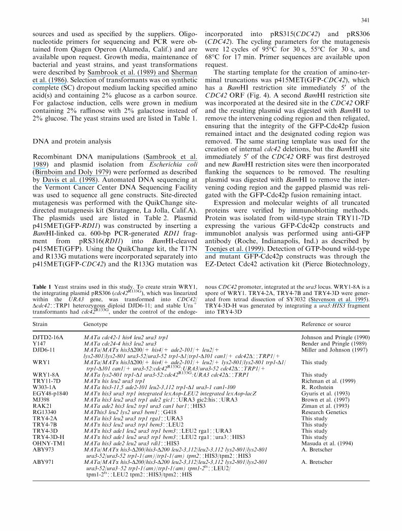
One possible candidate for a RhoGDI binding partnerthat might interact with the Rho-insert domain is theBni1p scaffold protein. To explore this possibility, wecarried out a random mutagenesis/two-hybrid proteininteraction study looking for mutations in Cdc42p thatspecifically abrogated the interactions between Cdc42pand Bni1p (see Materials and methods for details). Inthis study, the R133G mutation within the Rho-insertdomain was isolated. The R133G mutation abolishedinteractions with Bni1p, but retained wild-type interac-tions with Cla4p, Ste20p, Gic1p, and Gic2p (Fig. 3a),other effector proteins that had previously been shownto interact with Cdc42p through the switch I effectordomain. GFP-Cdc42R133Gp was capable of binding GTPwithin the cell (see Fig. 4c) and displayed normal clus-tering and membrane localization (data not shown),indicating that the Arg133 residue did not have a specificrole in nucleotide binding or localization. Expression ofcdc42R133G was able to complement the cdc42-1ts andcdc42D mutants, indicating that Cdc42R133Gp encoded afunctional protein. Cells harboring cdc42R133G as thesole copy of Cdc42p grew at 16, 23, 30, and 37�C and
Fig. 3 a The cdc42R133G mutation specifically blocked two-hybridprotein interactions with Bni1p. Plus sign indicates wild-type (WT)interaction, minus sign indicates no interaction. The D118Amutation keeps Cdc42p in a GDP-bound state and blocksinteractions with all tested effectors (Richman et al. 1999). bExpression of Cdc42R133Gp affected bud morphology at 37�C.Strain WRY1-8A was grown to early log phase at 23�C and halfthe culture was then shifted to 37�C for 7 h and observed bymicroscopy. Bar 10 lm
Fig. 4 a–c Amino-terminaltruncations and internaldeletions. a Lines represent theregions of Cdc42p fused toGFP and the box labeled D119–135 represents the internaldeletion of the Rho-insertdomain. b p415MET(GFP-CDC42) plasmids containingfull-length Cdc42p or thecodons for the residues listedwere transformed into wild-typestrain TRY11-7D andtransformants were observed byfluorescence microscopy.Double plus indicates properlocalization and strongfluorescence, single plusindicates proper localizationbut reduced fluorescence, minusindicates no specific targetingobserved. c GTP-binding ofGFP-Cdc42p mutant proteins.TRY11-7D cells containing theindicated GFP-Cdc42p mutantproteins were grown inSC)Leu+Met and cell lysateswere prepared and thenincubated with 0.1 mM GTPcSprior to incubation with GST-PBD and glutathione-agaroseresin. The T17N and D118Amutant proteins were negativecontrols that did not bind GTPand the D25–51 protein was anegative control that does notbind the GST-PBD. The resultis representative of threeindependent experiments
344
showed normal morphologies at all temperatures except37�C. Cells grown at 37�C exhibited abnormal mor-phologies, including 28% short-elongated budded cellsand 28% multi-budded cells (Fig. 3b, right panel), sug-gesting that Cdc42p–Bni1p interactions played animportant role in normal bud growth during thecell cycle. However, overexpression of Rdi1p led toGFP-Cdc42R133Gp removal from membranes to thesame extent as wild-type GFP-Cdc42p (data not shown),indicating that the R133G mutation did not significantlyaffect interactions with the Rdi1p RhoGDI and sug-gesting that Bni1p does not function in RhoGDI-dependent extraction of Cdc42p.
Cdc42p interactions with downstream effectors were notrequired for localization or clustering
Proper GFP-Cdc42p localization was previously ob-served in the cla4D, gic1D, gic2D, and bni1D effectormutants (Richman et al. 1999, 2002), suggesting thatother single or multiple interactions with downstreameffectors or regulators may be important for localiza-tion. To expand on these studies, GFP-Cdc42p locali-zation was observed in other mutant backgrounds.GFP-Cdc42p localized and clustered normally in a gic1Dgic2D double mutant and in cells deleted for the Bem1pscaffold protein (data not shown). Normal localizationand clustering was previously observed in myosinmyo1D, late secretion sec1ts and sec6ts, and septin cdc12-6ts mutants (Richman et al. 1999, 2002); and the GFP-Cdc42p expressed in either tropomyosin tpm2D singlemutants or tpm1-2ts tpm2D double mutants showed nodefects in localization or clustering (data not shown).
Interestingly, tpm1-2ts tpm2D mutant cells shifted torestrictive temperatures showed normal clustering for upto 1 h. Actin cables are disrupted within 10 min in thismutant (Pruyne et al. 1998), suggesting that actin cablesare not required for clustering. Taken together, theseresults indicated that interactions between Cdc42p andBni1p, Bem1p, Cla4p, Gic1p, and Gic2p are not re-quired for proper localization and that defects in theactin cytoskeleton or polarized secretion do not affectmembrane localization or clustering.
Truncated Cdc42p unable to bind guanine nucleotidesdoes not cluster
To uncover the region(s) or specific amino acid(s) re-quired for Cdc42p clustering, a series of Cdc42p amino-terminal truncations, internal deletions, and pointmutations were examined (Figs. 4, 5). Deletions weremade amino-terminal to the 183KKSKKCTIL domain,which was sufficient for membrane localization but notclustering (Richman et al. 2002). Sequences were sys-tematically deleted from residue 1 to residues 14, 63,119, 135, or 160, with each subsequent deletion repre-senting the removal of a structural domain (see Fig. 4).An internal deletion was also created through the re-moval of the Rho-insert domain (residues 119–135). Themolecular weight and expression of all constructs wereconfirmed by immunoblot analysis; and all constructswere expressed at levels comparable with wild-typeGFP-Cdc42p (data not shown). However, none of theamino-terminal truncations or the internal deletioncould complement the cdc42-1ts mutant (data notshown), indicating that removal of any domain fromCdc42p negatively affected its function.
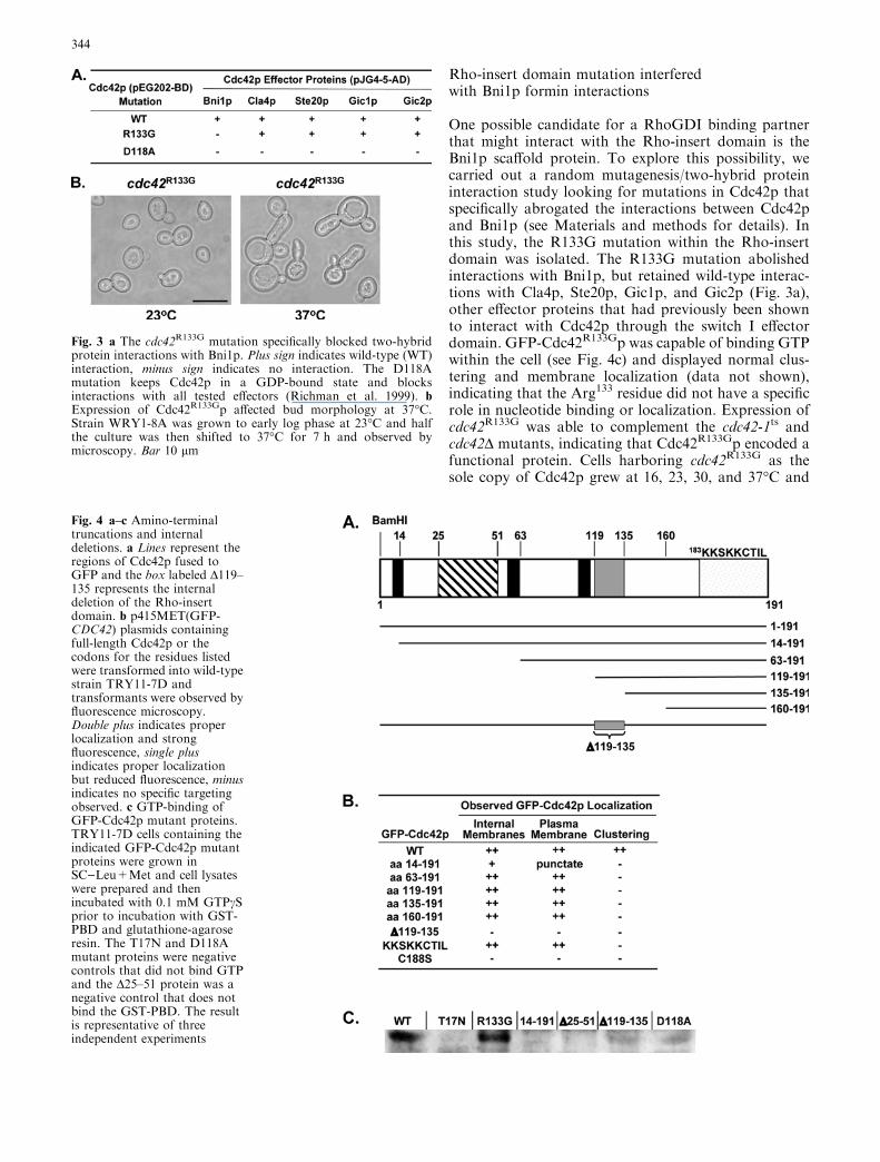
Truncated proteins containing either residues 63–191,119–191, 135–191, or 160–191, which lack guanine-nucleotide binding domains, showed membrane locali-zation patterns that were indistinguishable fromfull-length GFP-Cdc42p (Figs. 4, 5; data not shown),suggesting that nucleotide binding was not a prerequisitefor membrane binding. However, none of these trun-cated proteins exhibited cell-cycle specific clustering atpolarized growth sites, suggesting that residues 63–191were not sufficient for clustering. Two of these con-structs (63–191, 119–191) contained the Rho-insertdomain, indicating that this domain was also not suffi-cient for clustering. However, neither membrane locali-zation nor clustering was observed in cells expressingGFP-Cdc42D119–135p lacking the Rho-insert domain(Fig. 4), suggesting that the Rho-insert domain wasnecessary for localization. GFP-Cdc42D119–135p couldnot bind GTP within the cell (Fig. 4c), reinforcing therole of the Rho-insert domain in providing stability tothe nearby guanine-nucleotide binding domain thatcoordinates guanine ring binding (Hoffman et al. 2000).
Although cells containing GFP-Cdc4263–191p dis-played normal membrane localization, a fragmentedvacuolar phenotype was observed (Fig. 5), which was
Fig. 5 Localization of truncated proteins. p415MET vectorscontaining the indicated wild-type or truncation GFP-CDC42constructs were transformed into wild-type TRY11-7D cells. Bar10 lm
345

confirmed by the vacuolar stain FM4-64 (data notshown). The involvement of Cdc42p and its regulators inendocytosis and vacuolar fusion is well documented(Eitzen et al. 2001; Muller et al. 2001; Murray andJohnson 2001; White and Johnson 1997). Therefore,expression of Cdc4263–191p seemed to have a dominant-negative effect on Cdc42p function in regulating vacu-olar fusion.
GFP-Cdc4214–191p was unable to bind GTP within thecell (Fig. 4c), most probably because it lacked part of theP-loop that was necessary for GTP binding. It did notshow typical plasma membrane localization or cluster-ing, but instead displayed a punctate staining patternaround the periphery of the cell (Figs. 4b, 5). Thisstaining pattern may be due to proper localization ofGFP-Cdc4214–191p to the plasma membrane but aninability of GFP-Cdc4214–191p to form normal complexesat the plasma membrane. However, we cannot rule outthe possibility that improper folding of this larger trun-cated protein promoted the formation of aggregates,even though smaller truncated proteins (i.e., 63–191 or119–191) were localized properly to membranes.
Yellow fluorescent protein–Cdc24p appearedat the same time that cyan fluorescentprotein–Cdc42p began to cluster at the incipient bud siteand mother-bud neck region
Previous data (Richman et al. 2002) and data presentedherein suggested that nucleotide binding had a rolein Cdc42p clustering. If activation of Cdc42p to a
GTP-bound state was a prerequisite for clustering, thenclustering of Cdc42p and localization of its GEF Cdc24pshould temporally coincide at sites of polarized growth.To test this hypothesis, cells containing both yellowfluorescent protein (YFP)-Cdc24p and cyan fluorescentprotein (CFP)-Cdc42p were examined using time-lapsemicroscopy. Time-lapse experiments focused on clus-tering at mother-bud neck regions, because the separa-tion of Cdc24p-stained nuclei during anaphase served asan excellent signal for the imminent Cdc42p clusteringand Cdc24p targeting at the mother-bud neck region(Toenjes et al. 1999). Time-lapse photomicrographs ofnine mother–daughter cell pairs revealed that YFP-Cdc24p appeared at the same time that CFP-Cdc42pbegan to cluster at the mother-bud neck region (Fig. 6).Time-lapse experiments of clustering at incipient budsites were limited by the lack of a signal for the forth-coming appearance of CFP-Cdc42p and YFP-Cdc24p,which led to difficulties with GFP photobleachingupon multiple exposures. However, when incipientbud site co-localization was captured by time-lapse,YFP-Cdc24p appeared and CFP-Cdc42p clusteredwithin a 3-min interval (data not shown). Together withthe mother-bud neck region data, these results posi-tioned Cdc24p localization temporally and spatially withCdc42p clustering.
Discussion
The accumulation of GFP-Cdc42p in the cytosol uponoverexpression of Rdi1p supports a role for Rdi1p instimulating Cdc42p release from membranes, as previ-ously observed with mammalian RhoGDIs (Koch et al.1997; Leonard et al. 1992; Masuda et al. 1994). Thegeneral cytoplasmic localization of GFP-Rdi1p was alsoobserved with a HA-tagged Rdi1p (Koch et al. 1997)and is consistent with its role in extracting Cdc42p fromcellular membranes into the cytosol. The novel locali-zation of GFP-Rdi1p at polarized growth sites in smallbuds and at the mother-bud neck region during cytoki-nesis suggests that an increased localized amount of
Fig. 6 Time-lapse microscopy of CFP-Cdc42p and YFP-Cdc24pco-localization. Plasmids p416MET(CFP-CDC42) and p415ME-T(YFP-CDC24) were transformed into wild-type strain TRY11-7Dand cells were placed onto a thin-layered agar slab made with SC-Leu-Met medium. The zero time-point was set arbitrarily,representing the appearance of post-anaphase cells prior to Cdc42pclustering. YFP-Cdc24p appearance (upper panels, arrow) andCFP-Cdc42p clustering (lower panels, arrow) at the mother-budneck region occurs at approximately the same time (24 min time-point). YFP-Cdc24p targeting to nuclei was apparent at all time-points. Photomicrographs are representative of cells followed innine time-lapse experiments
346
Rdi1p is necessary for the extraction of the accumulated,clustered Cdc42p at these stages of the cell cycle (Rich-man et al. 2002). Interestingly, this cell-cycle specificRdi1p localization was not observed with a HA-taggedRdi1p (Koch et al. 1997), but this discrepancy may havebeen due to its intense cytoplasmic immunofluorescencesignal obscuring the membrane signal. Alternatively, theHA-specific antibodies may not have been able to bindto membrane-bound Rdi1p under the experimentalconditions tested, a phenomenon previously observedwith immunofluorescence localization of Cdc42p (Zimanet al. 1993).
Although deletion of the Rho-insert domain abro-gated the ability of Rdi1p to extract Cdc42p frommembranes, it is unlikely that this was due to a directinteraction between Rdi1p and the Rho-insert domain,based on the X-ray crystal structure of the mammalianCdc42p–RhoGDI complex (Hoffman et al. 2000).Therefore, interactions with other proteins may beimportant for this process. Analysis of the cdc42R133G
mutation assigned an additional function to the Rho-insert domain in mediating interactions with the poten-tial scaffold protein Bni1p, raising the possibility thatBni1p may be involved in Rdi1p function. However, theobservations that GFP-Cdc42R133Gp showed no defectsin localization and that Rdi1p could extractCdc42R133Gp from membranes suggested that interac-tions with Bni1p do not play a role in Rdi1p function.
Bni1p–Cdc42p interactions were not affected by var-ious switch I effector-domain mutations (Richman andJohnson 2000; Richman et al. 1999), suggesting that aregion other than the switch I effector domain was re-quired for these interactions. The isolation of the R133Gmutation from a screen designed to isolate mutationsthat specifically affected Cdc42p–Bni1p interactionsimplicated the Rho-insert domain as one of the Cdc42pregions involved in Cdc42p–Bni1p interactions. Theabnormal morphological phenotype of cdc42R133G cellsgrown at 37�C likewise suggested a functional role for theRho-insert domain in regulating cell polarity and wasconsistent with cell polarity defects associated with bni1mutants (Evangelista et al. 1997; Vallen et al. 2000).Taken together with previous studies showing that theRho-insert domain was involved in interactions withanother potential scaffold protein IQGAP (McCallumet al. 1996; Wu et al. 1997), these results raise the possi-bility that the Rho-insert domain is important formediating specific scaffold–Cdc42p interactions.
Multiple observations have implicated nucleotidebinding as being important for Cdc42p clustering butnot for membrane localization. First, the G12V muta-tion did not affect membrane localization, but did causean aberrant increase in clustering (Richman et al. 2002),raising the possibility that binding of GTP could pro-mote clustering. Also, Cdc42p and its guanine nucleo-tide exchange factor Cdc24p co-localized spatially(Richman et al. 2002) and temporally (Fig. 5) to sites ofpolarized growth, supporting a model in which Cdc24ppromotes clustering through activation of Cdc42p to a
GTP-bound state. Also, various truncated Cdc42 pro-teins predicted to be unable to bind nucleotides wereable to localize properly to cellular membranes but didnot cluster, supporting the importance of the nucleotide-bound state of Cdc42p for clustering. Although theregulation of nucleotide binding likely plays an impor-tant role in clustering, it is not clear whether binding ofGTP is important for the initial stimulation of cluster-ing, or the perpetuation of clustering, or both.
The results presented herein provide an importantinsight into the complicated nature of the relationshipsbetween Cdc42p localization, nucleotide binding, andprotein–protein interactions and the domains that reg-ulate these different functions. These data are consistentwith a model in which: (1) Cdc42p is anchored at theplasma membrane around the periphery of the cellthroughout the cell cycle, (2) its regional concentrationat sites of actin-dependent polarized growth is signifi-cantly increased (clustering) when activated to a GTP-bound state by Cdc24p, and (3) it is extracted from theplasma membrane by Rdi1p when polarized growth iscompleted at the bud tip and the mother-bud neck re-gion following cytokinesis. In this regard, Rdi1p wouldbe acting to turn off Cdc42p-dependent signaling path-ways at specific times in the cell cycle. However, theexact mechanisms by which Cdc42p is targeted to spe-cific membranes and clustered at sites of polarizedgrowth remain to be completely elucidated. Futurestudies should shed light on how multiple Cdc42pfunctional domains work together to control theseessential Cdc42p functions.
Acknowledgements We thank Anthony Bretscher for valuable re-agents, Herman Chen and Patti McClard for expert technicalassistance, and members of the Johnson Laboratory for valuablediscussions and critical comments on this manuscript. K.C.C. wassupported in part by an undergraduate fellowship from the UVM-Howard Hughes HELiX Program. This research was supported byNational Science Foundation grant MCB-0110138.
References
Benard V, Bohl BP, Bokoch GM (1999) Characterization of Racand Cdc42 activation in chemoattraction-stimulated humanneutrophils using a novel assay for active GTPases. J Biol Chem274:13198–13204
Bender A, Pringle JR (1989) Multicopy suppression of the cdc24budding defect in yeast by CDC42 and three newly identifiedgenes including the ras-related gene RSR1. Proc Natl Acad SciUSA 86:9976–9980
Birnboim HC, Doly J (1979) A rapid alkaline extraction procedurefor screening recombinant plasmid DNA. Nucleic Acids Res7:1513–1523
Brown JL, Jaquenoud M, Gulli M-P, Chant J, Peter M (1997)Novel Cdc42-binding proteins Gic1 and Gic2 control cellpolarity in yeast. Genes Dev 11:2972–2982
Butty AC, Perrinjaquet N, Petit A, Jaquenoud M, Segall JE,Hofmann K, Zwahlen C, Peter M (2002) A positive feedbackloop stabilizes the guanine-nucleotide exchange factor Cdc24 atsites of polarization. EMBO J 21:1565–1576
Chen G-C, Kim Y-J, Chan CSM (1997) The Cdc42 GTPase-asso-ciated proteins Gic1 and Gic2 are required for polarized cellgrowth in Saccharomyces cerevisiae. Genes Dev 11:2958–2971
347

Cvrckova F, De Virgilio C, Manser E, Pringle JR, Nasmyth K(1995) Ste20-like protein kinases are required for normallocalization of cell growth and for cytokinesis in budding yeast.Genes Dev 9:1817–1830
Davis CR, Richman TJ, Deliduka SB, Blaisdell JO, Collins CC,Johnson DI (1998) Analysis of the mechanisms of action ofthe Saccharomyces cerevisiae dominant lethal cdc42G12V anddominant negative cdc42D118A mutations. J Biol Chem273:849–858
Diebold BA, Bokoch GM (2001) Molecular basis for Rac2 regu-lation of phagocyte NADPH oxidase. Nat Immunol 2:211–215
Eitzen G, Thorngren N, Wickner W (2001) Rho1p and Cdc42p actafter Ypt7p to regulate vacuole docking. EMBO J 20:5650–5656
Etienne-Manneville S, Hall A (2002) Rho GTPases in cell biology.Nature 420:629–635
Evangelista M, Blundell K, Longtine MS, Chow CJ, Adames N,Pringle JR, Peter M, Boone C (1997) Bni1p, a yeast forminlinking Cdc42p and the actin cytoskeleton during polarizedmorphogenesis. Science 276:118–122
Finegold AA, Johnson DI, Farnsworth CC, Gelb MH, Judd SR,Glomset JA, Tamanoi F (1991) Protein geranylgeranyltrans-ferase of Saccharomyces cerevisiae is specific for Cys-Xaa-Xaa-Leu motif proteins and requires the CDC43 gene product, butnot the DPR1 gene product. Proc Natl Acad Sci USA 88:4448–4452
Gyuris J, Golemis E, Chertkov H, Brent R (1993) Cdi, a human G1and S phase protein phosphatase that associates with Cdk2.Cell 75:791–803
Hart MJ, Maru Y, Leonard D, Witte ON, Evans T, Cerione RA(1992) A GDP dissociation inhibitor that serves as a GTPaseinhibitor for the Ras-like protein CDC42Hs. Science 258:812–815
Hoffman GR, Nassar N, Cerione RA (2000) Structure of the Rhofamily GTP-binding protein Cdc42 in complex with the multi-functional regulator RhoGDI. Cell 100:345–356
Hori Y, Kikuchi A, Isomura M, Katayama M, Miura Y, FujiokaH, Kaibuchi K, Takai Y (1991) Post-translational modifica-tions of the C-terminal region of rho are important for itsinteraction with membranes and the stimulatory and inhibitoryGDP/GTP exchange proteins. Oncogene 6:515–522
Johnson DI (1999) Cdc42: an essential Rho-type GTPase control-ling eukaryotic cell polarity. Microbiol Mol Biol Rev 63:54–105
Johnson DI, Pringle JR (1990) Molecular characterization ofCDC42, a Saccharomyces cerevisiae gene involved in thedevelopment of cell polarity. J Cell Biol 111:143–152
Joneson T, Bar-Sagi D (1998) A Rac1 effector site controllingmitogenesis through superoxide production. J Biol Chem273:17991–17994
Koch G, Tanaka K, Masuda T, Yamochi W, Nonaka H, Takai Y(1997) Association of the Rho family small GTP-binding pro-teins with Rho GDP dissociation inhibitor (Rho GDI) in Sac-charomyces cerevisiae. Oncogene 15:417–422
Leonard D, Hart MJ, Platko JV, Eva A, Henzel W, Evans T,Cerione RA (1992) The identification and characterization of aGDP-dissociation inhibitor (GDI) for the CDC42Hs protein. JBiol Chem 267:22860–22868
Masuda T, Tanaka K, Nonaka H, Yamochi W, Maeda A, Takai Y(1994) Molecular cloning and characterization of yeast rhoGDP dissociation inhibitor. J Biol Chem 269:19713–19718
McCallum SJ, Wu WJ, Cerione RA (1996) Identification of aputative effector for Cdc42Hs with high sequence similarity tothe RasGAP-related protein IQGAP1 and a Cdc42Hs bindingpartner with similarity to IQGAP2. J Biol Chem 271:21732–21737
Merla A, Johnson DI (2000) The Cdc42p GTPase is targeted to thesite of cell division in the fission yeast Schizosaccharomycespombe. Eur J Cell Biol 79:469–477
Merla A, Johnson DI (2001) The Schizosaccharomyces pombeCdc42p GTPase signals through Pak2p and the Mkh1p–Pek1p–Spm1p MAP kinase pathway. Curr Genet 39:205–209
Miller PJ, Johnson DI (1997) Characterization of the S. cerevisiaecdc42-1ts allele and new temperature-conditional-lethal cdc42alleles. Yeast 13:561–572
Murray JM, Johnson DI (2001) The Cdc42p GTPase and its reg-ulators Nrf1p and Scd1p are involved in endocytic trafficking inthe fission yeast Schizosaccharomyces pombe. J Biol Chem276:3004–3009
Muller O, Johnson DI, Mayer A (2001) Cdc42p functions at thedocking stage of yeast vacuole membrane fusion. EMBO J20:5657–5665
Nern A, Arkowitz RA (1999) A Cdc24p–Far1p–Gbg proteincomplex required for yeast orientation during mating. J CellBiol 144:1187–1202
Nomanbhoy TK, Cerione RA (1996) Characterization of theinteraction between RhoGDI and Cdc42Hs using fluorescencespectroscopy. J Biol Chem 271:10004–10009
Nomanbhoy TK, Erikson JW, Cerione RA (1999) Kinetics ofCdc42 membrane extraction by Rho-GDI monitored by real-time fluorescence resonance energy transfer. Biochemistry38:1744–1750
Pruyne DW, Schott DH, Bretscher A (1998) Tropomyosin-con-taining actin cables direct the Myo2p-dependent polarizeddelivery of secretory vesicles in budding yeast. J Cell Biol143:1931–1945
Richman TJ, Johnson DI (2000) Saccharomyces cerevisiae Cdc42pGTPase is involved in preventing the recurrence of bud emer-gence during the cell cycle. Mol Cell Biol 20:8548–8559
Richman TJ, Sawyer MM, Johnson DI (1999) The Cdc42p GTPaseis involved in a G2/M morphogenetic checkpoint regulating theapical-isotropic switch and nuclear division in yeast. J BiolChem 274:16861–16870
Richman TJ, Sawyer MM, Johnson DI (2002) Saccharomyces ce-revisiae Cdc42p localizes to cellular membranes and clusters atsites of polarized growth. Eukaryot Cell 1:458–468
Sambrook J, Fritsch EF, Maniatis T (1989) Molecular cloning: alaboratory manual. Cold Spring Harbor Laboratory, ColdSpring Harbor, N.Y.
Sherman F, Fink GR, Hicks JB (1986) Methods in yeast genetics:laboratory manual. Cold Spring Harbor Laboratory, ColdSpring Harbor, N.Y.
Stevenson BJ, Ferguson B, De Virgilio C, Bi E, Pringle JR, Am-merer G, Sprague GF (1995) Mutation of RGA1, which encodesa putative GTPase-activating protein for the polarity-estab-lishment protein Cdc42p, activates the pheromone-responsepathway in the yeast Saccharomyces cerevisiae. Genes Dev9:2949–2963
Toenjes KA, Sawyer MM, Johnson DI (1999) The guanine-nucle-otide-exchange factor Cdc24p is targeted to the nucleus andpolarized growth sites. Curr Biol 9:1183–1186
Vallen EA, Caviston J, Bi E (2000) Roles of Hof1p, Bni1p, Bnr1p,and Myo1p in cytokinesis in Saccharomyces cerevisiae. MolBiol Cell 11:593–611
Vetter IR, Wittinghofer A (2001) Signal transduction—the guaninenucleotide-binding switch in three dimensions. Science294:1299–1304
Walker SJ, Brown HA (2002) Specificity of Rho insert-mediatedactivation of phospholipase D1. J Biol Chem 277:26260–26267
Walker SJ, Wu W-J, Cerione RA, Brown HA (2000) Activation ofphospholipase D1 by Cdc42 requires the Rho insert region.J Biol Chem 275:15665–15668
White WH, Johnson DI (1997) Characterization of synthetic-lethalmutants reveals a role for the Saccharomyces cerevisiae guanine-nucleotide exchange factor Cdc24p in vacuole function andNa+ tolerance. Genetics 147:43–55
Wu WJ, Leonard DA, Cerione RA, Manor D (1997) Interactionbetween Cdc42Hs and RhoGDI is mediated through the Rhoinsert region. J Biol Chem 272:26153–26158
Wu WJ, Lin R, Cerione RA, Manor D (1998) Transformationactivity of Cdc42 requires a region unique to Rho-related pro-teins. J Biol Chem 273:16655–16658
348
Ziman M, Johnson DI (1994) Genetic evidence for a functionalinteraction between S. cerevisiae CDC24 and CDC42. Yeast10:463–474
Ziman M, O’Brien JM, Ouellette LA, Church WR, Johnson DI(1991) Mutational analysis of CDC42Sc, a Saccharomycescerevisiae gene that encodes a putative GTP-binding protein
involved in the control of cell polarity. Mol Cell Biol 11:3537–3544
Ziman M, Preuss D, Mulholland J, O’Brien JM, Botstein D,Johnson DI (1993) Subcellular localization of Cdc42p, a Sac-charomyces cerevisiae GTP-binding protein involved in thecontrol of cell polarity. Mol Biol Cell 4:1307–1316
349