Electrorefining of a Base Metal Refinery residue copper alloy ...

Biochemical and Biophysical Research Communications 312 (2003) 755–760
BBRCwww.elsevier.com/locate/ybbrc
Analysis of a substrate specificity switch residueof cephalosporin acylase
Charles F. Sio, Linda G. Otten, Robbert H. Cool, and Wim J. Quax*
Pharmaceutical Biology, University Centre for Pharmacy, University of Groningen, Antonius Deusinglaan 1, 9713 AV Groningen, The Netherlands
Received 17 October 2003
Abstract
Residue Phe375 of cephalosporin acylase has been identified as one of the residues that is involved in substrate specificity.
A complete mutational analysis was performed by substituting Phe375 with the 19 other amino acids and characterising all purified
mutant enzymes. Several mutations cause a substrate specificity shift from the preferred substrate of the enzyme, glutaryl-7-ACA,
towards the desired substrate, adipyl-7-ADCA. The catalytic efficiency (kcat/Km) of mutant SY-77F375C towards adipyl-7-ADCA was
increased 6-fold with respect to the wild-type enzyme, due to a strong decrease of Km. The kcat of mutant SY-77F375H towards adipyl-
7-ADCA was increased 2.4-fold. The mutational effects point at two possible mechanisms by which residue 375 accommodates the
long side chain of adipyl-7-ADCA, either by a widening of a hydrophobic ring-like structure that positions the aliphatic part of the
side chain of the substrate, or by hydrogen bonding to the carboxylate head of the side chain.
� 2003 Elsevier Inc. All rights reserved.
Keywords: Cephalosporin acylase; Glutaryl acylase; Substrate specificity; Protein engineering; Site directed mutagenesis; Saturation mutagenesis;
Adipyl-7-ADCA; Semi-synthetic cephalosporins; Pseudomonas SY-77
Cephalosporin acylase is a crucial enzyme for
the development of new enzymatic pathways leadingto the production of semi-synthetic cephalosporins.
7-Aminocephalosporanic acid (7-ACA) and 7-amin-
odesacetoxycephalosporanic acid (7-ADCA) are impor-
tant intermediates in these pathways. Large quantities of
these compounds are needed, but the traditional pro-
duction routes are time-consuming, expensive, and/or
polluting [1]. An alternative route to obtain 7-A(D)CA is
the one-step enzymatic deacylation of either adipyl-7-ADCA [2] or cephalosporin C (CPC) [3]. However,
currently there is no biocatalyst available that is capable
of performing this task cost-efficiently [1]. A good
starting point for the generation of such an adipyl acyl-
ase or CPC acylase could be Pseudomonas SY-77 glutaryl
acylase [4,5], a cephalosporin acylase that is highly active
on a similar compound glutaryl-7-ACA (Fig. 1).
* Corresponding author. Fax: +31-50-363-3000.
E-mail address: [email protected] (W.J. Quax).
URL: http://www.farmbio.nl.
0006-291X/$ - see front matter � 2003 Elsevier Inc. All rights reserved.
doi:10.1016/j.bbrc.2003.10.180
Crystallisation studies of cephalosporin acylase with
and without glutaryl-7-ACA or glutarate showed thatPhe375 is part of the substrate binding site of the en-
zyme [6–8]. In an unbiased approach using directed
evolution of Pseudomonas SY-77 glutaryl acylase, mu-
tant SY-77F375L was found as one of the mutants that
significantly increases the activity of the enzyme on
adipyl-7-ADCA [9]. However, directed evolution by the
introduction of point mutations in the codon for Phe375
allows for the substitution by only six residues (Leu, Ile,Val, Cys, Tyr, and Ser) and consequently explores a
limited part of the sequence space at this position. Ad-
ditionally, the mutants were selected on an improved
hydrolysis of adipyl-7-ADCA analogues. Consequently,
neutral and negative effects and effects on the hydrolysis
of other b-lactam compounds that deepen the knowl-
edge of the cephalosporin acylase–substrate interaction
could not be observed. Therefore, it was decided to use asaturated mutagenesis approach to analyse the effects of
all possible residues at position 375 on the hydrolysis of
three b-lactam compounds. Mutant enzymes with all
natural amino acids at position 375 were overproduced

Fig. 1. The production of 7-A(D)CA from b-lactam compounds.
756 C.F. Sio et al. / Biochemical and Biophysical Research Communications 312 (2003) 755–760
and purified to determine their activities in the hydro-
lysis of glutaryl-7-ACA, adipyl-7-ADCA, and CPC, and
autocatalytic processing.
Several mutations were shown to improve the kinetic
parameters towards adipyl-7-ADCA. The highest in-
crease in kcat was due to the incorporation of a histidine,
whereas the largest decrease of the Km value was found
for mutant SY-77F375C that also shows the highest in-crease in catalytic efficiency. The effects of all mutations
on the kinetic parameters are discussed on the basis of
the crystal structure of the enzyme and allowed to elu-
cidate the possible modes by which residues at position
375 can modify substrate specificity.
Materials and methods
Chemicals. The b-lactam substrates glutaryl-7-ACA, adipyl-
7-ADCA, and CPC (as sodium salt) were gifts from DSM, The
Netherlands. Fluorescamine was from Sigma, BugBuster was from
Novagen, and 5-bromo-4-chloro-3-indolyl-phosphate and nitro blue
tetrazolium were from Duchefa, The Netherlands.
Mutagenesis of residue Phe375 of Pseudomonas SY-77 glutaryl
acylase. Mutagenesis was performed on plasmid pMcSY-2 [9], har-
bouring the gene encoding Pseudomonas SY-77 glutaryl acylase under
control of the tac-promoter. The random primer 50-atg cag gtg ccg acc
nng/c aac atc gtc tac gcc g was used to mutate the codon for Phe375 in
a megaprimer reaction with Pfu DNA polymerase (Stratagene) [10].
The resulting PCR product was ligated into pMcSY-2 by digestion
using the BglII and SstII restriction sites. After transformation into
Escherichia coli DH10B, cells were plated on LBagar plates containing
50 lg/ml chloramphenicol. Single colonies were picked for DNA se-
quence determination. Nine different mutants were obtained using this
method, the remaining 10 were produced by PCR using the site di-
rected version of the same primer, in which nng/c was replaced with the
codon for the desired residue.
A test was conducted to confirm that the transformants were still
able to produce acylase. Ten millilitres of 2� TY medium [11] sup-
plemented with 50lg/ml chloramphenicol and 0.1% glycerol was in-
oculated with 0.1ml of an overnight culture and grown at 30 �C for
24 h. Cells were harvested from 5ml of this culture and lysed with the
non-ionic detergent BugBuster [9]. The soluble fraction was spotted on
a nitrocellulose membrane, which was subsequently incubated with a
polyclonal rabbit antibody against purified Pseudomonas SY-77 glut-
aryl acylase (Eurogentec S.A.) and an alkaline phosphatase-conjugated
goat anti-rabbit antibody. The appearance of purple spots after incu-
bation with 5-bromo-4-chloro-3-indolyl-phosphate and nitro blue
tetrazolium confirmed the presence of acylase enzyme in the sample.
Transformants that did not produce acylase were discarded.
Characterisation of mutant enzymes.Mutant and WT enzymes were
produced in E. coli DH10B in 100ml cultures and purified by anion
exchange and hydrophobic interaction column chromatography as
described earlier [9]. The protein concentration in the purified samples
was determined using the DC protein assay (Bio-Rad) with BSA as the
reference protein. Analysis of the samples by SDS–PAGE [12] was done
using a 12.5% gel, which was stained with Coomassie brilliant blue.
The kinetic parameters kcat, Km, and kcat/Km of mutant and WT
enzyme towards glutaryl-7-ACA and adipyl-7-ADCA were determined
using the automated fluorescamine assay in 96-well format as de-
scribed earlier [9]. A concentration range of the b-lactam substrates
was incubated with a fixed amount of enzyme in 20mM phosphate
buffer, pH 7.5, at 37 �C. The reaction was stopped by the addition of
acetate buffer, pH 4.5, and the amount of product 7-A(D)CA formed
was determined by incubation with fluorescamine and measuring the
absorption at 380 nm. The kinetic parameters kcat and Km were cal-
culated from the rates of reaction at different concentrations of sub-
strate. Activity towards CPC was determined by the fluorescamine
assay at a high concentration of substrate [9], using approx. 20 lg of
purified enzyme and 5mM CPC in 200ll of reaction mixture. Aliquots
were taken after 24 and 48 h incubation at 37 �C and incubated with
fluorescamine to determine whether CPC had been deacylated.
Results
Production and autocatalytic processing of mutant
enzymes
All 19 mutant enzymes and WT could be expressed
and purified from E. coli DH10B at a yield of more than
10mg/L culture. On SDS–PAGE (Fig. 2), all purified
mutants displayed the bands corresponding to the ma-
ture a- and b-subunits, with the exception of mutant SY-
77F375P, which showed only the band corresponding tothe unprocessed propeptide consisting of the a-subunit,spacer peptide, and the b-subunit. Mutants SY-77F375I,
SY-77F375R, SY-77F375T, and SY-77F375V showed a band
corresponding to the unprocessed propeptide in addi-
tion to the a- and b-subunits, indicating that they un-
derwent partial processing. An impaired processing
could have also led to the accumulation of the poly-
peptide consisting of a-subunit plus spacer peptide, butno band corresponding to this polypeptide was seen in
any of the samples. Remarkably, the elution profiles of
all mutants were similar to that of the WT enzyme in all
column chromatography steps, indicating that the
folding of the maturated and the non-maturated en-
zymes is very similar, which has also been observed for
the S199A mutant of cephalosporin acylase [13].
Hydrolysis of glutaryl-7-ACA
None of the mutations of residue 375 improved the
kinetic parameters towards the preferred substrate of the
enzyme, glutaryl-7-ACA (Table 1 and Fig. 3). The best

Fig. 2. Maturation of WT and mutant cephalosporin acylases. Cepha-
losporin acylase is produced as a propeptide consisting of a signal se-
quence,a-subunit, spacer peptide, andb-subunit. Impairment of the first
maturation step leads to the accumulation of propeptide. Impairment of
the second maturation step would lead to accumulation of a-sub-unit + spacer peptide, visible as a band just above the band corre-
sponding to the a-subunit. Samples were incubated at 100 �C for 2min;
each lane contains 3 lg purified protein. The residues in which Phe375
has been mutated are indicated by the one-letter code. Ma, marker
proteins (Bio-Rad); a, a-subunit; b, b-subunit; and pp, propeptide.
C.F. Sio et al. / Biochemical and Biophysical Research Communications 312 (2003) 755–760 757
mutation was F375C, which lowered the catalytic effi-
ciency to about 50% of the value of WT due to a de-
crease of kcat. SY-77F375C was the only mutant that did
Table 1
Kinetic parameters of WT and mutant cephalosporin acylases
Residue Adipyl-7-ADCA
kcat (s�1) Km (mM)
Phe(WT) 0.49� 0.04 1.0� 0.2
Ala 0.20� 0.01 0.51� 0.08
Arg 0.018� 0.002 10� 1
Asn 0.9� 0.1 0.83� 0.09
Asp 0.015� 0.004 11� 3
Cys 0.55� 0.03 0.17� 0.03
Glu 0.08� 0.02 12� 2
Gln 0.68� 0.05 2.1� 0.3
Gly 0.073� 0.004 0.16� 0.02
His 1.16� 0.08 0.9� 0.2
Ile 0.097� 0.003 2.07� 0.03
Leu 0.67� 0.04 0.7� 0.1
Lys 0.036� 0.001 2.16� 0.07
Met 0.65� 0.02 0.65� 0.04
Pro N.D. N.D.
Ser 0.11� 0.01 0.99� 0.09
Thr 0.24� 0.04 0.9� 0.4
Trp 0.026� 0.001 1.5� 0.2
Tyr 0.43� 0.05 0.7� 0.1
Val 0.090� 0.003 1.37� 0.07
The kinetic parameters were calculated from Eadie–Hofstee plots. Valu
N.D., not detectable.
not significantly increase the Km towards glutaryl-7-ACA. The other sulphur residue Met caused a compa-
rable decrease of kcat accompanied by an 5-fold increase
of Km. The substitution of phenylalanine by tyrosine
lowered kcat only marginally, but it did cause a 2.5-fold
increase of Km. A similar effect was caused by the F375S
mutation, but the other hydroxyl residue Thr had a
stronger negative effect on the kinetic parameters. With
regard to the aliphatic residues, the smaller residues Glyand Ala caused a less drastic decline of kinetic param-
eters than the larger residues Val, Leu, and Ile. The in-
troduction of all other residues resulted in a drastic
decrease of catalytic efficiency.
Hydrolysis of adipyl-7-ADCA
All mutants showed unique kinetic parameters on the
desired substrate adipyl-7-ADCA. Five mutants showed
an increased catalytic efficiency, three a catalytic effi-
ciency similar to WT, and 11 a decreased catalytic effi-
ciency (Table 1 and Fig. 3). The catalytic efficiency
towards adipyl-7-ADCA was increased 6-fold by theintroduction of a Cys at position 375, mainly due to a
strong reduction of Km. The other sulphur-containing
residue, Met, increased the catalytic efficiency by a fac-
tor of 2 by slightly increasing kcat and decreasing Km.
The hydrophilic residues Asn, Gln, and His all increased
kcat. The catalytic efficiency was increased more than
2-fold by the introduction of Asn and His but not of
Gln. In contrast to Asn, Gln, and His, the hydrophilichydroxyl residues Ser and Thr significantly lowered kcatand did not affect Km. The aliphatic residue Leu
Glutaryl-7-ACA
kcat (s�1) Km (mM)
4.0� 0.3 0.031� 0.002
2.35� 0.07 0.07� 0.01
0.21� 0.05 0.6� 0.2
1.96� 0.05 0.18� 0.03
0.010� 0.002 0.22� 0.06
2.308� 0.007 0.038� 0.008
0.008� 0.001 0.2� 0.1
0.80� 0.02 0.7� 0.2
2.46� 0.08 0.066� 0.008
1.334� 0.007 0.11� 0.03
0.67� 0.04 0.10� 0.02
1.10� 0.09 0.26� 0.07
2.2� 0.4 0.7� 0.2
2.02� 0.09 0.17� 0.02
N.D. N.D.
3.1� 0.2 0.07� 0.02
1.8� 0.1 0.09� 0.03
0.47� 0.06 0.8� 0.1
3.64� 0.05 0.08� 0.01
1.02� 0.08 0.10� 0.02
es given are means�SD of at least three independent measurements.
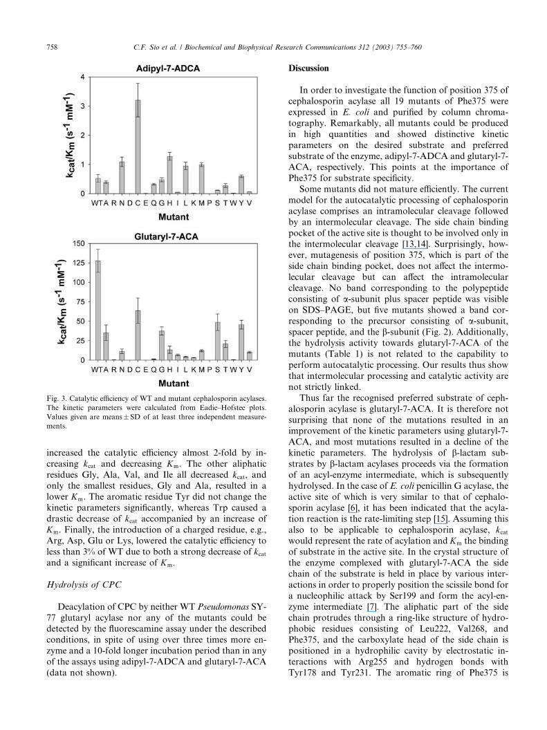
Fig. 3. Catalytic efficiency of WT and mutant cephalosporin acylases.
The kinetic parameters were calculated from Eadie–Hofstee plots.
Values given are means� SD of at least three independent measure-
ments.
758 C.F. Sio et al. / Biochemical and Biophysical Research Communications 312 (2003) 755–760
increased the catalytic efficiency almost 2-fold by in-
creasing kcat and decreasing Km. The other aliphatic
residues Gly, Ala, Val, and Ile all decreased kcat, andonly the smallest residues, Gly and Ala, resulted in a
lower Km. The aromatic residue Tyr did not change the
kinetic parameters significantly, whereas Trp caused a
drastic decrease of kcat accompanied by an increase of
Km. Finally, the introduction of a charged residue, e.g.,
Arg, Asp, Glu or Lys, lowered the catalytic efficiency to
less than 3% of WT due to both a strong decrease of kcatand a significant increase of Km.
Hydrolysis of CPC
Deacylation of CPC by neither WT Pseudomonas SY-
77 glutaryl acylase nor any of the mutants could bedetected by the fluorescamine assay under the described
conditions, in spite of using over three times more en-
zyme and a 10-fold longer incubation period than in any
of the assays using adipyl-7-ADCA and glutaryl-7-ACA
(data not shown).
Discussion
In order to investigate the function of position 375 of
cephalosporin acylase all 19 mutants of Phe375 were
expressed in E. coli and purified by column chroma-
tography. Remarkably, all mutants could be produced
in high quantities and showed distinctive kinetic
parameters on the desired substrate and preferred
substrate of the enzyme, adipyl-7-ADCA and glutaryl-7-ACA, respectively. This points at the importance of
Phe375 for substrate specificity.
Some mutants did not mature efficiently. The current
model for the autocatalytic processing of cephalosporin
acylase comprises an intramolecular cleavage followed
by an intermolecular cleavage. The side chain binding
pocket of the active site is thought to be involved only in
the intermolecular cleavage [13,14]. Surprisingly, how-ever, mutagenesis of position 375, which is part of the
side chain binding pocket, does not affect the intermo-
lecular cleavage but can affect the intramolecular
cleavage. No band corresponding to the polypeptide
consisting of a-subunit plus spacer peptide was visible
on SDS–PAGE, but five mutants showed a band cor-
responding to the precursor consisting of a-subunit,spacer peptide, and the b-subunit (Fig. 2). Additionally,the hydrolysis activity towards glutaryl-7-ACA of the
mutants (Table 1) is not related to the capability to
perform autocatalytic processing. Our results thus show
that intermolecular processing and catalytic activity are
not strictly linked.
Thus far the recognised preferred substrate of ceph-
alosporin acylase is glutaryl-7-ACA. It is therefore not
surprising that none of the mutations resulted in animprovement of the kinetic parameters using glutaryl-7-
ACA, and most mutations resulted in a decline of the
kinetic parameters. The hydrolysis of b-lactam sub-
strates by b-lactam acylases proceeds via the formation
of an acyl-enzyme intermediate, which is subsequently
hydrolysed. In the case of E. coli penicillin G acylase, the
active site of which is very similar to that of cephalo-
sporin acylase [6], it has been indicated that the acyla-tion reaction is the rate-limiting step [15]. Assuming this
also to be applicable to cephalosporin acylase, kcatwould represent the rate of acylation and Km the binding
of substrate in the active site. In the crystal structure of
the enzyme complexed with glutaryl-7-ACA the side
chain of the substrate is held in place by various inter-
actions in order to properly position the scissile bond for
a nucleophilic attack by Ser199 and form the acyl-en-zyme intermediate [7]. The aliphatic part of the side
chain protrudes through a ring-like structure of hydro-
phobic residues consisting of Leu222, Val268, and
Phe375, and the carboxylate head of the side chain is
positioned in a hydrophilic cavity by electrostatic in-
teractions with Arg255 and hydrogen bonds with
Tyr178 and Tyr231. The aromatic ring of Phe375 is

Fig. 4. Three-dimensional model of the binding of the side chain of glutaryl-7-ACA by cephalosporin acylase. Residues Leu222, Val268, and Phe375
(green) form a hydrophobic ring-like structure, through which the aliphatic part of the side chain of glutaryl-7-ACA (gold) protrudes, placing the
carboxylate head of the side chain in a hydrophilic cavity consisting of Tyr178, Tyr231, Gln248, and Arg255 (cyan). Ser 199 (magenta) performs the
nucleophilic attack on the scissile bond of the substrate. The Van der Waals radii of the residues forming the hydrophobic ring are shown in dots.
Phe375 interacts with C4 of the side chain and pushes the hydrophilic head into the cavity (Figure made with RasTop version 2.0.3, www.gen-
einfinity.org/rastop/, using the co-ordinates of PDB entry 1JVZ). (For interpretation of the references to colour in this figure legend, the reader is
referred to the web version of this paper.)
C.F. Sio et al. / Biochemical and Biophysical Research Communications 312 (2003) 755–760 759
positioned alongside the glutaryl side chain (Fig. 4).
This may explain why the incorporation of the small
hydrophobic residues Gly, Ala, and Cys causes the
smallest negative effects on the kinetic parameters
towards glutaryl-7-ACA. The incorporation of some
polarity can be overcome, as is demonstrated by therelatively small effects of the F375Y and F375S muta-
tions, but the incorporation of residues with larger side
chains results in a poor hydrolysis activity, probably due
to steric hindrance of the side chain.
In order to shift the substrate specificity of the en-
zyme towards the desired substrate adipyl-7-ADCA, the
longer adipyl side chain must be accommodated by the
side chain binding pocket while the scissile bond ismaintained in a favourable position with respect to
Ser199. Two strategies may be used: the hydrophobic
ring has to be widened in order to accommodate a
twisted aliphatic chain, or the carboxylate head has to
be pulled further into the side chain binding pocket,
which was proposed to be the mechanism of the im-
proved hydrolysis of adipyl-7-ADCA by mutant SY-
77Y178H [16]. The slightly different fit of glutaryl-7-ACAand glutarate in the substrate binding site [7] and the
alkylation of Trp202 by substrate analogues [17] indi-
cate that some flexibility in the binding of substrates
exists, a prerequisite for these two strategies.
Depending on the nature of the residue that substi-
tutes Phe375 either mechanism may apply. The substi-
tution of Phe375 by the smaller hydrophobic residues
Cys, Leu, and Met will expand the hydrophobic ringand generate extra space for the longer side chain. Ap-
parently, this is a very delicate procedure, since Val and
Ile have a negative influence on the kinetic parameters.
The data suggest that a greater expansion of the ring,
e.g., via substituting Phe375 by Gly or Ala, increases the
ability of the side chain binding pocket to accommodate
the adipyl side chain, as is indicated by a strong decrease
of Km. At the same time, the lower kcat values suggest
that the positioning of the scissile bond for hydrolysis is
less optimal in these mutants.
As for the second mechanism, the increase of kcatupon substituting Phe375 by the hydrogen-bond donors
Asn, Gln or His indicates that position 375 can also be
used to pull the adipyl side chain further into the side
chain binding pocket by means of direct or indirect
hydrogen-bonding to the carboxylate head. Again, the
structural alterations seem to be very delicate, since the
potentially hydrogen-bonding Ser and Thr have a neg-
ative effect on the kcat, and substitution by Tyr, which isbasically a phenylalanine with a hydrogen-bonding hy-
droxyl group, does not alter the kinetic parameters. The
effects of these mutations on the hydrolysis of glutaryl-7-
ACA are different and suggest that the carboxylate head
of the glutaryl side chain cannot use hydrogen bonding
to residue 375 to increase binding and hydrolysis. Ap-
parently, the shorter length of the side chain limits the
degrees of freedom and thereby prohibits alternativebinding modes.
The introduction of a charged residue (Arg, Lys, Glu,
and Asp) or the bulky Trp at position 375 seems to
block the passage of the charged carboxylate head of the
side chain through the hydrophobic ring and results in
very poor catalytic parameters towards both adipyl-7-
ADCA and glutaryl-7-ACA. Surprisingly, most of these
mutations do not impair the autocatalytic processing ofthe enzyme, another indication that processing and
catalytic activity are not as closely linked as has been
suggested by others.
This study shows that hydrolysis of adipyl-7-ADCA
can be increased by improving the hydrophobic

760 C.F. Sio et al. / Biochemical and Biophysical Research Communications 312 (2003) 755–760
interactions between amino acid 375 and the aliphaticside chain of the substrate, as well as by hydrogen
bonding of residue 375 to the carboxylate head of the
substrate. Furthermore, residue 375 can function as a
gatekeeper by blocking passage through the hydropho-
bic ring of the substrate binding site by steric or elec-
trostatic means. These results demonstrate that residue
375 is a key amino acid in the protein engineering of
cephalosporin acylase. The saturation mutagenesis, inwhich the effects of all 20 amino acids at this position
were analysed, was essential for the discovery of the
three different modes by which residue 375 can dictate
substrate specificity. Although the catalytic efficiency for
adipyl-7-ADCA can be improved 6-fold by mutagenesis
of Phe375, no activity on CPC could be detected. Ap-
parently, the amino moiety in the side chain of CPC still
comprises an insurmountable problem for an efficienthydrolysis, and additional mutations are required. Mu-
tation F375C decreases the Km towards adipyl-7-ADCA
by a factor 6 while the Km towards glutaryl-7-ACA re-
mains identical, indicating that the substrate specificity
of this mutant has been extended rather than shifted.
Mutant SY-77F375C may thus be regarded as the tem-
plate of choice for future mutagenesis studies.
Acknowledgments
This research was sponsored by contract GBI.4707 from STW,
which is part of the Dutch Organisation for Science and EU Grant
QLTR-2001-00519. R.H. Cool was supported by the EU-community
initiative Interreg IIIA.
References
[1] A. Bruggink, E.C. Roos, E. deVroom, Penicillin acylase in the
industrial production of b-lactam antibiotics, Org. Process Res.
Dev. 2 (1998) 128–133.
[2] L. Crawford, A.M. Stepan, P.C. McAda, J.A. Rambosek, M.J.
Conder, V.A. Vinci, C.D. Reeves, Production of cephalosporin
intermediates by feeding adipic acid to recombinant Penicillium
chrysogenum strains expressing ring expansion activity, Biotech-
nology N. Y. 13 (1995) 58–62.
[3] A. Matsuda, K. Matsuyama, K. Yamamoto, S. Ichikawa, K.
Komatsu, Cloning and characterization of the genes for two
distinct cephalosporin acylases from a Pseudomonas strain, J.
Bacteriol. 169 (1987) 5815–5820.
[4] S. Ichikawa, Y. Shibuya, K. Matsumoto, T. Fujii, K. Komatsu,
R. Kodaira, Purification and properties of 7b-(4-carboxybuta-namido) cephalosporanic acid acylase produced by mutants
derived from Pseudomonas, Agric. Biol. Chem. 45 (1981) 2231–
2236.
[5] Y. Shibuya, K. Matsumoto, T. Fujii, Isolation and properties of
7b-(4-carboxybutanamido) cephalosporanic acid acylase-produc-
ing bacteria, Agric. Biol. Chem. 45 (1981) 1561–1567.
[6] Y. Kim, K. Yoon, Y. Khang, S. Turley, W.G. Hol, The 2.0�AA
crystal structure of cephalosporin acylase, Structure 8 (2000)
1059–1068.
[7] Y. Kim, W.G. Hol, Structure of cephalosporin acylase in complex
with glutaryl-7-aminocephalosporanic acid and glutarate: insight
into the basis of its substrate specificity, Chem. Biol. 8 (2001)
1253–1264.
[8] K. Fritz-Wolf, K.P. Koller, G. Lange, A. Liesum, K. Sauber, H.
Schreuder, W. Aretz, W. Kabsch, Structure-based prediction of
modifications in glutarylamidase to allow single-step enzymatic
production of 7-aminocephalosporanic acid from cephalosporin
C, Protein Sci. 11 (2002) 92–103.
[9] L.G. Otten, C.F. Sio, J. Vrielink, R.H. Cool, W.J. Quax, Altering
the substrate specificity of cephalosporin acylase by directed
evolution of the b-subunit, J. Biol. Chem. 277 (2002) 42121–
42127.
[10] O. Landt, H.P. Grunert, U. Hahn, A general method for rapid
site-directed mutagenesis using the polymerase chain reaction,
Gene 96 (1990) 125–128.
[11] J. Sambrook, E.F. Fritsch, T. Maniatis, Molecular Cloning: A
Laboratory Manual, second ed., Cold Spring Harbor Laboratory,
Cold Spring Harbor, NY, 1989.
[12] U.K. Laemmli, Cleavage of structural proteins during the
assembly of the head of bacteriophage T4, Nature 227 (1970)
680–685.
[13] Y. Kim, S. Kim, T.N. Earnest, W.G. Hol, Precursor structure of
cephalosporin acylase. Insights into autoproteolytic activation in a
new N-terminal hydrolase family, J. Biol. Chem. 277 (2002) 2823–
2829.
[14] S. Kim, Y. Kim, Active site residues of cephalosporin acylase are
critical not only for enzymatic catalysis but also for post-
translational modification, J. Biol. Chem. 276 (2001) 48376–
48381.
[15] W.B. Alkema, R. Floris, D.B. Janssen, The use of chromogenic
reference substrates for the kinetic analysis of penicillin acylases,
Anal. Biochem. 275 (1999) 47–53.
[16] C.F. Sio, A.M. Riemens, J.M. van der Laan, R.M.D. Verhaert,
W.J. Quax, Directed evolution of a glutaryl acylase into an adipyl
acylase, Eur. J. Biochem. 269 (2002) 4495–4504.
[17] X. Huang, R. Zeng, X. Ding, X. Mao, Y. Ding, Z. Rao, Y.
Xie, W. Jiang, G. Zhao, Affinity alkylation of the Trp-B4
residue of the b-subunit of the glutaryl 7-aminocephalosporanic
acid acylase of Pseudomonas sp. 130, J. Biol. Chem. 277 (2002)
10256–10264.
Copyright © 2022 FDOKUMEN