An optimisation approach to increase DNA amplification success of otter faeces
10
RESEARCH ARTICLE An optimisation approach to increase DNA amplification success of otter faeces Simone Lampa Bernd Gruber Klaus Henle Marion Hoehn Received: 13 December 2006 / Accepted: 29 March 2007 / Published online: 25 April 2007 ȑ Springer Science+Business Media B.V. 2007 Abstract Faeces have proved to be a suitable non-inva- sive DNA source for microsatellite analysis in wildlife re- search. For the success of such studies it is essential to obtain the highest possible PCR amplification success rate. These rates are still relatively low in most carnivorous species, especially in the otter (Lutra lutra). We therefore optimised the entire microsatellite genotyping process by combining our findings with results from previous studies to gain a high rate of reliable genotypes. We investigated the influence of otter faecal quality in relation to the quantity of slimy secretions and three levels of storage periods at –20ŶC on amplification success. Further, we tested the cost- effective and time-saving Chelex extraction method against the profitable QIAamp Ȑ DNA Stool Kit (Qiagen), and compared three PCR methods - a standard single-step PCR protocol, a single-locus two-step PCR procedure and a multiplex two-step PCR procedure - regarding success rate and genotyping errors. The highest amplification success rate (median: 94%; mean: 78%) was achieved using faecal samples consisting only of jelly extracted with the QIA- amp Ȑ DNA Stool Mini Kit (Qiagen) immediately after collection and amplified following the time and cost effi- cient multiplex two-step PCR protocol. The two-step pro- cedure, also referred to as pre-amplification approach, turned out to be the main improvement as it increases amplification success about 11% and reduces genotyping errors about 53%, most notably allelic dropouts. Keywords Faecal DNA Á Lutra lutra Á Microsatellites Á Non-invasive samples Á Pre-amplification Introduction Microsatellite genotyping of non-invasive DNA sources like faeces is a novel and increasingly applied approach to analyse the genetic structure of species. So far it is the only available technique to study population structure, popula- tion size, genetic diversity, and relatedness of elusive animals (Amos and Pemberton 1992; Bruford and Wayne 1993; Queller et al. 1993; Kohn and Wayne 1997; Reed et al. 1997) such as otters (Lutra lutra). However, faecal samples typically contain low quantities of target DNA in a bacterial-enriched environment that includes PCR-inhibitors (Sidransky et al. 1992; Tschirch 1995; Murphy et al. 2000) and is likely exposed to hydro- lytic, oxidative, and enzymatic degradation (Kohn et al. 1995; Frantzen et al. 1998; Idaghdour et al. 2003). Thus, the success of a microsatellite analysis is significantly influ- enced by the age of the scat (Jansman et al. 2001; Dallas et al. 2003) and the exposure to weather conditions (Farrell et al. 2000; Murphy et al. 2000). It has been demonstrated that diet also affects the amplification success rate strongly being high for herbivorous species (Flagstad et al. 1999; Banks et al. 2002), intermediate for omnivorous species (Gerloff et al. 1995; Goossens et al. 2000; Frantz et al. 2003), and usually rather low in studies with carnivores (Reed et al. 1997; Kohn et al. 1999; Piggott and Taylor 2003). The first microsatellite studies analysing otter faeces from wild populations obtained amplification success rates S. Lampa (&) Á K. Henle Á M. Hoehn Department of Conservation Biology, UFZ—Helmholtz Centre for Environmental Research, Permoserstr. 15, Leipzig 04318, Germany e-mail: [email protected] B. Gruber Department of Computational Landscape Ecology, UFZ—Helmholtz Centre for Environmental Research, Permoserstr. 15, Leipzig 04318, Germany 123 Conserv Genet (2008) 9:201–210 DOI 10.1007/s10592-007-9328-9
Transcript of An optimisation approach to increase DNA amplification success of otter faeces

RESEARCH ARTICLE
An optimisation approach to increase DNA amplification successof otter faeces
Simone Lampa Æ Bernd Gruber Æ Klaus Henle ÆMarion Hoehn
Received: 13 December 2006 / Accepted: 29 March 2007 / Published online: 25 April 2007
� Springer Science+Business Media B.V. 2007
Abstract Faeces have proved to be a suitable non-inva-
sive DNA source for microsatellite analysis in wildlife re-
search. For the success of such studies it is essential to
obtain the highest possible PCR amplification success rate.
These rates are still relatively low in most carnivorous
species, especially in the otter (Lutra lutra). We therefore
optimised the entire microsatellite genotyping process by
combining our findings with results from previous studies to
gain a high rate of reliable genotypes. We investigated the
influence of otter faecal quality in relation to the quantity of
slimy secretions and three levels of storage periods at –20�C
on amplification success. Further, we tested the cost-
effective and time-saving Chelex extraction method against
the profitable QIAamp� DNA Stool Kit (Qiagen), and
compared three PCR methods - a standard single-step PCR
protocol, a single-locus two-step PCR procedure and a
multiplex two-step PCR procedure - regarding success rate
and genotyping errors. The highest amplification success
rate (median: 94%; mean: 78%) was achieved using faecal
samples consisting only of jelly extracted with the QIA-
amp� DNA Stool Mini Kit (Qiagen) immediately after
collection and amplified following the time and cost effi-
cient multiplex two-step PCR protocol. The two-step pro-
cedure, also referred to as pre-amplification approach,
turned out to be the main improvement as it increases
amplification success about 11% and reduces genotyping
errors about 53%, most notably allelic dropouts.
Keywords Faecal DNA � Lutra lutra � Microsatellites �Non-invasive samples � Pre-amplification
Introduction
Microsatellite genotyping of non-invasive DNA sources
like faeces is a novel and increasingly applied approach to
analyse the genetic structure of species. So far it is the only
available technique to study population structure, popula-
tion size, genetic diversity, and relatedness of elusive
animals (Amos and Pemberton 1992; Bruford and Wayne
1993; Queller et al. 1993; Kohn and Wayne 1997; Reed
et al. 1997) such as otters (Lutra lutra).
However, faecal samples typically contain low quantities
of target DNA in a bacterial-enriched environment that
includes PCR-inhibitors (Sidransky et al. 1992; Tschirch
1995; Murphy et al. 2000) and is likely exposed to hydro-
lytic, oxidative, and enzymatic degradation (Kohn et al.
1995; Frantzen et al. 1998; Idaghdour et al. 2003). Thus, the
success of a microsatellite analysis is significantly influ-
enced by the age of the scat (Jansman et al. 2001; Dallas
et al. 2003) and the exposure to weather conditions (Farrell
et al. 2000; Murphy et al. 2000). It has been demonstrated
that diet also affects the amplification success rate strongly
being high for herbivorous species (Flagstad et al. 1999;
Banks et al. 2002), intermediate for omnivorous species
(Gerloff et al. 1995; Goossens et al. 2000; Frantz et al.
2003), and usually rather low in studies with carnivores
(Reed et al. 1997; Kohn et al. 1999; Piggott and Taylor
2003). The first microsatellite studies analysing otter faeces
from wild populations obtained amplification success rates
S. Lampa (&) � K. Henle � M. Hoehn
Department of Conservation Biology,
UFZ—Helmholtz Centre for Environmental Research,
Permoserstr. 15, Leipzig 04318, Germany
e-mail: [email protected]
B. Gruber
Department of Computational Landscape Ecology,
UFZ—Helmholtz Centre for Environmental Research,
Permoserstr. 15, Leipzig 04318, Germany
123
Conserv Genet (2008) 9:201–210
DOI 10.1007/s10592-007-9328-9

of only 20% (Coxon et al. 1999; Dallas et al. 2003), which
is close to the lower end even for carnivorous species. Such a
low amplification success reduces considerably the suit-
ability of faecal samples for genetic studies in this species.
Therefore, it is of paramount importance to optimise the
genetic techniques to obtain an adequate amplification
success rate. The first attempt to optimise microsatellite
analysis for the otter was presented recently (Hajkova et al.
2006). Hajkova et al. (2006) investigated the impact of
collection temperature and sample type on amplification
success, tested three buffers and ethanol for their efficiency
in preservation and compared the extraction results of two
similar stool kits (Qiagen, Invitek). In our approach we tried
to systemise the optimisation by breaking down the whole
procedure of a microsatellite analysis into its relevant parts.
Four successive steps, each depending on the previous one,
are crucial for microsatellite analyses: (1) the collection of
samples, (2) the subsequent storage method, (3) the
extraction of DNA, and (4) the amplification of DNA using
PCR (polymerase chain reaction). In this study, we con-
centrated our optimisation effort on each of the four steps.
Hajkova et al. (2006) found a pronounced effect of
sample quality. However, even within the same species
sample quality can vary depending on study area, diet, time
of year, and also on the microsatellite loci and amplification
protocol used. Therefore, we tested the influence of faecal
quality on the amplification success as well. Hajkova et al.
(2006) reported that storage time had no effect on DNA
amplification success. This is in stark contrast to a number of
studies (Frantzen et al. 1998; Murphy et al. 2002) and needs
further investigation. Hence, we studied the impact of
storage periods on the success rate using three different
levels of storage time at –20�C (one day/one week/two
weeks). Although it has been shown that for the extraction of
faecal DNA stool kits (e.g. Qiagen) produce high success
rates (e.g. Goossens et al. 2000; Roeder et al. 2004;
Hajkova et al. 2006), they are also the most expensive. We
examined whether the cheap and time-saving Chelex
method can achieve comparable results and could be used
instead. Finally, to include recent advances in PCR tech-
niques, we optimised the PCR conditions of single-locus and
multiplex PCR and investigated whether a two-step ampli-
fication approach (similar to Piggott et al. 2004; Hedmark
and Ellegren 2006) yielded higher amplification success
rates and lower genotyping errors than standard PCR.
Materials and methods
Sample collection
Spraint and anal jelly samples were collected from a
wild otter population in Upper Lusatia, Saxony, Eastern
Germany. Collections were made mainly during the
morning hours on days without rain or frost. In all trials
only freshly deposited faeces from the previous night were
used. The external layer of the spraint which contains
sloughed gut cells was wiped off with a commercially
available cotton swab. Each cotton swab sample was stored
in a separate sterile 500 ml tube. A pilot study demon-
strated that this technique decreases the risk of sampling
prey hard parts, such as bones or fish scales, while
increasing the proportion of sloughed gut cells sampled. It
is also manageable in the field, and has a reduced risk of
contamination while maintaining a sufficient amplification
success rate relative to other methods.
To test for the effect of the sample quality on the
PCR amplification, 20 samples were classified into three
types of faeces according to the quantity of slimy secre-
tions: spraint (consisting of prey remains and almost no
mucus), spraint plus mucus (consisting of prey remains
and a layer of mucus), and jelly (gelatinous secretion of
anal scent glands without prey remains). Supplementary
data, such as weather conditions and collection time, were
recorded to test these factors for correlation with
DNA amplification success. Faecal samples were extracted
with the most reliable extraction method (see section
‘‘Comparison of extractions’’), PCR amplified at six loci
using several PCR protocols, and separated in an ABI
PRISM� 3100 Genetic Analyser (see section ‘‘PCR
amplification’’).
Storage time
The effect of storage time on genotyping success was
tested using 15 faecal samples. Three swabs were taken
from each faecal sample at three different positions of the
surface using a separate cotton swab each time. All swabs
were frozen at –20�C in a 500 ml tube within 10 h of
sampling. Amplification success could thus be tested for
each scat for each of the three storage times: (1) one day,
(2) one week, and (3) two weeks. To avoid any methodo-
logical bias the order of the subsamples was randomised
before proceeding with the following steps. DNA from all
45 subsamples was extracted with the most reliable
extraction method (see section ‘‘Comparison of extrac-
tions’’). Eight microsatellite loci with fragment length
127–211 bp (Lut 435, 457, 604, 615, 701, 717, 733, 832;
Dallas and Piertney 1998; Dallas et al. 2002) were ampli-
fied following the single-locus two-step PCR protocol (see
section ‘‘DNA amplification’’; annealing temperature for
Lut 717: 57�C; Lut 832: 55�C), and separated by electro-
phoresis on 1.75% agarose gels in TBE buffer (68.5 mM
Tris HCl, 89 mM boric acid, 2.5 mM EDTA). DNA was
ethidium bromide stained and visualised using BIO-RAD
Gel Doc 1000.
202 Conserv Genet (2008) 9:201–210
123

DNA extraction
DNA extractions were carried out in a separate laboratory
that was free of concentrated otter DNA or PCR products.
Aerosol resistant pipette tips were used in all working
steps. Negative controls were included in each extraction to
monitor contamination.
Two different extraction methods were tested: The
Chelex� 100 method is a very fast, simple, and cost-
effective technique that has been used in previous studies to
isolate DNA from hair (Walsh et al. 1991; Vigilant 1999;
Frantz et al. 2004) and faeces (Paxinos et al. 1997; Reed
et al. 1997; Palomares et al. 2002; Berry and Sarre,
unpublished). In contrast, the QIAamp� DNA Stool Mini
Kit (Qiagen) is more time-consuming and costly but pro-
duces relatively high-quality template DNA. The Qiagen kit
is based on the GuSCN/silica method (Frantz et al. 2003)
and has been applied to faecal DNA extractions several
times (Goossens et al. 2000; Morin et al. 2001; Frantz
et al. 2003; Nsubuga et al. 2004; Roeder et al. 2004).
The Chelex extraction protocol involved an initial wash
with 0.75 ml PBS puffer (pH 7.4), which was added to the
cotton swab in the 500 ml tube and homogenised by vor-
texing. 500 ll of the supernatant was transferred to a fresh
tube containing 500 ll 10% H2O-Chelex-solution and 4 ll
of proteinase K (10 mg/ml) was added. Samples were then
vortexed before incubation over night at 55�C with rotation.
The following day samples were briefly vortexed, then
boiled for 20 min followed by a 5 min centrifugation at
16,000g. The supernatant was removed into a new tube and
centrifuged again for 5 min at 16,000g before the super-
natant was transferred into a fresh tube and stored at –20�C.
The Qiagen kit extraction was carried out according to
the manufacturer’s instructions except for the initial steps.
Here the cotton swab was suspended in 1.7 ml of ASL
buffer (warmed to 70�C) in the 500 ml tube and vortexed
for 20 s. After 2 min of incubation at room temperature the
extraction was performed as from step 4 of the manufac-
turer’s instructions.
DNA was extracted on the day of collection from 47
faecal samples, which were each wiped at two different
positions with separate cotton swabs to allow a comparison
of both extraction methods for the same scat. Only samples
for which at least three microsatellites could be success-
fully amplified were included in the final comparison, this
was achieved for 24 samples. Amplification was carried out
with six microsatellites (Lut 435, 457, 604, 615, 701, 733;
Dallas and Piertney 1998; Dallas et al. 2002) following the
single-locus two-step PCR protocol (see section ‘‘DNA
amplification’’). PCR products were initially screened by
agarose gel electrophoresis and only successfully amplified
samples were genotyped on ABI PRISM� 3100 Genetic
Analyser (Applied Biosystems).
PCR amplification
PCRs were prepared using aerosol resistant pipette tips in a
DNA UV-cleaner box and all reactions included a PCR
negative control.
In a pilot study 12 microsatellites designed by Dallas
and Piertney (1998) and Dallas et al. (2002) (Lut 435, 457,
604, 615, 701, 715, 717, 733, 782, 818, 832, 833) were
tested for their amplification success rate, allelic richness
and heterozygosity in otter faecal DNA originating from
Saxony. Three of the 12 markers had a very low amplifi-
cation success rate (Lut 782: 14,9%; Lut 818: 12,8%; Lut
833: 10,6%). Moreover, Lut 782 turned out to be mono-
morphic.
Six of the remaining nine microsatellites had the same
optimal annealing temperature (58�C) and were hence
suitable for multiplex PCR. Conditions were optimised for
single and multiplex PCR of these six loci (combinations:
Lut 435, 604, 701; Lut 457, 615, 733) based on the original
PCR conditions described in Dallas et al. (1999). The major
difference between the original standard PCR protocol
and the optimised single-locus and multiplex protocols
described here is that two consecutive PCR reactions were
carried out, with PCR product from the first amplification
being used as the template for a second PCR reaction.
During the single-locus two-step procedure only one locus
was amplified per reaction, whereas the multiplex two-step
approach contained the primers for three markers in both
(first and second) PCR steps. Furthermore, compared to the
original PCR protocol the optimised protocols included
higher (Taq DNA Polymerase, primers) and lower (MgCl)
concentrations of PCR reagents, longer reaction times
during amplification (30 s vs. 15 s), the use of locus-spe-
cific annealing temperatures and a greater extent of cycles
(first PCR: 45, second PCR: 40 vs. 35). To test the effect of
performing two consecutive PCRs, the product from the
first amplification was also genotyped for all samples
identified as positive using the single-locus PCR protocol.
All three protocols (original, single-locus, multiplex)
were performed in 25 ll volumes containing 3 ll of DNA
extract (6 ll of PCR product for the second PCR). The final
reaction concentrations for both single-locus and multiplex
PCRs consisted of 1 · reaction buffer (Taq PCR Core Kit,
Qiagen) with 1.5 mM MgCl2, 200 lM of each dNTP,
0.6 lM of each primer (0.4 lM for the second PCR) and
0.5 units Taq Polymerase (Taq PCR Core Kit). Whereas the
original protocol uses a touch-down profile, we amplified at
the locus-specific annealing temperature. The PCR profile
was: initial 2 min 15 s at 90�C, and then cycles of 30 s at
90�C, 30 s at 58�C and 30 s at 72�C. The first PCR was
replicated for 45 cycles and the second PCR for 40 cycles.
Amplification ended with a final extension at 72�C for
1 min. Reactions were carried out in a BIOMETRA T3
Conserv Genet (2008) 9:201–210 203
123

Thermocycler. Forward primers were end-labelled at the
5¢-end with a fluorescent dye and the pigtail ‘GTTGCTT’
was added to the 5¢-end of reverse primers to enhance the 3¢adenosine overhang. This avoids typing error due to vari-
ability in non-templated nucleotide addition at the 3¢-end of
PCR products (Brownstein et al. 1996).
DNA from 20 faecal samples was extracted immediately
after collection using the most reliable extraction method
(see section ‘‘Comparison of extractions’’) and stored at –
20�C. For each of these 20 samples, three PCRs were
performed with the six loci, giving a total of 60 PCRs per
amplification protocol tested. PCR products were separated
and visualised in an ABI PRISM� 3100 Genetic Analyser
(Applied Biosystems) and analysed using ABI Prism�
GeneMapper Software V.3.0.
In all comparisons PCR amplification success rate was
used as an indicator for the quality of the particular
method and was estimated as the median percentage of
successfully amplified products either over all samples per
locus (see sections ‘‘Effect of storage time’’, ‘‘Compari-
son of extraction’’, ‘‘Comparison of amplification proto-
cols’’) or over all loci per sample (see sections ‘‘Sample
collection’’, ‘‘Comparison of amplification protocols’’).
Due to sample sizes between 15 and 24, non-parametric
tests (Kruskal–Wallis test, multiple comparisons and
Wilcoxon test) were used to test for significant differences
between methods (P = 0.05 after Bonferroni adjustments
for multiple testing, thus in three tests (see sections
‘‘Sample collection’’, ‘‘Effect of storage time’’, ‘‘Com-
parison of amplification protocols’’) the level of signifi-
cance is P = 0.0167).
In addition, the three PCR protocols (original, single-
locus, multiplex) were evaluated by comparing the rate of
false alleles and allelic dropout. Due to three replications of
each sample with each PCR protocol (nine PCRs per locus
and sample in total) the criteria established by Frantz et al.
(2003) could be used to obtain reliable genotypes for each
successfully amplified sample. Therefore, genotyping er-
rors were ascertained by comparing scored genotypes with
the reliable genotype. False alleles can occur in all positive
samples, whereas allelic dropout can only be detected in
positive heterozygous samples (Creel et al. 2003; Broquet
and Petit 2004). Hence, both rates were estimated consid-
ering only such genotypes.
Results
Sample collection
There was no correlation between collection time (within
the first 20 h after defecation) and amplification success
rate in any of the three trials (storage time, extraction,
amplification) (Kruskal–Wallis test: H (2, n = 59) = 2.82,
P = 0.24). Due to constant weather conditions during all
sample collections, predictions about weather effects on
PCR amplification could not be made. Rather, the quantity
of slimy secretions influenced amplification success sig-
nificantly in all three PCR protocols (Kruskal–Wallis test:
original: H (2, n = 20) = 9.92, P = 0.0070; single-locus: H
(2, n = 20) = 9.42, P = 0.0090; multiplex: H (2,
n = 20) = 11.13, P = 0.0038). PCR amplification success
was low for spraint and spraint plus mucus samples but
high for samples that consisted only of jelly (Table 1).
Pairwise comparisons using multiple comparisons showed
a significant increase in amplification success rate for jelly
samples compared with spraint samples. Compared with
spraint plus mucus samples only the multiplex protocol
achieved a significantly increased success rate for jelly
samples (Table 1).
Effect of storage time
On average, DNA was obtained from 80% of the samples
(n = 15) extracted after one day, while only 63% and 60%
of the same faecal samples (n = 15 each) that were ex-
tracted after one or two weeks amplified successfully
(represented as bands in agarose gel, Fig. 1). Thus, by
increasing the storage time we observed a significant de-
crease in amplification success rate, whereby the highest
decline was detected after one week (Wilcoxon tests:
1 day–1 week: Z = 2.366, P = 0.018; 1 day–2 weeks:
Z = 2.521, P = 0.012; 1 week–2 weeks: Z = 1.400,
P = 0.161).
Comparison of extractions
As shown in Fig. 2, the two extraction methods differ
significantly in amplification success rate (Wilcoxon test:
Z = 2.201, P = 0.028). Also, differences between extrac-
tion methods in successful PCRs were highly significant for
each microsatellite loci (Fisher test: P < 0.001, df = 1). All
six loci amplified in 17 out of 24 (70.8%) kit-extracted
samples, whereas none of the Chelex-extracted samples
yielded positive amplifications at all six loci simulta-
neously. Although positive extracts purified with the Che-
lex method often showed higher signal intensity as the
same kit-extracted sample, some negative Chelex-extracted
faeces did not even display primer dimers, which indicate
the presence of PCR inhibitors (Kohn et al. 1995; Reed
et al. 1997; Vigilant 1999; Palomares et al. 2002). Poten-
tial PCR inhibition, caused by components in faecal ex-
tracts, were tested in supplementary assays by amplifying
tissue DNA (already successfully amplified in former
PCRs) in which kit- or Chelex-extracted faecal DNA were
added. Negative amplifications were only noted from
204 Conserv Genet (2008) 9:201–210
123

mixtures of tissue and Chelex-extracted faecal DNA,
whereas all mixtures containing tissue and kit-extracted
faecal DNA resulted in positive PCRs.
Comparison of amplification protocols
Positive amplification products occurred in 27% of faecal
samples amplified using the original protocol compared
with 51% using the optimised single-locus protocol, and
47% using the multiplex protocol (Fig. 3). Thus, the opti-
mised single-locus two-step PCR protocol showed a near
significant improvement in amplification success compared
to the original protocol (Wilcoxon tests: Single-locus–
Original: Z = 2.201, P = 0.028; Multiplex–Original:
Z = 2.201, P = 0.028). Between the single-locus and the
multiplex protocol no significant difference was found
(Wilcoxon test: Z = 0.944, P = 0.345). Additionally, the
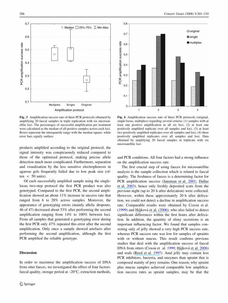
percentage of samples, in which a genotype could be ob-
tained at least once for all six loci simultaneously, declined
from 40% using the multiplex protocol to 35% using the
single-locus protocol to finally 25% using the original
protocol, thus highlighting the improved PCR conditions
(Fig. 4). A similar pattern was noted when the number of
positive replicates over all samples and microsatellite loci
was considered (Fig. 4).
When considering genotyping errors, false alleles dif-
fered between 0% following the original or the optimised
single-locus protocol and 0.02% using the multiplex pro-
tocol. Allelic dropout varied from 13 to 56% in the original
protocol (median 27%), from 16 to 48% in the single-locus
protocol (median 38%), and from 24 to 42% in the mul-
tiplex protocol (median 29%). In all positive amplification
Table 1 Median values and statistical significance of the amplification success rate corresponding to quantity of slimy secretions (spraint;
spraint plus mucus; jelly)
Median of amplification success rate Multiple comparisons
Spraint (1) Spraint plus mucus (2) Jelly (3) 1–2 1–3 2–3
Original 0.00 (n = 5) 0.06 (n = 6) 0.76 (n = 9) P = 1 P = 0.016 P = 0.076
Single-locus 0.22 (n = 5) 0.28 (n = 6) 1.00 (n = 9) P = 1 P = 0.014 P = 0.15
Multiplex 0.17 (n = 5) 0.19 (n = 6) 0.94 (n = 9) P = 1 P = 0.019 P = 0.018
1 day 1 week 2 weeks
Storage time
0,3
0,4
0,5
0,6
0,7
0,8
0,9
PC
R a
mpl
ifica
tion
succ
ess
rate
Median 25%-75% Min-Max
Fig. 1 Comparison of amplification success rate of three different
storage times at –20�C. Results are from 45 subsamples of 15 faecal
samples that were extracted using the Qiagen kit one day, one week,
or two weeks after storing at –20�C. Each sample from each treatment
was amplified once with eight microsatellite loci. The percentage of
successful amplification (detected as bands in an agarose gel) for each
locus in each storage time was calculated by pooling the data across
the 15 samples. Boxes represent the interquartile range with the
median square, while error bars signify outliers
Kit Chelex
Extraction method
0,0
0,2
0,4
0,6
0,8
1,0
PC
R a
mpl
ifica
tion
succ
ess
rate
Median 25%-75% Min-Max
Fig. 2 Median of the amplification success rate of two extraction
methods tested with 24 subsamples respectively. One amplification
with six microsatellite loci was carried out for each sample and each
extraction method. The amplification success rate (represented as
bands in an agarose gel and in an ABI PRISM� 3100 Genetic
Analyser) was obtained by averaging the positive samples over six
loci. Variance across loci is illustrated by a box plot chart. Boxes
represent the interquartile range with the median square, while error
bars signify outliers
Conserv Genet (2008) 9:201–210 205
123
products amplified according to the original protocol, the
signal intensity was conspicuously reduced compared to
those of the optimised protocol, making precise allele
detection much more complicated. Furthermore, separation
and visualisation by the less sensitive electrophoresis in
agarose gels frequently failed due to low peak size (of-
ten < 50 units).
Of each successfully amplified sample using the single-
locus two-step protocol the first PCR product was also
genotyped. Compared to the first PCR, the second ampli-
fication showed an about 11% increase in success rate that
ranged from 6 to 28% across samples. Moreover, the
appearance of genotyping errors (mainly allelic dropouts,
46 of 47) decreased about 53% after performing the second
amplification ranging from 14% to 100% between loci.
From all samples that generated a genotyping error during
the first PCR only 47% repeated this error after the second
amplification. Only once a sample showed artefacts after
performing the second amplification, although the first
PCR amplified the reliable genotype.
Discussion
In order to maximise the amplification success of DNA
from otter faeces, we investigated the effect of four factors:
faecal quality, storage period at –20�C, extraction methods,
and PCR conditions. All four factors had a strong influence
on the amplification success rate.
The first crucial step of using faeces for microsatellite
analysis is the sample collection which is related to faecal
quality. The freshness of faeces is a determining factor for
PCR amplification success (Jansman et al. 2001; Dallas
et al. 2003), hence only freshly deposited scats from the
previous night (up to 20 h after defecation) were collected.
However, within these approximately 20 h after defeca-
tion, we could not detect a decline in amplification success
rate. Comparable results were obtained by Coxon et al.
(1999) and Hajkova et al. (2006), who also failed to detect
significant differences within the first hours after defeca-
tion. In addition, the quantity of slimy secretions is an
important influencing factor. We found that samples con-
sisting only of jelly showed a very high PCR success rate,
whereas PCR success rate was low for samples of spraints
with or without mucus. This result confirms previous
studies that deal with the amplification success of faecal
DNA from otters (Coxon et al. 1999; Hajkova et al. 2006)
and seals (Reed et al. 1997). Anal jelly may contain less
PCR inhibitors, bacteria, and enzymes than spraint that is
composed mainly of prey remains. One reason, why spraint
plus mucus samples achieved comparable low amplifica-
tion success rates as spraint samples, may be that the
Multiplex Single Original
Amplification protocol
0,1
0,2
0,3
0,4
0,5
0,6
0,7P
CR
am
plifi
catio
n su
cces
s ra
te
Median 25%-75% Min-Max
Fig. 3 Amplification success rate of three PCR protocols obtained by
amplifying 20 faecal samples in triple replication with six microsat-
ellite loci. The percentages of successful amplification per treatment
were calculated as the median of all positive samples across each loci.
Boxes represent the interquartile range with the median square, while
error bars signify outliers
0
0,1
0,2
0,3
0,4
0,5
0,6
0,7
0,8
1 2 3 4
PC
R a
mpl
ifica
tion
succ
ess
rate
original
single
multiplex
Fig. 4 Amplification success rate of three PCR protocols (original,
single-locus, multiplex) regarding several criteria: (1) samples with at
least one positive amplification in all six loci, (2) at least one
positively amplified replicate over all samples and loci, (3) at least
two positively amplified replicates over all samples and loci, (4) three
positively amplified replicates over all samples and loci. Data
obtained by amplifying 20 faecal samples in triplicate with six
microsatellite loci
206 Conserv Genet (2008) 9:201–210
123

mucilage layer on spraint cannot be equated with the
secretion of anal scent glands called jelly. In the large
intestine mucus, secreted from goblet cells, is used as a
lubricant for faeces which must pass over membranes
(Liebich 1999; Welsch 2006). Whereas the secretion of
anal scent sacs either act as visual and olfactory stimuli
used in the social organisation of the population (Gorman
et al. 1978; Macdonald and Mason 1987) or is of gastric
origin being produced when otters have not eaten for 18–
24 h (Conroy and French 1991; Carss and Parkinson 1996).
Hajkova et al. (2006) chose the classification (i) spraint,
(ii) spraint with jelly, (iii) jelly and equated consequently
the thin layer of mucus on spraint with the anal jelly. In
contrast, we distinguish in our study between these two
types of slimy secretion, which is supported by the high
difference in amplification success between spraint plus
mucus and jelly samples. Also, climatic conditions can
have a high impact on the amplification rate of faeces
(Farrell et al. 2000; Murphy et al. 2000). Humidity, for
instance, might provide a better microclimate for bacteria
and enzymes, while longer periods of rainy weather might
wash away cells from the surface of the scat. Reduced
amplification success rate in wet periods were detected for
faeces from carnivores in western Venezuela (Farrell et al.
2000). During our pilot study we were able to observe the
same findings. Hence, we only sampled faeces during
periods of dry diurnal weather (i.e. without rain or frost).
Finally, the collection method itself might interfere with
the amplification success. It has been demonstrated that
homogenisation of faecal samples yields reduced PCR
success rates compared to surface wash or homogenisation
of surface scrape (Piggott and Taylor 2003). In light of this
and the results of our pilot study we used commercially
available cotton swabs for the sample collection.
Storing faecal samples in a freezer at –20�C is a fre-
quently practised method that should provide protection
against further degradation (Tikel et al. 1996; Reed et al.
1997; Frantzen et al. 1998; Wasser et al. 1997; Ernest et al.
2000; Frantz et al. 2003; Piggott and Taylor 2003).
Therefore, we considered this storage method to be suitable
in combination with our collection technique as reagents or
buffer solutions may remove cells from the cotton swab.
However, we could show that amplification success rate
decreased drastically with increased storage time (i.e. 20%
within two weeks). As a result, we recommend that DNA
extraction should be performed immediately after collec-
tion. Degradation during the thawing process is an unlikely
reason for this effect since all subsamples were treated in
the same manner. In contrast to our study, Hajkova et al.
(2006) recently reported that PCR success rate did not
decline in otter faecal samples that were preserved up to
234 days in a freezer at –20�C, stored in 96% ethanol or
buffers of kits. Our results demonstrated that the decline of
amplification success rate was highest after one week of
storage. Afterwards the decline was rather low. A possible
explanation for the results of Hajkova et al. (2006) may be
that they extracted most of the samples after the first week
of storing. However, contrasting results may also have
occurred because samples were stored in a buffer solution
which may preserve faecal samples over an intermediate
period of time.
In addition to collection and preservation, the extraction
of samples is the third factor strongly influencing quality
and quantity of template DNA (Wasser et al. 1997;
Flagstad et al. 1999; Banks et al. 2002). The efficiency of
DNA purification can vary greatly among species and even
among individuals (Taberlet and Luitkart 1999; Gossens
et al. 2000; Piggott and Taylor 2003). Due to low DNA
concentration in faeces (Gerloff et al. 1995; Murphy et al.
2000) and a high proportion of PCR inhibitors, nucleases,
bacteria, and enzymes (Deuter et al. 1995; Kohn et al.
1995; Reed et al. 1997; Frantzen et al. 1998) a rapid and
easy to handle (Reed et al. 1997) species specific protocol
should be used to isolate a maximum of DNA while
removing PCR inhibitors simultaneously. Therefore, two
extraction methods were tested. The crucial difference
between the quick, cheap, and simple Chelex method and
the more time-consuming QIAamp� DNA Stool Mini Kit
(Qiagen) is the thorough purification of extracts with the
Qiagen kit, whereas Chelex, as an alkaline chelating resin,
removes only polyvalent metal ions (Walsh et al. 1991;
Reed et al. 1997). In consideration of the 69% higher
amplification success rate obtained by extracting samples
with the Qiagen kit, we suspect that the washing and
purification steps of the Qiagen kit remove PCR inhibitors
to the greatest possible extent. The higher amplification
success justifies the time-consuming washing steps. In our
study the Chelex extractions must still have contained a
large number of PCR inhibitors, since some Chelex-ex-
tracted samples not only failed to amplify, but also showed
no sign of primer dimers, which indicate the presence
of PCR inhibitors (Kohn et al. 1995; Reed et al. 1997;
Vigilant 1999; Palomares et al. 2002). Moreover, control
amplifications that contained tissue extracts and Chelex-
purified faecal samples also failed to amplify. This indi-
cates that the addition of Chelex extracts to successfully
amplifying tissue samples can inhibit their amplification.
Other studies on otter faeces compared the Qiagen kit
with the similar Invitek kit whereby the Invitek kit
yielded a higher amplification success rate (Hajkova et al.
2006).
PCR conditions need to be adjusted according to the
quality and quantity of the DNA faecal extracts. For these
purposes we optimised the amplification protocol designed
for microsatellite loci by Dallas et al. (1999). The opti-
mised single-locus two-step PCR protocol achieved a 24%
Conserv Genet (2008) 9:201–210 207
123

(10% regarding positive PCRs at six loci) increase in
amplification success compared to the standard PCR con-
ditions outlined by Dallas et al. (1999). Major enhance-
ments are the performance of two consecutive PCR
reactions with the first PCR product being the template for
the second PCR reaction, the use of locus-specific
annealing temperatures, a greater extent of cycles, and
modifications in reagent concentrations. Based on the im-
proved protocol for single-locus PCR conditions a more
time and cost efficient multiplex protocol was developed,
despite previous opinions that multiplexing primers of
faecal DNA is difficult to achieve (Ernest et al. 2000). By
using less DNA extract the multiplex protocol allows to
analyse more loci and to perform the necessary PCR rep-
etitions. Genotyping errors (i.e. false alleles and allelic
dropout) occurred at a similar rate in all three PCR pro-
tocols and can only be overcome by a large number of
replicated amplifications (Taberlet et al. 1996; Kohn et al.
1999; Ernest et al. 2000; Frantz et al. 2003; Broquet and
Petit 2004). Due to the low signal intensity, the detection of
alleles was hampered when using the original protocol
instead of the optimised protocols. As we could demon-
strate the use of a two-step procedure, also referred as to
pre-amplification approach, offers an increase in quality
and quantity of the template DNA. This corroborates the
results of recent studies (Piggott et al. 2004; Hedmark and
Ellegren 2006) and highlights again the advantages of the
pre-amplification approach. Piggott et al. (2004) reported
an improvement in amplification success rate and geno-
typing error rate, whereas Hedmark and Ellegren (2006)
found that allelic dropouts generated during the first PCR
step were repeated to a high extent in the second amplifi-
cation. Our results are in line with the ones of Piggott et al.
(2004), as we observed that only 47% of allelic dropouts
from the first PCR appeared also in the second amplifica-
tion, while 53% of the samples generating a genotyping
error during the first step (46 allelic dropouts vs. 1 false
allele) amplified the reliable genotype after the second
PCR. Moreover, we were able to optimise the pre-ampli-
fication approach further and obtained a still more time and
cost effective protocol. Instead of performing an initial
multiplex PCR with all six primers followed by a second
separate amplification for each marker, we amplified three
markers at once in both consecutive amplifications. Thus,
the procedure outlined by Piggott et al. (2004) need seven
PCR reactions per sample to amplify six loci, using our
approach it demands merely four amplification steps per
sample for six markers.
For comparability purposes, the amplification success
rate is often calculated over all samples and loci in the
literature (e.g. Hajkova et al. 2006). We provide an addi-
tional amplification success rate that is based on the
amplification of all six microsatellites simultaneously
(Fig. 4). The restricted estimate of amplification success
offers more information about the value of primers when
dealing with questions such as population size estimates or
parentage analyses.
In summary, DNA of otter faeces is only available in
low concentrations (Gerloff et al. 1995; Murphy et al.
2000), is exposed to degradation (Frantzen et al. 1998),
contains a large number of PCR inhibitors (Deuter et al.
1995; Kohn et al. 1995), and is thus vulnerable to geno-
typing errors (Goossens et al. 2000; Broquet and Petit
2004). This may explain why previous PCR amplification
attempts have met limited success (Coxon et al. 1999;
Dallas et al. 2003). However, these difficulties can be
overcome with a suitable preservation technique that
avoids further degradation and an extraction method that
removes PCR inhibitors to a large extent. Efficient ampli-
fication conditions can compensate for low DNA concen-
trations while replicated PCRs can remediate genotyping
errors. We could assert that the highest amplification suc-
cess rate could be achieved by an extraction of jelly sam-
ples with the QIAamp� DNA Stool Mini Kit followed
immediately after collection and amplified using the opti-
mised multiplex PCR protocol. With this combination we
obtained a median amplification success of 94% (mean:
78%) compared to the 20% of the original methods de-
scribed by Dallas et al. (2003). To apply this method to
future studies it is important to know whether the quantity
of samples will be sufficient if only jelly samples can be
collected. However, even if all types of faecal samples are
collected and analysed the amplification success is still
47% (mean: 47%) over all samples per loci and 40% over
all locus per samples that obtained a genotype at least once
for all six loci. With such success rates, genetic analyses of
otter faeces can provide a powerful way to study otter
populations.
Acknowledgements We would like to thank R. Klenke for helpful
discussions and pertinent knowledge about the otter biology. A.
Vallentin and U. Hempel are thanked for collecting faecal samples.
Thanks to E. Kuster and the Department of Bioanalytical Ecotoxi-
cology and also to W. Durka and the Department of Community
Ecology for giving access to the laboratories. We are grateful to the
members of the Institute of Applied Ecology, University of Canberra
for proof-reading the manuscript and to the anonymous reviewers
who have greatly contributed to improve a first version of this paper.
We kindly acknowledge private landowners for their permission to
access the ponds.
References
Amos B, Pemberton J (1992) DNA fingerprinting in non-human
populations. Curr Opin Genet Dev 2:857–860
Banks SC, Piggott MP, Hansen BD, Robinson NA, Taylor AC (2002)
Wombat coprogenetics enumerating a common wombat popu-
lation by microsatellite analysis of faecal DNA. Aust J Zool
50:193–204
208 Conserv Genet (2008) 9:201–210
123

Broquet T, Petit E (2004) Quantifying genotyping errors in nonin-
vasive population genetics. Mol Ecol 13:3601–3608
Brownstein MJ, Carpten JD, Smith JR (1996) Modulation of non-
templated nucleotide addition by Taq DNA polymerase: primer
modifications that facilitate genotyping. Biotechniques 20:1004–
1006
Bruford MW, Wayne RK (1993) Microsatellites and their application
to population genetic studies. Curr Opin Genet Dev 3:939–943
Carss DN, Parkinson SG (1996) Errors associated with otter Lutralutra faecal analysis. 1. Assessing general diet from spraints.
J Zool 238:301–317
Conroy JWH, French DD (1991) Seasonal patterns in the sprainting
behaviour of otters (Lutra lutra L.) in Shetland. In: Reuther C,
Rochert R (eds) Proceedings of 5th International Otter Collo-
quium, Hankensbuttel, 1989
Coxon K, Chanin P, Dallas J, Sykes T (1999) The use of DNA
fingerprinting to study the population dynamics of otters (Lutralutra) in southern Britain a feasibility study. Environment
Agency R&D Project W1-025
Creel S, Spong G, Sands JL, Rotella J, Zeigle J, Joe L, Murphy KM,
Smith D (2003) Population size estimation in Yellowstone
wolves with error-prone noninvasive microsatellite genotypes.
Mol Ecol 12:2003–2009
Dallas JF, Piertney SB (1998) Microsatellite primers for the Eurasian
otter. Mol Ecol 7:1247–1263
Dallas JF, Bacon PJ, Carss DN, Conroy JWH, Green R, Jefferies DJ,
Kruuk H, Marshall F, Piertney SB, Racey PA (1999) Genetic
diversity in the Eurasian otterLutra lutra in Scotland Evidence
from microsatellite polymorphism. Biol J Linn Soc 68:73–86
Dallas JF, Marshall F, Piertney SB, Bacon PJ, Racey PA (2002)
Spatially restricted gene flow and reduced microsatellite poly-
morphism in the Eurasion otter Lutra lutra in Britain. Conserv
Genet 3:15–29
Dallas JF, Coxon KE, Sykes T, Chanin PRF, Marshall F, Carss DN,
Bacon PJ, Piertney SB, Racey PA (2003) Similar estimates of
population genetic composition and sex ratio derived from
carcasses and faeces of Eurasian otter Lutra lutra. Mol Ecol
12:275–282
Deuter R, Pietsch S, Hertel S, Muller O (1995) A method for
preparation of fecal DNA suitable for PCR. Nucleic Acids Res
23:3800–3801
Ernest HB, Penedo MCT, May BP, Syvanen M, Boyce WM (2000)
Molecular tracking of mountain lions in the Yosemite Valley
region in California genetic analysis using microsatellites and
faecal DNA. Mol Ecol 9:433–441
Farrell LE, Roman J, Sunquist ME (2000) Dietary separation of
sympatric carnivores identified by molecular analysis of scats.
Mol Ecol 9:1583–1590
Flagstad O, Roed K, Stacy JE, Jakobsen KS (1999) Reliable
noninvasive genotyping based on excremental PCR of nuclear
DNA purified with a magnetic bead protocol. Mol Ecol 8:879–
883
Frantz AC, Pope LC, Carpenter PJ, Roper TJ, Wilson GJ, Delahay RJ,
Burke T (2003) Reliable microsatellite genotyping of the
Eurasian badger (Meles meles) using faecal DNA. Mol Ecol
12:1649–1661
Frantz AC, Schaul M, Pope LC, Fack F, Schley L, Muller CP, Roper
TJ (2004) Estimating population size by genotyping remotely
plucked hair the Eurasian badger. J Appl Ecol 41:985–995
Frantzen MAJ, Silk JB, Fergusen JWH, Wayne RK, Kohn MH (1998)
Empirical evaluation of preservation methods for faecal DNA.
Mol Ecol 7:1423–1428
Gerloff U, Schlotterer C, Rassmann K, Rambold I, Hohmann G, Fruth
B, Tautz D (1995) Amplification of hypervariable simple
sequence repeats (microsatellites) from excremental DNA of
wild living bonobos (Pan paniscus). Mol Ecol 4:515–518
Goossens B, Chikhi L, Utami SS, de Ruiter J, Bruford MW (2000) A
multi-samples multi-extracts approach for microsatellite analysis
of faecal samples in an arboreal ape. Conserv Genet 1:157–162
Gorman ML, Jenkins D, Harper RJ (1978) The anal scent sacs of the
otter (Lutra lutra). J Zool 186:463–474
Hajkova P, Zemanova B, Bryja J, Hajek B, Roche K, Tkadlec E, Zima
J (2006) Factors affecting success of PCR amplification of
microsatellite loci from otter faeces. Mol Ecol Notes 6:559–562
Hedmark E, Ellegren H (2006) A test of the multiplex pre-
amplification approach in microsatellite genotyping of wolverine
faecal DNA. Conserv Genet 7:289–293
Idaghdour Y, Broderick D, Korrida A (2003) Faeces as a source of
DNA for molecular studies in a threatened population of great
bustards. Conser Genet 4:789–792
Jansman HAH, Chanin PRF, Dallas JF (2001) Monitoring otter
populations by DNA typing of spraints. IUCN Otter Spec Group
Bull 18:12–19
Kohn M, Knauer F, Stoffella A, Schroder W, Paabo S (1995)
Conservation genetics of the European brown bear—a study
using excremental PCR of nuclear and mitochondrial sequences.
Mol Ecol 4:95–103
Kohn MH, Wayne RK (1997) Facts from feces revisited. Trends Ecol
Evol 12:223–227
Kohn MH, York EC, Kamradt DA, Haught G, Sauvajot RM, Wayne
RK (1999) Estimating population size by genotyping faeces.
Proc R Soc Lond B 266:657–663
Liebich HG (1999) Funktionelle Histologie der Haussaugetiere:
Lehrbuch und Farbatlas fur Studium und Praxis, 3rd edn.
Schattauer, Stuttgart
Macdonald SM, Mason CF (1987) Seasonal marking in an otter
population. Acta Theriol 32:449–462
Morin PA, Chambers KE, Boesch C, Vigilant L (2001) Quanti-
tative polymerase chain reaction analysis of DNA from
noninvasive samples for accurate microsatellite genotyping
of wild chimpanzees (Pan troglodytes versus). Mol Ecol
10:1835–1844
Murphy MA, Waits LP, Kendall KC (2000) Quantitative evaluation
of fecal drying methods for brown bear DNA analysis. Wildlife
Soc B 28:951–957
Murphy MA, Waits LP, Kendall KC, Wasser SK, Higbee JA, Bogden
R (2002) An evaluation of long-term preservation methods for
brown bear (Ursus arctos) faecal DNA samples. Conserv Genet
3:435–440
Nsubuga AM, Robbins MM, Roeder AD, Morin PA, Boesch C,
Vigilant L (2004) Factors affecting the amount of genomic DNA
extracted from ape faeces and the identification of an improved
sample storage method. Mol Ecol 13:2089–2094
Palomares F, Godoy A, Piriz A, O’Brien J, Johnson WE (2002)
Faecal genetic analysis to determine the presence and distribu-
tion of elusive carnivores design and feasibility for the Iberian
lynx. Mol Ecol 11:2171–2182
Paxinos E, Mcintosh C, Ralls K, Fleischer R (1997) A noninvasive
method for distinguishing among canid species amplification and
enzyme restriction of DNA from dung. Mol Ecol 6:483–486
Piggott MP, Taylor AC (2003) Extensive evaluation of faecal
preservation and DNA extraction methods in Australian native
and introduced species. Aust J Zool 51:341–355
Piggott MP, Bellemain E, Taberlet P, Taylor AC (2004) A multiplex
pre-amplification method that significantly improves microsat-
ellite amplification and error rates for faecal DNA in limiting
conditions. Conserv Genet 5:417–420
Queller DC, Strassmann JE, Hughes CR (1993) Microsatellites and
kinship. Trends Ecol Evol 8:285–288
Reed JZ, Tollit DJ, Thompson PM, Amos W (1997) Molecular
scatology the use of molecular genetic analysis to assign species
sex and individual identity to seal faeces. Mol Ecol 6:225–234
Conserv Genet (2008) 9:201–210 209
123

Roeder AD, Archer FI, Poinar HN, Morin PA (2004) A novel method
for collection and preservation of faeces for genetic studies. Mol
Ecol Notes 4:761–764
Sidransky D (1992) Identification of rare mutations in the stool of
patients with curable colorectable tumors. Science 256:102–105
Taberlet P, Griffin S, Goossens B, Questiau S, Manceau V,
Escaravage N, Waits LP, Bouvet J (1996) Reliable genotyping
of samples with very low DNA quantities using PCR. Nucleic
Acids Res 24:3189–3194
Taberlet P, Luikart G (1999) Non-invasive genetic sampling and
individual identification. Biol J Linn Soc 68:41–55
Tikel D, Blair D, Marsh HD (1996) Marine mammal faeces as a
source of DNA. Mol Ecol 5:456–457
Tschirch W (1995) Neue biomedizinische Methoden fur die Feld-
forschung - dargestellt am Beispiel des Fischotters (Lutra lutra).
In: Stubbe M, Stubbe A, Heidecke D (eds) Methoden der
feldokologischen Saugetierforschung (Methods in mammalian
field ecology), vol 1. Martin-Luther-Universitat, Halle/Saale, pp
203–210
Vigilant L (1999) An evaluation of techniques for the extraction and
amplification of DNA from naturally shed hairs. Biol Chem
380:1329–1331
Walsh PS, Metzger DA, Higuchi R (1991) Chelex� 100 as a medium
for simple extraction of DNA for PCR-based typing form
forensic material. Biotechniques 10:506–513
Wasser SK, Houston CS, Koehler GM, Cadd GG, Fain SR (1997)
Techniques for application of faecal DNA methods to field
studies of ursids. Mol Ecol 6:1091–1097
Welsch U (2006) Lehrbuch Histologie, 2nd edn. Urban & Fischer,
Munchen
210 Conserv Genet (2008) 9:201–210
123