An interstitial deletion at 8q23.1-q24.12 associated with Langer-Giedion syndrome/...
6
CASE REPORT Open Access An interstitial deletion at 8q23.1-q24.12 associated with Langer-Giedion syndrome/ Trichorhinophalangeal syndrome (TRPS) type II and Cornelia de Lange syndrome 4 Nikoletta Selenti * , Maria Tzetis, Maria Braoudaki, Krinio Gianikou, Sofia Kitsiou-Tzeli and Helen Fryssira Abstract Background: There are three distinct subtypes of Trichorhinophalangeal syndrome (TRPS); TRPS type I, TRPS type II and TRPS type III. Features common to all three subtypes include sparse, slowly growing scalp hair, laterally sparse eyebrows, a bulbous tip of the nose (pear-shaped), and protruding ears. Langer–Giedion syndrome (LGS) or TRPS type II is a contiguous gene syndrome on 8q24.1, involving loss of functional copies of the TRPS1 and EXT1 genes. We report a male patient that was referred to the Department of Medical Genetics due to hypotonia and dysmorphic facial features. Results: Cytogenetic and array- Comparative Genomic Hybridization (aCGH) analysis revealed that the patient was a carrier of an interstitial deletion at 8q23.1-q24.12 of 12,5 Mb. Parental karyotype indicated that the father carried an apparently balanced insertion: 46, ΧΥ, der(10)ins(10;8)(q22;q23q24). Conclusions: This is the first report of an apparently balanced insertion including chromosomes 8 and 10 contributing to the etiology of LGS/ TRPS type II. Τimely diagnosis of parental balanced chromosomal rearrangements can reduce the risk of subsequent miscarriages as well as abnormal offspring. Background Langer–Giedion syndrome (LGS) or Trichorhinopha- langeal syndrome (TRPS) type II is a contiguous gene syndrome on 8q24.1, involving loss of functional copies of the TRPS1 and EXT1 genes, that was first described by Andreas Giedion, a Swiss pediatric radiologist. In 1966, he reported the association of slowly growing hair, a long pear-shaped nose with a bulbous tip, and finger deformities. In 1969, Giedion A. and Leonard O. Langer Jr. independently described a patient with these features as well as multiple exostoses. In 1974, the name LGS or TRP II was introduced subsequently by Hall et al. [1]. The diagnosis of TRPS type I, II and III is based on clinical and radiographic features as well as on genetic analysis that is helpful especially in the case of non-classical clinical presentation. TRPS type I caused by mutations in the TRPS1 gene located on chromosome 8q24.1, characterized by distinctive skeletal abnormalities and craniofacial dysmorphism. TRPS type II represents a contiguous gene syndrome involving loss of functional copies of the TRPS1 and EXT1 genes and is characterized by multiple exostoses and intellectual disability (ID) in addition to the classical triad of symptoms. TRPS type III is caused by mutations in TRPS1 gene and includes severe short stature and brachydactyly in the absence of exostoses [2]. It is postulated that nonsense mutations are usually re- sponsible for the TRPS type I phenotype, while missense mutations cause the TRPS type III phenotype [3]. The mode of inheritance in TRPS I and III is autosomal dominant. In contrast, TRPS II (LGS) is usually sporadic and caused by the deletion of contiguous genes in the long arm of chromosome 8 (8q24.11-13), involving loss * Correspondence: [email protected] Department of Medical Genetics, Aghia Sophia Childrens’ Hospital, Athens University, School of Medicine, Thivon and Levadeias 11527, Goudi, Athens, Greece © 2015 Selenti et al. Open Access This article is distributed under the terms of the Creative Commons Attribution 4.0 International License (http://creativecommons.org/licenses/by/4.0/), which permits unrestricted use, distribution, and reproduction in any medium, provided you give appropriate credit to the original author(s) and the source, provide a link to the Creative Commons license, and indicate if changes were made. The Creative Commons Public Domain Dedication waiver (http://creativecommons.org/publicdomain/zero/1.0/) applies to the data made available in this article, unless otherwise stated. Selenti et al. Molecular Cytogenetics (2015) 8:64 DOI 10.1186/s13039-015-0169-9
-
Upload
independent -
Category
Documents
-
view
1 -
download
0
Transcript of An interstitial deletion at 8q23.1-q24.12 associated with Langer-Giedion syndrome/...

CASE REPORT Open Access
An interstitial deletion at 8q23.1-q24.12associated with Langer-Giedion syndrome/Trichorhinophalangeal syndrome (TRPS)type II and Cornelia de Lange syndrome 4Nikoletta Selenti*, Maria Tzetis, Maria Braoudaki, Krinio Gianikou, Sofia Kitsiou-Tzeli and Helen Fryssira
Abstract
Background: There are three distinct subtypes of Trichorhinophalangeal syndrome (TRPS); TRPS type I, TRPS type IIand TRPS type III. Features common to all three subtypes include sparse, slowly growing scalp hair, laterally sparseeyebrows, a bulbous tip of the nose (pear-shaped), and protruding ears. Langer–Giedion syndrome (LGS) or TRPStype II is a contiguous gene syndrome on 8q24.1, involving loss of functional copies of the TRPS1 and EXT1 genes.We report a male patient that was referred to the Department of Medical Genetics due to hypotonia anddysmorphic facial features.
Results: Cytogenetic and array- Comparative Genomic Hybridization (aCGH) analysis revealed that the patient was acarrier of an interstitial deletion at 8q23.1-q24.12 of 12,5 Mb. Parental karyotype indicated that the father carried anapparently balanced insertion: 46, ΧΥ, der(10)ins(10;8)(q22;q23q24).Conclusions: This is the first report of an apparently balanced insertion including chromosomes 8 and 10 contributingto the etiology of LGS/ TRPS type II. Τimely diagnosis of parental balanced chromosomal rearrangements can reducethe risk of subsequent miscarriages as well as abnormal offspring.
BackgroundLanger–Giedion syndrome (LGS) or Trichorhinopha-langeal syndrome (TRPS) type II is a contiguous genesyndrome on 8q24.1, involving loss of functional copiesof the TRPS1 and EXT1 genes, that was first describedby Andreas Giedion, a Swiss pediatric radiologist. In1966, he reported the association of slowly growinghair, a long pear-shaped nose with a bulbous tip, andfinger deformities. In 1969, Giedion A. and Leonard O.Langer Jr. independently described a patient with thesefeatures as well as multiple exostoses. In 1974, thename LGS or TRP II was introduced subsequently byHall et al. [1]. The diagnosis of TRPS type I, II and IIIis based on clinical and radiographic features as well ason genetic analysis that is helpful especially in the caseof non-classical clinical presentation.
TRPS type I caused by mutations in the TRPS1 genelocated on chromosome 8q24.1, characterized bydistinctive skeletal abnormalities and craniofacialdysmorphism.TRPS type II represents a contiguous gene syndromeinvolving loss of functional copies of the TRPS1 andEXT1 genes and is characterized by multiple exostosesand intellectual disability (ID) in addition to theclassical triad of symptoms.TRPS type III is caused by mutations in TRPS1 geneand includes severe short stature and brachydactyly inthe absence of exostoses [2].
It is postulated that nonsense mutations are usually re-sponsible for the TRPS type I phenotype, while missensemutations cause the TRPS type III phenotype [3]. Themode of inheritance in TRPS I and III is autosomaldominant. In contrast, TRPS II (LGS) is usually sporadicand caused by the deletion of contiguous genes in thelong arm of chromosome 8 (8q24.11-13), involving loss
* Correspondence: [email protected] of Medical Genetics, Aghia Sophia Childrens’ Hospital, AthensUniversity, School of Medicine, Thivon and Levadeias 11527, Goudi, Athens,Greece
© 2015 Selenti et al. Open Access This article is distributed under the terms of the Creative Commons Attribution 4.0International License (http://creativecommons.org/licenses/by/4.0/), which permits unrestricted use, distribution, andreproduction in any medium, provided you give appropriate credit to the original author(s) and the source, provide alink to the Creative Commons license, and indicate if changes were made. The Creative Commons Public DomainDedication waiver (http://creativecommons.org/publicdomain/zero/1.0/) applies to the data made available in thisarticle, unless otherwise stated.
Selenti et al. Molecular Cytogenetics (2015) 8:64 DOI 10.1186/s13039-015-0169-9

of functional copies of the TRPS1 and the EXT1 genes[4, 5].In TRPS I and III, the eyebrows may be thickened
medially and thin or even absent laterally. In TRPS II,the eyebrows could be normal. Sparse eyelashes and sec-ondary sexual hairs may be evident. A long pear-shapednose with bulbous tip and a long philtrum are character-istic. Deformities of the fingers and toes can result fromcone-shaped epiphyses and irregular premature fusion,which could be mistaken as juvenile rheumatoid arthritis[1]. The middle phalanges are most commonly involved.Sometimes there is an abnormal patella with recurrentdislocation. Perthes-like change of the hip is commonand can lead to severe secondary osteoarthritis in theadult [1, 6]. In TRPS II, patients have additional featuresof multiple exostoses from early childhood, microceph-aly, skin and joint laxity. Mental impairment is common.Features common to all three disorders include sparse,
slowly growing scalp hair, laterally sparse eyebrows, abulbous tip of the nose (pear-shaped), and protrudingears. Further typical characteristics comprise a long flatphiltrum and a thin upper vermillion border. Distinctradiographic findings are often not detectable before2 years of age.
Clinical Phenotype/Genotype correlation in LGS/TRPS IIThe LGS/TRPII comprises the clinical features of twoseparate autosomal dominant diseases : the multiple her-editary exostoses (MHE) type I caused by mutation inthe gene encoding exostosin-1 (EXT1), and the TRPStype I caused by haploinsufficiency of the TRPS1 gene.The MHE is a genetically heterogeneous disorder whichcan be caused by mutations in the EXT1, EXT2 or EXT3gene [7].The LGS/TRPSII shows some clinical variability
depending on the loss of additional genes in the deletedregion [8, 9]. Mild to moderate intellectual disability,congenital nephrotic syndrome [10], hydrometrocolpos[11], conductive hearing loss [12], growth hormone defi-ciency [13], persistent cloaca and prune belly sequence[14], and a submucous cleft palate [15] have all been de-scribed as additional features of LGS/TRPSII dependingon the deletion size.Although deletions of the 8q region are rare and con-
siderably variable, the shortest region of deletion overlap(SRO) has been defined at 8q24.1, spanning 2 Mb, in-cluding TRPS1, EIF3S3, RAD21, OPG, CXIV and EXT1genes [16].Heterozygous mutations of the RAD21 gene at
8q24.11 were recently found to cause Cornelia deLange syndrome- 4 (CdLs-4), which is characterized bygrowth deficiency, mental retardation, microcephaly,bushy eyebrows and synophrys, depressed nasal bridge,
micrognathia, micromelia, hearing loss, anteverted nares,prominent symphysis and spurs in the anterior angle ofmandible, gastrointestinal problems (such us gastroesoph-ageal reflux, duplication of the gut, malrotation of colonwith volvulus and pyloric stenosis). There are occasionalseizures, congenital heart defects and inguinal hernias[17–19].TRPS type I, LGS/TRPSII and CdLs-4 share many fa-
cial features; some patients affected by LGS/TRPSII withdeletions involving RAD21 and not TRPS1, present typ-ical facial dysmorphisms. LGS/TRPSII patients’ facialhallmarks might be partially influenced by RAD21.Moreover, the phenotypic consequences of RAD21 dis-ruption might be underestimated in these patients [20].
Parental apparently-balanced chromosomalinsertion in the etiology of LGS/TRPSIIStructural aberrations in human chromosomes can be ini-tiated by chromosomal breakage and incomplete repair. Ifthe chromosomal aberration is balanced, copy number forall interrogated genomic regions is disomic. However,most chromosomal aberrations are unbalanced, resultingin duplication or deletion of genetic material at thechromosomal breakpoints. The frequency of insertionaltranslocation is estimated by karyotyping on severely men-tally retarded patients (4:40,000) [21]. Microscopically vis-ible insertional translocations occur with a frequency of1:80.000 in newborns [22]. However, the frequency is alsoestimated as high as 1:500 using array-based comparativegenome hybridization (aCGH) as a detection method inconjunction with FISH [23].The frequency of balanced chromosomal insertions is
much lower than reciprocal translocations because itrequires three chromosomal breaks. Parental balancedinsertions increase the possibility of copy number im-balances in the offspring resulting in congenital anom-alies of multiple organ systems and psychomotorretardation. aCGH analysis revealed that approximately2.1 % of parents with offspring having developmentalanomalies, harbor balanced insertions [24]. In this re-gard, genomic analysis of the parental DNA of a patientmay help us to exclude the possibility of parental bal-anced translocations as predisposing genetic risk.We report an LGS/TRPSII patient with an 8q23.1-
q24.12 microdeletion, caused by a parental apparentlybalanced insertion ( 46,XY, der(10)ins(10;8)(q22;q23q24).
Case presentationClinical reportOur male patient was the first child of healthy unrelatedparents of normal intelligence. He was born by caesariansection at 40 weeks of gestation after an uneventful preg-nancy. His birth weight was 3090 gr (25th centile), length
Selenti et al. Molecular Cytogenetics (2015) 8:64 Page 2 of 6

49 cm (25th centile) and head circumference 33 cm (25th
centile). Previously the mother had suffered a miscarriage.He was referred to our Department of Clinical Genetics
at 2 months due to hypotonia and dysmorphic facial fea-tures. On clinical examination the patient presented withbrachycephaly, low anterior hairline, a depressed and broadnasal bridge, a bulbous tip of the nose, a long and flat phil-trum, a thin upper lip vermilion, a high palate, retro-micrognathia and downslanting palpebral fissures (Fig. 1a).He had large laterally protruding ears (Fig. 1b), bilateralpre-axial polydactyly (Fig. 1c), long fingers and cryptorchid-ism. Heart and abdominal ultrasound revealed no patho-logical abnormalities. The brain MRI showed dysplasticcorpus callosum. The opthalmological examination wasnormal. The karyotype was found to be abnormal [46, XY,del(8)(q23→ q24) pat].
MethodsCytogenetic analysisCytogenetic analysis was performed on GTG-bandedmetaphases at a resolution of 500 bands approximately.The samples were subjected to blood lymphocyte cultureaccording to the standard cytogenetic protocol. The de-scription of karyotypes were made in accordance withthe ISCN 2013 (International System of Human Cyto-genetic Nomenclature).
Array-CGH analysis (aCGH)After informed consent from the parents, genomic DNAfrom the patient and the father was obtained from 3 mlof peripheral blood using the BioRobot® M48 System(Qiagen, Hilden, Germany) and the commercially avail-able kit MagAttract® DNA Blood Midi M48 Kit (Qiagen).The quality and quantity of the DNA samples was deter-mined using a NanoDrop ND-1000 UV–VIS spectro-photometer. Agilent Human Genome 4X180K CGH +SNP arrays were used in this study (Agilent Technolo-gies, Santa Clara, CA, www.agilent.com). Labelling andhybridization was carried out according to the manufac-turer’s recommendations. Data were processed usingFeature Extraction and analysed using Cytogenomicsvs.3.0 software (Agilent Technologies, CA) with the
following settings: Algorithm: ADM-1, Threshold: 6.7,with a minimum of 4 probes for a region to be included.Centralization and fuzzy zero corrections were appliedto remove putative variant intervals with small averagelog2 ratios.
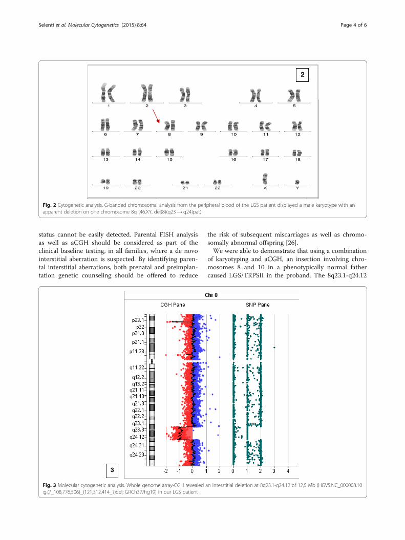
ResultsCytogenetic analysisG-banded chromosomal analysis from the peripheralblood of the patient displayed a male karyotype with anapparent deletion on one chromosome 8q (46,XY,del(8)(q23→ q24)pat) (Fig. 2). The maternal karyotypewas normal (46,XX), however the paternal karyotype re-vealed an apparently balanced insertion: 46, ΧΥ, der(10)ins(10;8)(q22;q23q24).
Array-CGH analysis (aCGH)Whole genome a-CGH revealed an interstitial deletion at8q23.1-q24.12 of 12,5 Mb (HGVS:NC_000008.10:g.(?_108,776,506)_(121,312,414_?)del; GRCh37/hg19). The deletionincluded the following genes: RSPO2, EIF3E, TTC35,TMEM74, TRHR, NUDCD1, ENY2, PKHD1L1, EBAG9,SYBU, KCNV1, CSMD3, MIR2053, TRPS1, EIF3H, UTP23,RAD21, NCRNA00255, MIR3610, C8orf85, SLC30A8,MED30, EXT1, SAMD12, TNFRSF11B, COLEC10, MAL2,NOV, ENPP2, TAF2, DSCC1, DEPTOR, COL14A1 (Fig. 3).The paternal result included only copy number polymor-phisms (CNPs), therefore the father carried a balancedinsertion.
Discussion and conclusionsParental balanced chromosomal rearrangement is an im-portant cause of copy number alteration in the offspring.A recent study revealed that approximately 2.1 % of du-plications and deletions associated with developmentaldelay originated from parental balanced chromosomalaberrations [24]. Repeat sequences, such as low copy re-peats (LCR) around the region of breakage are thoughtto mediate misalignment between the repeat regionsduring meiosis, leading to unequal recombination events[25]. However, the incidence of cytogenetically-visible in-sertions is low [21], and the potential parental carrier
cba
Fig. 1 Dysmorphic features in a LGS patient, that was referred to our Department of Medical Genetics. a A bulbous tip of the nose, a long andflat philtrum and a thin upper lip vermilion, b large laterally protruding ears and a depressed and broad nasal bridge and c pre-axial polydactyly
Selenti et al. Molecular Cytogenetics (2015) 8:64 Page 3 of 6
status cannot be easily detected. Parental FISH analysisas well as aCGH should be considered as part of theclinical baseline testing, in all families, where a de novointerstitial aberration is suspected. By identifying paren-tal interstitial aberrations, both prenatal and preimplan-tation genetic counseling should be offered to reduce
the risk of subsequent miscarriages as well as chromo-somally abnormal offspring [26].We were able to demonstrate that using a combination
of karyotyping and aCGH, an insertion involving chro-mosomes 8 and 10 in a phenotypically normal fathercaused LGS/TRPSII in the proband. The 8q23.1-q24.12
2
Fig. 2 Cytogenetic analysis. G-banded chromosomal analysis from the peripheral blood of the LGS patient displayed a male karyotype with anapparent deletion on one chromosome 8q (46,XY, del(8)(q23→ q24)pat)
3
Fig. 3 Molecular cytogenetic analysis. Whole genome array-CGH revealed an interstitial deletion at 8q23.1-q24.12 of 12,5 Mb (HGVS:NC_000008.10:g.(?_108,776,506)_(121,312,414_?)del; GRCh37/hg19) in our LGS patient
Selenti et al. Molecular Cytogenetics (2015) 8:64 Page 4 of 6

deletion of 12.5 Mb, contains 33 genes of which TRPS1,EXT1, and RAD21 could be considered responsible forthe core phenotypic features of the proband. Althoughdeletions of the 8q region are rare and considerably vari-able, the shortest region of deletion overlap (SRO) hasbeen identified as spanning 2 Mb, including TRPS1,EIF3S3, RAD21, OPG, CXIV, EXT1 genes [16]. To ourknowledge, this is the first report of an apparently bal-anced insertion including chromosomes 8 and 10 con-tributing to the etiology of LGS/TRPSII.Recently, defects in RAD21 have been associated with
CdLs-4. Although recognizable, the face and hair of pa-tients with LGS/TRPSII are variable and typically relatedto TRPS1 disruption [27]. In our patient, aCGH analysisrevealed the deletion of both TRPS1 and RAD21 geneswhich is in accordance with the dysmorphic facial fea-tures. TRPS type I, LGS/TRPSII and CdLs-4 share manyfacial features. Autosomal dominant missense mutationsresult in more severe functional, structural and cognitiveclinical findings compared to loss-of-function mutationsor deletions. In RAD21 haploinsufficiency or heterozy-gous loss-of-function mutation may cause the LGS/TRPSII affected individuals to have mild CDLS stigmatatoo [19].In LGS/TRPSII patients, the more distal the proximal
breakpoint is located from the TRPS1 gene, the fewertypical features of LGS/TRPSII are observed [28]. In thatcase, the TRPS1 gene expression may be altered if theproximal breakpoint occurs near the gene where a po-tential regulatory sequence, such as gene enhancer or re-pressor is located. Thus, the location of the proximalbreakpoint causes a functional disturbance of the TRPS1gene. The size of the critical region outside the TRPS1gene which contains the regulatory sequence is yet to bedetermined. Another possibility is that the newly formedchromosomal imbalance may cause a silencing effect onthe TRPS1 gene by placing it in close proximity to an-other gene [29].Nearly 70 % of LGS/TRPSII affected individuals may
exhibit mild to severe cognitive disability [30]. Althoughdelay in the LGS/TRPSII syndrome has been attributedto the deletion of genes outside the TRPS1-EXT1 inter-val, specific genes have yet to be identified. The size ofthe 8q deletion is directly correlated with intellectualdisability (ID)[13].In our patient, the 8q deletion spans 12,5 Mb and the
genes potentially involved were: CSMD3, MED30,SAMD12, EIF3H, RAD21 and TAF 2. In particular,CSMD3 has previously been suggested as a good candi-date for pathogenesis of developmental delay in LGS/TRPSII patients. RAD21 mutations or deletions havebeen observed in CdLs patients with very mild intellec-tual disability (ID) [19]. Mutations in the TAF2 gene arealso related with ID [31, 32].
Although more than 100 patients with LGS/TRPSIIhave been reported in the literature, there is little long-term follow-up and knowledge about the course and po-tential further health complications during adulthoodand advanced age. In our patient, clinical reevaluation atsix months revealed feeding difficulties and delayed psy-chomotor development.The number of distinct syndromes due to specific
chromosome imbalance is rapidly increasing, especiallysince a-CGH allows detection of submicroscopic aberra-tions of virtually any segment. Confirmation of LGS/TRPSII using the appropriate genetic analysis is neces-sary especially in cases of non-classical clinical presenta-tion [33].Skeletal and orthopedic problems continue to be an
important issue. Severe scoliosis and orthopedic prob-lems due to joint laxity, Perthes disease, asymmetry oflegs and joint immobility due to adjacent exostoses areamong the most frequent problems. On the other hand,eye, ear, and cardiac problems are rarely of major rele-vance [33].Τimely diagnosis of parental balanced chromosomal re-
arrangements using the appropriate techniques is import-ant and can reduce the risk of subsequent miscarriages aswell as abnormal offspring; in our case neither parentalkaryotype nor amniocentesis were performed for prenataltesting. The monosomy 8q23-q24 observed in the affectedchild resulted from segregation during paternal meiosis,due to the apparently balanced chromosomal insertion inthe father 46, ΧΥ, der(10)ins(10;8)(q22;q23q24).With better knowledge on the long-term course,
complications and molecular defects that cause LGS/TRPSII, genetic counseling is necessary. Clinical geneti-cists should provide information for the families, to ad-vise them about the course of the disease and how toovercome problems [33].
ConsentWritten informed consent was obtained from the patientfor publication of this Case report and any accompany-ing images. A copy of the written consent is available forreview by the Editor-in-Chief of this journal.
Competing interestsThe authors declare that they have no competing interests.
Authors’ contributionNS carried out the cytogenetic studies and wrote the manuscript, MT carriedout the molecular cytogenetic studies and helped to draft the manuscript, MBand KG carried out the molecular cytogenetic studies, SKT coordinated thestudy and HF made the clinical evaluation of the patient and helped to draftthe manuscript. All the authors have read and approved the manuscript.
Authors’ informationNS: Biologist Phd, Department of Medical Genetics, Athens University, Schoolof Medicine, Athens, GreeceMT: Assistant Prof. of Genetics, Department of Medical Genetics, AthensUniversity, School of Medicine, Athens, Greece
Selenti et al. Molecular Cytogenetics (2015) 8:64 Page 5 of 6

MB: Biologist Phd, Senior Postdoc, Department of Medical Genetics, AthensUniversity, School of Medicine, Athens, GreeceKG: Biologist Phd, Department of Medical Genetics, Athens University, Schoolof Medicine, Athens, GreeceSKT: Prof. of Medical Genetics, Department of Medical Genetics, AthensUniversity, School of Medicine, Athens, GreeceHF: Associate Prof. of Clinical Genetics, Department of Medical Genetics,Athens University, School of Medicine, Athens, Greece
Received: 30 April 2015 Accepted: 30 July 2015
References1. Tsang WK, Yang KW, Fong CM. Langer-Giedion syndrome: the evolving
imaging features in hands and beyond. Skeletal Radiol. 2014;43:251–5.2. Sidler JA, Filges I, Boesch N, Ramelli GP, Röthlisberger B, Huber AR, et al.
TRPS1 codon 952 constitutes a mutational hot spot intrichorhinophalangeal syndrome type I and could be associated withintellectual disability. Clin Dysmorphol. 2012;21:87–90.
3. Piccione M, Niceta M, Antona V, Di Fiore A, Cariola F, Gentile M, et al.Identification of two new mutations in TRPS 1 gene leading to the tricho-rhino-phalangeal syndrome type I and III. Am J Med Genet A.2009;149A:1837–41.
4. Shin HT, Chang MW. Trichorhinophalangeal syndrome, type II(Langer–Giedion syndrome). Dermatol Online J. 2001.
5. Sybert VP. Genetic skin disorders. Chapter hair shaft abnormalities,syndromic. Trichorhinophalangeal syndrome. 2nd ed. Oxford: OxfordUniversity Press; 2010. p. 225–8.
6. Sugiura Y. Tricho-rhino-phalangeal syndrome associated with Perthesdisease-like bone change and spondylolisthesis. Jinrui Idengaku Zasshi.1978;23–30.
7. Pereza N, Severinski S, Ostojić S, Volk M, Maver A, Dekanić KB, et al. Thirdcase of 8q23.3-q24.13 deletion in a patient with Langer-Giedion syndromephenotype without TRPS1 gene deletion. Am J Med Genet A.2012;158A:659–63.
8. Lüdecke HJ, Wagner MJ, Nardmann J, La Pillo B, Parrish JE, Willems PJ, et al.Molecular dissection of a contiguous gene syndrome: localization of thegenes involved in the Langer-Giedion syndrome. Hum Mol Genet.1995;31–36.
9. Lüdecke HJ, Schmidt O, Nardmann J, von Holtum D, Meinecke P, Muenke M,et al. Genes and chromosomal breakpoints in the Langer-Giedion syndromeregion on human chromosome 8. Hum Genet. 1999;105:619–28.
10. Lu FL, Hou JW, Tsai WS, Teng RJ, Yau KI, Wang TR. Tricho-rhino-phalangealsyndrome type II associated with epiglottic aplasia and congenitalnephrotic syndrome. J Formos Med Assoc. 1997;217–221.
11. Fryns JP. Trichorhinophalangeal syndrome type 2: another syndromic formof hydrometrocolpos. Am J Med Genet. 1997;73:233.
12. Vantrappen G, Feenstra L, Frijns JP. Conductive hearing loss in thetricho-rhino-phalangeal syndrome (TRP II) or in the Langer-Giedionsyndrome. Am J Med Genet. 1997;72:372–3.
13. Riedl S, Giedion A, Schweitzer K, M€ullner-Eidenb€ock A, Grill F, Frisch H,et al. Pronounced short stature in a girl with tricho-rhinophalangealsyndrome II (TRPS II, Langer-Giedion syndrome) and growth hormonedeficiency. Am J Med Genet Part A. 2004;131A:200–3.
14. Ramos FJ, McDonald-McGinn DM, Emanuel BS, Zackai EH. Trichorhino-phalangeal syndrome type II (Langer-Giedion) with persistent cloaca andprune belly sequence in a girl with 8q interstitial deletion.AmJ Med Genet. 1992;44:790–4.
15. Morioka D, Suse T, Shimizu Y, Ohkubo F, Hosaka Y. Langer-Giedionsyndrome associated with submucous cleft palate. Plast Reconstr Surg.1999;103:1458–63.
16. Lüdecke HJ, Johnson C, Wagner MJ, Wells DE, Turleau C, Tommerup N, et al.Molecular definition of the shortest region of deletion overlap in theLanger-Giedion syndrome. Am J Hum Genet. 1991;49:1197–206.
17. Chen CP, Lin SP, Liu YP, Chern SR, Wu PS, Su JW, et al. An interstitialdeletion of 8q23.3-q24.22 associated with Langer-Giedion syndrome,Cornelia de Lange syndrome and epilepsy. Gene. 2013;15:176–80.
18. Jones KL. Brachmann de Lange syndrome. In: Jones KL, editor. Smith'sRecognizable Patterns of Human Malformation. Philadelphia: ElsevierSaunders; 2006. p. 82–3.
19. Deardorff MA, Wilde JJ, Albrecht M, Dickinson E, Tennstedt S, Braunholz D,et al. RAD21 mutations cause a human cohesinopathy. Am J Hum Genet.2012;90:1014–27.
20. Cappuccio G, Genesio R, Ronga V, Casertano A, Izzo A, Riccio MP, et al.Complex chromosomal rearrangements causing Langer-Giedion syndromeatypical phenotype: genotype-phenotype correlation and literature review.Am J Med Genet A. 2014;164A:753–9.
21. Min BJ, Ko JM, Seo ME, Choi JS, Oh SK, Jeon J, et al. An interstitial,apparently-balanced chromosomal insertion in the etiology of Langer-Giedion syndrome in an Asian family. Eur J Med Genet. 2013;56:561–5.
22. Van Hemel JO, Eussen HJ. Interchromosomal insertions. Identification of fivecases and a review. Hum Genet. 2000;107:415–32.
23. Kang SH, Shaw C, Ou Z, Eng PA, Cooper ML, Pursley AN, et al. Insertionaltranslocation detected using FISH confirmation of arraycomparativegenomic hybridization (aCGH) results. Am J Med Genet A. 2010;152A:1111–26.
24. Nowakowska BA, de Leeuw N, Ruivenkamp CA, Sikkema-Raddatz B,Crolla JA, Thoelen R, et al. Parental insertional balanced translocations arean important cause of apparently de novo CNVs in patients withdevelopmental anomalies. Eur J Hum Genet. 2012;20:166–70.
25. Kitsiou-Tzeli S, Tzetis M, Sofocleous C, Vrettou C, Xaidara A, Giannikou K,et al. De novo interstitial duplication of the 15q11.2-q14 PWS/AS region ofmaternal origin: clinical description, array CGH analysis, and review of theliterature. Am J Med Genet A. 2010;152A:1925–32.
26. Carelle-Calmels N, Saugier-Veber P, Girard-Lemaire F, Rudolf G, Doray B,Guerin E, et al. Genetic compensation in a human genomic disorder. N EnglJ Med. 2009;360:1211–6.
27. Shanske AL, Patel A, Saukam S, Levy B, Lüdecke HJ. Clinical and molecularcharacterization of a patient with Langer–Giedion syndrome and mosaicdel(8)(q22. JT3q24.13). Am J Med Genet Part A. 2008;146A:3211–6.
28. McBrien J, Crolla JA, Huang S, Kelleher J, Gleeson J, Lynch SA. Further caseof microdeletion of 8q24 with phenotype overlapping Langer-Giedionwithout TRPS1 deletion. Am J Med Genet Part A. 2008;146A:1587–92.
29. van Karnebeek CD, Quik S, Sluijter S, Hulsbeek MM, Hoovers JM, HennekamRC. Further delineation of the chromosome 14q terminal deletionsyndrome. Am J Med Genet. 2002;110:65–72.
30. Wuyts W, Roland D, Lüdecke HJ, Wauters J, Foulon M, Van Hul W, et al.Multiple exostoses, mental retardation, hypertrichosis, and brainabnormalities in a boy with a de novo 8q24 submicroscopic interstitialdeletion. Am J Med Genet. 2002;113:326–32.
31. Hellman-Aharony S, Smirin-Yosef P, Halevy A, Pasmanik-Chor M, Yeheskel A,Har-Zahav A, et al. Microcephaly thin corpus callosum intellectual disabilitysyndrome caused by mutated TAF2. Pediatr Neurol. 2013;411–416.
32. Najmabadi H, Hu H, Garshasbi M, Zemojtel T, Abedini SS, Chen W, et al.Deep sequencing reveals 50 novel genes for recessive cognitive disorders.Nature. 2011;478(7367):57-63.
33. Schinzel A, Riegel M, Baumer A, Superti-Furga A, Moreira LM, Santo LD, et al.Long-term follow-up of four patients with Langer-Giedion syndrome:clinical course and complications. Am J Med Genet A. 2013;161 Α:2216-25.
Selenti et al. Molecular Cytogenetics (2015) 8:64 Page 6 of 6