An fMRI study of language processing in people at high genetic risk for schizophrenia
16
An fMRI study of language processing in people at high genetic risk for schizophrenia Xiaobo Li a , Craig A. Branch a,b , Hilary C. Bertisch c , Kyle Brown a , Kamila U. Szulc d , Babak A. Ardekani a,c , and Lynn E. DeLisi a,c,* a The Center for Advanced Brain Imaging at The Nathan S Kline Institute for Psychiatric Research, Orangeburg, New York 10962, United States bThe Albert Einstein School of Medicine, Bronx, NY, United States cThe Department of Psychiatry, New York University School of Medicine, New York, NY 10016, United States dThe Department of Radiology, New York University, School of Medicine, New York, New York 10016, United States Abstract Background—Abnormalities in language processing and the related brain structures have been reported in people with schizophrenia. It has been proposed that the brain pathways for language processing are anomalous in these individuals and form the underlying basis for the positive symptoms of the illness. If language pathway abnormalities can be detected early in people at high- risk for schizophrenia prior to the onset of symptoms, early treatment can ensue. Methods—Fifteen young adults at high genetic risk for developing schizophrenia were compared with 15 of their siblings with schizophrenia or schizoaffective disorder and 15 age and sex matched individuals at low risk for schizophrenia using a visual lexical decision task during fMRI. The data were analyzed by contrasting activation obtained during a real word–pseudoword discrimination task to activation obtained during a nonlinguistic discrimination task, and the differential activations were examined. Results—Patterns of brain activation while reading and discriminating between real and pseudowords differed across groups, with more bilateral activation in schizophrenia patients and their high-risk siblings than controls. In control subjects discrimination of words from psuedowords significantly activated Brodmann’s area 44 more strongly than when non-linguistic symbols were discriminated. However, high-risk subjects and their siblings with schizophrenia activated this region similarly for both language and non-language tasks. Conclusions—Normal individuals can be distinguished from subjects at high genetic risk for schizophrenia and patients with schizophrenia by their more lateralized and stronger activation of Brodmann’s area 44 to word compared with symbol discrimination tasks. Thus, evaluation of language processing by fMRI may be a valuable tool for use in the prediction of individual risk for developing schizophrenia. Keywords fMRI; Language; Early detection; Prodrome; High-risk; Endophenotype *Corresponding author. Center for Advance Brain Imaging and Professor of Psychiatry, New York University, The Nathan S. Kline Institute for Psychiatric Research, 140 Old Orangeburg Road, Orangeburg, New York 10962. Tel.: +1 845 398 5471; fax: +1 845 398 5472., E-mail address: [email protected] (L.E. DeLisi). NIH Public Access Author Manuscript Schizophr Res. Author manuscript; available in PMC 2007 September 17. Published in final edited form as: Schizophr Res. 2007 March ; 91(1-3): 62–72. NIH-PA Author Manuscript NIH-PA Author Manuscript NIH-PA Author Manuscript
-
Upload
independent -
Category
Documents
-
view
2 -
download
0
Transcript of An fMRI study of language processing in people at high genetic risk for schizophrenia

An fMRI study of language processing in people at high geneticrisk for schizophrenia
Xiaobo Lia, Craig A. Brancha,b, Hilary C. Bertischc, Kyle Browna, Kamila U. Szulcd, BabakA. Ardekania,c, and Lynn E. DeLisia,c,*a The Center for Advanced Brain Imaging at The Nathan S Kline Institute for Psychiatric Research,Orangeburg, New York 10962, United States
bThe Albert Einstein School of Medicine, Bronx, NY, United States
cThe Department of Psychiatry, New York University School of Medicine, New York, NY 10016, United States
dThe Department of Radiology, New York University, School of Medicine, New York, New York 10016, UnitedStates
AbstractBackground—Abnormalities in language processing and the related brain structures have beenreported in people with schizophrenia. It has been proposed that the brain pathways for languageprocessing are anomalous in these individuals and form the underlying basis for the positivesymptoms of the illness. If language pathway abnormalities can be detected early in people at high-risk for schizophrenia prior to the onset of symptoms, early treatment can ensue.
Methods—Fifteen young adults at high genetic risk for developing schizophrenia were comparedwith 15 of their siblings with schizophrenia or schizoaffective disorder and 15 age and sex matchedindividuals at low risk for schizophrenia using a visual lexical decision task during fMRI. The datawere analyzed by contrasting activation obtained during a real word–pseudoword discrimination taskto activation obtained during a nonlinguistic discrimination task, and the differential activations wereexamined.
Results—Patterns of brain activation while reading and discriminating between real andpseudowords differed across groups, with more bilateral activation in schizophrenia patients andtheir high-risk siblings than controls. In control subjects discrimination of words from psuedowordssignificantly activated Brodmann’s area 44 more strongly than when non-linguistic symbols werediscriminated. However, high-risk subjects and their siblings with schizophrenia activated this regionsimilarly for both language and non-language tasks.
Conclusions—Normal individuals can be distinguished from subjects at high genetic risk forschizophrenia and patients with schizophrenia by their more lateralized and stronger activation ofBrodmann’s area 44 to word compared with symbol discrimination tasks. Thus, evaluation oflanguage processing by fMRI may be a valuable tool for use in the prediction of individual risk fordeveloping schizophrenia.
KeywordsfMRI; Language; Early detection; Prodrome; High-risk; Endophenotype
*Corresponding author. Center for Advance Brain Imaging and Professor of Psychiatry, New York University, The Nathan S. KlineInstitute for Psychiatric Research, 140 Old Orangeburg Road, Orangeburg, New York 10962. Tel.: +1 845 398 5471; fax: +1 845 3985472., E-mail address: [email protected] (L.E. DeLisi).
NIH Public AccessAuthor ManuscriptSchizophr Res. Author manuscript; available in PMC 2007 September 17.
Published in final edited form as:Schizophr Res. 2007 March ; 91(1-3): 62–72.
NIH
-PA Author Manuscript
NIH
-PA Author Manuscript
NIH
-PA Author Manuscript

IntroductionAnomalies in language processing have been hypothesized to underlie the characteristicsymptoms of schizophrenia (Crow, 1998;DeLisi, 2001). The brain anatomical pathwaysassociating cortical regions for both perceptive and productive speech may be disrupted suchthat inner thoughts are perceived as auditory experiences and processed heard speech isdistorted as the basis for delusional perceptions. Speech is sometimes produced in adisorganized set of sentences in severe cases such that meaningful content is lacking.
An extensive previous literature documents various abnormalities of linguistic function inschizophrenia (Chaika, 1990; reviewed in DeLisi, 2001). Some past epidemiological andclinical reports indicate that this may have resulted from an early developmental problem asevidenced from noted delays in language acquisition and reading abilities (e.g. Crow et al.,1995;DeLisi et al., 1991). Recent fMRI studies provoking activation with a languageproduction paradigm in patients who already have the diagnosis of acute or chronicschizophrenia (Boksman et al., 2005;Kircher et al., 2001, 2005;Koeda et al., 2006;Kubicki etal., 2003;Sommer et al., 2001, 2003;Weiss et al., 2006) and in those during the prodromal stageprior to illness onset and/or at high-genetic risk for illness (Whalley et al., 2004, 2005, 2006)have shown disruption in the normal lateralized activation in the frontal and temporal corticalcircuits for language processing and further evidence that this pattern is heritable (Sommer etal., 2004). Other studies, mostly focusing on activation during tasks engaging working memory(Callicott et al., 2003;Keshavan et al., 2002;Seidman et al., 2006;Thermenos et al., 2004),attentional processes (Morey et al., 2005) in the prefrontal cortex, or facial expression andamygdala response (Habel et al., 2004), have suggested that these functional changes also occurearly on and could be vulnerability markers for the illness.
The current study is a further focus on language activation in subjects who are at high-risk forschizophrenia. We used a word/pseudoword discrimination task based on the tasks used inpreviously published studies that required either reading words silently or aloud (Binder et al.,2005;Mechelli et al., 2005;Paulesu et al., 2000;Xiao et al., 2005). This and similar tasks usedin normal control individuals have been shown to consistently activate Brodmann’s area 44and 45. Heim et al. (2005) recently demonstrated that the lexical decision task more stronglyactivated Brodmann’s area 44 and 45 than did a phonological decision task, and therefore weadopted a reading-only version of the lexical word/psuedoword discrimination task. Either thesentence completion task involving retrieval of appropriate words, as employed in the Whalleyet al. studies noted above or a task focused on recognition of words as employed in this studycould provide fMRI measures that may have future utility for early detection of schizophreniaand provide an understanding of the biological basis for why individuals are at high-risk fordeveloping schizophrenia. To our knowledge, the study presented here is the first study to usea word discrimination task to examine individuals at high-risk for developing schizophreniaby fMRI.
2. Methods2.1. Subjects
Subjects who were at high genetic risk for schizophrenia and age and sex matched subjectswho were at low risk were recruited for clinical and MRI evaluations. Individuals wereconsidered at high genetic risk if they originated from families in which at least one individualhad a diagnosis of schizophrenia or schizoaffective disorder by DSM-IV criteria and they werestill within the peak age of risk for schizophrenia (defined as ages 12–30; see review of age ofonset by DeLisi, 1992).
Li et al. Page 2
Schizophr Res. Author manuscript; available in PMC 2007 September 17.
NIH
-PA Author Manuscript
NIH
-PA Author Manuscript
NIH
-PA Author Manuscript

Individuals were considered at low risk for schizophrenia and were eligible for participationif they had no family history of any psychotic disorder, psychiatric hospitalization or suicidein a first or second-degree relative. The low-risk controls were not included if on a structuredinterview evaluation they were found to have evidence of a psychotic illness (schizophrenia,bipolar disorder or psychosis not otherwise specified). Siblings of the individuals at high-riskfor schizophrenia who had a diagnosis of schizophrenia or schizoaffective disorder were alsorecruited for comparison evaluations.
Recruitment of the high-risk cohort was possible by placing advertisements in newspapers andnewsletters distributed by multiple chapters of The National Alliance for The Mentally Ill(NAMI). In addition, families who previously participated in other genetic studies onschizophrenia conducted by Dr. DeLisi were contacted for eligibility for the current study(DeLisi et al., 2002). Those individuals with schizophrenia thus came from these families orthose newly recruited with a high-risk proband. Controls were solicited from the communityby public advertisement.
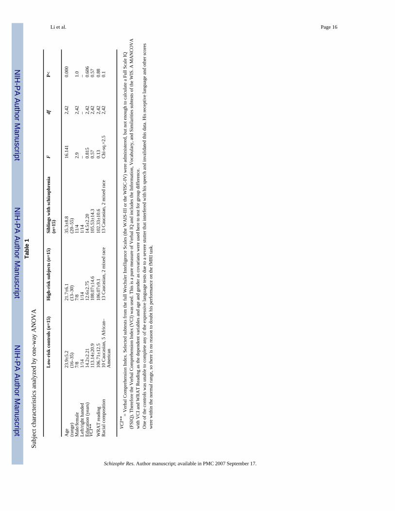
A total of 45 subjects were included in this study and categorized into 3 diagnostic groups with15 subjects in each group. Low-risk controls were age and sex matched as close as possible tothe individuals at high-risk for illness (Table 1). All subjects were interviewed using theDiagnostic Interview for Genetic Studies (DIGS; Nurnberger et al., 1994), information aboutthem was obtained from a family member, and as appropriate, when available, medical recordswere obtained. A diagnosis was made using DSM-IV criteria. In addition, all subjects hadverbal cognition tested during the preliminary evaluation and anyone with an IQ less than 85was not included in the study. Two measures of verbal cognition were included in the currentstudy (see Table 1), the Verbal Comprehension Index (VCI) from the Wechsler IntelligenceScales and the Wide Range Achievement Test-Reading (WRAT-Reading). Since only selectedsubtests from the full age appropriate Wechsler Scales (WAIS-III or WISC-IV; Wechsler,1997, 2004) were administered, but not enough to calculate a Verbal IQ (FSIQ), the VCI wasused. The VCI is a pure measure of Verbal IQ and includes the Information, Vocabulary, andSimilarities subtests of the WAIS. The WRAT-Reading was used as a standardized measurethat was close to the task used in the fMRI procedure (see below) and assesses differences inability to decode a list of progressively harder words. One of the controls was unable tocomplete any of the expressive language tests due to a severe stutter that interfered with hisspeech and invalidated these data.
This study received Institutional Review Board approval for human subjects’ research at theNathan S. Kline Institute for Psychiatric Research, a New York State Institution, and at NewYork University School of Medicine. All subjects gave written informed consent for theirparticipation after carefully being explained the nature of the study and its procedures.
2.2. Visual word discrimination taskSubjects were asked to perform a Visual Word Discrimination task using the block designillustrated in Fig. 1. Following a training and practice period outside the MRI unit, the taskwas repeated four times during the scan and fMRI data acquired as described below.
“A” blocks were Lexical Decision Blocks. In each “A” block, English language words (allconcrete nouns) were presented on a screen in front of the subject in a random fashion,interspersed with pseudowords matched to the real words for number of letters. “B” blockswere Non-Linguistic Control Blocks consisting of non-linguistic symbols that were not letters(Fig. 2), and “C” rest blocks contained a blank screen. Twenty-five different real English wordsand 25 different pseudowords were used, with 10 per Lexical Decision Block (5 real and 5pseudowords) and the same number of each was presented in each scanning sequence. The realwords were chosen from a public access database (http://www.psych.rl.ac.uk/) based on a
Li et al. Page 3
Schizophr Res. Author manuscript; available in PMC 2007 September 17.
NIH
-PA Author Manuscript
NIH
-PA Author Manuscript
NIH
-PA Author Manuscript

rating scale given for number of letters and word imagibility (range 550–700). Pseudowordswere chosen from Pexman et al. (2002). Concrete nouns were selected for the word stimuli.The range of “550–700” denotes the imagibility rating invented by linguists to assess whetherone word is easier to image internally than another. The ratings that we used in the currentwork refer to words that are highly imagible. These types of words were used in previoussimilar studies. Details of the imagibility rating are in Gilhooly and Logie (1980). The numberof letters in the pseudowords was matched to the number of letters in the words. However,pseudowords are not real words so they cannot be matched to words with respect to word type.Pseudowords are combination of letters that can be pronounced. They may sound like a word,but they do not have meaning. Subjects were instructed to use their right hand to press theleft button when a word appeared and to press the right button when the letters displayed werenot considered to be a real word.
During the Non-Linguistic Control Block, participants were presented with two different kindsof nonsense strings as shown in Fig. 2. The subjects were instructed to press the left button inresponse to the symbols in Fig. 2(a) and the right button in the response to the symbols in Fig.2(b). In one block, 5 left and 5 right symbols were randomly displayed on the screen in frontof the subject. This task was included to control for motor response and activity in the visualcortex.
During the rest block a blank screen was presented and subjects were instructed to keep theireyes open, remain relaxed and motionless.
The visual word discrimination task was repeated four times. Each block was comprised of 10stimuli, each presented for 1000 ms with an interstimulus interval of 3000 ms. Each sequenceconsisted of 8 s of initial fixation, followed by 11 stimulation blocks of 30 s each. The stimuliwere presented in random order within blocks. The order of blocks within sequences and theorder of trials within blocks differed across the three tasks, but were identical for all subjects.
Only the first three tasks for each subject were used in the final analysis. The fourth fMRIexperiment exhibited substantial head motion and little activation in all groups, and wastherefore excluded in subsequent analysis. No other data sets were excluded.
2.3. Magnetic resonance imaging protocolFunctional Imaging (fMRI) was performed on a 1.5T Siemens Vision system (ErlangenGermany). During each scan 169 functional volumes sensitive to blood oxygen level dependent(BOLD) contrast were acquired with a T2-weighted sequence (TR=2 ms, TE=50 ms, flipangle=85°, Matrix=64×64, FOV=224, pixel size= 3.5×3.5, time=5 min 38 s). Each volumecomprised 22 axial slices with 5 mm slice thickness and no gap. The first four volumes acquiredduring the initial fixation were discarded. Following the fourth fMRI acquisition series, a T2-weighted fast spin echo data set was acquired using the same slice orientation parameters asin the fMRI acquisition sequence but higher in-plane resolution. Finally, a high resolution 3-D magnetization prepared rapid gradient echo data set was acquired for spatial mapping offMRI data.
2.4. fMRI analysesData were analyzed using the FEAT fMRI Analysis tool in FSL3.3 (FMRIB’s SoftwareLibrary, www.fmrib.ox.ac.uk/fsl). In the first-level data analysis of each sequence, slice timingcorrection, intensity normalization and high pass temporal filtering were applied, non-brainstructures were removed using the BET Brain Extraction tool (Smith, 2002). To correct forhead motion, MCFLIRT was used, which is a linear registration tool applying rigid-bodytransformations (Jenkinson et al., 2002). Each image was smoothed with an 8-mm FWHM
Li et al. Page 4
Schizophr Res. Author manuscript; available in PMC 2007 September 17.
NIH
-PA Author Manuscript
NIH
-PA Author Manuscript
NIH
-PA Author Manuscript

Gaussian spatial filter. Time-series statistical analysis was carried out using FILM with localautocorrelation correction (Woolrich et al., 2001). Z (Gaussianized T/F) statistic images werethresholded using clusters determined by Z>2.3 and a corrected cluster significance thresholdof P<0.05 (Worsley et al., 1992). Scans were registered to an average T1-weighted braintemplate in the standard Talairach space with FLIRT (Jenkinson and Smith, 2001;Jenkinsonet al., 2002). Each of the first three fMRI acquisition sequences conducted on each subject wasanalyzed and the average response was obtained. In-Group analysis was carried out usingFLAME (FMRIB’s Local Analysis of Mixed Effects) (Beckmann et al., 2003;Woolrich et al.,2004). Z (Gaussianized T/F) statistic images were thresholded using clusters determined byZ>2.3 and a corrected cluster significance threshold of P<0.05 (Worsley et al., 1992).
The two blocks (A and B as described in Fig. 1) were analyzed for each individual, and a groupmean obtained for each group. In a first analysis we used an “exclusive mask” (A–B) by settingthe A event as 1 and B event as −1 in the contrasts and F-tests options. Meanwhile we maskedthis real contrast with positive A and positive B in the Contrast Masking options. This“exclusive mask analysis” differentiated the lexical from the non-linguistic control task andpermitted extraction of those regions predominantly activating with the lexical task. In a secondanalysis, we used an “inclusive mask” (activated A regions masked by activated regions of Bin the Contrast Masking options) that determined the regions of activation common to bothtasks (“inclusive mask analysis”). Thus, for each group, we obtained Z-thresholded activationmaps for the “A” task (word/pseudoword discrimination), the “B” task (non-linguisticdiscrimination), for “A–B”(exclusive mask) and for “A mask B” (inclusive mask).
For between-group analyses, the FSL FLAME package was utilized. The “A maskB” (inclusive mask) condition for the three groups were analyzed controlling for age and sex.Z (Gaussianized T/F) statistic images were thresholded at P =0.05 (cluster corrected). In thisstudy, all first order analyses were restricted to regions of positive activation. In this report,regions which exhibited negative activation are not considered.
3. ResultsThe siblings with schizophrenia were significantly older than the high-risk and low-risk groups;however, the groups did not differ significantly on sex, handedness, racial distribution, or levelof education (see Table 1). Age and sex were controlled for in analyses below.
In the within-group Exclusive Mask Analyses (language specific activity), the normal controlsshowed one cluster of significantly more activation in the left inferior frontal gyrus, i.e.Brodmann’s areas 44 (see Fig. 3). In the high-risk subjects and their siblings withschizophrenia, activation was similar for both the word discrimination and the non-linguistictasks. Thus no significantly activated brain region was found during this subtraction paradigm.
In the within-group Inclusive Mask Analyses, regions of activation common to both the non-linguistic control and linguistic blocks were examined (Fig. 4a,b,c). Control subjects hadsignificant activation on the left in Brodmann’s areas 44 and 45. However, both high-risk andsubjects with schizophrenia had significant activation during both linguistic and non-linguisticcontrol blocks in Brodmann’s areas 44 and 45 bilaterally, where activation on the right side(region of the right inferior frontal gyrus) was not present in the low-risk subjects. Thus, whencontrasted with the exclusive mask analysis, the high-risk subjects and patients experiencedless ‘task-specialization’ of Brodmann’s areas 44 and 45.
Three between-groups comparisons for the inclusive mask analysis were performed. First,high-risk subjects were compared with controls. Fig. 5 shows regions of significantly increasedand decreased activation in high-risk subjects relative to controls when the activation commonto both the linguistic and non-linguistic tasks were examined. Significantly increased activation
Li et al. Page 5
Schizophr Res. Author manuscript; available in PMC 2007 September 17.
NIH
-PA Author Manuscript
NIH
-PA Author Manuscript
NIH
-PA Author Manuscript

occurred in multiple regions in the right hemisphere (e.g. inferior frontal gyrus: Talairachcoordinates 52, 15,10; middle and superior temporal gyri: Talairach coordinates 63, −12, 7;and inferior parietal lobule: Talairach coordinates 44, −50, 48); whereas, regions in the rightfusiform gyrus (Talairach coordinates 40, −48, −18), and the right middle temporal gyrus(Talairach coordinates (64, −20, −11) had significantly decreased activation in the high-risksubjects compared with controls.
Secondly, the patients with schizophrenia were compared with controls. Fig. 6 showssignificantly increased and decreased regions of activation for the patients with schizophreniacompared with normal controls when the activation common to both the linguistic and non-linguistic tasks were analyzed. A number of regions showed significantly increased activationon the right side, including left superior temporal gyrus (Talairach coordinates −66, −7, 5),right and left side inferior parietal lobule (Talairach coordinates ±43, −45, 54) and right sideinferior frontal gyrus (Talairach coordinates 50, 11,10) in the patients. The regions withsignificantly decreased activation in the patients include left fusiform gyrus (−40, −42, −21)and right parahippocampal gyrus (26, −40, −2).
Thirdly, the high-risk subjects were compared with their siblings who had schizophrenia. Thecluster corrected results reported neither significantly increased nor significantly decreasedregions of activation, related to language processing, for the high-risk subjects compared withthe patients.
4. DiscussionThe current fMRI study used a visual word decision task to define regions of brain activationthat are significantly different between individuals who have schizophrenia compared withcontrols, and to determine whether these differences can also be seen in their relatives who arestill within the age of risk for schizophrenia and thus at a higher-risk than the general population.The data suggest that overall, our subjects with schizophrenia exhibited more right sided(bilateral) activation during the language task than did controls, as did their family membersat high-risk for the disorder. In addition, the same brain regions on the right were also activatedby a non-linguistic visual task. In contrast, some regions, such as the left inferior frontal gyrus,were activated in the controls significantly more during the linguistic than the control task,while they were activated by both tasks equally in the high-risk and schizophrenia subjects.Thus, the present data appear to show clear activation differences in controls when processinga linguistic compared with a non-linguistic task, but no regional activation differences specificto language in people at genetic high-risk for schizophrenia nor those with schizophrenia. Itcan be concluded that a more diffuse and bilateral set of regions are activated in both the high-risk and schizophrenia subjects than normal controls possibly signifying anomalous circuitryand less efficiency to the processing of language.
In addition, the current study shows some evidence of a difference in visual activation betweencontrols and the subjects with schizophrenia, but not those at high-risk. Visual processingdeficits have been found previously in studies of patients with schizophrenia (e.g. Kim et al.,2006) and in another study related to the genetic vulnerability for developing illness (Green etal., 2006). While our data do not support the latter, it is possible that visual activationdifferences could implicate visual processing pathways that are relevant to the recognition oflanguage and thus contribute to some of the changes then seen in the processing of words infrontal and temporal lobes.
The results of this study add to the increasing literature showing reduced language lateralizationin schizophrenia (e.g. Sommer et al., 2003; 2001) and previous publications (Whalley et al.,2005, 2006) showing that a more bilateral pattern of activation can also be detected in people
Li et al. Page 6
Schizophr Res. Author manuscript; available in PMC 2007 September 17.
NIH
-PA Author Manuscript
NIH
-PA Author Manuscript
NIH
-PA Author Manuscript

at high-risk for the disorder. These results also suggest that the underlying pathological basisfor schizophrenia may be related to anomalies in the pathways for language processing or otherfunctions as previously suggested (Crow, 1998;DeLisi, 2001;Morey et al., 2005).
Biological findings that distinguish individuals at high-risk for schizophrenia from controlsare particularly of interest. If in further studies they are shown to be both highly sensitive tothe prediction of who develops schizophrenia and highly specific to this disorder, then futureuse of any of these findings could be possible as a screening devise that may aid clinicians tomake decisions about treatment for people who show non-specific signs of illness and be ofprognostic value.
However, the current study was only an initial attempt at determining whether focusing on alanguage paradigm in fMRI may be a useful candidate method to develop further as a possiblescreening measure. Unfortunately in this first fMRI project aimed at distinguishing high-risksubjects from controls, we failed to save information on language task performance whilesubjects were performing the fMRI scan. However, in a prior training session, every subjectcould perform the task with ease and we were also unable to find any significant Verbal IQ orreading performance differences between groups when tested outside the scanner.Nevertheless, we do not know whether actual performance ability could have contributed tothe difference in activation patterns. Our current study was also not as well matched for racial/ethnic and age effects as they could be, since it was difficult to find these types of subjects forstudy. While we did not find effects of these variables on the pattern of activation (andcontrolled for age and sex in all analyses), they need to be more carefully matched in furtherstudies.
Future work will need a much larger cohort of high-risk individuals, followed longitudinally,to determine who eventually develops schizophrenia and whether the prior fMRi languagestudy would have been predictive of illness. In addition, a focus on individual variation andanalysis of the amount of activation in candidate brain regions detected by the current studywill be an important further analysis.
Acknowledgements
This project was partially supported by a grant from NIMH, R21 MH071720.
ReferencesBeckmann C, Jenkinson M, Smith SM. General multi-level linear modelling for group analysis in FMRI.
NeuroImage 2003;20:1052–1063. [PubMed: 14568475]Binder JR, Medler DA, Desai R, Conant LL, Liebenthal E. Some neurophysiological constraints on
models of word naming. NeuroImage 2005;27(3):677–693. [PubMed: 15921937]Boksman K, Theberge J, Williamson P, Drost DJ, Malla A, Densmore M, Takhar J, Pavlosky W, Menon
RS, Neufeld RW. Schizophrenia Research 2005;75:247–263. [PubMed: 15885517]Callicott JH, Egan MF, Mattay VS, Bertolino A, Bone AD, Vershinksi B, Weinberger DR. Abnormal
fMRI response of the dorsolateral prefrontal cortex in cognitively intact siblings of patients withschizophrenia. American Journal of Psychiatry 2003;160:709–719. [PubMed: 12668360]
Chaika, EO. Charles C. Thomas Publisher; Springfield, Illinois: 1990. Understanding Psychotic Speech:Beyond Freud and Chomsky.
Crow TJ. Nuclear schizophrenic symptoms as a window on the relationship between thought and speech.British Journal of Psychiatry 1998;173:303–309. [PubMed: 9926033]
Crow TJ, Done DJ, Sacker A. Childhood precursors of psychosis as clues to its evolutionary origins.European Archives of Psychiatry and Clinical Neuroscience 1995;245(2):61–69. [PubMed: 7654790]
Li et al. Page 7
Schizophr Res. Author manuscript; available in PMC 2007 September 17.
NIH
-PA Author Manuscript
NIH
-PA Author Manuscript
NIH
-PA Author Manuscript

DeLisi LE. Speech disorder in schizophrenia: review of the literature and new study of the relation touniquely human capacity for language. Schizophrenia Bulletin 2001;27:481–496. [PubMed:11596849]
DeLisi LE. The significance of the age of onset for schizophrenia. Schizophrenia Bulletin 1992;18:209–215. [PubMed: 1377833]
DeLisi LE, Boccio AM, Riordan H, Hoff AL, Dorfman A, McClelland J, Kushner M, Van Eyl O, OdenN. Familial thyroid disease and delayed language development in first admission patients withschizophrenia. Psychiatry Research 1991;38:39–50. [PubMed: 1946833]
DeLisi LE, Sherrington R, Shaw S, Nanthakumar B, Shields G, Smith AB, Wellman N, Larach NW,Loftus J, Razi K, Stewart J, Vita A, De Hurt M, Crow TJ, Sherrington R. A genome-wide scan of382 affected sibling-pairs with schizophrenia suggests linkage to chromosomes 2cen and 10p.American Journal of Psychiatry 2002;159:803–812. [PubMed: 11986135]
Gilhooly KJ, Logie RH. Age of acquisition, imagery, concreteness, familiarity and ambiguity measuresfor 1944 words. Behaviour Research Methods and Instrumentation 1980;12:395–427.
Green MF, Nuechterlein KH, Breitmeyer B, Mintz J. Forward and backward visual masking in unaffectedsiblings of schizophrenic patients. Biological Psychiatry 2006;59(5):446–451. [PubMed: 16139818]
Habel U, Klein M, Shah NJ, Toni I, Zilles K, Falkai P, Schneider F. Genetic load on amygdalahypofunction during sadness in nonaffected brothers of schizophrenia patients. American Journal ofPsychiatry 2004;161(10):1806–1813. [PubMed: 15465977]
Heim S, Alter K, Ischebeck AK, Amunts K, Eickhoff SB, Mohlberg H, Zilles K, von Cramon DY,Friederici AD. The role of the left Brodmann’s areas 44 and 45 in reading words and pseudowords.Cognitive Brain Research 2005;25(3):982–993. [PubMed: 16310346]
Jenkinson M, Smith SM. A global optimisation method for robust affine registration of brain images.Medical Image Analysis 2001;5(2):143–156. [PubMed: 11516708]
Jenkinson M, Bannister P, Brady M, Smith S. Improved optimisation for the robust and accurate linearregistration and motion correction of brain images. NeuroImage 2002;17(2):825–841. [PubMed:12377157]
Keshavan MS, Diwadkar VA, Spencer SM, Harenski KA, Luna B, Sweeney JA. A preliminary functionalmagnetic resonance imaging study in offspring of schizophrenic parents. Progress in Neuro-Psychopharmacology & Biological Psychiatry 2002;26(6):1143–1149. [PubMed: 12452537]
Kim D, Wylie G, Pasternak R, Butler PD, Javitt DC. Magnocellular contributions to impaired motionprocessing in schizophrenia. Schizophrenia Research 2006;82(1):1–8. [PubMed: 16325377]
Kircher TT, Bullmore ET, Brammer MJ, Williams SC, Broome MR, Murray RM, McGuire PK.Differential activation of temporal cortex during sentence completion in schizophrenic patients withand without formal thought disorder. Schizophrenia Research 2001;50:27–40. [PubMed: 11378312]
Kircher TT, Oh TM, Krammer MJ, McGuire PK. Neural correlates of syntax production in schizophrenia.British Journal of Psychiatry 2005;186:209–214. [PubMed: 15738501]
Koeda M, Takahashi H, Yahata N, Matsuura M, Asai K, Okubo Y, Tanaka H. Language processing andhuman voice perception in schizophrenia: a functional magnetic resonance imaging study. BiologicalPsychiatry 2006;59:948–957. [PubMed: 16616721]
Kubicki M, McCarley RW, Nestor PG, Huh T, Kikinis R, Shenton ME, Wible CG. An fMRI study ofsemantic processing in men with schizophrenia. NeuroImage 2003;20:1923–1933. [PubMed:14683698]
Mechelli A, Crinion JT, Long S, Friston KJ, Lambon Ralph MA, Patterson K, McClelland JL, Price CJ.Dissociating reading processes on the basis of neuronal interactions. Journal of CognitiveNeuroscience 2005;17(11):1753–1765. [PubMed: 16269111]
Morey RA, Inan S, Mitchell TV, Perkins DO, Lieberman JA, Belger A. Imaging frontostriatal functionin ultra-high-risk, early, and chronic schizophrenia during executive processing. Archives of GeneralPsychiatry 2005;62:254–262. [PubMed: 15753238]
Nurnberger JI, Blehar MC, Kaufmann CA, York-Cooler C, Simpson SG, Harkavy-Friedman J, SevereJB, Malaspina D, Reich T. Diagnostic interview for genetic studies: rationale, unique features andtraining. Archives of General Psychiatry 1994;51:849–862. [PubMed: 7944874]
Li et al. Page 8
Schizophr Res. Author manuscript; available in PMC 2007 September 17.
NIH
-PA Author Manuscript
NIH
-PA Author Manuscript
NIH
-PA Author Manuscript

Paulesu E, McCrory E, Fazio F, Menoncello L, Brunswick N, Cappa SF, Cotelli M, Cossu G, Corte F,Lorusso M, Pesenti S, Gallagher A, Perani D, Price C, Frith CD, Frith U. A cultural effect on brainfunction. Nature Neuroscience 2000;3(1):91–96.
Pexman P, Lupker S, Reggin L. Phonological effects in visual word recognition: investigating the impactof feedback activation. Journal of Experimental Psychology Learning, Memory, and Cognition2002;28:572–584.
Seidman LJ, Thermenos HW, Poldrack RA, Peace NK, Koch JK, Faraone SV, Tsuang MT. Altered brainactivation in dorsolateral prefrontal cortex in adolescents and young adults at genetic risk forschizophrenia: an fMRI study of working memory. Schizophrenia Research. 200610.1016/j.schres.2006.03.019.
Smith S. Fast robust automated brain extraction. Human Brain Mapping 2002;17(3):143–155. [PubMed:12391568]
Sommer IEC, Ramsey NE, Kahn RS. Language lateralization in schizophrenia, an fMRI study.Schizophrenia Research 2001;52:57–67. [PubMed: 11595392]
Sommer IE, Ramsey NF, Mandl RC, Kahn RS. Language lateralization in female patients withschizophrenia: an fMRI study. Schizophrenia Research 2003;60(23):183–190. [PubMed: 12591582]
Sommer IE, Ramsey NF, Mandl RC, van Oel CJ, Kahn RS. Language activation in monozygotic twinsdiscordant for schizophrenia. British Journal of Psychiatry 2004;184:128–135. [PubMed: 14754824][see comment]
Thermenos HW, Seidman LJ, Breiter H, Goldstein JM, Goodman JM, Poldrack R, Faraone SV, TsuangMT. Functional magnetic resonance imaging during auditory verbal working memory innonpsychotic relatives of persons with schizophrenia: a pilot study. Biological Psychiatry 2004;55(5):490–500. [PubMed: 15023577]
Wechsler, D. Wechsler Adult Intelligence Scale. Third Edition. The Psychological Corporation; SanAntonio, TX: 1997.
Wechsler, D. Wechsler Intelligence Scale for Children. Fourth Edition. Harcourt Assessment, Inc; SanAntonio, TX: 2004.
Weiss EM, Hofer A, Golaszewski S, Siedentopf C, Felber S, Fleischhacker WW. Language lateralizationin unmedicated patients during an acute episode of schizophrenia: a functional MRI study. PsychiatryResearch Neuroimaging 2006;146:185–190.
Whalley HC, Simonotto E, Flett S, Marshall I, Ebmeier KP, Owens DG, Goddard NH, Johnstone EC,Lawrie SM. fMRI correlates of state and trait effects in subjects at genetically enhanced risk ofschizophrenia. Brain 2004;127(Pt 3):478–490. [PubMed: 14749289]
Whalley HC, Simonotto E, Marshall I, Owens DG, Goddard NH, Johnstone EC, Lawrie SM. Functionaldisconnectivity in subjects at high genetic risk of schizophrenia. Brain 2005;128:2097–2108.[PubMed: 15930046]
Whalley HC, Simonotto E, Morehead W, McIntosh A, Marshall I, Ebmeier KP, Owens DG, GoddardNH, Johnstone EC, Lawrie SM. Functional imaging as a predictor of schizophrenia. BiologicalPsychiatry. 2006PMID: 16460690[Pubmed]
Woolrich MW, Ripley BD, Brady JM, Smith SM. Temporal autocorrelation in univariate linear modellingof FMRI data. NeuroImage 2001;14(6):1370–1386. [PubMed: 11707093]
Woolrich MW, Behrens TEJ, Beckmann CF, Jenkinson M, Smith SM. Multi-level linear modelling forFMRI group analysis using Bayesian inference. NeuroImage 2004;21(4):1732–1747. [PubMed:15050594]
Worsley KJ, Evans AC, Marrett S, Neelin P. A three-dimensional statistical analysis for CBF activationstudies in human brain. Journal of Cerebral Blood Flow and Metabolism 1992;12:900–918. [PubMed:1400644]
Xiao Z, Zhang JX, Wang X, Wu R, Hu X, Weng X, Tan LH. Differential activity in left inferior frontalgyrus for pseudowords and real words: an event-related fMRI study on auditory lexical decision.Human Brain Mapping 2005;25(2):212–221. [PubMed: 15846769]
Li et al. Page 9
Schizophr Res. Author manuscript; available in PMC 2007 September 17.
NIH
-PA Author Manuscript
NIH
-PA Author Manuscript
NIH
-PA Author Manuscript

Fig 1.The block design within one sequence. O — Initial Fixation (8 s). A — Lexical Decision TaskBlock. B — Non-linguistic Control Task Block. C — Rest block.
Li et al. Page 10
Schizophr Res. Author manuscript; available in PMC 2007 September 17.
NIH
-PA Author Manuscript
NIH
-PA Author Manuscript
NIH
-PA Author Manuscript

Fig 2.Visual stimuli in the non-linguistic motor control task.
Li et al. Page 11
Schizophr Res. Author manuscript; available in PMC 2007 September 17.
NIH
-PA Author Manuscript
NIH
-PA Author Manuscript
NIH
-PA Author Manuscript

Fig 3.Regions of significantly more brain activation for the word discrimination task compared withthe non-linguistic task in the control group. Note the activation of Brodmann’s area 44. Therewere no regions of significantly more activation for the word discrimination task in the subjectsat high-risk or their siblings with schizophrenia.
Li et al. Page 12
Schizophr Res. Author manuscript; available in PMC 2007 September 17.
NIH
-PA Author Manuscript
NIH
-PA Author Manuscript
NIH
-PA Author Manuscript

Fig 4.Significant regions of brain activation in the three groups when the Visual Word Decision Taskand the Non-word Control Task were analyzed using an inclusive (logical ‘and’ mask). Notethe laterality present in Brodmann’s area in the normal control subjects compared with thesubjects at high-risk for schizophrenia or subjects with schizophrenia. Conversely, visualactivation associated with the fMRI tasks resulted in bilateral activation in control subjects andschizophrenic patients, but not in subjects at high-risk for schizophrenia. (a) low-risk controls.(b) high-risk subjects. (c) subjects with schizophrenia.
Li et al. Page 13
Schizophr Res. Author manuscript; available in PMC 2007 September 17.
NIH
-PA Author Manuscript
NIH
-PA Author Manuscript
NIH
-PA Author Manuscript

Fig 5.Differences between subjects at high genetic risk for schizophrenia and low-risk controls(P<0.01) for the ‘inclusive mask analysis’. (a) High-risk>Controls. (b) High-risk<Controls.
Li et al. Page 14
Schizophr Res. Author manuscript; available in PMC 2007 September 17.
NIH
-PA Author Manuscript
NIH
-PA Author Manuscript
NIH
-PA Author Manuscript

Fig 6.Differences between subjects with schizophrenia and low-risk controls (P<0.01) for the‘inclusive mask analysis’ (P<0.01). (a) Patients>Controls. (b) Patients<Controls.
Li et al. Page 15
Schizophr Res. Author manuscript; available in PMC 2007 September 17.
NIH
-PA Author Manuscript
NIH
-PA Author Manuscript
NIH
-PA Author Manuscript
NIH
-PA Author Manuscript
NIH
-PA Author Manuscript
NIH
-PA Author Manuscript
Li et al. Page 16Ta
ble
1Su
bjec
t cha
ract
eris
tics a
naly
zed
by o
ne-w
ay A
NO
VA
Low
-ris
k co
ntro
ls (n
=15)
Hig
h-ri
sk su
bjec
ts (n
=15)
Sibl
ings
with
schi
zoph
reni
a(n
=15)
Fdf
P<
Age
23.9
±5.2
21.7
±6.1
35.3
±8.8
16.1
412,
420.
000
(ran
ge)
(16–
35)
(13–
30)
(20–
55)
Mal
e/fe
mal
e7/
87/
811
/42.
92,
421.
0Le
ft/rig
ht h
ande
d1/
141/
141/
14–
––
Educ
atio
n (y
ears
)14
.2±2
.21
12.6
±2.7
514
.5±2
.20
0.81
52,
420.
606
VCI*
*11
3.14
±20.
910
8.07
±14.
610
5.53
±14.
30.
572,
420.
57W
RA
T re
adin
g10
6.71
±12.
510
6.07
±9.1
102.
33±1
0.6
0.13
2,42
0.88
Rac
ial c
ompo
sitio
n10
Cau
casi
an, 5
Afr
ican
–A
mer
ican
13 C
auca
sian
, 2 m
ixed
race
13 C
auca
sian
, 2 m
ixed
race
Chi
-sq.
=2.5
2,42
0.1
VCI*
* = V
erba
l Com
preh
ensi
on In
dex.
Sel
ecte
d su
btes
ts fr
om th
e fu
ll W
echs
ler I
ntel
ligen
ce S
cale
s (th
e W
AIS
-III
or t
he W
ISC
-IV
) wer
e ad
min
iste
red,
but
not
eno
ugh
to c
alcu
late
a F
ull S
cale
IQ(F
SIQ
). Th
eref
ore
the
Ver
bal C
ompr
ehen
sion
Inde
x (V
CI)
was
use
d. T
his i
s a p
ure
mea
sure
of V
erba
l IQ
and
incl
udes
the
Info
rmat
ion,
Voc
abul
ary,
and
Sim
ilarit
ies s
ubte
sts o
f the
WIS
. A M
AN
CO
VA
with
VC
I and
WR
AT
Rea
ding
as t
he d
epen
dent
var
iabl
es a
nd a
ge a
nd g
ende
r as c
ovar
iate
s wer
e us
ed h
ere
to te
st fo
r gro
up d
iffer
ence
.O
ne o
f the
con
trols
was
una
ble
to c
ompl
ete
any
of th
e ex
pres
sive
lang
uage
test
s due
to a
seve
re st
utte
r tha
t int
erfe
red
with
his
spee
ch a
nd in
valid
ated
this
dat
a. H
is re
cept
ive
lang
uage
and
oth
er sc
ores
wer
e w
ithin
the
norm
al ra
nge,
so th
ere
is n
o re
ason
to d
oubt
his
per
form
ance
on
the
fMR
I tas
k.
Schizophr Res. Author manuscript; available in PMC 2007 September 17.


![[The relations between neurocognition and quality of life in people with schizophrenia]](https://static.fdokumen.com/doc/165x107/633d1d32b6ae16c04d04f64f/the-relations-between-neurocognition-and-quality-of-life-in-people-with-schizophrenia.jpg)