An ESAT-6:CFP10 DNA vaccine administered in ... - PubAg
12
Vaccine 25 (2007) 4735–4746 An ESAT-6:CFP10 DNA vaccine administered in conjunction with Mycobacterium bovis BCG confers protection to cattle challenged with virulent M. bovis Alexander C. Maue a , W. Ray Waters b , Mitchell V. Palmer b , Brian J. Nonnecke c , F. Chris Minion d , Wendy C. Brown e , Junzo Norimine e , Monica R. Foote f , Charles F.C. Scherer g , D. Mark Estes g,∗ a Department of Molecular Microbiology and Immunology, University of Missouri, Columbia, MO 65211, USA b United States Department of Agriculture, Agricultural Research Service, National Animal Disease Center, Bacterial Diseases of Livestock Research Unit, Ames, IA 50010, USA c United States Department of Agriculture, Agricultural Research Service, National Animal Disease Center, Periparturient Diseases of Cattle Research Unit, Ames, IA 50010, USA d College of Veterinary Medicine, Veterinary Microbiology and Preventive Medicine, Iowa State University, Ames, IA 50011, USA e Department of Veterinary Microbiology and Pathology, College of Veterinary Medicine, Washington State University, Pullman, WA 99164, USA f Department of Animal science, Iowa State University, Ames, IA 50011, USA g University of Texas Medical Branch, Department of Pediatrics and the Sealy Center for Vaccine Development, Galveston, TX 77555, USA Received 13 February 2007; received in revised form 24 March 2007; accepted 30 March 2007 Available online 24 April 2007 Abstract The potency of genetic immunization observed in the mouse has demonstrated the utility of DNA vaccines to induce cell-mediated and humoral immune responses. However, it has been relatively difficult to generate comparable responses in non-rodent species. The use of molecular adjuvants may increase the magnitude of these suboptimal responses. In this study, we demonstrate that the co-administration of plasmid-encoded GM-CSF and CD80/CD86 with a novel ESAT-6:CFP10 DNA vaccine against bovine tuberculosis enhances antigen-specific cell-mediated immune responses. ESAT-6:CFP10 + GM-CSF + CD80/CD86 DNA vaccinated animals exhibited significant (p < 0.01) antigen-specific proliferative responses compared to other DNA vaccinates. Increased expression (p ≤ 0.05) of CD25 on PBMC from ESAT-6:CFP10 + GM-CSF + CD80/CD86 DNA vaccinates was associated with increased proliferation, as compared to control DNA vaccinates. Significant (p <0.05) numbers of ESAT-6:CFP10-specific IFN- producing cells were evident from all ESAT-6:CFP10 DNA vaccinated animals compared to control DNA vaccinates. However, the greatest increase in IFN- producing cells was from animals vaccinated with ESAT-6:CFP10 + GM-CSF + CD80/CD86 DNA. In a low-dose aerosol challenge trial, calves vaccinated as neonates with Mycobacterium bovis BCG and ESAT-6:CFP10 + GM-CSF + CD80/CD86 DNA exhibited decreased lesion severity in the lung and lung-associated lymph nodes following viruluent M. bovis challenge compared to other vaccinated animals or non-vaccinated controls. These data suggest that a combined vaccine regimen of M. bovis BCG and a candidate ESAT-6:CFP10 DNA vaccine may offer greater protection against tuberculosis in cattle than vaccination with BCG alone. © 2007 Elsevier Ltd. All rights reserved. Keywords: Adjuvant; Neonate; Tuberculosis ∗ Corresponding author. Present address: Department of Pediatrics and the Sealy Center for Vaccine Development, University of Texas Medical Branch, 2.330F Children’s Hospital, 301 University Boulevard, Galveston, TX 77555-0372, USA. Tel.: +1 409 772 0434; fax: +1 409 772 0460. E-mail address: [email protected] (D.M. Estes). 0264-410X/$ – see front matter © 2007 Elsevier Ltd. All rights reserved. doi:10.1016/j.vaccine.2007.03.052
-
Upload
khangminh22 -
Category
Documents
-
view
1 -
download
0
Transcript of An ESAT-6:CFP10 DNA vaccine administered in ... - PubAg
A
hmpcaEvvvwlda©
K
0d
Vaccine 25 (2007) 4735–4746
An ESAT-6:CFP10 DNA vaccine administered in conjunctionwith Mycobacterium bovis BCG confers protection to
cattle challenged with virulent M. bovis
Alexander C. Maue a, W. Ray Waters b, Mitchell V. Palmer b, Brian J. Nonnecke c,F. Chris Minion d, Wendy C. Brown e, Junzo Norimine e, Monica R. Foote f,
Charles F.C. Scherer g, D. Mark Estes g,∗a Department of Molecular Microbiology and Immunology, University of Missouri, Columbia, MO 65211, USA
b United States Department of Agriculture, Agricultural Research Service, National Animal Disease Center,Bacterial Diseases of Livestock Research Unit, Ames, IA 50010, USA
c United States Department of Agriculture, Agricultural Research Service, National Animal Disease Center,Periparturient Diseases of Cattle Research Unit, Ames, IA 50010, USA
d College of Veterinary Medicine, Veterinary Microbiology and Preventive Medicine, Iowa State University, Ames, IA 50011, USAe Department of Veterinary Microbiology and Pathology, College of Veterinary Medicine, Washington State University, Pullman, WA 99164, USA
f Department of Animal science, Iowa State University, Ames, IA 50011, USAg University of Texas Medical Branch, Department of Pediatrics and the Sealy Center for Vaccine Development, Galveston, TX 77555, USA
Received 13 February 2007; received in revised form 24 March 2007; accepted 30 March 2007Available online 24 April 2007
bstract
The potency of genetic immunization observed in the mouse has demonstrated the utility of DNA vaccines to induce cell-mediated andumoral immune responses. However, it has been relatively difficult to generate comparable responses in non-rodent species. The use ofolecular adjuvants may increase the magnitude of these suboptimal responses. In this study, we demonstrate that the co-administration of
lasmid-encoded GM-CSF and CD80/CD86 with a novel ESAT-6:CFP10 DNA vaccine against bovine tuberculosis enhances antigen-specificell-mediated immune responses. ESAT-6:CFP10 + GM-CSF + CD80/CD86 DNA vaccinated animals exhibited significant (p < 0.01)ntigen-specific proliferative responses compared to other DNA vaccinates. Increased expression (p ≤ 0.05) of CD25 on PBMC fromSAT-6:CFP10 + GM-CSF + CD80/CD86 DNA vaccinates was associated with increased proliferation, as compared to control DNAaccinates. Significant (p < 0.05) numbers of ESAT-6:CFP10-specific IFN-� producing cells were evident from all ESAT-6:CFP10 DNAaccinated animals compared to control DNA vaccinates. However, the greatest increase in IFN-� producing cells was from animalsaccinated with ESAT-6:CFP10 + GM-CSF + CD80/CD86 DNA. In a low-dose aerosol challenge trial, calves vaccinated as neonatesith Mycobacterium bovis BCG and ESAT-6:CFP10 + GM-CSF + CD80/CD86 DNA exhibited decreased lesion severity in the lung and
ung-associated lymph nodes following viruluent M. bovis challenge compared to other vaccinated animals or non-vaccinated controls. Theseata suggest that a combined vaccine regimen of M. bovis BCG and a candidate ESAT-6:CFP10 DNA vaccine may offer greater protectiongainst tuberculosis in cattle than vaccination with BCG alone.
2007 Elsevier Ltd. All rights reserved.
eywords: Adjuvant; Neonate; Tuberculosis
∗ Corresponding author. Present address: Department of Pediatrics and the Sealy2.330F Children’s Hospital, 301 University Boulevard, Galveston, TX 77555-0372
E-mail address: [email protected] (D.M. Estes).
264-410X/$ – see front matter © 2007 Elsevier Ltd. All rights reserved.oi:10.1016/j.vaccine.2007.03.052
Center for Vaccine Development, University of Texas Medical Branch,, USA. Tel.: +1 409 772 0434; fax: +1 409 772 0460.
4 ccine 25
1
oc[tbisoratos
pmvafoesDIb[iioskabmotsCaDta
agmbantocr
feEpf
OcECtaevstovMrDGeDBscoetipga
2
2
wobow(uiuCF
736 A.C. Maue et al. / Va
. Introduction
DNA vaccination of mice has demonstrated the utilityf genetic immunization as a means of inducing robustell-mediated and humoral responses to encoded antigens1,2]. Development of successful, practical DNA vaccina-ion in humans and other outbred non-rodent species haseen hampered by several factors [3–5]. To induce signif-cant immune responses, vaccinates are often subjected toeveral immunizations comprised of relatively large amountsf plasmid DNA [6–8]. Individuals vaccinated with DNA areelatively weak responders [6,7]. Molecular adjuvants suchs cytokines, costimulatory molecules and immunostimula-ory DNA sequences have been used to increase the potencyf DNA vaccine-induced immune responses in non-rodentpecies [9–15].
Following intramuscular (IM) injection of a DNA vaccine,lasmid DNA is internalized by dendritic cells (DCs) anduscle cells. Myocytes express genes encoded on plasmids in
itro and in vivo [16,17]; however, these cells cannot initiaten immune response via naı̈ve T cells, even when trans-ected with an appropriate array of costimulatory moleculesr cytokines [18]. Effects of DNA vaccination are still appar-nt following ablation of tissue surrounding the injectionite [19]. Plasmid DNA has been isolated directly fromCs within the draining lymph nodes and skin following
M or intradermal inoculation [20]. In addition, DCs cane directly transfected following IM plasmid immunization21], suggesting that DCs are key mediators of DNA vaccine-nduced immune responses. Strategies to manipulate DCs toncrease the potency of DNA vaccination, include the usef growth factors such as granulocyte macrophage-colonytimulating factor (GM-CSF) [1,4,22] or fms-like tyrosineinase-3 ligand (Flt-3L) [9,23,24]. In mice and humans,dministration of these growth factors, used alone or in com-ination, increases numbers of DC’s [25–29]. Bovine bonearrow cells cultured with GM-CSF alone, GM-CSF + IL-4
r GM-CSF + IL-4 + Flt-3L display morphological charac-eristics typical of DCs [30]. Experiments in cattle havehown that the co-administration of plasmid-encoded GM-SF and Flt-3L enhances bovine CD4+ T cell responses ton Anaplasma marginale major surface protein-1 (MSP-1)NA vaccine [9]. The observed enhancement was attributed
o an increase in DC recruitment at the site of immunizationfter growth factor treatment [9].
Early secretory antigenic target-6 kDa (ESAT-6) proteinnd culture filtrate protein 10 (CFP10) are potent immuno-ens encoded by region of difference-1 (RD1) in tuberculousycobacteria [31] and are absent in Mycobactrium bovis
acille Calmette-Guerin (BCG) [32,33]. The loss of RD1 isssociated with the attenuation of BCG [34,35]. Althougho precise function has been attributed to these proteins,
heir expression correlates with an increased cytolytic abilityf M. tuberculosis [36]. ESAT-6 is recognized by bovine Tells during the early phase of infection [37], resulting in theelease of IFN-� [37,38], making it an attractive candidate(w6i
(2007) 4735–4746
or vaccine development. ESAT-6 and CFP10 exist as a het-rodimer in a 1:1 complex in vivo [39]. In vitro, the use of anSAT-6:CFP10 fusion protein induces high levels of IFN-�roduction [40] and robust proliferative responses by T cellsrom M. bovis-infected cattle.
In our previous study, we assessed the potential of CpGDN, CD80, CD86 and CD154 to enhance the efficacy of a
andidate bovine tuberculosis DNA vaccine directed againstSAT-6 [10]. Co-administration of CpG ODN with eitherD80/CD86 or CD154, in a single prime/boost vaccina-
ion regimen, enhanced ESAT-6-specific IFN-� responsess compared to non-vaccinated control animals [10]. How-ver, following aerosol challenge with M. bovis, only animalsaccinated with CD80/CD86 demonstrated decreased lesioneverity in the lungs and associated lymph nodes [10]. Collec-ively, this earlier study demonstrated that co-administrationf CD80/CD86 is superior to CD154 in augmenting DNAaccine-induced protection following aerosol challenge with. bovis [10]. In the present study, we evaluated immune
esponses generated against a candidate ESAT-6:CFP10NA vaccine and assessed the ability of plasmid-encodedM-CSF to further augment the adjuvant activity of plasmid-
ncoded CD80/CD86 in response to a novel ESAT-6:CFP10NA vaccine. In addition, we evaluated the use of a M. bovisCG prime + ESAT-6:CFP10 DNA vaccine prime/boost
trategy to induce protection against a low-dose aerosolhallenge. Our results indicate that the co-administrationf plasmids encoding GM-CSF and CD80/CD86 moleculesnhances bovine immune responses to genetic immuniza-ion and that this ESAT-6:CFP10 DNA vaccine administeredn conjunction with a M. bovis BCG prime confersrotection against experimental bovine tuberculosis to areater level than DNA vaccination or BCG vaccinationlone.
. Materials and methods
.1. Plasmid DNA and recombinant protein production
Plasmids encoding bovine CD80 and bovine GM-CSFere kindly provided by Dr. Chris J. Howard (Institutef Animal Health, Compton, UK). The construction ofovine CD80 and GM-CSF plasmids was described previ-usly [41,42]. For CD86 plasmid construction, total RNAas obtained from bovine PBMC using Trizol Reagent
Invitrogen, Carlsbad, CA) and reverse-transcribed to cDNAsing oligo dT 16 (Perkin-Elmer, Branchburg, NJ) accord-ng to the manufacturer’s instructions. The cDNA wassed to amplify an entire open reading frame of bovineD86 by polymerase chain reaction (PCR) using primersWD (5′-ACAGCAGAAATAACGAAAATGCG) and REV
5′-CATGGCGTTTACTCTTTAATTACA). PCR conditionsere 94 ◦C (10 min), followed by 35 cycles of 94 ◦C (15 s),0 ◦C (15 s) and 72 ◦C (2 min). PCR products were clonednto pCR3.1 using T4 ligase (Invitrogen). Sequence and
ccine 25 (2007) 4735–4746 4737
ds
uT6pGTpaT(fPim(ifboa
2
Pm(Mndgsm
2
wAdU
2
wtbbPOwwi
Fig. 1. Timeline detailing events during DNA vaccine trial #2. Calves receiv-ing Mycobacterium bovis BCG were immunized with 1 × 10⁶ CFU M. bovisBCG strain Pasteur at the time of primary immunization. Animals vacci-nated with BCG + DNA or DNA alone received a primary dose of plasmidsencoding ESAT-6:CFP10, GM-CSF, CD80 and CD86 in IFA, and an iden-tia
pamp((t(iDvEmdci
2
ai(tDtcieroMm
2.4. Aerosol challenge of vaccinated cattle
A.C. Maue et al. / Va
irection of the insert was confirmed by automated DNAequencing.
The ESAT-6:CFP10 vaccine plasmid was constructedsing the vector VR1020 (Vical Inc., San Diego, CA).he sequence encoding the protein fusion of ESAT-:CFP10 was amplified from plasmid pISM2202 [40] usingrimers “ES-CFP-U-Not” (5′-AATGCGGCCGCATAT-GGGGG) and “ES-CFP-L-EcoR1” (5′-GCTGAAT-CCGAAGCCCATTTGC). The PCR product was gelurified, digested with NotI and EcoRI and ligated into,
derivative of plasmid VR1020, plasmid pISM2214.he resulting plasmid was designated plasmid pISM2215
VR1020:6-His:ESAT-6:CFP10). The DNA sequence andrame orientation was confirmed by DNA sequence analysis.lasmid DNA for immunization was chemically transformed
nto competent Escherichia coli TOP10 (Invitrogen). Plas-id DNA was purified using the Qiagen Plasmid Giga kit
Qiagen, Valencia, CA) according to the manufacturer’snstructions. Purified recombinant protein was obtainedollowing induction of TOP10 cells containing pISM2202y metal chelate chromatography as described [40], dialyzedvernight at 4 ◦C in PBS and quantified by the Bradfordssay.
.2. Bacteria
M. bovis strain 1315 and attenuated M. bovis BCG strainasteur were grown in Middlebrook 7H9 media supple-ented with 10% oleic acid–albumin–dextrose complex
Difco, Detroit, MI) plus 0.05% Tween 80 (Sigma, St. Louis,O) as described previously [10]. M. bovis 1315 was origi-
ally isolated in 1995 from a naturally infected white-tailedeer [43]. Vaccine and challenge inocula consisted of mid-logrowth phase mycobacteria. Bacilli were enumerated usingerial dilution plate counting on Middlebrook 7H11 selectiveedia (Becton Dickinson, Cockeysville, MD).
.3. Animals and immunizations
All immunizations and challenge components of the studyere conducted at the National Animal Disease Center,mes, IA. Prior to experimentation, animal-related proce-ures were approved by the Institutional Animal Care andse Committee of the National Animal Disease Center.
.3.1. DNA vaccine trial #1Twenty-five castrated, 4-month-old Holstein bull calves,
ere selected for their negative reactivity to purified pro-ein derivative (PPD) from M. avium (PPDa) and M.ovis (PPDb), as well as rESAT-6:CFP10. Briefly, wholelood was incubated for 24 h in vitro in the presence ofPDa (10 �g/ml), PPDb (10 �g/ml) (Biocor Animal Health,
maha, NE) or rESAT-6:CFP10 (10 �g/ml). Supernatantsere harvested and antigen-specific IFN-� productionas determined using the Bovigam ELISA kit (Prion-cs Ag, Schlieren, Switzerland). Animals negative forMC
ical booster dose 6 weeks post-immunization. Sixteen weeks after primarymmunization, cattle received 103 CFU of virulent M. bovis 1315 by aerosolnd were euthanized 12 weeks after challenge.
revious exposure to mycobacterial antigens were thenssigned randomly to the following experimental treat-ent groups: (1) control DNA (empty plasmid vector
cDNA3.1) (n = 5); (2) ESAT-6:CFP10 DNA vaccinationn = 5); (3) ESAT-6:CFP10 + GM-CSF DNA vaccinationn = 5); (4) ESAT-6:CFP10 + CD80/CD86 DNA vaccina-ion (n = 5); (5) ESAT-6:CFP10 + GM-CSF + CD80/CD86n = 5). DNA vaccinates were immunized intramuscularlyn the right mid-cervical region with 2 mg of total plasmidNA emulsified in 2 ml of incomplete Freund’s adju-ant (IFA). All experimental vaccines contained 1 mg ofSAT-6:CFP10 encoding plasmid. The total amount of plas-id DNA was normalized to 2 mg by additional plasmids
epending on the treatment. Cattle received an identi-al booster dose of vaccine 20 days following primarymmunization.
.3.2. DNA vaccine trial #2Twenty-five Holstein calves less than 6 days of
ge were randomly assigned to the following exper-mental treatment groups: (1) non-vaccinated controlsn = 6); (2) M. bovis BCG strain Pasteur vaccina-ion (n = 5); (3) ESAT-6:CFP10 + GM-CSF + CD80/CD68NA vaccination (n = 5); (4) M. bovis BCG strain Pas-
eur + ESAT-6:CFP10 + GM-CSF + CD80/CD86 DNA vac-ination (n = 5). At 1 week of age, DNA vaccinates weremmunized subcutaneously with 2 mg of total plasmid DNAmulsified in 2 ml of IFA in the left mid-cervical region andeceived a booster dose at 6 weeks of age (Fig. 1). At the timef the first immunization, animals received 1.0 × 10⁶ CFU. bovis BCG strain Pasteur subcutaneously in the rightid-cervical region.
Low-dose aerosol challenge of cattle with 1.0 × 10³ CFU. bovis 1315 was conducted at the National Animal Diseaseenter as described previously [44].
4 ccine 25
2
ba(bPR2tC1(wPcCtScwMsp
2
vtp4tautiow
2
E�kPcoieEotm
2
ic4btLwPciPhbaUPPWswAECbifs
2
p6bs4acp0(sPaa((p
738 A.C. Maue et al. / Va
.5. Lymphocyte proliferation assays
PBMC were isolated via density centrifugation fromuffy-coat fractions of peripheral blood collected in 2×cid–citrate–dextrose. Peripheral blood mononuclear cellsPBMC) (5 × 10⁵) were cultured in triplicate wells of round-ottom 96-well plates (Falcon, Becton Dickinson, Lincolnark, NJ) in a total volume of 200 �l. Medium was completePMI 1640 (cRPMI) supplemented with 2 mM l-glutamine,5 mM HEPES buffer, 100 U/ml penicillin, 100 �g/ml strep-omycin, 1% non-essential amino acids (Sigma Chemicalo., St. Louis, MO), 2% essential amino acids (Sigma),% sodium pyruvate (Sigma), 50 �M 2-mercaptoethanolSigma), and 10% (v/v) fetal bovine sera (FBS). PBMCere stimulated in vitro with rESAT-6:CFP10 (5 �g/ml),PDb (5 �g/ml), pokeweed mitogen (PWM) (1 �g/ml) orRPMI alone. Cells were cultured for 6 days at 37 ◦C in 5%O2 humidified air with the addition of 0.5–1.0 �Ci [3H]-
hymidine (specific activity, 6.7 Ci/mM, Amersham Lifecience, Arlington Heights, IL) during the last 16–20 h ofulture. Well contents were harvested onto glass fiber filtersith a 96-well plate harvester (EG & G Wallac, Gaithersburg,D), and the incorporated radioactivity measured by liquid
cintillation counting. Treatments were run in triplicate andresented as mean cpm (±S.E.M.).
.6. IFN-γ ELISA
Cells were cultured in round-bottom 96-well plates in aolume of 200 �l. PBMC (4 × 10⁵) were cultured in vitro inhe presence of rESAT-6:CFP10 (5 �g/ml), PPDb (5 �g/ml),okeweed mitogen (PWM) (1 �g/ml) or cRPMI alone for8 h at 37 ◦C in 5% CO2 humidified air. Following incuba-ion, supernatants were removed and stored at −80 ◦C untilnalysis. Supernatants were assayed for IFN-� productionsing the Bovigam ELISA kit (Prionics) according to instruc-ions provided by the manufacturer. Concentrations (ng/ml)n test samples were quantified by comparing the absorbancef duplicate test samples with the absorbance of standardsithin a linear curve fit.
.7. IFN-γ ELISPOT assay
Antigen-specific IFN-� production was assayed using anLISPOT as described previously [10]. Anti-bovine IFN-monoclonal antibodies (MAb) CC302 and CC330 were
indly provided by Dr. Chris J. Howard. Briefly, 5 × 10⁵
BMC were added in 100 �l volumes containing eitherRPMI alone, rESAT-6:CFP10 (5 �g/ml), PPDb (5 �g/ml)r PWM (1 �g/ml). Plates were incubated for 36 h at 37 ◦Cn 5% CO2 humidified air. IFN-� spot-forming cells were
numerated using a standard dissection microscope or anLISPOT Reader (CTL). For each animal, the mean numberf spots of negative control (i.e. cRPMI alone) wells was sub-racted from the number of spots in test wells to determine theean number of antigen-specific IFN-� spot-forming cells.
It0ul
(2007) 4735–4746
.8. Detection of antigen-specific antibodies
Round-bottom 96-well plates (Falcon, Becton Dick-nson) were coated with rESAT-6:CFP10 (1 �g/ml) inarbonate–bicarbonate coating buffer pH 9.6 overnight at◦C. Plates were washed three times with 1× phosphateuffered saline plus 0.05% Tween 20 (PBST) (Sigma) andhen blocked for 1 h with milk diluent (Kirkegaard Perryaboratories, Gaithersburg, MD) at 37 ◦C. Plates were thenashed three times with PBST. Sera were diluted 1:100 inBS and 100 �l added to wells in duplicate for antigen-oated and control wells (i.e. coating buffer alone) for 1 hncubation at 37 ◦C. Plates were washed three times withBST before addition of 100 �l goat anti-bovine IgG (H + L)orseradish peroxidase-conjugated (KPL) secondary anti-ody diluted 1:10,000 in PBS + 0.1% fish gelatin. For isotypenalysis, 100 �l of sheep anti-bovine IgG1 (Serotec, Oxford,K) or sheep anti-bovine IgG2 (Serotec), diluted 1:5000 inBS + 0.1% fish gelatin, was added as secondary antibody.lates were incubated for 1 h at 37 ◦C and washed three times.ells were developed with the addition of 100 �l TMB sub-
trate (KPL) for 5 min at room temperature. The reactionas stopped by addition of 100 �l of 0.18 M sulfuric acid.450 of individual wells was measured with an automatedLISA plate reader (Molecular Devices, Menlo Park, CA)hanges in optical density (�OD) readings were calculatedy subtracting the mean OD readings for wells receiving coat-ng buffer alone (two replicates) from the mean OD readingsor antigen-coated wells (two replicates) receiving the sameerum sample.
.9. Flow cytometry
PBMC (4 × 10⁵ per well) were cultured in vitro in theresence of rESAT-6:CFP10 (5 �g/ml) or cRPMI alone fordays at 37 ◦C in 5% CO2 humidified air in a round-
ottom 96-well plate. Following incubation, cells wereubsequently pooled according to treatments. Approximately× 105 pooled cells in 200 �l of culture medium weredded to individual wells of round-bottom 96-well plates,entrifuged (2 min, 400 × g) and resuspended in 100 �l ofrimary antibody(s) (1 �g/ml in PBS containing 1% FBS and.1% sodium azide). Primary antibodies included anti-CD4GC50A1), anti-CD8� (BAQ111A), anti-�� TCR (GB21A,pecific for � chain) and anti-CD25 (CACT116A) (VMRD,ullman, WA). After 15 min incubation at room temper-ture, cells were centrifuged and stained with 100 �l ofppropriate secondary antibody [fluorescein isothiocyanateFITC, 1 �g/well)-conjugated goat anti-mouse IgG2b, IgMSouthern Biotechnology Associates, Birmingham, AL),hycoerythrin (PE, 1 �g/well)-conjugated goat anti-mousegG1]. Cells were incubated for 15 min at room tempera-
ure, centrifuged, washed and resuspended in PBS containing.04% sodium azide for acquisition (10,000 live gated events)sing a FACScan (Becton Dickinson, San Jose, CA; 488 nmaser, two color) flow cytometer. Data analysis was performed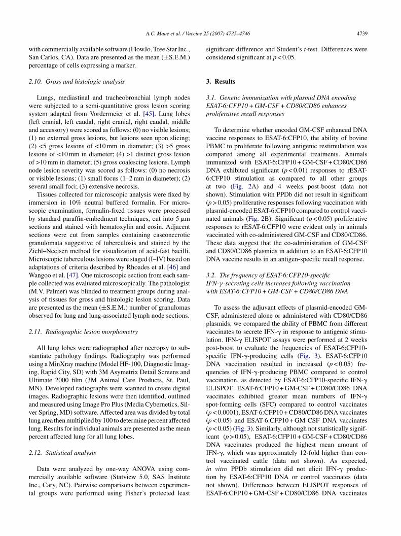
ccine 25
wSp
2
ws(a((lonos
isbssgZMaWp(yao
2
suiUMiavllp
2
mIt
sc
3
3Ep
vPciD6as(pnrvTaD
3Iw
CpvlpsDqvEvs(((iDIt
A.C. Maue et al. / Va
ith commercially available software (FlowJo, Tree Star Inc.,an Carlos, CA). Data are presented as the mean (±S.E.M.)ercentage of cells expressing a marker.
.10. Gross and histologic analysis
Lungs, mediastinal and tracheobronchial lymph nodesere subjected to a semi-quantitative gross lesion scoring
ystem adapted from Vordermeier et al. [45]. Lung lobesleft cranial, left caudal, right cranial, right caudal, middlend accessory) were scored as follows: (0) no visible lesions;1) no external gross lesions, but lesions seen upon slicing;2) <5 gross lesions of <10 mm in diameter; (3) >5 grossesions of <10 mm in diameter; (4) >1 distinct gross lesionf >10 mm in diameter; (5) gross coalescing lesions. Lymphode lesion severity was scored as follows: (0) no necrosisr visible lesions; (1) small focus (1–2 mm in diameter); (2)everal small foci; (3) extensive necrosis.
Tissues collected for microscopic analysis were fixed bymmersion in 10% neutral buffered formalin. For micro-copic examination, formalin-fixed tissues were processedy standard paraffin-embedment techniques, cut into 5 �mections and stained with hematoxylin and eosin. Adjacentections were cut from samples containing caseonecroticranulomata suggestive of tuberculosis and stained by theiehl–Neelsen method for visualization of acid-fast bacilli.icroscopic tuberculous lesions were staged (I–IV) based on
daptations of criteria described by Rhoades et al. [46] andangoo et al. [47]. One microscopic section from each sam-
le collected was evaluated microscopically. The pathologistM.V. Palmer) was blinded to treatment groups during anal-sis of tissues for gross and histologic lesion scoring. Datare presented as the mean (±S.E.M.) number of granulomasbserved for lung and lung-associated lymph node sections.
.11. Radiographic lesion morphometry
All lung lobes were radiographed after necropsy to sub-tantiate pathology findings. Radiography was performedsing a MinXray machine (Model HF-100, Diagnostic Imag-ng, Rapid City, SD) with 3M Asymetrix Detail Screens andltimate 2000 film (3M Animal Care Products, St. Paul,N). Developed radiographs were scanned to create digital
mages. Radiographic lesions were then identified, outlinednd measured using Image Pro Plus (Media Cybernetics, Sil-er Spring, MD) software. Affected area was divided by totalung area then multiplied by 100 to determine percent affectedung. Results for individual animals are presented as the meanercent affected lung for all lung lobes.
.12. Statistical analysis
Data were analyzed by one-way ANOVA using com-ercially available software (Statview 5.0, SAS Institute
nc., Cary, NC). Pairwise comparisons between experimen-al groups were performed using Fisher’s protected least
itnE
(2007) 4735–4746 4739
ignificant difference and Student’s t-test. Differences wereonsidered significant at p < 0.05.
. Results
.1. Genetic immunization with plasmid DNA encodingSAT-6:CFP10 + GM-CSF + CD80/CD86 enhancesroliferative recall responses
To determine whether encoded GM-CSF enhanced DNAaccine responses to ESAT-6:CFP10, the ability of bovineBMC to proliferate following antigenic restimulation wasompared among all experimental treatments. Animalsmmunized with ESAT-6:CFP10 + GM-CSF + CD80/CD86NA exhibited significant (p < 0.01) responses to rESAT-:CFP10 stimulation as compared to all other groupst two (Fig. 2A) and 4 weeks post-boost (data nothown). Stimulation with PPDb did not result in significantp > 0.05) proliferative responses following vaccination withlasmid-encoded ESAT-6:CFP10 compared to control vacci-ated animals (Fig. 2B). Significant (p < 0.05) proliferativeesponses to rESAT-6:CFP10 were evident only in animalsaccinated with co-administered GM-CSF and CD80/CD86.hese data suggest that the co-administration of GM-CSFnd CD80/CD86 plasmids in addition to an ESAT-6:CFP10NA vaccine results in an antigen-specific recall response.
.2. The frequency of ESAT-6:CFP10-specificFN-γ-secreting cells increases following vaccinationith ESAT-6:CFP10 + GM-CSF + CD80/CD86 DNA
To assess the adjuvant effects of plasmid-encoded GM-SF, administered alone or administered with CD80/CD86lasmids, we compared the ability of PBMC from differentaccinates to secrete IFN-� in response to antigenic stimu-ation. IFN-� ELISPOT assays were performed at 2 weeksost-boost to evaluate the frequencies of ESAT-6:CFP10-pecific IFN-�-producing cells (Fig. 3). ESAT-6:CFP10NA vaccination resulted in increased (p < 0.05) fre-uencies of IFN-�-producing PBMC compared to controlaccination, as detected by ESAT-6:CFP10-specific IFN-�LISPOT. ESAT-6:CFP10 + GM-CSF + CD80/CD86 DNAaccinates exhibited greater mean numbers of IFN-�pot-forming cells (SFC) compared to control vaccinatesp < 0.0001), ESAT-6:CFP10 + CD80/CD86 DNA vaccinatesp < 0.05) and ESAT-6:CFP10 + GM-CSF DNA vaccinatesp < 0.05) (Fig. 3). Similarly, although not statistically signif-cant (p > 0.05), ESAT-6:CFP10 + GM-CSF + CD80/CD86NA vaccinates produced the highest mean amount of
FN-�, which was approximately 12-fold higher than con-rol vaccinated cattle (data not shown). As expected,
n vitro PPDb stimulation did not elicit IFN-� produc-ion by ESAT-6:CFP10 DNA or control vaccinates (dataot shown). Differences between ELISPOT responses ofSAT-6:CFP10 + GM-CSF + CD80/CD86 DNA vaccinates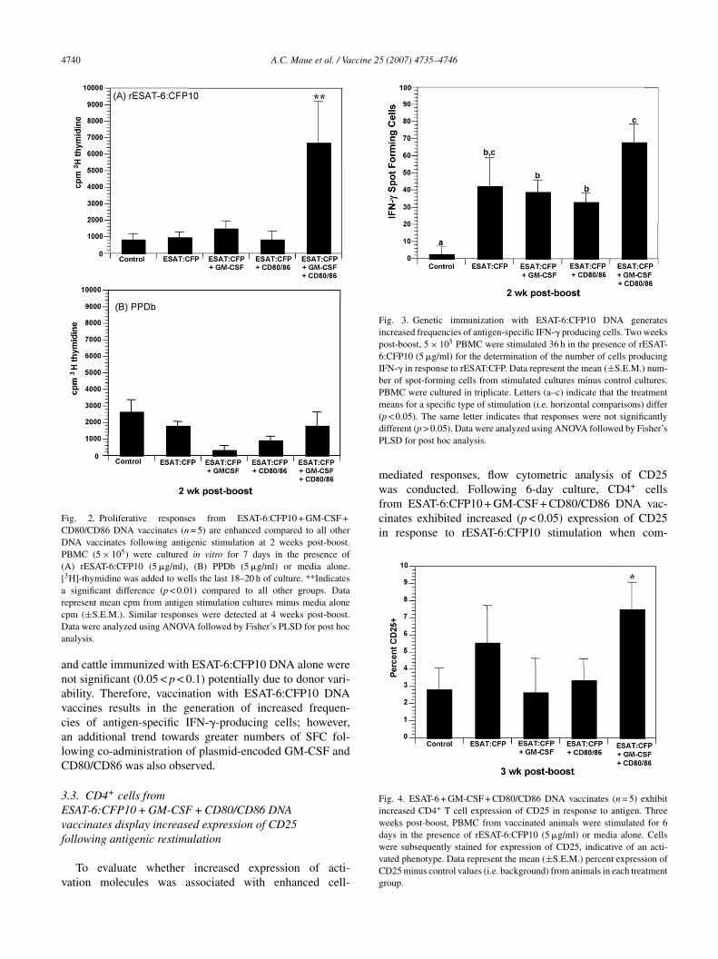
4740 A.C. Maue et al. / Vaccine 25 (2007) 4735–4746
Fig. 2. Proliferative responses from ESAT-6:CFP10 + GM-CSF +CD80/CD86 DNA vaccinates (n = 5) are enhanced compared to all otherDNA vaccinates following antigenic stimulation at 2 weeks post-boost.PBMC (5 × 10⁵) were cultured in vitro for 7 days in the presence of(A) rESAT-6:CFP10 (5 �g/ml), (B) PPDb (5 �g/ml) or media alone.[3H]-thymidine was added to wells the last 18–20 h of culture. **Indicatesa significant difference (p < 0.01) compared to all other groups. DatarcDa
anavcalC
3Evf
v
Fig. 3. Genetic immunization with ESAT-6:CFP10 DNA generatesincreased frequencies of antigen-specific IFN-� producing cells. Two weekspost-boost, 5 × 10⁵ PBMC were stimulated 36 h in the presence of rESAT-6:CFP10 (5 �g/ml) for the determination of the number of cells producingIFN-� in response to rESAT:CFP. Data represent the mean (±S.E.M.) num-ber of spot-forming cells from stimulated cultures minus control cultures.PBMC were cultured in triplicate. Letters (a–c) indicate that the treatmentmeans for a specific type of stimulation (i.e. horizontal comparisons) differ(dP
mwfrom ESAT-6:CFP10 + GM-CSF + CD80/CD86 DNA vac-cinates exhibited increased (p < 0.05) expression of CD25in response to rESAT-6:CFP10 stimulation when com-
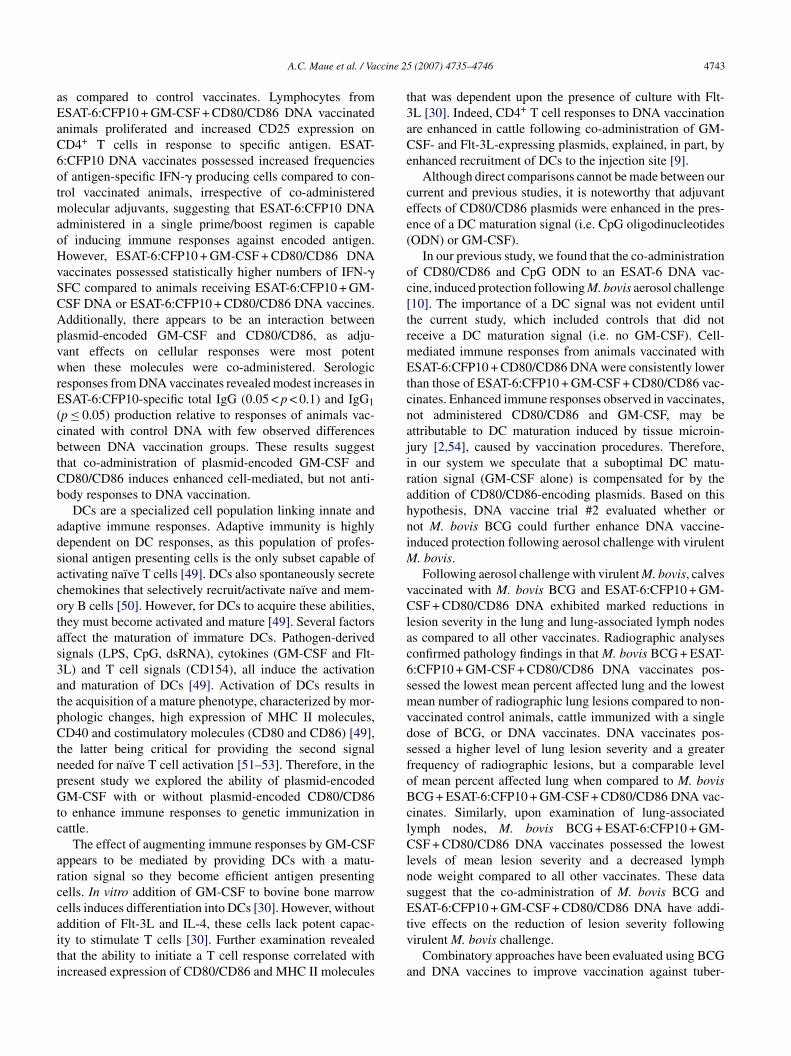
Fig. 4. ESAT-6 + GM-CSF + CD80/CD86 DNA vaccinates (n = 5) exhibitincreased CD4+ T cell expression of CD25 in response to antigen. Threeweeks post-boost, PBMC from vaccinated animals were stimulated for 6
epresent mean cpm from antigen stimulation cultures minus media alonepm (±S.E.M.). Similar responses were detected at 4 weeks post-boost.ata were analyzed using ANOVA followed by Fisher’s PLSD for post hoc
nalysis.
nd cattle immunized with ESAT-6:CFP10 DNA alone wereot significant (0.05 < p < 0.1) potentially due to donor vari-bility. Therefore, vaccination with ESAT-6:CFP10 DNAaccines results in the generation of increased frequen-ies of antigen-specific IFN-�-producing cells; however,n additional trend towards greater numbers of SFC fol-owing co-administration of plasmid-encoded GM-CSF andD80/CD86 was also observed.
.3. CD4+ cells fromSAT-6:CFP10 + GM-CSF + CD80/CD86 DNAaccinates display increased expression of CD25
ollowing antigenic restimulationTo evaluate whether increased expression of acti-ation molecules was associated with enhanced cell-
dwvCg
p < 0.05). The same letter indicates that responses were not significantlyifferent (p > 0.05). Data were analyzed using ANOVA followed by Fisher’sLSD for post hoc analysis.
ediated responses, flow cytometric analysis of CD25as conducted. Following 6-day culture, CD4+ cells
ays in the presence of rESAT-6:CFP10 (5 �g/ml) or media alone. Cellsere subsequently stained for expression of CD25, indicative of an acti-ated phenotype. Data represent the mean (±S.E.M.) percent expression ofD25 minus control values (i.e. background) from animals in each treatmentroup.

A.C. Maue et al. / Vaccine 25 (2007) 4735–4746 4741
Table 1Serum antibody responses to ESAT-6:CFP10 genetic immunizationa
Vaccination group �OD at A450
Total IgG day 0 Total IgG 2 weeks PB IgG1 day 0 IgG1 2 weeks PB IgG2 day 0 IgG2 2 weeks PB
Control (n = 5) 0.111 (0.035) 0.176 (0.034) 0.016 (0.016) 0.178 (0.072) 0.005 (0.005) 0.138 (0.022)ESAT-6:CFP10 (n = 20) 0.140 (0.024) 0.281* (0.029) 0.027 (0.009) 0.370** (0.044) 0.006 (0.002) 0.196 (0.023)ESAT-6:CFP10 alone (n = 5) 0.057 (0.021) 0.231 (0.043) 0.000 (0.000) 0.350 (0.094) 0.010 (0.003) 0.214 (0.048)ESAT-6:CFP10 + GM-CSF
(n = 5)0.182 (0.066) 0.300** (0.036) 0.052 (0.028) 0.384 (0.089) 0.000 (0.000) 0.233 (0.051)
ESAT-6:CFP10 + CD80/CD86(n = 5)
0.151 (0.051) 0.297 (0.093) 0.025 (0.019) 0.402* (0.080) 0.005 (0.003) 0.174 (0.034)
ESAT-6:CFP10 + GM-CSF + CD80/CD86(n = 5)
0.170 (0.039) 0.296 (0.061) 0.030 (0.016) 0.347 (0.116) 0.009 (0.004) 0.164 (0.055)
a Serum antibody responses to ESAT-6:CFP10 were assessed prior to vaccination (day 0) and at 2 weeks post-boost (PB). Sera were diluted 1:100 and addedt cal densf eadingsD nse by c
ppCciEso
3f
etaaba6aiipI6wrvarvts6cca
3Bvf
EetupagtoaTlaelma(tewmmlmvst
o ESAT-6:CFP10-coated wells (1 �g/ml) in duplicate. The changes in optior wells receiving coating buffer alone (two replicates) from the mean OD rata are presented as group mean (±S.E.M.). Differs from respective respo
ared to cattle vaccinated with control DNA at 3 weeksost-boost (Fig. 4). Differences in the expression ofD25 on CD8+ and �� TCR+ cells were not signifi-ant (p > 0.05) at this timepoint (data not shown). Thus,ncreased proliferative responses observed from PBMC ofSAT-6:CFP10 + GM-CSF + CD80/CD86 DNA vaccinatedubjects were associated with increased expression of CD25n CD4-bearing cells.
.4. Evaluation of antigen-specific humoral responsesollowing ESAT-6:CFP10 DNA vaccination
Antigen-specific serologic responses from animals werevaluated to determine the effects of genetic immuniza-ion on antibody responses. Responses (total IgG, IgG1nd IgG2) directed toward ESAT-6:CFP were negligiblet day 0 of the vaccine trial (Table 1). Analysis of anti-ody responses 2 weeks post-boost, revealed that increasedntibody production was more likely attributable to ESAT-:CFP10 DNA vaccination rather than the type of moleculardjuvant administered, although a significant (p ≤ 0.05)ncrease in total IgG against rESAT-6:CFP10 was seenn animals receiving ESAT-6:CFP10 + GM-CSF-encodinglasmids and a trend (0.05 < p < 0.1) toward increasedgG1 production observed in cattle vaccinated with ESAT-:CFP10 + CD80/CD86 DNA. The effect of immunizingith ESAT-6:CFP10 DNA vaccines was more telling when
esponses from all animals receiving ESAT-6:CFP10 DNAaccine were pooled. Two weeks following vaccine boost,trend (0.05 < p < 0.1) toward increased total IgG against
ESAT-6:CFP10 was evident in all ESAT-6:CFP10 DNAaccinates compared to control vaccinates (Table 1). Fur-her analysis of antigen-specific antibody isotypes revealed aignificant (p ≤ 0.05) IgG1 antibody response against ESAT-
:CFP10 from all DNA vaccinates relative to responses ofontrol animals (Table 1). These data suggest that DNA vac-ination with ESAT-6:CFP10 results in the production ofntigen-specific humoralresponses.nsDD
ity (�OD) readings were calculated by subtracting the mean OD readingsfor antigen-coated wells (two replicates) receiving the same serum sample.ontrols, *p < 0.1, **p ≤ 0.05.
.5. M. bovisCG + ESAT-6:CFP10 + GM-CSF + CD80/CD86 DNAaccinates display reduced lesion severity in the lungsollowing aerosol challenge with virulent M. bovis
Results from DNA vaccine trial #1 indicated thatSAT-6:CFP10 + GM-CSF + CD80/CD86 DNA vaccinationlicited more robust immune responses to encoded antigenhan all other DNA vaccinates. Therefore, we sought to eval-ate whether or not this vaccine combination could conferrotection in cattle aerosol challenged with virulent M. bovis,nd to assess if the addition of BCG enhances plasmid-enerated immunity. Previous experiments have determinedhe location of M. bovis after low-dose aerosol challengef cattle (∼120 days) to be primarily focused in the lungsnd regional lymph nodes draining the lung. As shown inable 2, BCG + ESAT-6:CFP10 DNA vaccinates possessed
ower gross pathology scores as compared to control animalsnd other vaccinates. To reinforce these findings, we nextmployed radiographic lesion morphometry to quantify lungesions. Non-vaccinated control animals exhibited the highest
ean number of radiographic lesions per lung, followed bynimals vaccinated with BCG alone and then DNA vaccinatesTable 2). BCG + ESAT-6:CFP10 DNA vaccinates possessedhe fewest mean number of lesions compared to all otherxperimental treatment groups. Similarly, cattle vaccinatedith BCG + ESAT-6:CFP10 DNA also displayed the lowestean percent affected lung following aerosol challenge. Ani-als vaccinated with ESAT-6:CFP10 possessed comparable
evels of percent affected lung and numbers of granulo-as upon histologic analysis to BCG + ESAT-6:CFP10 DNA
accinates, although exhibited a higher mean pathologycore and a greater frequency of radiographic lesions inhe lung. Numbers of granulomas of the various stages did
ot vary significantly between treatment groups (data nothown). These results suggest that BCG + ESAT-6:CFP10NA vaccination, and to some degree ESAT-6:CFP10NA vaccination, achieves reduced pathology of the lung
4742 A.C. Maue et al. / Vaccine 25 (2007) 4735–4746
Table 2Summary of lung pathology after challenge with virulent Mycobacterium bovis
Vaccination groupa Gross pathologyb Number ofradiographic lesions
Animals withradiographic lesions
% Affected lungc Histologicevaluationd
Control 1.32 ± 0.70 16.0 ± 9.36 5/5 1.29 ± 1.20 2.4 ± 1.3BCG 1.44 ± 0.74 8.80 ± 5.15 3/5 0.58 ± 0.30 3.4 ± 2.3ESAT-6:CFP10 1.20 ± 0.44 8.00 ± 2.72 4/4 0.16 ± 0.03 0.8 ± 0.4BCG + ESAT-6:CFP10 0.48 ± 0.27 3.60 ± 2.01 3/5 0.12 ± 0.10 0.6 ± 0.4
Data are presented as group mean (±S.E.M.).a Vaccine treatment groups were: non-vaccinated controls (n = 5), M. bovis BCG strain Pasteur vaccination (n = 5), ESAT-6:CFP10 + GM-CSF + CD80/CD68
DNA vaccination (n = 4), and M. bovis BCG strain Pasteur + ESAT-6:CFP10 + GM-CSF + CD80/CD86 DNA vaccination (n = 5).b Mean disease score: lungs were subjected to a semi-quantitative pathology scoring system described previously [45].
Lesionm 00 to d
tologic
cc
3r
apTsnoauiocdDlM
4
osCt[sdt[rauacw
TS
V
M
T
nD
[
c Lung lobes were removed at necropsy and individually radiographed.easured. Affected area was divided by total lung area then multiplied by 1d Mean number of granulomas detected per microscopic section upon his
ompared to control animals following virulent M. bovishallenge.
.6. BCG + ESAT-6:CFP10 DNA vaccinates exhibiteduced pathology of the lung-associated lymph nodes
Following aerosol challenge, tracheobronchial and medi-stinal lymph nodes were subjected to a previously describedathology scoring system [45]. Results are summarized inable 3. BCG + ESAT-6:CFP10 DNA vaccinates possessedignificantly reduced gross pathology scores of mediasti-al and tracheobronchial lymph nodes compared to allther animals (Table 3). Additionally, the mean weightsnd number of granulomas detected upon histologic eval-ation of the lung-associated lymph nodes were the lowestn animals receiving BCG + ESAT-6:CFP10 DNA. Numbersf granulomas of the various stages did not vary signifi-antly between treatment groups (data not shown). These
ata suggest that vaccination with BCG + ESAT-6:CFP10NA results in reduced pathology of the lung-drainingymph nodes following aerosol challenge with virulent. bovis.
e
nv
able 3ummary of pathology in the lung-associated lymph nodes after challenge with vir
accination groupa Gross pathologyb
ediastinal lymph nodeControl 2.70 ± 0.21BCG 2.00 ± 0.45ESAT-6:CFP10 2.00 ± 0.32BCG + ESAT-6:CFP10 0.60 ± 0.40
racheobronchial lymph nodeControl 2.20 ± 0.54BCG 1.80 ± 0.58ESAT-6:CFP10 2.00 ± 0.58BCG + ESAT-6:CFP10 0.60 ± 0.40
a Data are presented as group mean (±S.E.M.), n = 5/group. Vaccine treatment grouation (n = 5), ESAT-6:CFP10 + GM-CSF + CD80/CD68 DNA vaccination (n = 4), aNA vaccination (n = 5).b Mean disease score: mediastinal and tracheobronchial lymph nodes were subje
45].c Mean number of granulomas detected per microscopic section upon histologic
s were identified on digital images of scanned radiographs, outlined, andetermine percent affected lung.evaluation of lung tissue sections.
. Discussion
The goal of vaccination is to generate immunologic mem-ry that will provide rapid protection against exposure to apecific pathogen. Following antigenic priming, CD4+ andD8+ T cells undergo programmed division, resulting in
he generation of effector and memory T cell populations48]. This division occurs independently of further antigenictimulation [48]. However, factors such as the strength anduration of antigenic stimulation, and level of costimula-ion affect T cell differentiation and their functional qualities48]. In this study, we sought to generate improved recallesponses to a candidate DNA vaccine (ESAT-6:CFP10)gainst tuberculosis by addition of plasmid-encoded costim-latory molecules (CD80/CD86) and encoded GM-CSF. Inddition, we aimed to evaluate a vaccination regimen in whichalves were primed with M. bovis BCG and prime/boostedith an ESAT-6:CFP10 DNA vaccine containing plasmid-
ncoded costimulatory molecules.Following a single prime/boost regimen, animals immu-
ized with ESAT-6:CFP10 + GM-CSF + CD80/CD86 DNAaccine displayed increased recall responses to antigen
ulent M. bovis
Lymph node weight (g) Histologic evaluationc
29.62 ± 8.68 20.6 ± 7.925.12 ± 3.54 23.4 ± 6.121.42 ± 2.48 24.0 ± 8.614.66 ± 1.84 6.8 ± 4.9
10.36 ± 1.62 24.2 ± 5.911.22 ± 2.35 25.2 ± 8.310.12 ± 1.05 20.2 ± 3.8
7.46 ± 1.22 5.6 ± 3.9
ps were: non-vaccinated controls (n = 5), M. bovis BCG strain Pasteur vacci-nd M. bovis BCG strain Pasteur + ESAT-6:CFP10 + GM-CSF + CD80/CD86
cted to a semi-quantitative pathology scoring system described previously
evaluation of lymph node tissue sections.
ccine 25
aEaC6otmaoHvSCApvwrE(cbtCb
adsacotas3atpCtnpGtc
arccaiti
t3aCe
cee(
oc[trmEtcnajirahniM
vClac6smvdsfoBclClnsE
A.C. Maue et al. / Va
s compared to control vaccinates. Lymphocytes fromSAT-6:CFP10 + GM-CSF + CD80/CD86 DNA vaccinatednimals proliferated and increased CD25 expression onD4+ T cells in response to specific antigen. ESAT-:CFP10 DNA vaccinates possessed increased frequenciesf antigen-specific IFN-� producing cells compared to con-rol vaccinated animals, irrespective of co-administered
olecular adjuvants, suggesting that ESAT-6:CFP10 DNAdministered in a single prime/boost regimen is capablef inducing immune responses against encoded antigen.owever, ESAT-6:CFP10 + GM-CSF + CD80/CD86 DNAaccinates possessed statistically higher numbers of IFN-�FC compared to animals receiving ESAT-6:CFP10 + GM-SF DNA or ESAT-6:CFP10 + CD80/CD86 DNA vaccines.dditionally, there appears to be an interaction betweenlasmid-encoded GM-CSF and CD80/CD86, as adju-ant effects on cellular responses were most potenthen these molecules were co-administered. Serologic
esponses from DNA vaccinates revealed modest increases inSAT-6:CFP10-specific total IgG (0.05 < p < 0.1) and IgG1
p ≤ 0.05) production relative to responses of animals vac-inated with control DNA with few observed differencesetween DNA vaccination groups. These results suggesthat co-administration of plasmid-encoded GM-CSF andD80/CD86 induces enhanced cell-mediated, but not anti-ody responses to DNA vaccination.
DCs are a specialized cell population linking innate anddaptive immune responses. Adaptive immunity is highlyependent on DC responses, as this population of profes-ional antigen presenting cells is the only subset capable ofctivating naı̈ve T cells [49]. DCs also spontaneously secretehemokines that selectively recruit/activate naı̈ve and mem-ry B cells [50]. However, for DCs to acquire these abilities,hey must become activated and mature [49]. Several factorsffect the maturation of immature DCs. Pathogen-derivedignals (LPS, CpG, dsRNA), cytokines (GM-CSF and Flt-L) and T cell signals (CD154), all induce the activationnd maturation of DCs [49]. Activation of DCs results inhe acquisition of a mature phenotype, characterized by mor-hologic changes, high expression of MHC II molecules,D40 and costimulatory molecules (CD80 and CD86) [49],
he latter being critical for providing the second signaleeded for naı̈ve T cell activation [51–53]. Therefore, in theresent study we explored the ability of plasmid-encodedM-CSF with or without plasmid-encoded CD80/CD86
o enhance immune responses to genetic immunization inattle.
The effect of augmenting immune responses by GM-CSFppears to be mediated by providing DCs with a matu-ation signal so they become efficient antigen presentingells. In vitro addition of GM-CSF to bovine bone marrowells induces differentiation into DCs [30]. However, without
ddition of Flt-3L and IL-4, these cells lack potent capac-ty to stimulate T cells [30]. Further examination revealedhat the ability to initiate a T cell response correlated withncreased expression of CD80/CD86 and MHC II moleculestv
a
(2007) 4735–4746 4743
hat was dependent upon the presence of culture with Flt-L [30]. Indeed, CD4+ T cell responses to DNA vaccinationre enhanced in cattle following co-administration of GM-SF- and Flt-3L-expressing plasmids, explained, in part, bynhanced recruitment of DCs to the injection site [9].
Although direct comparisons cannot be made between oururrent and previous studies, it is noteworthy that adjuvantffects of CD80/CD86 plasmids were enhanced in the pres-nce of a DC maturation signal (i.e. CpG oligodinucleotidesODN) or GM-CSF).
In our previous study, we found that the co-administrationf CD80/CD86 and CpG ODN to an ESAT-6 DNA vac-ine, induced protection following M. bovis aerosol challenge10]. The importance of a DC signal was not evident untilhe current study, which included controls that did noteceive a DC maturation signal (i.e. no GM-CSF). Cell-ediated immune responses from animals vaccinated withSAT-6:CFP10 + CD80/CD86 DNA were consistently lower
han those of ESAT-6:CFP10 + GM-CSF + CD80/CD86 vac-inates. Enhanced immune responses observed in vaccinates,ot administered CD80/CD86 and GM-CSF, may bettributable to DC maturation induced by tissue microin-ury [2,54], caused by vaccination procedures. Therefore,n our system we speculate that a suboptimal DC matu-ation signal (GM-CSF alone) is compensated for by theddition of CD80/CD86-encoding plasmids. Based on thisypothesis, DNA vaccine trial #2 evaluated whether orot M. bovis BCG could further enhance DNA vaccine-nduced protection following aerosol challenge with virulent
. bovis.Following aerosol challenge with virulent M. bovis, calves
accinated with M. bovis BCG and ESAT-6:CFP10 + GM-SF + CD80/CD86 DNA exhibited marked reductions in
esion severity in the lung and lung-associated lymph nodess compared to all other vaccinates. Radiographic analysesonfirmed pathology findings in that M. bovis BCG + ESAT-:CFP10 + GM-CSF + CD80/CD86 DNA vaccinates pos-essed the lowest mean percent affected lung and the lowestean number of radiographic lung lesions compared to non-
accinated control animals, cattle immunized with a singleose of BCG, or DNA vaccinates. DNA vaccinates pos-essed a higher level of lung lesion severity and a greaterrequency of radiographic lesions, but a comparable levelf mean percent affected lung when compared to M. bovisCG + ESAT-6:CFP10 + GM-CSF + CD80/CD86 DNA vac-inates. Similarly, upon examination of lung-associatedymph nodes, M. bovis BCG + ESAT-6:CFP10 + GM-SF + CD80/CD86 DNA vaccinates possessed the lowest
evels of mean lesion severity and a decreased lymphode weight compared to all other vaccinates. These datauggest that the co-administration of M. bovis BCG andSAT-6:CFP10 + GM-CSF + CD80/CD86 DNA have addi-
ive effects on the reduction of lesion severity followingirulent M. bovis challenge.
Combinatory approaches have been evaluated using BCGnd DNA vaccines to improve vaccination against tuber-
4 ccine 25
crmc[DatisegcTitavbi[gad
aiTlroamstitnat
DtCinTTDtCaMDpi
A
LDmUaPU
R
[
[
[
[
744 A.C. Maue et al. / Va
ulosis. In cattle, a DNA prime-BCG boost immunizationegimen was shown to enhance protection following experi-ental challenge with M. bovis compared to non-vaccinated
ontrol animals and animals vaccinated with BCG alone55]. In addition, Cai et al. recently demonstrated that aNA vaccine prime followed by a BCG boost resulted in10–100-fold reduction of M. bovis in the lungs of cat-
le infected with virulent M. bovis compared to animalsmmunized with DNA alone or BCG alone [56]. In addition,everal experiments in mice have been reported to inducenhanced protection using BCG and DNA vaccine strate-ies. The enhancement of immune responses seen with theseombined approaches may be explained by several factors.he addition of DNA vaccines that encode proteins deleted
n the attenuation of BCG, may provide important vaccineargets that are present in virulent mycobacteria (i.e. ESAT-6nd CFP10). The inclusion of BCG also presumably pro-ides more T cell epitopes to which immune responses maye mounted. In addition, BCG provides a maturation signal tommature DCs through the involvement of toll-like receptors57], which may explain our enhancement of DNA vaccine-enerated immune responses in the presence of a moleculardjuvant. Further studies will be required to conclusivelyetermine this effect.
To date, M. bovis BCG is the only approved vaccinegainst tuberculosis. However, it has been demonstrated thatts efficacy in humans may range from 80% to 0% [58].he efficacy of BCG vaccination in cattle has been simi-
arly variable (reviewed in Refs. [59,60]). Recent progressegarding BCG vaccination of neonatal cattle and the usef combinatorial vaccines has been promising in findinglternative approaches to standard BCG vaccination. Thisay be important in controlling the spread of tuberculo-
is from wildlife reservoirs to cattle and thus, limiting theransmission of virulent mycobacteria to humans, particularlyn developing countries or countries with endemic bovineuberculosis. Natural transmission studies in the field will beeeded to accurately determine the efficacy of our vaccinend/or vaccine regimen in preventing the spread of bovineuberculosis.
In summary, our data demonstrate that ESAT-6:CFP10NA vaccination induces potent immune responses, and
hat co-administration of plasmid-encoded GM-CSF andD80/CD86 as a single prime/boost regimen enhances
mmune responses of cattle to ESAT-6:CFP10 genetic immu-ization, particularly at the level of cell-mediated immunity.he combination of a DC maturation/activation signal and acell activation signal appear to be optimal for generatingNA vaccine-induced immune responses in cattle. Finally
he combined M. bovis BCG and ESAT-6:CFP10 + GM-SF + CD80/CD86 DNA vaccine prime/boost strategyfforded vaccinates the highest degree of protection against
. bovis-induced pathology when compared to BCG or aNA vaccine administered separate of each other by possiblyroviding an adjuvant signal to potentiate DNA vaccine-nduced responses.[
(2007) 4735–4746
cknowledgments
The authors thank Don McDorman, Nancy Eischen, Peterasley and Josh Pitzer for technical assistance; Todd Holtz,ouglas Ewing, Jay Steffen and Dr. Jean Laufer for ani-al care and handling. This research was supported by thenited States Department of Agriculture Specific Cooper-
tive Agreement #58-1940-0-008 and the Program for therevention of Animal Infectious Diseases (PPAID) at theniversity of Missouri, Columbia, MO.
eferences
[1] Gurunathan S, Klinman DM, Seder RA. DNA vaccines: immunol-ogy, application, and optimization. Annu Rev Immunol 2000;18:927–74.
[2] Shedlock DJ, Weiner DB. DNA vaccination: antigen presentationand the induction of immunity. J Leukocyte Biol 2000;68(6):793–806.
[3] van Drunen Littel-van den Hurk S, Gerdts V, Loehr BI, Pontarollo R,Rankin R, Uwiera R, et al. Recent advances in the use of DNA vaccinesfor the treatment of diseases of farmed animals. Adv Drug Deliv Rev2000;43(1):13–28.
[4] Scheerlinck JY. Genetic adjuvants for DNA vaccines. Vaccine2001;19(17–19):2647–56.
[5] Beard CW, Mason PW. Out on the farm with DNA vaccines. NatBiotechnol 1998;16(13):1325–8.
[6] Wang R, Doolan DL, Le TP, Hedstrom RC, Coonan KM, Charoenvit Y,et al. Induction of antigen-specific cytotoxic T lymphocytes in humansby a malaria DNA vaccine. Science 1998;282(5388):476–80.
[7] Wang R, Epstein J, Baraceros FM, Gorak EJ, Charoenvit Y, Carucci DJ,et al. Induction of CD4(+) T cell-dependent CD8(+) type 1 responsesin humans by a malaria DNA vaccine. Proc Natl Acad Sci USA2001;98(19):10817–22.
[8] Arulkanthan A, Brown WC, McGuire TC, Knowles DP. Biasedimmunoglobulin G1 isotype responses induced in cattle withDNA expressing msp1a of Anaplasma marginale. Infect Immun1999;67(7):3481–7.
[9] Mwangi W, Brown WC, Lewin HA, Howard CJ, Hope JC, Baszler TV,et al. DNA-encoded fetal liver tyrosine kinase 3 ligand and granulocytemacrophage-colony-stimulating factor increase dendritic cell recruit-ment to the inoculation site and enhance antigen-specific CD4+ T cellresponses induced by DNA vaccination of outbred animals. J Immunol2002;169(7):3837–46.
10] Maue AC, Waters WR, Palmer MV, Whipple DL, Minion FC,Brown WC, et al. CD80 and CD86, but not CD154, augment DNAvaccine-induced protection in experimental bovine tuberculosis. Vac-cine 2004;23(6):769–79.
11] Manoj S, Griebel PJ, Babiuk LA, van Drunen Littel-van den Hurk S.Targeting with bovine CD154 enhances humoral immune responsesinduced by a DNA vaccine in sheep. J Immunol 2003;170(2):989–96.
12] Manoj S, Griebel PJ, Babiuk LA, van Drunen Littel-van den Hurk S.Modulation of immune responses to bovine herpesvirus-1 in cattle byimmunization with a DNA vaccine encoding glycoprotein D as a fusionprotein with bovine CD154. Immunology 2004;112(2):328–38.
13] Zhang Y, Palmer GH, Abbott JR, Howard CJ, Hope JC, Brown WC.CpG ODN 2006 and IL-12 are comparable for priming Th1 lympho-cyte and IgG responses in cattle immunized with a rickettsial outer
membrane protein in alum. Vaccine 2003;21(23):3307–18.14] Kim JJ, Bagarazzi ML, Trivedi N, Hu Y, Kazahaya K, Wilson DM,et al. Engineering of in vivo immune responses to DNA immuniza-tion via codelivery of costimulatory molecule genes. Nat Biotechnol1997;15(7):641–6.
ccine 25
[
[
[
[
[
[
[
[
[
[
[
[
[
[
[
[
[
[
[
[
[
[
[
[
[
[
[
[
[
[
[
[
[
[
A.C. Maue et al. / Va
15] Pontarollo RA, Babiuk LA, Hecker R, van Drunen Littel-van Den HurkS. Augmentation of cellular immune responses to bovine herpesvirus-1glycoprotein D by vaccination with CpG-enhanced plasmid vectors. JGen Virol 2002;83(Pt 12):2973–81.
16] Wolff JA, Ludtke JJ, Acsadi G, Williams P, Jani A. Long-term persis-tence of plasmid DNA and foreign gene expression in mouse muscle.Hum Mol Genet 1992;1(6):363–9.
17] Wolff JA, Dowty ME, Jiao S, Repetto G, Berg RK, Ludtke JJ, et al.Expression of naked plasmids by cultured myotubes and entry of plas-mids into T tubules and caveolae of mammalian skeletal muscle. J CellSci 1992;103(Pt 4):1249–59.
18] Iwasaki A, Stiernholm BJ, Chan AK, Berinstein NL, Barber BH.Enhanced CTL responses mediated by plasmid DNA immuno-gens encoding costimulatory molecules and cytokines. J Immunol1997;158(10):4591–601.
19] Torres CA, Iwasaki A, Barber BH, Robinson HL. Differential depen-dence on target site tissue for gene gun and intramuscular DNAimmunizations. J Immunol 1997;158(10):4529–32.
20] Casares S, Inaba K, Brumeanu TD, Steinman RM, Bona CA. Antigenpresentation by dendritic cells after immunization with DNA encodinga major histocompatibility complex class II-restricted viral epitope. JExp Med 1997;186(9):1481–6.
21] Chattergoon MA, Robinson TM, Boyer JD, Weiner DB. Specificimmune induction following DNA-based immunization through in vivotransfection and activation of macrophages/antigen-presenting cells. JImmunol 1998;160(12):5707–18.
22] Cohen AD, Boyer JD, Weiner DB. Modulating the immune responseto genetic immunization. FASEB J 1998;12(15):1611–26.
23] Hung CF, Hsu KF, Cheng WF, Chai CY, He L, Ling M, et al. Enhance-ment of DNA vaccine potency by linkage of antigen gene to a geneencoding the extracellular domain of Fms-like tyrosine kinase 3-ligand.Cancer Res 2001;61(3):1080–8.
24] Moore AC, Kong WP, Chakrabarti BK, Nabel GJ. Effects of antigen andgenetic adjuvants on immune responses to human immunodeficiencyvirus DNA vaccines in mice. J Virol 2002;76(1):243–50.
25] Daro E, Butz E, Smith J, Teepe M, Maliszewski CR, McKenna HJ.Comparison of the functional properties of murine dendritic cells gen-erated in vivo with Flt3 ligand, GM-CSF and Flt3 ligand plus GM-SCF.Cytokine 2002;17(3):119–30.
26] Kaplan G, Walsh G, Guido LS, Meyn P, Burkhardt RA, AbalosRM, et al. Novel responses of human skin to intradermal recombi-nant granulocyte/macrophage-colony-stimulating factor: Langerhanscell recruitment, keratinocyte growth, and enhanced wound healing.J Exp Med 1992;175(6):1717–28.
27] Shurin MR, Pandharipande PP, Zorina TD, Haluszczak C, SubbotinVM, Hunter O, et al. FLT3 ligand induces the generation of function-ally active dendritic cells in mice. Cell Immunol 1997;179(2):174–84.
28] Maraskovsky E, Brasel K, Teepe M, Roux ER, Lyman SD, Shortman K,et al. Dramatic increase in the numbers of functionally mature dendriticcells in Flt3 ligand-treated mice: multiple dendritic cell subpopulationsidentified. J Exp Med 1996;184(5):1953–62.
29] Maraskovsky E, Daro E, Roux E, Teepe M, Maliszewski CR, Hoek J,et al. In vivo generation of human dendritic cell subsets by Flt3 ligand.Blood 2000;96(3):878–84.
30] Hope JC, Werling D, Collins RA, Mertens B, Howard CJ. Flt-3 ligand, in combination with bovine granulocyte-macrophagecolony-stimulating factor and interleukin-4, promotes the growthof bovine bone marrow derived dendritic cells. Scand J Immunol2000;51(1):60–6.
31] Berthet FX, Rasmussen PB, Rosenkrands I, Andersen P, Gicquel B.A Mycobacterium tuberculosis operon encoding ESAT-6 and a novel
low-molecular-mass culture filtrate protein (CFP-10). Microbiology1998;144(Pt 11):3195–203.32] Behr MA, Wilson MA, Gill WP, Salamon H, Schoolnik GK, Rane S,et al. Comparative genomics of BCG vaccines by whole-genome DNAmicroarray. Science 1999;284(5419):1520–3.
[
[
(2007) 4735–4746 4745
33] Gordon SV, Brosch R, Billault A, Garnier T, Eiglmeier K, ColeST. Identification of variable regions in the genomes of tuberclebacilli using bacterial artificial chromosome arrays. Mol Microbiol1999;32(3):643–55.
34] Lewis KN, Liao R, Guinn KM, Hickey MJ, Smith S, Behr MA,et al. Deletion of RD1 from Mycobacterium tuberculosis mimicsbacille Calmette-Guerin attenuation. J Infect Dis 2003;187(1):117–23.
35] Pym AS, Brodin P, Brosch R, Huerre M, Cole ST. Loss of RD1contributed to the attenuation of the live tuberculosis vaccinesMycobacterium bovis BCG and Mycobacterium microti. Mol Microbiol2002;46(3):709–17.
36] Hsu T, Hingley-Wilson SM, Chen B, Chen M, Dai AZ, Morin PM, et al.The primary mechanism of attenuation of bacillus Calmette-Guerin isa loss of secreted lytic function required for invasion of lung interstitialtissue. Proc Natl Acad Sci USA 2003;100(21):12420–5.
37] Pollock JM, Andersen P. Predominant recognition of the ESAT-6 pro-tein in the first phase of interferon with Mycobacterium bovis in cattle.Infect Immun 1997;65(7):2587–92.
38] van Pinxteren LA, Ravn P, Agger EM, Pollock J, Andersen P. Diagnosisof tuberculosis based on the two specific antigens ESAT-6 and CFP10.Clin Diagn Lab Immunol 2000;7(2):155–60.
39] Renshaw PS, Panagiotidou P, Whelan A, Gordon SV, HewinsonRG, Williamson RA, et al. Conclusive evidence that the major T-cell antigens of the Mycobacterium tuberculosis complex ESAT-6and CFP-10 form a tight, 1:1 complex and characterization of thestructural properties of ESAT-6, CFP-10, and the ESAT-6*CFP-10complex. Implications for pathogenesis and virulence. J Biol Chem2002;277(24):21598–603.
40] Waters WR, Nonnecke BJ, Palmer MV, Robbe-Austermann S, Ban-nantine JP, Stabel JR, et al. Use of recombinant ESAT-6:CFP-10 fusionprotein for differentiation of infections of cattle by Mycobacteriumbovis and by M. avium subsp. avium and M. avium subsp. paratuber-culosis. Clin Diagn Lab Immunol 2004;11(4):729–35.
41] Parsons KR, Howard CJ. Cloning of cattle CD80. Immunogenetics1999;49(3):231–4.
42] Hope JC, Kwong LS, Sopp P, Collins RA, Howard CJ. Dendritic cellsinduce CD4+ and CD8+ T-cell responses to Mycobacterium bovis andM. avium antigens in Bacille Calmette Guerin vaccinated and nonvac-cinated cattle. Scand J Immunol 2000;52(3):285–91.
43] Bolin CA, Whipple DL, Khanna KV, Risdahl JM, Peterson PK, MolitorTW. Infection of swine with Mycobacterium bovis as a model of humantuberculosis. J Infect Dis 1997;176(6):1559–66.
44] Palmer MV, Waters WR, Whipple DL. Aerosol delivery ofvirulent Mycobacterium bovis to cattle. Tuberculosis (Edinb)2002;82(6):275–82.
45] Vordermeier HM, Chambers MA, Cockle PJ, Whelan AO, Simmons J,Hewinson RG. Correlation of ESAT-6-specific gamma interferon pro-duction with pathology in cattle following Mycobacterium bovis BCGvaccination against experimental bovine tuberculosis. Infect Immun2002;70(6):3026–32.
46] Rhoades ER, Frank AA, Orme IM. Progression of chronic pulmonarytuberculosis in mice aerogenically infected with virulent Mycobac-terium tuberculosis. Tuber Lung Dis 1997;78(1):57–66.
47] Wangoo A, Johnson L, Gough J, Ackbar R, Inglut S, Hicks D, etal. Advanced granulomatous lesions in Mycobacterium bovis-infectedcattle are associated with increased expression of type I procolla-gen, gammadelta (WC1+) T cells and CD 68+ cells. J Comp Pathol2005;133(4):223–34.
48] Seder RA, Ahmed R. Similarities and differences in CD4+ andCD8+ effector and memory T cell generation. Nat Immunol2003;4(9):835–42.
49] Banchereau J, Briere F, Caux C, Davoust J, Lebecque S, LiuYJ, et al. Immunobiology of dendritic cells. Annu Rev Immunol2000;18:767–811.
50] Dubois B, Massacrier C, Caux C. Selective attraction of naive and mem-ory B cells by dendritic cells. J Leukocyte Biol 2001;70(4):633–41.
4 ccine 25
[
[
[
[
[
[
[
[
[
2006;112(2–4):191–200.
746 A.C. Maue et al. / Va
51] Sharpe AH, Freeman GJ. The B7-CD28 superfamily. Nat Rev Immunol2002;2(2):116–26.
52] Carreno BM, Collins M. The B7 family of ligands and its receptors:new pathways for costimulation and inhibition of immune responses.Annu Rev Immunol 2002;20:29–53.
53] Chambers CA, Allison JP. Co-stimulation in T cell responses. CurrOpin Immunol 1997;9(3):396–404.
54] Porgador A, Irvine KR, Iwasaki A, Barber BH, Restifo NP, GermainRN. Predominant role for directly transfected dendritic cells in antigenpresentation to CD8+ T cells after gene gun immunization. J Exp Med1998;188(6):1075–82.
55] Skinner MA, Buddle BM, Wedlock DN, Keen D, de Lisle GW, Tascon
RE, et al. A DNA prime-Mycobacterium bovis BCG boost vaccinationstrategy for cattle induces protection against bovine tuberculosis. InfectImmun 2003;71(9):4901–7.56] Cai H, Yu DH, Hu XD, Li SX, Zhu YX. A combined DNAvaccine-prime, BCG-boost strategy results in better protection against
[
(2007) 4735–4746
Mycobacterium bovis challenge. DNA Cell Biol 2006;25(8):438–47.
57] Tsuji S, Matsumoto M, Takeuchi O, Akira S, Azuma I, Hayashi A,et al. Maturation of human dendritic cells by cell wall skeleton ofMycobacterium bovis bacillus Calmette-Guerin: involvement of toll-like receptors. Infect Immun 2000;68(12):6883–90.
58] Brewer TF. Preventing tuberculosis with bacillus Calmette-Guerin vac-cine: a meta-analysis of the literature. Clin Infect Dis 2000;31(Suppl3):S64–7.
59] Buddle BM, Wedlock DN, Denis M. Progress in the develop-ment of tuberculosis vaccines for cattle and wildlife. Vet Microbiol
60] Vordermeier HM, Chambers MA, Buddle BM, Pollock JM, Hewin-son RG. Progress in the development of vaccines and diagnosticreagents to control tuberculosis in cattle. Vet J 2006;171(2):229–44.