Genomic Characterization of Salmonella Typhimurium ... - MDPI

An
RIa
b
a
ARRAA
KBDDMS
1
rs(H2s(bp2nti
u
R
h0
Journal of Biotechnology 188 (2014) 9–16
Contents lists available at ScienceDirect
Journal of Biotechnology
j ourna l ho me page: www.elsev ier .com/ locate / jb io tec
n electrochemical genosensor for Salmonella typhi on goldanoparticles-mercaptosilane modified screen printed electrode
itu Dasa, Mukesh K. Sharmaa, Vepa K. Raoa, B.K. Bhattacharyaa,ti Garga,1, V. Venkateshb, Sanjay Upadhyaya,∗
Defence Research and Development Establishment, Jhansi Road, Gwalior 474002, IndiaDepartment of Chemistry, IIT Kanpur, Kanpur 208016, India
r t i c l e i n f o
rticle history:eceived 20 March 2014eceived in revised form 30 July 2014ccepted 1 August 2014vailable online 10 August 2014
eywords:iosensor
a b s t r a c t
In this work, we fabricated a system of integrated self-assembled layer of organosilane 3-mercaptopropyltrimethoxy silane (MPTS) on the screen printed electrode (SPE) and electrochemicallydeposited gold nanoparticle for Salmonella typhi detection employing Vi gene as a molecular marker. Thi-olated DNA probe was immobilized on a gold nanoparticle (AuNP) modified SPE for DNA hybridizationassay using methylene blue as redox (electroactive) hybridization indicator, and signal was monitored bydifferential pulse voltammetry (DPV) method. The modified SPE was characterized by cyclic voltammetry(CV), electrochemical impedance spectroscopy (EIS), and atomic force microscopy (AFM) method. The
ifferential pulse voltammetryNAercaptopropyl trimethoxy silane
creen printed electrode
DNA biosensor showed excellent performances with high sensitivity and good selectivity. The currentresponse was linear with the target sequence concentrations ranging from 1.0 × 10⁻¹¹ to 0.5 × 10⁻⁸ M andthe detection limit was found to be 50 (±2.1) pM. The DNA biosensor showed good discrimination abilityto the one-base, two-base and three-base mismatched sequences. The fabricated genosensor could alsobe regenerated easily and reused for three to four times for further hybridization studies.
© 2014 Elsevier B.V. All rights reserved.
. Introduction
Molecular-based (DNA) electrochemical biosensor has widelyeported in recent years due to the advantages such as high sen-itivity, specificity, simplicity, low cost, and easy to miniaturizeDrummond et al., 2003; Farjami et al., 2012; Ferapontova, 2011;ao et al., 2011; Henry et al., 2010; Jin et al., 2007; Lai et al.,006; Liu et al., 2008). In electrochemical DNA biosensor, thetability and amount of the immobilized single stranded DNAss-DNA) probe on the electrode surface as well as its accessi-ility toward the target DNA played an important role in theerformance of the biosensor (Cederquist et al., 2008; Liu et al.,005). With the advent of the nanoscience, various kinds of the
anoparticles are utilized for the DNA probe immobilization dueo their unique character such as high surface area, biocompat-bility and strong adsorption ability. Gold nanoparticles (AuNPs)∗ Corresponding author. Tel.: +91 751 2231859; fax: +91 751 2341148.E-mail addresses: [email protected],
[email protected] (S. Upadhyay).1 Present address: Defence Institute of Physiology & Allied Sciences, Lucknowoad, Delhi 110054, India.
ttp://dx.doi.org/10.1016/j.jbiotec.2014.08.002168-1656/© 2014 Elsevier B.V. All rights reserved.
have elegantly been used to immobilize the thiolated DNA probeon the electrode surface, due to their unique properties such asretaining biological activity, efficient conducting interfaces withelectrochemical signal transduction and for amplifying the elec-trical response (Elsholz et al., 2009; Li et al., 2010; Paredes et al.,2010). The self-assembly of AuNPs and silica core shell nanopar-ticles are reported for diverse applications (Cheung et al., 2003;Ming et al., 2002). The organosilane binds to the nanoparticle sur-face and thus forms the interface between core and shell. Moreover,gold has strong binding with thiol group containing moleculessuch as mercaptosuccinic acid, mercaptopropionic acid and cys-teamine, which may be considered due to the formation of strongthiol-gold bond (Bain et al., 1989; Giersig and Mulvaney, 1993;Gittins and Caruso, 2001; Ulman, 1996). The formation of self-assembly on the electrode surface sometimes blocks the electricalconductivity between nanoparticles and electrode surface which isnot favorable for the fabrication of biosensor. In this scenario, thedevelopment of simple and conductive electrode surface (nanopar-ticle modified) is highly desirable for the fabrication of biosensor.
The organosilane 3-mercaptopropyltrimethoxy silane (MPTS) hasoffered self-assembled monolayer on gold and proven to act effi-ciently between the electrode surface and sol–gel derived films(Cheung et al., 2003; Wang et al., 1998).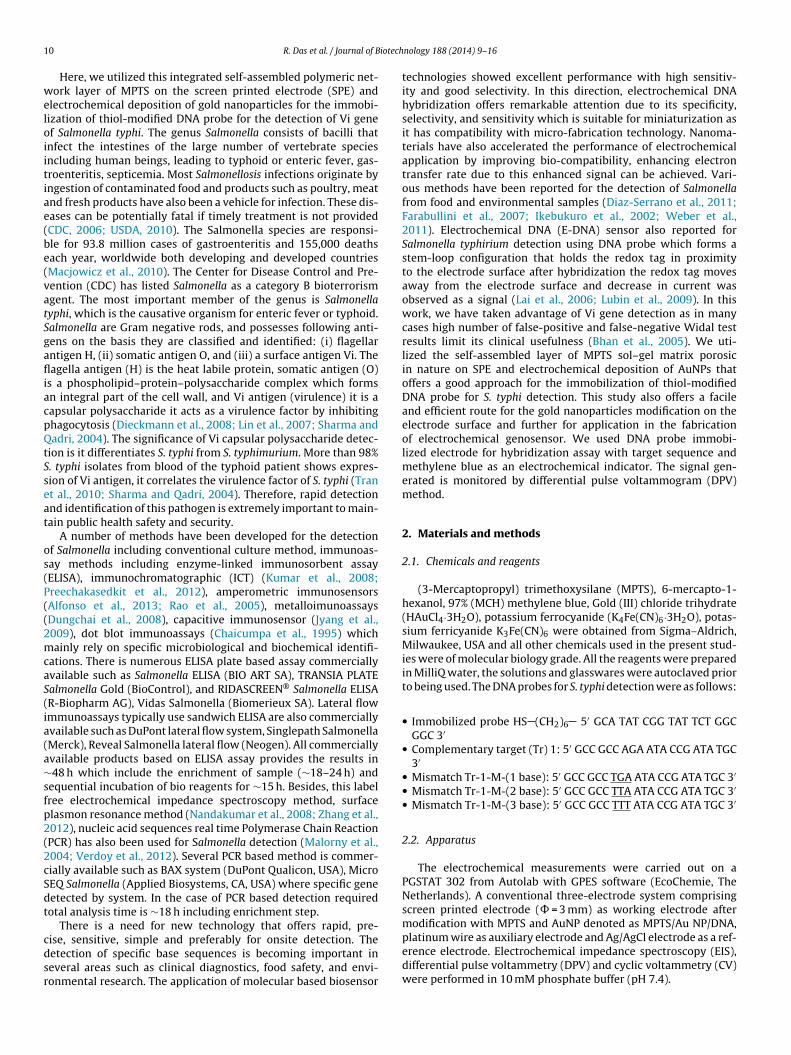
1 iotech
weloiitiae(be(vatSgafliacpQtSseat
os(P((2mcaS(ia(a∼sfp2(2cSdt
cdsr
0 R. Das et al. / Journal of B
Here, we utilized this integrated self-assembled polymeric net-ork layer of MPTS on the screen printed electrode (SPE) and
lectrochemical deposition of gold nanoparticles for the immobi-ization of thiol-modified DNA probe for the detection of Vi genef Salmonella typhi. The genus Salmonella consists of bacilli thatnfect the intestines of the large number of vertebrate speciesncluding human beings, leading to typhoid or enteric fever, gas-roenteritis, septicemia. Most Salmonellosis infections originate byngestion of contaminated food and products such as poultry, meatnd fresh products have also been a vehicle for infection. These dis-ases can be potentially fatal if timely treatment is not providedCDC, 2006; USDA, 2010). The Salmonella species are responsi-le for 93.8 million cases of gastroenteritis and 155,000 deathsach year, worldwide both developing and developed countriesMacjowicz et al., 2010). The Center for Disease Control and Pre-ention (CDC) has listed Salmonella as a category B bioterrorismgent. The most important member of the genus is Salmonellayphi, which is the causative organism for enteric fever or typhoid.almonella are Gram negative rods, and possesses following anti-ens on the basis they are classified and identified: (i) flagellarntigen H, (ii) somatic antigen O, and (iii) a surface antigen Vi. Theagella antigen (H) is the heat labile protein, somatic antigen (O)
s a phospholipid–protein–polysaccharide complex which formsn integral part of the cell wall, and Vi antigen (virulence) it is aapsular polysaccharide it acts as a virulence factor by inhibitinghagocytosis (Dieckmann et al., 2008; Lin et al., 2007; Sharma andadri, 2004). The significance of Vi capsular polysaccharide detec-
ion is it differentiates S. typhi from S. typhimurium. More than 98%. typhi isolates from blood of the typhoid patient shows expres-ion of Vi antigen, it correlates the virulence factor of S. typhi (Trant al., 2010; Sharma and Qadri, 2004). Therefore, rapid detectionnd identification of this pathogen is extremely important to main-ain public health safety and security.
A number of methods have been developed for the detectionf Salmonella including conventional culture method, immunoas-ay methods including enzyme-linked immunosorbent assayELISA), immunochromatographic (ICT) (Kumar et al., 2008;reechakasedkit et al., 2012), amperometric immunosensorsAlfonso et al., 2013; Rao et al., 2005), metalloimunoassaysDungchai et al., 2008), capacitive immunosensor (Jyang et al.,009), dot blot immunoassays (Chaicumpa et al., 1995) whichainly rely on specific microbiological and biochemical identifi-
ations. There is numerous ELISA plate based assay commerciallyvailable such as Salmonella ELISA (BIO ART SA), TRANSIA PLATEalmonella Gold (BioControl), and RIDASCREEN® Salmonella ELISAR-Biopharm AG), Vidas Salmonella (Biomerieux SA). Lateral flowmmunoassays typically use sandwich ELISA are also commerciallyvailable such as DuPont lateral flow system, Singlepath SalmonellaMerck), Reveal Salmonella lateral flow (Neogen). All commerciallyvailable products based on ELISA assay provides the results in48 h which include the enrichment of sample (∼18–24 h) and
equential incubation of bio reagents for ∼15 h. Besides, this labelree electrochemical impedance spectroscopy method, surfacelasmon resonance method (Nandakumar et al., 2008; Zhang et al.,012), nucleic acid sequences real time Polymerase Chain ReactionPCR) has also been used for Salmonella detection (Malorny et al.,004; Verdoy et al., 2012). Several PCR based method is commer-ially available such as BAX system (DuPont Qualicon, USA), MicroEQ Salmonella (Applied Biosystems, CA, USA) where specific geneetected by system. In the case of PCR based detection requiredotal analysis time is ∼18 h including enrichment step.
There is a need for new technology that offers rapid, pre-
ise, sensitive, simple and preferably for onsite detection. Theetection of specific base sequences is becoming important ineveral areas such as clinical diagnostics, food safety, and envi-onmental research. The application of molecular based biosensornology 188 (2014) 9–16
technologies showed excellent performance with high sensitiv-ity and good selectivity. In this direction, electrochemical DNAhybridization offers remarkable attention due to its specificity,selectivity, and sensitivity which is suitable for miniaturization asit has compatibility with micro-fabrication technology. Nanoma-terials have also accelerated the performance of electrochemicalapplication by improving bio-compatibility, enhancing electrontransfer rate due to this enhanced signal can be achieved. Vari-ous methods have been reported for the detection of Salmonellafrom food and environmental samples (Diaz-Serrano et al., 2011;Farabullini et al., 2007; Ikebukuro et al., 2002; Weber et al.,2011). Electrochemical DNA (E-DNA) sensor also reported forSalmonella typhirium detection using DNA probe which forms astem-loop configuration that holds the redox tag in proximityto the electrode surface after hybridization the redox tag movesaway from the electrode surface and decrease in current wasobserved as a signal (Lai et al., 2006; Lubin et al., 2009). In thiswork, we have taken advantage of Vi gene detection as in manycases high number of false-positive and false-negative Widal testresults limit its clinical usefulness (Bhan et al., 2005). We uti-lized the self-assembled layer of MPTS sol–gel matrix porosicin nature on SPE and electrochemical deposition of AuNPs thatoffers a good approach for the immobilization of thiol-modifiedDNA probe for S. typhi detection. This study also offers a facileand efficient route for the gold nanoparticles modification on theelectrode surface and further for application in the fabricationof electrochemical genosensor. We used DNA probe immobi-lized electrode for hybridization assay with target sequence andmethylene blue as an electrochemical indicator. The signal gen-erated is monitored by differential pulse voltammogram (DPV)method.
2. Materials and methods
2.1. Chemicals and reagents
(3-Mercaptopropyl) trimethoxysilane (MPTS), 6-mercapto-1-hexanol, 97% (MCH) methylene blue, Gold (III) chloride trihydrate(HAuCl4·3H2O), potassium ferrocyanide (K4Fe(CN)6·3H2O), potas-sium ferricyanide K3Fe(CN)6 were obtained from Sigma–Aldrich,Milwaukee, USA and all other chemicals used in the present stud-ies were of molecular biology grade. All the reagents were preparedin MilliQ water, the solutions and glasswares were autoclaved priorto being used. The DNA probes for S. typhi detection were as follows:
• Immobilized probe HS (CH2)6 5′ GCA TAT CGG TAT TCT GGCGGC 3′
• Complementary target (Tr) 1: 5′ GCC GCC AGA ATA CCG ATA TGC3′
• Mismatch Tr-1-M-(1 base): 5′ GCC GCC TGA ATA CCG ATA TGC 3′• Mismatch Tr-1-M-(2 base): 5′ GCC GCC TTA ATA CCG ATA TGC 3′• Mismatch Tr-1-M-(3 base): 5′ GCC GCC TTT ATA CCG ATA TGC 3′
2.2. Apparatus
The electrochemical measurements were carried out on aPGSTAT 302 from Autolab with GPES software (EcoChemie, TheNetherlands). A conventional three-electrode system comprisingscreen printed electrode (� = 3 mm) as working electrode aftermodification with MPTS and AuNP denoted as MPTS/Au NP/DNA,
platinum wire as auxiliary electrode and Ag/AgCl electrode as a ref-erence electrode. Electrochemical impedance spectroscopy (EIS),differential pulse voltammetry (DPV) and cyclic voltammetry (CV)were performed in 10 mM phosphate buffer (pH 7.4).
iotechnology 188 (2014) 9–16 11
2
rtst4pt3
2e
dt4tfwTtdftc2g
2
ldfwts6twrwasi
2
d1AbDlwtb
2
r
E/V vs Ag/AgCl
0.0 0.1 0. 2 0.3 0. 4 0.5 0. 6 0.7
I/μ μ
A
-80
-60
-40
-20
0
20
40
60
a
b
c
de
R. Das et al. / Journal of B
.3. Fabrication of screen-printed electrodes (SPE)
A semi-automatic screen-printing machine was used for the fab-ication of SPEs as reported elsewhere (Sharma et al., 2010). Forhis purpose, initially a layer of silver ink (conductive ink) wascreen-printed on the alumina substrate and subsequently appliedhe homemade carbon ink (polystyrene:graphite powder ratio is0%:60%) and dried. Finally, a layer of dielectric ink was screenrinted over the electrode to expose the area of working elec-rode and dried at 80◦ C for 2 h. The size of the electrodes was7 mm × 5 mm.
.4. Electrode modification with MPTS and AuNPslectrodeposition
The ∼50.0 mM MPTS sol was prepared by mixing MPTS withouble distilled water at a 0.5:100 ratio and 600 �L of 0.1 M HCl,he mixture was sonicated for 9.0 min followed by stirring for5.0 min until a clear homogeneous solution obtained. Then fromhis homogeneous MPTS sol 4 �L was dropped on the SPE and keptor 4.0–6.0 h to dry at room temperature (25◦ C). This ∼50 mM MPTSas optimum to expose the thiol groups ( SH) to bind the AuNPs.
he high concentrations of MPTS on the electrode surface lead tohe formation of multilayer of MPTS, which in turn to prevent theeposition of AuNPs. The MPTS concentration plays a crucial roleor the exposure of thiol-groups. On the MPTS modified SPE, con-rolled electrodeposition of AuNP was performed in 1 mM HAuCl4ontaining 0.5 M H2SO4 with potentiostaic method at −0.2 V for00 s. After the AuNPs electrodeposition on the MPTS modified SPE,ently washed with distilled water and dried at room temperature.
.5. Immobilization of DNA probe on AuNPs modified SPE
After the electrochemical deposition of AuNPs, 2.0 �L of thio-ated ss-DNA (10 �M) in 10 mM phosphate buffer (pH 7.4) wasropped on modified SPE. This electrode was incubated at 30 ◦Cor 1 h. Then the DNA modified electrode was thoroughly rinsedith 10 mM phosphate buffer (pH 7.4) followed by distilled water
o remove the weakly adsorbed DNA probe and dried. Thiolateds-DNA probe modified electrode was further immersed in 1.0 mM-mercapto-hexanol (MCH) for 1 h to block the uncovered elec-rode surface. Finally, the modified electrode was rinsed thoroughlyith 10 mM phosphate buffer (pH 7.4) and double distilled water,
espectively. To study the behavior of MB with ss-DNA, electrodeas immersed in 20 �M methylene blue (MB) for 5 min and is char-
cterized by DPV in 10 mM phosphate buffer (pH 7.4). The probes-DNA modified electrode is denoted as ss-DNA/AuNPs-MPTS/SPEn the text.
.6. DNA hybridization and detection using methylene blue
The DNA hybridization was performed by pipetting 2 �L ofifferent concentrations of target DNA (Tr-1) and mismatch (Tr--M) onto ss-DNA/AuNPs-MPTS/SPE for 1 h at 37 ◦C (Scheme 1).fter hybridization, the electrodes were washed with phosphateuffer carefully to remove the unbound DNA. In order to detectNA hybridization, the electrode was incubated in 20 �M methy-
ene blue (MB) for 5 min. After accumulation of MB, the electrodeas washed with 10 mM phosphate buffer thoroughly. Differen-
ial pulse voltammetry (DPV) was performed in 10 mM phosphateuffer (pH 7.4).
.7. Regeneration of the modified DNA hybridized electrode
The SPE obtained after DNA hybridization were regenerated byinsing the electrode surfaces with hot distilled water (90◦ C) for
Fig. 1. Cyclic voltammograms in 5 mM Fe(CN)63−/4− containing 0.1 M KCl at 10 mV/s
of different modified SPE: (a) un-modified SPE, (b) MPTS modified SPE, (c) AuNPs-MPTS/SPE, (d) ss-DNA/AuNPs-MPTS/SPE, and (e) ds-DNA/AuNPs-MPTS/SPE.
5 min followed by rapid cooling in an ice bath. The reusability of theSPE was monitored by repetitive hybridization with target ss-DNA.
3. Results and discussion
3.1. Electrochemical characterization of the modified SPE afterDNA hybridization
The electrochemical behavior of MPTS/AuNPs modified SPE wascharacterized by cyclic voltammogram (CV) and electrochemicalimpedance spectroscopy (EIS). In the present DNA biosensor, theeffect of each modification on SPE such as MPTS self assembly,AuNPs electrodeposition, ss-DNA immobilization and hybridiza-tion (ds-DNA) were characterized by CV in 5 mM Fe(CN)6
3−/4−
in phosphate buffer (Fig. 1). Before modification, CV of the SPEwas recorded and found well-defined redox peak current ofFe(CN)6
3−/4− (Fig. 1, curve a). After, the modification of SPE withMPTS sol no peak was observed. It reveals that the MPTS polymericnetwork hindered the electron transfer at the surface of the SPEfor diffusion of ferricyanide toward the electrode surface (Fig. 1,curve b). Here, the concentration of MPTS plays an important rolefor the exposure of thiol-groups on the electrode surface for fur-ther anchoring of the gold nanoparticles (Cheung et al., 2003; Minget al., 2002). After the electrochemical deposition of AuNPs on MPTSmodified (AuNPs-MPTS/SPE) electrode, the peak current increasedmore and peak potential (�Ep) became ∼150 mV (Fig. 1, curve c).
The result indicates that AuNPs has deposited on the MPTS mod-ified electrode that anchored with exposed thiol-groups leads toenhancing the electroactive surface area, and is responsible for anincrease in the peak current. The AuNPs provide an excellent elec-tron conducting pathways to the surface of MPTS and couplingbetween the MPTS surface and underlying SPE. As a result highexchange electron densities occurred and decrease in charge trans-fer resistance and increase in peak current is observed (Barfidokhtet al., 2013; Shein et al., 2009). The average value of the electroac-tive surface area was calculated according to the Randles–Sevickequation (Bard and Faulkner, 2000):
Ip = 2.69 × 10⁵ AD1/2n3/2v1/2C (1)
where n is the number of electrons participating in the redox reac-
tion, A is the area of the electrode (cm2), D is the diffusion coefficientof the molecule in solution (6.7 × 10⁻⁶ cm2 s−1), C is the concentra-tion of the probe molecule in the bulk solution (mol cm−3), and v isthe scan rate (V s−1). The electroactive surface area for unmodified
12 R. Das et al. / Journal of Biotechnology 188 (2014) 9–16
PTS-A
S(
DifcArrt
ipapclditadltmttrtvttrra
o
3
Sna
Scheme 1. The schematic representation of the M
PE and Au NPs modified electrode were (2.1 ± 0.3) × 10−4 cm2 and7.7 ± 0.2) × 10−3 cm2, respectively.
After, immobilization of the thiolated ss-DNA probe ss-NA/AuNPs-MPTS/SPE, peak current decreases and �Ep value
ncreases (Fig. 1, curve d) due to the electrostatic repulsion betweenerricyanide and ss-DNA probe on the electrode surface. It indi-ates that ss-DNA probe was immobilized on the electrode surface.fter hybridization with the complementary target the peak cur-ent further decreases and �Ep value increases due to the similareasons as that of in the case of ss-DNA probe immobilization onhe ds-DNA/AuNPs-MPTS/SPE (Fig. 1, curve e).
EIS was employed to study the surfaces of the modified SPEn 1.0 mM Fe(CN)6
3−/4− containing 0.1 M KCl solution and Nyquistlots were plotted with a frequency range of 0.01 Hz to 10 kHz. Themplitude of the applied potential was set at 0.2 V. In the Nyquistlot of impedance spectra, the semicircle portion at higher frequen-ies corresponds to the electron-transfer-limited process and theinear portion seen at lower frequencies may be ascribed to theiffusion. An increase in the diameter of the semicircle reflects the
ncrease in the electron transfer resistance (Rct). As shown in Fig. S1,he Rct value of the AuNPs modified SPE is ∼200 � (Fig. S1, curve). It is almost a straight line, which is a characteristic of a massiffusional limiting electron-transfer process. After the immobi-
ization of thiolated DNA probe, the value of Rct increased, andhe EIS showed a large interfacial resistance compared to AuNPs
odified SPE (Fig. S1, curve b). The increase in Rct value is dueo the immobilization of negatively charged probes on the elec-rode surface that electrostatically repels the negatively chargededox probe Fe(CN)6
3−/4− and, as a result, inhibits interfacial chargeransfer. The hybridization with target DNA induced a larger Rct
alue (Fig. S1, curve c) which further corroborates that more nega-ively charged DNA hybridizes on the probe immobilized SPE leado more repulsion of the negatively charged redox probe. Theseesults were consistent with the fact that the DNA sensor was fab-icated as expected and in corroboration with the CV results EISlso showed similar behavior.
Supplementary figure related to this article can be found, in thenline version, at http://dx.doi.org/10.1016/j.jbiotec.2014.08.002.
.2. AFM image of electrode after modification of electrode
Fig. 2(a) showed AFM image of Au NPs electrodeposition onPE, the topographical image clearly shows the presence of goldanoparticles on the SPE and it covers the whole surface of the SPEnd their Z-scale height is ∼30 nm and size of Au NPs is ∼20–40 nm.
uNPs modified SPE platform for the genosensor.
The deposition of AuNPs indicates the interaction between thiolgroups of MPTS that are freely available after modification of theSPE. After that DNA probe immobilized on the SPE, AFM imageclearly shows a change in morphology of the SPE and its Z-scaleheight is ∼75 nm (Fig. 2(b)). It shows that on the surface DNA probeis immobilized due to which change in morphology and increase inheight was observed as compared to Fig. 2(a). Further, on hybridiza-tion with target DNA change in morphology observed as well asits Z-scale height also increased to ∼260 nm Fig. 2(c). It confirmsqualitatively that the proper hybridization occurred on the elec-trode surface, and it could be visualized by increasing their Z-scaleheight.
3.3. Optimization of the experimental conditions
The hybridization temperature and time were optimized toobtain high sensitivity and reproducibility of the biosensor. TheDNA probe modified electrode was hybridized with target nucleicacid at different temperatures such as 25, 37, 45 and 55◦ C and dif-ferent times such as 20, 30, 45, and 60 min and DPV were monitored(Fig. S2). The increase in the current response at 25◦ C was less.While, at 37◦ C, an increase in the current response was more ascompared to 25◦ C. The hybridization time was optimized 60 min,after that the current response started to get saturated. Furtheron increasing the hybridization temperature to 45 and 55◦ C theincrease in the current response was observed, but it decreasedafter 20 min. The hybridization temperature is kept below the theo-retical melting temperature (Tm) of the probes (Mao et al., 2008). Tm
of the probe and target DNA is 52◦ C and 58◦ C, respectively. Abovethe Tm of the probes, it may get denatured/break the hybridizedDNA. Thus, the optimal hybridization temperature was chosen as37◦ C and hybridization time is 60 min for further experiments.
Supplementary figure related to this article can be found, in theonline version, at http://dx.doi.org/10.1016/j.jbiotec.2014.08.002.
3.4. Analytical performance of the DNA biosensor
The performance of the DNA biosensor was monitored byhybridization of ss-DNA/AuNPs-MPTS/SPE with different concen-trations of target ss-DNA sequence. Fig. 3(A) showed the DPVreductive signals of MB at the ss-DNA/AuNPs-MPTS/SPE (DNA
probe modified electrode) and after hybridization with differentconcentrations of target ss-DNA sequence. MB is an electrochem-ically active dye it has reversible redox characteristics. It giveswell-defined voltammetric response, the reduction potential of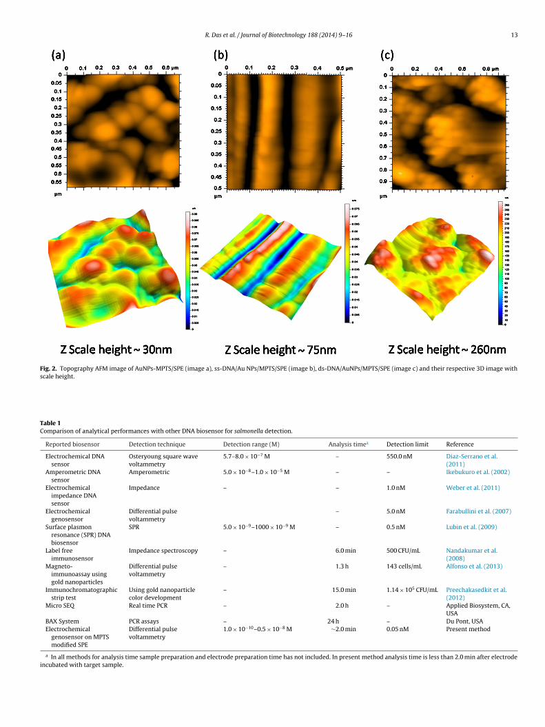
R. Das et al. / Journal of Biotechnology 188 (2014) 9–16 13
Fig. 2. Topography AFM image of AuNPs-MPTS/SPE (image a), ss-DNA/Au NPs/MPTS/SPE (image b), ds-DNA/AuNPs/MPTS/SPE (image c) and their respective 3D image withscale height.
Table 1Comparison of analytical performances with other DNA biosensor for salmonella detection.
Reported biosensor Detection technique Detection range (M) Analysis timea Detection limit Reference
Electrochemical DNAsensor
Osteryoung square wavevoltammetry
5.7–8.0 × 10⁻⁷ M – 550.0 nM Diaz-Serrano et al.(2011)
Amperometric DNAsensor
Amperometric 5.0 × 10⁻⁸–1.0 × 10⁻⁵ M – – Ikebukuro et al. (2002)
Electrochemicalimpedance DNAsensor
Impedance – – 1.0 nM Weber et al. (2011)
Electrochemicalgenosensor
Differential pulsevoltammetry
– 5.0 nM Farabullini et al. (2007)
Surface plasmonresonance (SPR) DNAbiosensor
SPR 5.0 × 10⁻⁹–1000 × 10−9 M – 0.5 nM Lubin et al. (2009)
Label freeimmunosensor
Impedance spectroscopy – 6.0 min 500 CFU/mL Nandakumar et al.(2008)
Magneto-immunoassay usinggold nanoparticles
Differential pulsevoltammetry
– 1.3 h 143 cells/mL Alfonso et al. (2013)
Immunochromatographicstrip test
Using gold nanoparticlecolor development
– 15.0 min 1.14 × 10⁵ CFU/mL Preechakasedkit et al.(2012)
Micro SEQ Real time PCR – 2.0 h – Applied Biosystem, CA,USA
BAX System PCR assays – 24 h – Du Pont, USAElectrochemical
genosensor on MPTSmodified SPE
Differential pulsevoltammetry
1.0 × 10⁻¹⁰–0.5 × 10⁻⁸ M ∼2.0 min 0.05 nM Present method
a In all methods for analysis time sample preparation and electrode preparation time has not included. In present method analysis time is less than 2.0 min after electrodeincubated with target sample.
14 R. Das et al. / Journal of Biotechnology 188 (2014) 9–16
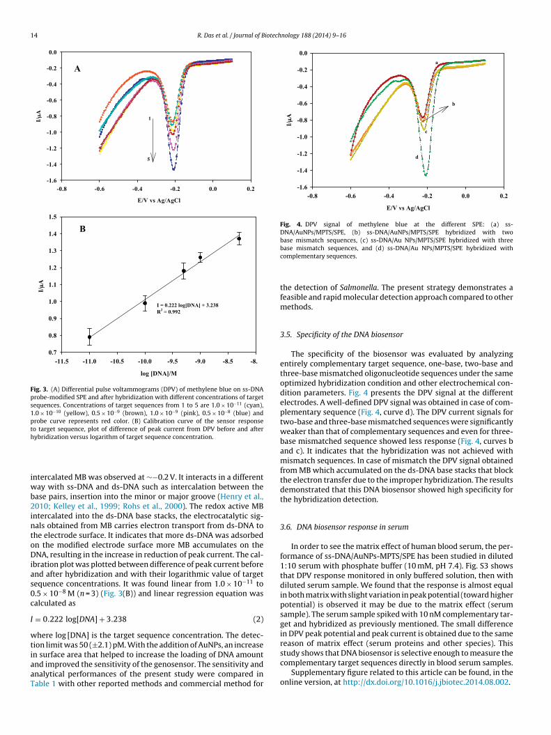
Fig. 3. (A) Differential pulse voltammograms (DPV) of methylene blue on ss-DNAprobe-modified SPE and after hybridization with different concentrations of targetsequences. Concentrations of target sequences from 1 to 5 are 1.0 × 10⁻¹¹ (cyan),1.0 × 10−10 (yellow), 0.5 × 10⁻⁹ (brown), 1.0 × 10⁻⁹ (pink), 0.5 × 10⁻⁸ (blue) andprobe curve represents red color. (B) Calibration curve of the sensor responseto target sequence, plot of difference of peak current from DPV before and afterh
iwb2intoDias0c
I
wtiaaT
E/V vs Ag/AgCl
-0.8 -0. 6 -0. 4 -0.2 0. 0 0.2
I/ μμA
-1.6
-1.4
-1.2
-1.0
-0.8
-0.6
-0.4
-0.2
0.0a
c
d
b
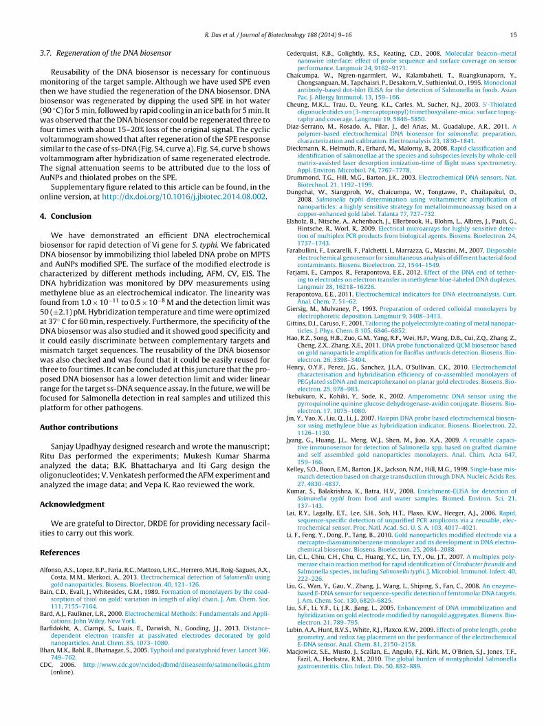
Fig. 4. DPV signal of methylene blue at the different SPE: (a) ss-DNA/AuNPs/MPTS/SPE, (b) ss-DNA/AuNPs/MPTS/SPE hybridized with twobase mismatch sequences, (c) ss-DNA/Au NPs/MPTS/SPE hybridized with three
ybridization versus logarithm of target sequence concentration.
ntercalated MB was observed at ∼−0.2 V. It interacts in a differentay with ss-DNA and ds-DNA such as intercalation between the
ase pairs, insertion into the minor or major groove (Henry et al.,010; Kelley et al., 1999; Rohs et al., 2000). The redox active MB
ntercalated into the ds-DNA base stacks, the electrocatalytic sig-als obtained from MB carries electron transport from ds-DNA tohe electrode surface. It indicates that more ds-DNA was adsorbedn the modified electrode surface more MB accumulates on theNA, resulting in the increase in reduction of peak current. The cal-
bration plot was plotted between difference of peak current beforend after hybridization and with their logarithmic value of targetequence concentrations. It was found linear from 1.0 × 10⁻¹¹ to.5 × 10−8 M (n = 3) (Fig. 3(B)) and linear regression equation wasalculated as
= 0.222 log[DNA] + 3.238 (2)
here log [DNA] is the target sequence concentration. The detec-ion limit was 50 (±2.1) pM. With the addition of AuNPs, an increase
n surface area that helped to increase the loading of DNA amountnd improved the sensitivity of the genosensor. The sensitivity andnalytical performances of the present study were compared inable 1 with other reported methods and commercial method forbase mismatch sequences, and (d) ss-DNA/Au NPs/MPTS/SPE hybridized withcomplementary sequences.
the detection of Salmonella. The present strategy demonstrates afeasible and rapid molecular detection approach compared to othermethods.
3.5. Specificity of the DNA biosensor
The specificity of the biosensor was evaluated by analyzingentirely complementary target sequence, one-base, two-base andthree-base mismatched oligonucleotide sequences under the sameoptimized hybridization condition and other electrochemical con-dition parameters. Fig. 4 presents the DPV signal at the differentelectrodes. A well-defined DPV signal was obtained in case of com-plementary sequence (Fig. 4, curve d). The DPV current signals fortwo-base and three-base mismatched sequences were significantlyweaker than that of complementary sequences and even for three-base mismatched sequence showed less response (Fig. 4, curves band c). It indicates that the hybridization was not achieved withmismatch sequences. In case of mismatch the DPV signal obtainedfrom MB which accumulated on the ds-DNA base stacks that blockthe electron transfer due to the improper hybridization. The resultsdemonstrated that this DNA biosensor showed high specificity forthe hybridization detection.
3.6. DNA biosensor response in serum
In order to see the matrix effect of human blood serum, the per-formance of ss-DNA/AuNPs-MPTS/SPE has been studied in diluted1:10 serum with phosphate buffer (10 mM, pH 7.4). Fig. S3 showsthat DPV response monitored in only buffered solution, then withdiluted serum sample. We found that the response is almost equalin both matrix with slight variation in peak potential (toward higherpotential) is observed it may be due to the matrix effect (serumsample). The serum sample spiked with 10 nM complementary tar-get and hybridized as previously mentioned. The small differencein DPV peak potential and peak current is obtained due to the samereason of matrix effect (serum proteins and other species). Thisstudy shows that DNA biosensor is selective enough to measure the
complementary target sequences directly in blood serum samples.Supplementary figure related to this article can be found, in theonline version, at http://dx.doi.org/10.1016/j.jbiotec.2014.08.002.
iotech
3
mtb(wfvsvTA
o
4
bDacDmf5aDimwtprfp
A
Raoa
A
i
R
A
B
B
B
B
C
R. Das et al. / Journal of B
.7. Regeneration of the DNA biosensor
Reusability of the DNA biosensor is necessary for continuousonitoring of the target sample. Although we have used SPE even
hen we have studied the regeneration of the DNA biosensor. DNAiosensor was regenerated by dipping the used SPE in hot water90 ◦C) for 5 min, followed by rapid cooling in an ice bath for 5 min. Itas observed that the DNA biosensor could be regenerated three to
our times with about 15–20% loss of the original signal. The cyclicoltammogram showed that after regeneration of the SPE responseimilar to the case of ss-DNA (Fig. S4, curve a). Fig. S4, curve b showsoltammogram after hybridization of same regenerated electrode.he signal attenuation seems to be attributed due to the loss ofuNPs and thiolated probes on the SPE.
Supplementary figure related to this article can be found, in thenline version, at http://dx.doi.org/10.1016/j.jbiotec.2014.08.002.
. Conclusion
We have demonstrated an efficient DNA electrochemicaliosensor for rapid detection of Vi gene for S. typhi. We fabricatedNA biosensor by immobilizing thiol labeled DNA probe on MPTSnd AuNPs modified SPE. The surface of the modified electrode isharacterized by different methods including, AFM, CV, EIS. TheNA hybridization was monitored by DPV measurements usingethylene blue as an electrochemical indicator. The linearity was
ound from 1.0 × 10⁻¹¹ to 0.5 × 10⁻⁸ M and the detection limit was0 (±2.1) pM. Hybridization temperature and time were optimizedt 37◦ C for 60 min, respectively. Furthermore, the specificity of theNA biosensor was also studied and it showed good specificity and
t could easily discriminate between complementary targets andismatch target sequences. The reusability of the DNA biosensoras also checked and was found that it could be easily reused for
hree to four times. It can be concluded at this juncture that the pro-osed DNA biosensor has a lower detection limit and wider linearange for the target ss-DNA sequence assay. In the future, we will beocused for Salmonella detection in real samples and utilized thislatform for other pathogens.
uthor contributions
Sanjay Upadhyay designed research and wrote the manuscript;itu Das performed the experiments; Mukesh Kumar Sharmanalyzed the data; B.K. Bhattacharya and Iti Garg design theligonucleotides; V. Venkatesh performed the AFM experiment andnalyzed the image data; and Vepa K. Rao reviewed the work.
cknowledgment
We are grateful to Director, DRDE for providing necessary facil-ties to carry out this work.
eferences
lfonso, A.S., Lopez, B.P., Faria, R.C., Mattoso, L.H.C., Herrero, M.H., Roig-Sagues, A.X.,Costa, M.M., Merkoci, A., 2013. Electrochemical detection of Salomenlla usinggold nanoparticles. Biosens. Bioelectron. 40, 121–126.
ain, C.D., Evall, J., Whitesides, G.M., 1989. Formation of monolayers by the coad-sorption of thiol on gold: variation in length of alkyl chain. J. Am. Chem. Soc.111, 7155–7164.
ard, A.J., Faulkner, L.R., 2000. Electrochemical Methods: Fundamentals and Appli-cations. John Wiley, New York.
arfidokht, A., Ciampi, S., Luais, E., Darwish, N., Gooding, J.J., 2013. Distance-dependent electron transfer at passivated electrodes decorated by gold
nanoparticles. Anal. Chem. 85, 1073–1080.han, M.K., Bahl, R., Bhatnagar, S., 2005. Typhoid and paratyphoid fever. Lancet 366,749–762.
DC, 2006. http://www.cdc.gov/ncidod/dbmd/diseaseinfo/salmonellosis.g.htm(online).
nology 188 (2014) 9–16 15
Cederquist, K.B., Golightly, R.S., Keating, C.D., 2008. Molecular beacon–metalnanowire interface: effect of probe sequence and surface coverage on sensorperformance. Langmuir 24, 9162–9171.
Chaicumpa, W., Ngren-ngarmlert, W., Kalambaheti, T., Ruangkunaporn, Y.,Chongsanguan, M., Tapchaisri, P., Desakorn, V., Suthienkul, O., 1995. Monoclonalantibody-based dot-blot ELISA for the detection of Salmonella in foods. AsianPac. J. Allergy Immunol. 13, 159–166.
Cheung, M.K.L., Trau, D., Yeung, K.L., Carles, M., Sucher, N.J., 2003. 5′-Thiolatedoligonucleotides on (3-mercaptopropyl) trimethoxysilane-mica: surface topog-raphy and coverage. Langmuir 19, 5846–5850.
Diaz-Serrano, M., Rosado, A., Pilar, J., del Arias, M., Guadalupe, A.R., 2011. Apolymer-based electrochemical DNA biosensor for salmonella: preparation,characterization and calibration. Electroanalysis 23, 1830–1841.
Dieckmann, R., Helmuth, R., Erhard, M., Malorny, B., 2008. Rapid classification andidentification of salmonellae at the species and subspecies levels by whole-cellmatrix-assisted laser desorption ionization-time of flight mass spectrometry.Appl. Environ. Microbiol. 74, 7767–7778.
Drummond, T.G., Hill, M.G., Barton, J.K., 2003. Electrochemical DNA sensors. Nat.Biotechnol. 21, 1192–1199.
Dungchai, W., Siangproh, W., Chaicumpa, W., Tongtawe, P., Chailapakul, O.,2008. Salmonella typhi determination using voltammetric amplification ofnanoparticles: a highly sensitive strategy for metalloimmunoassay based on acopper-enhanced gold label. Talanta 77, 727–732.
Elsholz, B., Nitsche, A., Achenbach, J., Ellerbrook, H., Blohm, L., Albres, J., Pauli, G.,Hintsche, R., Worl, R., 2009. Electrical microarrays for highly sensitive detec-tion of multiplex PCR products from biological agents. Biosens. Bioelectron. 24,1737–1743.
Farabullini, F., Lucarelli, F., Palchetti, I., Marrazza, G., Mascini, M., 2007. Disposableelectrochemical genosensor for simultaneous analysis of different bacterial foodcontaminants. Biosens. Bioelectron. 22, 1544–1549.
Farjami, E., Campos, R., Ferapontova, E.E., 2012. Effect of the DNA end of tether-ing to electrodes on electron transfer in methylene blue-labeled DNA duplexes.Langmuir 28, 16218–16226.
Ferapontova, E.E., 2011. Electrochemical indicators for DNA electroanalysis. Curr.Anal. Chem. 7, 51–62.
Giersig, M., Mulvaney, P., 1993. Preparation of ordered colloidal monolayers byelectrophoretic deposition. Langmuir 9, 3408–3413.
Gittins, D.I., Caruso, F., 2001. Tailoring the polyelectrolyte coating of metal nanopar-ticles. J. Phys. Chem. B 105, 6846–6852.
Hao, R.Z., Song, H.B., Zuo, G.M., Yang, R.F., Wei, H.P., Wang, D.B., Cui, Z.Q., Zhang, Z.,Cheng, Z.X., Zhang, X.E., 2011. DNA probe functionalized QCM biosensor basedon gold nanoparticle amplification for Bacillus anthracis detection. Biosens. Bio-electron. 26, 3398–3404.
Henry, O.Y.F., Perez, J.G., Sanchez, J.L.A., O’Sullivan, C.K., 2010. Electrochemcialcharacterisation and hybridisation efficiency of co-assembled monolayers ofPEGylated ssDNA and mercaptohexanol on planar gold electrodes. Biosens. Bio-electron. 25, 978–983.
Ikebukuro, K., Kohiki, Y., Sode, K., 2002. Amperometric DNA sensor using thepyrroquinoline quinine glucose dehydrogenase-avidin conjugate. Biosens. Bio-electron. 17, 1075–1080.
Jin, Y., Yao, X., Liu, Q., Li, J., 2007. Hairpin DNA probe based electrochemical biosen-sor using methylene blue as hybridization indicator. Biosens. Bioelectron. 22,1126–1130.
Jyang, G., Huang, J.L., Meng, W.J., Shen, M., Jiao, X.A., 2009. A reusable capaci-tive immunosensor for detection of Salmonella spp. based on grafted diamineand self assembled gold nanoparticles monolayers. Anal. Chim. Acta 647,159–166.
Kelley, S.O., Boon, E.M., Barton, J.K., Jackson, N.M., Hill, M.G., 1999. Single-base mis-match detection based on charge transduction through DNA. Nucleic Acids Res.27, 4830–4837.
Kumar, S., Balakrishna, K., Batra, H.V., 2008. Enrichment-ELISA for detection ofSalmonella typhi from food and water samples. Biomed. Environ. Sci. 21,137–143.
Lai, R.Y., Lagally, E.T., Lee, S.H., Soh, H.T., Plaxo, K.W., Heeger, A.J., 2006. Rapid,sequence-specific detection of unpurified PCR amplicons via a reusable, elec-trochemical sensor. Proc. Natl. Acad. Sci. U. S. A. 103, 4017–4021.
Li, F., Feng, Y., Dong, P., Tang, B., 2010. Gold nanoparticles modified electrode via amercapto-diazoaminobenzene monolayer and its development in DNA electro-chemical biosensor. Biosens. Bioelectron. 25, 2084–2088.
Lin, C.L., Chiu, C.H., Chu, C., Huang, Y.C., Lin, T.Y., Ou, J.T., 2007. A multiplex poly-merase chain reaction method for rapid identification of Citrobacter freundii andSalmonella species, including Salmonella typhi. J. Microbiol. Immunol. Infect. 40,222–226.
Liu, G., Wan, Y., Gau, V., Zhang, J., Wang, L., Shiping, S., Fan, C., 2008. An enzyme-based E-DNA sensor for sequence-specific detection of femtomolar DNA targets.J. Am. Chem. Soc. 130, 6820–6825.
Liu, S.F., Li, Y.F., Li, J.R., Jiang, L., 2005. Enhancement of DNA immobilization andhybridization on gold electrode modified by nanogold aggregates. Biosens. Bio-electron. 21, 789–795.
Lubin, A.A., Hunt, B.V.S., White, R.J., Plaxco, K.W., 2009. Effects of probe length, probegeometry, and redox tag placement on the performance of the electrochemical
E-DNA sensor. Anal. Chem. 81, 2150–2158.Macjowicz, S.E., Musto, J., Scallan, E., Angulo, F.J., Kirk, M., O’Brien, S.J., Jones, T.F.,Fazil, A., Hoekstra, R.M., 2010. The global burden of nontyphoidal Salmonellagastroenteritis. Clin. Infect. Dis. 50, 882–889.
1 iotech
M
M
M
N
P
P
R
R
S
6 R. Das et al. / Journal of B
alorny, B., Paccassoni, E., Fach, P., Bunge, C., Martin, A., Helmuth, R., 2004. Diagnos-tic real-time PCR for detection of Salmonella in food. Appl. Environ. Microbiol.70, 7046–7052.
ao, X., Jiang, J., Xu, X., Chu, X., Luo, Y., Shen, G., Yu, R., 2008. Enzymatic amplificationdetection of DNA based on molecular beacon biosensors. Biosens. Bioelectron.23, 1555–1561.
ing, M., Chen, Y., Katz, A., 2002. Synthesis and characterization of gold–silicananoparticles incorporating a mercaptosilane core–shell interface. Langmuir 18,8566–8572.
andakumar, V., Belle, L.T., Reed, J., Shah, M., Cochran, D., Joshi, L., Alford, T.L., 2008.A methodology for rapid detection of Salmonella typhimurium using label-freeelectrochemical impedance spectroscopy. Biosens. Bioelectron. 24, 1039–1042.
aredes, G.M., Begona, M., Garcia, G., Costa-Garcia, A., 2010. Genosensor for detectionof four pneumoniae bacteria using gold nanostructured screen-printed carbonelectrodes as transducers. Sens. Actuators B: Chem. 149, 329–335.
reechakasedkit, P., Pinwattana, K., Dunghai, W., Siangproh, W., Chaicumpa, W.,Tongtawe, P., Chailapakul, O., 2012. Development of a one-step immunochro-matographic strip test using gold nanoparticles for the rapid detection ofSalmonella typhi in human serum. Biosens. Bioelectron. 31, 562–566.
ao, V.K., Rai, G.P., Agarwal, G.S., Suresh, S., 2005. Amperometric immunosensor fordetection of antibodies of Salmonella typhi in patient serum. Anal. Chim. Acta531, 173–177.
ohs., R., Sklenar, H., Lavery, R., Roder, B., 2000. Methylene blue binding to DNA withalternating GC base: a modeling study. J. Am. Chem. Soc. 122, 2860–2866.
harma, A., Qadri, A., 2004. Vi polysaccharide of Salmonella typhi targets the pro-hibitin family of molecules in intestinal epithelial cells and suppresses earlyinflammatory responses. Proc. Natl. Acad. U. S. A. 101, 17492–17497.
nology 188 (2014) 9–16
Sharma, M.K., Agarwal, G.S., Rao, V.K., Upadhyay, S., Merwyn, S., Gopalan, N., Rai, G.P.,Vijayaraghavan, R., Prakash, S., 2010. Amperometric immunosensor based ongold nanoparticles/alumina sol–gel modified screen-printed electrodes for anti-bodies to Plasmodium falciparum histidine rich protein-2. Analyst 135, 608–614.
Shein, J.B., Lai, L.M.H., Eggers, P.K., Paddon-Row, M.N., Gooding, J.J., 2009. Formationof efficient electron transfer pathways by adsorbing gold nanoparticles to self-assembled monolayer modified electrodes. Langmuir 25, 11121–11128.
Tran, Q.T., Gomez, G., Khare, S., Lawhon, S.D., Raffatellu, M., Baumler, A.J., Ajith-doss, D., Dhavala, S., Adams, L.G., 2010. The Salmonella enteric serotype TyphiVi capsular antigen expressed after the bacterium enters the ileal mucosa. Infect.Immun. 78, 527–535.
USDA, 2010. Foodborne Illness & Disease, Salmonella and Salmonellosis Fact Sheets.http://www.fsis.usda.gov/factsheets/Salmonella/index.asp2010
Ulman, A., 1996. Formation and structure of self-assembled monolayers. Chem. Rev.96, 1533–1554.
Verdoy, D., Barrenetxea, Z., Berganzo, J., Agirregabiria, M., Ruano-Lopez, M.J., Mari-mon, J.M., Olabarria, G., 2012. A novel real time micro PCR based point-of-caredevice for Salmonella detection in human clinical samples. Biosens. Bioelectron.32, 259–265.
Wang, J., Pamidi, P.V.A., Zanette, D.R., 1998. Self-assembled sol–gel networks. J. Am.Chem. Soc. 120, 5852.
Weber, J.E., Pillai, S., Ram, M.K., Kumar, A., Singh, S.R., 2011. Electrochemical
impedance based-DNA sensor using a modified single walled carbon nanotubeelectrode. Mater. Sci. Eng. C 31, 821–825.Zhang, D., Yan, Y., Li, Q., Yu, T., Cheng, W., Wang, L., Ju, H., Ding, S., 2012. Label-freeand high-sensitive detection of Salmonella using a surface plasmon resonanceDNA-based biosensor. J. Biotechnol. 160, 123–128.
Copyright © 2022 FDOKUMEN