
An Antioxidant Response Phenotype Shared between Hereditary and Sporadic Type 2 Papillary Renal Cell...
13
Cancer Cell Article An Antioxidant Response Phenotype Shared between Hereditary and Sporadic Type 2 Papillary Renal Cell Carcinoma Aikseng Ooi, 1 Jing-Chii Wong, 2 David Petillo, 1 Douglas Roossien, 1 Victoria Perrier-Trudova, 1,3 Douglas Whitten, 4 Bernice Wong Hui Min, 2 Min-Han Tan, 2 Zhongfa Zhang, 1,9 Ximing J. Yang, 5 Ming Zhou, 6 Betty Gardie, 3 Vincent Molinie ´, 3 Ste ´ phane Richard, 3 Puay Hoon Tan, 7 Bin Tean Teh, 1,2, * and Kyle A. Furge 8, * 1 Laboratory of Cancer Genetics, Van Andel Research Institute, Grand Rapids, MI 49503, USA 2 NCCS-VARI Translational Cancer Research Laboratory, National Cancer Centre Singapore, 169610 Singapore 3 Ge ´ ne ´ tique Oncologique EPHE-INSERM U753 and Faculte ´ de Me ´ decine Paris-Sud, Le Kremlin-Bice ˆ tre, and Institut de Cance ´ rologie Gustave Roussy, 94805 Villejuif, France 4 Proteomics Core Facility, Michigan State University, East Lansing, MI 48824, USA 5 Department of Pathology, Northwestern Memorial Hospital, Northwestern University Feinberg School of Medicine, Chicago, IL 60611, USA 6 Department of Anatomic Pathology, Cleveland Clinic, Cleveland, OH 44195, USA 7 Department of Pathology, Singapore General Hospital, 169608 Singapore 8 Laboratory of Computational Biology, Van Andel Research Institute, Grand Rapids, MI 49503, USA 9 Present address: Center for Systems and Computational Biology, The Wistar Institute, Philadelphia, PA 19104, USA *Correspondence: [email protected] (B.T.T.), [email protected] (K.A.F.) DOI 10.1016/j.ccr.2011.08.024 SUMMARY Fumarate hydratase (FH) mutation causes hereditary type 2 papillary renal cell carcinoma (PRCC2). The main effect of FH mutation is fumarate accumulation. The current paradigm posits that the main consequence of fumarate accumulation is HIF-a stabilization. Paradoxically, FH mutation differs from other HIF-a stabilizing mutations, such as VHL and SDH mutations, in its associated tumor types. We identified that fumarate can directly up-regulate antioxidant response element (ARE)–controlled genes. We demonstrated that aldo-keto reductase family 1 member B10 (AKR1B10) is an ARE-controlled gene and is up-regulated upon FH knockdown as well as in FH null cell lines. AKR1B10 overexpression is also a prominent feature in both hereditary and sporadic PRCC2. This phenotype better explains the similarities between hereditary and sporadic PRCC2. INTRODUCTION Type 2 papillary renal cell carcinoma (PRCC2) is an aggressive subtype of kidney cancer that has no effective treatment. The hereditary form is associated with biallelic inactivation of the gene encoding the Kreb’s cycle enzyme fumarate hydratase (FH), which causes hereditary leiomyomatosis and renal cell cancer (HLRCC) (MIM ID #605839) that is characterized by kidney cancer, uterine fibroids, and cutaneous leiomyomatosis at high frequencies (Launonen et al., 2001; Tomlinson et al., 2002). Individuals who have HLRCC are also genetically predis- posed to bladder cancer, collecting-duct tumors, and adult Ley- dig cell tumors of the testis (Alam et al., 2003; Carvajal-Carmona et al., 2006; Kiuru and Launonen, 2004; Lehtonen et al., 2006). The current paradigm is that FH mutation causes the stabiliza- tion and accumulation of hypoxia-inducible factor alpha subunit (HIF-a), as well as increased expression of HIF-a target genes, such as vascular endothelial growth factor (VEGFA) and platelet-derived growth factor (PDGF)(Isaacs et al., 2005; Koivu- nen et al., 2007; O’Flaherty et al., 2010). However, this paradigm fails to explain the distinct clinical, biochemical, and genetic features apparent between tumors harboring FH mutation and Significance Type 2 papillary renal cell carcinoma (PRCC2) is an aggressive disease with no effective treatment. Despite the obvious morphological, genetic, and clinical differences, hereditary PRCC2 is thought to share similar pathway deregulation with its clear-cell counterpart (CCRCC) that arises as a result of SDH or VHL mutation. Furthermore, the robust response seen with anti-VEGF agents in CCRCC is not reproducible in PRCC2. This represents a distinct knowledge gap in the under- standing of PRCC2 biology. We identified deregulation of the KEAP1-NRF2 axis as a feature that distinguishes PRCC2 from CCRCC, but links both hereditary and sporadic PRCC2. Therefore, our finding provides a more complete under- standing of PRCC2 biology and lays the foundation for the development of effective treatment strategies. Cancer Cell 20, 511–523, October 18, 2011 ª2011 Elsevier Inc. 511
-
Upload
independent -
Category
Documents
-
view
1 -
download
0
Transcript of An Antioxidant Response Phenotype Shared between Hereditary and Sporadic Type 2 Papillary Renal Cell...

Cancer Cell
Article
An Antioxidant Response Phenotype Shared betweenHereditary and Sporadic Type 2 Papillary RenalCell CarcinomaAikseng Ooi,1 Jing-Chii Wong,2 David Petillo,1 Douglas Roossien,1 Victoria Perrier-Trudova,1,3 Douglas Whitten,4
Bernice Wong Hui Min,2 Min-Han Tan,2 Zhongfa Zhang,1,9 Ximing J. Yang,5 Ming Zhou,6 Betty Gardie,3
Vincent Molinie,3 Stephane Richard,3 Puay Hoon Tan,7 Bin Tean Teh,1,2,* and Kyle A. Furge8,*1Laboratory of Cancer Genetics, Van Andel Research Institute, Grand Rapids, MI 49503, USA2NCCS-VARI Translational Cancer Research Laboratory, National Cancer Centre Singapore, 169610 Singapore3Genetique Oncologique EPHE-INSERM U753 and Faculte de Medecine Paris-Sud, Le Kremlin-Bicetre, and Institut de Cancerologie
Gustave Roussy, 94805 Villejuif, France4Proteomics Core Facility, Michigan State University, East Lansing, MI 48824, USA5Department of Pathology, Northwestern Memorial Hospital, Northwestern University Feinberg School of Medicine, Chicago, IL 60611, USA6Department of Anatomic Pathology, Cleveland Clinic, Cleveland, OH 44195, USA7Department of Pathology, Singapore General Hospital, 169608 Singapore8Laboratory of Computational Biology, Van Andel Research Institute, Grand Rapids, MI 49503, USA9Present address: Center for Systems and Computational Biology, The Wistar Institute, Philadelphia, PA 19104, USA
*Correspondence: [email protected] (B.T.T.), [email protected] (K.A.F.)
DOI 10.1016/j.ccr.2011.08.024
SUMMARY
Fumarate hydratase (FH) mutation causes hereditary type 2 papillary renal cell carcinoma (PRCC2). Themaineffect of FH mutation is fumarate accumulation. The current paradigm posits that the main consequence offumarate accumulation is HIF-a stabilization. Paradoxically, FH mutation differs from other HIF-a stabilizingmutations, such as VHL and SDH mutations, in its associated tumor types. We identified that fumaratecan directly up-regulate antioxidant response element (ARE)–controlled genes. We demonstrated thataldo-keto reductase family 1 member B10 (AKR1B10) is an ARE-controlled gene and is up-regulated uponFH knockdown as well as in FH null cell lines. AKR1B10 overexpression is also a prominent feature in bothhereditary and sporadic PRCC2. This phenotype better explains the similarities between hereditary andsporadic PRCC2.
INTRODUCTION
Type 2 papillary renal cell carcinoma (PRCC2) is an aggressive
subtype of kidney cancer that has no effective treatment. The
hereditary form is associated with biallelic inactivation of the
gene encoding the Kreb’s cycle enzyme fumarate hydratase
(FH), which causes hereditary leiomyomatosis and renal cell
cancer (HLRCC) (MIM ID #605839) that is characterized by
kidney cancer, uterine fibroids, and cutaneous leiomyomatosis
at high frequencies (Launonen et al., 2001; Tomlinson et al.,
2002). Individuals who have HLRCC are also genetically predis-
Significance
Type 2 papillary renal cell carcinoma (PRCC2) is an aggressivmorphological, genetic, and clinical differences, hereditary PRits clear-cell counterpart (CCRCC) that arises as a result of SDwith anti-VEGF agents in CCRCC is not reproducible in PRCCstanding of PRCC2 biology. We identified deregulation of thefrom CCRCC, but links both hereditary and sporadic PRCC2standing of PRCC2 biology and lays the foundation for the de
C
posed to bladder cancer, collecting-duct tumors, and adult Ley-
dig cell tumors of the testis (Alam et al., 2003; Carvajal-Carmona
et al., 2006; Kiuru and Launonen, 2004; Lehtonen et al., 2006).
The current paradigm is that FHmutation causes the stabiliza-
tion and accumulation of hypoxia-inducible factor alpha subunit
(HIF-a), as well as increased expression of HIF-a target genes,
such as vascular endothelial growth factor (VEGFA) and
platelet-derived growth factor (PDGF) (Isaacs et al., 2005; Koivu-
nen et al., 2007; O’Flaherty et al., 2010). However, this paradigm
fails to explain the distinct clinical, biochemical, and genetic
features apparent between tumors harboring FH mutation and
e disease with no effective treatment. Despite the obviousCC2 is thought to share similar pathway deregulation withH or VHL mutation. Furthermore, the robust response seen2. This represents a distinct knowledge gap in the under-KEAP1-NRF2 axis as a feature that distinguishes PRCC2. Therefore, our finding provides a more complete under-velopment of effective treatment strategies.
ancer Cell 20, 511–523, October 18, 2011 ª2011 Elsevier Inc. 511

Cancer Cell
Antioxidant Response in Papillary Kidney Cancers
those harboring other HIF-a-accumulating mutations in genes
such as von Hippel–Lindau (VHL) and succinate dehydrogenase
(SDH). Both VHL and SDH mutations are associated with clear-
cell renal cell carcinoma (CCRCC), pheochromocytoma, and
paraganglioma (Vanharanta et al., 2004). Clinically, the robust
response of CCRCC to antiangiogenic agents that block VEGFA
has not been seen in either sporadic or hereditary PRCC2 (Mo-
lina et al., 2010). Furthermore, HIF-a-associated histology such
as higher vascular density is more apparent in hereditary
PRCC2 (Pollard et al., 2005a), suggesting that this model fails
to adequately describe the similarities between hereditary and
sporadic PRCC2.
The homeostasis of HIF-a is controlled at the protein level.
Under normoxic conditions, HIF-a is hydroxylated by prolyl
hydroxylase, marking it for ubiquitination by an E3 ubiquitin
ligase complex that contains VHL (Ivan et al., 2001). In cells
devoid of VHL function, the E3 ubiquitin ligase complex fails to
form and HIF-a accumulates. In cells lacking SDH function,
succinate accumulates and inhibits HIF-a prolyl hydroxylase in
a competitive manner (Pollard et al., 2006). VHL and SDH muta-
tions caused a similar spectrum of tumors. Overexpression of
HIF-a target genes is also a common feature of tumors harboring
either mutated gene (Dahia et al., 2005).
The accumulation of HIF-a resulting from FH mutation is
believed to be mediated by fumarate inhibition of prolyl hydrox-
ylase, in a manner similar to that of succinate (Isaacs et al., 2005;
Koivunen et al., 2007; O’Flaherty et al., 2010). FH catalyzes the
hydration of fumarate to malate in the Kreb’s cycle, and its loss
causes the accumulation of fumarate and succinate (Pollard
et al., 2005b). Both can escape into the cytosol, and the former
is believed to drive the development of HLRCC symptoms and
complications (Pollard et al., 2005b). An alternative theory is
that HIF-a accumulates as reactive oxygen species (ROS)
increase, which results from a glycolytic switch due to Kreb’s
cycle failure when FH is mutated (Sudarshan et al., 2009).
We sought a model that better describes both hereditary
PRCC2 (hereafter, HLRCC) and sporadic PRCC2 (hereafter,
PRCC2) through comprehensive analysis of transcriptome data.
RESULTS
The Expression Pattern of HIF-a-AssociatedProangiogenic Factors Differentiate CCRCCfrom HLRCC and PRCC2It is well accepted that HIF-a is stabilized and that its target
genes are overexpressed in HLRCC and CCRCC (reviewed
in Linehan et al., 2010). It has also been shown that pheochromo-
cytomas, arising from either VHL or SDH mutation, have HIF-a
accumulation (Dahia et al., 2005). To assess the differences in
HIF-a-mediated transcription changes in RCC subtypes, we
used gene expression data sets from pheochromocytoma
samples that arose from MEN2A, SDH, and VHL mutations.
We identified a set of genes commonly deregulated in pheochro-
mocytomas harboring VHL or SDH mutation (see Table S1,
which is available with this article online). Concordant with previ-
ously reported work, the gene expression profiles from SDH
mutant pheochromocytomas were markedly similar to those
derived from VHL mutation (p = 1.026149 3 10�22, mean-rank
gene set test; Table S1 and Figure 1A).
512 Cancer Cell 20, 511–523, October 18, 2011 ª2011 Elsevier Inc.
Next, we identified genes commonly up-regulated in pheo-
chromocytomas that had SDH or VHL mutations (Table S1)
and performed functional annotation clustering to identify genes
associated with the proangiogenic phenotype, common to SDH
and VHL loss of function. We found a subset of 10 genes asso-
ciated with angiogenesis (p < 0.05; enrichment score = 2.04)
(Figure 1B); seven of those were coordinately overlapped with
four angiogenesis GO terms. As expected, these seven genes
were also overexpressed in CCRCC relative to normal kidney
(Figure 1C), but their expression levels were not prominently
changed in either PRCC2 or HLRCC (Figure 1C; the demograph-
ical and mutation status of HLRCC samples used in this study
are shown in Table S2). In particular, the expression of VEGFA
in PRCC2 and in HLRCC was much lower than in CCRCC. The
VEGFAmRNA level was further validated by quantitative reverse
transcription PCR (qRT-PCR), and the result agreed with that
from our microarray (Figure S1). A similar analysis on proglyco-
lytic features of HLRCC tumors showed overexpression of other
HIF-a target genes such as hexokinase 2 and glucose trans-
porter type 1 (data not shown).
Gene Expression Signature Manifests the Lossof FH Function in Sporadic PRCC2The divergence in the expression of HIF-a-associated proangio-
genic genes among CCRCC, HLRCC, and PRCC2 suggests that
there may be deregulation of other pathways in papillary tumors.
We generated an FH loss-of-function gene signature by using
expression profiles previously generated from uterine fibroids
harboring FH mutations (Vanharanta et al., 2006). We identified
94 up-regulated and 76 down-regulated genes (minimum
twofold difference between FH�/�and FH+/+ fibroids; Figure 2A
and Table S3). A similar analysis found 1397 up-regulated and
985 down-regulated genes in HLRCC relative to normal kidney
tissues (Figure 2A and Table S3).
To cancel out tissue- and experiment-specific variations in
gene expression, we compared these two lists and identified
44 genes up-regulated in both FH�/� fibroids and HLRCC and
26 genes down-regulated in both (Figure 2A and Table S3); this
set was designated as the ‘‘FH signature.’’ As expected, when
the FH signature was used to interrogate the uterine fibroid
data via gene set enrichment analysis, the signature was specif-
ically enriched in FH�/� fibroid tissues (Figure 2B, left). In the
same test on an independent set (GSE2725) of FH�/� and
FH+/+ uterine fibroids and normal myometrial tissues, the signa-
ture was again specifically enriched in FH�/� fibroids (Figure 2B,
right). The FH signature was also enriched in FH+/� normal
kidney (Figure 2C), suggesting that heterozygous FH mutation
does alter the overall gene expression profile, possibly through
accumulation of fumarate. A change in the gene expression
profile of normal epithelial cells harboring heterozygous
BRCA1 or BRCA2 mutations was recently reported (Bellacosa
et al., 2010).
After validating the FH signature, we applied it to the PRCC2
data set and found that the signature was significantly enriched
(p = 3.60 3 10�9) (Figure 2D). Twenty-one genes overlapped
between the 2057 genes up-regulated in PRCC2 and the 44
up-regulated in our FH signature; 14 of 2251 genes down-regu-
lated in PRCC2 also overlapped (p = 4.02 3 10�10 and 6.84 3
10�08 for up- and down-regulation, respectively) (Figure 2E).

Figure 1. HIF-a-Associated Proangiogenic Factors of HLRCC and PRCC2 Differ from Those of CCRCC
(A) Heat map showing fold change of mRNA from 778 differentially regulated genes in pheochromocytomas harboring MEN2A, VHL, or SDHmutations. Red and
blue respectively represent higher and lower expression relative to the mean gene expression levels.
(B) Ten genes significantly associated with angiogenesis processes (p < 0.05; enrichment score = 2.04). Genes in red were coordinately overlapped with four
angiogenesis GO terms (blue).
(C) Relative fold change in mRNA abundance of individual angiogenic genes in kidney cancers subtypes (ChRCC = chromophobe renal cell carcinoma) relative to
the mean abundance in normal kidney tissues. See also Figure S1, Table S1, and Table S2.
Cancer Cell
Antioxidant Response in Papillary Kidney Cancers
Among the 21 up-regulated genes, the aldo-keto reductase
family 1 member B10 gene (AKR1B10) was the most up-regu-
lated, suggesting that there is a signal transduction defect result-
ing in such AKR1B10 expression in both PRCC2 and HLRCC
(Figure 2F and Table S4).
AKR1B10 as aGeneControlled by Antioxidant ResponseElementWe used an algorithm that predicts cis-acting elements to
identify potential transcription factors that control AKR1B10
expression (Figure S2). By coalescing cis-acting element motifs
associatedwith theAKR1B10 expression pattern, we isolated 13
candidates (Figure S2 and Figure 3A). Three of these elements
corresponded to sequences recognized by the nuclear factor
(erythroid-derived 2)-like 1 (NRF1) and nuclear factor
(erythroid-derived 2)-like 2 (NRF2) transcription factors, whereas
two others correspond to JUN, which is known to dimerize with
C
NRF and enable binding to a response element (Figure 3B)
(Venugopal and Jaiswal, 1998). Collectively, these cis-acting
elements correspond to the antioxidant response element
(ARE) as being a potential controller of AKR1B10 expression.
If this prediction is true, the expression pattern of AKR1B10
should correlate with those of other classic ARE-driven genes.
Thus, we performed a rank test on the AKR1B10 expression
pattern with the classic ARE-driven genes NAD(P)H dehydroge-
nase, quinone 1 (NQO1), and thioredoxin reductase 1 (TXNRD1)
(Hsieh et al., 2006; Sakurai et al., 2005). As expected, the
AKR1B10 expression pattern correlated well with both NQO1
and TXNRD1 in the RCC data set, with Kendall correlation
coefficients of 0.62 and 0.54, respectively (Pearson correlation
coefficient > 0.7; p < 0.05 in all correlation plots) (Figures 3C
and 3E). We also used independent data sets from lung small
airway cells (GSE994) and obtained similar results (Kendall
correlation coefficients of 0.67 and 0.50, respectively; Pearson
ancer Cell 20, 511–523, October 18, 2011 ª2011 Elsevier Inc. 513

Figure 2. The FH Signature in Sporadic PRCC2
(A) Venn diagram of genes up- or down-regulated in FH�/�
fibroids and HLRCC relative to FH+/+ normal tissues. Bold
designates the sets of up- and down-regulated genes
subsequently used as the ‘‘FH signature.’’
(B) The FH signature was specifically enriched in FH�/�
fibroids but not in normal myometria or FH+/+ fibroid
tissues.
(C) Enrichment of the FH signature in FH+/+ normal kidney,
FH+/� normal kidney, and FH�/� HLRCC tumor samples.
(D) FH signature enrichment in PRCC2 (p = 3.60 3 10�09).
(E) Venn diagram of genes up- and down-regulated in
PRCC2 relative to normal tissue. The overlaps were
statistically significant: with p values (hypergeometric test)
of 4.02 3 10�10 and 6.84 3 10�8 for up- and down-regu-
lated genes, respectively.
(F) Base 2 logarithmic fold increase in mRNA levels of
the 21 commonly up-regulated genes in diseased tissues
relative to corresponding controls. See also Tables S3
and S4.
Cancer Cell
Antioxidant Response in Papillary Kidney Cancers
correlation coefficient > 0.7; p < 0.05 in all correlation plots)
(Figures 3D and 3F). Furthermore, we identified a potential ARE
consensus sequence within the AKR1B10 gene.
The FH Signature Significantly Overlaps the GeneExpression Profile of KEAP1 KnockdownThe ARE enhancer is recognized by NRF1 and NRF2 (Ohtsuji
et al., 2008). These factors are negatively regulated by KEAP1
(although such regulation of NRF1 is not robustly established)
(Furukawa and Xiong, 2005; Hayes and McMahon, 2001; Zhang
et al., 2006). An electrophile sensor, KEAP1, binds to NRF tran-
scription factors under low-electrophile conditions and brings
them into close proximity with cullin 3 (CUL3) ubiquitin ligase
for ubiquitination (Furukawa and Xiong, 2005; Zhang et al.,
2005). The exposed cysteine residue of KEAP1, Cys-151, reacts
with intracellular electrophiles (Rachakonda et al., 2008; Zhang
and Hannink, 2003) by forming a covalent adduct, which induces
a conformational change that renders KEAP1 unable to bind
514 Cancer Cell 20, 511–523, October 18, 2011 ª2011 Elsevier Inc.
NRF2; thus, NRF2 accumulates and becomes
available to modulate ARE-controlled genes.
We used expression data from a KEAP1
knockdown experiment to assess whether the
KEAP1 inactivation profile shared features with
our FH signature (MacLeod et al., 2009). Of the
66 up-regulated genes following KEAP1 knock-
down, nine overlapped with the up-regulated
portion of the FH signature (Figure 4A and Table
S5); five of these nine were among the 20 most
strongly up-regulated genes (Figure 4B) (hyper-
geometric p = 6.70 � 10�16). The expression
levels of the nine genes were consistently higher
in FH�/� than in FH+/+ uterine fibroids (Fig-
ure 4C); their expression levels in RCC subtypes
are shown in Figure 4D. Consistent with their
clinical and morphological features, the expres-
sion patterns of the nine genes were similar
between HLRCC and PRCC2 and distinguished
them from CCRCC. qRT-PCR validation on
a subset of the nine genes is shown in Figure S3. The expression
pattern of AKR1B10 in data sets we used also correlated well
with other NRF2 targets such as aldo-keto reductase family 1,
member C1 (AKR1C1), glutamate-cysteine ligase, catalytic
subunit (GCLC), and sulfiredoxin 1 (SRXN1), which have been
identified as genes that become overexpressed upon KEAP1
knockdown (Figure S3) (MacLeod et al., 2009).
AKR1B10 mRNA Increased with Lower FH LevelGene expression analysis suggested that AKR1B10 overexpres-
sion is a prominent feature of the loss of FH function and is likely
an effect of NRF accumulation. To test this in a cellular system,
we assessed AKR1B10, NRF1, NRF2, and KEAP1 protein levels
in immortalized kidney tubular epithelial cells (HK-2) and an
HLRCC cell line, UOK-262 (Yang et al., 2010). AKR1B10,
NRF1, and NRF2 levels were higher in UOK-262 than in HK-2
cells (Figure 5A), whereas KEAP1 was lower. We then knocked
down FH using two different species of siRNA in immortalized

Figure 3. cis-Acting Element Motif Matching
Algorithm Prediction That AKR1B10 Is Controlled
by ARE
(A) Venn diagram of cis-acting element motifs correlated
to AKR1B10 gene expression pattern in data sets from
RCC (comprising normal kidney, PRCC1, and PRCC2),
lung small airway cells, and fibroid.
(B) The�log2 p values (hypergeometric test) of the overlap
between the cis-acting element motifs and the genes
identified through Kendall Rank Test in three different data
sets. Motif names in green were not assigned to any
transcription factor; names in red represent the ARE;
names in blue represent AP-1 or c-JUN binding sites.
Collectively, red plus blue motifs correspond to the
consensus motif of ARE.
(C–F) Correlation of the AKR1B10 expression pattern with
NQO1 (C and D) and TXNRD1 (E and F) in the RCCdata set
and in an independent data set from lung small airway
cells. See also Figure S2.
Cancer Cell
Antioxidant Response in Papillary Kidney Cancers
embryonic kidney cells (HEK293) and HK-2 cells. We achieved
over 60% knockdown of FH mRNA with each siRNA, but FH
protein abundance decreased only moderately by 72 hr after
knockdown (Figures 5B and 5C). This drop in FH protein was
accompanied by an increase in NRF1 and NRF2 concentrations
and a threefold increase in AKR1B10mRNA relative to that using
a scrambled siRNA control (Figure 5C). An experiment with non-
immortalized PCS-400 primary kidney tubular epithelial cells
produced similar results (Figure S4).
To further confirm the relationship between FH and AKR1B10
expression, we reintroducedwild-type FH into the FHmutant line
UOK-262 via lentivirus-mediated transgene knock-in. This gave
reductions in intracellular fumarate, NRF2 protein, and AKR1B10
mRNA (Figure 5D).
The most immediate effect of losing FH function is the accu-
mulation of intracellular fumarate (Pollard et al., 2005b). As ex-
pected, the intracellular fumarate level was also higher in the
FH null cell line UOK262 relative to the FH wild-type lines
Cancer Cell 20, 51
HEK293 and HK-2 (Figure 5E). Knocking down
FH in wild-type lines increased intracellular fu-
marate concentration, whereas knocking wild-
type FH into UOK-262 decreased AKR1B10
mRNA (Figure 5E).
Fumarate Can Increase NRF1 and NRF2Wehypothesized that AKR1B10 overexpression
is an effect of fumarate accumulation. Therefore,
we simulated fumarate accumulation by ex-
posing HK-2 cells to various concentrations of
the membrane-permeable form, dimethyl fuma-
rate (DMF), for 24 hr. The AKR1B10 mRNA level
increased with DMF concentration in a dose-
dependentmanner (Figure 6A), and this increase
was accompanied by an increase in NRF1 and
NRF2protein (Figure 6B). This effectwasevident
with only 12.5mMDMF, a concentration far lower
than that needed to stabilize HIF1a under similar
culture conditions (Isaacs et al., 2005; Koivunen
et al., 2007). Furthermore, reexpression of func-
tional FH in the FH null lines reduced the intracellular fumarate,
nuclear NRF2, and AKR1B10 expression levels (Figures 5D and
5E). These results support theprediction that fumarate is theelec-
trophile sensed by KEAP1, which leads to the stabilization of
NRF2 and in turn drives expression of AKR1B10.
The mechanism by which KEAP1 senses an electrophile
allows its direct detection through mass spectrometry. Thus,
we performed tandemmass spectrometry on HA-tagged human
KEAP1 that was ectopically expressed in FH knockdown
HEK293 cells and in scrambled-siRNA control cells. Cys-151
and Cys-288 of KEAP1 were modified by fumarate in FH knock-
down cells (Figures 6C and 6D and Table S6). Thismodification is
consistent with a nucleophilic addition reaction between fuma-
rate and a cysteine sulfhydryl group, forming a 2-succinyl
adduct. These modifications were not present in the scram-
bled-siRNA control. Additionally, a ubiquitination mark (GlyGly)
was found on Lys-615 of KEAP1 isolated from FH knockdown
cells (Figures S5A and S5B).
1–523, October 18, 2011 ª2011 Elsevier Inc. 515

Figure 4. Genes Overexpressed upon
KEAP1 Knockdown Overlap with Those
upon Loss of FH Gene Function
(A) The number of overlapping genes between the
FH signature and the genes overexpressed upon
KEAP1 knockdown; nine genes were overex-
pressed in common when either FH or KEAP1was
knocked down.
(B) The 20 most highly overexpressed genes
upon KEAP1 knockdown. Genes in red were in the
up-regulated component of the FH gene signa-
ture. This overlap is significant (p = 6.70 3 10�16
[hypergeometric test]).
(C) The relative mRNA levels of the nine overex-
pressed genes in FH�/� (red) and FH+/+ (green)
fibroids.
(D) The fold change in mRNA level for the nine
overexpressed genes in RCC subtypes relative
to the mean level in normal kidney. See also
Figure S3 and Table S5.
Cancer Cell
Antioxidant Response in Papillary Kidney Cancers
Ubiquitination of KEAP1 upon modification of its cysteines by
thiol-reactive compounds has been reported (Zhang et al.,
2005). Our mass spectrometry results also suggest that succi-
nated KEAP1 can become ubiquitinated. Furthermore, Western
blots detected a higher abundance of ubiquitin on HA-tagged
KEAP1 in FH knockdown cells than in scrambled siRNA-treated
HEK293 cells (Figure S5C). The amount of ubiquitinated KEAP1
increasedwithproteasome inhibitor (MG132) treatment, suggest-
ing proteasome-dependent degradation of ubiquitinated KEAP1.
Several studies have indirectly shown that AKR1B10 is induc-
ible by NRF2 inducers (Hubner et al., 2009) and is one of themost
up-regulated genes when KEAP1 is knocked down (MacLeod
et al., 2009), but AKR1B10 is not an established NRF2 target
gene. Therefore, we performed single or double knockdowns
in HK-2 cells using two pairs of siRNA. At 48 hr after siRNA trans-
fection, we induced the cells with 100 mM of DMF for 24 hr and
then measured the AKR1B10 mRNA levels.
The AKR1B10 mRNA level with use of a scrambled siRNA and
induced by DMF was 8.37-fold higher than that of a noninduced
scrambled-siRNA control. Knocking down NRF1 with either tar-
geting siRNA prior to DMF induction resulted in a 30% reduction
in the fumarate-induced AKR1B10 mRNA (5.87-fold increase
relative to noninduced scrambled control). More impressively,
a similar experiment with NRF2-targeting siRNA showed an
516 Cancer Cell 20, 511–523, October 18, 2011 ª2011 Elsevier Inc.
84% decrease in the DMF-induced
AKR1B10 mRNA (1.35-fold increase rela-
tive to noninduced scrambled control).
Knocking down both NRF1 and NRF2
totally abolished AKR1B10 transcription
induced by DMF (Figure 6E). These
results showed that both NRF1 and
NRF2 can drive the expression of
AKR1B10, with NRF2 playing a more
important role. This agrees with pub-
lished work in HaCaT keratinocytes
showing that AKR1B10 is up-regulated
upon KEAP1 knockdown and that the
effect is abolished upon NRF2 knock-
down (MacLeod et al., 2009).
We also identified an ARE consensus sequence at location
chr7:134,209,958-134,209,968 (Feb. 2009 [GRCh37/hg19])
within the enhancer region of the AKR1B10 gene. We used chro-
matin immunoprecipitation (ChIP) in UOK-262, DMF-induced
HK-2, and DMF-induced HEK293 cells to pull down DNA frag-
ments that were bound to NRF1 and NRF2. Using PCR primers
that specifically amplify DNA fragments from location chr7:134,
209,852-134,210,018, which cover the putative enhancer box,
we found that the DNA pulled down with both NRF1 and NRF2
was enriched with fragments from that location (Figure S5D). A
promoter reporter assay using pGL4-based reporter constructs
carrying a putative AKR1B10 promoter (including or excluding
the ARE sequence) also confirmed that the identified ARE is
inducible by FH knockdown (Figure 6F).
AKR1B10 Is Specifically Higher in PRCC2We used immunohistochemical (IHC) staining to detect
AKR1B10 in normal kidney tissue, type 1papillary renal cell carci-
noma (PRCC1), and PRCC2; the tissues were from three cohorts
of patients in Singapore, Ohio, and Michigan. In accord with our
genomic data, the AKR1B10 level was specifically increased in
PRCC2 in all three cohorts (Figures 7A–7G) and in HLRCC
samples (Figure 7H). These results imply that AKR1B10 could
be used as a diagnostic marker to differentiate HLRCC and

Figure 5. AKR1B10 mRNA Increased on Reduction in FH Level or Increase of Intracellular Fumarate
(A) Western blot showing the AKR1B10, NRF1, and KEAP1 levels in whole cell lysates and the NRF2 level in nuclear extracts of HK-2 and UOK-262 cells. b-actin
(ACTB) and TATA box binding protein (TBP) were loading controls.
(B) Western blot showing FH, NRF1, and KEAP1 levels in whole cell lysates and NRF2 level in nuclear extracts of the HK-2 and HEK293 cells transfected with
scrambled siRNA or siRNA1 and 2 targeting FH.
(C) Quantitative reverse transcription PCR showing the relative AKR1B10 mRNA increase upon siRNA-mediated knockdown of FH. Error bars represent SD.
(D) Right: Western blot of nuclear extracts from rescued and control cells shows a decrease in NRF2 in rescued cells; TBP was loading control. Left: qRT-PCR
showing the relative AKR1B10 mRNA decrease upon reexpression of wild-type FH (Rescue) in UOK-262 cells. Control cells were transduced with empty vector.
Error bars represent SD.
(E) Intracellular fumarate level and FH activity in study cell lines. Intracellular fumarate was much higher in UOK-262 relative to HK-2 and HEK293 cells.
Reexpression of wild-type FH in UOK-262 also decreased intracellular fumarate. RNAi-mediated knockdown of FH in HK-2 and HEK293 cells increased
intracellular fumarate levels. See also Figure S4.
Cancer Cell
Antioxidant Response in Papillary Kidney Cancers
PRCC2 from themore indolent PRCC1. Expression of the classic
NRF2 target geneNQO1was specifically up-regulated in HLRCC
and PRCC2, but showed only focal staining in normal kidney
tissues (Figures 7I–7K); NQO1 has been shown to be specifically
up-regulated in HLRCC (Linehan et al., 2007). IHC staining of
NRF2 was also more intense in HLRCC samples (Figure S6).
Hence, specific overexpression of AKR1B10 and NQO1, as well
as higher NRF2 levels in both HLRCC and PRCC2, support our
finding that overexpression of ARE-controlled genes is a bio-
chemical feature shared between HLRCC and PRCC2.
DISCUSSION
Hereditary forms of kidney cancer have provided important
insights into the pathways driving the development of kidney
C
cancer subtypes, but the biochemical features shared by the
hereditary and sporadic forms of PRCC2 have remained elusive.
Herein we report the overexpression of ARE-controlled genes as
a biochemical feature shared by both hereditary and sporadic
PRCC2.
The ARE controls a battery of phase II biotransformation
enzymes (Chan et al., 2001; McMahon et al., 2001). It is inducible
by many thiol-reactive compounds and by systemic conditions
such as hyperglycemia that produce advanced glycation prod-
ucts (He et al., 2010; Sango et al., 2006). As such, the expression
of ARE-controlled genes is high in liver, where xenobiotic detox-
ification takes place, as well as in rare isolated cases of normal
kidney samples (result not shown).
Our results suggest that in HLRCC, the induction of ARE-
controlled genes is mediated by fumarate, an electrophile that
ancer Cell 20, 511–523, October 18, 2011 ª2011 Elsevier Inc. 517

Figure 6. Fumarate Stabilization of NRF1 and NRF2
(A) qRT-PCR of AKR1B10 mRNA in cells exposed to DMF; error bars represent SD.
(B) Western blots of whole cell lysates show that cellular NRF1 increased in a dose-dependent manner with increasing concentration of DMF, as did NRF2 in
the nuclear lysate. ACTB and TBP were loading controls.
(C) Mass spectrum showing Cys-151 of human KEAP1 modified by a 2-succinyl group. (Fragmentation tables of the mass spectrum together and of a
nonsuccinated peptide are in Table S6).
(D) Mass spectrum showing Cys-288 of human KEAP1 was modified by a 2-succinyl group (fragmentation tables are in Table S6).
(E) qRT-PCR of NRF1, NRF2, and AKR1B10 mRNA in HK-2 cells subjected to single or double NRF1/NRF2 gene knockdown followed by induction with 100 mM
DMF for 24 hr. Error bars represent SD.
(F) Promoter reporter assay using Dual-Glo Luciferase Assay System (Promega) with pGL4.21 vector showed that the ARE at location chr7:134,209,958-
chr7:134,209,968 was inducible by siRNA knockdown (upper panel). Error bars represent SD pRL-TK was a transfection normalization control. Lower panel
shows the DNA fragments cloned into the pGL4.21 vectors. Chromosomal locations are shown in blue; red indicates the AKR1B10 transcription start site
(chr7:134,212,344). The ARE sequence is also shown. See also Figure S5 and Table S6.
Cancer Cell
Antioxidant Response in Papillary Kidney Cancers
can react with a cysteine sulfhydryl through a nucleophilic addi-
tion reaction (Alderson et al., 2006). This posttranslational modi-
fication has been demonstrated (Blatnik et al., 2008; Frizzell
et al., 2010; Frizzell et al., 2009; Nagai et al., 2007). Therefore,
it is likely that fumarate reacts with Cys-151 and Cys-288 of
KEAP1, leading to stabilization of NRF2, which drives the tran-
scription of ARE-controlled genes. This mechanism also
explains the observation that fumarate (and its derivative) but
not succinate can induce an increase in NQO1 and gluta-
518 Cancer Cell 20, 511–523, October 18, 2011 ª2011 Elsevier Inc.
thione-S-transferase (GST) activities in rodent cells and tissues
(Spencer et al., 1990).
In PRCC2, the expression phenotype suggests that somatic
mutation of genes involving electrophilic metabolic inter-
mediates could contribute to the increased expression of
ARE-controlled genes. Furthermore, loss- or gain-of-function
mutations to KEAP1 or NRF2, respectively, could have the
same effect. Several examples of ARE-controlled gene induction
by electrophilic metabolites include metabolites from fatty acid

Figure 7. AKR1B10 Protein Increase in PRCC2
(A–F) H&E (A, C, and E) and AKR1B10 IHC staining (B, D, and F) of normal
kidney (A and B), PRCC1 (C and D), and PRCC2 (E and F).
(G) The number of positive AKR1B10 IHC stains in three cohorts providing
papillary RCC samples. The specificity and sensitivity of AKR1B10 staining in
differentiating type 2 from type 1 tumor were consistent in all three cohorts.
(H) AKR1B10 staining in an HLRCC sample (scale bar = 0.1 mm).
(I–K) NQO1 staining in HLRCC, PRCC2, and normal kidney samples (scale
bar = 0.1 mm). See also Figure S6.
Cancer Cell
Antioxidant Response in Papillary Kidney Cancers
metabolism (Gao et al., 2007; Siow et al., 2007; Wang et al.,
2009), polyamine metabolism (Kwak et al., 2003), and L-trypto-
phan metabolism (Pae et al., 2006). Loss-of-function mutations
and epigenetic silencing of metabolic enzymes that cause elec-
trophilic metabolites to accumulate have been associated with
C
increased cancer risk. For example, mutation of fumarylacetoa-
cetate hydrolase, causing accumulation of fumarylacetoacetate,
is associated with hepatocellular carcinoma (Russo and
O’Regan, 1990). Epigenetic silencing of 3-hydroxyanthranilate-
3,4-dioxygenase (HAAO), which oxidizes the electrophilic
L-tryptophan metabolite 3-hydroxyanthranilic acid, is found in
endometrial and ovarian carcinoma (Huang et al., 2009; Huang
et al., 2010). Hence, we propose that reduced KEAP1 function
causes the increased expression of ARE-controlled genes in
both HLRCC and PRCC2 (Figure 8).
Reduced KEAP1 function will produce NRF2 stabilization and
the overexpression of ARE-controlled genes, which neutralize
oxidative stresses and thus provide a growth advantage (re-
viewed in Lau et al., 2008). KEAP1-inactivating and NRF2-acti-
vating mutations are found in many lung cancers (Ohta et al.,
2008; Shibata et al., 2008; Singh et al., 2006; Wang et al.,
2008), and KEAP1-inactivating mutations are markers of poor
prognosis in other cancers (Nioi and Nguyen, 2007). The abilities
of ARE-controlled genes to neutralize oxidative stress have also
been shown in cells that harbor mutations in genes that produce
electrophilic intermediates (Lau et al., 2008).
In this study, we demonstrated that increased AKR1B10
expression occurs with only amoderate reduction in the FH level,
suggesting that in the context of HLRCC, the stabilization of
NRF2 occurs prior to neoplasia. This is particularly important in
HLRCC, which depends on glucose as the primary energy
source, creating high ROS stress (Sudarshan et al., 2009).
NRF2 stabilization may enable precancerous cells to endure
the oxidative stresses of subsequent transforming events. Alter-
natively, deregulation of the KEAP1-NRF2 axis introduces
another mechanism for stabilizing HIF-a in HLRCC. The thiore-
doxin reductase/thioredoxin pathway can stabilizeHIF-a through
PKCd (Koshikawa et al., 2009; Malec et al., 2010), a mechanism
consistent with previous work (Sudarshan et al., 2009). Hence,
this pathway, together with direct inhibition of HIF prolyl hydrox-
ylase by fumarate and succinate, could contribute to the
elevatedHIF-a level in HLRCC. Finally, KEAP1 is a negative regu-
lator of NFkB: KEAP1 E3 ligase activity is involved in ubiq-
uitination of IKKb (Lee et al., 2009). Therefore, our finding
predicts activation of the NFkB pathway in HLRCC and PRCC2.
The identification of KEAP1-NRF2 deregulation suggests that
a single therapeutic intervention might be possible for both
HLRCC and PRCC2 in terms of ARE-controlled genes. One
possibility is directly inhibiting genes such as AKR1B10 to
reduce cellular detoxification capability. siRNA-mediated knock-
down of AKR1B10 in HLRCC cell lines has halted proliferation
(preliminary results, data not shown). Thioredoxin reductase I
is also a potential therapeutic target (Holmgren and Lu, 2010).
The TXNRD1 inhibitor cisplatin is effective in controlling HLRCC
cell growth in vitro and in vivo through increasing the cellular
ROS level (Sourbier et al., 2010). Thioredoxin reductase could
also be targeted using curcumin or dinitrochlorobenzene
(DNCB), which can modify the enzyme to enhance its NADPH
oxidase activity and hence elevate ROS to a level not tolerable
by the cancer cells (Holmgren and Lu, 2010). Incidentally, both
in vitro and in vivo studies have shown that the proteosome
inhibitor bortezomib, which inhibits NFkB (Kashkar et al.,
2007), can effectively reduce HLRCC cell proliferation (Sourbier
et al., 2010). All of these are potential treatment strategies
ancer Cell 20, 511–523, October 18, 2011 ª2011 Elsevier Inc. 519
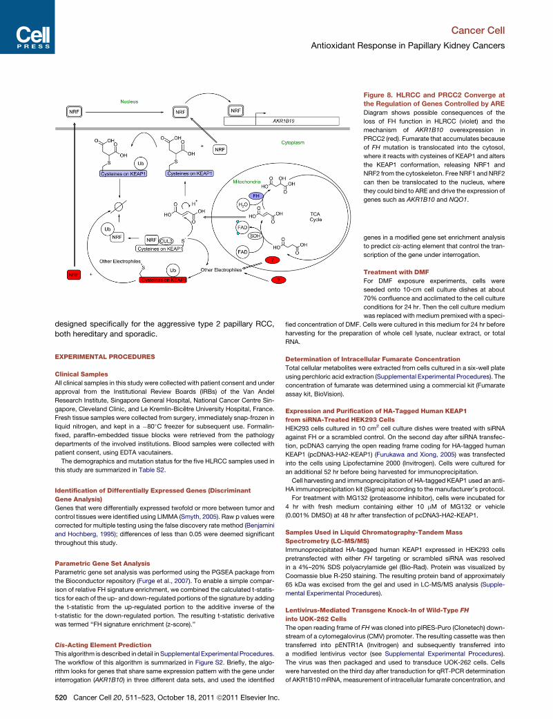
Figure 8. HLRCC and PRCC2 Converge at
the Regulation of Genes Controlled by ARE
Diagram shows possible consequences of the
loss of FH function in HLRCC (violet) and the
mechanism of AKR1B10 overexpression in
PRCC2 (red). Fumarate that accumulates because
of FH mutation is translocated into the cytosol,
where it reacts with cysteines of KEAP1 and alters
the KEAP1 conformation, releasing NRF1 and
NRF2 from the cytoskeleton. Free NRF1 andNRF2
can then be translocated to the nucleus, where
they could bind to ARE and drive the expression of
genes such as AKR1B10 and NQO1.
Cancer Cell
Antioxidant Response in Papillary Kidney Cancers
designed specifically for the aggressive type 2 papillary RCC,
both hereditary and sporadic.
EXPERIMENTAL PROCEDURES
Clinical Samples
All clinical samples in this study were collected with patient consent and under
approval from the Institutional Review Boards (IRBs) of the Van Andel
Research Institute, Singapore General Hospital, National Cancer Centre Sin-
gapore, Cleveland Clinic, and Le Kremlin-Bicetre University Hospital, France.
Fresh tissue samples were collected from surgery, immediately snap-frozen in
liquid nitrogen, and kept in a �80�C freezer for subsequent use. Formalin-
fixed, paraffin-embedded tissue blocks were retrieved from the pathology
departments of the involved institutions. Blood samples were collected with
patient consent, using EDTA vacutainers.
The demographics and mutation status for the five HLRCC samples used in
this study are summarized in Table S2.
Identification of Differentially Expressed Genes (Discriminant
Gene Analysis)
Genes that were differentially expressed twofold or more between tumor and
control tissues were identified using LIMMA (Smyth, 2005). Raw p values were
corrected for multiple testing using the false discovery rate method (Benjamini
and Hochberg, 1995); differences of less than 0.05 were deemed significant
throughout this study.
Parametric Gene Set Analysis
Parametric gene set analysis was performed using the PGSEA package from
the Bioconductor repository (Furge et al., 2007). To enable a simple compar-
ison of relative FH signature enrichment, we combined the calculated t-statis-
tics for each of the up- and down-regulated portions of the signature by adding
the t-statistic from the up-regulated portion to the additive inverse of the
t-statistic for the down-regulated portion. The resulting t-statistic derivative
was termed ‘‘FH signature enrichment (z-score).’’
Cis-Acting Element Prediction
This algorithm is described in detail in Supplemental Experimental Procedures.
The workflow of this algorithm is summarized in Figure S2. Briefly, the algo-
rithm looks for genes that share same expression pattern with the gene under
interrogation (AKR1B10) in three different data sets, and used the identified
520 Cancer Cell 20, 511–523, October 18, 2011 ª2011 Elsevier Inc.
genes in a modified gene set enrichment analysis
to predict cis-acting element that control the tran-
scription of the gene under interrogation.
Treatment with DMF
For DMF exposure experiments, cells were
seeded onto 10-cm cell culture dishes at about
70% confluence and acclimated to the cell culture
conditions for 24 hr. Then the cell culture medium
was replaced with medium premixed with a speci-
fied concentration of DMF. Cells were cultured in this medium for 24 hr before
harvesting for the preparation of whole cell lysate, nuclear extract, or total
RNA.
Determination of Intracellular Fumarate Concentration
Total cellular metabolites were extracted from cells cultured in a six-well plate
using perchloric acid extraction (Supplemental Experimental Procedures). The
concentration of fumarate was determined using a commercial kit (Fumarate
assay kit, BioVision).
Expression and Purification of HA-Tagged Human KEAP1
from siRNA-Treated HEK293 Cells
HEK293 cells cultured in 10 cm2 cell culture dishes were treated with siRNA
against FH or a scrambled control. On the second day after siRNA transfec-
tion, pcDNA3 carrying the open reading frame coding for HA-tagged human
KEAP1 (pcDNA3-HA2-KEAP1) (Furukawa and Xiong, 2005) was transfected
into the cells using Lipofectamine 2000 (Invitrogen). Cells were cultured for
an additional 52 hr before being harvested for immunoprecipitation.
Cell harvesting and immunoprecipitation of HA-tagged KEAP1 used an anti-
HA immunoprecipitation kit (Sigma) according to the manufacturer’s protocol.
For treatment with MG132 (proteasome inhibitor), cells were incubated for
4 hr with fresh medium containing either 10 mM of MG132 or vehicle
(0.001% DMSO) at 48 hr after transfection of pcDNA3-HA2-KEAP1.
Samples Used in Liquid Chromatography-Tandem Mass
Spectrometry (LC-MS/MS)
Immunoprecipitated HA-tagged human KEAP1 expressed in HEK293 cells
pretransfected with either FH targeting or scrambled siRNA was resolved
in a 4%–20% SDS polyacrylamide gel (Bio-Rad). Protein was visualized by
Coomassie blue R-250 staining. The resulting protein band of approximately
65 kDa was excised from the gel and used in LC-MS/MS analysis (Supple-
mental Experimental Procedures).
Lentivirus-Mediated Transgene Knock-In of Wild-Type FH
into UOK-262 Cells
The open reading frame of FH was cloned into pIRES-Puro (Clonetech) down-
stream of a cytomegalovirus (CMV) promoter. The resulting cassette was then
transferred into pENTR1A (Invitrogen) and subsequently transferred into
a modified lentivirus vector (see Supplemental Experimental Procedures).
The virus was then packaged and used to transduce UOK-262 cells. Cells
were harvested on the third day after transduction for qRT-PCR determination
of AKR1B10mRNA, measurement of intracellular fumarate concentration, and
Cancer Cell
Antioxidant Response in Papillary Kidney Cancers
Western blot analysis. Lentivirus packaged from an empty vector (carrying the
CMV promoter and the puromycin resistance cassette from pIRES-Puro) was
used as a transduction control. A schematic vector map is in the Supplemental
Experimental Procedures section.
ACCESSION NUMBER
Microarray data generated in this study have been deposited at the NCBI Gene
Expression Omnibus, accession number GSE26574.
SUPPLEMENTAL INFORMATION
Supplemental Information includes six figures, six tables, and Supplemental
Experimental Procedures and may be found with this article online at
doi:10.1016/j.ccr.2011.08.024.
ACKNOWLEDGMENTS
This paper is dedicated to the memory of Thean-Poh Ooi.
We thank Sabrina Noyes for administrative support and David Nadziejka for
technical editing. We acknowledge the Van Andel Research Foundation, the
Singapore Millennium Foundation, and Fondation IGR & INCa (PNES Rein
and Centre Expert National Cancers Rares PREDIR) for funding. A.O., K.F.,
and B.T. designed the study; A.O. performed all experiments, bioinformatics,
and data analysis; J.C.W. and D.R. performed IHC staining; D.P. performed
microarray experiments; D.W. performed tandem mass spectrometry; and
X.J.Y. and M.Z. performed histological evaluation. B.T., Z.Z., M.Z., M.H.T.,
B.H.W., V.P.T., P.H.T., B.G., V.M., and S.R. provided samples and clinical
data. A.O. and K.F. prepared the manuscript. Mass spectrometry and data
analysis were performed in the Michigan State University, Proteomic Core
Facility.
Received: December 22, 2010
Revised: June 4, 2011
Accepted: August 30, 2011
Published: October 17, 2011
REFERENCES
Alam, N.A., Rowan, A.J., Wortham, N.C., Pollard, P.J., Mitchell, M., Tyrer, J.P.,
Barclay, E., Calonje, E., Manek, S., Adams, S.J., et al. (2003). Genetic and
functional analyses of FHmutations inmultiple cutaneous and uterine leiomyo-
matosis, hereditary leiomyomatosis and renal cancer, and fumarate hydratase
deficiency. Hum. Mol. Genet. 12, 1241–1252.
Alderson, N.L., Wang, Y., Blatnik, M., Frizzell, N., Walla, M.D., Lyons, T.J., Alt,
N., Carson, J.A., Nagai, R., Thorpe, S.R., and Baynes, J.W. (2006).
S-(2-Succinyl)cysteine: a novel chemical modification of tissue proteins by
a Krebs cycle intermediate. Arch. Biochem. Biophys. 450, 1–8.
Bellacosa, A., Godwin, A.K., Peri, S., Devarajan, K., Caretti, E., Vanderveer, L.,
Bove, B., Slater, C., Zhou, Y., Daly, M., et al. (2010). Altered gene expression in
morphologically normal epithelial cells from heterozygous carriers of BRCA1
or BRCA2 mutations. Cancer Prev Res (Phila) 3, 48–61.
Benjamini, Y., and Hochberg, Y. (1995). Controlling the false discovery rate:
a practical and powerful approach to multiple testing. J. R. Stat. Soc. B 57,
289–300.
Blatnik, M., Thorpe, S.R., and Baynes, J.W. (2008). Succination of proteins
by fumarate: mechanism of inactivation of glyceraldehyde-3-phosphate
dehydrogenase in diabetes. Ann. N Y Acad. Sci. 1126, 272–275.
Carvajal-Carmona, L.G., Alam, N.A., Pollard, P.J., Jones, A.M., Barclay, E.,
Wortham, N., Pignatelli, M., Freeman, A., Pomplun, S., Ellis, I., et al. (2006).
Adult leydig cell tumors of the testis caused by germline fumarate hydratase
mutations. J. Clin. Endocrinol. Metab. 91, 3071–3075.
Chan, K., Han, X.D., and Kan, Y.W. (2001). An important function of Nrf2 in
combating oxidative stress: detoxification of acetaminophen. Proc. Natl.
Acad. Sci. USA 98, 4611–4616.
C
Dahia, P.L., Ross, K.N., Wright, M.E., Hayashida, C.Y., Santagata, S.,
Barontini, M., Kung, A.L., Sanso, G., Powers, J.F., Tischler, A.S., et al.
(2005). A HIF1alpha regulatory loop links hypoxia and mitochondrial signals
in pheochromocytomas. PLoS Genet. 1, 72–80.
Frizzell, N., Lima, M., and Baynes, J.W. (2010). Succination of proteins in dia-
betes. Free Radic. Res. 45, 101–109.
Frizzell, N., Rajesh, M., Jepson, M.J., Nagai, R., Carson, J.A., Thorpe, S.R.,
and Baynes, J.W. (2009). Succination of thiol groups in adipose tissue proteins
in diabetes: succination inhibits polymerization and secretion of adiponectin.
J. Biol. Chem. 284, 25772–25781.
Furge, K.A., Tan, M.H., Dykema, K., Kort, E., Stadler, W., Yao, X., Zhou, M.,
and Teh, B.T. (2007). Identification of deregulated oncogenic pathways in renal
cell carcinoma: an integrated oncogenomic approach based on gene expres-
sion profiling. Oncogene 26, 1346–1350.
Furukawa, M., and Xiong, Y. (2005). BTB protein Keap1 targets antioxidant
transcription factor Nrf2 for ubiquitination by the Cullin 3-Roc1 ligase. Mol.
Cell. Biol. 25, 162–171.
Gao, L., Wang, J., Sekhar, K.R., Yin, H., Yared, N.F., Schneider, S.N., Sasi, S.,
Dalton, T.P., Anderson, M.E., Chan, J.Y., et al. (2007). Novel n-3 fatty acid
oxidation products activate Nrf2 by destabilizing the association between
Keap1 and Cullin3. J. Biol. Chem. 282, 2529–2537.
Hayes, J.D., and McMahon, M. (2001). Molecular basis for the contribution of
the antioxidant responsive element to cancer chemoprevention. Cancer Lett.
174, 103–113.
Holmgren, A., and Lu, J. (2010). Thioredoxin and thioredoxin reductase:
current research with special reference to human disease. Biochem.
Biophys. Res. Commun. 396, 120–124.
Hsieh, T.C., Lu, X., Wang, Z., andWu, J.M. (2006). Induction of quinone reduc-
tase NQO1 by resveratrol in human K562 cells involves the antioxidant
response element ARE and is accompanied by nuclear translocation of
transcription factor Nrf2. Med. Chem. 2, 275–285.
Huang, Y.W., Jansen, R.A., Fabbri, E., Potter, D., Liyanarachchi, S., Chan,
M.W., Liu, J.C., Crijns, A.P., Brown, R., Nephew, K.P., et al. (2009).
Identification of candidate epigenetic biomarkers for ovarian cancer detection.
Oncol. Rep. 22, 853–861.
Huang, Y.W., Luo, J., Weng, Y.I., Mutch, D.G., Goodfellow, P.J., Miller, D.S.,
and Huang, T.H. (2010). Promoter hypermethylation of CIDEA, HAAO and
RXFP3 associated with microsatellite instability in endometrial carcinomas.
Gynecol. Oncol. 117, 239–247.
Hubner, R.H., Schwartz, J.D., De Bishnu, P., Ferris, B., Omberg, L., Mezey,
J.G., Hackett, N.R., and Crystal, R.G. (2009). Coordinate control of expression
of Nrf2-modulated genes in the human small airway epithelium is highly
responsive to cigarette smoking. Mol. Med. 15, 203–219.
Isaacs, J.S., Jung, Y.J., Mole, D.R., Lee, S., Torres-Cabala, C., Chung, Y.L.,
Merino, M., Trepel, J., Zbar, B., Toro, J., et al. (2005). HIF overexpression
correlates with biallelic loss of fumarate hydratase in renal cancer: novel role
of fumarate in regulation of HIF stability. Cancer Cell 8, 143–153.
Ivan, M., Kondo, K., Yang, H., Kim, W., Valiando, J., Ohh, M., Salic, A., Asara,
J.M., Lane, W.S., and Kaelin, W.G., Jr. (2001). HIFalpha targeted for
VHL-mediated destruction by proline hydroxylation: implications for O2
sensing. Science 292, 464–468.
Kashkar, H., Deggerich, A., Seeger, J.M., Yazdanpanah, B., Wiegmann, K.,
Haubert, D., Pongratz, C., and Kronke, M. (2007). NF-kappaB-independent
down-regulation of XIAP by bortezomib sensitizes HL B cells against cytotoxic
drugs. Blood 109, 3982–3988.
Kiuru, M., and Launonen, V. (2004). Hereditary leiomyomatosis and renal cell
cancer (HLRCC). Curr. Mol. Med. 4, 869–875.
Koivunen, P., Hirsila, M., Remes, A.M., Hassinen, I.E., Kivirikko, K.I., and
Myllyharju, J. (2007). Inhibition of hypoxia-inducible factor (HIF) hydroxylases
by citric acid cycle intermediates: possible links between cell metabolism
and stabilization of HIF. J. Biol. Chem. 282, 4524–4532.
Koshikawa, N., Hayashi, J., Nakagawara, A., and Takenaga, K. (2009).
Reactive oxygen species-generating mitochondrial DNA mutation up-regu-
lates hypoxia-inducible factor-1alpha gene transcription via
ancer Cell 20, 511–523, October 18, 2011 ª2011 Elsevier Inc. 521

Cancer Cell
Antioxidant Response in Papillary Kidney Cancers
phosphatidylinositol 3-kinase-Akt/protein kinase C/histone deacetylase
pathway. J. Biol. Chem. 284, 33185–33194.
Kwak, M.K., Kensler, T.W., and Casero, R.A., Jr. (2003). Induction of phase 2
enzymes by serum oxidized polyamines through activation of Nrf2: effect of
the polyamine metabolite acrolein. Biochem. Biophys. Res. Commun. 305,
662–670.
Lau, A., Villeneuve, N.F., Sun, Z., Wong, P.K., and Zhang, D.D. (2008). Dual
roles of Nrf2 in cancer. Pharmacol. Res. 58, 262–270.
Launonen, V., Vierimaa, O., Kiuru, M., Isola, J., Roth, S., Pukkala, E., Sistonen,
P., Herva, R., and Aaltonen, L.A. (2001). Inherited susceptibility to uterine leio-
myomas and renal cell cancer. Proc. Natl. Acad. Sci. USA 98, 3387–3392.
Lee, D.F., Kuo, H.P., Liu, M., Chou, C.K., Xia, W., Du, Y., Shen, J., Chen, C.T.,
Huo, L., Hsu, M.C., et al. (2009). KEAP1 E3 ligase-mediated downregulation of
NF-kappaB signaling by targeting IKKbeta. Mol. Cell 36, 131–140.
Lehtonen, H.J., Kiuru, M., Ylisaukko-Oja, S.K., Salovaara, R., Herva, R.,
Koivisto, P.A., Vierimaa, O., Aittomaki, K., Pukkala, E., Launonen, V., and
Aaltonen, L.A. (2006). Increased risk of cancer in patients with fumarate hydra-
tase germline mutation. J. Med. Genet. 43, 523–526.
Linehan, W.M., Bratslavsky, G., Pinto, P.A., Schmidt, L.S., Neckers, L.,
Bottaro, D.P., and Srinivasan, R. (2010). Molecular diagnosis and therapy of
kidney cancer. Annu. Rev. Med. 61, 329–343.
Linehan, W.M., Pinto, P.A., Srinivasan, R., Merino, M., Choyke, P., Choyke, L.,
Coleman, J., Toro, J., Glenn, G., Vocke, C., et al. (2007). Identification of the
genes for kidney cancer: opportunity for disease-specific targeted therapeu-
tics. Clin. Cancer Res. 13, 671s–679s.
MacLeod, A.K., McMahon, M., Plummer, S.M., Higgins, L.G., Penning, T.M.,
Igarashi, K., and Hayes, J.D. (2009). Characterization of the cancer chemopre-
ventive NRF2-dependent gene battery in human keratinocytes: demonstration
that the KEAP1-NRF2 pathway, and not the BACH1-NRF2 pathway, controls
cytoprotection against electrophiles as well as redox-cycling compounds.
Carcinogenesis 30, 1571–1580.
Malec, V., Gottschald, O.R., Li, S., Rose, F., Seeger, W., and Hanze, J. (2010).
HIF-1 alpha signaling is augmented during intermittent hypoxia by induction of
the Nrf2 pathway in NOX1-expressing adenocarcinoma A549 cells. Free
Radic. Biol. Med. 48, 1626–1635.
McMahon, M., Itoh, K., Yamamoto, M., Chanas, S.A., Henderson, C.J.,
McLellan, L.I., Wolf, C.R., Cavin, C., and Hayes, J.D. (2001). The Cap‘n’Collar
basic leucine zipper transcription factor Nrf2 (NF-E2 p45-related factor 2)
controls both constitutive and inducible expression of intestinal detoxification
and glutathione biosynthetic enzymes. Cancer Res. 61, 3299–3307.
Molina, A.M., Feldman, D.R., Ginsberg, M.S., Kroog, G., Tickoo, S.K., Jia, X.,
Georges, M., Patil, S., Baum, M.S., Reuter, V.E., and Motzer, R.J. (2010).
Phase II trial of sunitinib in patients with metastatic non-clear cell renal cell
carcinoma. Invest. New Drugs. [Epub ahead of print].
Nagai, R., Brock, J.W., Blatnik, M., Baatz, J.E., Bethard, J., Walla, M.D.,
Thorpe, S.R., Baynes, J.W., and Frizzell, N. (2007). Succination of protein thiols
during adipocyte maturation: a biomarker of mitochondrial stress. J. Biol.
Chem. 282, 34219–34228.
Nioi, P., and Nguyen, T. (2007). A mutation of Keap1 found in breast cancer
impairs its ability to repress Nrf2 activity. Biochem. Biophys. Res. Commun.
362, 816–821.
O’Flaherty, L., Adam, J., Heather, L.C., Zhdanov, A.V., Chung, Y.L., Miranda,
M.X., Croft, J., Olpin, S., Clarke, K., Pugh, C.W., et al. (2010). Dysregulation of
hypoxia pathways in fumarate hydratase-deficient cells is independent of
defective mitochondrial metabolism. Hum. Mol. Genet. 19, 3844–3851.
Ohta, T., Iijima, K., Miyamoto, M., Nakahara, I., Tanaka, H., Ohtsuji, M., Suzuki,
T., Kobayashi, A., Yokota, J., Sakiyama, T., et al. (2008). Loss of Keap1 func-
tion activates Nrf2 and provides advantages for lung cancer cell growth.
Cancer Res. 68, 1303–1309.
Ohtsuji, M., Katsuoka, F., Kobayashi, A., Aburatani, H., Hayes, J.D., and
Yamamoto, M. (2008). Nrf1 and Nrf2 play distinct roles in activation of antiox-
idant response element-dependent genes. J. Biol. Chem. 283, 33554–33562.
Pae, H.O., Oh, G.S., Lee, B.S., Rim, J.S., Kim, Y.M., and Chung, H.T. (2006).
3-Hydroxyanthranilic acid, one of L-tryptophanmetabolites, inhibits monocyte
522 Cancer Cell 20, 511–523, October 18, 2011 ª2011 Elsevier Inc.
chemoattractant protein-1 secretion and vascular cell adhesion molecule-1
expression via heme oxygenase-1 induction in human umbilical vein endothe-
lial cells. Atherosclerosis 187, 274–284.
Pollard, P., Wortham, N., Barclay, E., Alam, A., Elia, G., Manek, S., Poulsom,
R., and Tomlinson, I. (2005a). Evidence of increased microvessel density
and activation of the hypoxia pathway in tumours from the hereditary leiomyo-
matosis and renal cell cancer syndrome. J. Pathol. 205, 41–49.
Pollard, P.J., Briere, J.J., Alam, N.A., Barwell, J., Barclay, E., Wortham, N.C.,
Hunt, T., Mitchell, M., Olpin, S., Moat, S.J., et al. (2005b). Accumulation of
Krebs cycle intermediates and over-expression of HIF1alpha in tumours which
result from germline FH and SDHmutations. Hum. Mol. Genet. 14, 2231–2239.
Pollard, P.J., El-Bahrawy, M., Poulsom, R., Elia, G., Killick, P., Kelly, G., Hunt,
T., Jeffery, R., Seedhar, P., Barwell, J., et al. (2006). Expression of HIF-1alpha,
HIF-2alpha (EPAS1), and their target genes in paraganglioma and pheochro-
mocytoma with VHL and SDH mutations. J. Clin. Endocrinol. Metab. 91,
4593–4598.
Rachakonda, G., Xiong, Y., Sekhar, K.R., Stamer, S.L., Liebler, D.C., and
Freeman, M.L. (2008). Covalent modification at Cys151 dissociates the elec-
trophile sensor Keap1 from the ubiquitin ligase CUL3. Chem. Res. Toxicol.
21, 705–710.
Russo, P., and O’Regan, S. (1990). Visceral pathology of hereditary tyrosine-
mia type I. Am. J. Hum. Genet. 47, 317–324.
Sakurai, A., Nishimoto, M., Himeno, S., Imura, N., Tsujimoto,M., Kunimoto,M.,
and Hara, S. (2005). Transcriptional regulation of thioredoxin reductase 1
expression by cadmium in vascular endothelial cells: role of NF-E2-related
factor-2. J. Cell. Physiol. 203, 529–537.
Sango, K., Suzuki, T., Yanagisawa, H., Takaku, S., Hirooka, H., Tamura, M.,
and Watabe, K. (2006). High glucose-induced activation of the polyol pathway
and changes of gene expression profiles in immortalized adult mouse
Schwann cells IMS32. J. Neurochem. 98, 446–458.
Shibata, T., Ohta, T., Tong, K.I., Kokubu, A., Odogawa, R., Tsuta, K., Asamura,
H., Yamamoto, M., and Hirohashi, S. (2008). Cancer relatedmutations in NRF2
impair its recognition by Keap1-Cul3 E3 ligase and promote malignancy. Proc.
Natl. Acad. Sci. USA 105, 13568–13573.
Singh, A., Misra, V., Thimmulappa, R.K., Lee, H., Ames, S., Hoque, M.O.,
Herman, J.G., Baylin, S.B., Sidransky, D., Gabrielson, E., et al. (2006).
Dysfunctional KEAP1-NRF2 interaction in non-small-cell lung cancer. PLoS
Med. 3, e420.
Siow, R.C., Ishii, T., and Mann, G.E. (2007). Modulation of antioxidant gene
expression by 4-hydroxynonenal: atheroprotective role of the Nrf2/ARE tran-
scription pathway. Redox Rep. 12, 11–15.
Smyth,G.K. (2005). Limma: linearmodels formicroarray data. InBioinformatics
and Computational Biology Solutions using R and Bioconductor, R.
Gentleman, V. Carey, S. Dudoit, R.A. Irizarry, and W. Huber, eds. (New York:
Springer), pp. 397–420.
Sourbier, C., Valera-Romero, V., Giubellino, A., Yang, Y., Sudarshan, S.,
Neckers, L., and Linehan, W.M. (2010). Increasing reactive oxygen species
as a therapeutic approach to treat hereditary leiomyomatosis and renal cell
carcinoma. Cell Cycle 9, 4183–4189.
Spencer, S.R., Wilczak, C.A., and Talalay, P. (1990). Induction of glutathione
transferases and NAD(P)H:quinone reductase by fumaric acid derivatives in
rodent cells and tissues. Cancer Res. 50, 7871–7875.
Sudarshan, S., Sourbier, C., Kong, H.S., Block, K., Valera Romero, V.A., Yang,
Y., Galindo, C., Mollapour, M., Scroggins, B., Goode, N., et al. (2009).
Fumarate hydratase deficiency in renal cancer induces glycolytic addiction
and hypoxia-inducible transcription factor 1alpha stabilization by glucose-
dependent generation of reactive oxygen species. Mol. Cell. Biol. 29, 4080–
4090.
Tomlinson, I.P., Alam, N.A., Rowan, A.J., Barclay, E., Jaeger, E.E., Kelsell, D.,
Leigh, I., Gorman, P., Lamlum, H., Rahman, S., et al; Multiple Leiomyoma
Consortium. (2002). Germline mutations in FH predispose to dominantly in-
herited uterine fibroids, skin leiomyomata and papillary renal cell cancer.
Nat. Genet. 30, 406–410.

Cancer Cell
Antioxidant Response in Papillary Kidney Cancers
Vanharanta, S., Buchta, M., McWhinney, S.R., Virta, S.K., Peczkowska, M.,
Morrison, C.D., Lehtonen, R., Januszewicz, A., Jarvinen, H., Juhola, M.,
et al. (2004). Early-onset renal cell carcinoma as a novel extraparaganglial
component of SDHB-associated heritable paraganglioma. Am. J. Hum.
Genet. 74, 153–159.
Vanharanta, S., Pollard, P.J., Lehtonen, H.J., Laiho, P., Sjoberg, J., Leminen,
A., Aittomaki, K., Arola, J., Kruhoffer, M., Orntoft, T.F., et al. (2006). Distinct
expression profile in fumarate-hydratase-deficient uterine fibroids. Hum.
Mol. Genet. 15, 97–103.
Venugopal, R., and Jaiswal, A.K. (1998). Nrf2 and Nrf1 in association with Jun
proteins regulate antioxidant response element-mediated expression and
coordinated induction of genes encoding detoxifying enzymes. Oncogene
17, 3145–3156.
Wang, R., An, J., Ji, F., Jiao, H., Sun, H., and Zhou, D. (2008). Hypermethylation
of the Keap1 gene in human lung cancer cell lines and lung cancer tissues.
Biochem. Biophys. Res. Commun. 373, 151–154.
Wang, R., Kern, J.T., Goodfriend, T.L., Ball, D.L., and Luesch, H. (2009).
Activation of the antioxidant response element by specific oxidized metabo-
lites of linoleic acid. Prostaglandins Leukot. Essent. Fatty Acids 81, 53–59.
C
Yang, Y., Valera, V.A., Padilla-Nash, H.M., Sourbier, C., Vocke, C.D., Vira,
M.A., Abu-Asab, M.S., Bratslavsky, G., Tsokos, M., Merino, M.J., et al.
(2010). UOK 262 cell line, fumarate hydratase deficient (FH-/FH-) hereditary
leiomyomatosis renal cell carcinoma: in vitro and in vivo model of an aberrant
energy metabolic pathway in human cancer. Cancer Genet. Cytogenet. 196,
45–55.
Zhang, D.D., and Hannink, M. (2003). Distinct cysteine residues in Keap1 are
required for Keap1-dependent ubiquitination of Nrf2 and for stabilization of
Nrf2 by chemopreventive agents and oxidative stress. Mol. Cell. Biol. 23,
8137–8151.
Zhang, D.D., Lo, S.C., Sun, Z., Habib, G.M., Lieberman,M.W., andHannink,M.
(2005). Ubiquitination of Keap1, a BTB-Kelch substrate adaptor protein for
Cul3, targets Keap1 for degradation by a proteasome-independent pathway.
J. Biol. Chem. 280, 30091–30099.
Zhang, Y., Crouch, D.H., Yamamoto, M., and Hayes, J.D. (2006). Negative
regulation of the Nrf1 transcription factor by its N-terminal domain is indepen-
dent of Keap1: Nrf1, but not Nrf2, is targeted to the endoplasmic reticulum.
Biochem. J. 399, 373–385.
ancer Cell 20, 511–523, October 18, 2011 ª2011 Elsevier Inc. 523