Alzheimer's Presenilin Mutation Sensitizes Neural Cells to Apoptosis Induced by Trophic Factor...
11
Alzheimer’s Presenilin Mutation Sensitizes Neural Cells to Apoptosis Induced by Trophic Factor Withdrawal and Amyloid b-Peptide: Involvement of Calcium and Oxyradicals Qing Guo, 1 Bryce L. Sopher, 2 Katsutoshi Furukawa, 1 Dao G. Pham, 2 Nic Robinson, 1 George M. Martin, 2 and Mark P. Mattson 1 1 Sanders-Brown Research Center on Aging and Department of Anatomy and Neurobiology, University of Kentucky, Lexington, Kentucky 40536, and 2 Department of Pathology, University of Washington, Seattle, Washington 98195-7470 Most autosomal dominant inherited forms of early onset Alz- heimer’s disease (AD) are caused by mutations in the presenilin-1 (PS-1) gene on chromosome 14. PS-1 is an integral membrane protein with six to nine membrane-spanning do- mains and is expressed in neurons throughout the brain wherein it is localized mainly in endoplasmic reticulum (ER). The mechanism or mechanisms whereby PS-1 mutations promote neuron degeneration in AD are unknown. Recent findings sug- gest links among deposition of amyloid b-peptide (Ab), oxida- tive stress, disruption of ion homeostasis, and an apoptotic form of neuron death in AD. We now report that expression of the human PS-1 L286V mutation in PC12 cells increases their susceptibility to apoptosis induced by trophic factor withdrawal and Ab. Increases in oxidative stress and intracellular calcium levels induced by the apoptotic stimuli were exacerbated greatly in cells expressing the PS-1 mutation, as compared with control cell lines and lines overexpressing wild-type PS-1. The antiapoptotic gene product Bcl-2 prevented apoptosis after NGF withdrawal from differentiated PC12 cells expressing mu- tant PS-1. Elevations of [Ca 21 ] i in response to thapsigargin, an inhibitor of the ER Ca 21 -ATPase, were increased in cells ex- pressing mutant PS-1, and this adverse effect was abolished in cells expressing Bcl-2. Antioxidants and blockers of calcium influx and release from ER protected cells against the adverse consequences of the PS-1 mutation. By perturbing cellular calcium regulation and promoting oxidative stress, PS-1 muta- tions may sensitize neurons to apoptotic death in AD. Key words: Alzheimer’s disease; antioxidant; bcl-2; dan- trolene; endoplasmic reticulum; fura-2; nerve growth factor Alzheimer’s disease (AD) is a progressive and always fatal neu- rodegenerative disorder characterized by the death of neurons in brain regions involved in learning and memory processes (for review, see Selkoe, 1989). Accumulations of insoluble fibrillar aggregates of a protein called amyloid b-peptide (Ab) are impli- cated in the pathogenesis of AD. Ab is associated with degener- ating neurons in AD brain, and mutations in the gene for the amyloid precursor protein (the source of Ab) cause a small percentage of cases of inherited familial AD (for review, see Mullan and Crawford, 1993). Moreover, mice genetically engi- neered to produce mutated human amyloid precursor protein exhibit Ab deposition and cognitive impairments (Games et al., 1995; Hsaio et al., 1996). Ab damages and kills cultured neurons by a mechanism involving oxidative stress and disruption of cel- lular calcium homeostasis (Mattson et al., 1992; Behl et al., 1994; Fukuyama et al., 1994; Goodman and Mattson, 1994; Mark et al., 1995, 1997a). Apoptosis is a form of “programmed” cell death characterized by plasma membrane phospholipid alterations, cell shrinkage, and nuclear DNA condensation and fragmentation (Bredesen, 1995; Thompson, 1995). Ab induces apoptosis in cul- tured neurons (Forloni et al., 1993; Loo et al., 1993), and studies of postmortem brain tissue suggest that neuronal apoptosis occurs in AD (Su et al., 1994; Smale et al., 1995). The molecular and cellular mechanisms that predispose neurons to apoptotic death in AD are unknown. An important step toward elucidating the cause or causes of neuron degeneration in AD was made with the identification of the genes responsible for the majority of cases of autosomal dominant inherited forms of early onset AD. The genes, called presenilin-1 (PS-1; chromosome 14) and presenilin-2 (PS-2; chro- mosome 1), encode proteins predicted to be integral membrane proteins with six to nine membrane-spanning domains (Levy- Lahad et al., 1995; Rogaev et al., 1995; Sherrington et al., 1995; Doan et al., 1996; Li and Greenwald, 1996). Immunohistochem- ical analyses indicate that PS-1 is expressed in neurons throughout the brain (Cook et al., 1996; Cribbs et al., 1996; Elder et al., 1996); PS-1 has been localized to both degenerating (Murphy et al., 1996) and nondegenerating (Giannakopoulos et al., 1997) neu- rons in AD brain. In cultured cells PS-1 localizes to subcellular compartments and seems to be at particularly high levels in the endoplasmic reticulum (ER) (Kovacs et al., 1996; Walter et al., 1996). PS-1 can be processed endoproteolytically (Thinakaran et al., 1996), although the significance of such processing is un- known. Because PS-1 mutations account for the majority of cases of inherited early onset forms of AD, understanding the normal functions of PS-1 and how PS-1 mutations promote neuron de- generation are central issues in the AD field. We report that expression of a human PS-1 mutation in PC12 cells increases their vulnerability to apoptosis induced by trophic factor withdrawal and Ab. The mechanism whereby mutant PS-1 promotes apopto- Received Feb. 12, 1997; revised March 20, 1997; accepted March 25, 1997. This work was supported by Grants to M.P.M. from National Institutes of Health (NS30583 and AG10836) and the Alzheimer’s Association, to G.M.M. from National Institutes of Health (AG10917), and to B.L.S. from the University of Washington Nathan Shock Center for Excellence in the Basic Biology of Aging. We thank J. Begley, S. Bose, R. Pelfrey, and J. Xie for technical assistance. Correspondence should be addressed to Dr. Mark P. Mattson, 211 Sanders-Brown Building, University of Kentucky, Lexington, KY 40536-0230. Copyright © 1997 Society for Neuroscience 0270-6474/97/174212-11$05.00/0 The Journal of Neuroscience, June 1, 1997, 17(11):4212– 4222
-
Upload
independent -
Category
Documents
-
view
1 -
download
0
Transcript of Alzheimer's Presenilin Mutation Sensitizes Neural Cells to Apoptosis Induced by Trophic Factor...
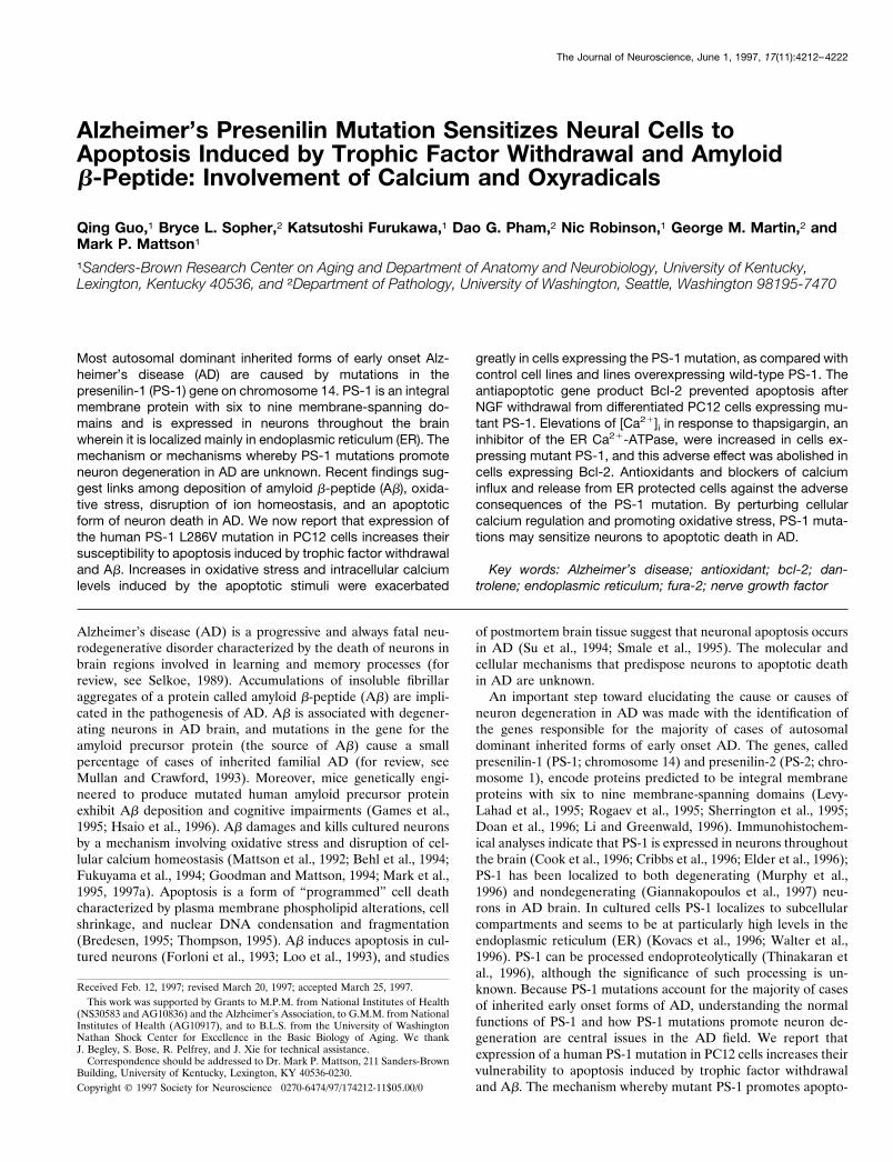
Alzheimer’s Presenilin Mutation Sensitizes Neural Cells toApoptosis Induced by Trophic Factor Withdrawal and Amyloidb-Peptide: Involvement of Calcium and Oxyradicals
Qing Guo,1 Bryce L. Sopher,2 Katsutoshi Furukawa,1 Dao G. Pham,2 Nic Robinson,1 George M. Martin,2 andMark P. Mattson1
1Sanders-Brown Research Center on Aging and Department of Anatomy and Neurobiology, University of Kentucky,Lexington, Kentucky 40536, and 2Department of Pathology, University of Washington, Seattle, Washington 98195-7470
Most autosomal dominant inherited forms of early onset Alz-heimer’s disease (AD) are caused by mutations in thepresenilin-1 (PS-1) gene on chromosome 14. PS-1 is an integralmembrane protein with six to nine membrane-spanning do-mains and is expressed in neurons throughout the brainwherein it is localized mainly in endoplasmic reticulum (ER). Themechanism or mechanisms whereby PS-1 mutations promoteneuron degeneration in AD are unknown. Recent findings sug-gest links among deposition of amyloid b-peptide (Ab), oxida-tive stress, disruption of ion homeostasis, and an apoptoticform of neuron death in AD. We now report that expression ofthe human PS-1 L286V mutation in PC12 cells increases theirsusceptibility to apoptosis induced by trophic factor withdrawaland Ab. Increases in oxidative stress and intracellular calciumlevels induced by the apoptotic stimuli were exacerbated
greatly in cells expressing the PS-1 mutation, as compared withcontrol cell lines and lines overexpressing wild-type PS-1. Theantiapoptotic gene product Bcl-2 prevented apoptosis afterNGF withdrawal from differentiated PC12 cells expressing mu-tant PS-1. Elevations of [Ca21]i in response to thapsigargin, aninhibitor of the ER Ca21-ATPase, were increased in cells ex-pressing mutant PS-1, and this adverse effect was abolished incells expressing Bcl-2. Antioxidants and blockers of calciuminflux and release from ER protected cells against the adverseconsequences of the PS-1 mutation. By perturbing cellularcalcium regulation and promoting oxidative stress, PS-1 muta-tions may sensitize neurons to apoptotic death in AD.
Key words: Alzheimer’s disease; antioxidant; bcl-2; dan-trolene; endoplasmic reticulum; fura-2; nerve growth factor
Alzheimer’s disease (AD) is a progressive and always fatal neu-rodegenerative disorder characterized by the death of neurons inbrain regions involved in learning and memory processes (forreview, see Selkoe, 1989). Accumulations of insoluble fibrillaraggregates of a protein called amyloid b-peptide (Ab) are impli-cated in the pathogenesis of AD. Ab is associated with degener-ating neurons in AD brain, and mutations in the gene for theamyloid precursor protein (the source of Ab) cause a smallpercentage of cases of inherited familial AD (for review, seeMullan and Crawford, 1993). Moreover, mice genetically engi-neered to produce mutated human amyloid precursor proteinexhibit Ab deposition and cognitive impairments (Games et al.,1995; Hsaio et al., 1996). Ab damages and kills cultured neuronsby a mechanism involving oxidative stress and disruption of cel-lular calcium homeostasis (Mattson et al., 1992; Behl et al., 1994;Fukuyama et al., 1994; Goodman and Mattson, 1994; Mark et al.,1995, 1997a). Apoptosis is a form of “programmed” cell deathcharacterized by plasma membrane phospholipid alterations, cellshrinkage, and nuclear DNA condensation and fragmentation(Bredesen, 1995; Thompson, 1995). Ab induces apoptosis in cul-tured neurons (Forloni et al., 1993; Loo et al., 1993), and studies
of postmortem brain tissue suggest that neuronal apoptosis occursin AD (Su et al., 1994; Smale et al., 1995). The molecular andcellular mechanisms that predispose neurons to apoptotic deathin AD are unknown.
An important step toward elucidating the cause or causes ofneuron degeneration in AD was made with the identification ofthe genes responsible for the majority of cases of autosomaldominant inherited forms of early onset AD. The genes, calledpresenilin-1 (PS-1; chromosome 14) and presenilin-2 (PS-2; chro-mosome 1), encode proteins predicted to be integral membraneproteins with six to nine membrane-spanning domains (Levy-Lahad et al., 1995; Rogaev et al., 1995; Sherrington et al., 1995;Doan et al., 1996; Li and Greenwald, 1996). Immunohistochem-ical analyses indicate that PS-1 is expressed in neurons throughoutthe brain (Cook et al., 1996; Cribbs et al., 1996; Elder et al., 1996);PS-1 has been localized to both degenerating (Murphy et al.,1996) and nondegenerating (Giannakopoulos et al., 1997) neu-rons in AD brain. In cultured cells PS-1 localizes to subcellularcompartments and seems to be at particularly high levels in theendoplasmic reticulum (ER) (Kovacs et al., 1996; Walter et al.,1996). PS-1 can be processed endoproteolytically (Thinakaran etal., 1996), although the significance of such processing is un-known. Because PS-1 mutations account for the majority of casesof inherited early onset forms of AD, understanding the normalfunctions of PS-1 and how PS-1 mutations promote neuron de-generation are central issues in the AD field. We report thatexpression of a human PS-1 mutation in PC12 cells increases theirvulnerability to apoptosis induced by trophic factor withdrawaland Ab. The mechanism whereby mutant PS-1 promotes apopto-
Received Feb. 12, 1997; revised March 20, 1997; accepted March 25, 1997.This work was supported by Grants to M.P.M. from National Institutes of Health
(NS30583 and AG10836) and the Alzheimer’s Association, to G.M.M. from NationalInstitutes of Health (AG10917), and to B.L.S. from the University of WashingtonNathan Shock Center for Excellence in the Basic Biology of Aging. We thankJ. Begley, S. Bose, R. Pelfrey, and J. Xie for technical assistance.
Correspondence should be addressed to Dr. Mark P. Mattson, 211 Sanders-BrownBuilding, University of Kentucky, Lexington, KY 40536-0230.Copyright © 1997 Society for Neuroscience 0270-6474/97/174212-11$05.00/0
The Journal of Neuroscience, June 1, 1997, 17(11):4212–4222
sis seems to involve disruption of calcium homeostasis and in-creased oxidative stress.
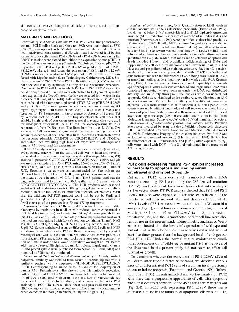
MATERIALS AND METHODSExpression of wild-type and mutant PS-1 in PC12 cells. Rat pheochromo-cytoma (PC12) cells (Black and Greene, 1982) were maintained at 37°C(5% CO2 atmosphere) in RPMI-1640 medium supplemented 10% withheat-inactivated horse serum and 5% with heat-inactivated fetal bovineserum. A full-length human PS-1 cDNA and a PS-1 cDNA containing theL286V mutation were cloned into either the expression vector pTRE inthe Tet-off expression system (Clontech, Cambridge, UK) or pRc/CMVto produce pTRE-PS1 and pTRE-PS1L286V or pCMV-PS1 and pCMV-PS1L286V, where, in both cases, the expression of PS-1 and PS-1 L286VcDNAs is under the control of CMV promoter. PC12 cells were trans-fected with Lipofectamine (Life Technologies, Gaithersburg, MD). Sta-ble expression of PS-1 L286V in PC12 cells with the pRc/CMV vector didnot affect cell viability significantly during the G418 selection procedure.Double-stable PC12 cell lines in which PS-1 and PS-1 L286V expressioncould be suppressed or induced were established by first generating stablelines expressing the Tet-off system (cells were selected for 4 weeks in thepresence of 0.8 mg/ml of G418). G418-resistant clones were isolated andcotransfected with the response plasmids pTRE-PS1 or pTRE-PS1L286Vand pTK-Hyg. Cells were grown in selection medium containing 0.4mg/ml hygromycin, and stable clones were isolated after 4 weeks andscreened for PS-1 expression in the presence or absence of 2 mg/ml Tetby Western blot or RT-PCR. Resulting double-stable cell lines thatexhibited high levels of expression after removal of tetracycline were usedfor subsequent experiments. A PC12 cell line overexpressing Bcl-2 (agenerous gift from D. Bredesen, The Burnaham Institute, La Jolla, CA;Kane et al., 1993) was used to generate stable lines expressing the Tet-offsystem as described above. The latter lines then were cotransfected withthe response plasmids pTRE-PS1 or pTRE-PS1L286V and pTK-Hyg,and stable lines exhibiting high levels of expression of wild-type andmutant PS-1 were used for experiments.
RT-PCR analysis was performed as described previously (Guo et al.,1996). Briefly, mRNA from the cultured cells was isolated and reverse-transcribed via the reverse transcription system (Promega, Madison, WI)and the 39 primer 59-GCTTCCCATTCCTCACTGAA-39. cDNA (2.5 ml)was used as a template in a 50 ml PCR, using 15–40 cycles of 94°C (1 min),60°C (2 min), and 72°C (2 min) with a final extension time of 10 min at72°C. Reaction mixtures were as recommended for Taq polymerase(Perkin-Elmer Cetus, Oak Brook, IL), except that Taq was added afterthe mixtures were heated to 95°C for 7 min. The 39 primer used was theoligonucleotide used to prime the cDNA synthesis; the 59 primer was 59-GTGGCTGTTTTGTGTCCGAA-39. The PCR products were resolvedand visualized by electrophoresis in 3% agarose gel stained with ethidiumbromide. Because the Leu to Val mutation at codon 286 creates a PvuIIsite, the wild-type RT-PCR product could not be cut by PvuII andgenerated a single 251-bp fragment, whereas the mutation resulted inPvuII cleavage of the product into 79 and 172 bp fragments.
Experimental treatments. Cells were differentiated to a neuron-likephenotype by incubation in medium with reduced serum concentration(2% fetal bovine serum) and containing 50 ng/ml nerve growth factor(NGF) (Black et al., 1982). Immediately before experimental treatmentthe medium was replaced with Locke’s solution containing (in mM): NaCl154, KCl 5.6, CaCl2 2.3, MgCl2 1.0, NaHCO3 3.6, glucose 5, and HEPES5, pH 7.2. Serum withdrawal from undifferentiated PC12 cells and NGFwithdrawal from differentiated PC12 cells were accomplished by repeatedwashing of cells with Locke’s solution. Synthetic Ab25–35 was purchasedfrom Bachem (Torrence, CA), and stocks were prepared at a concentra-tion of 1 mM in water and allowed to incubate overnight at 37°C beforeaddition to cultures. Nifedipine, sodium dantrolene, thapsigargin, vitaminE, and propyl gallate were purchased from Sigma (St. Louis, MO) andprepared as 5003 stocks in ethanol.
Generation of PS-1 antibodies and Western blot analysis. Affinity-purifiedpolyclonal antibody was isolated from serum of rabbits injected with asynthetic peptide with a sequence (NH2-NDDGGFSEEWEAQRD-COOH) corresponding to amino acids 331–345 of the loop region ofhuman PS-1. Preliminary studies showed that this antibody recognizesboth wild-type and PS-1 L286V. For Western blot analysis solubilized cellproteins were separated by electrophoresis in a 12% polyacrylamide gel,transferred to a nitrocellulose sheet, and immunoreacted with PS-1antibody (1:100). The nitrocellulose sheet was processed further withHRP-conjugated anti-mouse secondary antibody and a chemilumines-cence detection method (Amersham, Arlington Heights, IL).
Analyses of cell death and apoptosis. Quantification of LDH levels inculture medium was done as described previously (Bruce et al., 1996).Levels of cellular 3-(4,5-dimethylthiazol-2-yl)-2,5-diphenyltetrazoliumbromide (MTT) reduction, a measure of mitochondrial redox status andfunction (Shearman et al., 1994), were quantified as described previously(Mattson et al., 1995). Briefly, MTT solution (5 mg/ml PBS) was added tocultures (1:10, v:v; MTT solution/culture medium) and allowed to incu-bate for 3 hr. The cells were washed three times with Locke’s solution andsolubilized in dimethylsulfoxide; the absorbance in each culture well wasquantified with a plate reader. Methods used to establish apoptotic celldeath included Hoescht and propidium iodide staining of DNA andsuppression of cell death by macromolecular synthesis inhibitors. ForHoescht and propidium iodide staining, cells were fixed in 4% parafor-maldehyde, membranes were permeabilized with 0.2% Triton X-100, andcells were stained with the fluorescent DNA-binding dyes Hoescht 33342or propidium iodide, as described previously (Mark et al., 1995; Krumanet al., 1996). Hoescht-stained cultures were used to quantify the percent-age of “apoptotic” cells; cells with condensed and fragmented DNA wereconsidered apoptotic, whereas cells in which the DNA was distributeddiffusely and uniformly throughout the nucleus were considered notapoptotic. Cells were visualized under epifluorescence illumination (340nm excitation and 510 nm barrier filter) with a 403 oil immersionobjective. Cells were counted in four random 403 fields per culture;counts were made without knowledge of cell type or treatment history.Images of propidium iodide-stained cells were acquired with a confocallaser scanning microscope (488 nm excitation and 510 nm barrier filter;Molecular Dynamics, Sunnyvale, CA) with a 603 oil immersion objective.
Measurements of intracellular peroxide and calcium levels. Peroxidelevels were measured by using the dye 2,7-dichlorofluorescein diacetate(DCF) as described previously (Goodman and Mattson, 1994; Mattson etal., 1995). Ratiometric imaging of the calcium indicator dye fura-2 wasperformed as described previously (Mattson et al., 1992, 1993a). Formeasurements of DCF fluorescence and [Ca21]i after exposure to Ab,cells were loaded with DCF or fura-2 and maintained in the presence ofAb during imaging.
RESULTSPC12 cells expressing mutant PS-1 exhibit increasedvulnerability to apoptosis induced by serumwithdrawal and amyloid b-peptideRat neural (PC12) cells were stably transfected with a DNAconstruct encoding PS-1 containing the 286 leu–val mutation(L286V), and additional lines were transfected with wild-typePS-1 or vector alone. RT-PCR analysis showed that PS-1 and PS-1L286V mRNAs were expressed at variable levels in each of thetransfected cell lines isolated (data not shown) (cf. Guo et al.,1996). Levels of PS-1 expression were established in Western blotanalyses (Fig. 1); clonal lines expressing moderately high levels ofwild-type PS-1 (n 5 3) or PS1L286V (n 5 3), one vector-transfected line, and the untransfected parent cell line were cho-sen for use in the present study. Densitometric analyses of West-ern blots showed that the levels of expression of wild-type andmutant PS-1 in the clones chosen were very similar and were atleast five times greater than the background level of endogenousPS-1 (Fig. 1B). Under the normal culture maintenance condi-tions, overexpression of wild-type or mutant PS-1 at the levels ofthe lines used in the present study did not seem to affect cellsurvival or growth.
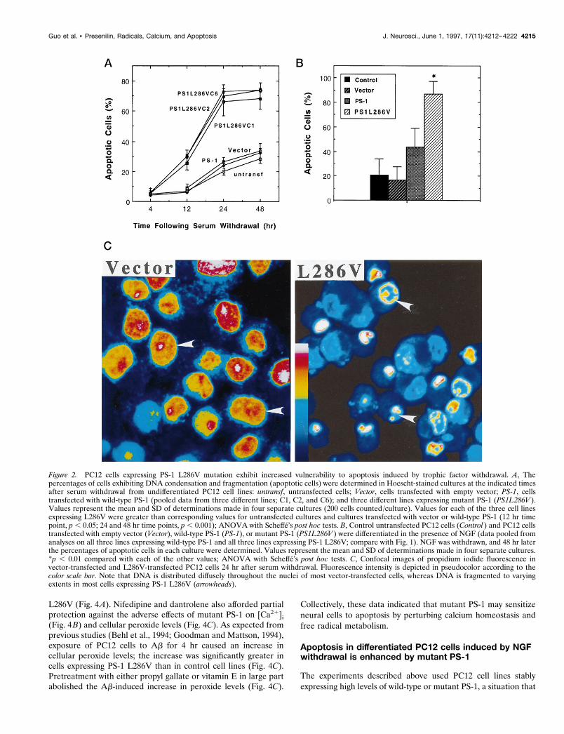
To determine whether the expression of PS-1 L286V affectedcell death after trophic factor withdrawal, we deprived variouslines of undifferentiated PC12 cells of serum, an insult previouslyshown to induce apoptosis (Bastitatou and Greene, 1991; Ruken-stein et al., 1991). In untransfected and vector-transfected PC12cells there was a progressive appearance of cells with apoptoticnuclei that occurred between 12 and 48 hr after serum withdrawal(Fig. 2A). In PC12 cells expressing PS-1 L286V there was adramatic increase in the numbers of apoptotic cells present at 12,
Guo et al. • Presenilin, Radicals, Calcium, and Apoptosis J. Neurosci., June 1, 1997, 17(11):4212–4222 4213

24, and 48 hr after serum withdrawal (Fig. 2A,C). In another setof experiments cells were differentiated into a neuron-like phe-notype by chronic exposure to NGF, and then the NGF was
withdrawn. Apoptosis induced by NGF withdrawal in cell linesexpressing mutant PS-1 was significantly greater than in untrans-fected cells, vector-transfected cells, and cells overexpressing wild-type PS-1 (Fig. 2B). NGF withdrawal induced apoptosis in ;20%of cells in untransfected and vector-transfected cell lines and in;80% of the cells in lines expressing mutant PS-1. The level ofapoptosis after NGF withdrawal in cells overexpressing wild-typePS-1 was somewhat higher than that in vector-transfected anduntransfected lines, although the difference did not reach statis-tical significance (Fig. 2B).
Previous studies showed that Ab can induce apoptosis in cul-tured primary neurons (Loo et al., 1993) and PC12 cells(Gschwind and Huber, 1995). Exposure of undifferentiated PC12cells to Ab for 24 hr induced apoptotic nuclear changes, and thepercentage of cells undergoing apoptosis was significantly greaterin cells expressing PS-1 L286V than in untransfected cells, vector-transfected cells, and cells overexpressing wild-type PS-1 (Fig.3A). Nuclear condensation and fragmentation induced by Ab
(Fig. 3A) and serum withdrawal (data not shown) were preventedby the protein synthesis inhibitor cycloheximide, consistent withan apoptotic mechanism of cell death. It was reported previouslythat Ab causes a relatively rapid (minutes to hours) impairment ofmitochondrial function as measured by MTT reduction that oc-curs very early in the apoptotic process (Shearman et al., 1994;Kruman et al., 1996). Exposure of PC12 cells to Ab resulted in adecrease in levels of MTT reduction in all cell lines examined, andthe magnitude of the decrease in MTT reduction was significantlygreater in each line expressing PS-1 L286V than in untransfectedcells, cells transfected with empty vector, and lines overexpressingwild-type PS-1 (Fig. 3B).
Proapoptotic action of PS-1 mutation involvesdisruption of calcium homeostasis and induction ofoxidative stressTo test the hypothesis that PS-1 mutations promote cell death byincreasing oxidative stress, we determined whether antioxidantswould protect cells against death induced by Ab and measuredlevels of peroxides in the different cell lines after exposure to Ab.When cultures were pretreated with the antioxidants propyl gal-late or vitamin E before exposure to Ab, cell death 24 hr later wasreduced significantly, and the death-enhancing effect of L286Vwas prevented (Fig. 4A). In light of previous data implicatingdisruption of cellular calcium homeostasis in the mechanism ofAb cytotoxicity (Mattson et al., 1992, 1993a; Mark et al., 1995)and apoptosis (Takei and Endo, 1994; Ciutat et al., 1995), wedetermined whether cells expressing PS-1 L286V exhibit in-creased sensitivity to Ab-induced elevation of [Ca21]i andwhether agents that suppress calcium influx would protect cellsagainst the adverse effects of PS-1 L286V. Ab induced an increasein [Ca21]i during a 4 hr exposure period; the elevation of [Ca21]i
was significantly greater in cells expressing PS-1 L286V, as com-pared with control cell lines (Fig. 4B). Ab caused [Ca21]i toincrease to 150–170 nM in control cell lines and to .300 nM inL286V-expressing cells. Cell death induced by Ab, and the death-enhancing effect of PS-1 L286V was attenuated significantly incultures pretreated with nifedipine, a blocker of L-type voltage-dependent calcium channels (Fig. 4A). Dantrolene, an inhibitorof calcium release from ER stores, also in large part preventedAb-induced cell death in cells expressing L286V, suggesting a rolefor altered calcium release in the proapoptotic action of PS-1
Figure 1. Expression of wild-type and mutant PS-1 in PC12 cells. A,Western blot showing PS-1 protein in untransfected and vector-transfected PC12 cells and in three different lines of cells expressingwild-type (PS-1) and mutant (PS1L286V ) PS-1. Equivalent amounts ofprotein (50 mg/ lane) from cell homogenates of the indicated cell lines(untransfected cells, vector-transfected cells, PS-1, and PS1L286V)were separated by SDS-PAGE, transferred to nitrocellulose, andprobed with PS-1 antibody. Similar to results of other investigators(Thinakaran et al., 1996), in addition to recognizing full-length PS-1(46 kDa band), our anti-loop PS-1 antibody also recognized presump-tive proteolytic products of PS-1 ;32 and 19 kDa. B, Densitometricanalysis of relative levels of PS-1 protein in PC12 cell lines stablyexpressing wild-type (PS-1) and mutant (PS1L286V ) PS-1. Note thatbasal PS-1 levels are relatively low in PC12 cells and that levels ofexpression of wild-type and mutant PS-1 were similar among the linesshown.
4214 J. Neurosci., June 1, 1997, 17(11):4212–4222 Guo et al. • Presenilin, Radicals, Calcium, and Apoptosis
L286V (Fig. 4A). Nifedipine and dantrolene also afforded partialprotection against the adverse effects of mutant PS-1 on [Ca21]i
(Fig. 4B) and cellular peroxide levels (Fig. 4C). As expected fromprevious studies (Behl et al., 1994; Goodman and Mattson, 1994),exposure of PC12 cells to Ab for 4 hr caused an increase incellular peroxide levels; the increase was significantly greater incells expressing PS-1 L286V than in control cell lines (Fig. 4C).Pretreatment with either propyl gallate or vitamin E in large partabolished the Ab-induced increase in peroxide levels (Fig. 4C).
Collectively, these data indicated that mutant PS-1 may sensitizeneural cells to apoptosis by perturbing calcium homeostasis andfree radical metabolism.
Apoptosis in differentiated PC12 cells induced by NGFwithdrawal is enhanced by mutant PS-1
The experiments described above used PC12 cell lines stablyexpressing high levels of wild-type or mutant PS-1, a situation that
Figure 2. PC12 cells expressing PS-1 L286V mutation exhibit increased vulnerability to apoptosis induced by trophic factor withdrawal. A, Thepercentages of cells exhibiting DNA condensation and fragmentation (apoptotic cells) were determined in Hoescht-stained cultures at the indicated timesafter serum withdrawal from undifferentiated PC12 cell lines: untransf, untransfected cells; Vector, cells transfected with empty vector; PS-1, cellstransfected with wild-type PS-1 (pooled data from three different lines; C1, C2, and C6); and three different lines expressing mutant PS-1 (PS1L286V ).Values represent the mean and SD of determinations made in four separate cultures (200 cells counted /culture). Values for each of the three cell linesexpressing L286V were greater than corresponding values for untransfected cultures and cultures transfected with vector or wild-type PS-1 (12 hr timepoint, p , 0.05; 24 and 48 hr time points, p , 0.001); ANOVA with Scheffe’s post hoc tests. B, Control untransfected PC12 cells (Control ) and PC12 cellstransfected with empty vector (Vector), wild-type PS-1 (PS-1), or mutant PS-1 (PS1L286V ) were differentiated in the presence of NGF (data pooled fromanalyses on all three lines expressing wild-type PS-1 and all three lines expressing PS-1 L286V; compare with Fig. 1). NGF was withdrawn, and 48 hr laterthe percentages of apoptotic cells in each culture were determined. Values represent the mean and SD of determinations made in four separate cultures.*p , 0.01 compared with each of the other values; ANOVA with Scheffe’s post hoc tests. C, Confocal images of propidium iodide fluorescence invector-transfected and L286V-transfected PC12 cells 24 hr after serum withdrawal. Fluorescence intensity is depicted in pseudocolor according to thecolor scale bar. Note that DNA is distributed diffusely throughout the nuclei of most vector-transfected cells, whereas DNA is fragmented to varyingextents in most cells expressing PS-1 L286V (arrowheads).
Guo et al. • Presenilin, Radicals, Calcium, and Apoptosis J. Neurosci., June 1, 1997, 17(11):4212–4222 4215

could perturb metabolic pathways nonspecifically in the cells.Moreover, because wild-type and mutant PS-1 were expressedthroughout the period of NGF-induced cell differentiation, wecould not rule out the possibility that mutant PS-1 affects thedifferentiation process. To examine further the proapoptotic ac-tions of PS-1 mutations, we therefore established PC12 linesexpressing wild-type and mutant PS-1 under the control of atetracycline-suppressible (Tet-off) promoter. For these experi-ments cells were maintained in the presence of tetracycline duringthe period of differentiation with NGF. After differentiation and48 hr before NGF withdrawal, tetracycline concentration wasreduced to induce PS-1 wild-type or mutant expression. As ex-pected, levels of PS-1 expression increased with decreasing con-centrations of tetracycline in the culture medium (Fig. 5A); sub-sequent experiments were performed in cultures induced toexpress wild-type or mutant PS-1 at comparable levels (Fig.5B). Withdrawal of NGF from differentiated untransfected,vector-transfected, and Tet-off-transfected PC12 cells resultedin apoptosis of ;40% of the cells during a 48 hr period (Fig.6 A). In contrast, apoptosis did not occur after NGF withdrawalin PC12 cells expressing Bcl-2. NGF withdrawal-induced apo-ptosis was enhanced significantly in PC12 cells expressing mu-tant PS-1 L286V (Fig. 6C), as compared with cells expressingwild-type PS-1 (Fig. 6 B). Cells expressing PS-1 L286V alsoexhibited a modest increase in basal levels of apoptosis in thepresence of NGF. PC12 cells expressing Bcl-2 in combinationwith wild-type or mutant PS-1 were completely resistant toapoptosis induced by NGF withdrawal (Fig. 6C). Collectively,these data indicate that mutant PS-1 possesses an adverseproperty not present in wild-type PS-1 that sensitizes neuronsto apoptosis.
Enhanced oxidative stress after NGF withdrawal andperturbed calcium homeostasis in differentiated PC12cells expressing mutant PS-1Withdrawal of NGF from differentiated untransfected and vector-transfected PC12 cells resulted in an increase in levels of cellularperoxides that occurred within 3 hr (Fig. 7A). An increase incellular peroxides also occurred after NGF withdrawal from PC12cells expressing Bcl-2, suggesting that the antiapoptotic action ofBcl-2 occurred downstream of the oxidative stress (cf. Greenlundet al., 1995). NGF withdrawal-induced peroxide accumulation wasenhanced greatly in PC12 cells expressing the mutant PS-1 L286V,as compared with cells expressing wild-type PS-1 (Fig. 7B). Basallevels of peroxides in cells expressing PS-1 L286V were somewhathigher than in control cells, although the difference did not reachstatistical significance. PC12 cells expressing Bcl-2 in combinationwith mutant PS-1 exhibited peroxide levels after NGF withdrawalthat were lower than in cells lacking Bcl-2, but the difference didnot reach statistical significance (Fig. 7C).
PS-1 is localized to the ER, and a recent study showed thatPC12 cells expressing mutant PS-1 exhibit altered ER calciumregulation (Guo et al., 1996). We therefore examined calciumresponses to thapsigargin, an inhibitor of the ER Ca21–ATPasein differentiated PC12 cells expressing wild-type or mutantPS-1, with or without Bcl-2. Rest [Ca21]i was ;60 nM in controlcells and was elevated to 80 –110 nM in cells expressing PS-1L286V (Fig. 8). In PC12 cells not expressing PS-1, thapsigargincaused an increase in [Ca21]i to 160 –180 nM, and the responsewas attenuated by ;50% in cells expressing Bcl-2 (Fig. 8 A).PC12 cells expressing PS-1 L286V exhibited a markedly en-
Figure 3. Mutant PS-1 increases PC12 cell vulnerability to apoptosisand mitochondrial dysfunction induced by amyloid b-peptide. A, Theindicated cell lines were exposed to vehicle (Vehicle), 50 mM Ab (Ab),or 10 mM cycloheximide plus 50 mM Ab (CHX 1 Ab) for 24 hr. Thencells were stained with Hoescht dye, and the percentages of cellsexhibiting DNA condensation and fragmentation were determined.Values represent the mean and SD of determinations made in fourseparate cultures (data pooled from analyses on all three lines express-ing wild-type PS-1 and all three lines expressing PS-1 L286V; comparewith Fig. 1). For all cell lines the values for cells exposed to Ab weresignificantly greater than values for vehicle or CHX plus Ab-treatedcell lines ( p , 0.01). *p , 0.01 compared with each of the other celllines exposed to Ab; ANOVA with Scheffe’s post hoc tests. B, Parallelcultures of untransfected control cells (Untransf ), vector-transfectedcells (Vector), three lines of cells transfected with wild-type PS-1 (PS-1;pooled data), and three lines of mutant PS-1 cells (PS1L286V; pooleddata) were exposed for 4 hr to 50 mM Ab, and relative levels of MTTreduction (a measure of mitochondrial function) were quantified. Val-ues are the mean and SD of determinations made in four separatecultures and are expressed as a percentage of vehicle-treated control(vector-transfected) cells (data pooled from analyses on all three linesexpressing wild-type PS-1 and all three lines expressing PS-1 L286V;compare with Fig. 1). There were no differences in basal levels of MTTreduction among the various control, wild-type PS-1-expressing, andmutant PS-1-expressing lines (data not shown). *p , 0.01 comparedwith corresponding values for untransfected, vector-transfected, andWT PS-1-transfected lines exposed to Ab; ANOVA with Scheffe’s posthoc tests.
4216 J. Neurosci., June 1, 1997, 17(11):4212–4222 Guo et al. • Presenilin, Radicals, Calcium, and Apoptosis

Figure 4. PC12 cells expressing PS-1 L286V mutation exhibit increasedlevels of oxidative stress and intracellular calcium after exposure to Ab:attenuation by antioxidants and blockers of calcium influx and releasefrom intracellular stores. A, Cultures were pretreated for 24 hr with 50 mMvitamin E (VitE) or for 2 hr with 5 mM propyl gallate (PG), 1 mM nifedipine
Figure 5. Controlled expression of PS-1 in PC12 cells with the use of atetracyline-responsive transactivator. A, A PC12 cell line expressing the“Tet-off” construct was stably transfected with a pTRE-derived plasmidexpressing PS-1 L286V gene. Cells were incubated for 48 hr in thepresence of 2.0, 0.004, 0.002, and 0 mg/ml tetracycline (lanes 1–4, respec-tively). Cell proteins were separated by SDS-PAGE (100 mg/ lane), trans-ferred to a nitrocellulose sheet, and immunoreacted with PS-1 antibody.Note that, as the concentration of tetracycline was decreased, the levels ofmutant PS-1 expression increased. B, Western blot showing that in theabsence of tetracycline a double-stable PS-1 L286V cell line (C1) and adouble-stable PS-1 cell line (C3) show significantly higher levels of PS-1expression than do vector-transfected or untransfected cell lines.
4
(Nifedipine), or 1 mM dantrolene (DTL). Then cultures were exposed to 50 mMAb for 24 hr, and the medium was removed for LDH assay (cells exposed toAb first undergo apoptosis, followed by secondary necrosis, the latter beingdetected by LDH release assay). Values are expressed as a percentage of themaximal LDH release (mean and SD of 6–8 cultures); maximal LDH releasewas determined in parallel cultures (of each cell line) subjected to freeze–thaw. Values for each of the Ab-treated cell lines expressing PS-1 L286Vwere significantly greater than each of the values for control (untransfected)and vector-transfected cell lines ( p , 0.01) and for each of the values in PS-1L286V cultures pretreated with vitamin E, propyl gallate, nifedipine, ordantrolene ( p , 0.01 in each case); ANOVA with Scheffe’s post hoc tests. B,C, Cultures were pretreated with antioxidants or Ca21 flux blockers asdescribed for A and then exposed to vehicle or Ab for 4 hr. Then the relative[Ca21]i (fura-2 imaging) (B) and levels of peroxides (DCF Fluorescence) (C)in individual cells were quantified. Values are the mean and SD of determi-nations made in three to four cultures (15–25 cells for [Ca21]i measurementsand 40–60 cells /culture for DCF measurements). Values for each of theAb-treated cell lines expressing L286V were significantly greater than each ofthe values in the vehicle-treated cultures ( p , 0.01), each of the values in thecultures pretreated with vitamin E or propyl gallate ( p , 0.01), and each ofthe values for L286V cells in the cultures pretreated with nifedipine ordantrolene ( p , 0.05); ANOVA with Scheffe’s post hoc tests.
Guo et al. • Presenilin, Radicals, Calcium, and Apoptosis J. Neurosci., June 1, 1997, 17(11):4212–4222 4217

hanced peak [Ca21]i response to thapsigargin, with levels risingto ;400 nM (Fig. 8C). The enhanced [Ca21]i response tothapsigargin was abolished completely in PC12 cells expressingBcl-2 (Fig. 8C). Taken together with the data above showingthat dantrolene protects PC12 cells against the proapoptoticactions of PS-1 L286V, these findings suggest that altered ERcalcium regulation is involved mechanistically in the apoptoticaction of mutant PS-1.
DISCUSSIONPrevious studies showed that both Ab (Rabizadeh et al., 1994;Gschwind and Huber, 1995) and trophic factor withdrawal (Bas-titatou and Greene, 1991; Rukenstein et al., 1991) induce apo-ptosis in PC12 cells. Similarly, Ab (Forloni et al., 1993; Loo et al.,1993) and trophic factor deprivation (Prehn et al., 1994) induceapoptosis in neurons in primary cultures established from brainregions (e.g., hippocampus, neocortex, and basal forebrain) that
Figure 7. Oxidative stress induced by NGF withdrawal is enhanced in PC12 cells expressing mutant PS-1. Cultures of differentiated PC12 cells wereincubated for 3 hr in serum-free medium containing or lacking NGF, and levels of cellular peroxides were quantified by confocal laser scanningmicroscope image analysis of DCF fluorescence. Values are the mean and SEM of determinations made in at least four separate cultures. A, Analysesin various control PC12 cell lines: WT, untransfected wild-type cells; puro, vector-transfected cells; Tet-off, cells transfected with the Tet-off plasmid; puroTet-off, cells doubly transfected with empty vector and Tet-off plasmid; bcl-2 puro, cells expressing Bcl-2. Note that NGF withdrawal induced a similar levelof peroxide accumulation in all control lines and in cells overexpressing Bcl-2. Each value for NGF2 cultures was significantly greater than thecorresponding value for NGF1 cells ( p , 0.01). B, Bcl-2 does not prevent NGF withdrawal-induced accumulation of peroxides in PC12 cellsoverexpressing wild-type PS-1. Two lines of control cells expressing wild-type PS-1 (C3 and C7 ) and two different lines of cells expressing both Bcl-2 andPS-1 (C11 and C13) were analyzed. Each value for NGF2 cultures was significantly greater than the corresponding value for NGF1 cells ( p , 0.01). C,Mutant PS-1 enhances accumulation of peroxides in PC12 cells deprived of NGF. Two lines of control cells expressing PS-1 L286V and two lines of cellsexpressing both Bcl-2 and PS-1 L286V were analyzed. *p , 0.05 compared with corresponding values for each line expressing wild-type PS-1 (B)and p , 0.01 compared with corresponding values for cells maintained in the presence of NGF; ANOVA with Scheffe’s post hoc tests for pair-wisecomparisons).
Figure 6. Mutant PS-1 increases the vulnerability of differentiated PC12 cells to NGF withdrawal-induced apoptosis. Cultures of differentiated PC12 cellswere incubated for 48 hr in serum-free medium containing or lacking NGF, and the percentage of cells exhibiting nuclear condensation and fragmentationwas quantified. Values are the mean and SEM of determinations made in at least four separate cultures. A, Analyses in various control PC12 cell lines:WT, untransfected wild-type cells; puro, vector-transfected cells (pBabe-puro vector used for Bcl-2 expression); Tet-off, cells transfected with the Tet-offplasmid; puro Tet-off, cells doubly transfected with pBabe-puro vector and Tet-off plasmid; bcl-2 puro, cells expressing Bcl-2. Note that NGF withdrawalinduced a similar level of apoptosis in all control lines, whereas cells expressing Bcl-2 were resistant to NGF withdrawal-induced apoptosis. *p , 0.01compared with corresponding values for NGF1 cultures and the value for Bcl-2 NGF2 cells. B, Bcl-2 protects PC12 cells overexpressing wild-type PS-1against NGF withdrawal-induced apoptosis. Two lines of control cells expressing wild-type PS-1 (C3 and C7 ) and two different lines of cells expressingboth Bcl-2 and PS-1 (C11 and C13) were analyzed. *p , 0.01 compared with corresponding values for NGF1 cells and compared with the value for theNGF2 line expressing Bcl-2. C, Mutant PS-1 enhances vulnerability of PC12 cells to apoptosis induced by NGF withdrawal: protection by Bcl-2. Two linesof control cells expressing PS-1 L286V and two lines of cells expressing both Bcl-2 and PS-1 L286V were analyzed. *p , 0.05 compared with correspondingvalues for each line expressing wild-type PS-1 (B), p , 0.01 compared with corresponding values for cells maintained in the presence of NGF, and p ,0.01 compared with NGF2 lines coexpressing Bcl-2; ANOVA with Scheffe’s post hoc tests for pair-wise comparisons.
4218 J. Neurosci., June 1, 1997, 17(11):4212–4222 Guo et al. • Presenilin, Radicals, Calcium, and Apoptosis

are affected in AD. We found that both undifferentiated anddifferentiated PC12 cells expressing PS-1 L286V were extremelysensitive to apoptotic cell death when compared with variouscontrol cell lines. These findings suggest that the mutated PS-1protein possesses an adverse proapoptotic property. Overexpres-sion of wild-type PS-1, at levels similar to or greater than mutantPS-1 levels, did not result in increased vulnerability of PC12 cellsto apoptosis, indicating that the proapoptotic action of PS-1L286V was not simply the consequence of increased levels of PS-1protein. Although the specific nature of that adverse property ofthe PS-1 mutation was not established in the present study, thedata suggest an action on systems that regulate free radical me-tabolism and/or calcium homeostasis. Thus, levels of cellularperoxides induced by Ab were increased greatly in cells expressingthe PS-1 mutation, as compared with control lines, and twodifferent antioxidants (vitamin E and propyl gallate) protectedPC12 cells against cell death induced by Ab. The antioxidants alsosuppressed Ab-induced increases in intracellular peroxide andcalcium levels, consistent with previous data suggesting that themechanism of Ab neurotoxicity involves membrane lipid peroxi-dation and impairment of membrane ion transport systems andcalcium influx (Mattson et al., 1992; Behl et al., 1994; Goodmanand Mattson, 1994; Mark et al., 1995, 1997a,b).
Previous studies of mechanisms of neuron death induced bytrophic factor withdrawal and Ab have implicated reactive oxygenspecies (ROS) (Hockenbery et al., 1993; Kane et al., 1993; Green-lund et al., 1995). We found that levels of oxidative stress andapoptosis after NGF withdrawal from differentiated PC12 cellswere enhanced in cells expressing mutant PS-1, but not in cellsoverexpressing wild-type PS-1. These effects of mutant PS-1 werenot attributable to changes that occurred during the process ofcellular differentiation, because we allowed the cells to differen-tiate before induction of PS-1 expression by using the Tet-offsystem. Considerable data indicate that levels of oxidative stressare increased in AD brain, particularly in the environment ofneuritic plaques and in neurofibrillary tangles (for review, see
Benzi and Moretti, 1995; Smith et al., 1995); levels of oxidativestress also are increased in the brain during normal aging (Stadt-man, 1992). The present findings suggest the possibility that PS-1mutations may promote oxidative stress and thereby sensitizeneurons to decrements in trophic factor support and increasedaccumulations of Ab that occur in the aging brain. The data alsomay provide at least a partial explanation for the increased pro-duction of Ab documented in blood and other tissues from humancarriers of PS-1 mutations (Scheuner et al., 1996), because studieshave shown that manipulations that promote metabolic stress andincrease [Ca21]i in cultured neurons can alter proteolytic process-ing of b-amyloid precursor protein (bAPP) in cultured cells infavor of increased Ab production (Gabuzda et al., 1994; Quer-furth and Selkoe, 1994).
PS-1 seems to be localized to the ER in several cell types,including neurons (Guo et al., 1996; Kovacs et al., 1996; Walter etal., 1996). Calcium imaging studies of cultured PC12 expressingPS-1 L286V have shown that this mutation alters calcium releasefrom ER stores such that calcium responses to agonists thatactivate the IP3 pathway (e.g., muscarinic cholinergic agonists andbradykinin) are enhanced greatly (Guo et al., 1996). The per-turbed calcium homeostasis observed in PC12 cells expressingmutant PS-1 is consistent with reports that calcium signaling isaltered in cultured fibroblasts taken from carriers of PS-1 muta-tions (McCoy et al., 1993; Ito et al., 1994). Our data suggest thatdisruption of calcium homeostasis by mutant PS-1 could be linkedmechanistically to its proapoptotic action because dantrolene, anagent that blocks calcium release from ER, protected cells againstthe death-promoting effect of the PS-1 mutation. Recent findingsin studies of non-neuronal cells have linked ER calcium regula-tion to apoptosis. For example, Lam et al. (1993) showed thatglucocorticoids induce release of calcium from ER, which iscorrelated with subsequent DNA fragmentation and apoptosis,and Khan et al. (1996) provided evidence that lymphocyte apo-ptosis is mediated by increased expression of IP3 receptors. Wefound that thapsigargin-induced increases of [Ca21]i were en-hanced in PC12 cells expressing mutant PS-1 and that Bcl-2
Figure 8. Elevations of [Ca21]i induced by thapsigargin are enhanced significantly in PC12 cells expressing mutant PS-1: attenuation by Bcl-2. PC12cells were incubated in serum-free medium and basal [Ca21]i (Tpg2), and the peak [Ca21]i after exposure to 1 mM thapsigargin (Tpg1) was quantified (cf.Guo et al., 1996). Values are the mean and SEM of determinations made in at least four separate cultures (15–20 cells /culture). A, Analyses in variouscontrol PC12 cell lines: WT, untransfected wild-type cells; puro, vector-transfected cells (vector for Bcl-2-expressing line); Tet-off, cells transfected withthe Tet-off plasmid; puro Tet-off, cells doubly transfected with pBabe-puro and Tet-off plasmid; bcl-2 puro, cells expressing Bcl-2. *p , 0.01 compared withcorresponding values for Tpg2 cultures, and p , 0.05 compared with the Tpg1 value in cells expressing Bcl-2. B, The [Ca21]i response to thapsigarginin cells expressing wild-type PS-1 is attenuated in cells coexpressing Bcl-2. *p , 0.01 compared with corresponding values for Tpg2, and p , 0.05compared with Tpg1 values in cells expressing Bcl-2. C, Mutant PS-1 enhances [Ca21]i responses to thapsigargin: attenuation by Bcl-2. *p , 0.001compared with corresponding values for Tpg2, p , 0.01 compared with Tpg1 values in cells expressing Bcl-2, and p , 0.01 compared with Tpg1– PS1Tet-off values (B); ANOVA with Scheffe’s post hoc tests for pair-wise comparisons.
Guo et al. • Presenilin, Radicals, Calcium, and Apoptosis J. Neurosci., June 1, 1997, 17(11):4212–4222 4219
prevented the enhanced response in cells expressing mutant PS-1.Lam et al. (1994) showed that Bcl-2 protects lymphoma cellsagainst apoptosis induced by thapsigargin and suppresses calciumrelease from ER. The localization of mutant PS-1 to ER (Guo etal., 1996) suggests that its effects on ER calcium homeostasis andapoptosis may be linked mechanistically, a possibility supportedby our data. The ability of dantrolene to suppress the increasedperoxide accumulation induced by Ab in PC12 cells expressingPS-1 L286V suggests that calcium release from ER contributes tothe enhanced oxidative stress associated with mutant PS-1 expres-sion. However, in addition to calcium release from ER, calciuminflux through voltage-dependent plasma membrane channelsalso may be involved, because nifedipine protected cells againstAb toxicity and suppressed elevation of [Ca21]i and peroxideaccumulation induced by Ab. These data are consistent withprevious studies of primary neurons and PC12 cells showing thatremoval of extracellular calcium or treatment with nifedipineattenuates Ab toxicity (Mattson et al., 1993a; Weiss et al., 1994).
The observation that both antioxidants and calcium channelblockers protected neurons against the adverse effects of the PS-1mutation are consistent with a scenario in which Ab induces avicious cycle in which oxidative stress disrupts ion homeostasis,which, in turn, promotes further oxidative stress. There is nowabundant evidence to support the involvement of such recipro-cating cytotoxic cascades in many different neurodegenerativeconditions (Mattson et al., 1992; Zhang et al., 1993; Goodman etal., 1996). Thus, although both the normal function of PS-1 andthe exact alteration that results from PS-1 mutations are un-known, our data indicate that PS-1 mutations promote neurode-generative apoptotic cascades involving perturbed ion homeosta-sis and oxidative stress.
Recent data from studies of brain tissue, blood, and culturedfibroblasts from carriers of PS mutations and studies of trans-fected cell lines indicate that cells expressing PS mutations pro-duce greater than normal levels of Ab1–42 (Borchelt et al., 1996;Lemere et al., 1996; Scheuner et al., 1996). In addition, studies oftransgenic mice that express mutant PS-1 suggest that levels ofAb1–42 are increased in brain tissue (Duff et al., 1996). The latterdata suggest that PS-1 mutations may cause early onset AD byaltering bAPP processing in ways that lead to increased Abproduction. The increased vulnerability of neuronal cells express-ing PS-1 L286V to Ab toxicity and trophic factor withdrawaldocumented in the present study is unlikely to result from in-creased Ab production because PC12 cells are a rat cell line and,in contrast to human Ab, rat Ab is neither amyloidogenic norneurotoxic (Otvos et al., 1993). However, it is conceivable thataltered processing of bAPP could reduce levels of neuroprotectivesecreted forms of APP, which have been shown to protect neuronsagainst oxidative apoptotic insults, including Ab toxicity and glu-cose withdrawal (Mattson et al., 1993b; Goodman and Mattson,1994; Furukawa et al., 1996).
Collectively, our data suggest that mutant PS-1 protein pos-sesses an adverse proapoptotic property. Wolozin et al. (1996)recently reported that PC12 cells overexpressing wild-type PS-2exhibit increased apoptosis after trophic factor withdrawal andthat a PS-2 mutant enhanced basal levels of apoptosis. Deng et al.(1996) reported that overexpression of wild-type PS-2 increasedvulnerability of PC12 cells to apoptosis induced by staurosporineor hydrogen peroxide. ALG-3, the mouse homolog of PS-2, wasshown to modulate apoptosis in T lymphocytes (Vito et al.,1996), although in the latter case ALG-3 prevented apoptosis.We observed neither increased nor reduced apoptosis in PC12
cells overexpressing wild-type PS-1, suggesting that PS-1 doesnot function directly in an apoptotic pathway and that mutantPS-1 acquires a novel adverse property. However, we cannotrule out the possibility that mutant PS-1 interferes with a normaltrophic property of endogenous PS-1 in a loss-of-functionscenario.
REFERENCESBastitatou A, Greene LA (1991) Aurintricarboxylic acid rescues PC12
cells and sympathetic neurons from cell death caused by nerve growthfactor deprivation: correlation with suppression of endonuclease activ-ity. J Cell Biol 115:461–471.
Behl C, Davis JB, Lesley R, Schubert D (1994) Hydrogen peroxidemediates amyloid b-protein toxicity. Cell 77:817–827.
Benzi G, Moretti A (1995) Are reactive oxygen species involved in Alz-heimer’s disease? Neurobiol Aging 16:661–674.
Black MM, Greene LA (1982) Changes in the colchicine susceptibility ofmicrotubules associated with neurite outgrowth: studies with nervegrowth factor-responsive PC12 pheochromocytoma cells. J Cell Biol95:379–386.
Borchelt DR, Thinakaran G, Eckman CB, Lee MK, Davenport F, Rato-vitsky T, Prada C-M, Kim G, Seekins S, Yager D, Slunt HH, Wang R,Seeger M, Levey AI, Gandy SE, Copeland NG, Jenkins NA, Price DL,Younkin SG, Sisodia SS (1996) Familial Alzheimer’s disease-linkedpresenilin-1 variants elevate Ab1–42/1–40 ratio in vitro and in vivo.Neuron 17:1005–1013.
Bredesen DE (1995) Neural apoptosis. Ann Neurol 38:839–851.Bruce AJ, Malfroy B, Baudry M (1996) b-amyloid toxicity in organotypic
cultures: protection by EUK-8, a synthetic catalytic free radical scaven-ger. Proc Natl Acad Sci USA 88:3633–3636.
Ciutat D, Esquerda JE, Caldero J (1995) Evidence for calcium regulationof spinal cord motoneuron death in the chick embryo in vivo. Dev BrainRes 86:167–179.
Cook DB, Sung JC, Golde TE, Felsenstein KM, Wojczyk BS, Tanzi RE,Trojanowski JQ, Lee VMY, Doms RW (1996) Expression and analysisof presenilin-1 in a human neuronal system: localization in cell bodiesand dendrites. Proc Natl Acad Sci USA 93:9223–9228.
Cribbs DH, Chen L, Bendle SM, La Ferla FM (1996) Widespread neu-ronal expression of the presenilin-1 early-onset Alzheimer’s disease inthe murine brain. Am J Pathol 148:1797–1806.
Deng G, Pike CJ, Cotman CW (1996) Alzheimer-associated presenilin-2confers increased sensitivity to apoptosis in PC12 cells. FEBS Lett397:50–54.
Doan A, Thinakaran G, Borchelt DR, Slunt HH, Ratovitsky T, PodlisnyM, Selkoe DJ, Seeger M, Gandy SE, Price DL, Sisodia SS (1996)Protein topology of presenilin-1. Neuron 17:1023–1030.
Duff K, Eckman C, Zehr C, Yu X, Prada C-M, Perez-Tur J, Hutton M,Buee L, Harigaya Y, Yager D, Morgan D, Gordon MN, Holcomb L,Refolo L, Zenk B, Hardy J, Younkin S (1996) Increased amyloid-b42(43) in brains of mice expressing mutant presenilin-1. Nature383:710–713.
Elder GA, Tezapsidis N, Carter J, Shioi J, Bouras C, Li D, Johnston JM,Efthimiopoulos S, Friedrich Jr VL, Robakis NK (1996) Identificationand neuron-specific expression of the S182/presenilin-1 protein in hu-man and rodent brains. J Neurosci Res 45:308–320.
Forloni G, Chiesa R, Smiroldo S, Verga L (1993) Apoptosis-mediatedneurotoxicity induced by chronic application of b-amyloid fragment25–35. NeuroReport 4:523–526.
Fukuyama R, Wadhwani KC, Galdzicki Z, Rapoport SI, Ehrenstein G(1994) b-Amyloid polypeptide increases calcium uptake in PC12 cells:a possible mechanism for its cellular toxicity in Alzheimer’s disease.Brain Res 667:269–272.
Furukawa K, Sopher B, Rydel RE, Begley JG, Martin GM, Mattson MP(1996) Increased activity-regulating and neuroprotective efficacy ofa-secretase-derived secreted APP is conferred by a C-terminal heparin-binding domain. J Neurochem 67:1882–1896.
Gabuzda D, Busciglio J, Chen L, Matsudaira P, Yankner BA (1994)Inhibition of energy metabolism alters the processing of amyloid pre-cursor protein and induces a potentially amyloidogenic derivative. J BiolChem 269:13623–13628.
Games D, Adams D, Alessandrinl R, Barbour R, Berthelette P, BlackwellC, Carr T, Clemens J, Donaldson T, Gillespie F, Guido T, Hagoplan S,
4220 J. Neurosci., June 1, 1997, 17(11):4212–4222 Guo et al. • Presenilin, Radicals, Calcium, and Apoptosis

Johnson-Wood K, Khan K, Lee M, Lelbowitz E, McConlogue S,Montoya-Zavala M, Mucke L, Paganini L, Penniman E, Power M,Schenk D, Seubert P, Snyder B, Soriano F, Tan H, Vitale J, WadsworthS, Wolozin B, Zhao J (1995) Alzheimer-type neuropathology in trans-genic mice overexpressing V717F b-amyloid precursor protein. Nature373:523–527.
Giannakopoulos P, Bouras C, Kovari E, Shioi J, Tezapsidis N, Hof PR,Robakis NK (1997) Presenilin-1 immunoreactive neurons are pre-served in late-onset Alzheimer’s disease. Am J Pathol 150:429–436.
Goodman Y, Mattson MP (1994) Secreted forms of b-amyloid precursorprotein protect hippocampal neurons against amyloid b-peptide-induceoxidative injury. Exp Neurol 128:1–12.
Goodman Y, Bruce AJ, Cheng B, Mattson MP (1996) Estrogens atten-uate and corticosterone exacerbates excitotoxicity, oxidative injury, andamyloid b-peptide toxicity in hippocampal neurons. J Neurochem66:1836–1844.
Greenlund LJ, Deckwerth TL, Johnson EM (1995) Superoxide dis-mutase delays neuronal apoptosis: a role for reactive oxygen species inprogrammed neuronal death. Neuron 14:303–315.
Gschwind M, Huber G (1995) Apoptotic cell death induced by b-amyloid1–42 peptide is cell type-dependent. J Neurochem 65:292–300.
Guo Q, Furukawa K, Sopher BL, Pham DG, Robinson N, Martin GM,Mattson MP (1996) Alzheimer’s PS-1 mutation perturbs calcium ho-meostasis and sensitizes PC12 cells to death induced by amyloidb-peptide. NeuroReport 8:379–383.
Hockenbery D, Oltvai ZN, Yin XM, Milliman CL, Korsmeyer SH (1993)Bcl-2 functions in an antioxidant pathway to prevent apoptosis. Cell75:241–251.
Hsaio K, Chapman P, Nilsen S, Eckman C, Harigaya Y, Younkin S, YangF, Cole G (1996) Correlative memory deficits, Ab elevation, and amy-loid plaques in transgenic mice. Science 274:99–103.
Ito E, Oka K, Etcheberrigaray R, Nelson TJ, McPhie DL, Tofel-Grehl B,Gibson GE, Alkon DL (1994) Internal Ca21 mobilization is altered infibroblasts from patients with Alzheimer disease. Proc Natl Acad SciUSA 91:534–538.
Kane DJ, Sarafian TA, Anton R, Hahn H, Gralla EB, Valentine JS, OrdT, Bredesen DE (1993) Bcl-2 inhibition of neural death: decreasedgeneration of reactive oxygen species. Science 262:1274–1277.
Kovacs DM, Fausett HJ, Page KJ, Kim T-W, Moir RD, Merriam DE,Hollister RD, Hallmark OG, Mancini R, Felsenstein KM, Hyman BT,Tanzi RE, Wasco W (1996) Alzheimer-associated presenilins 1 and 2:neuronal expression in brain and localization to intracellular mem-branes in mammalian cells. Nat Med 2:224–229.
Kruman I, Guo Q, Bruce AJ, Bredesen DE, Mattson MP (1996) Hy-droxynonenal may mediate apoptotic neuronal death induced by trophicfactor withdrawal and oxidative insults. Soc Neurosci Abstr 22:1481.
Lam M, Dubyak G, Distelhorst CW (1993) Effect of glucocorticosteroidtreatment on intracellular calcium homeostasis in mouse lymphomacells. Mol Endocrinol 7:686–693.
Lemere CA, Lopera F, Kosik KS, Lendon CL, Ossa J, Saido TC, Yamagu-chi H, Ruiz A, Martinez A, Madrigal L, Hincapie L, Arango JC,Anthony DC, Koo EH, Goate AM, Selkoe DJ, Arango VJC (1996)The E280A presenilin-1 Alzheimer mutation produces increased Ab42deposition and severe cerebellar pathology. Nat Med 2:1146–1150.
Levy-Lahad E, Wasco W, Poorkaj P, Romano DM, Oshima J, PettingellWH, Yu C-E, Jondro PD, Schmidt SD, Wang K, Crowley AC, Fu Y-H,Guenette SY, Galas D, Nemens E, Wijsman EM, Bird TD, Schellen-berg GD, Tanzi RE (1995) Candidate gene for the chromosome 1familial Alzheimer’s disease locus. Science 269:973–977.
Li X, Greenwald I (1996) Membrane topology of the C. elegans SEL-12presenilin. Neuron 17:1015–1021.
Loo DT, Copani A, Pike CJ, Whittemore ER, Walencewicz AJ, CotmanCW (1993) Apoptosis is induced by b-amyloid in cultured centralnervous system neurons. Proc Natl Acad Sci USA 90:7951–7955.
Mark RJ, Hensley K, Butterfield DA, Mattson MP (1995) Amyloidb-peptide impairs ion-motive ATPase activities: evidence for a role inloss of neuronal Ca21 homeostasis and cell death. J Neurosci15:6239–6249.
Mark RJ, Lovell MA, Markesbery WR, Uchida K, Mattson MP (1997a) Arole for 4-hydroxynonenal in disruption of ion homeostasis and neuronaldeath induced by amyloid b-peptide. J Neurochem 68:255–264.
Mark RJ, Pang Z, Geddes JW, Mattson MP (1997b) Amyloid b-peptideimpairs glucose uptake in hippocampal and cortical neurons: involve-ment of membrane lipid peroxidation. J Neurosci 17:1046–1054.
Mattson MP, Cheng B, Davis D, Bryant K, Lieberburg I, Rydel RE (1992)
b-Amyloid peptides destabilize calcium homeostasis and render humancortical neurons vulnerable to excitotoxicity. J Neurosci 12:376–389.
Mattson MP, Tomaselli K, Rydel RE (1993a) Calcium-destabilizing andneurodegenerative effects of aggregated b-amyloid peptide are attenu-ated by basic FGF. Brain Res 621:35–49.
Mattson MP, Cheng B, Culwell A, Esch F, Lieberburg I, Rydel RE(1993b) Evidence for excitoprotective and intraneuronal calcium-regulating roles for secreted forms of b-amyloid precursor protein.Neuron 10:243–254.
Mattson MP, Barger SW, Begley JG, Mark RJ (1995) Calcium, freeradicals, and excitotoxic neuronal death in primary cell culture. Meth-ods Cell Biol 46:187–216.
McCoy KR, Mullins RD, Newcomb TG, Ng GM, Pavlinkova G, PolinskyRJ, Nee LE, Sisken JE (1993) Serum- and bradykinin-induced calciumtransients in familial Alzheimer’s fibroblasts. Neurobiol Aging14:447–455.
Mullan M, Crawford F (1993) Genetic and molecular advances in Alz-heimer’s disease. Trends Neurosci 16:398–403.
Murphy GM, Forno LS, Ellis WG, Nochlin D, Levy-Lahad E, Poorkaj P,Bird TD, Jiang Z, Cordell B (1996) Antibodies to presenilin proteinsdetect neurofibrillary tangles in Alzheimer’s disease. Am J Pathol149:1839–1846.
Otvos L, Szendrei GI, Lee VM, Mantsch HH (1993) Human and rodentAlzheimer b-amyloid peptides acquire distinct conformations inmembrane-mimicking solvents. Eur J Biochem 211:249–257.
Prehn JH, Bindokas VP, Marcuccilli CJ, Krajewski S, Reed JC, Miller RJ(1994) Regulation of neuronal Bcl2 protein expression and calciumhomeostasis by transforming growth factor type beta confers wide-ranging protection on rat hippocampal neurons. Proc Natl Acad SciUSA 91:12599–12603.
Querfurth HW, Selkoe DJ (1994) Calcium ionophore increases amyloidb-peptide production by cultured cells. Biochemistry 33:4550–4561.
Rabizadeh S, Bitler CM, Butcher LL, Bredesen DE (1994) Expression ofthe low-affinity nerve growth factor receptor enhances b-amyloid pep-tide toxicity. Proc Natl Acad Sci USA 91:10703–10706.
Rogaev EI, Sherrington R, Rogaeva EA, Levesque G, Ikeda M, Liang Y,Chi H, Lin C, Holman K, Tsuda T, Mar L, Sorbi S, Nacmias B,Piacentini S, Amaducci L, Chumakov I, Cohen D, Lannfelt L, FraserPE, Rommens JM, St. George-Hyslop PH (1995) Familial Alzheimer’sdisease in kindreds with missense mutations in a gene on chromosome1 related to the Alzheimer’s disease type 3 gene. Nature 376:775–778.
Rukenstein A, Rydel RE, Greene LA (1991) Multiple agents rescuePC12 cells from serum-free cell death by translation- and transcription-independent mechanisms. J Neurosci 11:2552–2563.
Scheuner D, Eckman C, Jensen M, Song X, Citron M, Suzuki N, Bird TD,Hardy J, Hutton M, Kukull W, Larson E, Levy-Lahad E, Viitanen M,Peskind E, Poorkaj P, Schellenberg G, Tanzi R, Wasco W, Lannfelt L,Selkoe D, Younkin S (1996) The amyloid b-protein deposited in thesenile plaques of Alzheimer’s disease is increased in vivo by the prese-nilin 1 and 2 and APP mutations linked to familial Alzheimer’s disease.Nat Med 2:864–870.
Selkoe DJ (1989) Biochemistry of altered brain proteins in Alzheimer’sdisease. Annu Rev Neurosci 12:463–490.
Shearman MS, Ragan CI, Iversen LL (1994) Inhibition of PC12 cellredox activity is a specific, early indicator of the mechanism ofb-amyloid-mediated cell death. Proc Natl Acad Sci USA 91:1470–1474.
Sherrington R, Rogaev EI, Liang Y, Rogaeva EA, Levesque G, Ikeda M,Chi H, Lin C, Li G, Holman K, Tsuda T, Mar L, Foncin J-F, Bruni AC,Montesi MP, Sorbi S, Rainero I, Pinessi L, Nee L, Chumakov I, PollenD, Brookes A, Sansequ P, Polinsky RJ, Wasco W, Da Silva HAR,Haines JL, Pericak-Vance MA, Tanzi RE, Roses AD, Fraser PE,Rommens JM, St. George-Hyslop PH (1995) Cloning of a gene bear-ing missense mutations in early-onset familial Alzheimer’s disease.Nature 375:754–760.
Smale G, Nichols NR, Brady DR (1995) Evidence for apoptotic celldeath in Alzheimer’s disease. Exp Neurol 133:225–230.
Smith MA, Sayre LM, Monnier VM, Perry G (1995) Radical aging inAlzheimer’s disease. Trends Neurosci 18:172–176.
Stadtman E (1992) Protein oxidation and aging. Science 257:1220–1224.Su JH, Anderson AJ, Cummings B, Cotman CW (1994) Immunocyto-
chemical evidence for apoptosis in Alzheimer’s disease. NeuroReport5:2529–2533.
Takei N, Endo Y (1994) Ca21 ionophore-induced apoptosis on culturedembryonic rat cortical neurons. Brain Res 652:65–70.
Thinakaran G, Borchelt DR, Lee MK, Slunt HH, Spitzer L, Kim G,
Guo et al. • Presenilin, Radicals, Calcium, and Apoptosis J. Neurosci., June 1, 1997, 17(11):4212–4222 4221

Ratovitsky T, Davenport F, Norstedt C, Seeger M, Hardy J, Levey A,Gandy SE, Jenkins NA, Copeland NG, Price DL, Sisodia SA (1996)Endoproteolysis of presenilin-1 and accumulation of processed deriva-tives in vivo. Neuron 17:181–190.
Thompson CB (1995) Apoptosis in the pathogenesis and treatment ofdisease. Science 267:1456–1462.
Vito P, Lacan E, D’Adamio L (1996) Interfering with apoptosis: Ca21-binding protein ALG-2 and Alzheimer’s disease gene ALG-3. Science271:521–525.
Walter J, Capell A, Grunberg J, Pesold B, Schindzielorz A, Prior R,Podlisny MB, Fraser P, St. George-Hyslop PH, Selkoe DJ, Haass C(1996) The Alzheimer’s disease-associated presenilins are differentially
phosphorylated proteins located predominantly within the endoplasmicreticulum. Mol Med 2:673–691.
Weiss JH, Pike CJ, Cotman CW (1994) Ca21 channel blockers attenuateb-amyloid peptide toxicity to cortical neurons in culture. J Neurochem62:372–375.
Wolozin B, Iwasaki K, Vito P, Ganjei JK, Lacana E, Sunderland T, ZhaoB, Kusiak JW, Wasco W, D’Adamio L (1996) Participation ofpresenilin-2 in apoptosis: enhanced basal activity conferred by an Alz-heimer mutation. Science 274:1710–1713.
Zhang Y, Tatsuno T, Carney J, Mattson MP (1993) Basic FGF, NGF,and IGFs protect hippocampal neurons against iron-induced degener-ation. J Cereb Blood Flow Metab 13:378–388.
4222 J. Neurosci., June 1, 1997, 17(11):4212–4222 Guo et al. • Presenilin, Radicals, Calcium, and Apoptosis