Allergy - Hypersensitivity
28
■ Gell and Coombs Classification ■ IgE-Mediated (Type I) Hypersensitivity ■ Antibody-Mediated Cytotoxic (Type II) Hypersensitivity ■ Immune Complex–Mediated (Type III) Hypersensitivity ■ Type IV or Delayed-Type Hypersensitivity (DTH) A Second Exposure to Poison Oak May Result in Delayed-Type Hypersensitivity Hypersensitive Reactions A effector molecules that act to remove antigen by various mechanisms described in previous chap- ters. Generally, these effector molecules induce a localized inflammatory response that eliminates antigen without extensively damaging the host’s tissue. Under certain cir- cumstances, however, this inflammatory response can have deleterious effects, resulting in significant tissue damage or even death. This inappropriate immune response is termed hypersensitivity or allergy. Although the word hypersensi- tivity implies an increased response, the response is not always heightened but may, instead, be an inappropriate im- mune response to an antigen. Hypersensitive reactions may develop in the course of either humoral or cell-mediated responses. The ability of the immune system to respond inappro- priately to antigenic challenge was recognized early in this century. Two French scientists, Paul Portier and Charles Richet, investigated the problem of bathers in the Mediter- ranean reacting violently to the stings of Portuguese Man of War jellyfish. Portier and Richet concluded that the localized reaction of the bathers was the result of toxins. To counteract this reaction, the scientists experimented with the use of isolated jellyfish toxins as vaccines. Their first attempts met with disastrous results. Portier and Richet injected dogs with the purified toxins, followed later by a booster of toxins. Instead of reacting to the booster by producing antibodies against the toxins, the dogs immediately reacted with vomit- ing, diarrhea, asphyxia, and, in some instances, death. Clear- ly this was an instance where the animals “overreacted” to the antigen. Portier and Richet coined the term anaphylaxis, loosely translated from Greek to mean the opposite of prophylaxis, to describe this overreaction. Richet was subse- quently awarded the Nobel Prize in Physiology or Medicine in 1913 for his work on anaphylaxis. We currently refer to anaphylactic reactions within the humoral branch initiated by antibody or antigen-antibody complexes as immediate hypersensitivity, because the symp- toms are manifest within minutes or hours after a sensitized recipient encounters antigen. Delayed-type hypersensitiv- ity (DTH) is so named in recognition of the delay of symp- toms until days after exposure. This chapter examines the mechanisms and consequences of the four primary types of hypersensitive reactions. Gell and Coombs Classification Several forms of hypersensitive reaction can be distin- guished, reflecting differences in the effector molecules gen- erated in the course of the reaction. In immediate hypersen- sitive reactions, different antibody isotypes induce different immune effector molecules. IgE antibodies, for example, induce mast-cell degranulation with release of histamine and other biologically active molecules. IgG and IgM anti- bodies, on the other hand, induce hypersensitive reactions by activating complement. The effector molecules in the complement reactions are the membrane-attack complex and such complement split products as C3a, C4a, and C5a. In delayed-type hypersensitivity reactions, the effector molecules are various cytokines secreted by activated T H or T C cells. chapter 16
Transcript of Allergy - Hypersensitivity

■ Gell and Coombs Classification
■ IgE-Mediated (Type I) Hypersensitivity
■ Antibody-Mediated Cytotoxic (Type II)Hypersensitivity
■ Immune Complex–Mediated (Type III)Hypersensitivity
■ Type IV or Delayed-Type Hypersensitivity (DTH)
A Second Exposure to Poison Oak May Result in Delayed-Type Hypersensitivity
HypersensitiveReactions
A
effector molecules that act to remove antigen byvarious mechanisms described in previous chap-
ters. Generally, these effector molecules induce a localizedinflammatory response that eliminates antigen without extensively damaging the host’s tissue. Under certain cir-cumstances, however, this inflammatory response can havedeleterious effects, resulting in significant tissue damage oreven death. This inappropriate immune response is termedhypersensitivity or allergy. Although the word hypersensi-tivity implies an increased response, the response is notalways heightened but may, instead, be an inappropriate im-mune response to an antigen. Hypersensitive reactions maydevelop in the course of either humoral or cell-mediatedresponses.
The ability of the immune system to respond inappro-priately to antigenic challenge was recognized early in thiscentury. Two French scientists, Paul Portier and CharlesRichet, investigated the problem of bathers in the Mediter-ranean reacting violently to the stings of Portuguese Man ofWar jellyfish. Portier and Richet concluded that the localizedreaction of the bathers was the result of toxins. To counteractthis reaction, the scientists experimented with the use ofisolated jellyfish toxins as vaccines. Their first attempts metwith disastrous results. Portier and Richet injected dogs withthe purified toxins, followed later by a booster of toxins.Instead of reacting to the booster by producing antibodiesagainst the toxins, the dogs immediately reacted with vomit-ing, diarrhea, asphyxia, and, in some instances, death. Clear-ly this was an instance where the animals “overreacted” tothe antigen. Portier and Richet coined the term anaphylaxis,loosely translated from Greek to mean the opposite ofprophylaxis, to describe this overreaction. Richet was subse-quently awarded the Nobel Prize in Physiology or Medicinein 1913 for his work on anaphylaxis.
We currently refer to anaphylactic reactions within thehumoral branch initiated by antibody or antigen-antibodycomplexes as immediate hypersensitivity, because the symp-toms are manifest within minutes or hours after a sensitizedrecipient encounters antigen. Delayed-type hypersensitiv-ity (DTH) is so named in recognition of the delay of symp-toms until days after exposure. This chapter examines themechanisms and consequences of the four primary types ofhypersensitive reactions.
Gell and Coombs ClassificationSeveral forms of hypersensitive reaction can be distin-guished, reflecting differences in the effector molecules gen-erated in the course of the reaction. In immediate hypersen-sitive reactions, different antibody isotypes induce differentimmune effector molecules. IgE antibodies, for example,induce mast-cell degranulation with release of histamineand other biologically active molecules. IgG and IgM anti-bodies, on the other hand, induce hypersensitive reactionsby activating complement. The effector molecules in thecomplement reactions are the membrane-attack complexand such complement split products as C3a, C4a, and C5a.In delayed-type hypersensitivity reactions, the effector molecules are various cytokines secreted by activated TH orTC cells.
chapter 16

As it became clear that several different immune mecha-nisms give rise to hypersensitive reactions, P. G. H. Gell andR. R. A. Coombs proposed a classification scheme in whichhypersensitive reactions are divided into four types. Threetypes of hypersensitivity occur within the humoral branchand are mediated by antibody or antigen-antibody complexes:IgE-mediated (type I), antibody-mediated (type II), and im-mune complex–mediated (type III). A fourth type of hyper-sensitivity depends on reactions within the cell-mediatedbranch, and is termed delayed-type hypersensitivity, or DTH(type IV). Each type involves distinct mechanisms, cells, andmediator molecules (Figure 16-1). This classification schemehas served an important function in identifying the mecha-nistic differences among various hypersensitive reactions,
but it is important to point out that secondary effects blur theboundaries between the four categories.
IgE-Mediated (Type I) HypersensitivityA type I hypersensitive reaction is induced by certain types ofantigens referred to as allergens, and has all the hallmarks ofa normal humoral response. That is, an allergen induces ahumoral antibody response by the same mechanisms asdescribed in Chapter 11 for other soluble antigens, resultingin the generation of antibody-secreting plasma cells andmemory cells. What distinguishes a type I hypersensitiveresponse from a normal humoral response is that the plasma
362 P A R T I I I Immune Effector Mechanisms
V I S U A L I Z I N G C O N C E P T S
Type I
IgE-Mediated Hypersensitivity
Ag induces crosslinking ofIgE bound to mast cells andbasophils with release ofvasoactive mediators
Typical manifestations includesystemic anaphylaxis andlocalized anaphylaxis such ashay fever, asthma, hives, food allergies, and eczema
Typical manifestations includeblood transfusion reactions,erythroblastosis fetalis, andautoimmune hemolyticanemia
Typical manifestations includecontact dermatitis, tubercularlesions and graft rejection
Typical manifestations includelocalized Arthus reaction andgeneralized reactions suchas serum sickness, necrotizingvasculitis, glomerulnephritis,rheumatoid arthritis, andsystemic lupus erythematosus
Ab directed against cell surfaceantigens meditates cell destruction via complementactivation or ADCC
Ag-Ab complexes depositedin various tissues inducecomplement activation andan ensuing inflammatory response mediated by massive infiltration of neutrophils
Sensitized TH1 cells releasecytokines that activatemacrophages or TC cells whichmediate direct cellular damage
IgG-Mediated CytotoxicHypersensitivity
Immune Complex-MediatedHypersensitivity
Cell-Mediated Hypersensitivity
Type II Type III Type IV
Allergen
Allergen-specificIgE
Fc receptorfor IgE
Fc receptor
Degranulation
C3b
C3b
C3b
Antigen
Immunecomplex
Complementactivation
Complementactivation
Immunecomplex
C
CNeutrophil
Activated macrophage
Cytokines
Sensitized TDTH
ADCC
Cytotoxiccell
SurfaceantigenTarget
cell
FIGURE 16-1 The four types of hypersensitive responses.

cells secrete IgE. This class of antibody binds with high affin-ity to Fc receptors on the surface of tissue mast cells andblood basophils. Mast cells and basophils coated by IgE aresaid to be sensitized. A later exposure to the same allergencross-links the membrane-bound IgE on sensitized mast cellsand basophils, causing degranulation of these cells (Figure16-2). The pharmacologically active mediators released fromthe granules act on the surrounding tissues. The principaleffects—vasodilation and smooth-muscle contraction—maybe either systemic or localized, depending on the extent ofmediator release.
There Are Several Components of Type I Reactions As depicted in Figure 16-2, several components are critical todevelopment of type I hypersensitive reactions. This sectionwill consider these components first and then describe themechanism of degranulation.
ALLERGENS
The majority of humans mount significant IgE responsesonly as a defense against parasitic infections. After an indi-vidual has been exposed to a parasite, serum IgE levels in-
crease and remain high until the parasite is successfullycleared from the body. Some persons, however, may have anabnormality called atopy, a hereditary predisposition to thedevelopment of immediate hypersensitivity reactions againstcommon environmental antigens. The IgE regulatory defectssuffered by atopic individuals allow nonparasitic antigens tostimulate inappropriate IgE production, leading to tissue-damaging type I hypersensitivity. The term allergen refersspecifically to nonparasitic antigens capable of stimulatingtype I hypersensitive responses in allergic individuals.
The abnormal IgE response of atopic individuals is at leastpartly genetic—it often runs in families.Atopic individuals haveabnormally high levels of circulating IgE and also more thannormal numbers of circulating eosinophils. These individualsare more susceptible to allergies such as hay fever, eczema, andasthma. The genetic propensity to atopic responses has beenmapped to several candidate loci. One locus, on chromosome5q, is linked to a region that encodes a variety of cytokines,including IL-3, IL-4, IL-5, IL-9, IL-13, and GM-CSF. A secondlocus,on chromosome 11q, is linked to a region that encodes the� chain of the high-affinity IgE receptor. It is known that inher-ited atopy is multigenic and that other loci probably also areinvolved. Indeed, as information from the Human GenomeProject is analyzed, other candidate genes may be revealed.
Hypersensitive Reactions C H A P T E R 16 363
Sensitized mast cellMemory cell Plasma cell
B cell TH cell
Allergen
CD4
IL-4
Allergen-specific
IgE
Fc receptorfor IgE
+ Allergen
Allergen
Eosinophil
Sensory–nerveendings
Blood platelets
Mucous gland
Vasoactiveamines
Small blood vessel
Smooth muscle cell
Degranulation
FIGURE 16-2 General mechanism underlying a type I hypersensi-tive reaction. Exposure to an allergen activates B cells to form IgE-secreting plasma cells. The secreted IgE molecules bind to IgE-specific Fc receptors on mast cells and blood basophils. (Many mol-ecules of IgE with various specificities can bind to the IgE-Fc recep-
tor.) Second exposure to the allergen leads to crosslinking of thebound IgE, triggering the release of pharmacologically active media-tors, vasoactive amines, from mast cells and basophils. The media-tors cause smooth-muscle contraction, increased vascular perme-ability, and vasodilation.

Most allergic IgE responses occur on mucous membranesurfaces in response to allergens that enter the body by eitherinhalation or ingestion. Of the common allergens listed inTable 16-1, few have been purified and characterized. Thosethat have include the allergens from rye grass pollen, ragweedpollen, codfish, birch pollen, timothy grass pollen, and beevenom. Each of these allergens has been shown to be a multi-antigenic system that contains a number of allergenic com-ponents. Ragweed pollen, a major allergen in the UnitedStates, is a case in point. It has been reported that a squaremile of ragweed yields 16 tons of pollen in a single season.Indeed, all regions of the United States are plagued by rag-weed pollen as well as pollen from trees indigenous to theregion. The pollen particles are inhaled, and their toughouter wall is dissolved by enzymes in the mucous secretions,releasing the allergenic substances. Chemical fractionation ofragweed has revealed a variety of substances, most of whichare not allergenic but are capable of eliciting an IgM or IgGresponse. Of the five fractions that are allergenic (i.e., able toinduce an IgE response), two evoke allergenic reactions inabout 95% of ragweed-sensitive individuals and are calledmajor allergens; these are designated the E and K fractions.The other three, called Ra3, Ra4, and Ra5, are minor allergensthat induce an allergic response in only 20% to 30% of sensi-tive subjects.
Why are some pollens (e.g., ragweed) highly allergenic,whereas other equally abundant pollens (e.g., nettle) arerarely allergenic? No single physicochemical property seemsto distinguish the highly allergenic E and K fractions of rag-weed from the less allergenic Ra3, Ra4, and Ra5 fractions andfrom the nonallergenic fractions. Rather, allergens as a groupappear to possess diverse properties. Some allergens, includ-ing foreign serum and egg albumin, are potent antigens; oth-ers, such as plant pollens, are weak antigens. Although most
allergens are small proteins or protein-bound substanceshaving a molecular weight between 15,000 and 40,000, at-tempts to identify some common chemical property of theseantigens have failed. It appears that allergenicity is a conse-quence of a complex series of interactions involving not onlythe allergen but also the dose, the sensitizing route, some-times an adjuvant, and—most important, as noted above—the genetic constitution of the recipient.
REAGINIC ANTIBODY (IGE)
As described in Chapter 4, the existence of a human serumfactor that reacts with allergens was first demonstrated by K. Prausnitz and H. Kustner in 1921. The local wheal andflare response that occurs when an allergen is injected into asensitized individual is called the P-K reaction. Because theserum components responsible for the P-K reaction dis-played specificity for allergen, they were assumed to be anti-bodies, but the nature of these P-K antibodies, or reagins,was not demonstrated for many years.
Experiments conducted by K. and T. Ishizaka in the mid-1960s showed that the biological activity of reaginic antibodyin a P-K test could be neutralized by rabbit antiserum againstwhole atopic human sera but not by rabbit antiserum specificfor the four human immunoglobulin classes known at thattime (IgA, IgG, IgM, and IgD) (Table 16-2). In addition, whenrabbits were immunized with sera from ragweed-sensitiveindividuals, the rabbit antiserum could inhibit (neutralize) apositive ragweed P-K test even after precipitation of the rabbitantibodies specific for the human IgG, IgA, IgM, and IgD iso-types. The Ishizakas called this new isotype IgE in reference tothe E antigen of ragweed that they used to characterize it.
Serum IgE levels in normal individuals fall within therange of 0.1–0.4 �g/ml; even the most severely allergic indi-viduals rarely have IgE levels greater than 1 �g/ml. These lowlevels made physiochemical studies of IgE difficult; it was notuntil the discovery of an IgE myeloma by S. G. O. Johanssonand H. Bennich in 1967 that extensive chemical analysis ofIgE could be undertaken. IgE was found to be composed oftwo heavy � and two light chains with a combined molecularweight of 190,000. The higher molecular weight as comparedwith IgG (150,000) is due to the presence of an additionalconstant-region domain (see Figure 4-13). This additionaldomain (CH4) contributes to an altered conformation of theFc portion of the molecule that enables it to bind to glyco-protein receptors on the surface of basophils and mast cells.Although the half-life of IgE in the serum is only 2–3 days,once IgE has been bound to its receptor on mast cells andbasophils, it is stable in that state for a number of weeks.
MAST CELLS AND BASOPHILS
The cells that bind IgE were identified by incubating humanleukocytes and tissue cells with either 125I-labeled IgE mye-loma protein or 125I-labeled anti-IgE. In both cases, autoradi-ography revealed that the labeled probe bound with highaffinity to blood basophils and tissue mast cells. Basophils are
364 P A R T I I I Immune Effector Mechanisms
TABLE 16-1 Common allergens associatedwith type I hypersensitivity
Proteins FoodsForeign serum NutsVaccines Seafood
EggsPlant pollens Peas, beansRye grass MilkRagweedTimothy grass Insect productsBirch trees Bee venom
Wasp venomDrugs Ant venomPenicillin Cockroach calyxSulfonamides Dust mitesLocal anestheticsSalicylates Mold spores
Animal hair and dander

granulocytes that circulate in the blood of most vertebrates;in humans, they account for 0.5%–1.0% of the circulatingwhite blood cells. Their granulated cytoplasm stains withbasic dyes, hence the name basophil. Electron microscopy re-veals a multilobed nucleus, few mitochondria, numerous glycogen granules, and electron-dense membrane-boundgranules scattered throughout the cytoplasm that containpharmacologically active mediators (see Figure 2-10c).
Mast-cell precursors are formed in the bone marrow dur-ing hematopoiesis and are carried to virtually all vascularizedperipheral tissues, where they differentiate into mature cells.Mast cells are found throughout connective tissue, particu-larly near blood and lymphatic vessels. Some tissues, includ-ing the skin and mucous membrane surfaces of the respira-tory and gastrointestinal tracts, contain high concentrationsof mast cells; skin, for example, contains 10,000 mast cells per
mm3. Electron micrographs of mast cells reveal numerousmembrane-bounded granules distributed throughout thecytoplasm, which, like those in basophils, contain pharmaco-logically active mediators (Figure 16-3). After activation, thesemediators are released from the granules, resulting in the clin-ical manifestations of the type I hypersensitive reaction.
Mast cell populations in different anatomic sites differ sig-nificantly in the types and amounts of allergic mediators theycontain and in their sensitivity to activating stimuli andcytokines. Mast cells also secrete a large variety of cytokinesthat affect a broad spectrum of physiologic, immunologic,and pathologic processes (see Table 12-1).
IgE-BINDING Fc RECEPTORS
The reaginic activity of IgE depends on its ability to bind to areceptor specific for the Fc region of the � heavy chain. Two
Hypersensitive Reactions C H A P T E R 16 365
TABLE 16-2 Identification of IgE based on reactivity of atopic serum in P-K test
Serum Treatment Allergen added P-K reaction at skin site
Atopic None – –Atopic None + +Nonatopic None + –Atopic Rabbit antiserum to human atopic serum* + –Atopic Rabbit antiserum to human IgM, IgG, IgA, and IgD† + +
*Serum from an atopic individual was injected into rabbits to produce antiserum against human atopic serum. When this antiserum was reacted with human atopicserum, it neutralized the P-K reaction.†Serum from an atopic individual was reacted with rabbit antiserum to the known classes of human antibody (IgM, IgA, IgG, and IgD) to remove these isotypes from the atopic serum. The treated atopic serum continued to give a positive P-K reaction, indicating that a new immunoglobulin isotype was responsible for this reactivity.
SOURCE: Based on K. Ishizaka and T. Ishizaka, 1967, J. Immunol. 99:1187.
(a) (b) (c)
FIGURE 16-3 (a) Electron micrograph of a typical mast cell revealsnumerous electron-dense membrane-bounded granules prior to degranulation. (b) Close-up of intact granule underlying the plasma
membrane of a mast cell. (c) Granule releasing its contents (towardstop left) during degranulation. [From S. Burwen and B. Satir, 1977, J. Cell Biol. 73:662.]

classes of Fc�R been identified, designated Fc�RI and Fc�RII,which are expressed by different cell types and differ by 1000-fold in their affinity for IgE.
HIGH-AFFINIT Y RECEPTOR (FC�RI) Mast cells and baso-phils express Fc�RI, which binds IgE with a high affinity (KD
= 1–2 � 10–9 M). The high affinity of this receptor enables it to bind IgE despite the low serum concentration of IgE (1 � 10–7). Between 40,000 and 90,000 Fc�RI molecules havebeen shown to be present on a human basophil.
The Fc�RI receptor contains four polypeptide chains: an� and a � chain and two identical disulfide-linked � chains(Figure 16-4a). The external region of the � chain containstwo domains of 90 amino acids that are homologous with theimmunoglobulin-fold structure, placing the molecule in theimmunoglobulin superfamily (see Figure 4-19). Fc�RI inter-acts with the CH3/CH3 and CH4/CH4 domains of the IgEmolecule via the two Ig-like domains of the � chain. The �chain spans the plasma membrane four times and is thoughtto link the � chain to the � homodimer. The disulfide-linked� chains extend a considerable distance into the cytoplasm.Each � chain has a conserved sequence in its cytosolic do-main known as an immunoreceptor tyrosine-based activa-tion motif (ITAM). As described earlier, two other mem-
brane receptors that have this motif are CD3 and the asso-ciated � chains of the T-cell receptor complex (see Figure 10-10) and the Ig-�/Ig-� chains associated with membraneimmunoglobulin on B cells (see Figure 11-7). The ITAMmotif on these three receptors interacts with protein tyrosinekinases to transduce an activating signal to the cell. Allergen-mediated crosslinkage of the bound IgE results in aggrega-tion of the Fc�RI receptors and rapid tyrosine phosphoryla-tion, which initiates the process of mast-cell degranulation.The role of Fc�RI in anaphylaxis is confirmed by experimentsconducted in mice that lack Fc�RI. These mice have normallevels of mast cells but are resistant to localized and systemicanaphylaxis.
LOW-AFFINIT Y RECEPTOR (FC�RII) The other IgE recep-tor, designated Fc�RII (or CD23), is specific for the CH3/CH3 domain of IgE and has a lower affinity for IgE (KD = 1 � 10–6M) than does Fc�RI (Figure 16-4b). The Fc�RIIreceptor appears to play a variety of roles in regulating theintensity of the IgE response. Allergen crosslinkage of IgEbound to Fc�RII has been shown to activate B cells, alveolarmacrophages, and eosinophils. When this receptor is blockedwith monoclonal antibodies, IgE secretion by B cells isdiminished. A soluble form of Fc�RII (or sCD23), which is
366 P A R T I I I Immune Effector Mechanisms
NH2
Ig-likedomains
Extracellularspace
Plasmamembrane
Cytoplasm
ITAM
S
S
COOH COOH
COOHCOOH
NH2
α
β
S S
γγNH2H2N
S
S
NH2
SolubleCD23
S
S
S
S
S S
COOH
Proteolyticcleavage
(a) FcεRI: High-affinity IgE receptor
(b) FcεRII (CD23): Low-affinity IgE receptor
FIGURE 16-4 Schematic diagrams of the high-affinity Fc�RI andlow-affinity Fc�RII receptors that bind the Fc region of IgE. (a) Each �chain of the high-affinity receptor contains an ITAM, a motif also pre-sent in the Ig-�/Ig-� heterodimer of the B-cell receptor and in the
CD3 complex of the T-cell receptor. (b) The low-affinity receptor is un-usual because it is oriented in the membrane with its NH2-terminusdirected toward the cell interior and its COOH-terminus directed to-ward the extracellular space.

generated by autoproteolysis of the membrane receptor, hasbeen shown to enhance IgE production by B cells. Interest-ingly, atopic individuals have higher levels of CD23 on theirlymphocytes and macrophages and higher levels of sCD23 intheir serum than do nonatopic individuals.
IgE Crosslinkage Initiates DegranulationThe biochemical events that mediate degranulation of mastcells and blood basophils have many features in common.For simplicity, this section presents a general overview ofmast-cell degranulation mechanisms without calling atten-tion to the slight differences between mast cells and baso-phils. Although mast-cell degranulation generally is initiatedby allergen crosslinkage of bound IgE, a number of otherstimuli can also initiate the process, including the anaphyla-toxins (C3a, C4a, and C5a) and various drugs. This sectionfocuses on the biochemical events that follow allergencrosslinkage of bound IgE.
RECEPTOR CROSSLINKAGE
IgE-mediated degranulation begins when an allergen cross-links IgE that is bound (fixed) to the Fc receptor on the sur-face of a mast cell or basophil. In itself, the binding of IgE toFc�RI apparently has no effect on a target cell. It is only afterallergen crosslinks the fixed IgE-receptor complex that de-granulation proceeds. The importance of crosslinkage is in-dicated by the inability of monovalent allergens, which can-not crosslink the fixed IgE, to trigger degranulation.
Experiments have revealed that the essential step in de-granulation is crosslinkage of two or more Fc�RI mole-cules—with or without bound IgE. Although crosslinkage isnormally effected by the interaction of fixed IgE with diva-lent or multivalent allergen, it also can be effected by a vari-ety of experimental means that bypass the need for allergenand in some cases even for IgE (Figure 16-5).
Intracellular Events Also Regulate Mast-Cell DegranulationThe cytoplasmic domains of the � and � chains of Fc�RI areassociated with protein tyrosine kinases (PTKs). Crosslink-age of the Fc�RI receptors activates the associated PTKs,resulting in the phosphorylation of tyrosines within theITAMs of the � subunit as well as phosphorylation of resi-dues on the � subunit and on phospholipase C. These phos-phorylation events induce the production of a number ofsecond messengers that mediate the process of degranulation(Figure 16-6).
Within 15 s after crosslinkage of Fc�RI, methylation ofvarious membrane phospholipids is observed, resulting in anincrease in membrane fluidity and the formation of Ca2+
channels. An increase of Ca2+ reaches a peak within 2 min ofFc�RI crosslinkage (Figure 16-7). This increase is due both tothe uptake of extracellular Ca2+ and to a release of Ca2+ from
intracellular stores in the endoplasmic reticulum (see Figure16-6). The Ca2+ increase eventually leads to the formation ofarachidonic acid, which is converted into two classes ofpotent mediators: prostaglandins and leukotrienes (see Fig-ure 16-6). The increase of Ca2+ also promotes the assembly of microtubules and the contraction of microfilaments, bothof which are necessary for the movement of granules to theplasma membrane. The importance of the Ca2+ increase inmast-cell degranulation is highlighted by the use of drugs,such as disodium cromoglycate (cromolyn sodium), thatblock this influx as a treatment for allergies.
Concomitant with phospholipid methylation and Ca2+ in-crease, there is a transient increase in the activity of membrane-bound adenylate cyclase, with a rapid peak of its reaction prod-uct, cyclic adenosine monophosphate (cAMP), reached about1 min after crosslinkage of Fc�RI (see Figure 16-7).The effects ofcAMP are exerted through the activation of cAMP-dependent
Hypersensitive Reactions C H A P T E R 16 367
(a) Allergen crosslinkage ofcell-bound IgE
(b) Antibody crosslinkageof IgE
(c) Chemical crosslinkageof IgE
(d) Crosslinkage of IgEreceptors byanti-receptor antibody
(e) Enhanced Ca2+ influxby ionophore thatincreases membranepermeability to Ca2+
IgEFc receptor IgE
Allergen
Mast cell
Anti-isotype Ab
Anti-idiotype Ab
Crosslinking chemical
Anti-receptorAb
IonophoreCa2+
FIGURE 16-5 Schematic diagrams of mechanisms that can triggerdegranulation of mast cells. Note that mechanisms (b) and (c) do notrequire allergen; mechanisms (d) and (e) require neither allergen norIgE; and mechanism (e) does not even require receptor crosslinkage.

protein kinases, which phosphorylate proteins on the granulemembrane, thereby changing the permeability of the granulesto water and Ca2+ (see Figure 16-6). The consequent swellingof the granules facilitates their fusion with the plasma mem-brane, releasing their contents. The increase in cAMP is tran-sient and is followed by a drop in cAMP to levels below base-line (see Figure 16-7). This drop in cAMP appears to benecessary for degranulation to proceed; when cAMP levels areincreased by certain drugs, the degranulation process isblocked. Several of these drugs are given to treat allergic disor-ders and are considered later in this section.
Several Pharmacologic Agents Mediate Type I Reactions
The clinical manifestations of type I hypersensitive reactionsare related to the biological effects of the mediators releasedduring mast-cell or basophil degranulation. These mediatorsare pharmacologically active agents that act on local tissuesas well as on populations of secondary effector cells, includ-ing eosinophils, neutrophils, T lymphocytes, monocytes, andplatelets. The mediators thus serve as an amplifying terminaleffector mechanism, much as the complement system serves
368 P A R T I I I Immune Effector Mechanisms
Swollengranule
Allergen
IgE
Adenylatecyclase
PMT II
Phospho-
lipase C
PKC
PKC
SS
SS
PIP2
DAG
Active
Inactiv
e
Ca2+
Ca2+
Ca2+
cAMP (transient)ATP
Protein kinaseinactive
Protein kinaseactive
IP3
Endoplasmic reticulum
PC
PE PSPMT I
Lyso PCPhospho-
DegranulationFusogens
Microtubules
and microfilam
ents
Arachidonic acid
Ca2+
Ca2+
Granule
Prostaglandin D2(PGD2)
Leukotriene A4
LTB4LTC4LTD4LTE4
SRS-A
Secretion Secretion
lipase A2
Mediators(e.g., histamine)
PTK
PTKPTK
1
2
6
34
7
5
PKC
FCεRIFCεRI
FIGURE 16-6 Diagrammatic overview of biochemical events inmast-cell activation and degranulation. Allergen crosslinkage of boundIgE results in Fc�RI aggregation and activation of protein tyrosine ki-nase (PTK). (1) PTK then phosphorylates phospholipase C, which con-verts phosphatidylinositol-4,5 bisphosphate (PIP2) into diacylglycerol(DAG) and inositol triphosphate (IP3). (2) DAG activates protein ki-nase C (PKC), which with Ca2+ is necessary for microtubular assemblyand the fusion of the granules with the plasma membrane. IP3 is a po-tent mobilizer of intracellular Ca2+ stores. (3) Crosslinkage of Fc�RI alsoactivates an enzyme that converts phosphatidylserine (PS) into phos-phatidylethanolamine (PE). Eventually, PE is methylated to form phos-phatidylcholine (PC) by the phospholipid methyl transferase enzymes Iand II (PMT I and II). (4) The accumulation of PC on the exterior sur-
face of the plasma membrane causes an increase in membrane fluidityand facilitates the formation of Ca2+ channels. The resulting influx ofCa2+ activates phospholipase A2, which promotes the breakdown of PC into lysophosphatidylcholine (lyso PC) and arachidonic acid. (5) Arachidonic acid is converted into potent mediators: the leuko-trienes and prostaglandin D2. (6) Fc�RI crosslinkage also activates themembrane adenylate cyclase, leading to a transient increase of cAMPwithin 15 s. A later drop in cAMP levels is mediated by protein kinaseand is required for degranulation to proceed. (7) cAMP-dependent pro-tein kinases are thought to phosphorylate the granule-membrane pro-teins, thereby changing the permeability of the granules to water andCa2+. The consequent swelling of the granules facilitates fusion with theplasma membrane and release of the mediators.

as an amplifier and effector of an antigen-antibody interac-tion. When generated in response to parasitic infection, thesemediators initiate beneficial defense processes, includingvasodilation and increased vascular permeability, whichbrings an influx of plasma and inflammatory cells to attackthe pathogen. On the other hand, mediator release inducedby inappropriate antigens, such as allergens, results in unnec-essary increases in vascular permeability and inflammationwhose detrimental effects far outweigh any beneficial effect.
The mediators can be classified as either primary or sec-ondary (Table 16-3). The primary mediators are producedbefore degranulation and are stored in the granules. Themost significant primary mediators are histamine, proteases,eosinophil chemotactic factor, neutrophil chemotactic fac-tor, and heparin. The secondary mediators either are synthe-sized after target-cell activation or are released by the break-down of membrane phospholipids during the degranulationprocess. The secondary mediators include platelet-activatingfactor, leukotrienes, prostaglandins, bradykinins, and variouscytokines. The differing manifestations of type I hypersensi-tivity in different species or different tissues partly reflectvariations in the primary and secondary mediators present.The main biological effects of several of these mediators aredescribed briefly in the next sections.
HISTAMINE
Histamine, which is formed by decarboxylation of the aminoacid histidine, is a major component of mast-cell granules,accounting for about 10% of granule weight. Because it isstored—preformed—in the granules, its biological effects areobserved within minutes of mast-cell activation. Once re-leased from mast cells, histamine initially binds to specific
receptors on various target cells. Three types of histamine re-ceptors—designated H1, H2, and H3—have been identified;these receptors have different tissue distributions and medi-ate different effects when they bind histamine.
Most of the biologic effects of histamine in allergic reac-tions are mediated by the binding of histamine to H1 recep-tors. This binding induces contraction of intestinal and bron-chial smooth muscles, increased permeability of venules, andincreased mucus secretion by goblet cells. Interaction of his-tamine with H2 receptors increases vasopermeability anddilation and stimulates exocrine glands. Binding of hista-mine to H2 receptors on mast cells and basophils suppressesdegranulation; thus, histamine exerts negative feedback onthe release of mediators.
LEUKOTRIENES AND PROSTAGLANDINS
As secondary mediators, the leukotrienes and prostaglandinsare not formed until the mast cell undergoes degranulationand the enzymatic breakdown of phospholipids in theplasma membrane. An ensuing enzymatic cascade generatesthe prostaglandins and the leukotrienes (see Figure 16-6). Ittherefore takes a longer time for the biological effects of thesemediators to become apparent. Their effects are more pro-nounced and longer lasting, however, than those of histamine.The leukotrienes mediate bronchoconstriction, increased vas-cular permeability, and mucus production. Prostaglandin D2
causes bronchoconstriction.The contraction of human bronchial and tracheal smooth
muscles appears at first to be mediated by histamine, but,within 30–60 s, further contraction is mediated by the leuko-trienes and prostaglandins. Being active at nanomole levels,the leukotrienes are as much as 1000 times more potent as
Hypersensitive Reactions C H A P T E R 16 369
45C
a u
pta
ke, c
pm
× 1
0–3 /
106
cells
(
)
His
tam
ine
rele
ase,
% (
)
8
6
4
2
50
30
10
Methylation
cAMP
Ca2+ uptake
Anti-IgE Fab
Histamine release
1 2 3 5 8 10
Time, min
[3H
] M
eth
yl in
corp
ora
tio
n, c
pm
× 1
0–3
/106
cel
ls (
)
cAM
P, p
mo
l/10
6 ce
lls (
)
6
4
2
6
5
4
3
2
FIGURE 16-7 Kinetics of major bio-chemical events that follow crosslinkageof bound IgE on cultured human ba-sophils with F(ab)2 fragments of anti-IgE. Curves are shown for phospholipidmethylation (solid blue), cAMP produc-tion (solid black), Ca2+ influx (dashedblue), and histamine release (dashedblack). In control experiments withanti–IgE Fab fragments, no significantchanges were observed. [Adapted fromT. Ishizaka et al., 1985, Int. Arch. AllergyAppl. Immunol. 77:137.]

bronchoconstrictors than histamine is, and they are alsomore potent stimulators of vascular permeability and mucussecretion. In humans, the leukotrienes are thought to con-tribute to the prolonged bronchospasm and buildup of mu-cus seen in asthmatics.
CYTOKINES
Adding to the complexity of the type I reaction is the varietyof cytokines released from mast cells and eosinophils. Someof these may contribute to the clinical manifestations of typeI hypersensitivity. Human mast cells secrete IL-4, IL-5, IL-6,and TNF-� These cytokines alter the local microenviron-ment, eventually leading to the recruitment of inflammatorycells such as neutrophils and eosinophils. IL-4 increases IgEproduction by B cells. IL-5 is especially important in therecruitment and activation of eosinophils. The high concen-trations of TNF-� secreted by mast cells may contribute toshock in systemic anaphylaxis. (This effect may parallel therole of TNF-� in bacterial septic shock and toxic-shock syn-drome described in Chapter 12.)
Type I Reactions Can Be Systemic or LocalizedThe clinical manifestations of type I reactions can range fromlife-threatening conditions, such as systemic anaphylaxis andasthma, to hay fever and eczema, which are merely annoying.
SYSTEMIC ANAPHYLAXIS
Systemic anaphylaxis is a shock-like and often fatal statewhose onset occurs within minutes of a type I hypersensitive
reaction. This was the response observed by Portier andRichet in dogs after antigenic challenge. Systemic anaphy-laxis can be induced in a variety of experimental animals andis seen occasionally in humans. Each species exhibits charac-teristic symptoms, which reflect differences in the distribu-tion of mast cells and in the biologically active contents oftheir granules. The animal model of choice for studying sys-temic anaphylaxis has been the guinea pig. Anaphylaxis canbe induced in guinea pigs with ease, and its symptoms closelyparallel those observed in humans.
Active sensitization in guinea pigs is induced by a singleinjection of a foreign protein such as egg albumin. After anincubation period of about 2 weeks, the animal is usuallychallenged with an intravenous injection of the same pro-tein. Within 1 min, the animal becomes restless, its respira-tion becomes labored, and its blood pressure drops. As thesmooth muscles of the gastrointestinal tract and bladdercontract, the guinea pig defecates and urinates. Finally bron-chiole constriction results in death by asphyxiation within2–4 min of the injection. These events all stem from the sys-temic vasodilation and smooth-muscle contraction broughton by mediators released in the course of the reaction. Post-mortem examination reveals that massive edema, shock, andbronchiole constriction are the major causes of death.
Systemic anaphylaxis in humans is characterized by a sim-ilar sequence of events. A wide range of antigens have beenshown to trigger this reaction in susceptible humans, includ-ing the venom from bee, wasp, hornet, and ant stings; drugs,such as penicillin, insulin, and antitoxins; and seafood andnuts. If not treated quickly, these reactions can be fatal. Epi-nephrine is the drug of choice for systemic anaphylactic reac-tions. Epinephrine counteracts the effects of mediators such
370 P A R T I I I Immune Effector Mechanisms
TABLE 16-3 Principal mediators involved in type I hypersensitivity
Mediator Effects
PRIMARY
Histamine, heparin Increased vascular permeability; smooth-muscle contractionSerotonin Increased vascular permeability; smooth-muscle contractionEosinophil chemotactic factor (ECF-A) Eosinophil chemotaxisNeutrophil chemotactic factor (NCF-A) Neutrophil chemotaxisProteases Bronchial mucus secretion; degradation of blood-vessel basement membrane;
generation of complement split products
SECONDARY
Platelet-activating factor Platelet aggregation and degranulation; contraction of pulmonary smooth musclesLeukotrienes (slow reactive substance
of anaphylaxis, SRS-A) Increased vascular permeability; contraction of pulmonary smooth musclesProstaglandins Vasodilation; contraction of pulmonary smooth muscles; platelet aggregationBradykinin Increased vascular permeability; smooth-muscle contractionCytokinesIL-1 and TNF-� Systemic anaphylaxis; increased expression of CAMs on venular endothelial cellsIL-2, IL-3, IL-4, IL-5, IL-6, TGF-�, and GM-CSF Various effects (see Table 12-1)

as histamine and the leukotrienes by relaxing the smoothmuscles and reducing vascular permeability. Epinephrinealso improves cardiac output, which is necessary to preventvascular collapse during an anaphylactic reaction. In addi-tion, epinephrine increases cAMP levels in the mast cell,thereby blocking further degranulation.
LOCALIZED ANAPHYLAXIS (ATOPY)
In localized anaphylaxis, the reaction is limited to a specifictarget tissue or organ, often involving epithelial surfaces atthe site of allergen entry. The tendency to manifest localizedanaphylactic reactions is inherited and is called atopy. Atopicallergies, which afflict at least 20% of the population in devel-oped countries, include a wide range of IgE-mediated disor-ders, including allergic rhinitis (hay fever), asthma, atopicdermatitis (eczema), and food allergies.
ALLERGIC RHINITIS The most common atopic disorder,affecting 10% of the U.S. population, is allergic rhinitis, com-monly known as hay fever. This results from the reaction ofairborne allergens with sensitized mast cells in the conjuncti-vae and nasal mucosa to induce the release of pharmacologi-cally active mediators from mast cells; these mediators thencause localized vasodilation and increased capillary perme-ability. The symptoms include watery exudation of the con-junctivae, nasal mucosa, and upper respiratory tract, as wellas sneezing and coughing.
ASTHMA Another common manifestation of localized ana-phylaxis is asthma. In some cases, airborne or blood-borneallergens, such as pollens, dust, fumes, insect products, orviral antigens, trigger an asthmatic attack (allergic asthma);in other cases, an asthmatic attack can be induced by exerciseor cold, apparently independently of allergen stimulation(intrinsic asthma). Like hay fever, asthma is triggered bydegranulation of mast cells with release of mediators, butinstead of occurring in the nasal mucosa, the reaction devel-ops in the lower respiratory tract. The resulting contractionof the bronchial smooth muscles leads to bronchoconstric-tion. Airway edema, mucus secretion, and inflammationcontribute to the bronchial constriction and to airway ob-struction. Asthmatic patients may have abnormal levels ofreceptors for neuropeptides. For example, asthmatic patientshave been reported to have increased expression of receptorsfor substance P, a peptide that contracts smooth muscles, anddecreased expression of receptors for vasoactive intestinalpeptide, which relaxes smooth muscles.
Most clinicians view asthma as primarily an inflammatorydisease. The asthmatic response can be divided into early andlate responses (Figure 16-8). The early response occurs withinminutes of allergen exposure and primarily involves hista-mine, leukotrienes (LTC4), and prostaglandin (PGD2). Theeffects of these mediators lead to bronchoconstriction, vaso-dilation, and some buildup of mucus. The late response oc-curs hours later and involves additional mediators, includingIL-4, IL-5, IL-16, TNF-�, eosinophil chemotactic factor (ECF),
and platelet-activating factor (PAF). The overall effects ofthese mediators is to increase endothelial cell adhesion aswell as to recruit inflammatory cells, including eosinophilsand neutrophils, into the bronchial tissue.
The neutrophils and eosinophils are capable of causingsignificant tissue injury by releasing toxic enzymes, oxygenradicals, and cytokines. These events lead to occlusion of thebronchial lumen with mucus, proteins, and cellular debris;sloughing of the epithelium; thickening of the basementmembrane; fluid buildup (edema); and hypertrophy of thebronchial smooth muscles. A mucus plug often forms andadheres to the bronchial wall. The mucus plug contains clus-ters of detached epithelial-cell fragments, eosinophils, someneutrophils, and spirals of bronchial tissue known as Cursch-mann’s spirals. Asthma is increasing in prevalence in theUnited States, particularly among children in inner-city envi-ronments (see Clinical Focus on page 376).
FO OD ALLERGIES Various foods also can induce localizedanaphylaxis in allergic individuals. Allergen crosslinking ofIgE on mast cells along the upper or lower gastrointestinaltract can induce localized smooth-muscle contraction andvasodilation and thus such symptoms as vomiting or diar-rhea. Mast-cell degranulation along the gut can increase thepermeability of mucous membranes, so that the allergenenters the bloodstream. Various symptoms can ensue, de-pending on where the allergen is deposited. For example,some individuals develop asthmatic attacks after ingestingcertain foods. Others develop atopic urticaria, commonlyknown as hives, when a food allergen is carried to sensitizedmast cells in the skin, causing swollen (edematous) red (ery-thematous) eruptions; this is the wheal and flare response, orP-K reaction, mentioned earlier.
ATOPIC DERMATITIS Atopic dermatitis (allergic eczema) isan inflammatory disease of skin that is frequently associatedwith a family history of atopy. The disease is observed mostfrequently in young children, often developing during in-fancy. Serum IgE levels are often elevated. The allergic individ-ual develops skin eruptions that are erythematous and filledwith pus. Unlike a delayed-type hypersensitive reaction, whichinvolves TH1 cells, the skin lesions in atopic dermatitis haveTH2 cells and an increased number of eosinophils.
Late-Phase Reactions Induce LocalizedInflammatory ReactionsAs a type I hypersensitive reaction begins to subside, media-tors released during the course of the reaction often inducelocalized inflammation called the late-phase reaction. Dis-tinct from the late response seen in asthma, the late-phasereaction begins to develop 4–6 h after the initial type I reac-tion and persists for 1–2 days. The reaction is characterized byinfiltration of neutrophils, eosinophils, macrophages, lymph-ocytes, and basophils. The localized late-phase response alsomay be mediated partly by cytokines released from mast cells.
Hypersensitive Reactions C H A P T E R 16 371

372 P A R T I I I Immune Effector Mechanisms
Mast cell
Mucussecretion
Mucousglands
Blood vessel
Inflammatorycells (eosinophils;
neutrophils)
Thickenedbasementmembrane
LATE RESPONSEEARLY RESPONSE
Earlyresponse
Lateresponse
Histamine VasodilationBronchoconstrictionMucus secretion
PGD2LTC4
Increased endothelial cell adhesionIL-4, TNF-α, LTC4Leukocyte migrationPAF, IL-5, ECFLeukocyte activationIL-4, IL-5
LTC4
IL-4
IL-5
TNF-α
ECFNCF
PAFIL-4
TH 2APC
Recruitment of inflammatory cells
Epithelialinjury
Eosinophils
PGD2
LTC4
EARLY RESPONSE (minutes) LATE RESPONSE (hours)
Curschmann's spirals
Broncho-constriction
Vasodilation
Histamine
FIGURE 16-8 The early and late inflammatory responses in asthma.The immune cells involved in the early and late responses are repre-
sented at the top. The effects of various mediators on an airway, repre-sented in cross section, are illustrated in the center.

Both TNF-� and IL-1 increase the expression of cell-adhesionmolecules on venular endothelial cells, thus facilitating thebuildup of neutrophils, eosinophils, and monocytes that char-acterizes the late-phase response.
Eosinophils play a principal role in the late-phase reac-tion, accounting for some 30% of the cells that accumulate.Eosinophil chemotactic factor, released by mast cells duringthe initial reaction, attracts large numbers of eosinophils tothe affected site. Various cytokines released at the site, includ-ing IL-3, IL-5, and GM-CSF, contribute to the growth anddifferentiation of the eosinophils. Eosinophils express Fc receptors for IgG and IgE isotypes and bind directly to antibody-coated allergen. Much as in mast-cell degranula-tion, binding of antibody-coated antigen activates eosino-phils, leading to their degranulation and release of inflam-matory mediators, including leukotrienes, major basicprotein, platelet-activation factor, eosinophil cationic pro-tein (ECP), and eosinophil-derived neurotoxin. The releaseof these eosinophil-derived mediators may play a protectiverole in parasitic infections. However, in response to allergens,these mediators contribute to extensive tissue damage in thelate-phase reaction. The influx of eosinophils in the late-phase response has been shown to contribute to the chronicinflammation of the bronchial mucosa that characterizespersistent asthma.
Neutrophils are another major participant in late-phasereactions, accounting for another 30% of the inflammatorycells. Neutrophils are attracted to the area of a type I reactionby neutrophil chemotactic factor, released from degranulat-ing mast cells. In addition, a variety of cytokines released atthe site, including IL-8, have been shown to activate neu-trophils, resulting in release of their granule contents, includ-ing lytic enzymes, platelet-activating factor, and leukotrienes.
Type I Responses Are Regulated by Many FactorsAs noted earlier, the antigen dose, mode of antigen presenta-tion, and genetic constitution of an animal influence the levelof the IgE response induced by an antigen (i.e., its allergenic-ity). Breeding experiments with mice have shown that thisgenetic variation is not linked to the MHC. A genetic compo-nent also has been shown to influence susceptibility to type Ihypersensitive reactions in humans. If both parents are aller-gic, there is a 50% chance that a child will also be allergic;when only one parent is allergic, there is a 30% chance that achild will manifest some kind of type I reaction.
The effect of antigen dosage on the IgE response is illus-trated by immunization of BDF1 mice. Repeated low doses of an appropriate antigen induce a persistent IgE response in these mice, but higher antigen doses result in transient IgE production and a shift toward IgG. The mode of antigenpresentation also influences the development of the IgE re-sponse. For example, immunization of Lewis-strain rats withkeyhole limpet hemocyanin (KLH) plus aluminum hydrox-
ide gel or Bordetella pertussis as an adjuvant induces a strongIgE response, whereas injection of KLH with complete Fre-und’s adjuvant produces a largely IgG response. Infection ofmice with the nematode Nippostrongylus brasiliensis (Nb), likecertain adjuvants, preferentially induces an IgE response. Forexample, Nb-infected mice develop higher levels of IgE spe-cific for an unrelated antigen than do uninfected control mice.
The relative levels of the TH1 and TH2 subsets also are keyto the regulation of type I hypersensitive responses. TH1 cellsreduce the response, whereas TH2 cells enhance it. Cytokinessecreted by TH2 cells—namely, IL-3, IL-4, IL-5, and IL-10—stimulate the type I response in several ways. IL-4 enhancesclass switching to IgE and regulates the clonal expansion ofIgE-committed B cells; IL-3, IL-4, and IL-10 enhance mast-cell production; and IL-3 and IL-5 enhance eosinophil matu-ration, activation, and accumulation. In contrast, TH1 cellsproduce IFN-� which inhibits the type I response.
The pivotal role of IL-4 in regulation of the type I responsewas demonstrated in experiments by W. E. Paul and co-workers. When these researchers activated normal, unprimedB cells in vitro with the bacterial endotoxin lipopolysaccharide(LPS), only 2% of the cells expressed membrane IgG1 andonly 0.05% expressed membrane IgE. However, when un-primed B cells were incubated with LPS plus IL-4, the per-centage of cells expressing IgG1 increased to 40%–50% andthe percentage expressing IgE increased to 15%–25%. In anattempt to determine whether IL-4 plays a role in regulatingIgE production in vivo, Paul primed Nb-infected mice withthe harmless antigen TNP-KLH in the presence and absenceof monoclonal antibody to IL-4. The antibody to IL-4 re-duced the production of IgE specific for TNP-KLH in theseNb-infected mice to 1% of the level in control animals.
Further support for the role of IL-4 in the IgE responsecomes from the experiments of K. Rajewsky and coworkerswith IL-4 knockout mice. These IL-4–deficient mice wereunable to mount an IgE response to helminthic antigens.Furthermore, increased levels of CD4+ TH2 cells and in-creased levels of IL-4 have been detected in atopic individu-als. When allergen-specific CD4+ T cells from atopic individ-uals are cloned and added to an autologous B-cell culture, theB cells synthesize IgE, whereas allergen-specific CD4+ T cellsfrom nonatopic individuals do not support IgE production.
In contrast to IL-4, IFN-� decreases IgE production, sug-gesting that the balance of IL-4 and IFN-� may determine theamount of IgE produced (Figure 16-9). Since IFN-� is se-creted by the TH1 subset and IL-4 by the TH2 subset, the rela-tive activity of these subsets may influence an individual’sresponse to allergens. According to this proposal, atopic andnonatopic individuals would exhibit qualitatively differenttype I responses to an allergen: the response in atopic individ-uals would involve the TH2 subset and result in production ofIgE; the response in nonatopic individuals would involve theTH1 subset and result in production of IgM or IgG. To testthis hypothesis, allergen-specific T cells were cloned fromatopic and nonatopic individuals. The cloned T cells from the
Hypersensitive Reactions C H A P T E R 16 373

atopic individuals were predominantly of the TH2 phenotype(secreting IL-4), whereas the cloned T cells from nonatopic in-dividuals were predominantly of the TH1 phenotype (secret-ing IFN-�). Needless to say, there is keen interest in down-regulating IL-4 as a possible treatment for allergic individuals.
Several Methods Are Used to Detect Type IHypersensitivity Reactions
Type I hypersensitivity is commonly identified and assessedby skin testing. Small amounts of potential allergens areintroduced at specific skin sites either by intradermal injec-tion or by superficial scratching. A number of tests can beapplied to sites on the forearm or back of an individual at onetime. If a person is allergic to the allergen, local mast cellsdegranulate and the release of histamine and other mediatorsproduces a wheal and flare within 30 min (Figure 16-10). Theadvantage of skin testing is that it is relatively inexpensiveand allows screening of a large number of allergens at onetime. The disadvantage of skin testing is that it sometimessensitizes the allergic individual to new allergens and in somerare cases may induce systemic anaphylactic shock. A fewindividuals also manifest a late-phase reaction, which comes4–6 h after testing and sometimes lasts for up to 24 h. Asnoted already, eosinophils accumulate during a late-phasereaction, and release of eosinophil-granule contents con-tributes to the tissue damage in a late-phase reaction site.
Another method of assessing type I hypersensitivity is todetermine the serum level of total IgE antibody by theradioimmunosorbent test (RIST). This highly sensitive tech-nique, based on the radioimmunoassay, can detect nanomo-lar levels of total IgE. The patient’s serum is reacted withagarose beads or paper disks coated with rabbit anti-IgE.After the beads or disks are washed, 125I-labeled rabbit anti-IgE is added. The radioactivity of the beads or disks, mea-
sured with a gamma counter, is proportional to the level ofIgE in the patient’s serum (Figure 16-11a).
The similar radioallergosorbent test (RAST) detects theserum level of IgE specific for a given allergen. The allergen iscoupled to beads or disks, the patient’s serum is added, and
374 P A R T I I I Immune Effector Mechanisms
Ind
uce
d I
gE s
ynth
esis
, ng/
ml
543210IL-4, ng/ml
10
5
1
2
3
4
(a)
Ind
uce
d I
gE s
ynth
esis
, ng/
ml
IFN-γ, µ/ml
20010050403020100
10
5
1
2
3
4
(b)
FIGURE 16-9 Effect of IL-4 and IFN-� on in vitro production of IgE.These plots show the amount of IgE produced by plasma cells cul-
tured in the presence of various concentrations of IL-4 (a) or IFN-�(b). [Adapted from G. Del Prete, 1988, J. Immunol. 140:4193.]
FIGURE 16-10 Skin testing by intradermal injection of allergensinto the forearm. In this individual, a weal and flare response devel-oped within a few minutes at the site where grass was injected, indi-cating that the individual is allergic to grass. [From L. M. Lichtenstein,1993, Sci. Am. 269(2):117. Used with permission.]

unbound antibody is washed away. The amount of specificIgE bound to the solid-phase allergen is then measured byadding 125I-labeled rabbit anti-IgE, washing the beads, andcounting the bound radioactivity (Figure 16-11b).
Type I Hypersensitivities Can Be Controlled MedicallyThe obvious first step in controlling type I hypersensitivitiesis to avoid contact with known allergens. Often the removalof house pets, dust-control measures, or avoidance of offend-ing foods can eliminate a type I response. Elimination of in-halant allergens (such as pollens) is a physical impossibility,however, and other means of intervention must be pursued.
Immunotherapy with repeated injections of increasingdoses of allergens (hyposensitization) has been known forsome time to reduce the severity of type I reactions, or even
eliminate them completely, in a significant number of indi-viduals suffering from allergic rhinitis. Such repeated intro-duction of allergen by subcutaneous injections appears tocause a shift toward IgG production or to induce T-cell–mediated suppression (possibly by a shift to the TH1 subsetand IFN-� production) that turns off the IgE response (Fig-ure 16-12). In this situation, the IgG antibody is referred to asblocking antibody because it competes for the allergen, bindsto it, and forms a complex that can be removed by phagocy-tosis; as a result, the allergen is not available to crosslink thefixed IgE on the mast-cell membranes, and allergic symp-toms decrease.
Another form of immunotherapy is the use of humanizedmonoclonal anti-IgE. These antibodies bind to IgE, but only ifIgE is not already bound to Fc�RI; the latter would lead to his-tamine release. In fact, the monoclonal antibodies are specifi-cally selected to bind membrane IgE on IgE-expressing B cells.
Hypersensitive Reactions C H A P T E R 16 375
+
Radiolabeledanti–IgE
Count boundlabel
Patient IgEAnti–IgE coupledto solid phase
Paper disk oragarose bead
(a)
Allergen coupledto solid phase
+
Count boundlabel
Patient IgE
Bound allergen–specific IgE
Radiolabeled anti–IgE
Nonspecific IgEis washed away
(b)
FIGURE 16-11 Procedures for assessing type I hypersensitivity. (a) Radioimmunosorbent test (RIST) can quantify nanogram amounts
of total serum IgE. (b) Radioallergosorbent test (RAST) can quantifynanogram amounts of serum IgE specific for a particular allergen.

These antibodies are humanized by the genetic engineering ofthe genes encoding the H and L chains; mouse frameworkregions are replaced with human framework sequences and
the end result is a mouse/human chimeric monoclonal that isnot likely to be recognized as foreign by the human immunesystem. When injected into people suffering from allergy, theseantibodies can bind free IgE as well as down-regulate IgE pro-duction in B cells. This results in lower serum IgE concentra-tion which, in turn, reduces the sensitivity of basophils. Thisform of immunotherapy is useful in treating many forms ofallergies, especially crippling food allergies.
Another approach for treating allergies stems from thefinding that soluble antigens tend to induce a state of anergyby activating T cells in the absence of the necessary co-stimulatory signal (see Figure 10-15). Presumably, a soluble
376 P A R T I I I Immune Effector Mechanisms
siveness. Atopic individuals, those with apredisposition to the type I hypersensitiveresponse, are most susceptible to the de-velopment of bronchial hyperresponsive-ness and asthma, but only 10%–30% ofatopic individuals actually develop asthma.The evidence that asthma has a geneticcomponent originally was derived fromfamily studies, which estimated that therelative contribution of genetic factors toatopy and asthma is 40%–60%. While ge-netic factors are important, further studieshave indicated that environmental factorsalso play a large role. Additionally, asthmais a complex genetic disease, controlled byseveral genes, so that susceptibility to it islikely to involve the interaction of multiplegenetic and environmental factors.
How do we determine which genescontribute to a complex multigenic dis-ease such as this? One approach is thecandidate-gene approach, in which a hypo-thesis suggests that a particular gene or set
of genes may have some relation to the dis-ease. After such a gene has been identified,families with apparent predisposition to thedisease are examined for polymorphic alle-les of the gene in question. Comparing fam-ily members who do or do not have thedisease allows correlation between a partic-ular allele and the presence of the disease.The problem with this approach is its biastoward identification of genes already sus-pected to play a role in the disease, whichprecludes identification of new genes. Agood example of the use of the candidate-gene approach is the identification of a re-gion on chromosome 5, region 5q31–33,that appears to be linked to the develop-ment of asthma. Using a candidate-geneapproach, this region was investigated be-cause it includes a cluster of cytokinegenes, among them the genes that en-code IL-3, -4, -5, -9, and -13, as well as the gene that encodes granulocytemacro-phage colony-stimulating factor. IL-4 isthought to be a good candidate gene, sinceit induces the Ig class-switch to IgE. Severalgroups of investigators have examined thisregion in different populations and con-cluded that there is a polymorphism associ-
Asthma affects almost 5%of the population of the United States.For reasons that are still unclear, the inci-dence of asthma recently has increaseddramatically in developed countries.Even more alarming is that the severity ofthe disease also appears to be increasing.The increase in asthma mortality is high-est among children, and in the UnitedStates the mortality is highest amongAfrican-American children of the innercity. In 1999, 7.7 million children hadasthma and more than 2000 of themdied of the disease. These statistics areincreasing each year. In addition to itshuman costs, asthma imposes high fi-nancial costs on society. During 2000,the cost for the treatment of asthma in theUnited States was more than $12 billion.
Asthma is commonly defined as an in-flammatory disease of the airway, and it ischaracterized by bronchial hyperrespon-
C L I N I C A L F O C U S
The Genetics of Asthma
Seru
m t
iter
4
1,000
10,000
128
1970 1972
2004 8 12 4
1971
IgE
IgG
FIGURE 16-12 Hyposensitization treatment of type I allergy. In-jection of ragweed antigen periodically for 2 years into a ragweed-sensitive individual induced a gradual decrease in IgE levels and adramatic increase in IgG. Both antibodies were measured by a ra-dioimmunoassay. [Adapted from K. Ishizaka and T. Ishizaka, 1973, in Asthma Physiology, Immunopharmacology and Treatment, K. F.Austen and L. M. Lichtenstein (eds.), Academic Press.]

antigen is internalized by endocytosis, processed, and pre-sented with class II MHC molecules, but fails to induceexpression of the requisite co-stimulatory ligand (B7) onantigen-presenting cells.
Knowledge of the mechanism of mast-cell degranulationand the mediators involved in type I reactions opened theway to drug therapy for allergies. Antihistamines have beenthe most useful drugs for symptoms of allergic rhinitis. Thesedrugs act by binding to the histamine receptors on target cellsand blocking the binding of histamine. The H1 receptors areblocked by the classical antihistamines, and the H2 receptorsby a newer class of antihistamines.
Several drugs block release of allergic mediators by inter-fering with various biochemical steps in mast-cell activationand degranulation (Table 16-4). Disodium cromoglycate(cromolyn sodium) prevents Ca2+ influx into mast cells.Theophylline, which is commonly administered to asthmat-ics orally or through inhalers, blocks phosphodiesterase,which catalyzes the breakdown of cAMP to 5-AMP. Theresulting prolonged increase in cAMP levels blocks degranu-lation. A number of drugs stimulate the �-adrenergic systemby stimulating �-adrenergic receptors. As mentioned earlier,
Hypersensitive Reactions C H A P T E R 16 377
More recently, a large genome-widescreen for loci linked to asthma suscepti-bility was conducted in ethnically diversepopulations that included Caucasians,Hispanics, and African-Americans. Thisstudy, published by a large collaborativegroup from medical centers throughoutthe United States identified many candi-date loci associated with asthma. One lo-cus on chromosome 5 coincided with thealready identified region at 5q31–33. In-terestingly, however, this locus was associ-ated with asthma in Caucasians but not inHispanics or African-Americans. Similarly,some loci appeared to have a high cor-relation with asthma in Hispanics only,and other loci were identified as unique to African-Americans. Another interestingconclusion was that the association be-tween chromosome 11q and atopy didnot appear to be correlated with asthma.This could indicate that asthma and atopyhave different molecular bases. More im-portant, it suggests that genetic linkage toatopy should not be confused with geneticlinkage to asthma. Overall, this studyidentified several genes linked to asthmaand found that the number and relative
importance of these genes may differamong ethnic groups. This suggests thatgenetic differences as well as differences inenvironment may be the underlying basisof the differences observed in the preva-lence as well as the severity of the diseaseamong ethnic groups in the United States.
It is well documented that a higher thanaverage percentage of African-American inner-city children have serious complica-tions with asthma. This has raised the ques-tion whether there is a genetic predis-position for asthma in African-Americans.Recently, however, a report from Rosen-streich and colleagues has indicated an im-portant environmental linkage to asthma inthe inner city. This group assessed the role ofallergies to the cockroach in the develop-ment of asthma; they found that a combina-tion of cockroach allergy and exposure tohigh levels of cockroach allergen can help ex-plain the high frequency of asthma-relatedhealth problems in inner-city children. Thesedata also point to defects in the public-healthsystems in large cities. Clearly, a concertedeffort by public agencies to eradicate insectinfestations would benefit the health ofthose who live in inner-city communities.
ated with predisposition to asthma thatmaps to the promotor region of IL-4. Addi-tionally, two alleles of IL-9 associated withatopy have been identified.
Another approach to identifying genesassociated with a particular disease is arandom genomic search. In this method,the entire genome is scanned for polymor-phisms associated with the disease inquestion. Using the random genomic ap-proach, a British study (Lympany et al.,1992) identified a linkage between a poly-morphism on chromosome 11—morespecifically, region 11q13—associated withatopy in British families. This region mapsto the vicinity of the � subunit of the high-affinity IgE receptor (Fc�RI�). This asso-ciation is exciting, since we know how important IgE is in mediating type I reac-tions. However, some caution in interpret-ing these results is necessary. This studylooked at associations between polymor-phisms and atopy, but most individualswho are atopic do not develop asthma.Therefore this association, while impor-tant in identifying factors in developingatopy, may not be relevant to the develop-ment of asthma.
TABLE 16-4 Mechanism of action of some drugsused to treat type I hypersensitivity
Drug Action
Antihistamines Block H1 and H2 receptors on targetcells
Cromolyn sodium Blocks Ca2+ influx into mast cells
Theophylline Prolongs high cAMP levels in mast cells by inhibiting phosphodiesterase, whichcleaves cAMP to 5-AMP*
Epinephrine Stimulates cAMP production by binding (adrenalin) to �-adrenergic receptors on mast cells*
Cortisone Reduces histamine levels by blocking conversion of histidine to histamine and stimulates mast-cell production of cAMP*
*Although cAMP rises transiently during mast-cell activation, degranulation isprevented if cAMP levels remain high.

epinephrine (also known as adrenaline) is commonly ad-ministered during anaphylactic shock. It acts by binding to�-adrenergic receptors on bronchial smooth muscles andmast cells, elevating the cAMP levels within these cells. Theincreased levels of cAMP promote relaxation of the bron-chial muscles and decreased mast-cell degranulation. Anumber of epinephrine analogs have been developed thatbind to selected �-adrenergic receptors and induce cAMPincreases with fewer side effects than are seen with epineph-rine. Cortisone and various other anti-inflammatory drugsalso have been used to reduce type I reactions.
Antibody-Mediated Cytotoxic (Type II)HypersensitivityType II hypersensitive reactions involve antibody-mediateddestruction of cells. Antibody can activate the complementsystem, creating pores in the membrane of a foreign cell (seeFigure 13-5), or it can mediate cell destruction by antibody-dependent cell-mediated cytotoxicity (ADCC). In this pro-cess, cytotoxic cells with Fc receptors bind to the Fc region ofantibodies on target cells and promote killing of the cells (seeFigure 14-12). Antibody bound to a foreign cell also can serveas an opsonin, enabling phagocytic cells with Fc or C3b re-ceptors to bind and phagocytose the antibody-coated cell(see Figure 13-12).
This section examines three examples of type II hypersen-sitive reactions. Certain autoimmune diseases involve auto-antibody–mediated cellular destruction by type II mecha-nisms. These diseases are described in Chapter 20.
Transfusion Reactions Are Type II ReactionsA large number of proteins and glycoproteins on the mem-brane of red blood cells are encoded by different genes, eachof which has a number of alternative alleles. An individualpossessing one allelic form of a blood-group antigen can rec-ognize other allelic forms on transfused blood as foreign andmount an antibody response. In some cases, the antibodieshave already been induced by natural exposure to similarantigenic determinants on a variety of microorganisms pre-sent in the normal flora of the gut. This is the case with theABO blood-group antigens (Figure 16-13a).
Antibodies to the A, B, and O antigens, called isohemag-glutinins, are usually of the IgM class. An individual withblood type A, for example, recognizes B-like epitopes on in-testinal microorganisms and produces isohemagglutinins tothe B-like epitopes. This same individual does not respond toA-like epitopes on the same intestinal microorganisms be-cause these A-like epitopes are too similar to self and a state ofself-tolerance to these epitopes should exist (Figure 16-13b).If a type A individual is transfused with blood containing typeB cells, a transfusion reaction occurs in which the anti-B iso-hemagglutinins bind to the B blood cells and mediate their
378 P A R T I I I Immune Effector Mechanisms
Galactose(a)
N–Acetylglucosamine
Lipid or protein
N–Acetylgalactosamine Galactose
O antigen
A antigen B antigen
Fucose
(b)
GenotypeBlood–groupphenotype
Antigens on erythrocytes(agglutinins)
Serum antibodies(isohemagglutinins)
AA or AOBB or BOABOO
ABABO
ABA and BNone
Anti–BAnti–ANoneAnti–A and anti–B
FIGURE 16-13 ABO blood group. (a) Structure of terminal sug-ars, which constitute the distinguishing epitopes, in the A, B, and O
blood antigens. (b) ABO genotypes and corresponding phenotypes,agglutinins, and isohemagglutinins.

destruction by means of complement-mediated lysis. Anti-bodies to other blood-group antigens may result fromrepeated blood transfusions because minor allelic differencesin these antigens can stimulate antibody production. Theseantibodies are usually of the IgG class.
The clinical manifestations of transfusion reactions resultfrom massive intravascular hemolysis of the transfused redblood cells by antibody plus complement. These manifesta-tions may be either immediate or delayed. Reactions thatbegin immediately are most commonly associated with ABOblood-group incompatibilities, which lead to complement-mediated lysis triggered by the IgM isohemagglutinins.Within hours, free hemoglobin can be detected in the plas-ma; it is filtered through the kidneys, resulting in hemoglo-binuria. Some of the hemoglobin gets converted to bilirubin,which at high levels is toxic. Typical symptoms include fever,chills, nausea, clotting within blood vessels, pain in the lowerback, and hemoglobin in the urine. Treatment involvesprompt termination of the transfusion and maintenance ofurine flow with a diuretic, because the accumulation ofhemoglobin in the kidney can cause acute tubular necrosis.
Delayed hemolytic transfusion reactions generally occurin individuals who have received repeated transfusions ofABO-compatible blood that is incompatible for other blood-group antigens. The reactions develop between 2 and 6 daysafter transfusion, reflecting the secondary nature of thesereactions. The transfused blood induces clonal selection andproduction of IgG against a variety of blood-group mem-brane antigens, most commonly Rh, Kidd, Kell, and Duffy.The predominant isotype involved in these reactions is IgG,which is less effective than IgM in activating complement.For this reason, complement-mediated lysis of the transfusedred blood cells is incomplete, and many of the transfusedcells are destroyed at extravascular sites by agglutination, op-sonization, and subsequent phagocytosis by macrophages.Symptoms include fever, low hemoglobin, increased biliru-bin, mild jaundice, and anemia. Free hemoglobin is usuallynot detected in the plasma or urine in these reactions becauseRBC destruction occurs in extravascular sites.
Hemolytic Disease of the Newborn Is Caused by Type II ReactionsHemolytic disease of the newborn develops when maternalIgG antibodies specific for fetal blood-group antigens crossthe placenta and destroy fetal red blood cells. The conse-quences of such transfer can be minor, serious, or lethal.Severe hemolytic disease of the newborn, called erythroblas-tosis fetalis, most commonly develops when an Rh+ fetus ex-presses an Rh antigen on its blood cells that the Rh– motherdoes not express.
During pregnancy, fetal red blood cells are separated fromthe mother’s circulation by a layer of cells in the placentacalled the trophoblast. During her first pregnancy with anRh+ fetus, an Rh– woman is usually not exposed to enoughfetal red blood cells to activate her Rh-specific B cells. At the
time of delivery, however, separation of the placenta from theuterine wall allows larger amounts of fetal umbilical-cordblood to enter the mother’s circulation. These fetal red bloodcells activate Rh-specific B cells, resulting in production ofRh-specific plasma cells and memory B cells in the mother.The secreted IgM antibody clears the Rh+ fetal red cells fromthe mother’s circulation, but the memory cells remain, athreat to any subsequent pregnancy with an Rh+ fetus. Acti-vation of these memory cells in a subsequent pregnancyresults in the formation of IgG anti-Rh antibodies, whichcross the placenta and damage the fetal red blood cells (Fig-ure 16-14). Mild to severe anemia can develop in the fetus,sometimes with fatal consequences. In addition, conversionof hemoglobin to bilirubin can present an additional threatto the newborn because the lipid-soluble bilirubin may accu-mulate in the brain and cause brain damage.
Hemolytic disease of the newborn caused by Rh incom-patibility in a subsequent pregnancy can be almost entirelyprevented by administering antibodies against the Rh anti-gen to the mother within 24–48 h after the first delivery.These antibodies, called Rhogam, bind to any fetal red bloodcells that enter the mother’s circulation at the time of deliveryand facilitate their clearance before B-cell activation andensuing memory-cell production can take place. In a subse-quent pregnancy with an Rh+ fetus, a mother who has beentreated with Rhogam is unlikely to produce IgG anti-Rh anti-bodies; thus, the fetus is protected from the damage thatwould occur when these antibodies crossed the placenta.
The development of hemolytic disease of the newborncaused by Rh incompatibility can be detected by testing ma-ternal serum at intervals during pregnancy for antibodies to theRh antigen. A rise in the titer of these antibodies as pregnancyprogresses indicates that the mother has been exposed to Rhantigens and is producing increasing amounts of antibody. Thepresence of maternal IgG on the surface of fetal red blood cellscan be detected by a Coombs test. Isolated fetal red blood cellsare incubated with the Coombs reagent, goat antibody tohuman IgG antibody. If maternal IgG is bound to the fetal redblood cells, the cells agglutinate with the Coombs reagent.
If hemolytic disease caused by Rh incompatibility is de-tected during pregnancy, the treatment depends on theseverity of the reaction. For a severe reaction, the fetus can begiven an intrauterine blood-exchange transfusion to replacefetal Rh+ red blood cells with Rh– cells. These transfusionsare given every 10–21 days until delivery. In less severe cases,a blood-exchange transfusion is not given until after birth,primarily to remove bilirubin; the infant is also exposed tolow levels of UV light to break down the bilirubin and pre-vent cerebral damage. The mother can also be treated duringthe pregnancy by plasmapheresis. In this procedure, a cell-separation machine is used to separate the mother’s bloodinto two fractions, cells and plasma. The plasma containingthe anti-Rh antibody is discarded, and the cells are reinfusedinto the mother in an albumin or fresh-plasma solution.
The majority of cases (65%) of hemolytic disease of thenewborn have minor consequences and are caused by ABO
Hypersensitive Reactions C H A P T E R 16 379

blood-group incompatibility between the mother and fetus.Type A or B fetuses carried by type O mothers most com-monly develop these reactions. A type O mother is mostlikely to develop IgG antibody to the A or B blood-groupantigens either through natural exposure or through expo-sure to fetal blood-group A or B antigens in successive preg-nancies. Usually the fetal anemia resulting from this incom-patibility is mild; the major clinical manifestation is a slightelevation of bilirubin, with jaundice. Depending on the sever-ity of the anemia and jaundice, a blood-exchange transfusionmay be required in these infants. In general the reaction ismild, however, and exposure of the infant to low levels of UVlight is enough to break down the bilirubin and avoid cere-bral damage.
Drug-Induced Hemolytic Anemia Is a Type II ResponseCertain antibiotics (e.g., penicillin, cephalosporin, and strep-tomycin) can adsorb nonspecifically to proteins on RBCmembranes, forming a complex similar to a hapten-carriercomplex. In some patients, such drug-protein complexesinduce formation of antibodies, which then bind to the
adsorbed drug on red blood cells, inducing complement-mediated lysis and thus progressive anemia. When the drug iswithdrawn, the hemolytic anemia disappears. Penicillin isnotable in that it can induce all four types of hypersensitivitywith various clinical manifestations (Table 16-5).
380 P A R T I I I Immune Effector Mechanisms
B cell
DEVELOPMENT OF ERYTHROBLASTOSIS FETALIS (WITHOUT RHOGAM) PREVENTION (WITH RHOGAM)
Placenta
Maternalcirculation
RBCswith Rhantigen
1st Pregnancy
2nd Pregnancy
Delivery
Mother
Mother(treated with Rhogam)
Rh-specific B cell Memory cell
Memory cell
Plasmacells
Plasma cells
Anti-RhIgM Rhogam
PreventsB-cell activationand memory cellformation
IgG
IgG anti-Rh Ab crosses placentaand attacks fetal RBCs causingerythroblastosis fetalis
FIGURE 16-14 Development of erythroblastosis fetalis (hemolyticdisease of the newborn) caused when an Rh– mother carries an Rh+
fetus (left), and effect of treatment with anti-Rh antibody, or Rhogam(right).
TABLE 16-5 Penicillin-induced hypersensitivereactions
Antibody orType of lymphocytes Clinicalreaction induced manifestations
I IgE Urticaria, systemicanaphylaxis
II IgM, IgG Hemolytic anemia
III IgG Serum sickness,glomerulonephritis
IV TDTH cells Contact dermatitis

Immune Complex–Mediated (Type III) HypersensitivityThe reaction of antibody with antigen generates immunecomplexes. Generally this complexing of antigen with anti-body facilitates the clearance of antigen by phagocytic cells.In some cases, however, large amounts of immune complexescan lead to tissue-damaging type III hypersensitive reactions.The magnitude of the reaction depends on the quantity ofimmune complexes as well as their distribution within thebody. When the complexes are deposited in tissue very nearthe site of antigen entry, a localized reaction develops. Whenthe complexes are formed in the blood, a reaction can de-velop wherever the complexes are deposited. In particular,complex deposition is frequently observed on blood-vesselwalls, in the synovial membrane of joints, on the glomerularbasement membrane of the kidney, and on the choroidplexus of the brain. The deposition of these complexes initi-ates a reaction that results in the recruitment of neutrophilsto the site. The tissue there is injured as a consequence ofgranular release from the neutrophil.
Type III hypersensitive reactions develop when immunecomplexes activate the complement system’s array of im-mune effector molecules (see Figure 13-2). As explained inChapter 13, the C3a, C4a, and C5a complement split prod-ucts are anaphylatoxins that cause localized mast-cell de-granulation and consequent increase in local vascular per-meability. C3a, C5a, and C5b67 are also chemotactic factorsfor neutrophils, which can accumulate in large numbers atthe site of immune-complex deposition. Larger immune com-plexes are deposited on the basement membrane of blood-vessel walls or kidney glomeruli, whereas smaller complexesmay pass through the basement membrane and be depositedin the subepithelium. The type of lesion that results dependson the site of deposition of the complexes.
Much of the tissue damage in type III reactions stemsfrom release of lytic enzymes by neutrophils as they attemptto phagocytose immune complexes. The C3b complementcomponent acts as an opsonin, coating immune complexes.A neutrophil binds to a C3b-coated immune complex bymeans of the type I complement receptor, which is specificfor C3b. Because the complex is deposited on the basement-membrane surface, phagocytosis is impeded, so that lyticenzymes are released during the unsuccessful attempts of theneutrophil to ingest the adhering immune complex. Furtheractivation of the membrane-attack mechanism of the com-plement system can also contribute to the destruction of tis-sue. In addition, the activation of complement can induceaggregation of platelets, and the resulting release of clottingfactors can lead to formation of microthrombi.
Type III Reactions Can Be LocalizedInjection of an antigen intradermally or subcutaneously intoan animal that has high levels of circulating antibody specificfor that antigen leads to formation of localized immune
complexes, which mediate an acute Arthus reaction within4–8 h (Figure 16-15). Microscopic examination of the tissuereveals neutrophils adhering to the vascular endotheliumand then migrating into the tissues at the site of immune-complex deposition. As the reaction develops, localized tissueand vascular damage results in an accumulation of fluid(edema) and red blood cells (erythema) at the site. The sever-ity of the reaction can vary from mild swelling and redness totissue necrosis.
After an insect bite, a sensitive individual may have a rapid,localized type I reaction at the site. Often, some 4–8 h later, atypical Arthus reaction also develops at the site, with pro-nounced erythema and edema. Intrapulmonary Arthus-type
Hypersensitive Reactions C H A P T E R 16 381
1
2
Skin
Neutrophil
Antigen
C3b
CR1C3b
C3bLytic
enzymes
Lyticenzymes
3Immunecomplex
Complementactivation
Neutrophil
Histaminereceptor
Mast cell
C3a C5aC5b67
C4aC3a C5a
FIGURE 16-15 Development of a localized Arthus reaction (type IIIhypersensitive reaction). Complement activation initiated by immunecomplexes (classical pathway) produces complement intermediatesthat (1) mediate mast-cell degranulation, (2) chemotactically attractneutrophils, and (3) stimulate release of lytic enzymes from neu-trophils trying to phagocytose C3b-coated immune complexes.
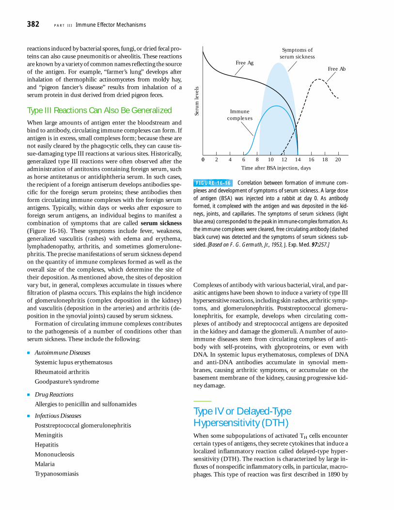
reactions induced by bacterial spores, fungi, or dried fecal pro-teins can also cause pneumonitis or alveolitis. These reactionsare known by a variety of common names reflecting the sourceof the antigen. For example, “farmer’s lung” develops afterinhalation of thermophilic actinomycetes from moldy hay,and “pigeon fancier’s disease” results from inhalation of aserum protein in dust derived from dried pigeon feces.
Type III Reactions Can Also Be Generalized When large amounts of antigen enter the bloodstream andbind to antibody, circulating immune complexes can form. Ifantigen is in excess, small complexes form; because these arenot easily cleared by the phagocytic cells, they can cause tis-sue-damaging type III reactions at various sites. Historically,generalized type III reactions were often observed after theadministration of antitoxins containing foreign serum, suchas horse antitetanus or antidiphtheria serum. In such cases,the recipient of a foreign antiserum develops antibodies spe-cific for the foreign serum proteins; these antibodies thenform circulating immune complexes with the foreign serumantigens. Typically, within days or weeks after exposure toforeign serum antigens, an individual begins to manifest acombination of symptoms that are called serum sickness(Figure 16-16). These symptoms include fever, weakness,generalized vasculitis (rashes) with edema and erythema,lymphadenopathy, arthritis, and sometimes glomerulone-phritis. The precise manifestations of serum sickness dependon the quantity of immune complexes formed as well as theoverall size of the complexes, which determine the site oftheir deposition. As mentioned above, the sites of depositionvary but, in general, complexes accumulate in tissues wherefiltration of plasma occurs. This explains the high incidenceof glomerulonephritis (complex deposition in the kidney)and vasculitis (deposition in the arteries) and arthritis (de-position in the synovial joints) caused by serum sickness.
Formation of circulating immune complexes contributesto the pathogenesis of a number of conditions other thanserum sickness. These include the following:
■ Autoimmune Diseases
Systemic lupus erythematosus
Rheumatoid arthritis
Goodpasture’s syndrome
■ Drug Reactions
Allergies to penicillin and sulfonamides
■ Infectious Diseases
Poststreptococcal glomerulonephritis
Meningitis
Hepatitis
Mononucleosis
Malaria
Trypanosomiasis
Complexes of antibody with various bacterial, viral, and par-asitic antigens have been shown to induce a variety of type IIIhypersensitive reactions, including skin rashes, arthritic symp-toms, and glomerulonephritis. Poststreptococcal glomeru-lonephritis, for example, develops when circulating com-plexes of antibody and streptococcal antigens are depositedin the kidney and damage the glomeruli. A number of auto-immune diseases stem from circulating complexes of anti-body with self-proteins, with glycoproteins, or even withDNA. In systemic lupus erythematosus, complexes of DNAand anti-DNA antibodies accumulate in synovial mem-branes, causing arthritic symptoms, or accumulate on thebasement membrane of the kidney, causing progressive kid-ney damage.
Type IV or Delayed-TypeHypersensitivity (DTH)When some subpopulations of activated TH cells encountercertain types of antigens, they secrete cytokines that induce alocalized inflammatory reaction called delayed-type hyper-sensitivity (DTH). The reaction is characterized by large in-fluxes of nonspecific inflammatory cells, in particular, macro-phages. This type of reaction was first described in 1890 by
382 P A R T I I I Immune Effector Mechanisms
0
Time after BSA injection, days
20
Free Ab
Seru
m le
vels
Free Ag
Immunecomplexes
Symptoms ofserum sickness
0 2 4 6 8 10 12 14 16 18
FIGURE 16-16 Correlation between formation of immune com-plexes and development of symptoms of serum sickness. A large doseof antigen (BSA) was injected into a rabbit at day 0. As antibodyformed, it complexed with the antigen and was deposited in the kid-neys, joints, and capillaries. The symptoms of serum sickness (lightblue area) corresponded to the peak in immune-complex formation. Asthe immune complexes were cleared, free circulating antibody (dashedblack curve) was detected and the symptoms of serum sickness sub-sided. [Based on F. G. Germuth, Jr., 1953, J. Exp. Med. 97:257.]

Robert Koch, who observed that individuals infected withMycobacterium tuberculosis developed a localized inflamma-tory response when injected intradermally with a filtratederived from a mycobacterial culture. He called this localizedskin reaction a “tuberculin reaction.” Later, as it became ap-parent that a variety of other antigens could induce thisresponse (Table 16-6), its name was changed to delayed-typeor type IV hypersensitivity in reference to the delayed onsetof the reaction and to the tissue damage (hypersensitivity)that is often associated with it. The term hypersensitivity issomewhat misleading, for it suggests that a DTH response isalways detrimental. Although in some cases a DTH responsedoes cause extensive tissue damage and is in itself pathologic,in many cases tissue damage is limited, and the responseplays an important role in defense against intracellular patho-gens and contact antigens. The hallmarks of a type IV reac-tion are the delay in time required for the reaction to developand the recruitment of macrophages as opposed to neu-trophils, as found in a type III reaction. Macrophages are themajor component of the infiltrate that surrounds the site ofinflammation.
There Are Several Phases of the DTH ResponseThe development of the DTH response begins with an initialsensitization phase of 1–2 weeks after primary contact withan antigen. During this period, TH cells are activated andclonally expanded by antigen presented together with therequisite class II MHC molecule on an appropriate antigen-presenting cell (Figure 16-17a). A variety of antigen-presentingcells have been shown to be involved in the activation of aDTH response, including Langerhans cells and macrophages.Langerhans cells are dendritic cells found in the epidermis.These cells are thought to pick up antigen that enters throughthe skin and transport it to regional lymph nodes, where
T cells are activated by the antigen. In some species, includinghumans, the vascular endothelial cells express class II MHCmolecules and also function as antigen-presenting cells in thedevelopment of the DTH response. Generally, the T cells ac-tivated during the sensitization phase are CD4+, primarily ofthe TH1 subtype, but in a few cases CD8+ cells have also beenshown to induce a DTH response. The activated T cells pre-viously were called TDTH cells to denote their function in theDTH response, although in reality they are simply a subset ofactivated TH1 cells (or, in some cases, TC cells).
A subsequent exposure to the antigen induces the effectorphase of the DTH response (see Figure 16-17b). In the effec-tor phase, TH1 cells secrete a variety of cytokines that recruitand activate macrophages and other nonspecific inflamma-tory cells. A DTH response normally does not become appar-ent until an average of 24 h after the second contact with theantigen; the response generally peaks 48–72 h after secondcontact. The delayed onset of this response reflects the timerequired for the cytokines to induce localized influxes ofmacrophages and their activation. Once a DTH response be-gins, a complex interplay of nonspecific cells and mediatorsis set in motion that can result in tremendous amplification.By the time the DTH response is fully developed, only about5% of the participating cells are antigen-specific TH1 cells;the remainder are macrophages and other nonspecific cells.
Macrophages are the principal effector cells of the DTHresponse. Cytokines elaborated by TH1 cells induce bloodmonocytes to adhere to vascular endothelial cells and mi-grate from the blood into the surrounding tissues. Duringthis process the monocytes differentiate into activated macro-phages. As Chapter 2 described, activated macrophages ex-hibit increased levels of phagocytosis and an increased abilityto kill microorganisms through various cytotoxic mediators.In addition, activated macrophages express increased levelsof class II MHC molecules and cell-adhesion molecules andtherefore function more effectively as antigen-presentingcells.
The influx and activation of macrophages in the DTH re-sponse is important in host defense against parasites andbacteria that live within cells, where circulating antibodiescannot reach them. The heightened phagocytic activity andthe buildup of lytic enzymes from macrophages in the area ofinfection lead to nonspecific destruction of cells, and thus ofthe intracellular pathogen. Generally, the pathogen is clearedrapidly with little tissue damage. However, in some cases,especially if the antigen is not easily cleared, a prolongedDTH response can itself become destructive to the host as theintense inflammatory response develops into a visible granu-lomatous reaction. A granuloma develops when continuousactivation of macrophages induces the macrophages to ad-here closely to one another, assuming an epithelioid shapeand sometimes fusing to form multinucleated giant cells(Figure 16-18). These giant cells displace the normal tissuecells, forming palpable nodules, and release high concentra-tions of lytic enzymes, which destroy surrounding tissue. Inthese cases, the response can damage blood vessels and lead
Hypersensitive Reactions C H A P T E R 16 383
TABLE 16-6Intracellular pathogens and contactantigens that induce delayed-type(type IV) hypersensitivity
Intracellular bacteria Intracellular virusesMycobacterium tuberculosis Herpes simplex virusMycobacterium leprae Variola (smallpox)Listeria monocytogenes Measles virusBrucella abortus
Intracellular fungi Contact antigensPneumocystis carinii PicrylchlorideCandida albicans Hair dyesHistoplasma capsulatum Nickel saltsCryptococcus neoformans Poison ivy
Intracellular parasites Poison oak
Leishmania sp.

to extensive tissue necrosis. The response to Mycobacteriumtuberculosis illustrates the double-edged nature of the DTHresponse. Immunity to this intracellular bacterium involves aDTH response in which activated macrophages wall off theorganism in the lung and contain it within a granuloma-typelesion called a tubercle. Often, however, the concentrated re-lease of lytic enzymes from the activated macrophages withintubercles damages lung tissue. Some examples of truly hyper-sensitive conditions, in which tissue damage far outweighsany beneficial effects, are described in Chapter 17.
Numerous Cytokines Participate in the DTH Reaction
Among the cytokines produced by TH1 cells are a numberthat attract and activate macrophages to the site of infection.IL-3 and GM-CSF induce localized hematopoiesis of thegranulocyte-monocyte lineage. IFN-� and TNF-� (togetherwith macrophage-derived TNF-� and IL-1) act on nearbyendothelial cells, inducing a number of changes that facilitateextravasation of monocytes and other nonspecific inflam-
384 P A R T I I I Immune Effector Mechanisms
V I S U A L I Z I N G C O N C E P T S
(a) Sensitization phase
(b) Effector phase
Restingmacrophage
MembraneTNF-β
SecretedIFN-γ
Effects of macrophageactivation: ↑ Class II MHC molecules ↑ TNF receptors ↑ Oxygen radicals ↑ Nitric oxide
Antigen-presenting cells: Macrophages Langerhans cells
DTH-mediating cells: TH1 cells generally CD8 cells occasionally
TH1 secretions:Cytokines: IFN-γ, TNF-β, IL-2,
IL-3, GM-CSFChemokines: IL-8, MCAF, MIF
APC
Intracellularbacteria
CD4+ TH
TH1 cells(generally)
SensitizedTH1
Class IIMHC
TNFreceptor
Activatedmacrophage
FIGURE 16-17 Overview of the DTH response. (a) In the sensiti-zation phase after initial contact with antigen (e.g., peptides derivedfrom intracellular bacteria), TH cells proliferate and differentiate intoTH1 cells. Cytokines secreted by these T cells are indicated by the darkblue balls. (b) In the effector phase after subsequent exposure of sen-
sitized TH1 cells to antigen, the TH1 cells secrete a variety of cytokinesand chemokines. These factors attract and activate macrophages andother nonspecific inflammatory cells. Activated macrophages aremore effective in presenting antigen, thus perpetuating the DTH re-sponse, and function as the primary effector cells in this reaction.

matory cells. Circulating neutrophils and monocytes adhereto the adhesion molecules displayed on the vascular endothe-lial cells and extravasate into the tissue spaces. Neutrophilsappear early in the reaction, peaking by about 6 h and thendeclining in numbers. The monocyte infiltration occurs be-tween 24 and 48 h after antigen exposure.
As the monocytes enter the tissues to become macro-phages, they are chemotactically drawn to the site of theDTH response by chemokines such as monocyte chemotac-tic and activating factor (MCAF). Another chemokine calledmigration-inhibition factor (MIF) inhibits macrophages frommigrating beyond the site of a DTH reaction. As macro-phages accumulate at the site of a DTH reaction, they areactivated by cytokines, particularly IFN-� and membrane-bound TNF-� produced by TH1 cells. As noted earlier,macrophages become more effective as antigen-presentingcells upon activation. Thus, the activated macrophages canefficiently mediate activation of more T cells, which in turnsecrete more cytokines that recruit and activate even moremacrophages. This self-perpetuating response, however, is adouble-edged sword, with a fine line existing between a ben-eficial, protective response and a detrimental response char-acterized by extensive tissue damage.
A report of experiments with knockout mice that couldnot produce IFN-� demonstrated the importance of thiscytokine in the DTH response. When these knockout micewere infected with an attenuated strain of Mycobacteriumbovis known as BCG (Bacille Calmette Guérin), nearly all theanimals died within 60 days, whereas wild-type mice sur-vived (Figure 16-19). Macrophages from the IFN-� knockoutmice were shown to have reduced levels of class II MHC mol-ecules and of bactericidal metabolites such as nitric oxideand superoxide anion.
The DTH Reaction Is Detected with a Skin TestThe presence of a DTH reaction can be measured experi-mentally by injecting antigen intradermally into an animaland observing whether a characteristic skin lesion developsat the injection site. A positive skin-test reaction indicatesthat the individual has a population of sensitized TH1 cellsspecific for the test antigen. For example, to determinewhether an individual has been exposed to M. tuberculosis,PPD, a protein derived from the cell wall of this mycobac-terium, is injected intradermally. Development of a red,slightly swollen, firm lesion at the site between 48 and 72 hlater indicates previous exposure. The skin lesion resultsfrom intense infiltration of cells to the site of injection duringa DTH reaction; 80%–90% of these cells are macrophages.Note, however, that a positive test does not allow one to conclude whether the exposure was to a pathogenic form ofM. tuberculosis or to a vaccine form received through immu-nization, which is performed in some parts of the world.
Contact Dermatitis Is a Type of DTH ResponseMany contact-dermatitis reactions, including the responses toformaldehyde, trinitrophenol, nickel, turpentine, and activeagents in various cosmetics and hair dyes, poison oak, and poi-son ivy, are mediated by TH1 cells. Most of these substancesare small molecules that can complex with skin proteins.
Hypersensitive Reactions C H A P T E R 16 385
Intracellularbacteria
Activatedmacrophage
Epithelioid cell
Multinucleatedgiant cell
TH1 cell
FIGURE 16-18 A prolonged DTH response can lead to formation ofa granuloma, a nodule-like mass. Lytic enzymes released from activatedmacrophages in a granuloma can cause extensive tissue damage.
100
80
60
40
20
050 60403020
Surv
ival
, %
Days after BCG infection
Wild type mice
IFN-γ knock-out mice
FIGURE 16-19 Experimental demonstration of the role of IFN-� inhost defense against intracellular pathogens. Knockout mice wereproduced by introducing a targeted mutation in the gene encodingIFN-�. The mice were then infected with 107 colony-forming units ofattenuated Mycobacterium bovis (BCG) and their survival monitored.[Adapted from D. K. Dalton et al., 1993, Science 259:1739.]

This complex is internalized by antigen-presenting cells inthe skin (e.g., Langerhans cells), then processed and pre-sented together with class II MHC molecules, causing activa-tion of sensitized TH1 cells. In the reaction to poison oak, forexample, a pentadecacatechol compound from the leaves ofthe plant forms a complex with skin proteins. When TH cellsreact with this compound appropriately displayed by localantigen-presenting cells, they differentiate into sensitizedTH1 cells. A subsequent exposure to pentadecacatechol willelicit activation of TH1 cells and induce cytokine production(Figure 16-20). Approximately 48–72 h after the secondexposure, the secreted cytokines cause macrophages to accu-mulate at the site. Activation of these macrophages andrelease of lytic enzymes result in the redness and pustulesthat characterize a reaction to poison oak.
SUMMARY
■ Hypersensitive reactions are inflammatory reactions with-in the humoral or cell-mediated branches of the immunesystem that lead to extensive tissue damage or even death.The four types of hypersensitive reaction generate charac-teristic effector molecules and clinical manifestations.
■ A type I hypersensitive reaction is mediated by IgE anti-bodies, whose Fc region binds to receptors on mast cells orblood basophils. Crosslinkage of the fixed IgE by allergenleads to mast cell or basophil degranulation with release ofpharmacologically active mediators. The principal effectsof these mediators are smooth-muscle contraction andvasodilation. Clinical manifestations of type I reactions in-clude potentially life-threatening systemic anaphylaxis andlocalized responses such as hay fever and asthma.
■ A type II hypersensitive reaction occurs when antibody re-acts with antigenic determinants present on the surface ofcells, leading to cell damage or death through complement-mediated lysis or antibody-dependent cell-mediated cyto-toxicity (ADCC). Transfusion reactions and hemolytic dis-ease of the newborn are type II reactions.
■ A type III hypersensitive reaction is mediated by the for-mation of immune complexes and the ensuing activationof complement. Complement split products serve as im-mune effector molecules that elicit localized vasodilationand chemotactically attract neutrophils. Deposition of im-mune complexes near the site of antigen entry can inducean Arthus reaction, in which lytic enzymes released by theaccumulated neutrophils and the complement membrane-attack complex cause localized tissue damage.
■ A type IV hypersensitive reaction involves the cell-mediatedbranch of the immune system. Antigen activation of sensi-tized TH1 cells induces release of various cytokines thatcause macrophages to accumulate and become activated.The net effect of the activation of macrophages is to releaselytic enzymes that cause localized tissue damage.
ReferencesAnsari, A. A., et al. 1989. Human immune responsiveness to
Lolium perenne pollen allergen Lol p III (rye III) is associatedwith HLA-DR3 and DR5. Hum. Immunol. 25:59.
Aubry, J. P., et al. 1992. CD21 is a ligand for CD23 and regulatesIgE production. Nature 358:505.
Barnes, K. C., and D. G. Marsh. 1998. The genetics and complex-ity of allergy and asthma. Immunol. Today 19:325.
Bonnefoy, J. Y., et al. 1993. A new pair of surface moleculesinvolved in human IgE regulation. Immunol. Today 14:1.
386 P A R T I I I Immune Effector Mechanisms
Langerhans cell (APC)
Monocyte
MIFMCF
Lyticenzymes
Tissue macrophage
Tissue macrophage
Self-protein
IFN-γ
Sensitized TH1
Skin
Pentadecacatechol
Poison oak(Toxicodendron radicans)
FIGURE 16-20 Development of delayed-type hypersensitivity reac-tion after a second exposure to poison oak. Cytokines such as IFN-�,macrophage-chemotactic factor (MCF), and migration-inhibition fac-tor (MIF) released from sensitized TH1 cells mediate this reaction.Tissue damage results from lytic enzymes released from activatedmacrophages.

Borish, L. 1999. Genetics of allergy and asthma. Ann. AllergyAsthma Immunol. 82:413.
Busse, W., and W. Neaville. 2001. Anti-immunoglobulin E for thetreatment of allergic disease. Curr. Opin. in Allergy & Immunol.1:105.
Chang, T. W. 2000. The pharmacological basis of anti-IgE ther-apy. Nat. Biotech. 18:157.
Daser, A., et al. 1995. Role and modulation of T-cell cytokines inallergy. Curr. Opin. Immunol. 7:762.
Finkelman, F. D., et al. 1988. IL-4 is required to generate and sus-tain in vivo IgE response. J. Immunol. 141:2335.
Holt, P. G. 1994. Immunoprophylaxis of atopy: light at the endof the tunnel? Immunol. Today 15:484.
Hoyne, G. F., et al. 1995. Peptide modulation of allergen-specificimmune responses. Curr. Opin. Immunol. 7:757.
Kuhn, R., K. Rajewsky, and W. Muller. 1991. Generation andanalysis of interleukin-4 deficient mice. Science 254:707.
Lympany, P., et al. 1992. Genetic analysis of the linkage betweenchromosome 11q and atopy. Clin. Exp. Allergy 22:1085.
Marsh, D. G., et al. 1994. Linkage analysis of IL-4 and other chro-mosome 5q31.1 markers and total serum immunoglobulin Econcentrations. Science 264:1152.
Marsh, D. G., et al. The Collaborative Study on the Genetics ofAsthma (CSGA). 1997. A genome-wide search for asthma sus-ceptibility loci in ethnically diverse populations. Nat. Genet.15:389.
Metzger, H. 1999. It’s spring, and thoughts turn to . . . allergies.Cell 97:287.
Novak, N., S. Kraft, and T. Bieber. 2001. IgE receptors. Curr.Opinion in Immunol. 13:721.
Paul-Eugène, N., et al. 1993. Functional interaction between �2-adrenoceptor agonists and interleukin-4 in the regulationof CD23 expression and release and IgE production in hu-mans. Molec. Immunol. 30:157.
Razin, E., I. Pecht, and J. Rivera. 1995. Signal transduction in theactivation of mast cells and basophils. Immunol. Today 16:370.
Romagnani, S. 2001. T-cell responses in allergy and asthma.Curr. Opin. in Allergy & Clin. Immunol. 1:73.
Rosenstreich, D. L., et al. 1997. The role of cockroach allergy andexposure to cockroach allergen in causing morbidity amonginner-city children with asthma. N. Engl. J. Med. 336:1356.
Teixeira, M. M., T. J. Williams, and P. G. Hellewell. 1995. Mecha-nisms and pharmacological manipulation of eosinophil accu-mulation. Trends Pharmacol. Sci. 16:418.
Thomas, P., et al. 1992. Glycosylation-inhibiting factor from hu-man T cell hybridomas constructed from peripheral blood lym-phocytes of a bee venom–sensitive allergic patient. J. Immunol.148:729.
Wills-Karp, M., J. Santeliz, and C. L. Karp. 2001. The germlesstheory of allergic disease: revisiting the hygiene hypothesis.Nature Rev. Immunol. 1:69.
USEFUL WEB SITES
http://www.niaid.nih.gov/
National Institute of Allergy and Infectious Diseases home-page. NIAID is the NIH Institute that sponsors research ininfectious diseases. Their Web site provides a number of linksto other relevant sites.
http://allergy.mcg.edu/home.html
A site maintained by the American College of Allergy, Asthma& Immunology. An excellent source of patient informationabout many allergies. This site contains many valuable links.
http//:www.glaxowellcome.co.uk/health/actiontb/
Action TB—A review of tuberculosis for a general audiencethat appears on the Glaxo-Wellcome company’s Web site.
http//:www.aaaai.org/
The American Association of Allergy, Asthma and ImmunologyWeb site. A good site for exploring the many aspects of asthma.
Study QuestionsCLINICAL FOCUS QUESTION Discuss why IL-4 and Fc�RI� areexcellent candidate genes involved in the genetic susceptibility toasthma.
1. Indicate whether each of the following statements is true orfalse. If you think a statement is false, explain why.
a. Mice infected with Nippostrongylus brasiliensis exhibitdecreased production of IgE.
b. IL-4 decreases IgE production by B cells.
c. The initial step in the process of mast-cell degranulationis crosslinking of Fc receptors.
d. Antihistamines are effective for the treatment of type IIIhypersensitivity.
e. Most pollen allergens contain a single allergenic compo-nent.
f. Babies can acquire IgE-mediated allergies by passive trans-fer of maternal antibody.
g. Transfusion reactions are a manifestation of type IIhypersensitivity.
2. In an immunology laboratory exercise, you are studying theresponse of mice injected intradermally with complete antibod-ies to the IgE Fc receptor (Fc�R1) or with Fab fragments of suchantibodies.
a. Predict the response expected with each type of antibody.
b. Would the responses observed depend on whether themice were allergic? Explain.
Hypersensitive Reactions C H A P T E R 16 387A
U:“
D.G
.”or
“D
.W.”
?
Go to www.whfreeman.com/immunology Self-TestReview and quiz of key terms

3. Serum sickness can result when an individual is given a largedose of antiserum such as a mouse antitoxin to snake venom.How could you take advantage of recent technological advancesto produce an antitoxin that would not produce serum sicknessin patients who receive it?
4. What immunologic mechanisms most likely account for aperson’s developing each of the following reactions after an insectbite?
a. Within 1–2 min after being bitten, swelling and rednessappear at the site and then disappear by 1 h.
b. 6–8 h later, swelling and redness again appear and persistfor 24 h.
c. 72 h later, the tissue becomes inflamed, and tissue necro-sis follows.
5. Indicate which type(s) of hypersensitive reaction (I–IV)apply to the following characteristics. Each characteristic canapply to one, or more than one, type.
a. Is an important defense against intracellular pathogens.b. Can be induced by penicillin.c. Involves histamine as an important mediator.d. Can be induced by poison oak in sensitive individuals.e. Can lead to symptoms of asthma.f. Occurs as result of mismatched blood transfusion.g. Systemic form of reaction is treated with epinephrine.h. Can be induced by pollens and certain foods in sensitive
individuals.i. May involve cell destruction by antibody-dependent cell-
mediated cytotoxicity.j. One form of clinical manifestation is prevented by
Rhogam.k. Localized form characterized by wheal and flare reaction.
6. In the table below, indicate whether each immunologic eventlisted does (+) or does not (–) occur in each type of hypersensi-tive response.
388 P A R T I I I Immune Effector Mechanisms
Hypersensitivity
Immunologic event Type I Type II Type III Type IV
IgE-mediated degranulationof mast cells
Lysis of antibody-coatedblood cells by complement
Tissue destruction inresponse to poison oak
C3a- and C5a-mediatedmast-cell degranulation
Chemotaxis of neutrophils
Chemotaxis of eosinophils
Activation of macrophagesby IFN-�
Deposition of antigen-antibody complexes onbasement membranesof capillaries
Sudden death due tovascular collapse (shock) shortly after injection oringestion of antigen