Allelic Variation Within the Putative Autism Spectrum Disorder Risk Gene Homeobox A1 and Cerebellar...
12
Allelic Variation within the Putative Autism Spectrum Disorder Risk Gene Homeobox-A-1 (HOXA1) and Cerebellar Maturation in Typically Developing Children and Adolescents Armin Raznahan 1,2 , Yohan Lee 1 , Catherine Vaituzis 1 , Lan Tran 1 , Susan Mackie 1 , Henning Tiemeier 1 , LIv Clasen 1 , Francois Lalonde 1 , Dede Greenstein 1 , Ron Pierson 1,3 , and Jay N Giedd 1 1 Child Psychiatry Branch, National Institute of Mental Health, National Institutes of Health, Bethesda, MD, USA 2 Child Psychiatry Department, Kings College London, Institute of Psychiatry, London, UK 3 Brain Image Analysis, LLC Abstract LAY ABSTRACT—Autism is known to be highly heritable, and has been associated with abnormalities in the development of several brain structures, including the cerebellum. Previous research has hinted that a gene controlling the development of posterior brain regions such as the cerebellum, may influence risk for autism. This gene is called Homeobox Domain A1 (HOXA1), and the variant within HOXA1 that has been most studied in relation to autism (A218G) falls within a gene region that is important for HOXA1 protein functioning. Although we know that autism appear to influence the dynamics of brain development, and that cerebellar anatomy continues to change over the lifespan – we do not know if A218G genotype influences cerebellar development over time. We studied this issue in typically developing controls who had a total of 296 repeat structural brain scans taken between ages 5 and 23 years of age. The volume of multiple cerebellar components was measures by hand in each scan, and we related developmental changes in these volumes to A218G genotype. We found that, in a part of the cerebellum implicated in autism, A218G genotype modified the rate of cerebellar growth. This suggests for the first time that the putative ASD risk gene HOXA1 has the capacity to modify the longitudinal development of cerebellar systems implicated in ASD neurobiology. SCIENTIFIC ABSTRACT—Homeobox-A-1 (HOXA1) has been proposed as a candidate gene for autism spectrum disorder (ASD) as it regulates embryological patterning of hind-brain structures implicated in autism neurobiology. In line with this notion, a non-synonymous single nucleotide polymorphism within a highly conserved domain of HOXA1 - A218G (rs10951154) - has been linked to both ASD risk, and cross-sectional differences in superior posterior lobar cerebellar anatomy in late adulthood. Despite evidence for early onset and developmentally dynamic cerebellar involvement in ASD, little is known of the relationship between A218G genotype and maturation of the cerebellum over early development. We addressed this issue using 296 longitudinally acquired structural magnetic resonance imaging brain scans from 116 healthy individuals between 5 and 23 years of age. Mixed models were used to compare the relationship between age and semi-automated measures of cerebellar volume in A-homozygotes (AA) and carriers of the G allele (Gcar). Total cerebellar volume increased between ages 5 and 23 years in both groups. However, this was accelerated in the Gcar relative to the AA group (Genotype-by- age interaction term, p=0.03), and driven by genotype-dependent differences in the rate of bilateral Corresponding Author: Armin Raznahan, [email protected]. NIH Public Access Author Manuscript Autism Res. Author manuscript; available in PMC 2013 April 01. Published in final edited form as: Autism Res. 2012 April ; 5(2): 93–100. doi:10.1002/aur.238. NIH-PA Author Manuscript NIH-PA Author Manuscript NIH-PA Author Manuscript
-
Upload
hms-harvard -
Category
Documents
-
view
2 -
download
0
Transcript of Allelic Variation Within the Putative Autism Spectrum Disorder Risk Gene Homeobox A1 and Cerebellar...
Allelic Variation within the Putative Autism Spectrum DisorderRisk Gene Homeobox-A-1 (HOXA1) and Cerebellar Maturation inTypically Developing Children and Adolescents
Armin Raznahan1,2, Yohan Lee1, Catherine Vaituzis1, Lan Tran1, Susan Mackie1, HenningTiemeier1, LIv Clasen1, Francois Lalonde1, Dede Greenstein1, Ron Pierson1,3, and Jay NGiedd1
1Child Psychiatry Branch, National Institute of Mental Health, National Institutes of Health,Bethesda, MD, USA2Child Psychiatry Department, Kings College London, Institute of Psychiatry, London, UK3Brain Image Analysis, LLC
AbstractLAY ABSTRACT—Autism is known to be highly heritable, and has been associated withabnormalities in the development of several brain structures, including the cerebellum. Previousresearch has hinted that a gene controlling the development of posterior brain regions such as thecerebellum, may influence risk for autism. This gene is called Homeobox Domain A1 (HOXA1),and the variant within HOXA1 that has been most studied in relation to autism (A218G) fallswithin a gene region that is important for HOXA1 protein functioning. Although we know thatautism appear to influence the dynamics of brain development, and that cerebellar anatomycontinues to change over the lifespan – we do not know if A218G genotype influences cerebellardevelopment over time. We studied this issue in typically developing controls who had a total of296 repeat structural brain scans taken between ages 5 and 23 years of age. The volume ofmultiple cerebellar components was measures by hand in each scan, and we related developmentalchanges in these volumes to A218G genotype. We found that, in a part of the cerebellumimplicated in autism, A218G genotype modified the rate of cerebellar growth. This suggests forthe first time that the putative ASD risk gene HOXA1 has the capacity to modify the longitudinaldevelopment of cerebellar systems implicated in ASD neurobiology.
SCIENTIFIC ABSTRACT—Homeobox-A-1 (HOXA1) has been proposed as a candidate genefor autism spectrum disorder (ASD) as it regulates embryological patterning of hind-brainstructures implicated in autism neurobiology. In line with this notion, a non-synonymous singlenucleotide polymorphism within a highly conserved domain of HOXA1 - A218G (rs10951154) -has been linked to both ASD risk, and cross-sectional differences in superior posterior lobarcerebellar anatomy in late adulthood. Despite evidence for early onset and developmentallydynamic cerebellar involvement in ASD, little is known of the relationship between A218Ggenotype and maturation of the cerebellum over early development. We addressed this issue using296 longitudinally acquired structural magnetic resonance imaging brain scans from 116 healthyindividuals between 5 and 23 years of age. Mixed models were used to compare the relationshipbetween age and semi-automated measures of cerebellar volume in A-homozygotes (AA) andcarriers of the G allele (Gcar). Total cerebellar volume increased between ages 5 and 23 years inboth groups. However, this was accelerated in the Gcar relative to the AA group (Genotype-by-age interaction term, p=0.03), and driven by genotype-dependent differences in the rate of bilateral
Corresponding Author: Armin Raznahan, [email protected].
NIH Public AccessAuthor ManuscriptAutism Res. Author manuscript; available in PMC 2013 April 01.
Published in final edited form as:Autism Res. 2012 April ; 5(2): 93–100. doi:10.1002/aur.238.
NIH
-PA Author Manuscript
NIH
-PA Author Manuscript
NIH
-PA Author Manuscript
superior posterior lobar volume change with age (p=0.002). Resultantly, although superiorposterior lobar volume did not differ significantly between genotype groups at age 5 (p=0.9), byage 23 it was 12% greater in Gcar than AA (p=0.002). Our results suggest that common geneticvariation within this putative ASD risk gene has the capacity to modify the development ofcerebellar systems implicated in ASD neurobiology.
KeywordsAutism; HOXA1; Cerebellum; Gene; Brain; MRI
INTRODUCTIONIn recent years there has been increasing interest in using combinations of neuroimaging andgenetics to delineate the biological pathways through which specific genetic variants maymodify risk for heritable neurodevelopmental conditions (Hariri & Weinberger, 2003;Meyer-Lindenberg, 2010). To date however, relatively few studies have applied thisapproach to study putative risk genes for Autism Spectrum Disorder (ASD) (Raznahan et al.,2010; Raznahan et al., 2009; Wassink et al., 2007). Here, we use such an “ImagingGenetics” approach to relate genetic variation within a candidate risk gene for ASD tolongitudinal measures of regional structural cerebellar maturation within a large sample oftypically developing controls.
Interest in regional cerebellar maturation as a potential marker of genetic risk for ASD stemsfrom several observations. First, reduced cerebellar purkinje cell count has become one ofthe most consistent findings in ASD post-mortem studies (Palmen, van, Hof, & Schmitz,2004), which suggests that disruption of cerebellar development in ASD may be evidentvery early in life. Second, there are multiple structural magnetic resonance imaging (sMRI)reports of abnormal cerebellar anatomy in children and adults with ASD (Stanfield et al.,2008). Such reports have examined diverse anatomical indices including volume and mid-sagittal cerebellar surface area. The best-replicated finding in these studies is abnormalitywithin lobule VI of the cerebellum (Mitchell et al., 2009; Stanfield et al., 2007; Webb et al.,2009) – arguing that a regional approach to measurement of cerebellar anatomy is desirablein ASD neurobiology. Furthermore, several cross-sectional reports suggest that cerebellarabnormalities in ASD may vary as a function of age (Cleavinger et al., 2008; Courchesne etal., 2001; Stanfield et al., 2007) – indicating that candidate cerebellar markers of ASD riskwould be best studied using longitudinal study designs. Finally, the notion that genetic riskfactors may shape cerebellar alterations in a developmentally dynamic manner within ASDis supported by evidence from twin and family neuroimaging studies demonstrating that themagnitude of genetic contribution may vary with age (Posthuma et al., 2000; Wallace et al.,2006). Despite the above lines of evidence, the relationship between specific putative ASDrisk alleles and developmental changes in regional cerebellar anatomy remains unexamined.
The strongest candidate gene for such a longitudinal imaging genetics approach isHomeobox-A-1 (HOXA1), which encodes a protein of known importance for hind-brainpatterning in vertebrates. Homeobox-A-1 (HOXA1) is one of a family of evolutionarilyconserved genes that govern morphogenesis and cellular differentiation of the centralnervous system through their function as transcription factors (Akin & Nazarali, 2005). Theinitial impetus for examining HOXA1 as a candidate gene for ASD came from overlap in thehind-brain abnormalities seen in post-mortem studies of people with idiopathic ASD(Bauman & Kemper, 2005), hind-brain alterations in both animal HOXA1 knockout models,and post-mortem studies of people with ASD in the context of teratogenic syndromes causedby molecules known to impact HOXA1 signaling (e.g. sodium valproate (Ingram et al.,2000; Rodier, Ingram, Tisdale, Nelson, & Romano, 1996)). Subsequently, case-series of
Raznahan et al. Page 2
Autism Res. Author manuscript; available in PMC 2013 April 01.
NIH
-PA Author Manuscript
NIH
-PA Author Manuscript
NIH
-PA Author Manuscript
humans with rare HOXA1 mutations found that learning disability and ASD were central tothe resulting syndrome, which was accompanied by radiological evidence of hind-brainmalformation (Tischfield et al., 2005).
To date, three independent groups have linked a common non-synonymous mutation with anevolutionarily conserved HOXA1 exon to risk for ASD (Conciatori et al., 2004; Ingram etal., 2000) or related traits (Chakrabarti et al., 2009) [although a different allele wasimplicated as imparting risk in each study, and several negative association studies also exist(Collins, Schroer, Bird, & Michaelis, 2003; Devlin et al., 2002; Romano et al., 2003; Sen etal., 2007; Talebizadeh et al., 2002)]. This single nucleotide polymorphism (SNP) (A218G,rs10951154) results in a His->Arg amino acid sequence change in a highly conservedpolyhistidine tail HOXA1 protein domain. Although there are no published studies thatdirectly test A218G functionality, sequence variation within this polyhistidine domain isknown to result in increased cell death and impaired neuronal differentiation – suggestingthat allelic variation at A218G may impact HOXA1 functioning and brain development(Paraguison et al., 2005; Paraguison et al., 2007). Give this, it is notable that allelic variationat the A218G SNP has been shown to modulate head circumference in people with ASDdiagnoses (Conciatori et al., 2004), and in typically developing children (Muscarella et al.,2007). The lack of a relationship between A218G genotype and head circumference inhealthy adults (Muscarella et al., 2007) suggests that the relationship between this SNP andbrain anatomy may change with development. This possibility has never been explored, butis supported by a recent observation that HOXA1 expression in human brain tissue variesdramatically as a function of age (Siegmund et al., 2007).
Despite extensive evidence that HOXA1 plays a role in hind-brain development, only onestudy has related HOXA1 A218G genotype to sMRI measures of hind-brain anatomy. Canuet al (Canu et al., 2009) found that carriage of the G allele was associated with regionalreductions of cerebellar volume within the anterior and superior posterior lobes in twoindependent cohorts of healthy adults (Canu et al., 2009). Because this was a cross-sectionalstudy limited to adults, it remains unknown if A218G influences cerebellar anatomy inchildhood, and whether the relationship between A218G genotype and anatomy changeswith age. As outlined above, these questions are especially relevant to the potential role ofHOXA1 in ASD neurobiology given meta-analytic evidence that differences in cerebellaranatomy between people with ASD and TDCs vary with age (Stanfield et al., 2007).Therefore, in order to more fully assess the potential for HOXA1 to influence structuralcerebellar phenotypes in ASD, we examined the relationship between A218G genotype andlongitudinal measures of regional cerebellar anatomy in a large sample of typicallydeveloping children, adolescents and young adults.
METHODSSample
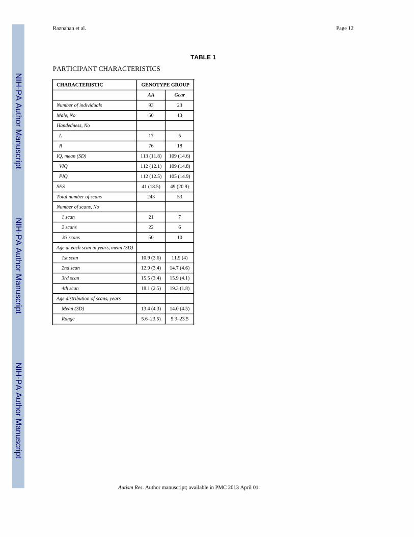
Please see Table 1 for full details of participant demographics and genotype composition.We included 296 structural magnetic imaging brain scans that had been acquiredlongitudinally on 116 healthy unrelated Caucasian children and adolescents aged betweenages 5 and 23 years or age. Participants were recruited through local advertisement. Theabsence of neurological or psychiatric illness was established through completion of ascreening questionnaire (Child Behavior Checklist) and a structured diagnostic interviewadministered by a child psychiatrist (Giedd et al., 1999). Participants were of mixedhandedness (handedness established using Physical and Neurological Examination of SoftSigns).
Raznahan et al. Page 3
Autism Res. Author manuscript; available in PMC 2013 April 01.
NIH
-PA Author Manuscript
NIH
-PA Author Manuscript
NIH
-PA Author Manuscript

All participants had a full-scale intelligence quotient (FSIQ) of greater than 80 asdetermined using age-appropriate Wechsler Intelligence Scales including WISC-R, WAIS-Rand WASI (Shaw et al., 2006)). Socioeconomic status (SES) was quantified usingHollingshead scales (Hollingshead, 1975). The institutional review board of the NationalInstitutes of Health approved the research protocol employed in this study and writteninformed consent and assent to participate in the study were obtained from parents andchildren respectively.
GenotypingFor each participant, DNA was extracted from previously prepared lymphoblastoid cell linesusing standard methods (Qiagen, MD, USA). It has been established that conversion of cellsinto lymphoblastoid lines does not cause errors into SNP genotyping (Herbeck et al., 2009).Genotyping was performed by Prevention Genetics (Marshfield, WI, USA), using sub-microlitre allele-specific polymerase-chain-reactions (Hawkins, Khripin, Valdes, & Weaver,2002). DNA sequencing of positive controls was conducted to ensure correct assignment ofgenotypes. Allele frequencies were A 0.89 and G 0.11 - in keeping with reference CEPHdata for populations of European descent. Participants were grouped as G-carriers (20%) orA-homozygotes (80%). Genotype frequencies did not deviate from Hardy-Weinbergequilibrium.
NeuroimagingOf all 116 participants with at least one brain sMRI scan, 60% had 2 or more, and 15% had3 or more scans. Scans were acquired at approximately 2-year intervals. All sMRI scanswere T-1 weighted images with contiguous 1.5 mm axial slices and 2.0 mm coronal slices,obtained on the same 1.5-T General Electric (Milwaukee, WI) Signa scanner using a 3Dspoiled gradient recalled echo sequence with the following parameters: Echo time, 5ms;Repetition time, 24ms; flip angle 45 (DEG); acquisition matrix, 256 × 192; number ofexcitations, 1; and field of view, 24 cm. Head placement was standardized as describedpreviously.
All measures of cerebellar anatomy were derived using the semi-automated methoddescribed in Pierson et al (Pierson et al., 2002). Briefly, 31 pre-specified cerebellarlandmarks were defined in each scan. Each landmark in the cerebellar co-ordinate space wasaveraged over all scans in which landmarks had been defined. This created a referencelandmark standard to which all subsequent scans could be warped prior to segmentation.Segmentation was achieved using an artificial neural network (ANN) approach (Magnotta etal., 2002), which was “trained” on 30 different cerebella that had been manually segmentedinto the following structures: Corpus Medullare (Cerebellar white matter and output nuclei),Cerbellar Vermis, Anterior Lobe (comprising lobules I, II, III, IV and V), Superior PosteriorLobe (comprising lobules VI, and VIIA-crus I) and Inferior Posterior Lobe (comprisinglobules VIIA-crus II, VIIB, VIII, IX and X). Each cerebellum to be segmented wassubmitted to the ANN for labeling, and the output then hand-edited to conform withreference tracing standards. Final overlap and intra-class correlation coefficients betweensemi-automated and manual approaches, as established in the original methodologicalpublication for this method were favorable. For all sub-regions, overlap values exceeded82%, and ICCs were greater than 0.9. This technique was implemented within our studycohort with high inter- and intra-rater reliabilities (all intra-class correlation coefficients over0.7). As was reported elsewhere (Mackie et al., 2007), for cerebellar lobes, initial intra-raterand interr-ater intraclass correlations (ICCs) were greater than 0.80 for all 5 tracers. For thevermis, initial intra-rater ICCs were greater than 0.80, and inter-rater ICCs were greater than0.68. Following the initial phase of measuring, two tracers continued measuring cerebellarlobes over a four-year span. Longitudinal lobar (left and right anterior, superior posterior
Raznahan et al. Page 4
Autism Res. Author manuscript; available in PMC 2013 April 01.
NIH
-PA Author Manuscript
NIH
-PA Author Manuscript
NIH
-PA Author Manuscript

and inferior posterior) intra-rater ICCs over this period ranged from 0.69 (superior vermis)to 0.99. All of these ICCs were approximately or at least 0.70, which is a qualitativelyacceptable (good) level of reliability (based on Fleiss (1986) who proposes a standard of0.40 to 0.75 as fair to good reliability, and above 0.75 as excellent). Concurrent lobar inter-rater ICCs were excellent and ranged from 0.91 to 0.99, and longitudinal lobar inter-raterICCs were good to excellent and ranged from 0.72 to 0.96. ICCs were calculated using the“one -way random” option in SPSS. In addition to calculating ICCs, measurements weregraphed to examine tracer drift and systematic biases within tracers. In the current sample,one tracer completed all of the vermis measurements that followed the initial phase ofmeasuring. Longitudinal ICCs for the vermis were 0.92 for the total vermis, and ranged from0.70 to 0.98 for the three vermal sub-regions (anterior, superior and inferior).
Statistical AnalysisLongitudinal mixed-models were used to estimate the effects of genotype on the rate oflinear volumetric change over time. We used mixed-models as these permit the inclusion ofmultiple measurements per person at different ages, and irregular intervals betweenmeasurements, thereby increasing statistical power (Pinheiro & DM, 2000). A nestedrandom effects term that modeled within family and within person dependence ofobservations was included. All models were run with and without sex, handedness, IQ andSES status as both main effects and in interaction with genotype terms. These terms werenot included in the final model because (i) their inclusion did not add significantly to thepredictive power of the model (as determined using a likelihood ratio test), (ii) their additiondid not alter the findings. Linear age terms were used for all cerebellar measures aspreliminary analyses established that higher-order age terms could not significantly predictvolumetric measures within the smaller genotype sub-group. This is likely to reflect sample-size limitations because curvilinear volume changes in cerebellar volume have beenidentified in larger samples (Tiemeier et al., 2010). Use of linear age protected against therisk of finding trajectory differences between genotype groups as an artifact of differences ingroup-size impacting the ability to identify significant higher-order age effects. We alsoprotected against artifactual effects of differences in genotype group-sizes, by confirmingthat all significant findings in the whole sample held when re-examined within a smallersubsample where each G-carrier were longitudinally matched 1:1 with an AA homozygotes.
We analyzed the relationship between HOXA1 genotype and cerebellar maturation in astaged manner, beginning with total cerebellar volume change as our developmentalphenotype of interest. Next we distinguished vermal and non-vermal (i.e cereballarhemispheres) structures given evidence for a difference in their embryonic origins (Yachnis& Rorke, 1999). Finally, we separately analyzed genetic influences on each of three vermalsub-components - Anterior Lobe (encompassing lobules I, II, III, IV and V), SuperiorPosterior Lobe (encompassing lobules VI and Crus I of lobule VIIA) and Inferior PosteriorLobe (encompassing Crus II of lobule VIIA and lobules VIIB, VIII, IX and X). Non-vermalcerebellar volume was divided into the same three lobar compartments and the corpusmedullare (central cerebellar white matter and output nuclei).
RESULTSAs has been previously shown in another longitudinal study of cerebellar development(Tiemeier et al., 2010), total cerebellar volume increased between ages 5 and 23 years.However, this was accelerated in G-carriers relative to A-homozygotes (Genotype-by-ageinteraction term, p=0.03). The interaction was driven by non-vermal rather than rather thanvermal structures (Genotype-by-age interaction terms p=0.01 and p=0.3 respectively).Analysis of non-vermal subdivisions revealed that allelic effects on cerebellar anatomy werelocalized to the Superior Posterior Lobe (p=0.002) only (Anterior Lobe p=0.2, Inferior
Raznahan et al. Page 5
Autism Res. Author manuscript; available in PMC 2013 April 01.
NIH
-PA Author Manuscript
NIH
-PA Author Manuscript
NIH
-PA Author Manuscript
Posterior Lobe p=0.5, Corpus Medullare p=0.1). Genotypic effects on bilateral SuperiorPosterior Lobe volumes held in both hemispheres (Left p=0.01, Right p= 0.002).Resultantly, although bilateral Superior Posterior Lobe volume did not differ significantlybetween genotype groups at age 5 (p=0.9), by age 23 it was 12% greater in G-carriers thanA-homozygotes (p=0.002). There were no statistically significant relationships betweenA218G genotype and vermal volume – either as a main effect (p=0.11), or in interactionwith age (p=0.32).
DISCUSSIONOur results suggest that there are significant differences in bilateral Superior Posterior Lobegrowth according to A218G genotype. As a result, although there were no statisticallysignificant differences in the volume of this cerebellar sub-region between genotype groupsin childhood, a highly significant volume increase in G-carriers relative to A-homozygotesemerged by the third decade of life. Cross-sectional analyses in two independent groups ofhealthy adults in the sixth decade of life have also linked A218G genotype to SuperiorPosterior lobar volume, although G-carriers had volumetric reductions compared to A-homozygotes (Canu et al., 2009). Different “directions” of genotype effects in our currentstudy and that of Canu et al (Canu et al., 2009) could be due to differences in the age-rangesstudied. The upper limit of the age-range examined in our study was 23 years, whereas thesample studies by Canu et al began at age 40 years, and extended to beyond 60 years of age.Therefore, it is possible that the relationship between A218G genotype and SuperiorPosterior Lobe anatomy changes between the third and sixth decades of life, such thatexaggerated growth in G carriers relative to A homozygotes during the early decades of lifewhen cerebellar volume is known to increase in age (Tiemeier et al., 2010), gives way toexaggerated volume loss in G carriers relative to A homozygotes in later life, whencerebellar volume is known to reduce with age (Raz, Dupuis, Briggs, McGavran, & Acker,1998). This hypothesis is in keeping with existing evidence for developmentally dynamicrelationships between A218G genotype and another anatomical index of relevance to thecentral nervous system – head circumference (Conciatori et al., 2004; Muscarella et al.,2007).
Although the molecular mechanism and cellular pathways through which allelic variation atthe A218G SNP might modify cerebellar development remain unclear, our findings arguefor a close assessment of the potential functional consequences of this non-synonymousSNP. While the canonical understanding that HOXA1 expression is restricted to hind-brainregions including and caudal to rhombomere 4 argues against the possibility that afunctional HOXA1 polymorphim might modify cerebellar structures (which derive fromrhombomere 1), recent evidence for more rostral HOXA1 expression suggests severalmechanisms through which genetically-determined variation in HOXA1 functioning couldimpact cerebellar development (McClintock et al., 2003; Wang & Zoghbi, 2001). In additionto arguing that HOXA1 may play a role in normal human cerebellar development, ourfindings raise the possibility that the tentative links that have been made between functionalvariation within HOXA1 signaling pathways and ASD risk (Rasalam et al., 2005) may bemediated by the capacity of HOXA1 to modify development of the cerebellum. Thisargument is strengthened by the fact that our study localizes A218G genotype influences oncerebellar maturation to the superior posterior cerebellar lobe, which includes the cerebellarlobule that has most consistently shown abnormalities in ASD - lobule VI (Courchesne etal., 1994; Hashimoto et al., 1995; Kaufmann et al., 2003; Stanfield et al., 2007; Webb et al.,2009).
An important next step would be to determine if our findings can be replicated inindependent samples of typically developing individuals. To more directly examine if
Raznahan et al. Page 6
Autism Res. Author manuscript; available in PMC 2013 April 01.
NIH
-PA Author Manuscript
NIH
-PA Author Manuscript
NIH
-PA Author Manuscript

HOXA1 regulation of cerebellar development is relevant to ASD pathogenesis, it will alsobe necessary to compare the relationship between HOXA1 genotype and cerebellar anatomyin people with ASD and typically developing controls. Disruption of normative relationshipsbetween HOXA1 genotype and cerebellar development in people with ASD could eitherreflect the action of primary risk factors for ASD, or the secondary consequences of havingASD. Theoretically, these two possibilities would be best distinguished by studying fullydiscordant monozygotic twin pairs, although this is not practically possible in ASD due tovery high monozygotic concordance rates (Folstein & Rutter, 1977). Moreover, sample sizerestrictions of ASD twin studies would make it very hard to longitudinally modeldevelopmental changes in cerebellar anatomy. An alternative approach would be to studygene-brain anatomy in a longitudinal sample of probands with ASD and their unaffectedsiblngs. This paradigm has been used with some success in psychiatric conditions other thanASD (Raznahan et al., 2011), but comes with its own compilations. These include theinability of sibling studies to distinguish shared genetic and environmental risks for ASD;the fact genetic imaging findings in unaffected siblings of probands with ASD may not onlyreflect the action of primary ASD risk factors in the absence of active disease, but could alsoreflect causes of resilience in healthy siblings that are absent in affected probands; and theinadequacy of sib-designs for defining markers of genetic risk in non-polygenic scenarios ofASD causation (Frazer, Murray, Schork, & Topol, 2009)(Frazer et al., 2009).
Our study has several limitations. Firstly, our sample of typically developing controls isrestricted to people of European descent, and while this reduced the likelihood of populationstratification, it limits the generalizability of out findings. Secondly, our parcellation of thecerebellum did not distinguish white and gray matter, which could show differentrelationships with HOXA1 genotype. Thirdly, our sample size was such that we were notable to model the curvilinear changes in cerebellar volume with age that have beendescribed in larger longitudinal studies (Tiemeier et al., 2010).
Despite these limitations, our study represents the largest, and only longitudinal, imaging-genetic investigation of HOXA1 to date. Our findings suggest that this putative ASD riskgene has the capacity to modify the development of cerebellar systems implicated in ASDneurobiology.
AcknowledgmentsAR was funded by the UK Medical Research Council Fellowship (G0701370) while carrying out aspects of thiswork. AR would like to thanks Profs Patrick Bolton, Declan Murphy and Gareth Barker for their helpful commentson earlier versions of this manuscript.
REFERENCESAkin ZN, Nazarali AJ. Hox genes and their candidate downstream targets in the developing central
nervous system. Cellular and Molecular Neurobiology. 2005; 25(3–4):697–741. [PubMed:16075387]
Bauman ML, Kemper TL. Neuroanatomic observations of the brain in autism: A review and futuredirections. International Journal of Developmental Neuroscience. 2005; 23(2–3):183–187.[PubMed: 15749244]
Canu E, Boccardi M, Ghidoni R, Benussi L, Duchesne S, Testa C, et al. HOXA1 A218Gpolymorphism is associated with smaller cerebellar volume in healthy humans. Journal ofNeuroimaging. 2009; 19(4):353–358. [PubMed: 19018953]
Chakrabarti B, Dudbridge F, Kent L, Wheelwright S, Hill-Cawthorne G, Allison C, et al. Genes relatedto sex steroids, neural growth, and social-emotional behavior are associated with autistic traits,empathy, and Asperger syndrome. Autism Res. 2009; 2(3):157–177. [PubMed: 19598235]
Raznahan et al. Page 7
Autism Res. Author manuscript; available in PMC 2013 April 01.
NIH
-PA Author Manuscript
NIH
-PA Author Manuscript
NIH
-PA Author Manuscript

Cleavinger HB, Bigler ED, Johnson JL, Lu J, McMahon W, Lainhart JE. Quantitative magneticresonance image analysis of the cerebellum in macrocephalic and normocephalic children andadults with autism. Journal of the International Neuropsychological Society. 2008; 14(3):401–413.[PubMed: 18419839]
Collins JS, Schroer RJ, Bird J, Michaelis RC. The HOXA1 A218G polymorphism and autism: lack ofassociation in white and black patients from the South Carolina Autism Project. Journal of Autism& Developmental Disorders. 2003; 33(3):343–348. [PubMed: 12908836]
Conciatori M, Stodgell CJ, Hyman SL, O'Bara M, Militerni R, Bravaccio C, et al. Association betweenthe HOXA1 A218G Polymorphism and Increased Head Circumference in Patients with Autism.[References]. Biological Psychiatry. 2004; 55(4):413–419. [PubMed: 14960295]
Courchesne E, Karns CM, Davis HR, Ziccardi R, Carper RA, Tigue ZD, et al. Unusual brain growthpatterns in early life in patients with autistic disorder: an MRI study. Neurology. 2001; 57(2):245–254. [PubMed: 11468308]
Courchesne E, Saitoh O, Yeung-Courchesne R, Press GA, Lincoln AJ, Haas RH, et al. Abnormality ofcerebellar vermian lobules VI and VII in patients with infantile autism: identification of hypoplasticand hyperplastic subgroups with MR imaging. AJR Am.J.Roentgenol. 1994; 162(1):123–130.[PubMed: 8273650]
Devlin B, Bennett P, Cook EH Jr, Dawson G, Gonen D, Grigorenko EL, et al. No evidence for linkageof liability to autism to HOXA1 in a sample from the CPEA network. American Journal ofMedical Genetics. 2002; 114(6):667–672. [PubMed: 12210285]
Folstein S, Rutter M. Genetic influences and infantile autism. Nature. 1977; 265(5596):726–728.[PubMed: 558516]
Frazer KA, Murray SS, Schork NJ, Topol EJ. Human genetic variation and its contribution to complextraits. Nat Rev Genet. 2009; 10(4):241–251. [PubMed: 19293820]
Giedd JN, Blumenthal J, Jeffries NO, Castellanos FX, Liu H, Zijdenbos A, et al. Brain developmentduring childhood and adolescence: a longitudinal MRI study. Nature Neuroscience. 1999; 2(10):861–863.
Hariri AR, Weinberger DR. Imaging genomics. British Medical Bulletin. 2003; 65(1):259–270.[PubMed: 12697630]
Hashimoto T, Tayama M, Murakawa K, Yoshimoto T, Miyazaki M, Harada M, et al. Development ofthe brainstem and cerebellum in autistic patients. Journal of Autism & Developmental Disorders.1995; 25(1):1–18. [PubMed: 7608030]
Hawkins JR, Khripin Y, Valdes AM, Weaver TA. Miniaturized sealed-tube allele-specific PCR. HumMutat. 2002; 19(5):543–553. [PubMed: 11968087]
Herbeck JT, Gottlieb GS, Wong K, Detels R, Phair JP, Rinaldo CR, et al. Fidelity of SNP arraygenotyping using Epstein Barr virus-transformed B-lymphocyte cell lines: implications forgenome-wide association studies. PLoS One. 2009; 4(9):e6915. [PubMed: 19730697]
Hollingshead, AB. Four-factor index for social status. New Haven: Yale UP; 1975.Ingram JL, Stodgell CJ, Hyman SL, Figlewicz DA, Weitkamp LR, Rodier PM. Discovery of allelic
variants of HOXA1 and HOXB1: genetic susceptibility to autism spectrum disorders. Teratology.2000; 62(6):393–405. [PubMed: 11091361]
Kaufmann WE, Cooper KL, Mostofsky SH, Capone GT, Kates WR, Newschaffer CJ, et al. Specificityof cerebellar vermian abnormalities in autism: a quantitative magnetic resonance imaging study.Journal of Child Neurology. 2003; 18(7):463–470. [PubMed: 12940651]
Magnotta VA, Harris G, Andreasen NC, O'Leary DS, Yuh WTC, Heckel D. Structural MR imageprocessing using the BRAINS2 toolbox. Computerized Medical Imaging & Graphics. 2002; 26(4):251–264. [PubMed: 12074920]
Mark M, Lufkin T, Vonesch JL, Ruberte E, Olivo JC, Dolle P, et al. Two rhombomeres are altered inHoxa-1 mutant mice. Development. 1993; 119(2):319–338. [PubMed: 8287791]
McClintock JM, Jozefowicz C, Assimacopoulos S, Grove EA, Louvi A, Prince VE. Conservedexpression of Hoxa1 in neurons at the ventral forebrain/midbrain boundary of vertebrates.Development Genes and Evolution. 2003; 213(8):399–406. [PubMed: 12748854]
Raznahan et al. Page 8
Autism Res. Author manuscript; available in PMC 2013 April 01.
NIH
-PA Author Manuscript
NIH
-PA Author Manuscript
NIH
-PA Author Manuscript
McIntosh AM, Baig BJ, Hall J, Job D, Whalley HC, Lymer GK, et al. Relationship of catechol-O-methyltransferase variants to brain structure and function in a population at high risk of psychosis.Biological Psychiatry. 2007; 61(10):1127–1134. [PubMed: 17014827]
Meyer-Lindenberg A. Intermediate or brainless phenotypes for psychiatric research? PsychologicalMedicine. 2010; 40(7):1057–1062. [PubMed: 20540175]
Mitchell SR, Reiss AL, Tatusko DH, Ikuta I, Kazmerski DB, Botti JA, et al. Neuroanatomic alterationsand social and communication deficits in monozygotic twins discordant for autism disorder.American Journal of Psychiatry. 2009; 166(8):917–925. [PubMed: 19605538]
Muscarella LA, Guarnieri V, Sacco R, Militerni R, Bravaccio C, Trillo S, et al. HOXA1 gene variantsinfluence head growth rates in humans. Am.J.Med.Genet.B Neuropsychiatr.Genet. 2007; 144B(3):388–390. [PubMed: 17171652]
Palmen SJ, van EH, Hof PR, Schmitz C. Neuropathological findings in autism. [Review] [117 refs].Brain. 2004; 127(Pt 12):2572–2583. [PubMed: 15329353]
Paraguison RC, Higaki K, Sakamoto Y, Hashimoto O, Miyake N, Matsumoto H, et al. Polyhistidinetract expansions in HOXA1 result in intranuclear aggregation and increased cell death.Biochemical and Biophysical Research Communications. 2005; 336(4):1033–1039. [PubMed:16168961]
Paraguison RC, Higaki K, Yamamoto K, Matsumoto H, Sasaki T, Kato N, et al. Enhanced autophagiccell death in expanded polyhistidine variants of HOXA1 reduces PBX1-coupled transcriptionalactivity and inhibits neuronal differentiation. Journal of Neuroscience Research. 2007; 85(3):479–487. [PubMed: 17131398]
Pierson R, Corson PW, Sears LL, Alicata D, Magnotta V, Oleary D, et al. Manual and semiautomatedmeasurement of cerebellar subregions on MR images. Neuroimage. 2002; 17(1):61–76. [PubMed:12482068]
Pinheiro, J.; DM, B. Mixed-effects models in S and S-PLUS. New York: Springer; 2000.Posthuma D, De Geus EJ, Neale MC, Hulshoff Pol HE, Baare WEC, Kahn RS, et al. Multivariate
genetic analysis of brain structure in an extended twin design. Behavior Genetics. 2000; 30(4):311–319. [PubMed: 11206086]
Rasalam AD, Hailey H, Williams JH, Moore SJ, Turnpenny PD, Lloyd DJ, et al. Characteristics offetal anticonvulsant syndrome associated autistic disorder. Developmental Medicine and ChildNeurology. 2005; 47(8):551–555. [PubMed: 16108456]
Raz N, Dupuis JH, Briggs SD, McGavran C, Acker JD. Differential effects of age and sex on thecerebellar hemispheres and the vermis: a prospective MR study. AJNR. American Journal ofNeuroradiology. 1998; 19(1):65–71. [PubMed: 9432159]
Raznahan A, Greenstein D, Lee Y, Long R, Clasen L, Gochman P, et al. Catechol-o-methyl transferase(COMT) val158met polymorphism and adolescent cortical development in patients withchildhood-onset schizophrenia, their non-psychotic siblings, and healthy controls. Neuroimage.2011; 57(4):1517–1523. [PubMed: 21620981]
Raznahan A, Lee Y, Long R, Greenstein D, Clasen L, Addington A, et al. Common functionalpolymorphisms of DISC1 and cortical maturation in typically developing children and adolescents.Molecular Psychiatry. 2010 2010/07/16 ed.
Raznahan A, Toro R, Proitsi P, Powell J, Paus T, Bolton P, et al. A functional polymorphism of thebrain derived neurotrophic factor gene and cortical anatomy in autism spectrum disorder. Journalof Neurodevelopmental Disorders. 2009; 1(3):215–223. [PubMed: 21547716]
Rodier PM, Ingram JL, Tisdale B, Nelson S, Romano J. Embryological origin for autism:developmental anomalies of the cranial nerve motor nuclei. Journal of Comparative Neurology.1996; 370(2):247–261. [PubMed: 8808733]
Romano V, Cali F, Mirisola M, Gambino G, D AR, Di RP, et al. Lack of association of HOXA1 andHOXB1 mutations and autism in Sicilian (Italian) patients. Molecular Psychiatry. 2003; 8(8):716–717. [PubMed: 12888798]
Sen B, Sinha S, Ahmed S, Ghosh S, Gangopadhyay PK, Usha R. Lack of association of HOXA1 andHOXB1 variants with autism in the Indian population. Psychiatric Genetics. 2007; 17(1):1.[PubMed: 17167333]
Raznahan et al. Page 9
Autism Res. Author manuscript; available in PMC 2013 April 01.
NIH
-PA Author Manuscript
NIH
-PA Author Manuscript
NIH
-PA Author Manuscript

Shaw P, Greenstein D, Lerch J, Clasen L, Lenroot R, Gogtay N, et al. Intellectual ability and corticaldevelopment in children and adolescents. Nature. 2006; 440(7084):676–679. [PubMed: 16572172]
Siegmund KD, Connor CM, Campan M, Long TI, Weisenberger DJ, Biniszkiewicz D, et al. DNAmethylation in the human cerebral cortex is dynamically regulated throughout the life span andinvolves differentiated neurons. PLoS One. 2007; 2(9):e895. [PubMed: 17878930]
Stanfield AC, McIntosh AM, Spencer MD, Philip R, Gaur S, Lawrie SM. Towards a neuroanatomy ofautism: A systematic review and meta-analysis of structural magnetic resonance imaging studies.Eur.Psychiatry. 2007
Stanfield AC, McIntosh AM, Spencer MD, Philip R, Gaur S, Lawrie SM. Towards a neuroanatomy ofautism: a systematic review and meta-analysis of structural magnetic resonance imaging studies.Eur Psychiatry. 2008; 23(4):289–299. [PubMed: 17765485]
Talebizadeh Z, Bittel DC, Miles JH, Takahashi N, Wang CH, Kibiryeva N, et al. No associationbetween HOXA1 and HOXB1 genes and autism spectrum disorders (ASD). Journal of MedicalGenetics. 2002; 39(11):e70. [PubMed: 12414832]
Tiemeier H, Lenroot RK, Greenstein DK, Tran L, Pierson R, Giedd JN. Cerebellum developmentduring childhood and adolescence: a longitudinal morphometric MRI study. Neuroimage. 2010;49(1):63–70. [PubMed: 19683586]
Tischfield MA, Bosley TM, Salih MA, Alorainy IA, Sener EC, Nester MJ, et al. Homozygous HOXA1mutations disrupt human brainstem, inner ear, cardiovascular and cognitive development. NatureGenetics. 2005; 37(10):1035–1037. [PubMed: 16155570]
Trainor PA, Krumlauf R. Patterning the cranial neural crest: hindbrain segmentation and Hox geneplasticity. Nat Rev Neurosci. 2000; 1(2):116–124. [PubMed: 11252774]
Wallace GL, Eric Schmitt J, Lenroot R, Viding E, Ordaz S, Rosenthal MA, et al. A pediatric twinstudy of brain morphometry. J Child Psychol Psychiatry. 2006; 47(10):987–993. [PubMed:17073977]
Wang VY, Zoghbi HY. Genetic regulation of cerebellar development. Nat Rev Neurosci. 2001; 2(7):484–491. [PubMed: 11433373]
Wassink TH, Hazlett HC, Epping EA, Arndt S, Dager SR, Schellenberg GD, et al. Cerebral corticalgray matter overgrowth and functional variation of the serotonin transporter gene in autism.Archives of General Psychiatry. 2007; 64(6):709–717. [PubMed: 17548752]
Webb SJ, Sparks BF, Friedman SD, Shaw DW, Giedd J, Dawson G, et al. Cerebellar vermal volumesand behavioral correlates in children with autism spectrum disorder. Psychiatry Research. 2009;172(1):61–67. [PubMed: 19243924]
Yachnis AT, Rorke LB. Cerebellar and brainstem development: an overview in relation to Joubertsyndrome. Journal of Child Neurology. 1999; 14(9):570–573. [PubMed: 10488901]
Raznahan et al. Page 10
Autism Res. Author manuscript; available in PMC 2013 April 01.
NIH
-PA Author Manuscript
NIH
-PA Author Manuscript
NIH
-PA Author Manuscript

Figure 1. HOXA1 genotype modulates cerebellar developmentRelationship Between Genotype at HOXA1 A218G Polymorphism and DevelopmentalChanges in Cerebellar Volume. Geneotypic influences on cerebellar anatomy are presentedin a staged manner. Shaded coronal and then sagittal views of the cerebellum are used tohighlight successive localization of significant HOXA1 modulation of cerebellar maturation,moving from total cerebellar volume, through non-vermal cerebellar volume to posteriorsuperior lobe volume. P values for the interaction between age and A218G genotype areprovided by each line plot.
Raznahan et al. Page 11
Autism Res. Author manuscript; available in PMC 2013 April 01.
NIH
-PA Author Manuscript
NIH
-PA Author Manuscript
NIH
-PA Author Manuscript
NIH
-PA Author Manuscript
NIH
-PA Author Manuscript
NIH
-PA Author Manuscript
Raznahan et al. Page 12
TABLE 1
PARTICIPANT CHARACTERISTICS
CHARACTERISTIC GENOTYPE GROUP
AA Gcar
Number of individuals 93 23
Male, No 50 13
Handedness, No
L 17 5
R 76 18
IQ, mean (SD) 113 (11.8) 109 (14.6)
VIQ 112 (12.1) 109 (14.8)
PIQ 112 (12.5) 105 (14.9)
SES 41 (18.5) 49 (20.9)
Total number of scans 243 53
Number of scans, No
1 scan 21 7
2 scans 22 6
≥3 scans 50 10
Age at each scan in years, mean (SD)
1st scan 10.9 (3.6) 11.9 (4)
2nd scan 12.9 (3.4) 14.7 (4.6)
3rd scan 15.5 (3.4) 15.9 (4.1)
4th scan 18.1 (2.5) 19.3 (1.8)
Age distribution of scans, years
Mean (SD) 13.4 (4.3) 14.0 (4.5)
Range 5.6–23.5) 5.3–23.5
Autism Res. Author manuscript; available in PMC 2013 April 01.