Repair of congenital sternal cleft in infants and adolescents
Upload
independentCategory
view
1download
0
The LaryngoscopeLippincott Williams & Wilkins, Inc.© 2006 The American Laryngological,Rhinological and Otological Society, Inc.
Alar Base Flap and Suspending Suture:A Strategy to Restore Symmetry to theNasal Alar Contour in PrimaryCleft-Lip Rhinoplasty
William Numa, MD; Kyle Eberlin, BA; Usama S. Hamdan, MD, FICS
Objective: Patients presenting with cleft-lip de-formity usually present with a characteristic nasaldeformity. We describe the mechanism and contribu-tion of different surgical techniques to restore alarsymmetry in primary cleft-lip rhinoplasty. Study De-sign: We evaluate surgical results using a retrospec-tive, randomized, blinded surgical grading system.We describe a surgical technique designed to restorenasal symmetry in patients undergoing primary cleft-lip rhinoplasty. Patients were selected retrospec-tively. Methods: A series of patients were identifiedwith nasal asymmetry associated with cleft-lip defor-mity. All patients underwent cleft-lip repair with con-current primary cleft-lip rhinoplasty. Patients whounderwent alar base flap suspending suture (ABF-SS)were grouped and selected consecutively after a mod-ification in the senior author’s surgical technique. Acontrol group was matched for age, sex, and cleftcharacteristics. Primary rhinoplasty was carried outconcurrently for both study groups while undergoingunilateral cleft-lip repair. The control group did notundergo the described ABF-SS technique. All patientswere operated on by the same surgeon over a periodof 5 years. Surgical outcomes were evaluated by apanel including lay people as well as trained healthcare workers experienced in the critical evaluation ofesthetic results after cleft-lip rhinoplasty. Results:Forty-six records were reviewed of patients undergo-ing complete unilateral cleft-lip repair. After applyingstrict inclusion/exclusion criteria, nine patients un-derwent the described ABF-SS technique. All patients
in the preoperative group had a clinically and statis-tically comparable degree of deformity (P > .05).There was a clinical and statistically significant im-provement in nostril size, shape, symmetry, alar basesymmetry, and nasal tip/dome symmetry for patientsundergoing repair with the described technique com-pared with the control group. No clinical or statisti-cally significant difference was observed in the scar-ring scores between groups. Conclusions: Patientspresenting with cleft-lip deformity usually presentwith a characteristic nasal deformity. Executionof the described surgical techniques restores na-sal alar symmetry in patients undergoing concur-rent primary cleft-lip rhinoplasty. Key Words: Uni-lateral, cleft, lip, rhinoplasty, suspension, suture,alar base flap.
Laryngoscope, 116:2171–2177, 2006
INTRODUCTIONThe nasal deformity associated with the unilateral
cleft lip continues to present a significant challenge togeneral and facial plastic surgeons. As outcomes in surgi-cal management of the cleft lip continue to improve, re-construction of the nose has become perhaps one of themost important functional and esthetic components of sur-gical repair. Despite a plethora of technical approachesaimed at correcting the defect, no one procedure has beenuniversally satisfactory in the repair of nasal deformitiesassociated with cleft-lip abnormalities.1
Primary rhinoplasty at the time of unilateral cleft-liprepair has become the increasingly accepted surgical op-tion for the care of most patients presenting with thisdeformity. However, complete nasal reconstruction is usu-ally avoided because of the potential interference withnasal, septal, and midface development. Some long-termstudies have found that primary rhinoplasty had no ad-verse long-term effects on nasal growth.2
The nasal deformity associated with a unilateral cleftlip is the result of the complex geometry involving both thefacial skeleton as well as the changes in the surroundingsoft tissues, including aberrant muscle insertion points.
From the Department of Otolaryngology–Head and Neck Surgery(W.M.), Tufts-New England Medical Center, Boston, Massachusetts, U.S.A.,Boston University School of Medicine (K.E.), Boston, Massachusetts,U.S.A., and the Department of Otology and Laryngology, Harvard MedicalSchool, Department of Otolaryngology, Boston University School of Medi-cine, and Department of Otolaryngology (U.S.H.), Tufts University School ofMedicine, Boston, Massachusetts, U.S.A.
Editor’s Note: This Manuscript was accepted for publication August23, 2006.
Send correspondence to Dr. Usama Hamdan, 28 Martingale Lane,Westwood, MA 02090.
DOI: 10.1097/01.mlg.0000244153.78495.e8
Laryngoscope 116: December 2006 Numa et al.: Alar Base Flap and Suspending Suture
2171

This nasal deformity typically exhibits the following ana-tomic characteristics on the cleft side (Fig. 1): 1) an out-stretched and laterally displaced alar base, 2) a shortenedcolumella, 3) a long and thinned lateral crus with a short-ened medial crus, 4) flattening of the nasal dome, 5) poornasal tip definition and support, and 6) asymmetry of thepyriform apertures secondary to maxillary hypoplasia.3 Inaddition, the appearance of the nasal deformity changesover time, with nasal asymmetry peaking in the postpu-bertal growth stage.4
One of the most widely used surgical techniques forrepair of the cleft lip is the Millard rotation-advancementtechnique, first presented in 1955 and since championed
and modified by many experts in the field.5 The basictechnique involves the advancement of a mucocutaneousflap from the affected lateral lip medially to cover the cleftdefect, attempting to conceal scarring along the philtralcolumns and nasal sill. This technique has providedwidely acceptable results for lip repair.
However, despite several attempts to incorporate pri-mary rhinoplasty into the Millard unilateral lip repairtechnique, several obstacles remain to the adequate res-toration of nasal symmetry. Seemingly excellent initialresults can change over time because of scar formationand anatomic discrepancies between the cleft and noncleftsides.7 The lateral crus on the cleft side frequently be-comes displaced and asymmetric because the scarring vec-tors of tension tend to distort tissues away from theirintended intraoperative positioning and the direction ofthe advancement flap. As the incisions heal and the childgrows, the nasal deformity can become accentuated. This
Fig. 1. Nasal characteristics associated with unilateral completecleft-lip nasal deformity: 1) outstretched and laterally displaced alarbase, 2) shortened columella, 3) long and thinned lateral crus withshortened medial crus, 4) flattening of nasal dome, 5) poor nasal tipdefinition and support, and 6) asymmetry of pyriform aperturessecondary to maxillary hypoplasia.3
Fig. 2. Skin incisions addressing primary lip deformity remain largelyunmodified from described Millard technique. Area of underminingin plane between nasal cartilages and nasal superficial musculoapo-neurotic system (dashed line).
Fig. 3. Skin incisions are limited to lowermost aspect of alar creaseat its junction with upper lip.
Fig. 4. Dissection of soft tissue envelope overlying nasal cartilagesis carried out in closed fashion using curved strabismus scissors.Desired dissection plane is between nasal cartilages and nasalsuperficial musculoaponeurotic system.
Laryngoscope 116: December 2006 Numa et al.: Alar Base Flap and Suspending Suture
2172
may be caused, in part, by insufficient underlying supportof the alar base. We propose to correct the void in thisunderlying support intraoperatively through the tech-niques described below. We describe a modification of theMillard rotation-advancement technique to create greaternostril symmetry and minimize long-term lateral dis-placement of the nasal ala on the cleft side after primarycleft-lip rhinoplasty.
These techniques were developed as a result of theconstant critical observation of our own surgical outcomes,during which we identified lateralization (widening) of thealar base in patients undergoing concurrent primary rhi-noplasty during cleft-lip repair. This result was docu-mented approximately 1 to 2 years after the initial proce-dure, when patients presented for cleft-palate repair.
MATERIALS AND METHODSA retrospective review was made of our experience treating
patients with varying degrees of nasal asymmetry associatedwith unilateral cleft-lip deformity. All patients underwent cleft-lip repair with concurrent primary cleft-lip rhinoplasty.
Patients in the study group were selected consecutively overa 3 year period, after a modification in the senior author’s surgicaltechnique (U.S.H.). For the patients in the study group, the alarbase flap and suspending suture (ABF-SS) technique was used. Acontrol group was also selected from the senior author’s practice,matching for age, sex, and cleft characteristics. The control group
underwent primary rhinoplasty concurrently with the initialcleft-lip repair without use of the described technique (ABF-SS).All patients in the control group underwent surgical interventionbefore the development of the described technique.
All procedures were performed as part of an American-based humanitarian surgical team. No orthodontic or prosthodon-tic treatment was available preoperatively or postoperatively toeither group of patients given the limited resources available inthe countries where these procedures take place.
Both groups were operated on by a team led by the samesurgeon (U.S.H.). Preoperative and postoperative digital photo-graphic documentation was obtained on each patient at a dis-tance of approximately 1 m. Frontal, lateral, and base views wereavailable for review. Surgical results on each patient were eval-uated by a panel of 10 observers. The panel was composed of bothlay people (5) as well as trained health care professionals (5)experienced in the critical evaluation of esthetic results aftercleft-lip rhinoplasty. The panel was blinded to the type of proce-dure performed and was asked to qualitatively evaluate nasalsymmetry in categories including nostril size, shape, scarring,and alar symmetry. The panel was asked to focus on the nasalcharacteristics while ignoring the lip characteristics. They wereasked to assign a numeric value in a visual analogue ordinal scaleranging from 0 to 4 (symmetric, nearly symmetric, mild defor-mity, moderate deformity, and severe deformity, respectively).Patients were evaluated independently, with degree of deformityassessed. Each patient was compared pre- and postoperatively(study group vs. the control group). Nonparametric statisticalanalysis was performed by comparing assessed degree of symme-try using Mann-Whitney analysis, thus comparing the distribu-tion of variance both within and between each group.
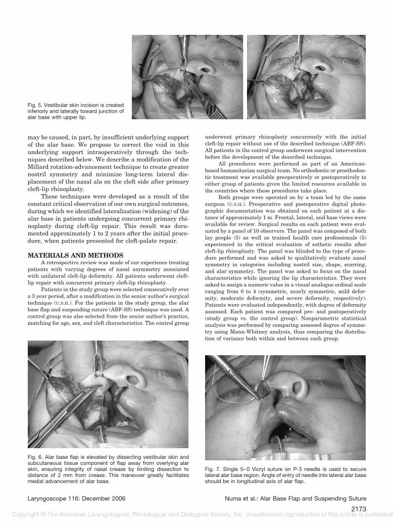
Fig. 5. Vestibular skin incision is createdinferiorly and laterally toward junction ofalar base with upper lip.
Fig. 6. Alar base flap is elevated by dissecting vestibular skin andsubcutaneous tissue component of flap away from overlying alarskin, ensuring integrity of nasal crease by limiting dissection todistance of 2 mm from crease. This maneuver greatly facilitatesmedial advancement of alar base.
Fig. 7. Single 5–0 Vicryl suture on P-3 needle is used to securelateral alar base region. Angle of entry of needle into lateral alar baseshould be in longitudinal axis of alar flap.
Laryngoscope 116: December 2006 Numa et al.: Alar Base Flap and Suspending Suture
2173

Only patients with unilateral complete cleft-lip deformitywere included. Patients with incomplete cleft-lip deformity, bilat-eral clefts, inadequate pre/postoperative photodocumentation, orinsufficient follow-up time to assess the described nasal charac-teristics were excluded from the study group. All patients hadvarying degrees of cleft palate, and postoperative follow-up wasobtained as part of the preoperative assessment for cleft-palatecorrection.
TechniqueThe general sequence of the operative procedure is as fol-
lows: skin incisions, primary rhinoplasty (medial portion), cleft-lip flaps, and, finally, the retrograde (lateral portion) of therhinoplasty. Primary cleft rhinoplasty refers to the procedureto correct the nasal deformity concurrently with cleft-lip re-pair. Its aim historically has been to free the nasal structures
from the overlying cutaneous soft tissue envelope in one oper-ative procedure.
Skin Incisions. The skin incisions addressing the primarylip deformity remain largely unmodified from the described Mil-lard technique (Fig. 2). The initial medial approach to the nose iscombined with a lateral (retrograde) approach that serves as theunderlying principle for this repair. Laterally, the ABF-SS tech-nique is based on the following steps:
● Limiting the extent of skin incisions to the lowermostaspect of the alar crease at its junction with the upper lip(Fig. 3). This limitation evolved from the observation thatvarying degrees of alar notching resulted secondary to theextension of the skin incision along the base of the alarcrease in cephalic and medial directions. The ABF-SStechnique allows ample rotation and advancement of thealar flap into a medial position based on the extensivesubcutaneous dissection, obviating the need for extendingthe skin incision along the base of the alar crease.
Primary Rhinoplasty, Medial Portion. After the skinincisions, dissection of the soft tissue envelope overlying the nasalcartilages is carried out in a closed fashion using curved strabis-mus scissors (Fig. 4). The overlying soft tissues of the nose are
Fig. 8. Suture is then passed and secured to subcutaneous tissue atpoint just lateral to subnasale on noncleft side, thereby anchoringalar base to exactly desired location. This can be tailored to meetconfiguration of nonaffected ala that serves as intraoperative com-parison template.
Fig. 9. As in Figure 8, suture is then passed and secured to subcu-taneous tissue at point just lateral to subnasale on noncleft side,thereby anchoring alar base to exactly desired location. This can betailored to meet configuration of nonaffected ala that serves asintraoperative comparison template.
Fig. 10. Pre- and postoperative photographs.
Fig. 11. Pre- and postoperative photographs.
Laryngoscope 116: December 2006 Numa et al.: Alar Base Flap and Suspending Suture
2174

dissected off from the underlying prolabium, columella, dome,and lateral crus. The dissection plane is developed superficial tothe superficial musculoaponeurotic system (SMAS) layer invest-ing the nose. The area separating the medial crura of the lowerlateral cartilages is dissected, with care taken not to separate thecrura from the quadrangular cartilage. The ligamentous attach-ments of the upper lateral and lower lateral cartilages (scrollregion) as well as all other tip support mechanisms are preserved.The superior limit of the dissection is the scroll area. The laterallimit of the dissection is the posterior border of the lateral crus.
Cleft-lip Flaps. The details pertinent to our modificationsin the flaps to address the cleft-lip repair are the subject of aseparate paper, currently under submission. In brief, wide surgi-cal undermining and extensive freeing of the aberrant attach-ments of the orbicularis oris muscle fibers and nasal soft tissueenvelope is carried out beyond the borders of some previouslydescribed methods.
Primary Rhinoplasty, Lateral Portion (ABF-SS)ALAR BASE FLAP. Dissection is carried out laterally on the
cleft side, with care taken not to disrupt the alar crease, asoutlined in the following steps:
● A vestibular skin incision is created inferiorly and later-ally toward the junction of the alar base with the upper lip(Fig. 5). The cephalomedial border of the incision is 1 to 2mm lateral to the posterior edge of the lateral crus on thecleft side. The incision is placed 2 to 3 mm lateral to themucocutaneous junction in the vestibular skin area in an
attempt to preserve the functionality of the alar compo-nent of the nasal valve and to prevent nasal stenosis inthat region. This issue has been overlooked in some pre-vious corrective surgical techniques, thus disrupting theintegrity of the internal aspect of the lateral alar region.
● The alar flap is then elevated by dissecting the vestibularskin and subcutaneous tissue component of the flap awayfrom the overlying alar skin, ensuring the integrity of thenasal crease by limiting the dissection to a distance of 2mm from the crease (Fig. 6). This maneuver greatly facil-itates the medial advancement of the alar base.
● By avoiding creation of incisions in the vestibular muco-cutaneous junction, three goals are accomplished: 1)avoidance of alar collapse, 2) avoidance of recruitment ofexcess tissue into the nostril, and 3) avoidance of theintroduction of vestibular mucosa into the skin flap of thereconstructed nasal sill.
SUSPENDING SUTURE. Once the dissection planes are devel-oped, the skin flap is anchored to the subcutaneous tissue at apoint just lateral to the subnasale on the noncleft side. A single5–0 Vicryl suture on a P-3 needle (Ethicon, Inc., Somerville, NJ)is used to secure the lateral alar base region. The angle of entryof the needle into the lateral alar base should be in the longitu-dinal axis of the alar flap (Fig. 7). Doing so ensures that thesymmetrical medialization of the alar flap is accomplished as oneanatomic unit. It also provides the necessary support to maintainthe alar flap suspension. This prevents future lateralization ofthe flap and alar base.
The suture is then passed and secured to the subcutaneoustissue at a point just lateral to the subnasale on the noncleft side,thereby anchoring the alar base to exactly the desired location(Figs. 8 and 9). This can be tailored to meet the configuration ofthe nonaffected ala that serves as an intraoperative comparisontemplate. Overcorrection should be avoided by all means becausethis will contribute to long-term nasal stenosis. The resultingdead space overlying the alar flap is obliterated by using one totwo transfixion sutures. The authors currently use 5–0 Chromicon a P-3 needle (Ethicon, Inc.). This maneuver secures the lateralcrus in its medialized and anatomically correct position and alsoprevents future thickening of the ala secondary to cicatricialtissue formation. Finally, a 6–0 Vicryl subcutaneous invertedsuture is used to position the vertex of the alar flap into thecreated nasal sill. Preoperative and postoperative photographsare shown in Figures 10 and 11.
RESULTSThis technique has been carried out on 46 patients
with varying degrees of cleft-lip deformity (27 males and19 females) to date during a period of 3 years. After re-
TABLE I.Patient Characteristics.
MillardRotation-Advancement
With PrimaryRhinoplasty
ABF-SS TechniqueWith PrimaryRhinoplasty
Sex
Male 5 7
Female 4 2
Age at surgery, mo(range)
10.6 (10–13) 11.8 (3–36)
Cleft lip
Complete (%) 9 (100) 9 (100)
Cleft palate 9 (100) 9 (100)
Average follow-up time,mo (range)
19 (9–60) 14 (6–28)
Comorbidities n/a n/a
ABF-SS � alar base flap and suspending suture.
TABLE II.Preoperative Assessment.
Millard Repair ABF-SS Technique P Value,Mann-Whitney Rank
Sum TestMean Median Mode Mean Median Mode
Nostril size 2.80 3 3 2.87 3 3 .737
Nostril shape 2.99 3 3 2.89 3 3 .721
Nostril symmetry 2.99 3 4 3.11 3 4 .409
Alar base symmetry 3.00 3 4 3.01 3 3 .998
Nasal tip/dome symmetry 2.50 3 3 2.27 2 2 .118
Scarring 0.00 0 0 0.00 0 0 .999
ABF-SS � alar base flap and suspending suture.
Laryngoscope 116: December 2006 Numa et al.: Alar Base Flap and Suspending Suture
2175

viewing all preoperative and postoperative photodocu-mentation and adhering to the previously defined inclu-sion/exclusion criteria, a total of nine patients wereselected for the study group. Patient age at the time ofsurgery averaged 11.8 (3–36) months. The follow-up timeafter surgery was 14 (6–28) months. A matched controlgroup was also selected (Table I). All procedures wereperformed by the same surgeon (U.S.H.). No major compli-cations were seen. No nasal stenosis, flap necrosis, orhypertrophic scars were seen in either group.
The panel of observers found no statistical differencein the varying degrees of deformity and nasal asymmetryamong both preoperative groups. The severity scoresranged from 0 (symmetric) to 4 (severe deformity), withlower scored indicating a better esthetic appearance (Ta-bles II and III). A significant difference was found betweenboth postoperative groups when evaluating nostril size,nostril shape, nostril symmetry, alar base symmetry, andnasal tip/dome symmetry. Patients undergoing repairwith use of the ABF-SS technique showed a statisticallysignificant improved degree of perceived symmetry in nos-tril size (P � .001), nostril shape (P � .001), nostril sym-metry (P � .001), alar base symmetry (P � .004), andnasal tip/dome symmetry (P � .001). No statistically sig-nificant difference was found when evaluating scarringbetween groups (P � .24) (Figs. 12 and 13).
DISCUSSIONWe deliberately limit the extent of the primary rhi-
noplasty to the subcutaneous freeing of the lower lateralcartilage from the overlying skin on the medial aspect ofthe cleft nasal deformity and to mobilize the laterallydisplaced dome and lateral crus to a medial and moresymmetric position. This technique avoids any circumfer-ential rim or marginal incisions that can predispose thehealing nostril to future concentric stenosis. In addition,the extent of the lateral dissection is carried out as previ-ously described to ensure proper mobilization of the alarbase and nasal symmetry.
This technique is different from previously describedsuspending suturing techniques by the following points7,8:
1. An increased extent of lateral dissection in creating thealar flap.
2. The alar base suture is positioned in a longitudinalaxis.
3. Preservation of the structural integrity of the lowerlateral cartilage.
4. Alar flap/crease transfixion sutures.
It is the nasal ala, and not the lip, characteristics thatare the aim of this technique. This technique can be easilyincorporated into the concurrent management of primaryrhinoplasty in the surgical repair of unilateral cleft-lip
Fig. 13. Postoperative Assessment.
TABLE III.Postoperative Assessment.
Millard Repair ABF-SS Technique P Value,Mann-Whitney Rank
Sum TestMean Median Mode Mean Median Mode
Nostril size 1.73 2 1 0.94 1 0 �.001
Nostril shape 2.02 2 2 1.18 1 1 �.001
Nostril symmetry 2.17 2 1 1.29 1 1 �.001
Alar base symmetry 1.59 2 2 1.06 1 0 .004
Nasal tip/dome symmetry 1.48 1 1 0.83 0 0 �.001
Scarring 1.10 1 0 1.24 1 1 .242
ABF-SS � alar base flap and suspending suture.
Fig. 12. Preoperative Assessment.
Laryngoscope 116: December 2006 Numa et al.: Alar Base Flap and Suspending Suture
2176

deformity. This technique is not aimed at obviating theneed for secondary septorhinoplasty once nasal and septaldevelopment has occurred. However, by restoring the na-sal symmetry while preserving the integrity of the lowerlateral cartilage, this approach facilitates secondary rhi-noplasty at a later stage. The results of the study displaystatistical and clinical superiority only for unilateral com-plete clefts. However, this technique can also be easilyextrapolated and incorporated in the repair of the cleft-liprhinoplasty in patients with incomplete cleft-lip deformityor isolated nasal deformity revisions.
Clearly, a certain degree of selection bias is presentbecause the study group has been operated on by an in-creasingly experienced surgeon. Longer follow-up will berequired to confirm these results. The control group wasselected from pictures predating the development of thedescribed technique, which inevitably increases the ten-dency to show the best results of the time.
If a given repair universally corrected all clinical pre-sentations of this deformity effectively, a consensus wouldexist on the repair method to be used. The multiple ap-proaches and iterations to these techniques is a testament tothe complexity and variation of the underlying anatomicdeformity and the surgical repair necessary to correct it.
CONCLUSIONIn our experience, the described technique achieves
excellent cosmetic results by recreating a naturally ap-pearing nostril size, shape, symmetry, as well as restoringalar base symmetry and nasal dome/tip symmetry. It canbe easily incorporated in the concurrent primary rhino-plasty technique in the surgical repair of the unilateralcleft-lip deformity. These improvements are appreciated
by both lay people and seasoned health care personnelexperienced in the critical evaluation of esthetic resultsafter cleft-lip rhinoplasty.
It is the opinion of the authors that overly aggressive,early single-stage approaches to cleft-lip rhinoplasty maydisrupt subsequent normal growth and development of thealar cartilage. We believe in the importance of limiting thescope of the initial intervention to a cartilage-sparing tech-nique that alters the underlying cartilaginous architecturewithout dividing or suturing the lower lateral cartilage. Wetherefore choose not to perform direct cartilaginous incisionsor circumferential nasal skin incisions, relying only on theones created to address the primary lip deformity.
BIBLIOGRAPHY1. Brusse CA, Van der Werff JF, Stevens HP, et al. Symmetry
and morbidity assessment of unilateral complete cleft lipnose corrected with or without primary nasal correction.Cleft Palate Craniofac J 1999;36:361–366.
2. McComb HK, Coghlan BA. Primary repair of the unilateralcleft lip nose: completion of a longitudinal study. CleftPalate Craniofac J 1996;33:23–30.
3. Shih CW, Sykes JM, Correction of the cleft-lip nasal deformity.Facial Plast Surg 2002;18:253–262.
4. Kyrkanides S, Bellohusen R, Subtelny JD. Asymmetries of theupper lip and nose in noncleft and postsurgical unilateralcleft lip and palate individuals. Cleft Palate Craniofac J1996;33:306–311.
5. Millard DR. Extensions of the rotation-advancement principlefor wide unilateral cleft lips. Plast Reconstr Surg 1968;42:535–544.
6. Trenite GJ, Paping RH, Trenning AH. Rhinoplasty in the cleftlip patient. Cleft Palate Craniofac J 1997;34:63–68.
7. Millard, DR Jr. Refinements in rotation-advancement cleft liptechnique. Plast Reconstr Surg 1964;33:26.
8. Millard DR Jr. Cleft Craft: The Evolution of its Surgery. Bos-ton: Little, Brown, 1976.
Laryngoscope 116: December 2006 Numa et al.: Alar Base Flap and Suspending Suture
2177
Copyright © 2022 FDOKUMEN


![4* • Pharingeal grooves/cleft : 4 • [Pharyngeal membrane]](https://static.fdokumen.com/doc/165x107/6334ea00b9085e0bf5093ec7/4-pharingeal-groovescleft-4-pharyngeal-membrane.jpg)