ajnmmi0131897.pdf - e-Century Publishing Corporation
10
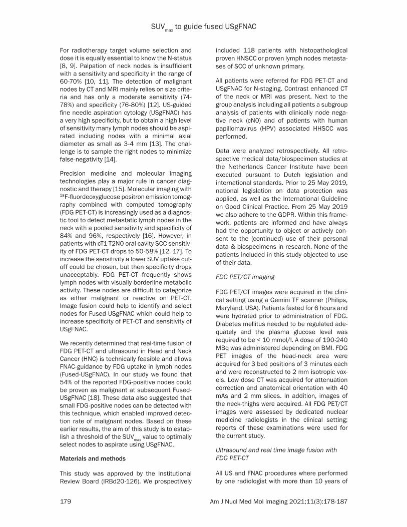
Am J Nucl Med Mol Imaging 2021;11(3):178-187 www.ajnmmi.us /ISSN:2160-8407/ajnmmi0131897 Original Article SUV max values at FDG PET-CT to predict malignancy in lymph nodes aspirated by real time image fused USgFNAC in head and neck squamous cell carcinoma Petra K de Koekkoek-Doll 1 , Wouter Vogel 2,3 , Monique Maas 1 , Jonas Castelijns 1 , Laura Smit 4 , Joannis Zavrakidis 5 , Regina Beets-Tan 1 , Michiel van den Brekel 6,7 Departments of 1 Radiology, 2 Nuclear Medicine, 3 Radiation Oncology, 4 Pathology, 5 Epidemiology & Biostatistics, 6 Head and Neck Surgery & Oncology, The Netherlands Cancer Institute, Amsterdam, Netherlands; 7 Department of Maxillofacial Surgery, Amsterdam University Medical Center, Amsterdam, Netherlands Received February 15, 2021; Accepted May 17, 2021; Epub June 15, 2021; Published June 30, 2021 Abstract: 18 F-fluordeoxyglucose (FDG) positron emission tomography combined with computed tomography (PET-CT) and ultrasound guided fine-needle aspiration cytology (USgFNAC) are commonly used to detect nodal metastases in head and neck squamous cell carcinoma (HNSCC). FDG PET-CT helps to guide selection of borderline suspicious nodes to aspirate using USgFNAC. Real time image fusion of FDG PET-CT with US is a new available technique and can improve this selection. The aim of this study was to determine optimal SUV max values for USgFNAC node selec- tion to improve USgFNAC sensitivity. 118 patients, with histopathological proven HNSCC or proven lymph nodes metastases of SCC of unknown primary, referred for staging of HNSCC with FDG PET-CT and ultrasound, were pro- spectively included. Additionally to standard USgFNAC of suspicious nodes fusion was performed to confirm that USgFNAC took place in FDG-positive nodes and to add Fused-USgFNAC in missed FDG-positive nodes. Fusion was performed on nodes with reported having metabolic activity. SUV max values were measured in all Fused-USgFNAC nodes. The reference standard was cytology. In 118 patients USgFNAC was performed in 281 nodes. At fusion 22/281 (8%) nodes were FDG-negative. Out of 259 FDG-positive nodes 253 (98%) nodes were fused successfully. USgFNAC had conclusive results in 237/253 nodes (94%). In 126/237 nodes (53%) cytology proved to be tumor positive. Below SUV max of 2.87 no fused FDG-positive nodes proved to be tumor positive at cytology. To improve sen- sitivity, only FDG-positive nodes with SUV max values above 2.87 should be selected for USgFNAC. Image fusion can identify those nodes for USgFNAC selection. Keywords: Head and neck cancer, lymph node metastasis staging, hybrid imaging, real-time image fusion, ultra- sound FDG-PET, SUV max Introduction The prognosis of head and neck squamous cell carcinoma (HNSSC) depends on many factors. Especially in HPV negative tumors the pres- ence or absence of metastatic lymph nodes (N-stage) is one of the most important predic- tors for loco regional control and risk for distant metastasis and thereby highly influences the patient’s management [1]. The number of metastases, their laterality, the involved node levels and the presence of extracapsular spread are important predictive parameters [2]. In a systematic review Lodder et al. report- ed that increasing volume of involved nodes is correlated with worsening outcome [3]. Management of cervical nodes often includes neck dissection (ND), radiotherapy (RT) or che- mo-radiation (CRT). The extent of ND and RT as well as the dose of RT depend on the N-stage. Elective treatment is not always necessary if the risk of occult metastases is very low. To minimize treatment morbidity, accurate staging is thus very important [4, 5]. A systematic review and meta-analysis of elective neck dissection vs active surveillance of cT1-T2N0 squamous cell carcinoma (SCC) of the oral cavity showed that elective neck dissection (END) results in fewer regional recurrences than active surveil- lance [6]. Still, watchful waiting is considered a treatment option in case of a low risk for metas- tases and a very close follow-up of the neck [7].
-
Upload
khangminh22 -
Category
Documents
-
view
1 -
download
0
Transcript of ajnmmi0131897.pdf - e-Century Publishing Corporation
Am J Nucl Med Mol Imaging 2021;11(3):178-187www.ajnmmi.us /ISSN:2160-8407/ajnmmi0131897
Original ArticleSUVmax values at FDG PET-CT to predict malignancy in lymph nodes aspirated by real time image fused USgFNAC in head and neck squamous cell carcinoma
Petra K de Koekkoek-Doll1, Wouter Vogel2,3, Monique Maas1, Jonas Castelijns1, Laura Smit4, Joannis Zavrakidis5, Regina Beets-Tan1, Michiel van den Brekel6,7
Departments of 1Radiology, 2Nuclear Medicine, 3Radiation Oncology, 4Pathology, 5Epidemiology & Biostatistics, 6Head and Neck Surgery & Oncology, The Netherlands Cancer Institute, Amsterdam, Netherlands; 7Department of Maxillofacial Surgery, Amsterdam University Medical Center, Amsterdam, Netherlands
Received February 15, 2021; Accepted May 17, 2021; Epub June 15, 2021; Published June 30, 2021
Abstract: 18F-fluordeoxyglucose (FDG) positron emission tomography combined with computed tomography (PET-CT) and ultrasound guided fine-needle aspiration cytology (USgFNAC) are commonly used to detect nodal metastases in head and neck squamous cell carcinoma (HNSCC). FDG PET-CT helps to guide selection of borderline suspicious nodes to aspirate using USgFNAC. Real time image fusion of FDG PET-CT with US is a new available technique and can improve this selection. The aim of this study was to determine optimal SUVmax values for USgFNAC node selec-tion to improve USgFNAC sensitivity. 118 patients, with histopathological proven HNSCC or proven lymph nodes metastases of SCC of unknown primary, referred for staging of HNSCC with FDG PET-CT and ultrasound, were pro-spectively included. Additionally to standard USgFNAC of suspicious nodes fusion was performed to confirm that USgFNAC took place in FDG-positive nodes and to add Fused-USgFNAC in missed FDG-positive nodes. Fusion was performed on nodes with reported having metabolic activity. SUVmax values were measured in all Fused-USgFNAC nodes. The reference standard was cytology. In 118 patients USgFNAC was performed in 281 nodes. At fusion 22/281 (8%) nodes were FDG-negative. Out of 259 FDG-positive nodes 253 (98%) nodes were fused successfully. USgFNAC had conclusive results in 237/253 nodes (94%). In 126/237 nodes (53%) cytology proved to be tumor positive. Below SUVmax of 2.87 no fused FDG-positive nodes proved to be tumor positive at cytology. To improve sen-sitivity, only FDG-positive nodes with SUVmax values above 2.87 should be selected for USgFNAC. Image fusion can identify those nodes for USgFNAC selection.
Keywords: Head and neck cancer, lymph node metastasis staging, hybrid imaging, real-time image fusion, ultra-sound FDG-PET, SUVmax
Introduction
The prognosis of head and neck squamous cell carcinoma (HNSSC) depends on many factors. Especially in HPV negative tumors the pres-ence or absence of metastatic lymph nodes (N-stage) is one of the most important predic-tors for loco regional control and risk for distant metastasis and thereby highly influences the patient’s management [1]. The number of metastases, their laterality, the involved node levels and the presence of extracapsular spread are important predictive parameters [2]. In a systematic review Lodder et al. report-ed that increasing volume of involved nodes is correlated with worsening outcome [3].
Management of cervical nodes often includes neck dissection (ND), radiotherapy (RT) or che-mo-radiation (CRT). The extent of ND and RT as well as the dose of RT depend on the N-stage. Elective treatment is not always necessary if the risk of occult metastases is very low. To minimize treatment morbidity, accurate staging is thus very important [4, 5]. A systematic review and meta-analysis of elective neck dissection vs active surveillance of cT1-T2N0 squamous cell carcinoma (SCC) of the oral cavity showed that elective neck dissection (END) results in fewer regional recurrences than active surveil-lance [6]. Still, watchful waiting is considered a treatment option in case of a low risk for metas-tases and a very close follow-up of the neck [7].
SUVmax to guide fused USgFNAC
179 Am J Nucl Med Mol Imaging 2021;11(3):178-187
For radiotherapy target volume selection and dose it is equally essential to know the N-status [8, 9]. Palpation of neck nodes is insufficient with a sensitivity and specificity in the range of 60-70% [10, 11]. The detection of malignant nodes by CT and MRI mainly relies on size crite-ria and has only a moderate sensitivity (74-78%) and specificity (76-80%) [12]. US-guided fine needle aspiration cytology (USgFNAC) has a very high specificity, but to obtain a high level of sensitivity many lymph nodes should be aspi-rated including nodes with a minimal axial diameter as small as 3-4 mm [13]. The chal-lenge is to sample the right nodes to minimize false-negativity [14].
Precision medicine and molecular imaging technologies play a major rule in cancer diag-nostic and therapy [15]. Molecular imaging with 18F-fluordeoxyglucose positron emission tomog-raphy combined with computed tomography (FDG PET-CT) is increasingly used as a diagnos-tic tool to detect metastatic lymph nodes in the neck with a pooled sensitivity and specificity of 84% and 96%, respectively [16]. However, in patients with cT1-T2N0 oral cavity SCC sensitiv-ity of FDG PET-CT drops to 50-58% [12, 17]. To increase the sensitivity a lower SUV uptake cut-off could be chosen, but then specificity drops unacceptably. FDG PET-CT frequently shows lymph nodes with visually borderline metabolic activity. These nodes are difficult to categorize as either malignant or reactive on PET-CT. Image fusion could help to identify and select nodes for Fused-USgFNAC which could help to increase specificity of PET-CT and sensitivity of USgFNAC.
We recently determined that real-time fusion of FDG PET-CT and ultrasound in Head and Neck Cancer (HNC) is technically feasible and allows FNAC-guidance by FDG uptake in lymph nodes (Fused-USgFNAC). In our study we found that 54% of the reported FDG-positive nodes could be proven as malignant at subsequent Fused-USgFNAC [18]. These data also suggested that small FDG-positive nodes can be detected with this technique, which enabled improved detec-tion rate of malignant nodes. Based on these earlier results, the aim of this study is to estab-lish a threshold of the SUVmax value to optimally select nodes to aspirate using USgFNAC.
Materials and methods
This study was approved by the Institutional Review Board (IRBd20-126). We prospectively
included 118 patients with histopathological proven HNSCC or proven lymph nodes metasta-ses of SCC of unknown primary.
All patients were referred for FDG PET-CT and USgFNAC for N-staging. Contrast enhanced CT of the neck or MRI was present. Next to the group analysis including all patients a subgroup analysis of patients with clinically node nega-tive neck (cN0) and of patients with human papillomavirus (HPV) associated HHSCC was performed.
Data were analyzed retrospectively. All retro-spective medical data/biospecimen studies at the Netherlands Cancer Institute have been executed pursuant to Dutch legislation and international standards. Prior to 25 May 2019, national legislation on data protection was applied, as well as the International Guideline on Good Clinical Practice. From 25 May 2019 we also adhere to the GDPR. Within this frame-work, patients are informed and have always had the opportunity to object or actively con-sent to the (continued) use of their personal data & biospecimens in research. None of the patients included in this study objected to use of their data.
FDG PET/CT imaging
FDG PET/CT images were acquired in the clini-cal setting using a Gemini TF scanner (Philips, Maryland, USA). Patients fasted for 6 hours and were hydrated prior to administration of FDG. Diabetes mellitus needed to be regulated ade-quately and the plasma glucose level was required to be < 10 mmol/l. A dose of 190-240 MBq was administered depending on BMI. FDG PET images of the head-neck area were acquired for 3 bed positions of 3 minutes each and were reconstructed to 2 mm isotropic vox-els. Low dose CT was acquired for attenuation correction and anatomical orientation with 40 mAs and 2 mm slices. In addition, images of the neck-thighs were acquired. All FDG PET/CT images were assessed by dedicated nuclear medicine radiologists in the clinical setting; reports of these examinations were used for the current study.
Ultrasound and real time image fusion with FDG PET-CT
All US and FNAC procedures where performed by one radiologist with more than 10 years of
SUVmax to guide fused USgFNAC
180 Am J Nucl Med Mol Imaging 2021;11(3):178-187
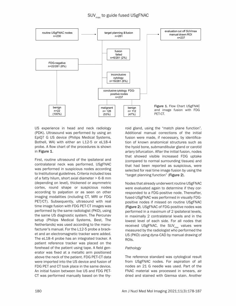
US experience in head and neck radiology (PDK). Ultrasound was performed by using an EpiQ7 G US device (Philips Medical Systems, Bothell, WA) with either an L12-5 or eL18-4 probe. A flow chart of the procedures is shown in Figure 1.
First, routine ultrasound of the ipsilateral and contralateral neck was performed. USgFNAC was performed in suspicious nodes according to Institutional guidelines. Criteria included loss of a fatty hilum, short axial diameter > 6-8 mm (depending on level), thickened or asymmetric cortex, round shape or suspicious nodes according to palpation or as seen on other imaging modalities (including CT, MRI or FDG PET/CT). Subsequently, ultrasound with real time image fusion with FDG PET-CT images was performed by the same radiologist (PKD), using the same US diagnostic system. The Percunav setup (Philips Medical Systems, Best, The Netherlands) was used according to the manu-facturer’s manual. For the L12-5 probe a brack-et and an electromagnetic tracker were added. The eL18-4 probe has an integrated tracker. A patient reference tracker was placed on the forehead of the patient using tape. A field gen-erator was fixed at a metallic arm positioned above the neck of the patient. FDG PET-CT data were imported into the US device and fusion of FDG PET and CT took place in the same device. An initial fusion between live US and FDG PET-CT was performed manually based on the thy-
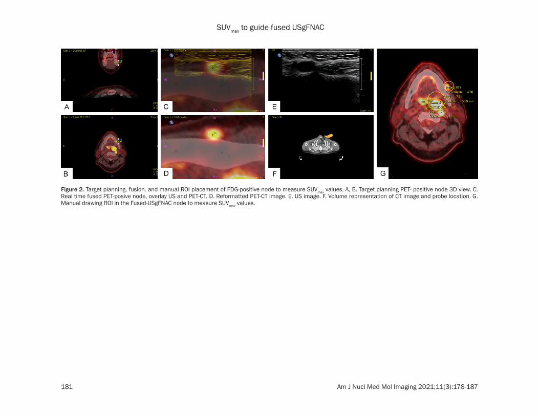
roid gland, using the “match plane function”. Additional manual corrections of the initial fusion were made, if necessary, by identifica-tion of known anatomical structures such as the hyoid bone, submandibular gland or carotid artery bifurcation. After the initial fusion, nodes that showed visible increased FDG uptake (compared to normal surrounding tissues) and that had been reported as suspicious, were selected for real time image fusion by using the “target planning function” (Figure 2).
Nodes that already underwent routine USgFNAC were evaluated again to determine if they cor-responded to a FDG-positive node. Thereafter, fused-USgFNAC was performed in visually FDG-positive nodes if missed on routine USgFNAC (Figure 2). USgFNAC of FDG-positive nodes was performed in a maximum of 2 ipsilateral levels, in maximally 2 contralateral levels and in the lowest level of each side. For all nodes that received USgFNAC, the SUVmax values were measured by the radiologist who performed the US (PKD) using dyna-CAD by manual drawing of ROIs.
Pathology
The reference standard was cytological result from USgFNAC nodes. For aspiration of all nodes an 21 G needle was used. Part of the FNAC material was processed in smears, air dried and stained with Giemsa stain. Another
Figure 1. Flow Chart USgFNAC and image fusion with FDG PET-CT.
SUVmax to guide fused USgFNAC
181 Am J Nucl Med Mol Imaging 2021;11(3):178-187
Figure 2. Target planning. fusion. and manual ROI placement of FDG-positive node to measure SUVmax values. A, B. Target planning PET- positive node 3D view. C. Real time fused PET-posive node, overlay US and PET-CT. D. Reformatted PET-CT image. E. US image. F. Volume representation of CT image and probe location. G. Manual drawing ROI in the Fused-USgFNAC node to measure SUVmax values.

SUVmax to guide fused USgFNAC
182 Am J Nucl Med Mol Imaging 2021;11(3):178-187
part of every aspirate was fixed in 10 ml 4% for-malin and embedded in paraffin for further immunohistochemistry if necessary, according to routine diagnostic workup. For clinically stag-ing all samples were evaluated by experienced head and neck pathologists, the cytological results of the clinically setting were used retro-spectively for the current study. HPV status was immunohistochemically assessed on formalin-fixed paraffin-embedded tissue samples from tumor biopsies or resections during standard routine diagnostic procedures. Antibodies for p53 (DO-7, 1/7000, DAKO) and p16 (E6H4; ready to use, Ventana Medical systems/Roche) were used in a Benchmark ULTRA autostainer (Ventane Medical systemsReactions were detected using OptiView DAB Detection kit (#760-700; Roche) for visualization p16 and p53. Finally, the slides were counterstained with Hematoxylin II and Bluing Reagent (Ventana Medical Systems).
Statistical evaluation
Calculation of sensitivity and specificity of the techniques was not possible as not all patients had a neck dissection with a pathology report. We therefore scored the percentage of positive cytology in relation to the SUV values in all nodes that were visually FDG-positive and aspi-rated at USgFNAC using image fusion. PET-negative nodes, nodes with failed fusion and nodes with inconclusive cytological results were excluded for further evaluation. Baseline characteristics were evaluated by descriptive statistics. A two-sided Independent samples t-test was used to compare the groups and sub-groups and the two groups with positive and negative cytology based on their short axis diameter and SUVmax. A p-value of </= 0.05 was considered statistically significant.
Results
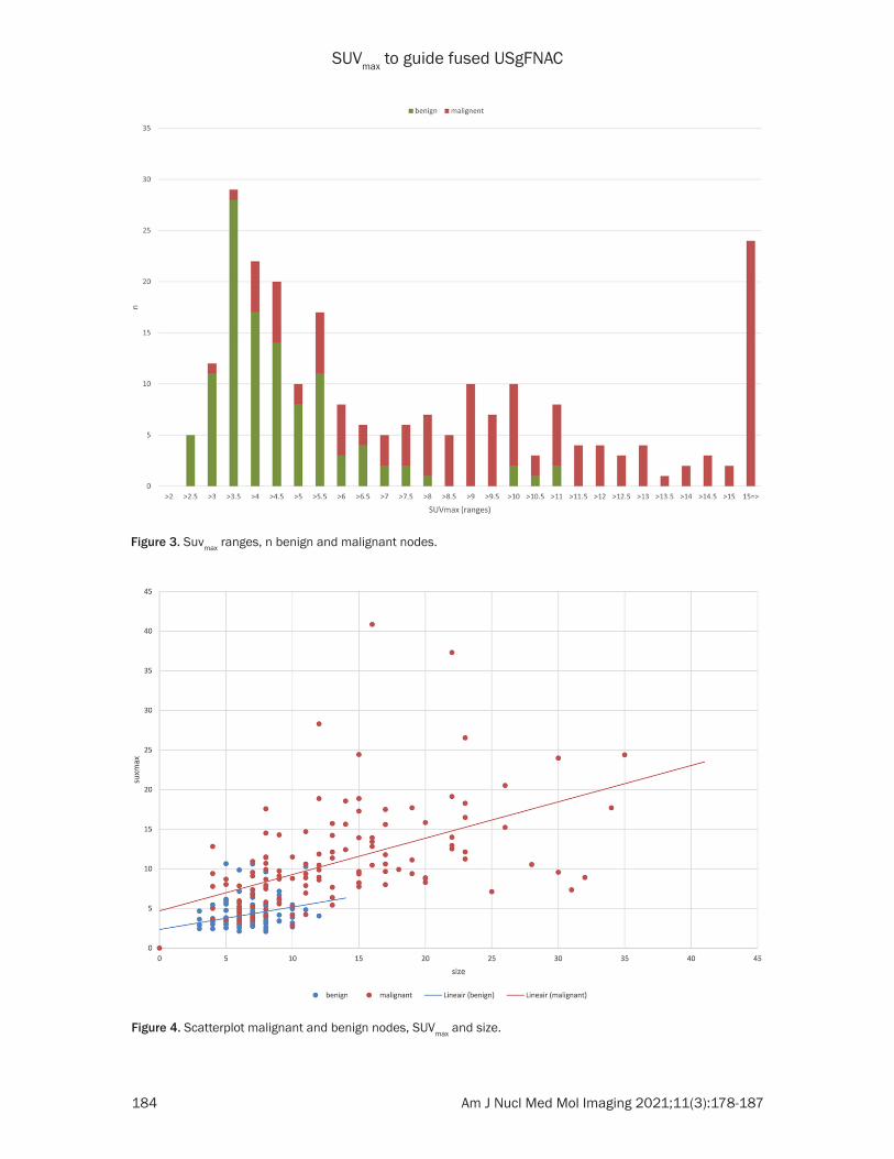
A total of 118 patients (median age of 63; range 32-89) with HNSCC were included. Diagnoses are shown in Table 1. In these 118 patients USgFNAC was performed in 281 nodes (Figure 1) Out of 281 nodes, 22 (8%) nodes were FDG-negative and all of these 22 nodes proved to be tumor negative at cytology. Image fusion failed in another 6 nodes (2%). Of the remaining 253 FDG-positive nodes, 16 (6%) had inconclusive cytological findings. These nodes were excluded from further evaluation. From the remaining, with image fusion con-firmed 237 FDG-positive nodes, 126 (53%) had malignant cytology. The median size (diameter short axis) of all FDG-positive evaluated nodes was 10.3 mm (range 3-35 mm). The average size of nodes with malignant cytology was sig-nificantly larger than nodes with benign cytolo-gy, with a median size (diameter short axis) of 13.4 mm (range 4-35 mm) compared to 6.7 mm (range 3-15 mm), p-value < 0.0001. The mean SUVmax of all evaluated FDG-positive nodes was 7.8 (SD 5.8). The mean SUVmax in nodes with malignant cytology was also signifi-cant higher with an average 11.0 (SD 6.3) com-pared to benign nodes with an average 4.3 (SD 1.7), p-value < 0.0001 (Table 2). The lowest SUVmax in nodes with malignant cytology was 2.87 and the highest SUVmax in nodes with benign cytology was 10.7 (Table 3; Figures 3, 4).
Subgroup analysis of clinically node negative neck (cN0) patients
Clinically 35/118 (30%) patients had a cN0 neck. In these patients, USgFNAC was per-formed in 71 FDG-positive nodes and 20 out of these (28%) had malignant cytology. The medi- The medi-The medi-
Table 1. Diagnosis of all patientsDiagnose n % HPV HPV+ % HPV- %SCC unkown primary 12 12.2% 12 6 50% 6 50%SCC oral cavity 25 21.1%SCC oropharyngeal 33 28.0% 33 18 54% 15 46%SCC hypopharyngeal 12 10.2%SCC laryngeal 23 19.5%SCC nasal cavity paranasal sinuses 6 5.1% 4 2 50% 2 50%SCC nasopharyngeal 6 5.1% 4 2 50% 2 50%SCC cutaneus 1 0.8%total 118 100% 53 28 53% 23 47%
SUVmax to guide fused USgFNAC
183 Am J Nucl Med Mol Imaging 2021;11(3):178-187
Table 2. Mean SUVmax and size (short-axis diameter) in FDG-posi-tive nodes of all HNSCC patients
nodes n %size SUVmax
median min max median min maxmalignant 125 53% 13.4 4 35 11.0 2.87 40.84benign 112 47% 6.7 3 15 4.3 1.95 10.7all 237 100% 10.3 3 35 7.8 1.95 40.84
Table 3. SUVmax in ranges and number of malignant and benign nodes
SUVmax nodes benign malignant
> ≤ n % mean mm n % n %2 0 0% 0 0% 0 0%
2 2.5 5 2% 5.80 5 5% 0 0%2.5 3 12 5% 6.42 11 10% 1 1%3 3.5 29 12% 6.55 28 25% 1 1%3.5 4 22 9% 6.14 17 15% 5 4%4 4.5 20 8% 7.95 14 13% 6 5%4.5 5 10 4% 6.80 8 7% 2 2%5 5.5 17 7% 7.24 11 10% 6 5%5.5 6 8 3% 7.00 3 3% 5 4%6 6.5 6 3% 8.00 4 4% 2 2%6.5 7 5 2% 8.20 2 2% 3 2%7 7.5 6 3% 14.33 2 2% 4 3%7.5 8 7 3% 9.29 1 1% 6 5%8 8.5 5 2% 14.40 0 0% 5 4%8.5 9 10 4% 13.90 0 0% 10 8%9 9.5 7 3% 10.57 0 0% 7 6%9.5 10 10 4% 13.00 2 2% 8 6%10 10.5 3 1% 13.00 1 1% 2 2%10.5 11 8 3% 12.50 2 2% 6 5%11 11.5 4 2% 15.75 0 0% 4 3%11.5 12 4 2% 11.75 0 0% 4 3%12 12.5 3 1% 16.67 0 0% 3 2%12.5 13 4 2% 16.00 0 0% 4 3%13 13.5 1 0% 16.00 0 0% 1 1%13.5 14 2 1% 15.50 0 0% 2 2%14 14.5 3 1% 14.67 0 0% 3 2%14.5 15 2 1% 9.50 0 0% 2 2%15 24 10% 19.63 0 0% 24 19%
an short axis of nodes in cN0 was significantly smaller, p-value 0.001 (7.8 mm, range 3-23) than in cN+ (10.6 mm, range 3-35). The median short axis of malignant cN0 nodes (10.1 range 4-23) was significant smaller, value 0.039 than in cN+ nodes (13.5, range 4-35). The mean SUVmax of cN0 nodes with malignant cytology
was significantly higher than in nodes with benign cytology, with an average of 8.3 (SD 5.0) versus 4.45 (SD 1.8), p value < 0.003.
The mean SUVmax of malignant nodes in all cN+ necks was 13.5 (SD 7.2) while it was 8.3 (SD 5.0) in cN0 necks, which was significantly lower, p-value 0.002. The lowest SUVmax in nodes with malignant cytology was 3.6 and the highest SUVmax in nodes with benign cytology was 10.6.
Subgroup cut-off SUVmax values in HPV associated SSC of the oropharynx, nasopharynx and unknown primary
Immunohistochemically results of HPV related tumors were present in 53 patients.
In 28/53 patients the tumor was associated with HPV (HPV+) and USgFNAC was per-formed in 73 nodes. 48/73 nodes were malignant at cytol-ogy. Mean short axis was 13.8 mm (SD 7.1) and mean SUVmax was 9.6 (SD 4.8). 25/78 nodes were benign at cytology. Mean short axis was 7.2 mm (SD 1.99) and mean SUVmax was 4.96 (SD 1.99). The lowest SUVmax in nodes with malignant cytology was 3.39 and the high-est SUVmax in nodes with benign cytology was 10.27.
In 25/53 patients the tumor was HPV negative (HPV-) and USgFNAC was performed in 45 nodes. 26/45 nodes were malignant at cytology. Mean
short axis was 12.2 mm (SD 7.2), mean SUVmax was 10.9 (SD 5.1). 19/45 nodes were benign at cytology. Mean short axis was 7.4 mm (SD 2.2), mean SUVmax was 4.2 (SD 2.0).
The lowest SUVmax in nodes with malignant cytology was 2.87 and the highest SUVmax in
SUVmax to guide fused USgFNAC
184 Am J Nucl Med Mol Imaging 2021;11(3):178-187
Figure 3. Suvmax ranges, n benign and malignant nodes.
Figure 4. Scatterplot malignant and benign nodes, SUVmax and size.
SUVmax to guide fused USgFNAC
185 Am J Nucl Med Mol Imaging 2021;11(3):178-187
nodes with benign cytology was 10.6. Mean SUVmax in malignant HPV- nodes was 10.6 and in malignant HPV+ nodes 9.6, statistically not significant p value = 0.279.
Discussion
The presence of cervical lymph node metasta-ses in HNSCC has a large impact on patients prognosis and treatment [19, 20]. To detect occult nodal metastases in clinically node neg-ative neck FDG PET-CT is slightly superior to MRI, CT or ultrasound [16, 21]. On the other hand, however, regarding the detection of cervi-cal lymph node metastases in cN0 HNSCC patients, a recent meta-analysis still showed a low sensitivity and moderate specificity of FDG PET-CT [17]. This can be explained by the non-specific nature of FDG uptake in small nodes and the difficulty to distinguish between benign, reactive or inflammatory on the one hand and malignant nodes other hand. Although ultra-sound enables detection of enlarged nodes and enables evaluation of additional morpho-logical features of nodes, it does not enable detection of micro-metastases in small nodes [22]. In our study only 22/281 nodes which underwent USgFNAC where FDG-negative, none of these nodes proved to be malignant. On ultrasound it might be difficult to identify tiny FDG-positive nodes with regular morpholo-gy and the major challenge performing USgFNAC is to select the right tiny FDG-positive node to puncture (based on imaging features on US but also other modalities), whereas the major issue in FDG PET-CT is, how borderline SUV values should be interpreted. Generally, a node with a SUVmax value above 4.5 is consid-ered to be metastatic, whereas the nature of nodes with a SUV-value below this value remains uncertain [23].
In our study malignant FDG-positive nodes in cN0 HNSCC patients were significantly smaller than in cN+ HNSCC patients and had a signifi-cant lower mean SUVmax value.
These small nodes are the nodes that are diffi-cult to accurately select for aspiration with rou-tine US. Due to image fusion we were able to localize small FDG-positive nodes with a nor-mal morphological appearance on ultrasound to perform fused-USgFNAC. Out of the real time image fused guided FDG-positive FNAC nodes
53% proved to be malignant at cytology. Based on Fused USgFNAC none of the FDG-positive nodes with a SUVmax of </= 2.87 was malignant. Our data are comparable to a recent study in which comparing PET-CT-data with cytological results, below a SUVmax of 2.2 no malignant nodes were found [24]. Therefore, a threshold SUVmax > 2.87 can be used to guide US-FNAC. Using this value to select nodes for fused-USgF-NAC will likely increase the accuracy of detec-tion of malignant nodes in staging of HNSCC.
Based on Fused-USgFNAC at a SUVmax of 10.7 or higher all FDG-PET positive nodes were malignant and in fact in these nodes, aspira-tion might not be needed. A SUVmax value between 2.87 and 4.5 poses the major clinical problem, and in these cases, but also up to SUVmax 10.7 fused USgFNAC might be helpful to make the diagnosis.
Our results are also comparable with those in a study of Payabvash et al. who reported that at a SUVmax ≥ 2.5 sensitivity for detection of malig-nant nodes was 100%, with histopathological results as reference standard [25]. Other stud-ies that correlated with histology found higher cut-offs for SUVmax values for malignant nodes. Dequanter et al. compared FDG-PET-CT with histology after neck dissection and proposed a SUVmax cut-off-value of 4.05 to detect malignant nodes [26]. Using this cut-off-value in the cur-rent study 13 malignant nodes would have been missed. Because accurate N-stage is essential for treatment planning, a lower cut-off SUVmax, like >/= 2.87, is preferred to guide Fused-USgFNAC. To minimalize false negative results because of sampling errors, USgFNAC should be repeated in nodes with benign cyto-logical results and a SUVmax of >/= 2.87, if rele-vant for treatment decision.
SUVmax values are reported significantly higher in HPV-negative nodes than in HPV-positive nodes [27]. In a meta-analysis of Fleming et al, a higher SUVmax value in HPV-negative tumors was found but PET SUVmax scores were unable to reliable differentiate between HPV-positive and HPV-negative tumors [28]. These results are comparable to our results with a statisti-cally not significant higher mean SUVmax value in malignant HPV-negative nodes than in malig-nant HPV-positive nodes.
SUVmax to guide fused USgFNAC
186 Am J Nucl Med Mol Imaging 2021;11(3):178-187
Limitations
Because all Fused-USgFNAC and measure-ments were performed by one radiologist, the interobserver variability of the procedure is unknown.
In this study histopathological results of neck dissection as a gold standard are not available; therefore sampling error, leading to false nega-tive results of cytology cannot be excluded.
Conclusion
To improve the sensitivity, for FDG-positive nodes with a benign morphological appearance on ultrasound, a SUVmax value ≥ 2.87 should be used to select nodes for USgFNAC. Those FDG-positive nodes can be identified by real time image fusion and Fused-USgFNAC can be performed.
Acknowledgements
The authors would like to acknowledge Pedro Sanches, PhD and Frans Gleuwink (Philips Benelux, Boschdijk 525, 5621JG Eindhoven) for providing knowledge and training on Percunav image fusion (MR, CT, PET).
Disclosure of conflict of interest
None.
Address correspondence to: Dr. Petra K de Koekkoek-Doll, Department of Radiology, The Netherlands Cancer Institute, Plesmanlaan 121, 1066 CX, Amsterdam, The Netherlands. Tel: +31 20 5122003; E-mail: [email protected]
References
[1] Godény M. Prognostic factors in advanced pharyngeal and oral cavity cancer; Significance of multimodality imaging in terms of 7th edi-tion of TNM. Cancer Imaging 2014; 14: 1-13.
[2] Woolgar JA. Histopathological prognosticators in oral and oropharyngeal squamous cell carci-noma. Oral Oncol 2006; 42: 229-239.
[3] Lodder WL, Pameijer FA, Rasch CR, van den Brekel MW and Balm AJ. Prognostic signifi-cance of radiologically determined neck node volume: a systematic review. Oral Oncol 2012; 48: 298-302.
[4] Al-Mamgani A, Verheij M and van den Brekel MWM. Elective unilateral nodal irradiation in
head and neck squamous cell carcinoma: a paradigm shift. Eur J Cancer 2017; 82: 1-5.
[5] Nieuwenhuis EJ, Castelijns JA, Pijpers R, van den Brekel MW, Brakenhoff RH, van der Waal I, Snow GB and Leemans CR. Wait-and-see poli-cy for the N0 neck in early-stage oral and oro-pharyngeal squamous cell carcinoma using ultrasonography-guided cytology: is there a role for identification of the sentinel node? Head Neck 2002; 24: 282-9.
[6] Abu-Ghanem S, Yehuda M, Carmel NN, Leshno M, Abergel A, Gutfeld O and Fliss DM. Elective neck dissection vs observation in early-stage squamous cell carcinoma of the oral tongue with no clinically apparent lymph node metas-tasis in the neck. JAMA Otolaryngol Neck Surg 2016; 142: 857.
[7] Koyama LKS, Matos LL, Kulcsar MAV, de Araú-jo Filho VJF and Cernea CR. Oral cancer treat-ment: still an indication for elective neck dis-section? ORL 2018; 80: 96-102.
[8] van den Bosch S, Vogel WV, Raaijmakers CP, Dijkema T, Terhaard CHJ, Al-Mamgani A and Kaanders JHAM. Implications of improved di-agnostic imaging of small nodal metastases in head and neck cancer: radiotherapy target vol-ume transformation and dose de-escalation. Radiother Oncol 2018; 128: 472-478.
[9] Navran A, Heemsbergen W, Janssen T, Ham-ming-Vrieze O, Jonker M, Zuur C, Verheij M, Re-meijer P, Sonke JJ, van den Brekel M and Al-Mamgani A. The impact of margin reduction on outcome and toxicity in head and neck cancer patients treated with image-guided volumetric modulated arc therapy (VMAT). Radiother On-col 2019; 130: 25-31.
[10] van den Brekel MW, Castelijns JA, Stel HV, van der Waal I, Tiwari RM and Snow GB. Diagnostiek van lymfekliermetastasen van hoofd-halscarcinomen. Ned Tijdschr Geneeskd 1995; 139: 1075-8.
[11] Geetha NT, Hallur N, Goudar G, Sikkerimath BC and Gudi SS. Cervical lymph node metastasis in oral squamous carcinoma preoperative as-sessment and histopathology after neck dis-section. J Maxillofac Oral Surg 2010; 9: 42-7.
[12] Kyzas PA, Evangelou E, Denaxa-Kyza D and Io-annidis JP. 18F-fluorodeoxyglucose positron emission tomography to evaluate cervical node metastases in patients with head and neck squamous cell carcinoma: a meta-analy-sis. J Natl Cancer Inst 2008; 100: 712-720.
[13] van den Brekel MW and Castelijns JA. What the clinician wants to know: surgical perspective and ultrasound for lymph node imaging of the neck. Cancer Imaging 2005; 5 Spec No: 0-16.
[14] Borgemeester MC, van den Brekel MW, van Tinteren H, Smeele LE, Pameijer FA, van Velthuysen ML and Balm AJ. Ultrasound-guid-
SUVmax to guide fused USgFNAC
187 Am J Nucl Med Mol Imaging 2021;11(3):178-187
ed aspiration cytology for the assessment of the clinically N0 neck: factors influencing its accuracy. Head Neck 2008; 30: 1505-1513.
[15] Ghasemi M, Nabipour I, Omrani A, Alipour Z and Assadi M. Precision medicine and molecu-lar imaging: new targeted approaches toward cancer therapeutic and diagnosis. Am J Nucl Med Mol Imaging 2016; 6: 310-327.
[16] Sun R, Tang X, Yang Y and Zhang C. (18)FDG-PET/CT for the detection of regional nodal me-tastasis in patients with head and neck can-cer: a meta-analysis. Oral Oncol 2015; 51: 314-20.
[17] Kim SJ, Pak K and Kim K. Diagnostic accuracy of F-18 FDG PET or PET/CT for detection of lymph node metastasis in clinically node nega-tive head and neck cancer patients; A system-atic review and meta-analysis. Am J Otolaryn-gol 2019; 40: 297-305.
[18] de Koekkoek-Doll PK, Maas M, Vogel W, Castelijns J, Smit L, Zavrakidis I, Beets-Tan R and van den Brekel M. Real-time ultrasound image fusion with FDG-PET/CT to perform fused image-guided fine-needle aspiration in neck nodes: feasibility and diagnostic value. AJNR Am J Neuroradiol 2021; 42: 566-572.
[19] Snow GB, Annyas AA, van Slooten EA, Bartelink H and Hart AA. Prognostic factors of neck node metastasis. Clin Otolaryngol Allied Sci 1982; 7: 185-92.
[20] Leemans CR, Tiwari R, Nauta JJ, van der Waal I and Snow GB. Regional lymph node involve-ment and its significance in the development of distant metastases in head and neck carci-noma. Cancer 1993; 71: 452-6.
[21] Yoon DY, Hwang HS, Chang SK, Rho YS, Ahn HY, Kim JH and Lee IJ. CT, MR, US, 18F-FDG PET/CT, and their combined use for the as-sessment of cervical lymph node metastases in squamous cell carcinoma of the head and neck. Eur Radiol 2009; 19: 634-642.
[22] Richards PS and Peacock TE. The role of ultra-sound in the detection of cervical lymph node metastases in clinically NO squamous cell car-cinoma of the head and neck. Cancer Imaging 2007; 7: 167-178.
[23] Lim RSM, Ramdave S, Beech P, Billah B, Karim MN, Smith JA, Safdar A and Sigston E. Utility of SUVmax on 18 F-FDG PET in detecting cervical nodal metastases. Cancer Imaging 2016; 16: 1-8.
[24] Peltenburg B, de Keizer B, Willem Dankbaar J, de Boer M, Willems SM, P Philippens ME, J Ter-haard CH and de Bree R. Acta Oncologica Pre-diction of ultrasound guided fine needle aspi-ration cytology results by FDG PET-CT for lymph node metastases in head and neck squamous cell carcinoma patients Prediction of ultra-sound guided fine needle aspiration cytology results by FDG PET-CT for lymph node metasta-ses in head and neck squamous cell carcino-ma patients. Acta Oncol 2018; 57: 1687-1692.
[25] Payabvash S, Meric K and Cayci Z. Differentia-tion of benign from malignant cervical lymph nodes in patients with head and neck cancer using PET/CT imaging. Clin Imaging 2016; 40: 101-105.
[26] Dequanter D, Shahla M, Aubert C, Deniz Y and Lothaire P. Prognostic value of FDG PET/CT in head and neck squamous cell carcinomas. Onco Targets Ther 2015; 8: 2279-2283.
[27] Tahari AK, Alluri K, Quon H, Koch W, Wahl RL, Subramaniam RM, Subramaniam R and Mor-gan RH. FDG PET/CT imaging of Oropharyn-geal SCC: characteristics of HPV positive and negative tumors. Clin Nucl Med 2014; 39: 225-231.
[28] Fleming JC, Woo J, Moutasim K, Mellone M, Frampton SJ, Mead A, Ahmed W, Wood O, Rob-inson H, Ward M, Woelk CH, Ottensmeier CH, King E, Kim D, Blaydes JP and Thomas GJ. HPV, tumour metabolism and novel target identification in head and neck squamous cell carcinoma. Br J Cancer 2019; 120: 356-367.