Agouti-related protein-deficient mice display an age-related lean phenotype
7
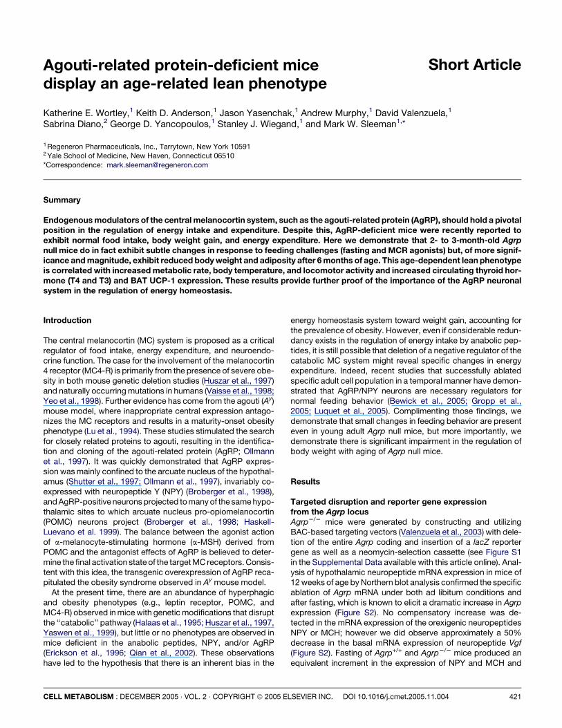
Short Article Agouti-related protein-deficient mice display an age-related lean phenotype Katherine E. Wortley, 1 Keith D. Anderson, 1 Jason Yasenchak, 1 Andrew Murphy, 1 David Valenzuela, 1 Sabrina Diano, 2 George D. Yancopoulos, 1 Stanley J. Wiegand, 1 and Mark W. Sleeman 1, * 1 Regeneron Pharmaceuticals, Inc., Tarrytown, New York 10591 2 Yale School of Medicine, New Haven, Connecticut 06510 *Correspondence: [email protected] Summary Endogenous modulators of the central melanocortin system, such as the agouti-related protein (AgRP), should hold a pivotal position in the regulation of energy intake and expenditure. Despite this, AgRP-deficient mice were recently reported to exhibit normal food intake, body weight gain, and energy expenditure. Here we demonstrate that 2- to 3-month-old Agrp null mice do in fact exhibit subtle changes in response to feeding challenges (fasting and MCR agonists) but, of more signif- icance and magnitude, exhibit reduced body weight and adiposity after 6 months of age. This age-dependent lean phenotype is correlated with increased metabolic rate, body temperature, and locomotor activity and increased circulating thyroid hor- mone (T4 and T3) and BAT UCP-1 expression. These results provide further proof of the importance of the AgRP neuronal system in the regulation of energy homeostasis. Introduction The central melanocortin (MC) system is proposed as a critical regulator of food intake, energy expenditure, and neuroendo- crine function. The case for the involvement of the melanocortin 4 receptor (MC4-R) is primarily from the presence of severe obe- sity in both mouse genetic deletion studies (Huszar et al., 1997) and naturally occurring mutations in humans (Vaisse et al., 1998; Yeo et al., 1998). Further evidence has come from the agouti (A y ) mouse model, where inappropriate central expression antago- nizes the MC receptors and results in a maturity-onset obesity phenotype (Lu et al., 1994). These studies stimulated the search for closely related proteins to agouti, resulting in the identifica- tion and cloning of the agouti-related protein (AgRP; Ollmann et al., 1997). It was quickly demonstrated that AgRP expres- sion was mainly confined to the arcuate nucleus of the hypothal- amus (Shutter et al., 1997; Ollmann et al., 1997), invariably co- expressed with neuropeptide Y (NPY) (Broberger et al., 1998), and AgRP-positive neurons projected to many of the same hypo- thalamic sites to which arcuate nucleus pro-opiomelanocortin (POMC) neurons project (Broberger et al., 1998; Haskell- Luevano et al. 1999). The balance between the agonist action of a-melanocyte-stimulating hormone (a-MSH) derived from POMC and the antagonist effects of AgRP is believed to deter- mine the final activation state of the target MC receptors. Consis- tent with this idea, the transgenic overexpression of AgRP reca- pitulated the obesity syndrome observed in A y mouse model. At the present time, there are an abundance of hyperphagic and obesity phenotypes (e.g., leptin receptor, POMC, and MC4-R) observed in mice with genetic modifications that disrupt the ‘‘catabolic’’ pathway (Halaas et al., 1995; Huszar et al., 1997, Yaswen et al., 1999), but little or no phenotypes are observed in mice deficient in the anabolic peptides, NPY, and/or AgRP (Erickson et al., 1996; Qian et al., 2002). These observations have led to the hypothesis that there is an inherent bias in the energy homeostasis system toward weight gain, accounting for the prevalence of obesity. However, even if considerable redun- dancy exists in the regulation of energy intake by anabolic pep- tides, it is still possible that deletion of a negative regulator of the catabolic MC system might reveal specific changes in energy expenditure. Indeed, recent studies that successfully ablated specific adult cell population in a temporal manner have demon- strated that AgRP/NPY neurons are necessary regulators for normal feeding behavior (Bewick et al., 2005; Gropp et al., 2005; Luquet et al., 2005). Complimenting those findings, we demonstrate that small changes in feeding behavior are present even in young adult Agrp null mice, but more importantly, we demonstrate there is significant impairment in the regulation of body weight with aging of Agrp null mice. Results Targeted disruption and reporter gene expression from the Agrp locus Agrp 2/2 mice were generated by constructing and utilizing BAC-based targeting vectors (Valenzuela et al., 2003) with dele- tion of the entire Agrp coding and insertion of a lacZ reporter gene as well as a neomycin-selection cassette (see Figure S1 in the Supplemental Data available with this article online). Anal- ysis of hypothalamic neuropeptide mRNA expression in mice of 12 weeks of age by Northern blot analysis confirmed the specific ablation of Agrp mRNA under both ad libitum conditions and after fasting, which is known to elicit a dramatic increase in Agrp expression (Figure S2). No compensatory increase was de- tected in the mRNA expression of the orexigenic neuropeptides NPY or MCH; however we did observe approximately a 50% decrease in the basal mRNA expression of neuropeptide Vgf (Figure S2). Fasting of Agrp +/+ and Agrp 2/2 mice produced an equivalent increment in the expression of NPY and MCH and CELL METABOLISM : DECEMBER 2005 $ VOL. 2 $ COPYRIGHT ª 2005 ELSEVIER INC. DOI 10.1016/j.cmet.2005.11.004 421
-
Upload
independent -
Category
Documents
-
view
3 -
download
0
Transcript of Agouti-related protein-deficient mice display an age-related lean phenotype
Short ArticleAgouti-related protein-deficient micedisplay an age-related lean phenotype
Katherine E. Wortley,1 Keith D. Anderson,1 Jason Yasenchak,1 Andrew Murphy,1 David Valenzuela,1
Sabrina Diano,2 George D. Yancopoulos,1 Stanley J. Wiegand,1 and Mark W. Sleeman1,*
1 Regeneron Pharmaceuticals, Inc., Tarrytown, New York 105912 Yale School of Medicine, New Haven, Connecticut 06510
*Correspondence: [email protected]
Summary
Endogenous modulators of the central melanocortin system, such as the agouti-related protein (AgRP), should hold a pivotalposition in the regulation of energy intake and expenditure. Despite this, AgRP-deficient mice were recently reported toexhibit normal food intake, body weight gain, and energy expenditure. Here we demonstrate that 2- to 3-month-old Agrpnull mice do in fact exhibit subtle changes in response to feeding challenges (fasting and MCR agonists) but, of more signif-icance and magnitude, exhibit reduced body weight and adiposity after 6 months of age. This age-dependent lean phenotypeis correlated with increased metabolic rate, body temperature, and locomotor activity and increased circulating thyroid hor-mone (T4 and T3) and BAT UCP-1 expression. These results provide further proof of the importance of the AgRP neuronalsystem in the regulation of energy homeostasis.
Introduction
The central melanocortin (MC) system is proposed as a criticalregulator of food intake, energy expenditure, and neuroendo-crine function. The case for the involvement of the melanocortin4 receptor (MC4-R) is primarily from the presence of severe obe-sity in both mouse genetic deletion studies (Huszar et al., 1997)and naturally occurring mutations in humans (Vaisse et al., 1998;Yeo et al., 1998). Further evidence has come from the agouti (Ay)mouse model, where inappropriate central expression antago-nizes the MC receptors and results in a maturity-onset obesityphenotype (Lu et al., 1994). These studies stimulated the searchfor closely related proteins to agouti, resulting in the identifica-tion and cloning of the agouti-related protein (AgRP; Ollmannet al., 1997). It was quickly demonstrated that AgRP expres-sion was mainly confined to the arcuate nucleus of the hypothal-amus (Shutter et al., 1997; Ollmann et al., 1997), invariably co-expressed with neuropeptide Y (NPY) (Broberger et al., 1998),and AgRP-positive neurons projected to many of the same hypo-thalamic sites to which arcuate nucleus pro-opiomelanocortin(POMC) neurons project (Broberger et al., 1998; Haskell-Luevano et al. 1999). The balance between the agonist actionof a-melanocyte-stimulating hormone (a-MSH) derived fromPOMC and the antagonist effects of AgRP is believed to deter-mine the final activation state of the target MC receptors. Consis-tent with this idea, the transgenic overexpression of AgRP reca-pitulated the obesity syndrome observed in Ay mouse model.
At the present time, there are an abundance of hyperphagicand obesity phenotypes (e.g., leptin receptor, POMC, andMC4-R) observed in mice with genetic modifications that disruptthe ‘‘catabolic’’ pathway (Halaas et al., 1995; Huszar et al., 1997,Yaswen et al., 1999), but little or no phenotypes are observed inmice deficient in the anabolic peptides, NPY, and/or AgRP(Erickson et al., 1996; Qian et al., 2002). These observationshave led to the hypothesis that there is an inherent bias in the
CELL METABOLISM : DECEMBER 2005 $ VOL. 2 $ COPYRIGHT ª 2005 E
energy homeostasis system toward weight gain, accounting forthe prevalence of obesity. However, even if considerable redun-dancy exists in the regulation of energy intake by anabolic pep-tides, it is still possible that deletion of a negative regulator of thecatabolic MC system might reveal specific changes in energyexpenditure. Indeed, recent studies that successfully ablatedspecific adult cell population in a temporal manner have demon-strated that AgRP/NPY neurons are necessary regulators fornormal feeding behavior (Bewick et al., 2005; Gropp et al.,2005; Luquet et al., 2005). Complimenting those findings, wedemonstrate that small changes in feeding behavior are presenteven in young adult Agrp null mice, but more importantly, wedemonstrate there is significant impairment in the regulation ofbody weight with aging of Agrp null mice.
Results
Targeted disruption and reporter gene expressionfrom the Agrp locusAgrp2/2 mice were generated by constructing and utilizingBAC-based targeting vectors (Valenzuela et al., 2003) with dele-tion of the entire Agrp coding and insertion of a lacZ reportergene as well as a neomycin-selection cassette (see Figure S1in the Supplemental Data available with this article online). Anal-ysis of hypothalamic neuropeptide mRNA expression in mice of12 weeks of age by Northern blot analysis confirmed the specificablation of Agrp mRNA under both ad libitum conditions andafter fasting, which is known to elicit a dramatic increase in Agrpexpression (Figure S2). No compensatory increase was de-tected in the mRNA expression of the orexigenic neuropeptidesNPY or MCH; however we did observe approximately a 50%decrease in the basal mRNA expression of neuropeptide Vgf(Figure S2). Fasting of Agrp+/+ and Agrp2/2 mice produced anequivalent increment in the expression of NPY and MCH and
LSEVIER INC. DOI 10.1016/j.cmet.2005.11.004 421

S H O R T A R T I C L E
increased Vgf expression, such that the difference in basal ex-pression of Vgf observed between the Agrp+/+ and Agrp2/2
mice was lost (Figure S2). Reporter expression was detectedspecifically in neurons of the arcuate nucleus and median emi-nence of the hypothalamus (Figure 1A), but not elsewhere inthe brain. Numerous cells in the adrenal medulla (Figure 1B)also exhibited specific reporter expression. Additional expres-sion was observed in the kidney in glomeruli-like structures(data not shown), consistent with previous reports of endoge-nous AgRP (Shutter et al., 1997), although no functional distur-bance in kidney function (serum creatinine; data not shown)could be detected in Agrp2/2 mice.
Agrp deletion does not impair metabolicparameters in young miceAnalysis in F2 generation mice revealed that Agrp2/2 mice wereviable and displayed no significant differences in the bodyweights of male or female as compared to their Agrp+/+ litter-mates (Figure 2A). Consistent with this, there were no significantdifferences in 24 hr food intake (Figure 2B) or metabolic rate, asassessed by oxygen consumption (VO2, Figure 2C), respiratoryquotient, or energy expenditure (data not shown) in mice at 8–10weeks of age. Locomotor activity was also similar between bothgroups of mice (Figure 2D).
Since changes in the expression of hypothalamic orexigenicneuropeptides, including Agrp, are thought to have importantphysiological consequences, such as stimulating reflex hyper-phagia following a fast, we were interested in monitoring this re-sponse in the Agrp2/2 mice. Following a 24 hr fast, Agrp+/+ miceshowed a small but significant increase in food intake at 1, 4, and24 hr during the re-feeding period (data not shown). In contrast,the Agrp2/2 mice showed no significant increase in food intakeat 1 or 4 hr post-fast. Food intake in the Agrp2/2 mice was sig-nificantly increased at 24 hr post-fast, although to a lesser extentthan in the Agrp+/+ mice (data not shown). Once again, the met-abolic parameters (VO2, respiratory quotient, and locomotor ac-tivity) during and after this period of food deprivation did not sig-nificantly differ between groups (data not shown). Body weightsof the Agrp+/+and Agrp2/2 mice at the start of the fast (Agrp+/+:21.1 6 0.51 versus Agrp2/2: 21.61 6 0.76) and at the end of there-feeding period (Agrp+/+: 20.25 6 0.66 versus Agrp2/2: 20.78g) were also similar.
If AgRP is indeed the endogenous antagonist of the melano-cortin system then its absence should cause a potentiated ano-rectic response induced by receptor agonists. To directly evalu-ate this, Agrp2/2 mice and Agrp+/+ littermates were placed inmetabolic cages and administered the melanocortin agonist,melanotan II (MT-II; 3 mg/kg i.p.), or phosphate-buffered saline(PBS) 2 hr before the onset of dark phase of the light cycle. InAgrp+/+ mice, MT-II potently inhibited food intake during the first1–4 hr of the nocturnal feeding period. This effect rapidly atten-uated over time such that in Agrp+/+ mice cumulative 24 hr foodintake in MT-II-treated animals was not significantly differentfrom intake in those injected with PBS (Figure 3A, ANOVA:F1,21 = 0.383; NS). The magnitude of the same dose of MT-IIon food intake was similar in Agrp2/2 mice over the first 4 hr ofthe dark period but was of prolonged duration (Figure 3B) suchthat cumulative food intake at 24 hr was significantly reduced(ANOVA effect of time x treatment: F1,21 = 4.236; p < 0.0001).
In addition, repeated administration of 3 mg/kg MT-II for 4consecutive days prevented Agrp2/2 mice from gaining weight
422
over the 4 day period (Figure 3C, effect of time x genotype: F1,4 =6.54; p = 0.0005). Whereas central administration of MT-II po-tently induces body weight loss, this chronic peripheral dosingregimen of MT-II was based on a previous study (Pierroz et al.,2002) and was intended to create a submaximal stimulation ofthe melanocortin pathway that would reveal a difference in sen-sitivity to melanocortin receptor stimulation in the KO mice. Wealso tested the anorectic response induced by the serotonin-releasing agent, fenfluramine, which mediates its anorectic ef-fect by its action on melanocortin neurons in the hypothalamus(Heisler et al., 2002). Under these test conditions, Agrp+/+ miceshowed only a modest reduction in food intake at 1 or 4 hr intothe dark period or after 24 hr (NS at any time point). In contrast,Agrp2/2 mice exhibited a strong suppression of food intake at1 hr into the dark period, which was statistically significant(data not shown), and interestingly, these mice appeared tocompensate for the anorectic response by overeating at 24 hr.Agrp2/2 mice did not exhibit a general hypersensitivity to all an-orectic compounds, as the effects of PYY(3–36) and CCK wereequally variable in Agrp+/+ and Agrp2/2 mice using the same testparadigm (data not shown).
Figure 1. Expression analysis of the lacZ reporter gene in the endogenous Agrp
loci
A) Staining of whole brain and coronal sections reveals specific reporter gene ex-
pression (lacZ reporter, in blue) of neurons in the arcuate nucleus of Agrp2/2 mice.
B) Analysis of whole adrenal gland and sections revealed lacZ reporter expression
in the medulla (M) of the adrenal gland.
CELL METABOLISM : DECEMBER 2005

Age-related lean phenotype in AgRP KO mice
Figure 2. 8- to 10-week-old Agrp null mice display
no deficit in metabolic parameters
No differences in (A) body weight (top left), (B) spon-
taneous food intake (top right), (C) basal metabolic
rate (oxygen consumption), and (D) locomotor activ-
ity were evident in male or female (data not shown)
Agrp2/2 mice at 8–10 weeks of age. Data points rep-
resent the mean 6 SEM, n = 8/genotype.
Agrp2/2 mice exhibit reduced body weight and elevatedmetabolic rate with ageSince the lack of effect of AgRP deletion on body weight andspontaneous food intake was surprising, given the potent ef-fects of exogenous AgRP on food intake and adiposity (Ollmannet al., 1997), we backcrossed heterozygote mice to C57BL6/Jmice for five generations (which represents 96.9% C57BL6/Jbackground strain). Both backcrossed Agrp+/+ and Agrp2/2
mice showed no significant differences in body weight at 3–5months of age, similar to the results from F2 generation miceshown above. However, at 6 months of age, male Agrp2/2
mice showed a statistically significant reduction in body weight,which became more pronounced as the animals aged(Figure 4A, ANOVA effect of time x genotype over 12 months:F1, 25 = 7.09; p < 0.0001). Although body weight was decreased,no difference in body length was apparent between Agrp+/+ andAgrp2/2 mice (Figure 4A). We examined metabolic parametersin these mice at 10 months of age. Consistent with their reduc-tion in body weight, Agrp2/2 mice exhibited a significant in-crease in metabolic rate (VO2) during the dark phase of the lightcycle (Figure 4B), a significant reduction in respiratory quotientduring the light phase, indicating greater lipid utilization(Figure 4C, ANOVA effect of time x genotype: F1, 26 = 2.25; p =0.0007), and a significant increase in energy expenditure (datanot shown). Locomotor activity was also increased in Agrp2/2
mice during the dark phase (Figure 4D, ANOVA effect of time xgenotype: F1, 26 = 1.57; p = 0.0411). The reduction in bodyweight in the 10-month-old Agrp2/2 mice was primarily due toa significant reduction in body fat mass, while lean mass re-mained unchanged (Figure 4E and data not shown). Absolutefood intake was not altered in Agrp2/2 mice (average intakeover 4 days: Agrp+/+, 5.8 6 0.2 g versus Agrp2/2, 5.4 6 0.2 g,NS) even when corrected for body weight (Agrp+/+, 0.165 60.0054 g/g body weight versus Agrp2/2, 0.177 6 0.0046 g/gbody weight, NS). Core body temperature was increased in
CELL METABOLISM : DECEMBER 2005
a separate cohort of 12-month-old Agrp2/2 mice compared toage-matched control mice (Figure 4F). An analysis of serumdid reveal a marked increase in thyroid hormone levels (totalT3 and T4) in both male and female Agrp2/2 mice compared
Figure 3. Agrp deletion alters the response to melanocortin agonist challenges
Detailed assessment of food intake following an acute MT-II injection (3.0 mg/kg
i.p.) showed that (B) Agrp2/2 mice exhibit a more pronounced decrease in food in-
take over the following 24 hr than (A) Agrp+/+ mice (wt). In a separate experiment,
prolonged administration of MT-II (3 mg/kg/day, i.p. for 4 days) significantly re-
duced body weight (C). Data points represent the mean 6 SEM, n = 8/genotype.
423

S H O R T A R T I C L E
Figure 4. Aging Agrp-deficient mice are lean and
exhibit reduced adiposity but increased thyroid hor-
mone levels
A) Body weight, but not body length (insert), was ob-
served to be significantly reduced in Agrp2/2 (N5F2
generation) male mice from 6 months of age on-
ward. Metabolic analysis revealed (B) significantly
increased oxygen consumption (dark period), (C) re-
duced respiratory quotient (light period), and (D) ele-
vated locomotor activity in 10-month-old Agrp2/2
mice compared to Agrp+/+ littermates. (E) Absolute
fat mass was significantly reduced in 10-month-old
Agrp2/2 mice compared to Agrp+/+ littermates, while
lean mass was unchanged from analysis by PIXimus.
For (A)–(E), *p < 0.05, zp < 0.01, and the data points
represent the mean 6 SEM of n = 5 Agrp+/+ and 8
Agrp2/2 male mice. F) Body temperature was in-
creased in male 12-month-old Agrp2/2 mice. *p <
0.05 and the data points represent the mean 6
SEM of n = 9 Agrp+/+ and 5 Agrp2/2 male mice. G)
Analyses of serum samples showed an increase in
basal (4 hr fast) circulating total T3 and (H) total T4
in 10-month-old male Agrp2/2 mice compared to
Agrp+/+ littermates. *p < 0.05, zp < 0.01. The data
points represent the mean 6 SEM of n = 5 Agrp+/+
and 8 Agrp2/2 male mice.
to Agrp+/+ littermates (Figure 4G and 4H). Consistent with themarked reduction in adiposity, Agrp2/2 mice also showed anapproximately 3-fold decrease in circulating leptin levels (datanot shown). Cholesterol levels were also significantly reducedin both male and female Agrp2/2 mice, whereas insulin, gluca-gon, glucose, triglycerides, nonesterified fatty acids (NEFA),and corticosterone remained unchanged (Table S1).
Discussion
We demonstrate here that Agrp null mice do have small butdetectable changes in feeding responses, but more importantlythey develop a late-onset lean phenotype that is associated withincreased energy expenditure. The fact that this is also associ-
424
ated with an increased T3/T4 lends support for the hypothesisthat the MC system utilizes the HPT axis as a critical part of itsrepertoire to balance energy homeostasis (Kim et al., 2000; Fe-kete et al., 2002) and may have important implications in our un-derstanding and treatment of mature-onset obesity in humans.These results are consistent with recent AgRP-specific neuronalablation (toxin-induced) studies that have clearly demonstratedthe importance of NPY/AgRP neuronal system to the adult ani-mal for the normal regulation of food intake and body weight(Gropp et al., 2005, Luquet et al., 2005).
The disparity between the behavioral effects observed withexogenous administration of AgRP and the phenotype of Agrpnull mouse lines (Qian et al., 2002) has largely been ascribedto developmental compensatory mechanisms. Perhaps more
CELL METABOLISM : DECEMBER 2005

Age-related lean phenotype in AgRP KO mice
solid evidence for this has come from the recent studies re-ported by Luquet et al., 2005, where only neonates and not adultmice are able to withstand toxin-induced AgRP neuronal abla-tion without becoming moribund. Whatever the nature of thecompensation mechanism(s), Agrp null mice commonly showno differences in body weight, basal spontaneous food intake,or metabolic parameters between the different lines in mice 2–3 months of age. In contrast, we do demonstrate here thatAgrp2/2 mice exhibit impaired behavioral responses to certainstresses, such as reflexive hyperphagia. One variable that mayhave previously masked this effect might be the severity of thefasting paradigm, bringing into play additional compensatorypathways; e.g., in our studies we observe a normalization ofVgf in Agrp2/2 mice with prolonged fasting. Similar discrepan-cies in the detection and magnitude of a reflexive hyperphagicresponse have also been reported in the evaluation of Npy2/2
mice (Bannon et al., 2000; Segal-Lieberman et al., 2003) andhave been attributed to parameters such as the timing of fast-ing/refeeding. Both experiments were conducted with mice ofsimilar mixed background (129/C57Bl6), so if any difference ex-ists it may lie at a more molecular level since we do note that dif-ferent ES cells were used to generate these lines. Whatever thecause, it is clear that AgRP, like NPY, can play a role in modulat-ing re-feeding responses under certain situations (mild stress),but the magnitude of these effects is small.
A wealth of data exists documenting the actions of AgRP onMC receptors both in vivo and in vitro (Ollmann et al., 1997),and as such a reduction in endogenous AgRP levels should becorrelated with enhanced action of MC ligands or pharmacolog-ical mimetics. Indeed, a prolonged sensitivity to the anorecticactions of MT-II was observed in Agrp null mice. The idea ofan increase in MC receptor tone is further supported by the find-ing that fenfluramine, which is believed to activate POMC neu-rons in the arcuate nucleus via its action on 5-HT2C receptors,reduced food intake in free-fed Agrp2/2 but not in Agrp+/+
mice at this dose. It is interesting to note that with AgRP cell ab-lation technique (Gropp et al., 2005), there is a clear increase inPOMC expression (Gropp et al., 2005), suggesting a clear loss ofNPY/AgRP inhibitory input. Whether this is evident in the Agrpnull line reported in these studies has not been determined.
Whereas the phenotype of 2- to 3-month-old Agrp2/2 micewas revealed only when the mice were presented with a physio-logical or pharmacological challenge, 6-month-old Agrp2/2
mice exhibited a spontaneous reduction in body weight andbody fat mass. The reduction in body weight was progressive,such that at 12 months of age, the Agrp2/2 mice were 13% ligh-ter than their Agrp+/+ littermates. The magnitude and direction ofthis effect on body weight is consistent with the AgRP/NPY neu-ronal ablation studies (Bewick et al., 2005; Luquet et al., 2005)and opposite to that for the deletion of POMC neurons (Groppet al., 2005). Our studies into the physiological basis of thislean phenotype show it is associated with elevated circulatinglevels of T3 and T4 and suggest that the absence of AgRPmay play some role in modulating the activity of the HPT axis.Such a phenomenon would be consistent with previous studiesshowing that NPY/AgRP neurons send inhibitory projectionsonto paraventricular TRH neurons (Diano et al., 1998), that exog-enous AgRP administration has a central inhibitory action on theHPT axis (Fekete et al., 2002), and that this effect of AgRP is ab-sent in MC4-R KC mice (Fekete et al., 2004). Consistent withtheir lean phenotype, indirect calorimetry studies revealed an in-
CELL METABOLISM : DECEMBER 2005
crease in lipid utilization in the aging Agrp2/2 mice specificallyduring the resting phase of the light/dark cycle, when animalsswitch to lipid substrate utilization. Oxygen consumption wasalso increased in the aging Agrp2/2 mice, specifically duringthe active period, when animals require more oxygen for physi-cal activity. These findings are indicative of sympathetic nervoussystem activation, which is a well-documented effect of in-creased thyroid function (Silva, 1995). Both UCP-1 mRNA ex-pression in brown adipose tissue and core body temperaturewere also increased in Agrp2/2 mice, supporting an increasedactivation of the sympathetic nervous system and subsequentincreased metabolic rate and energy expenditure.
In parallel with indirect calorimetry, we monitored locomotoractivity of the Agrp2/2 mice. Compared with Agrp+/+ mice,Agrp2/2 mice showed increased activity specifically during theactive phase of the light-dark period, consistent with the in-crease in oxygen consumption seen during this period. Until re-cently, little was known about the contribution of spontaneousactivity and non-exercise activity thermogenesis (NEAT) to en-ergy expenditure. Studies with other hyperthyroid animals(Levine et al., 2003) identified specific changes in spontaneousactivity that are consistent with our findings in Agrp2/2 miceand opposite in direction to the recently documented effect ofexogenous AgRP administration to reduce locomotor activity(Tang-Christensen et al., 2004). Pronounced hyperphagia is of-ten associated with hyperthyroidism, although we observed nochange in absolute intake and only a small difference in food in-take per gram of body weight. The absence of changes in foodintake in this setting, however, may be indicative of the relativelymild activation of the thyroid axis, consistent with our previousobservations (Macdonald et al., 2005).
Of interest in the present study is the finding that the lean phe-notype of the Agrp2/2 mice and the associated changes in met-abolic parameters only presented themselves in aging mice. Ag-ing in rodents is associated with increased adiposity, leptinresistance (Gabriely et al., 2002; Zhang et al., 2004), and de-creased thyroid function (Pekary et al., 1987; Waner and Nyska1988; Penzes et al., 1991). Thus, it would seem that with the de-letion of AgRP, mice are able to maintain their thyroid function asthey age and subsequently maintain metabolic parameters suchas oxygen consumption, which would allow the maintenance ofa lean body composition.
In summary, we show that genetic deletion of AgRP eventuallyleads to the maintenance of a lean phenotype in aging mice.These findings, along with the association recently found be-tween leanness and polymorphisms in the human AgRP gene(Marks et al., 2004) suggest that effective treatment of obesityand related metabolic disorders by modulation of the melano-cortin system may require both the inhibition of endogenousAgRP as well as the administration of an MC3/4-R agonist.
Experimental procedures
Generation of Agrp-deficient mice and experimental procedures
Mice were generated using bacterial artificial chromosome (BAC)-based tar-
geting vectors, in which the coding region of the Agrp locus (from ATG initi-
ation codon to the termination codon) was precisely deleted and replaced
with an in-frame lacZ reporter gene and neomycin-selectable marker, and
electroporated into embryonic stem (ES) cells. After germline transmission
was established, F1 heterozygous mice were bred together to generate F2
Agrp+/+ and Agrp2/2 mice. Also, mice were repeatedly backcrossed to
C57BL6/J to generate N5 breeding heterozygote pairs that were used to
425
S H O R T A R T I C L E
generate N5F2 Agrp+/+ and Agrp2/2 mice. All experiments reported were
conducted on either F2 or N5F2 littermates that were housed under 12 hr
of light per day in a temperature-controlled environment. All procedures
were conducted in compliance with protocols approved by the Regeneron
Institutional Animal Care and Use Committee. Animals had free access to
standard chow (Purina # 5020; St Louis, MO). All drugs were dissolved in
sterile PBS and injected either s.c. or i.p., as indicated, 2 hr before dark onset.
MT-II was obtained from Bachem Bioscience Inc. (King of Prussia, PA). All in-
jection studies were carried out in freely feeding mice.
Indirect calorimetry, food intake, and body composition
Metabolic parameters were obtained using an Oxymax (Columbus Instru-
ments International Corp., Columbus, OH) open circuit indirect calorimetry
system, and body composition by dual emission X-ray absorption (PIXImus;
Lunar, Madison, WI) by previously described methods (Macdonald et al.,
2005).
Serum chemistry and tissue analysis
Basal serum samples were taken between 14:00 and 14:30 hr and analyzed
for glucose, triglycerides, and cholesterol utilizing the Bayer 1650 blood
chemistry analyzer (Bayer, Tarrytown, NY). Nonesterified free fatty acids
(NEFA) were analyzed using a diagnostic kit (WAKO, Richmond, VA). Insulin,
leptin, and glucagon were assessed using the Mouse Endocrine Immunoas-
say Panel (LincoPlex, Linco, St. Charles, MO). T3 and T4 were measured us-
ing T3 (total) and T4 (total) EIA kits from Alpco Diagnostics (Windham, NH).
Corticosterone was assessed using a corticosterone EIA kit from R & D sys-
tems (Minneapolis, MN). Serum was diluted 1:16 for corticosterone analysis.
HistologyFor b-galactosidase staining, adult mice were deeply anesthetized (240 mg/
kg ketamine, 48 mg/kg xylazine, i.m.) and exsanguinated with ice-cold hep-
arinized saline. Tissue was fixed by transcardial perfusion of 2% paraformal-
dehyde in 0.1 M phosphate buffer, postfixed for 2 hr and then cryoprotected
for at least 24 hr in two changes of buffered 30% sucrose at 4ºC with agitation
before sectioning.
Data and statistical analyses
Data are expressed as mean 6 SEM and comparisons carried out using
a paired or unpaired t test or repeated measures analysis of variance
(ANOVA) where appropriate using the program STATVIEW. When a signifi-
cant F ratio was obtained (significance p < 0.05), post hoc analysis was con-
ducted between groups using a multiple comparison procedure with Bonfer-
roni/Dunn post hoc comparison. p values < 0.05 were considered significant.
Supplemental data
Supplemental data include four figures and one table and are available with
this article online at http://www.cellmetabolism.org/cgi/content/full/2/6/421/
DC1/.
Acknowledgments
We thank the Velocigene core for the preparation of targeting vectors, blas-
tocyst injections, and genotyping of mice and Tom DeChiara and Mary Sim-
mons for the coordinated breeding of mice. We also acknowledge technical
assistance from Jane Murray, Karen Garcia, Lubomira Malinova, Marshena
Moncrieffe, Rong Liu, Hilary Cox, Anna Coppola, and Karen Thabet and art-
work by Vicki Lan and Brian Ephraim.
Received: April 18, 2005
Revised: August 12, 2005
Accepted: November 11, 2005Published: December 6, 2005
References
Bannon, A.W., Seda, J., Carmouche, M., Francis, J.M., Norman, M.H., Kar-bon, B., and McCaleb, M.L. (2000). Behavioral characterization of neuropep-tide Y knockout mice. Brain Res. 868, 79–87.
426
Bewick, G.A., Gardiner, J.V., Dhillo, W.S., Kent, A.S., White, N.E., Webster,Z., Ghatei, M.A., and Bloom, S.R. (2005). Post-embryonic ablation of AgRPneurons in mice leads to a lean, hypophagic phenotype. FASEB J. 19,1680–1682.
Broberger, C., Johansen, J., Johansson, C., Schalling, M., and Hokfelt, T.(1998). The neuropeptide Y/agouti gene-related protein (AGRP) brain cir-cuitry in normal, anorectic, and monosodium glutamate-treated mice.Proc. Natl. Acad. Sci. USA 95, 15043–15048.
Diano, S., Naftolin, F., Goglia, F., and Horvath, T.L. (1998). Segregation of theintra-and extrahypothalamic neuropeptide Y and catecholaminergic inputson paraventricular neurons, including those producing thyrotropin-releasinghormone. Regul. Pept. 75–76, 117–126.
Erickson, J.C., Clegg, K.E., and Palmiter, R.D. (1996). Sensitivity to leptin andsusceptibility to seizures of mice lacking neuropeptide Y. Nature 381, 415–421.
Fekete, C., Sarkar, S., Rand, W.M., Harney, J.W., Emerson, C.H., Bianco,A.C., and Lechan, R.M. (2002). Agouti-related protein (AGRP) has a centralinhibitory action on the hypothalamic-pituitary-thyroid (HPT) axis; compari-sons between the effect of AGRP and neuropeptide Y on energy homeosta-sis and the HPT axis. Endocrinology 143, 3846–3853.
Fekete, C., Marks, D.L., Sarkar, S., Emerson, C.H., Rand, W.M., Cone, R.D.,and Lechan, R.M. (2004). Effect of Agouti-related protein in regulation of thehypothalamic-pituitary-thyroid axis in the melanocortin 4 receptor knockoutmouse. Endocrinology 145, 4816–4821.
Gabriely, I., Ma, X.H., Yang, X.M., Rossetti, L., and Barzilai, N. (2002). Leptinresistance during aging is independent of fat mass. Diabetes 51, 1016–1021.
Gropp, E., Shanabrough, M., Borok, E., Xu, A.W., Janoschek, R., Buch, T.,Plum, L., Balthasar, N., Hampel, B., Waisman, A., et al. (2005). Agouti-relatedpeptide-expressing neurons are mandatory for feeding. Nat. Neurosci. 8,1289–1291.
Halaas, J.L., Gajiwala, K.S., Maffei, M., Cohen, S.L., Chait, B.T., Rabinowitz,D., Lallone, R.L., Burley, S.K., and Friedman, J.M. (1995). Weight-reducingeffects of the plasma protein encoded by the obese gene. Science 269,543–546.
Haskell-Luevano, C., Chen, P., Li, C., Chang, K., Smith, M.S., Cameron, J.L.,and Cone, R.D. (1999). Characterization of the neuroanatomical distributionof agouti-related protein immunoreactivity in the rhesus monkey and the rat.Endocrinology 140, 1408–1415.
Heisler, L.K., Cowley, M.A., Tecott, L.H., Fan, W., Low, M.J., Smart, J.L., Ru-binstein, M., Tatro, J.B., Marcus, J.N., Holstege, H., et al. (2002). Activation ofcentral melanocortin pathways by fenfluramine. Science 297, 609–611.
Huszar, D., Lynch, C.A., Fairchild-Huntress, V., Dunmore, J.H., Fang, Q.,Berkemeier, L.R., Gu, W., Kesterson, R.A., Boston, B.A., Cone, R.D., et al.(1997). Targeted disruption of the melanocortin-4 receptor results in obesityin mice. Cell 88, 131–141.
Kim, M.S., Small, C.J., Stanley, S.A., Morgan, D.G., Seal, L.J., Kong, W.M.,Edwards, C.M., Abusnana, S., Sunter, D., Ghatei, M.A., et al. (2000). The cen-tral melanocortin system affects the hypothalamo-pituitary thyroid axis andmay mediate the effect of leptin. J. Clin. Invest. 105, 1005–1011.
Levine, J.A., Nygren, J., Short, K.R., and Nair, K.S. (2003). Effect of hyperthy-roidism on spontaneous physical activity and energy expenditure in rats. J.Appl. Physiol. 94, 165–170.
Lu, D., Willard, D., Patel, I.R., Kadwell, S., Overton, L., Kost, T., Luther, M.,Chen, W., Woychik, R.P., Wilkison, W.O., et al. (1994). Agouti protein is an an-tagonist of the melanocyte-stimulating-hormone receptor. Nature 371, 799–802.
Luquet, S., Perez, F.A., Hnasko, T.S., and Palmiter, R.D. (2005). NPY/AgRPneurons are essential for feeding in adult mice but can be ablated in neo-nates. Science 310, 683–685.
Macdonald, L.E., Wortley, K.E., Gowen, L.C., Anderson, K.D., Murray, J.D.,Poueymirou, W.T., Simmons, M.V., Barber, D., Valenzuela, D.M., Econo-mides, A.N., et al. (2005). Resistance to diet-induced obesity in mice globallyover expressing OGH/GPB5. Proc. Natl. Acad. Sci. USA 102, 2496–2501.
CELL METABOLISM : DECEMBER 2005
Age-related lean phenotype in AgRP KO mice
Marks, D.L., Boucher, N., Lanouette, C.M., Perusse, L., Brookhart, G., Co-muzzie, A.G., Chagnon, Y.C., and Cone, R.D. (2004). Ala67Thr polymorphismin the Agouti-related peptide gene is associated with inherited leanness inhumans. Am. J. Med. Genet. A 126, 267–271.
Ollmann, M.M., Wilson, B.D., Yang, Y.K., Kerns, J.A., Chen, Y., Gantz, I., andBarsh, G.S. (1997). Antagonism of central melanocortin receptors in vitro andin vivo by agouti-related protein. Science 278, 135–138.
Pekary, A.E., Mirell, C.J., Turner, L.F., Jr., Walfish, P.G., and Hershman, J.M.(1987). Hypothalamic secretion of thyrotropin releasing hormone declines inaging rats. J. Gerontol. 42, 447–450.
Penzes, L., Izsak, J., Kranz, D., Schubert, K., Noble, R.C., and Beregi, E.(1991). Effect of aging on cold tolerance and thyroid activity in CBA/Ca inbredmice. Exp. Gerontol. 26, 601–608.
Pierroz, D.D., Ziotopoulou, M., Ungsunan, L., Moschos, S., Flier, J.S., andMantzoros, C.S. (2002). Effects of acute and chronic administration of themelanocortin agonist MTII in mice with diet-induced obesity. Diabetes 51,1337–1345.
Qian, S., Chen, H., Weingarth, D., Trumbauer, M.E., Novi, D.E., Guan, X., Yu,H., Shen, Z., Feng, Y., Frazier, E., et al. (2002). Neither agouti-related proteinnor neuropeptide Y is critically required for the regulation of energy homeo-stasis in mice. Mol. Cell. Biol. 22, 5027–5035.
Segal-Lieberman, G., Trombly, D.J., Juthani, V., Wang, X., and Maratos-Flier,E. (2003). NPY ablation in C57BL/6 mice leads to mild obesity and to an im-paired refeeding response to fasting. Am. J. Physiol. Endocrinol. Metab. 284,E1131–E1139.
Shutter, J.R., Graham, M., Kinsey, A.C., Scully, S., Luthy, R., and Stark, K.L.(1997). Hypothalamic expression of ART, a novel gene related to agouti,
CELL METABOLISM : DECEMBER 2005
is up-regulated in obese and diabetic mutant mice. Genes Dev. 11,593–602.
Silva, J.E. (1995). Thyroid hormone control of thermogenesis and energy bal-ance. Thyroid 5, 481–492.
Tang-Christensen, M., Vrang, N., Ortmann, S., Bidlingmaier, M., Horvath,T.L., and Tschop, M. (2004). Central administration of ghrelin and agouti-related protein (83–132) increases food intake and decreases spontaneouslocomotor activity in rats. Endocrinology 145, 4645–4652.
Vaisse, C., Clement, K., Guy-Grand, B., and Froguel, P. (1998). A frameshiftmutation in human MC4R is associated with a dominant form of obesity. Nat.Genet. 20, 113–114.
Valenzuela, D.M., Murphy, A.J., Frendewey, D., Gale, N.W., Economides,A.N., Auerbach, W., Poueymirou, W.T., Adams, N.C., Rojas, J., Yasenchak,J., et al. (2003). High-throughput engineering of the mouse genome coupledwith high-resolution expression analysis. Nat. Biotechnol. 21, 652–659.
Waner, T., and Nyska, A. (1988). Thyroxine (T4) and triiodothyronine (T3) lev-els in the Fischer 344 inbred rat. Lab. Anim. 22, 276–280.
Yaswen, L., Diehl, N., Brennan, M.B., and Hochgeschwender, U. (1999).Obesity in the mouse model of pro-opiomelanocortin deficiency respondsto peripheral melanocortin. Nat. Med. 5, 1066–1070.
Yeo, G.S., Farooqi, I.S., Aminian, S., Halsall, D.J., Stanhope, R.G., and O’Ra-hilly, S. (1998). A frameshift mutation in MC4R associated with dominantly in-herited human obesity. Nat. Genet. 20, 111–112.
Zhang, Y., Matheny, M., Tumer, N., and Scarpace, P.J. (2004). Aged-obeserats exhibit robust responses to a melanocortin agonist and antagonist de-spite leptin resistance. Neurobiol. Aging 25, 1349–1360.
427