
Age-related differences in processing irrelevant information: Evidence from event-related potentials
36
Age and irrelevant information 1 Running Head: Age and irrelevant information Author’s copy of an article published in Neuropsychologia. Please cite this work as follows: Vallesi A., Stuss D.T., McIntosh A.R., Picton T.W. (2009). Age-related differences in processing irrelevant information: evidence from event-related potentials. Neuropsychologia, Vol 47 (2), pp. 577–586. DOI: 10.1016/j.neuropsychologia.2008.10.018. This material is presented to ensure timely dissemination of scholarly and technical work. Copyright and all rights therein are retained by the authors or by other copyright holders. All persons copying this information are expected to adhere to the terms and constraints invoked by each author's copyright. In most cases, these works may not be reposted without the explicit permission of the copyright holder. AGE-RELATED DIFFERENCES IN PROCESSING IRRELEVANT INFORMATION: EVIDENCE FROM EVENT-RELATED POTENTIALS Antonino Vallesi 1,# , Donald T. Stuss 1,2,3 , Anthony R. McIntosh 1,2 , Terence W. Picton 1,2,3 1 Rotman Research Institute at Baycrest, Toronto, Canada 2 Department of Psychology, University of Toronto, Canada 3 Department of Medicine, University of Toronto, Canada # Corresponding Author: Antonino Vallesi Rotman Research Institute - Baycrest Centre for Geriatric Care 3560 Bathurst St., Toronto, ON, Canada, M6A 2E1 Telephone: (416) 785-2500 ext. 3509 Fax: (416) 785-2862
Transcript of Age-related differences in processing irrelevant information: Evidence from event-related potentials
Age and irrelevant information 1
Running Head: Age and irrelevant information
Author’s copy of an article published in Neuropsychologia. Please cite this work as follows: Vallesi A., Stuss D.T., McIntosh A.R., Picton T.W. (2009). Age-related differences in processing irrelevant information: evidence from ev ent-related potentials. Neuropsychologia, Vol 47 (2), pp. 577–586. DOI: 10. 1016/j.neuropsychologia.2008.10.018. This material is presented to ensure timely dissemination of scholarly and technical work. Copyright and all rights therein are retained by the authors or by other copyright holders. All persons copying this information are expected to adhere to the terms and constraints invoked by each author's copyright. In most cases, these works may not be reposted without the explicit permission of the copyright holder.
AGE-RELATED DIFFERENCES IN PROCESSING IRRELEVANT
INFORMATION: EVIDENCE FROM EVENT-RELATED POTENTIALS
Antonino Vallesi1,#, Donald T. Stuss1,2,3, Anthony R. McIntosh1,2, Terence W.
Picton1,2,3
1 Rotman Research Institute at Baycrest, Toronto, Canada
2 Department of Psychology, University of Toronto, Canada
3 Department of Medicine, University of Toronto, Canada
#Corresponding Author:
Antonino Vallesi
Rotman Research Institute - Baycrest Centre for Geriatric Care
3560 Bathurst St., Toronto, ON, Canada, M6A 2E1
Telephone: (416) 785-2500 ext. 3509
Fax: (416) 785-2862

Age and irrelevant information 3
Abstract
Ignoring irrelevant information becomes more difficult with increasing age. The present
cross-sectional study addressed this issue by investigating age-related differences in the
ability to withhold a response to non-target stimuli. Fourteen young (20-34 years) and 14
elderly (60-80 years) participants performed two go/nogo tasks (simple vs. complex). In
the simple task the subjects responded to red O and blue X (target go stimuli) while
withholding responses to the blue O and red X (conflict nogo stimuli) and to numbers of
either color (irrelevant nogo stimuli). In the complex version, 4 vowels and 4 consonants
were used instead of O and X. Accuracy, response times (RTs) and event-related
potentials (ERPs) were recorded. Both young and elderly groups made more commission
errors to conflict nogo stimuli (mean 5 and 8 % in the simple and complex tasks,
respectively, age differences not significant) than to irrelevant nogo stimuli (mean < 1%),
indicating difficulty in withholding a response when a pertinent stimulus feature (letter
identity) was shared with the go stimuli. In addition to later RTs to go stimuli and later P3
waves for the conflicting stimuli than the young group, elderly participants showed a very
prominent left posterior P2 and a large pre-central P3 to the irrelevant nogo stimuli.
These findings suggest that elderly have difficulty in ignoring irrelevant nogo stimuli
even when they are easily distinguishable from the go stimuli.
Keywords: Go/nogo, aging, irrelevant stimuli, cognitive interference, Event-related
Potentials.
4
Several cognitive functions decline when people become older (Craik & Salthouse, 2008).
Since frontal cortex deterioration also occurs with aging (Raz, 2000; Tisserand & Jolles, 2003),
some authors have related these age-related cognitive impairments to specific frontal lobe functions
(Bugaiska et al., 2007; West, 1996; 2000). In support of this view, elderly adults may have selective
problems in inhibition (Dempster, 1992; Hasher & Zacks, 1988), a function traditionally attributed
to the frontal lobes (Knight, Staines, Swick, & Chao, 1999). Inhibitory deficits may cause irrelevant
and potentially distracting information to interfere with the contents of working memory, and thus
lead cognitive processing and action away from current goals (Hasher, Zacks, & May, 1999).
Interference from irrelevant information can be resolved both by enhancing the processing of
task-relevant information and by suppressing the processing of irrelevant information (Gazzaley,
Cooney, McEvoy, Knight, & D'Esposito, 2005a). Consistent with the inhibition deficit hypothesis
(Hasher & Zacks, 1988), suppressing the neural activity associated with cognitive processing of
irrelevant information is more problematic in the elderly than enhancing the neural activity for
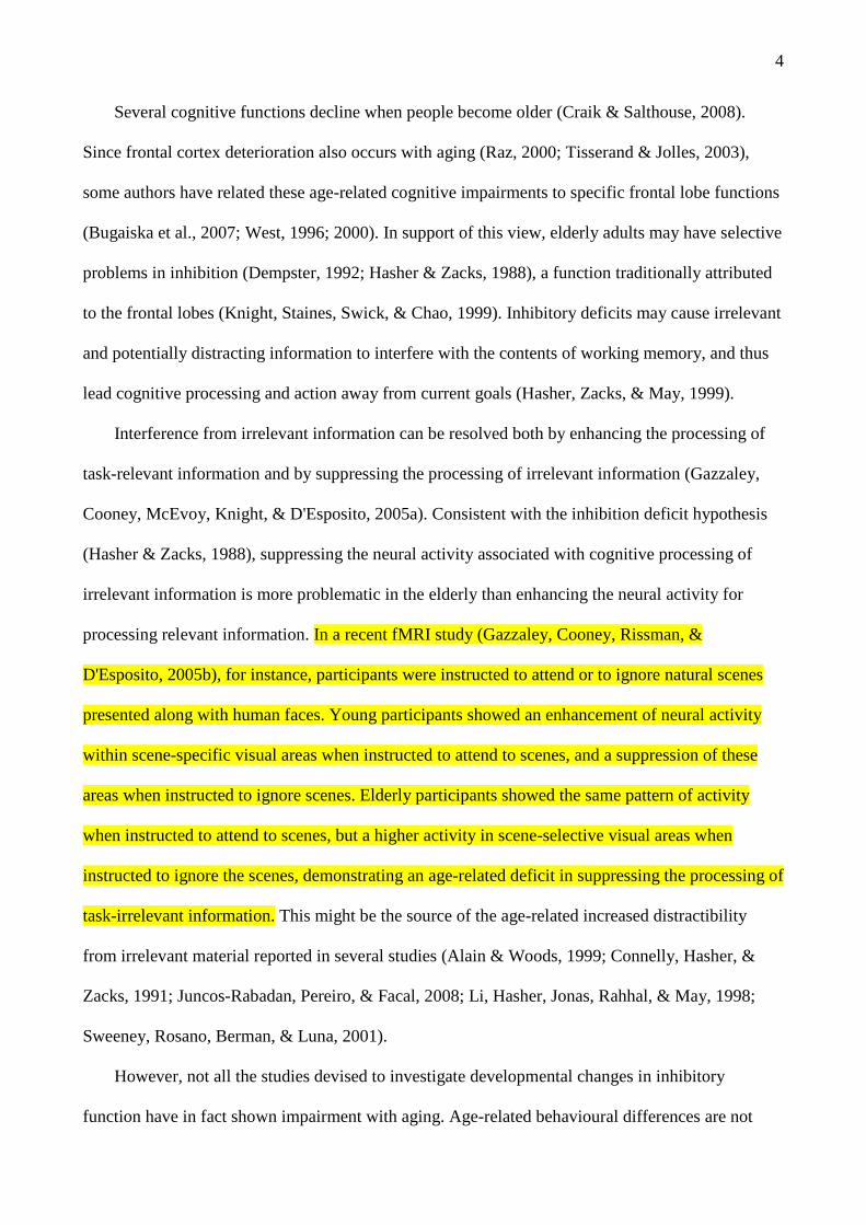
processing relevant information. In a recent fMRI study (Gazzaley, Cooney, Rissman, &
D'Esposito, 2005b), for instance, participants were instructed to attend or to ignore natural scenes
presented along with human faces. Young participants showed an enhancement of neural activity
within scene-specific visual areas when instructed to attend to scenes, and a suppression of these
areas when instructed to ignore scenes. Elderly participants showed the same pattern of activity
when instructed to attend to scenes, but a higher activity in scene-selective visual areas when
instructed to ignore the scenes, demonstrating an age-related deficit in suppressing the processing of
task-irrelevant information. This might be the source of the age-related increased distractibility
from irrelevant material reported in several studies (Alain & Woods, 1999; Connelly, Hasher, &
Zacks, 1991; Juncos-Rabadan, Pereiro, & Facal, 2008; Li, Hasher, Jonas, Rahhal, & May, 1998;
Sweeney, Rosano, Berman, & Luna, 2001).
However, not all the studies devised to investigate developmental changes in inhibitory
function have in fact shown impairment with aging. Age-related behavioural differences are not

5
generally reported when only a few distractors are present in visual search tasks (Hommel, Li, & Li,
2004), or when distracting stimuli are easily distinguishable from targets on the basis of prominent
perceptual (Carlson, Hasher, Zacks, & Connelly, 1995; Greenwood, Parasuraman, & Haxby, 1993;
Scialfa, Esau, & Joffe, 1998) or semantic features (Connelly, et al., 1991; Li et al., 1998; Li, 1999).
Furthermore, even in conditions when cognitive interference is supposed to be high, such as in
the Stroop task, results showing inhibition deficits in elderly people have been inconsistent (Basak
& Verhaeghen, 2003), and often cancelled out once age-related differences in general speed are
taken into account (see Verhaeghen & De Meersman, 1998, for a meta-analysis; but see Juncos-
Rabadan et al., 2008; Rush, Barch, & Braver, 2006, for opposite evidence). Moreover, when elderly
individuals are provided enough time to process the relevant stimulus features, for example with the
aid of a precue (Ryan, Shen, Reingold, 2006), or after practice (Davidson, Zacks, & Williams,
2003; Dulaney & Rogers, 1994), interference effects are usually significantly reduced (Kramer,
Hahn, & Gopher, 1999). Some researchers therefore explain age-related problems by means of
more general constructs, such as a non-specific slowing of processing (Birren & Fisher, 1995;
Salthouse, 1996), or loss of information along the processing flow (Myerson, Hale, Wagstaff, Poon,
& Smith, 1990).
It thus appears that elderly adults can successfully suppress potentially interfering information
if this is readily distinguishable from target information or if there is enough processing time or
previous practice. However, even without behavioural signs of interference, irrelevant information
may be still processed differently as a function of age at the neural level. In particular, elderly
individuals may still have difficulties in ignoring information irrelevant for the current goals. In
support of this distractibility hypothesis of aging (see Healey, Campbell, & Hasher, 2008, for a
review), it has been shown that items that have been attended but are no longer relevant, or should
be actively forgotten, produce stronger proactive interference in elderly than in young individuals
(Bowles & Salthouse, 2003; Kane & Hasher, 1995). Paradoxically, if previously distracting

6
information becomes relevant for the current task, elderly adults may show a benefit and even
outperform young controls (Rowe, Valderrama, Hasher, & Lenartowicz, 2006).
Given the lack of a consensus in the literature, the present study was designed to test how aging
influences the processing of to-be-ignored items. We used an adapted version of the go/nogo task, a
paradigm widely used to study response inhibition (Butter, 1969; Passingham, 1972). In a typical
go/nogo task, participants are instructed either to respond (go) or not to respond (nogo) to
predefined sets of stimuli. In the present study, we used as targets a red O and a blue X. A first type
of nogo stimuli (conflict nogo – blue O and red X) was characterized by complementary
combinations of the two features (i.e., color and letter identity) defining the target go stimuli. A
second type of nogo stimuli (irrelevant nogo), instead, shared the color feature with go stimuli, but
was easily distinguishable from them because it belonged to a different semantic category, namely
numbers as opposed to letters. In the case of conflict nogo stimuli, participants have to integrate
letter identity with color identity and associate the result of this feature integration with the less
prepotent but correct nogo response. In the case of irrelevant nogo, participants could rely on the
identity of the stimuli only (i.e., numbers as opposite to letters), without the need to combine that
feature with the color feature.
With this task, participants might develop the capacity to perceive the conjunction between
features (letter and color) for the go stimuli (“red O” and “blue X”) as “singletons” (Maljkovic &
Nakayama, 1994), thus bypassing the need to suppress a prepotent response for a complementary
combination of features after some practice, in the case of conflict nogo stimuli (Vallesi, McIntosh,
Alexander, & Stuss, in prep.). To avoid this potential problem, a second task version used 4 colored
“consonants” and 4 colored “vowels” as go and conflict nogo stimuli. With this version, it was less
likely that participants could perceive go stimuli as singletons, due to the number of letters that
were presented (8 instead of 2).
Since behavioural measures alone do not allow the full assessment of the timing of underlying
neural mechanisms, especially if no overt response is required (such as with nogo stimuli), event-
7
related potentials (ERPs) were also recorded. When ERPs have been recorded with the go/nogo
paradigm, two components have been reported to be consistently more pronounced for nogo
stimuli: a fronto-central negative deflection developing around 200-400 ms post-stimulus (nogo
N2), and a subsequent positivity (nogo P3) usually maximal at central sites (Eimer, 1993;
Falkenstein, Hoormann, & Hohnsbein, 1999; Kok, 1986; Pfefferbaum, Ford, Weller, & Kopell,
1985; Simson, Vaughan, & Ritter, 1977). The functional meaning of the nogo N2 is still debated
(Smith, Johnstone, & Barry, 2007; Falkenstein, 2006), for instance because it is different for visual
and auditory nogo stimuli (Falkenstein, Koshlykova, Kiroj, Hoormann, & Hohnsbein, 1995). This
finding casts doubt on the possibility that the nogo N2 reflects a central modality-independent
inhibitory process.
On the other hand, the nogo P3 has more consistently been interpreted as indicating the
effectiveness of the inhibition (Falkenstein et al., 1995; Pfefferbaum et al., 1985; Roberts, Rau,
Lutzenberger, & Birbaumer, 1994). Accordingly, the nogo P3, but not the nogo N2, is larger for
nogo stimuli following a cue that invalidly prompted the subject to prepare a go response (Smith et
al., 2007). Furthermore, less impulsive individuals have larger nogo P3 amplitude than highly
impulsive ones (Ruchsow et al., in press). However, the functional meaning of the nogo P3
component is also disputed. Interpretation of the go/nogo difference can be confounded by factors
such as the different frequency or motor requirements of go and nogo stimuli. The latter are
particularly problematic when recording ERPs, because motor-related deflections can add to
go/nogo waveform differences, especially in the latency range of the nogo P3 (Simson et al., 1977;
Verleger, Paehge, Kolev, Yordanova, & Jaskowski, 2006; but see Roberts et al., 1994). We
compared two types of equally frequent nogo stimuli (conflicting and irrelevant) in order to
circumvent these difficulties.
In this paradigm, however, the functional significance of the nogo P3 component might be
dependent on the nature of the distracting information, whether conflict and irrelevant nogo stimuli.
When elicited by conflict nogo stimuli, it would probably measure the inhibitory function (cf. Smith
8
et al., 2007). A nogo P3 evoked by the irrelevant stimulus might be related to distraction. In the
three-stimulus odd-ball paradigm studied in young adults (Katayama & Polich, 1998; Sawaki &
Katayama, 2007; 2008), rare target stimuli (requiring a response) are presented together with
frequent standard stimuli and rare distractors. These studies showed that, when the standard-target
discrimination was difficult, there was a large P3 for simple distractors (similar to the irrelevant
nogo stimuli here). This finding suggests attentional capture: attention may be allocated to
information that deviates from the context, even though this information is not relevant to ongoing
activity (Sawaki & Katayama, 2008).
Some predictions can be derived from the existing cognitive hypotheses of aging. On the
inhibition deficit hypothesis, one would expect more commission errors behaviorally, and a smaller
and later P3 complex for conflict nogo stimuli (an index of inhibition efficiency) in the elderly than
in the young group. However, the P3 to irrelevant nogo stimuli (numbers) will be crucial to
understand any age-related change in the processing of irrelevant material. If elderly people are
generally more distractible than the young individuals, then their attention will be captured even
more by irrelevant nogo stimuli. This should be traceable electrophysiologically by exaggerated,
rather than attenuated, ERP components (primarily the P3) to these nogo stimuli (cf. Sawaki &
Katayama, 2007). However, given the ease of the discrimination required in this nogo condition
(numbers vs. letters), we did not expect a higher error rate in the elderly.
Method
Participants
Fourteen young (8 females; mean age: 27 years, range: 20-34) and fourteen elderly (9 females;
mean age: 71 years, range: 60-80) healthy volunteers took part in the study after providing informed
consent. The participants had normal or corrected-to-normal vision and no history of neurological
or psychiatric disorders. All subjects were right handed, with the average Edinburgh Handedness
Inventory score (Oldfield, 1971) being 87 (66-100) and 84 (33-100) for young and elderly subjects,

9
respectively. Elderly participants were also screened for dementia using the Mini Mental State
examination (average score was 29/30, range: 28-30). The two groups were matched for education
(for both, 17 years on average). Participants were familiar with the task, since the ERP session
occurred 1-7 days after another session with the same tasks (inside an MRI scanner for most of the
subjects). Participants received 10 dollars per hour for participating in this ERP experiment, which
lasted 2-3 hours.
Procedure and Task
Subjects were seated in a sound-attenuated semi-dark room facing a monitor placed 60 cm
from their eyes. Visual stimuli were presented in the centre of a computer screen against a
constantly grey background. Go-nogo stimuli were letters and numbers written in Times Roman
font and colored in blue or red (50% each). In the simple version of the task, go stimuli were “red
O” and “blue X”, and nogo stimuli were either “blue O” and “red X” (conflict nogo) or red and blue
numbers 2 and 3 (irrelevant nogo). In the complex version, go stimuli were “red vowels” (A, E, I,
U) and “blue consonants” (L, N, P, Z), conflict nogo stimuli were “blue vowels” and “red
consonants”, and irrelevant nogo were red and blue numbers (4, 5, 6, 7). At each level of task
complexity, the association between color and go/nogo letters was reversed for half of the subjects.
Thus, the experiment consisted of a 2 task (simple vs. complex) by 3 go/nogo stimulus type (go,
conflict nogo, irrelevant nogo) factorial design.
Each trial began with a go/nogo stimulus lasting for 300 ms. A blank screen followed the
stimulus presentation. Inter-Stimulus-Intervals varied randomly and continuously between 2.2 and
4.2 sec. Participants performed 2 runs for each task. Each run had 64 go stimuli (50%), 32 conflict
nogo (25%) and 32 irrelevant nogo (25%). The total number of test trials was 512. Participants were
instructed to press the “B” key of a computer keyboard with the index finger of their dominant hand
as soon as they saw a go stimulus, and to refrain from responding when a nogo stimulus appeared.
The importance of speed and accuracy were equally stressed in the instructions. Go responses were

10
accepted with a deadline of 2 sec after the onset of the go stimulus. Six familiarization trials
preceded each run. During the presentation of these initial trials, which were not included in the
analyses, participants received visual feedback about their performance.
Behavioral data analysis
The first trial was discarded from further analysis. Performance to the irrelevant nogo condition
was at ceiling in both age groups. This condition was therefore discarded from further analyses.
Accuracy data were analyzed through a 2x2x2 ANOVA with task (simple vs. complex) and
stimulus type (conflict nogo vs. go) as the within subject factors, and age group (young vs. elderly)
as the between subjects factor. To assess response speed, RTs to the go stimuli were analyzed by
means of a 2x2 ANOVA with task as the within subject factor, and age group as the between
subjects factor.
Electrophysiological recording and pre-processing
Scalp voltages were recorded using ElectroCaps with 64 Ag/AgCl electrodes (10/20 system),
which included electrodes on the external canthus and infra-orbital ridge. The acquisition software
was NeuroScan SynAmps (El Paso, TX). The online reference electrode was the vertex, with the
median frontal electrode AFz used as the ground. Electrode impedances were maintained below 5
kΩ. The electrical signals were filtered with a bandwidth 0.1-70 Hz and digitized with a 250 Hz
sampling rate.
The software used for analyzing the recorded signals was Brain Electrical Source Analysis
(BESA 5.2; Germany). Data were re-referenced to an average reference, and digitally filtered (0.1-
30 Hz). Eye artifacts (i.e., eye-blinks, lateral and vertical movements) were compensated from the
ERP waveforms using source components (Ille et al. 2002) derived from the recordings obtained
before and after the performance of the task (Picton et al., 2000). Stimulus-locked ERP data from
correct trials were averaged as a function of the 6 conditions obtained by crossing 2 task (simple vs.

11
complex) by 3 stimulus type (go, conflict nogo, irrelevant nogo). Each ERP was averaged over a
1100 ms period beginning 100 ms before the stimulus and corrected to the pre-stimulus baseline.
The final ERPs (averaged across the runs) were based on a total of 51-64 nogo stimuli, and 114-128
go stimuli in each task
Spatio-temporal PLS Analysis
ERP data were analyzed using Partial Least Square (PLS, http://www.rotman-
baycrest.on.ca/pls) software for multi-modality imaging data analysis (Edgington, 1980; McIntosh,
Bookstein, Haxby, & Grady, 1996). PLS is a multivariate technique which has already been
successfully used for analysis of ERP data (Lobaugh, West, & McIntosh, 2001). It describes the
relation between one set of independent variables, based on the groups and conditions of the
experimental design, and a large set of dependent measures, such as scalp ERPs. We used a spatio-
temporal PLS analysis, which detects where the strongest experimental effects are expressed in the
scalp and when these occur in time.
The term Partial Least Square refers to the computation of the optimal least-squares fit to part
of a cross-block covariance matrix (Wold, 1982). This matrix is composed of subsequent time-
points for each electrode as columns, and subjects within each group within each experimental
condition as rows. The matrix undergoes Singular Value Decomposition to yield a set of Latent
Variables (LVs). Each LV describes how strongly a certain pattern of experimental conditions
(design scores) are expressed by each electrode at each time point (electrode saliences). Design
scores express the experimental effect of a particular LV’s electrode salience. For each electrode,
the polarity and magnitude of the electrode saliences denote the direction and strength of the
identified differences among the experimental conditions, as shown in the design scores. The
number of LVs extracted is equal to the experimental conditions. The LVs account for the matrix in
decreasing order of magnitude (i.e., the first LV will account for most of the variance).

12
Additional measures extracted from PLS are the scalp scores for each LV. Scalp scores are
obtained by multiplying the electrode saliences by the raw waveforms of each subject. Thus, scalp
scores provide a summary statistic that indicates how strongly each individual subject contributes to
the patterns depicted by the LV.
Statistical significance of the spatiotemporal pattern expressed by each LV was assessed by a
permutation test using 1000 permutations across the different experimental conditions (Edgington,
1980; McIntosh et al., 1996). Permutations consist of sampling without replacement to reassign the
order of conditions for each subject. PLS is recalculated for each new permuted sample, and the
number of times the permuted singular values exceeded the observed singular values in each LV is
calculated and expressed as a probability. A LV was considered significant at p < 0.05.
To protect from effects of possible outliers, the stability of the saliences identified on each LV
for each electrode and time point was assessed by a bootstrap test over 200 bootstrap samples
(Efron & Tibshirani, 1986). Bootstrap samples are produced by sampling with replacement and
keeping the assignment of experimental conditions to each subject fixed. PLS is recomputed for
each bootstrap sample. The ratio of the salience to its standard error estimated through the bootstrap
procedure approximately corresponds to a z-score (Efron & Tibshirani, 1986). Bootstrap ratios
equal to or greater than 3.3 (roughly corresponding to a p level ≤ .001) were chosen as the cut-off
for stable non-zero saliences. The main purpose of the bootstrap procedure is to detect those
portions of the ERP waveforms that express robust experimental effects across subjects.
A first PLS analysis including 12 task conditions obtained by crossing the 2 group x 2 task x 3
stimulus type factorial design yielded complex interactions that were difficult to interpret. A more
limited PLS analysis was therefore carried out. Go trials were omitted to avoid possible problems in
the interpretation of the go/nogo differences derived from the differential contributions of motor
electrophysiological components in these two kinds of conditions (Simson et al., 1977; Verleger et
al., 2006). After exclusion of the go conditions, this analysis considered 8 conditions: 2 group x 2
task x 2 nogo stimulus type (conflict and irrelevant nogo).

13
Analysis of the P3 to irrelevant stimuli. Visual inspection showed that the P3 to irrelevant
stimuli was different in the two groups. A direct comparison between these two age groups for this
component did not emerge in the PLS analysis, since the design scores for LVs 1 and 2 mainly
describe ERP effects that occurred either in the elderly or in the young group, respectively (see
Figure 2), and do not contrast the two groups directly. To focus on this effect more directly, we
used a classical peak latency and amplitude approach on this component. Since the P3 scalp
amplitude was maximal at Cz for the irrelevant nogo stimuli in the elderly, we focused our analysis
on that electrode. To attenuate effects of noise on peak detection, data were first low-pass filtered at
8 Hz (slope: 12 dB/octave, type: zero phase). For each of the two groups and two tasks, the peak
latency was found in the grand average waveform. The average of these 4 values was 474 ms. A
search time-window was established ±100 ms around this average peak latency. Peak latency and
amplitude were detected within this time window for each subject and task. Both the latency and
the amplitude of this peak were submitted to a 2x2 ANOVA with age group as the between subjects
factor and task as the repeated measure.
Results
Behavioral results
Accuracy. Table 1 shows accuracy data. There were more commission errors to conflict nogo
(6.1%) than misses to go stimuli (2.1%) [condition main effect, F(1,26) = 24.9, p < .001]. The
difference in the percentage of errors to conflict nogo and target go stimuli was higher in the
complex task (5.7%) than in the simple one (2.4%) [task x condition interaction, F(1,26) = 9.6, p <
.01]. No other significant effect was found. In particular, accuracy was not affected by the age
group factor.
-------- Insert Table 1 about here --------

14
Response Times. Mean RTs to go stimuli are shown in Table 2. Elderly participants responded
to go stimuli more slowly than young ones [768 vs. 667 ms; age main effect, F(1,26) = 11.7, p <
.01]. Moreover, RTs were longer for the complex than for the simple task [752 vs. 684 ms; task
main effect, F(1,26) = 38.5, p < .001]. No group by task interaction was obtained (p = .36).
-------- Insert Table 2 about here --------
ERP Data
ERP associated to each condition and group in two representative electrodes (Cz and Oz) are
displayed in Figure 1. We shall first describe the main ERP components and then report the PLS
results. The stimuli elicited sensory evoked potentials that were maximally recorded over the
posterior scalp regions. The main peaks were a small P1 wave, followed by a large N1 wave (170
ms) and a smaller P2 wave. The N1 inverted its polarity over more anterior electrodes. The sensory
potentials were followed by a series of late waves that were maximally recorded in the fronto-
central regions: an N2 peaking near 280 ms, and a P3, which peaked at around 480 ms for irrelevant
nogo stimuli and later for conflict nogo stimuli. Age effects comprised (i) a later and longer lasting
P3 wave to the conflict nogo in the elderly individuals; (ii) prominent P2 and P3 waves to the
irrelevant nogo stimuli in the elderly group.
-------- Insert Figure 1 about here --------
In the spatio-temporal PLS analysis, the first two LVs only were significant by permutation test
(ps < .0001). Design scores (Figure 2) indicated that the first LV reflected waveform differences
between conflict (positive scores) and irrelevant (negative scores) nogo stimuli mainly in the
elderly group (accounted cross-block variance = 46.5%). The same contrast was expressed by the
15
second LV (explained cross-block variance = 29.6%), but mainly in the young group. That is, each
LV represented the ERP changes between the two nogo stimulus types for one group of subjects,
and that a similar difference occurred (within each group) for the simple and complex tasks. These
LVs are described in detail below.
-------- Insert Figure 2 about here --------
Conflict vs. irrelevant nogo in the elderly group. For the first LV, we will mainly focus on
three latency-windows in which saliences were consistently stable. Saliences at all electrodes have
been plotted in a color map at the bottom panels of Figure 3, to show a full picture of the
topographical distribution of the saliences. However, ERP waveforms are only shown for the
electrodes with the peak salience. Peak electrode saliences for the first LV are shown in the line at
the top panels of Figure 3. The circles at the top of each channel plot indicate when in time the
saliences for this LV were stable (i.e., bootstrap ratio ≥ 3.3). Negative saliences indicate when
waveforms were more negative (or less positive) for conflict nogo than for irrelevant nogo in the
elderly group. Positive saliences indicate when waveforms were more positive (or less negative) for
conflict nogo than for irrelevant nogo in the elderly participants.
-------- Insert Figure 3 about here --------
The first effect occurred in the 244-348 ms latency-window, when a modulation of the left
posterior P2 was observed. The effect peaked at CB1 electrode, and polarity reversed at frontal
(mainly right) electrodes. The P2 component was greater for irrelevant than for conflict nogo
stimuli in both tasks (no such an effect was detected in the young group). Second, the nogo P3 was
more pronounced for irrelevant than for conflict nogo stimuli in the 316-596 ms latency-window.
This effect was greatest at electrode C1, with a polarity inversion peaking at FP2. Finally, in a later

16
latency-window (740-928) a nogo P3 was still present for conflict nogo and went back to baseline
for irrelevant nogo in the elderly. This effect was stable in the majority of the time-points belonging
to this latency-window. The saliences were greatest at C3 (796 ms) and later at C1 (916 ms). In
summary, the elderly group showed a more pronounced posterior P2 and central P3 to irrelevant
(vs. conflict) nogo stimuli, and a later and long lasting P3 to conflict (vs. irrelevant) nogo stimuli.
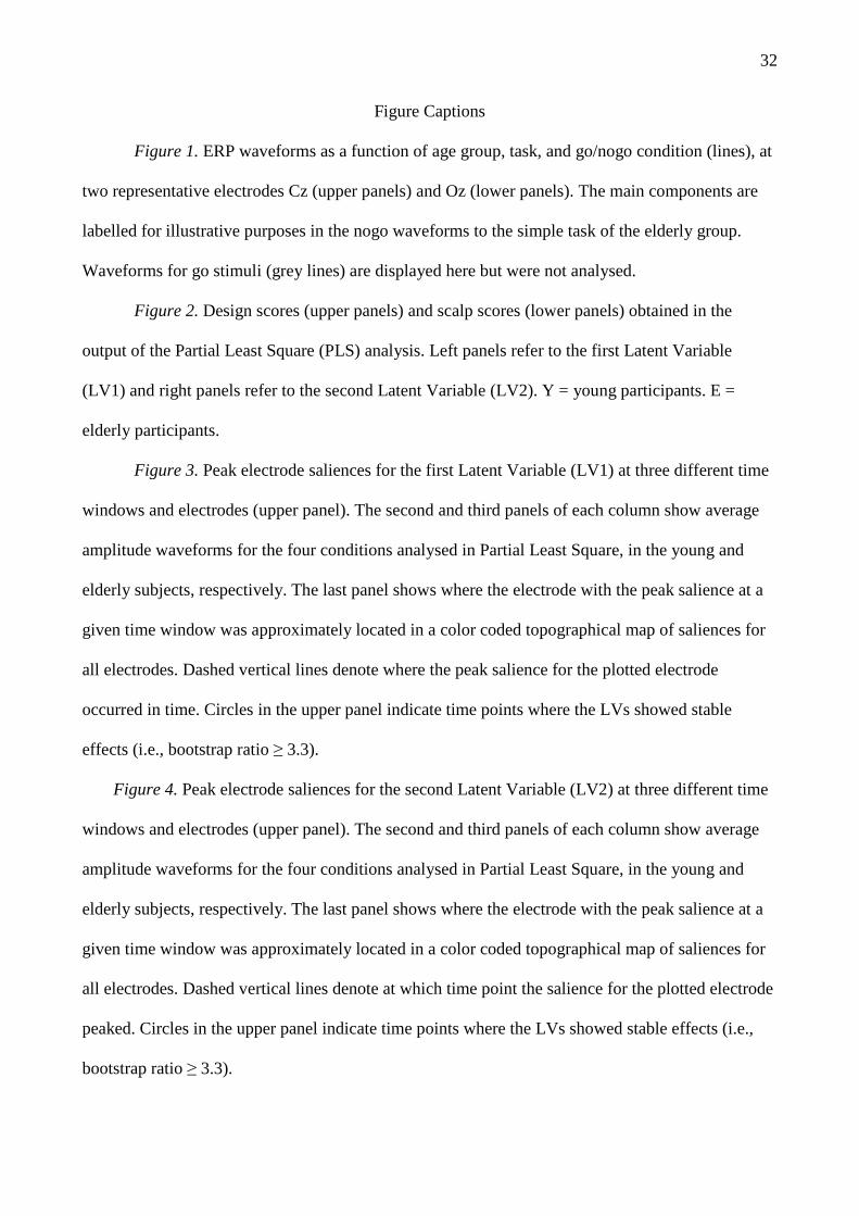
Conflict vs. irrelevant nogo in the young group. For the second LV, we will also focus on three
latency-windows where saliences were consistently stable. Again, only the electrodes with the peak
salience at each of these time windows are shown in detail. The topographical distribution of the
saliences across the scalp can be appreciated at the bottom panels of Figure 4. Peak electrode
saliences for the second LV are shown in the line at the top panels of Figure 4. The circles at the top
of each channel plot indicate when in time the saliences for this LV were stable (bootstrap ratio ≥
3.3). Salience direction was the same as in the previous LV for the elderly group.
-------- Insert Figure 4 about here --------
Saliences in the first latency-window (344-452 ms) reached their maximum value at FCz, even
though they showed a generally asymmetric left distribution in all fronto-central electrodes. Polarity
reversed at the most posterior electrodes, peaking at PO3. In terms of ERP amplitudes, this effect
indicates that, in the young group, a P3 for irrelevant nogo occurs in this time-window whereas the
N2 deflection for conflict nogo has not fully resolved yet. Peak saliences in the second latency-
window (620-672 ms) occurred at Cz. The meaning of this effect from the point of view of scalp
amplitudes is that, in the young group, the P3 was still present for conflict nogo in this time-
window, whereas it already went back to baseline for irrelevant nogo. In a final latency-window
(860-908 ms), saliences peaked at CPz, with a polarity inversion at TP10. This effect was due to a
late ERP negativity developing for conflict nogo. This component probably shares similarities with
a preceding negativity present for irrelevant nogo on the same electrode (636-656), whose
17
authenticity is however difficult to disentangle from the effect of the much bigger P3 for conflict
nogo, mostly present at the vertex at the same time. In summary, the most relevant ERP effects in
the young group were an earlier P3 to irrelevant nogo stimuli, and a later P3 to conflict nogo
stimuli, which however still occurred earlier than in the elderly group.
Age-related differences in the P3 to irrelevant nogo stimuli. The ANOVA on the latency of the
P3 to irrelevant nogo stimuli at Cz produced no significant effects. The ANOVA on the amplitude
of the same component yielded a significant age group main effect [F(1, 26) = 8.4, p < .01]: elderly
participants had a larger P3 (4.3 µV) than young ones (2.1 µV). There was no other significant
effect.
Discussion
The present study investigated age-related changes in processing irrelevant stimuli in two
go/nogo tasks (simple and complex) by means of behavioural and electrophysiological measures.
Two types of non-target stimuli were used. One type (conflict nogo) was defined by combinations
of colors and letters complementary to those used in the go stimuli. These were assumed to produce
cognitive conflict. The identity of the other type of nogo stimuli (irrelevant nogo) was completely
different from the target stimuli (numbers rather than letters). These were expected not to produce
conflict, since they could be easily distinguished from the go stimuli. Finally, go stimuli were
expected to elicit a prepotent response, because they occurred twice as frequently as either type of
nogo stimuli.
Accuracy data supported these assumptions, by showing that both age groups made more
commission errors to the conflict nogo stimuli than misses to the go stimuli, while performance was
at ceiling for the irrelevant nogo stimuli. RTs were generally higher than in previous go/nogo tasks
(e.g., Falkenstein et al., 1999), but this difference can be explained by the more demanding Stroop-
like task employed here and by the equal emphasis on speed and accuracy in the task instructions.
RT data indicated that elderly participants were slower than young participants in their responses to

18
go stimuli, confirming the existing literature on the processing speed slowing in aging (Salthouse,
1996). Both age groups suffered equally from interference especially in the complex task (more
errors to conflict nogo), and their RTs to the go stimuli were also equally affected by task
complexity (slower RTs for the complex task).
PLS analysis on the ERP data yielded two significant LVs. These LVs expressed spatio-
temporal ERP patterns distinguishing conflict and irrelevant nogo stimuli in a distinct manner for
either age group. In both tasks and groups, the P3 occurred earlier for irrelevant than for conflict
nogo stimuli. The young group showed a later parietal negativity associated with the conflict nogo
stimuli, which was not expected a priori. More relevant for the present purposes, conflict nogo
stimuli were associated with a much later P3 in the elderly than in the young participants. Finally,
P3 to irrelevant nogo stimuli was more pronounced in the elderly, as confirmed by univariate
analyses.
Although many models explain the cognitive effects of aging in terms of inhibition deficits
(Hasher & Zacks, 1988), only a handful of studies have used the go/nogo paradigm with elderly
while recording ERPs. These studies usually show an age related delay in the latency of the P3 to
nogo stimuli (Horvath, Czigler, Birkas, Winkler, & Gervai, in press; Pfefferbaum & Ford, 1988;
Tachibana, Aragane, & Sugita, 1996; but see Fallgatter, Mueller, & Strik, 1999). However, since
the P3 and RTs to the go stimuli are usually also delayed (Falkenstein et al., 2002), this has been
taken as evidence of a general slowing with aging, rather than a specific slowing of inhibition. A
general slowing view is further supported by the lack of any age-related differences in the
commission error rates (Falkenstein et al., 2002; Rush et al., 2006). A similar general slowing
account could explain age-related differences not only in the RTs but also in the latency of P3 to
conflict nogo stimuli found in the present study. Our task instructions equally emphasized speed
and accuracy, thus allowing more time for the elderly individuals to process the conflict nogo
stimuli. It would be interesting in future studies to manipulate time pressure and investigate how
19
this manipulation affects age-related differences in both the error rate and the P3 to conflict nogo
stimuli.
A general slowing account, however, does not apply to the pattern of results found here for the
irrelevant nogo stimuli. Differential processing of irrelevant information in the elderly started as
early as a posterior P2 component and was subsequently reflected by a pre-central middle-left P3.
Increases in the P2 amplitude have been associated with the detection of visual features in selective
attention tasks (Hillyard & Muente, 1984; Luck & Hillyard, 1994). An increase in the P2 amplitude
has been occasionally observed with age with auditory odd-ball tasks (Amenedo & Diaz, 1998; see
Crowley & Colrain, 2004, for a review). However, the P2 described in the above mentioned studies
has a centro-frontal topography, and probably corresponds to a “P3a”. On the contrary, the age
effect was found here at left occipital electrodes, a location which suggests a modality-specific
modulation. Few studies have reported modulations of a similar component. One of them found
that, in simple visual discrimination tasks, amplitude of a P2 component over the left occipito-
parietal scalp region increases with practice and better performance (Ding, Song, Fan, Qu, & Chen,
2003). The age effect on the P2 may thus reflect more bottom-up processing in a condition when
visual discrimination (numbers vs. letters) is very easy, as suggested by performance at ceiling.
This suggests a deficit in the capacity to withdraw processing resources from irrelevant stimuli as
early as the perceptual stages of analysis (see Gazzaley et al., 2005b, for analogous fMRI evidence).
Based on the amplitude of this component, elderly individuals seem to process irrelevant visual
information more than necessary.
The finding of an irrelevant nogo P3 with higher amplitude in the elderly than in the young
group might seem in contrast with previous P3 literature on aging. Previous studies showed a
delayed and less pronounced P3 in elderly both in the nogo condition of the go/nogo task
(Pfefferbaum & Ford, 1988) and, more generally, in the more classical odd-ball task (Fjell &
Walhovd, 2001; Picton, Stuss, Champagne, & Nelson, 1984; Polich, 1997).
20
Task context however may explain the differences between our results and those in preceding
ERP literature on aging. The importance of the context to interpret outcomes of distractor P3 has
been stressed by previous studies (Katayama & Polich, 1998; Sawaki & Katayama, 2007). By using
the three-stimulus odd-ball paradigms while recording ERPs, these studies showed an enhancement
of the P3 for simple distractors when a difficult discrimination between targets and standard stimuli
is required, possibly because attention is captured more by these irrelevant deviant events in such a
context (Sawaki & Katayama, 2007; 2008). Analogous interpretations have also been proposed to
explain the P3a elicited by unexpected irrelevant stimuli in other cognitive paradigms (e.g.,
Friedman, Cycowicz, & Gaeta, 2001; Vallesi, Mapelli, Cherubini, under review). In our case,
irrelevant nogo stimuli were semantically deviant (numbers vs. letters) and elicited an earlier nogo
P3 than conflict nogo stimuli. Importantly, this nogo P3 was much greater in the elderly than in the
young group. In the light of recent work (Sawaki & Katayama, 2008), the present data may suggest
that attentional capture by irrelevant deviant stimuli is increased in the normal aging population.
In the present go/nogo study, however, bottom-up attentional capture is more likely to be
reflected by the earlier posterior P2 component to irrelevant nogo, while the P3 might indicate
increased compensatory top-down inhibition of responses to these stimuli (Smith et al., 2007). In
favour of this interpretation, the elderly participants did not show increased responses to the
irrelevant nogo stimuli. A motor inhibition hypothesis is also favoured by the topography of this
component, which is mainly present over the left premotor scalp, especially in the elderly (see
Figure 3, middle panels). This over-recruitment of neural activity at different processing stages for
unnecessary deviant information suggests a less efficient goal-directed activity (Nessler, Friedman,
Johnson, & Bersick, 2007; Gazzaley et al., 2005b; Milham et al., 2002).
Although the current results did not show any behavioral differences between the two age
groups for the irrelevant nogo condition, and thus do not allow a definitive interpretation of the
ERP pattern in terms of an age-related deficit, previous studies have demonstrated that the extra
processing of irrelevant material can be detrimental when the experimental situation changes, for

21
instance when previously attended material has to be suppressed, or previously suppressed material
becomes relevant (Bowles & Salthouse, 2003; Healey, Campbell, & Hasher, 2008; Kane & Hasher,
1995).
An alternative interpretation of the current ERP results would be that they reflect a strategic
rather than compensatory shift with aging (Band & Kok, 2000; Gottlob & Madden, 1999). For
example, they may indicate an increased level of prefrontally-mediated cognitive control
(Velanova, Lustig, Jacoby, & Buckner, 2007). Elderly participants may have strategically decided
to pay more attention to the irrelevant stimuli (nogo P2), and then to actively suppress responses to
them (nogo P3). However, although strategic differences between the two age groups cannot be
completely discarded, it is not clear why elderly individuals would have chosen such a “strategy”:
the irrelevant nogo condition does not seem to require demanding discrimination and response
inhibition, as demonstrated by almost perfect accuracy levels in both groups. Thus, it is more likely
that elderly subjects fail to suppress the processing of contextually-deviant, irrelevant information
at relatively early stages of perceptual analysis, as suggested by the increased posterior P2, and then
have to actively suppress responses to those stimuli, as reflected by the enhanced left central nogo
P3.
A comment on the lack of an effect of task complexity is owed. The complex task used here
probably had the same working memory load as the simpler one, as far as remembering the task
rules is concerned (i.e., to respond to “red vowels” and “blue consonants” were presented, instead
of “red O” and “blue X”). However, the task was more complex: an extra operation was required in
order to make a go/nogo decision (i.e., judging the vowel vs. consonant status of the stimulus).
Many studies have shown impairing effects of task complexity with aging (Verhaeghen, Cerella, &
Basak, 2006), for instance by enhancing effects of distractors in disrupting working memory
function (Gazzaley, Sheridan, Cooney, & D'Esposito, 2007; Viskontas, Morrison, Holyoak,
Hummel, & Knowlton, 2004). Accordingly, conflict nogo stimuli should have exerted more age-
related interfering effects in the complex task than in the simple one. However, although a main

22
effect of task complexity is detected behaviourally and suggested by visual inspection of ERPs,
neither behavioural measures nor ERP data showed a task complexity by age interaction. This lack
of interaction could be due to the high level of education for both groups of participants (17 years).
Another important factor could be the familiarity with the task. Interactions between task
complexity and age group were indeed detectable when participants performed the tasks in the first
session 1-7 days before the ERP session (Vallesi, McIntosh, & Stuss, in preparation). It is possible
that these differential age-related effects of task complexity are only detectable in the learning
phase of tasks producing cognitive interference (cf. Dulaney & Rogers, 1994).
In summary, the present study sheds light on some of the possible mechanisms underlying
distraction problems in aging. Apart from an age-related delay in latency of RTs and endogenous
ERP components to conflicting stimuli, the key finding was a disproportional processing of
irrelevant, “to be ignored” stimuli, when those were easily distinguishable from the stimuli
pertinent to the current goals, that occurred in the elderly individuals. These findings suggest
increased bottom-up distractibility followed by a compensatory top-down cognitive control in the
elderly.

23
Acknowledgements
This research was supported by: postdoctoral fellowship funding from Heart and Stroke
Foundation Centre for Stroke Recovery and Canadian Institute of Health Research (CIHR, MFE-
87658) to AV; CIHR grants to DTS (MT-12853, GR-14974) J.S. McDonnell foundation grants to
ARM (220020082) and DTS (21002032); and from Canadian Foundation for Innovation (CFI) and
Ontario Innovation Trust to University of Toronto Functional Imaging Network (DTS, #1226).
Authors wish to thank Patricia Van Roon for her kind assistance in data collection.
24
References
Alain, C. & Woods, D. L. (1999). Age-related changes in processing auditory stimuli during visual
attention: evidence for deficits in inhibitory control and sensory memory. Psychol.Aging, 14,
507-519.
Amenedo, E. & Diaz, F. (1998). Aging-related changes in processing of non-target and target
stimuli during an auditory oddball task. Biol.Psychol., 48, 235-267.
Band, G. P. & Kok, A. (2000). Age effects on response monitoring in a mental-rotation task.
Biol.Psychol., 51, 201-221.
Basak, C. & Verhaeghen, P. (2003). Subitizing speed, subitizing range, counting speed, the Stroop
effect, and aging: capacity differences and speed equivalence. Psychol.Aging, 18, 240-249.
Birren, J. E. & Fisher, L. M. (1995). Aging and speed of behavior: possible consequences for
psychological functioning. Annu.Rev.Psychol., 46, 329-353.
Bowles, R. P. & Salthouse, T. A. (2003). Assessing the age-related effects of proactive interference
on working memory tasks using the Rasch model. Psychol.Aging, 18, 608-615.
Bugaiska, A., Clarys, D., Jarry, C., Taconnat, L., Tapia, G., Vanneste, S. et al. (2007). The effect of
aging in recollective experience: the processing speed and executive functioning hypothesis.
Conscious.Cogn, 16, 797-808.
Butter, C. M. (1969). Perseveration in extinction and in discriminative reversal tasks following
selective frontal ablations in Macaca mulatta. Physiol Behav, 4, 163-171.
Carlson, M. C., Hasher, L., Zacks, R. T., & Connelly, S. L. (1995). Aging, distraction, and the
benefits of predictable location. Psychol.Aging, 10, 427-436.
Connelly, S. L., Hasher, L., & Zacks, R. T. (1991). Age and reading: the impact of distraction.
Psychol.Aging, 6, 533-541.
Craik, F.I.M., Salthouse, T.A. (Eds.) (2008). The Handbook of Aging and Cognition. Third Ed. New
York, NY: Psychology Press.
Crowley, K. E. & Colrain, I. M. (2004). A review of the evidence for P2 being an independent
component process: age, sleep and modality. Clin.Neurophysiol., 115, 732-744.
Davidson, D. J., Zacks, R. T., & Williams, C. C. (2003). Stroop interference, practice, and aging.
Neuropsychol.Dev.Cogn B Aging Neuropsychol.Cogn, 10, 85-98.
Dempster, F. N. (1992). The rise and fall of the inhibitory mechanism: toward a unified theory of
cognitive development and aging. Developmental review, 12, 45-75.
Ding, Y., Song, Y., Fan, S., Qu, Z., & Chen, L. (2003). Specificity and generalization of visual
perceptual learning in humans: an event-related potential study. NeuroReport, 14, 587-590.

25
Dulaney, C. L. & Rogers, W. A. (1994). Mechanisms underlying reduction in Stroop interference
with practice for young and old adults. J.Exp.Psychol.Learn.Mem.Cogn, 20, 470-484.
Edgington, E. S. (1980). Randomization Tests. New York: Marcel Dekker.
Efron, B. & Tibshirani, R. (1986). Bootstrap methods for standard errors, confidence intervals and
other measures of statistical accuracy. Stat.Sci., 1, 54-77.
Eimer, M. (1993). Effects of attention and stimulus probability on ERPs in a Go/Nogo task.
Biol.Psychol., 35, 123-138.
Falkenstein, M., Koshlykova, N. A., Kiroj, V. N., Hoormann, J., & Hohnsbein, J. (1995). Late ERP
components in visual and auditory go/nogo tasks. Electroencephalogr.Clin Neurophysiol., 96,
36-43.
Falkenstein, M. (2006). Inhibition, conflict and the Nogo-N2. Clin.Neurophysiol., 117, 1638-1640.
Falkenstein, M., Hoormann, J., & Hohnsbein, J. (1999). ERP components in Go/Nogo tasks and
their relation to inhibition. Acta Psychol.(Amst), 101, 267-291.
Falkenstein, M., Hoormann, J., & Hohnsbein, J. (2002). Inhibition-related ERP components:
Variations with modality, age, and time-on-task. Journal of Psychophysiology, 16, 167-175.
Fallgatter, A. J., Mueller, T. J., & Strik, W. K. (1999). Age-related changes in the brain electrical
correlates of response control. Clin.Neurophysiol., 110, 833-838.
Fjell, A. M. & Walhovd, K. B. (2001). P300 and neuropsychological tests as measures of aging:
scalp topography and cognitive changes. Brain Topogr., 14, 25-40.
Friedman, D., Cycowicz, Y. M., & Gaeta, H. (2001). The novelty P3: an event-related brain
potential (ERP) sign of the brain's evaluation of novelty. Neurosci Biobehav.Rev., 25, 355-373.
Gazzaley, A., Cooney, J. W., McEvoy, K., Knight, R. T., & D'Esposito, M. (2005a). Top-down
enhancement and suppression of the magnitude and speed of neural activity. J.Cogn Neurosci.,
17, 507-517.
Gazzaley, A., Cooney, J. W., Rissman, J., & D'Esposito, M. (2005b). Top-down suppression deficit
underlies working memory impairment in normal aging. Nat.Neurosci., 8, 1298-1300.
Gazzaley, A., Sheridan, M. A., Cooney, J. W., & D'Esposito, M. (2007). Age-related deficits in
component processes of working memory. Neuropsychology, 21, 532-539.
Gottlob, L. R. & Madden, D. J. (1999). Age differences in the strategic allocation of visual
attention. J.Gerontol.B Psychol.Sci.Soc.Sci., 54, 165-172.
Greenwood, P. M., Parasuraman, R., & Haxby, J. V. (1993). Changes in visuospatial attention over
the adult lifespan. Neuropsychologia, 31, 471-485.

26
Hasher, L. & Zacks, R. T. (1988). Working memory, comprehension, and aging: A review and a
new view. In G.H.Bower (Ed.), The Psychology of Learning and Motivation (Vol. 22, pp. 193-
225). New York: Academic Press.
Hasher, L., Zacks, R. T., & May, C. P. (1999). Inhibitory control, circadian arousal, and age. In
D.Gopher & A. Koriat (Eds.), Attention and Performance XVII, Cognitive Regulation of
Performance: Interaction of Theory and Application (pp. 653-675). Cambridge, MA: MIT
Press.
Healey, M.K., Campbell, K.L., & Hasher, L. (2008). Cognitive aging and increased distractibility:
Costs and potential benefits. In W.S. Sossin, J.C. Lacaille, V.F. Castellucci & S. Belleville
(Eds.), Progress in Brain Research, (Vol. 169, pp. 353-363). Elsevier B.V.
Hillyard, S.A., Muente T. F. (1984). Selective attention to color and location: an analysis with
event-related brain potentials. Perception Psychophysics 36, 185–98.
Hommel, B., Li, K. Z., & Li, S. C. (2004). Visual search across the life span. Dev.Psychol., 40, 545-
558.
Horvath, J., Czigler, I., Birkas, E., Winkler, I., & Gervai, J. (in press). Age-related differences in
distraction and reorientation in an auditory task. Neurobiol.Aging. DOI:
10.1016/j.neurobiolaging.2007.10.003.
Ille, N., Berg, P. & Scherg, M. (2002). Artifact correction of the ongoing EEG using spatial filters
based on artifact and brain signal topographies. J. Clin. Neurophysiol. 19, 113-124.
Juncos-Rabadan, O., Pereiro, A. X., & Facal, D. (2008). Cognitive interference and aging: insights
from a spatial stimulus-response consistency task. Acta Psychol.(Amst), 127, 237-246.
Katayama, J. & Polich, J. (1998). Stimulus context determines P3a and P3b. Psychophysiology, 35,
23-33.
Kane, M. J. & Hasher, L. (1995). Interference. In G.Maddox (Ed.), Encyclopedia of Aging (2nd ed.,
pp. 514-516). New York: Springer.
Knight, R. T., Staines, W. R., Swick, D., & Chao, L. L. (1999). Prefrontal cortex regulates
inhibition and excitation in distributed neural networks. Acta Psychol.(Amst), 101, 159-178.
Kok, A. (1986). Effects of degradation of visual stimulation on components of the event-related
potential (ERP) in go/nogo reaction tasks. Biol.Psychol., 23, 21-38.
Kramer, A. F., Hahn, S., & Gopher, D. (1999). Task coordination and aging: explorations of
executive control processes in the task switching paradigm. Acta Psychol.(Amst), 101, 339-378.
Li, K. Z., Hasher, L., Jonas, D., Rahhal, T. A., & May, C. P. (1998). Distractibility, circadian
arousal, and aging: a boundary condition? Psychol.Aging, 13, 574-583.

27
Li, K. Z. (1999). Selection from Working Memory: On the Relationship between Processing and
Storage Components. Aging, Neuropsychology, and Cognition, 6, 99-116.
Lobaugh, N. J., West, R., & McIntosh, A. R. (2001). Spatiotemporal analysis of experimental
differences in event-related potential data with partial least squares. Psychophysiology, 38, 517-
530.
Luck, S. J. & Hillyard, S. A. (1994). Electrophysiological correlates of feature analysis during
visual search. Psychophysiology, 31, 291-308.
Maljkovic, V. & Nakayama, K. (1994). Priming of pop-out: I. Role of features. Mem.Cognit., 22,
657-672.
McIntosh, A. R., Bookstein, F. L., Haxby, J. V., & Grady, C. L. (1996). Spatial pattern analysis of
functional brain images using Partial Least Squares. Neuroimage, 3, 143-157.
Milham, M. P., Erickson, K. I., Banich, M. T., Kramer, A. F., Webb, A., Wszalek, T. et al. (2002).
Attentional control in the aging brain: insights from an fMRI study of the stroop task. Brain
Cogn, 49, 277-296.
Myerson, J., Hale, S., Wagstaff, D., Poon, L. W., & Smith, G. A. (1990). The information-loss
model: a mathematical theory of age-related cognitive slowing. Psychol.Rev., 97, 475-487.
Nessler, D., Friedman, D., Johnson, R., Jr., & Bersick, M. (2007). ERPs suggest that age affects
cognitive control but not response conflict detection. Neurobiol.Aging, 28, 1769-1782.
Oldfield, R. C. (1971). The assessment and analysis of handedness: the Edinburgh inventory.
Neuropsychologia, 9, 97-113.
Passingham, R. E. (1972). Non-reversal shifts after selective prefrontal ablations in monkeys
(Macaca mulatta). Neuropsychologia, 10, 41-46.
Pfefferbaum, A. & Ford, J. M. (1988). ERPs to stimuli requiring response production and
inhibition: effects of age, probability and visual noise. Electroencephalogr.Clin.Neurophysiol.,
71, 55-63.
Pfefferbaum, A., Ford, J. M., Weller, B. J., & Kopel, B. S. (1985). ERPs to response production and
inhibition. Electroencephalogr.Clin Neurophysiol., 60, 423-434.
Picton, T. W., Bentin, S., Berg, P., Donchin, E., Hillyard, S. A., Johnson, R., et al. (2000).
Guidelines for using human event-related potentials to study cognition: recording standards and
publication criteria. Psychophysiology, 37, 127-152.
Picton, T. W., Stuss, D. T., Champagne, S. C., & Nelson, R. F. (1984). The effects of age on human
event-related potentials. Psychophysiology, 21, 312-325.
Polich, J. (1997). On the relationship between EEG and P300: individual differences, aging, and
ultradian rhythms. Int.J.Psychophysiol., 26, 299-317.
28
Raz, N. (2000) Aging of the brain and its impact on cognitive performance: integration of structural
and functional findings. In: Handbook of aging and cognition (Craik, F.I.M., Salthouse, T.A.,
Eds.), vol. 2, pp. 1-90. Mahwah, NJ: Erlbaum.
Ryan, J. D., Shen, J., & Reingold, E. M. (2006). Modulation of distraction in ageing. Br.J.Psychol.,
97, 339-351.
Roberts, L. E., Rau, H., Lutzenberger, W., & Birbaumer, N. (1994). Mapping P300 waves onto
inhibition: Go/No-Go discrimination. Electroencephalogr.Clin Neurophysiol., 92, 44-55.
Rowe, G., Valderrama, S., Hasher, L., & Lenartowicz, A. (2006). Attentional disregulation: a
benefit for implicit memory. Psychol.Aging, 21, 826-830.
Ruchsow, M., Groen, G., Kiefer, M., Hermle, L., Spitzer, M., & Falkenstein, M. (in press).
Impulsiveness and ERP components in a Go/Nogo task. J.Neural Transm.
Rush, B. K., Barch, D. M., & Braver, T. S. (2006). Accounting for cognitive aging: context
processing, inhibition or processing speed? Neuropsychol.Dev.Cogn B Aging
Neuropsychol.Cogn, 13, 588-610.
Salthouse, T. A. (1996). The processing-speed theory of adult age differences in cognition.
Psychol.Rev., 103, 403-428.
Sawaki, R. & Katayama, J. (2007). Difficulty of discrimination modulates attentional capture for
deviant information. Psychophysiology, 44, 374-382.
Sawaki, R. & Katayama, J. (2008). Distractor P3 is associated with attentional capture by stimulus
deviance. Clin.Neurophysiol., 119, 1300-1309.
Scialfa, C. T., Esau, S. P., & Joffe, K. M. (1998). Age, target-distractor similarity, and visual
search. Exp.Aging Res., 24, 337-358.
Simson, R., Vaughan, H. G., & Ritter, W. (1977). The scalp topography of potentials in auditory
and visual Go/NoGo tasks. Electroencephalogr.Clin.Neurophysiol., 43, 864-875.
Smith, J. L., Johnstone, S. J., & Barry, R. J. (2007). Response priming in the Go/NoGo task: the N2
reflects neither inhibition nor conflict. Clin.Neurophysiol., 118, 343-355.
Sweeney, J. A., Rosano, C., Berman, R. A., & Luna, B. (2001). Inhibitory control of attention
declines more than working memory during normal aging. Neurobiol.Aging, 22, 39-47.
Tachibana, H., Aragane, K., & Sugita, M. (1996). Age-related changes in event-related potentials in
visual discrimination tasks. Electroencephalogr.Clin.Neurophysiol., 100, 299-309.
Tisserand, D. J. & Jolles, J. (2003). On the involvement of prefrontal networks in cognitive ageing.
Cortex, 39, 1107-1128.
Vallesi A., Mapelli D., Cherubini P. (under review). Neural correlates of inference-driven attention
in perceptual and symbolic tasks: an Event-related Potential study.

29
Velanova, K., Lustig, C., Jacoby, L. L., & Buckner, R. L. (2007). Evidence for frontally mediated
controlled processing differences in older adults. Cereb.Cortex, 17, 1033-1046.
Verhaeghen, P., Cerella, J., & Basak, C. (2006). Aging, task complexity, and efficiency modes: the
influence of working memory involvement on age differences in response times for verbal and
visuospatial tasks. Neuropsychol.Dev.Cogn B Aging Neuropsychol.Cogn, 13, 254-280.
Verhaeghen, P. & De Meersman, L. (1998). Aging and the negative priming effect: a meta-analysis.
Psychol.Aging, 13, 435-444.
Verleger, R., Paehge, T., Kolev, V., Yordanova, J., & Jaskowski, P. (2006). On the relation of
movement-related potentials to the go/no-go effect on P3. Biol.Psychol., 73, 298-313.
Viskontas, I. V., Morrison, R. G., Holyoak, K. J., Hummel, J. E., & Knowlton, B. J. (2004).
Relational integration, inhibition, and analogical reasoning in older adults. Psychol.Aging, 19,
581-591.
West, R. L. (1996). An application of prefrontal cortex function theory to cognitive aging.
Psychological Bulletin, 120, 272-292.
West, R. (2000). In defense of the frontal lobe hypothesis of cognitive aging. Journal of the
International Neuropsychological Society, 6, 727-729.
Wold, H. (1982). Soft modeling: the basic design and some extensions. In H.Wold (Ed.), Systems
Under Indirect Observation: Causality-Structure-Prediction: Part II. (pp. 1-54). Amsterdam:
North-Holland Publishing Company.

30
Table 1. Mean Percentage of correct responses (and standard errors of the mean) as a function of
task, go/nogo condition, and age groups.
Simple Task Complex Task
Young Elderly Young Elderly
Go stimulus 97.5 (0.9) 97.5 (1.0) 97.9 (0.8) 98.7 (0.6)
Conflict nogo 94.6 (1.2) 95.5 (1.4) 92.3 (0.9) 93.0 (1.5)
Irrelevant nogo 99.4 (0.8) 100 (0) 99.9 (0.1) 99.7 (0.2)

31
Table 2. Mean Response Times (and standard errors of the mean) of correct responses (in
milliseconds) to go stimuli as a function of task and age groups.
Age Group
Young Elderly
Task
Simple 639 (23) 696 (27)
Complex 729 (19) 807 (19)
32
Figure Captions
Figure 1. ERP waveforms as a function of age group, task, and go/nogo condition (lines), at
two representative electrodes Cz (upper panels) and Oz (lower panels). The main components are
labelled for illustrative purposes in the nogo waveforms to the simple task of the elderly group.
Waveforms for go stimuli (grey lines) are displayed here but were not analysed.
Figure 2. Design scores (upper panels) and scalp scores (lower panels) obtained in the
output of the Partial Least Square (PLS) analysis. Left panels refer to the first Latent Variable
(LV1) and right panels refer to the second Latent Variable (LV2). Y = young participants. E =
elderly participants.
Figure 3. Peak electrode saliences for the first Latent Variable (LV1) at three different time
windows and electrodes (upper panel). The second and third panels of each column show average
amplitude waveforms for the four conditions analysed in Partial Least Square, in the young and
elderly subjects, respectively. The last panel shows where the electrode with the peak salience at a
given time window was approximately located in a color coded topographical map of saliences for
all electrodes. Dashed vertical lines denote where the peak salience for the plotted electrode
occurred in time. Circles in the upper panel indicate time points where the LVs showed stable
effects (i.e., bootstrap ratio ≥ 3.3).
Figure 4. Peak electrode saliences for the second Latent Variable (LV2) at three different time
windows and electrodes (upper panel). The second and third panels of each column show average
amplitude waveforms for the four conditions analysed in Partial Least Square, in the young and
elderly subjects, respectively. The last panel shows where the electrode with the peak salience at a
given time window was approximately located in a color coded topographical map of saliences for
all electrodes. Dashed vertical lines denote at which time point the salience for the plotted electrode
peaked. Circles in the upper panel indicate time points where the LVs showed stable effects (i.e.,
bootstrap ratio ≥ 3.3).

33
Figure 1

34
Figure 2

35
Figure 3

36
Figure 4